Harnessing biomaterials for Nanomedicine: Preparation, toxicity and applications Janos Szebeni & Chezy Barenholz Copyright © 2009 by Pan Stanford Publishing Pte Ltd www.panstanford.com
Chapter 12
Adverse Immune Effects of Liposomes:
Complement Activation, Immunogenicity
and Immune Suppression
Janos Szebeni and Yechezkel (Chezy) Barenholz Nanomedicine Research and Education Center, Bay Zoltan Foundation for Applied Research and Semmelweis University Medical School, Budapest, and Dept of Health Sciences, Miskolc University, Hungary, and SeroScience Inc., Budapest [email protected] Y.B., Laboratory of Membrane and liposome Research, Institute of Medical Research Israel Canada (IMRIC), The Hebrew University-Hadassah Medical School, Jerusalem 91120, Israel [email protected]; [email protected]
Some therapeutically relevant liposomes are recognized by the immune system
as foreign, and the resulting innate or specific immune response can be adverse
to the host. The innate response involves activation of the complement (C)
system which, via liberation of anaphylatoxins (C5a, C3a), underlies an acute
hypersensitivity syndrome called C activation-related pseudoallergy (CARPA).
CARPA represents a potential barrier to the clinical use of reactogenic liposomes
in cardiac patients, as a main manifestation of C activation in the body is
cardiopulmory distress. The adverse immune response to liposomes involving
specific immunity is exemplified by PEGylated nano-liposome induced transient
IgM production, which causes accelerated blood clearance (ABC). This chapter
highlights some common and specific causes of adverse immune effects of
liposomes, summarizes the latest information on CARPA, the ABC phenomenon
and immunosuppression by anticancer liposomes, and outlines the immune
mechanisms involved in these phenomena.

2 Adverse Immune Effects of Liposomes: Complement Activation, Immunogenicity and Immune
Suppression
1. INTRODUCTION
Today, 46 years after the discovery of liposomes [11], and nearly a quarter
century after their first infusion in man [71], the immune effects of liposomes
represents a relatively poorly explored territory within ”liposomology”. This
will, however, likely change in the near future, as the rising number of advanced
drug carrier liposomes, which are complex and contain protein or other ligands,
carry increased risk of immune recognition. The absence of adverse immune
effects, a unique asset of simple liposomes, will probably become the exception
rather than the rule. Liposomal immunology will extend its present focus, the
use of liposomes as vaccines, to mapping an uncharted network of
hypersensitivity and immunogenicity reaction pathways. As the safety of
medicinal nanoparticles comes more to the fore, reactogenicity and
immunogenicity testing may join the list of toxicity assays required by regulatory
agencies. A deeper insight into the fine immunomodulatory effects of complex
liposomes may also lead to a need to revise our views on immune suppression,
as is the case with DoxilTM, one of the points addressed later in this chapter.
This looks like a rough way not only for future R & D of liposomes but for
all nanomedicines; a toll we need to pay for nursing conventional, small-
molecular medicines into the nano dimension, where the size, shape or surface
features of drug particles get the attention of the immune system (Fig. 1).
Fig. 1. Immune recognition of nanoparticles. Map of different nanoparticles on a diameter vs. Mw chart; blue shaded area is the region of immune recognition. Liposomes and carbon nanotubes, with their length, fall into the “sight” of the immune system, while smaller nanoparticles (fullerenes, dendrimers, micelles and complex polymeric particles (e.g., nanolatex) are, in theory, below the size and Mw thresholds of immune recognition. The green “Lipinsky box” shows the position of traditional small-molecular weight drugs
[58].
3
2. TYPES AND FEATURES OF IMMUNE RESPONSES TO LIPOSOMES
Immune responses to liposomes can be stimulatory or inhibitory, weak,
moderate or severe, all with a broad variation in their time of onset and duration
(Table 1).
Table 1. Variations of liposome-induced immune changes according to the time of onset and duration
Type of
change Time of onset Duration Example & Reference
Stimulation
Immediate (within
seconds to minutes) Minutes to hours
Hypersensitivity
(infusion) reactions caused
by liposomes [79-81,84] Delayed (within
hours)
Late (within days to
months) Weeks to years
Immunity to liposomal
antigens, e.g. influenza or
hepatitis B [3,13]
Inhibition
Short-term Hours to days
Liposomal alendronate
[22]
Doxil induced
immunosuppression[2,32,
74]. Long-term Days to months
In additions to the above time-related differences, the immune effects of
liposomes also differ in the part of the immune system primarily affected, i.e.,
whether the nonspecific, innate, or the specific, adaptive arm of immunity
undergo overstimulation or suppression. The term reactogenicity is used to imply
a broad activation process that involves the innate as well as the adaptive arm of
the immune system, while immunogenicity usually refers to specific antibody
induction with B and T cell responses, not necessarily involving innate
immunity. Of note, antigenicity, i.e., the capability of liposomes to carry antigens,
does not necessarily mean reactogenicity or immunogenicity, as antigens may
remain unrecognized or can induce immune suppression (tolerance).
The impact of liposomes on the immune system was explored in two
periods; first, in the late 1960s, the late-onset, long-lasting immunogenic
properties were recognized, opening the field of liposomal vaccines. Since then
numerous vaccine candidates have reached advanced clinical trials, and one
product, an influenza virus-like liposome expressing hepatitis B antigen
(“immunopotentiating reconstituted influenza virosomes”, Epaxal-Berna®), has
reached the market as a vaccine against hepatitis B.
The “bad” side of immune stimulation, i.e., the rise of immune responses
with adverse consequences, has been known only since the mid-1990s, following
the introduction to date of a dozen liposomal drugs [59], including Doxil
[12,29,31,72].
This chapter focuses only on the latter, adverse immune responses to
liposomes; the vaccine applications have been amply reviewed elsewhere [3-5,
13,19]. It was our goal to highlight the common grounds of immune stimulation
4 Adverse Immune Effects of Liposomes: Complement Activation, Immunogenicity and Immune
Suppression
by liposomes, to provide examples of reactogenicity, immunogenicity and
immune suppression, and to outline recent theories about the mechanism of
these phenomena.
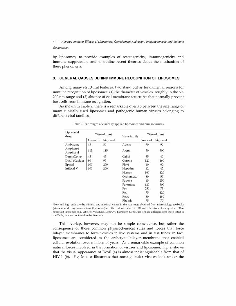
3. GENERAL CAUSES BEHIND IMMUNE RECOGNITION OF LIPOSOMES
Among many structural features, two stand out as fundamental reasons for
immune recognition of liposomes: (1) the diameter of vesicles, roughly in the 50-
200 nm range and (2) absence of cell membrane structures that normally prevent
host cells from immune recognition.
As shown in Table 2, there is a remarkable overlap between the size range of
many clinically used liposomes and pathogenic human viruses belonging to
different viral families.
Table 2. Size ranges of clinically applied liposomes and human viruses
Liposomal
drug *Size (d, nm)
Virus family *Size (d, nm)
low end high end low end high end
Ambisome 45 80 Adeno 70 90
Amphotec
Amphocyl 115 115 Arena 50 300
DaunoXome 45 45 Calici 35 40
Doxil (Caelyx) 80 95 Corona 120 160
Epaxal 100 200 Flavi 40 60
Inflexal V 100 200 Hepadna 42 42
Herpes 100 120
Orthomyxo 80 55
Papova 45 250
Paramyxo 120 300
Pox 250 75
Reo 75 120
Retro 80 180
Rhabdo 75 70
*Low and high ends are the minimal and maximal values in the size range obtained from microbiology textbooks
(viruses), and drug informations (liposomes) or other internet sources. Of note, the sizes of many other FDA-
approved liposomes (e.g., Abelcet, Visudyne, DepoCyt, Extrasorb, DepoDur) [59] are different from those listed in
the Table, or were not found in the literature.
This overlap, however, may not be simple coincidence, but rather the
consequence of those common physicochemical rules and forces that force
bilayer membranes to form vesicles in live systems and in test tubes; in fact,
liposomes are considered as the archetype bilayer membrane that enabled
cellular evolution over millions of years. As a remarkable example of common
natural forces involved in the formation of viruses and liposomes, Fig. 2. shows
that the visual appearance of Doxil (a) is almost indistinguishable from that of
HIV-1 (b). Fig 2c also illustrates that most globular viruses look under the

5
electron microscope very much like large unilamellar vesicles (LUV), and
paramyxoviruses look like typical multilamellar vesicles (MLV).
Fig. 2. Electron microscopic images of Doxil (a), HIV-1 (b) and other common human viruses (c) scaled to each other in the nm region, illustrating the morphological similarity of liposomes and viruses.
In addition to viruses, liposomes also resemble ectosomes, i.e. membrane
vesicles detached from cells, as well as most other ecto-organelles and cellular
debris that form upon cell death. Nanobacteria, the smallest self-replicating
pathogens are also in the liposome size range (100-200 nm) [18]. In essense,
liposomes mimic the size and shape of pathogenic microbes and some
subcellular structures against which nature developed strong eliminatory
mechanisms via humoral and cellular immune responses.
The second major cause underlying the immune recognition of liposomes is
the lack of self-discriminating molecules on the bilayer membrane. Fig. 3 shows
those that are engaged in protecting cells from attack by the C system, referred to
as C control proteins (CCPs). These include C receptor type 1 (CR1), decay
accelerating factor (DAF), membrane cofactor protein (MCP) and CD59. Fig 3.
illustrates the activities of these proteins [41]: CR1 binds C3 convertase subunits,
C3b and C4b, and promotes their physical dissociation (decay accelerating
activity). Furthermore, CR1 also enhances the catalytic lysis of C3 convertases by
the plasma serine protease, factor I (fI) (cofactor activity).

6 Adverse Immune Effects of Liposomes: Complement Activation, Immunogenicity and Immune
Suppression
Fig. 3. Complement control proteins on blood cells, endothelial and other cells that normally prevent host damage by C attack. These all are not present on current liposomes (reproduced from [41] with permission).
Furthermore, CR1 binds C3b-opsonised particles and immune complexes,
extracting them from the activation cascade and carrying them to –among others,
to cells of the RES. DAF binds C3 convertase and accelerates its decay to
subunits; MCP displays cofactor activity, while CD59, as last defense line against
C damage, interferes with C9 unfolding and polymerizing to form the membrane
attack complex (MAC) [41]. In absence of the above membrane proteins, which
normally prevent host damage by C attack, the mistake of the immune system to
recognize beneficial liposomal medicines becomes irreversible “friendly fire”.
The ensuing inflammatory reaction causes harm to both the body and the
liposomal drug.
4. CONSEQUENCES OF IMMUNE RECOGNITION OF LIPOSOMES
4.1. Acute hypersensitivity reactions
4.1.1. Symptoms and occurrence
Acute hypersensitivity reactions (HSRs) to liposomes have been reported
from time to time, ever since 1986, the first clinical study wherein large doses of
liposomes were infused in cancer patients [71]. All kinds of liposomes and lipid-
based drugs can cause such reactions, as follows from the large variety of
vesicles reported to be reactogenic [15,23,25,27,34-36,38,65,70]. The reported
frequency following premedication and/or other precautionary measures, such
7
as the extension of treatment time with very slow initial rate of infusion usually
remains below 10% [78,79,88]. Without premedication, however, HSRs were
observed in 45% of patients treated for the first time with Doxil [16,75].
Recommendation for ready access to emergency equipment to treat HSRs
reactions has recently become part of the box insert of Doxil [88].
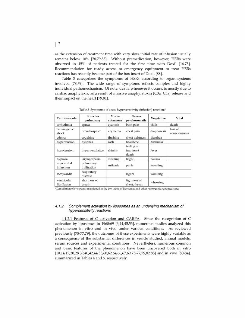
Table 3 categorizes the symptoms of HSRs according to organ systems
involved [78,79]. The wide range of symptoms reflects complex and highly
individual pathomechanism. Of note, death, whenever it occurs, is mostly due to
cardiac anaphylaxis, as a result of massive anaphylatoxin (C3a, C5a) release and
their impact on the heart [79,81].
Table 3 Symptoms of acute hypersensitivity (infusion) reactions*
Cardiovascular Broncho-
pulmonary
Muco-
cutaneous
Neuro-
psychosomatic Vegetative Vital
arrhythmia apnea cyanosis back pain chills death
carcinogenic
shock bronchospasm erythema chest pain diaphoresis
loss of
consciousness
edema coughing flushing chest tightness diarrhea
hypertension dyspnea rash headache dizziness
hypotension hyperventilation rhinitis
feeling of
imminent
death
fever
hypoxia laryngospasm swelling fright nausea
myocardial
infarction
pulmonary
infiltration urticaria panic sweating
tachycardia respiratory
distress rigors vomiting
ventricular
fibrillation
shortness of
breath
tightness of
chest, throat wheezing
*Compilation of symptoms mentioned in the box labels of liposomes and other reactogenic nanomedicines
4.1.2. Complement activation by liposomes as an underlying mechanism of hypersensitivity reactions
4.1.2.1 Features of C activation and CARPA. Since the recognition of C
activation by liposomes in 1968/69 [6,44,45,53], numerous studies analyzed this
phenomenon in vitro and in vivo under various conditions. As reviewed
previously [75-77,79], the outcomes of these experiments were highly variable as
a consequence of the substantial differences in vesicle studied, animal models,
serum sources and experimental conditions. Nevertheless, numerous common
and basic features of the phenomenon have been uncovered both in vitro
[10,14,17,20,28,39,40,42,44,53,60,62,64,66,67,69,75-77,79,82,85] and in vivo [80-84],
summarized in Tables 4 and 5, respectively.

8 Adverse Immune Effects of Liposomes: Complement Activation, Immunogenicity and Immune
Suppression
Table 4. Observations on C activation by liposomes in vitro
• All types of liposomes can activate C, with neutral small unilamellar vesicles (SUV)
being the least reactogenic.
• The sensitivity of human serum for C activation by different liposomes shows
substantial individual variation.
• A serum sensitive to a certain liposome is not necessarily sensitive to another
liposome: individual sensitivity is formulation specific.
• C activation may proceed on both the classical and the alternative pathways.
• Activation triggers include the binding to the vesicles of IgG, IgM, CRP*, C1q, C3, and
potentially, MBL** and ficolin.
• C activation is enhanced by:
o positive or negative surface charge,
o increasing liposome size in the 70-300 nm range**
o inhomogeneity
o endotoxin contamination
o presence of aggregates
o presence of doxorubicin or similar drugs that can bind to & aggregate
liposomes/lipids
o presence of cholesterol in the bilayer membrane at ~ >70%
o PEGylation with PEG-PE
• C activation can be inhibited by known C inhibitors***
* CRP, C reactive protein
** MBL, mannose binding lectin
***For example, by soluble C receptor type I (sCR1) [84,85]
4.1.2.2. Mechanisms involved in C activation and CARPA. In commenting
on the mechanisms of some of the effects listed in Table 4, surface charge is
perhaps the best known enhancer of liposomal C activation. A recent study
mapped the effect to the phosphate oxygen moiety of mPEG-phospholipid
conjugates in mPEGylated dipalmitoylphosphatidylcholine (DPPC) liposomes.
The evidence included the lack of C activation by non-PEGylated vesicles and
the inhibition of C activation by methylation of the oxygen moiety on mPEG-
DPPE phosphate [63]. Yet another study pointed to the specificity of acidic
phospholipids causing C activation, inasmuch as vesicles containing carboxylic
acid derivatives failed to activate C [73].
Morphological evidence of the presence of oval, elongated and irregular
liposomes and aggregates in Doxil, taken together with a recent report on the
critical impact of minor differences in liposome surface curvature on C activation
via the classical pathway (via IgM binding) [68] raise the possibility that
doxorubicin could be an indirect contributing factor to C activation by Doxil [82]
via modifying the surface of liposomes.
4.1.3. Mechanism of liposome reactions and its individual variation
Among the symptoms of HSRs (Table 3) there is no single, or unique
symptom that would be liposome-specific; just as many more micellar and
particulate drugs, radiocontrast agents, monoclonal antibodies and enzyme

9
Table 5. Observations on C activation-related pseudoallergy caused by liposomes
• All types of liposomes can cause CARPA in animals and man, with neutral SUV being
the least reactogenic.
• The sensitivity of different animals for different liposomes shows substantial
variation.
• The sensitivity of different species to liposomal CARPA decreases in the following
order: pig> dog> rabbit> sheep > rat> mouse.
• The minimum effective reactogenic dose of liposomes in rats is 10-100-times higher
than that in pigs or dogs.
• The individual variation of the cardiopulmonary changes associated with porcine
liposome-induced CARPA is lower than that of dogs.
• In pigs, pulmonary hypertension, while in dogs, systemic hypotension are the
dominating cardiopulmonary symptoms of CARPA.
• Both in pigs and dogs the cardiopulmonary changes can decrease or entirely
disappear after the second or third dosing, a reflection of tachyphylaxis (tolerance
induction).
• The latter phenomenon allows the development of desensitization protocols using
empty (placebo) liposomes.
• Both in pigs and dogs leukopenia followed by leukocytosis and thrombocytopenia are
varying hematological abnormalities associated with CARPA.
• The rise of plasma thromboxane A2 (measured as TXB2) closely parallels CARPA in
pigs, indicating that it is a rate limiting mediator. TXB2 also rises in other species
during CARPA.
• CARPA can be inhibited in pigs with C inhibitors (e.g., sCR1, anti porcine C5
antibody, indomethacin).
• Based on the minimal effective reactogenic dose, porcine and canine CARPA may
represent a model of human CARPA.
therapeutics, liposomes are “only” triggers of a complex chain of events
involving C activation, binding of anaphylatoxins to their specific receptors on
mast cells, basophils and other anaphylatoxin-receptor-positive cells
(macrophages), activation of the latter cells to release a host of vasoactive
mediators, including histamine, tryptase, PAF, LTB2, LTB4, LTC4, LTD4, LTE4,
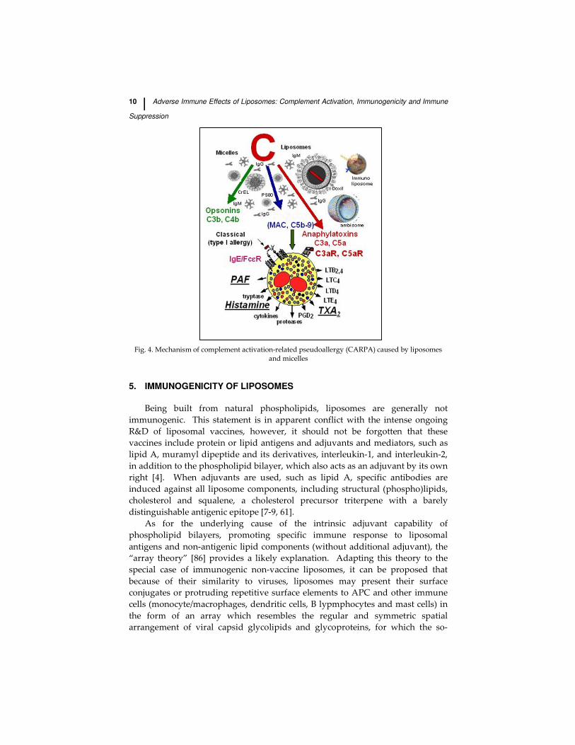
TXA2, PGD2 and TXD4 [37, 57, 87] (Fig. 4).
Some of these mediators (e.g., PAF, histamine, tryptase and TXA2) are
preformed and liberate from the cells immediately upon activation, while others
are de novo synthesized and, hence, liberate slower. This differential, multistep
release of allergomedins from anaphylatoxin-responsive cells may explain the
individual variation in the start of clinical symptoms. Specifically, activation of
H1 receptors leads to vasoconstriction and vascular leakage, and is responsible
for the cardiovascular and cutaneous symptoms of anaphylaxis. H2 receptors, in
turn, increase cellular cAMP levels and cause vasodilation, increased heart rate
and pulse pressure [56]. Another potentially important factor in individual
variation of HSR symptoms is the relative abundance of reactive cells in different
organs of response, i.e., the skin, lung, heart, bowel, etc. [33].
10 Adverse Immune Effects of Liposomes: Complement Activation, Immunogenicity and Immune
Suppression
Fig. 4. Mechanism of complement activation-related pseudoallergy (CARPA) caused by liposomes and micelles
5. IMMUNOGENICITY OF LIPOSOMES
Being built from natural phospholipids, liposomes are generally not
immunogenic. This statement is in apparent conflict with the intense ongoing
R&D of liposomal vaccines, however, it should not be forgotten that these
vaccines include protein or lipid antigens and adjuvants and mediators, such as
lipid A, muramyl dipeptide and its derivatives, interleukin-1, and interleukin-2,
in addition to the phospholipid bilayer, which also acts as an adjuvant by its own
right [4]. When adjuvants are used, such as lipid A, specific antibodies are
induced against all liposome components, including structural (phospho)lipids,
cholesterol and squalene, a cholesterol precursor triterpene with a barely
distinguishable antigenic epitope [7-9, 61].
As for the underlying cause of the intrinsic adjuvant capability of
phospholipid bilayers, promoting specific immune response to liposomal
antigens and non-antigenic lipid components (without additional adjuvant), the
“array theory” [86] provides a likely explanation. Adapting this theory to the
special case of immunogenic non-vaccine liposomes, it can be proposed that
because of their similarity to viruses, liposomes may present their surface
conjugates or protruding repetitive surface elements to APC and other immune
cells (monocyte/macrophages, dendritic cells, B lypmphocytes and mast cells) in
the form of an array which resembles the regular and symmetric spatial
arrangement of viral capsid glycolipids and glycoproteins, for which the so-
11
called “pattern recognition receptors” (e.g., LPS and Toll-like receptors, TLRs) on
the above cells readily react, generating innate and subsequent specific immune
responses. Originally TLRs recognize molecule arrays that are broadly shared by
pathogens (called pathogen-associated molecular patterns, PAMPs, such as LPS,
lipoproteins, lipopeptides, flagellin, double-stranded RNA or the unmethylated
CpG islands of bacterial and viral DNA), however, “liposomal arrays” also
trigger “danger” signaling within these cells despite the absence of PAMPs,
which ultimately leads to antibody production against the “pseudo-PAMPs” and
their phospholipid support. The resultant immune response may or may not
differ from a standard immune response to vaccines, depending on the pathway
of immune activation.
An example of non-standard, partial immunogenicity is the so-called “ABC
phenomenon”, i.e., accelerated blood clearance of PEGylated liposomes, a
phenomenon that has great clinical relevance [21]. It has been discovered by
Ishida and colleagues [46-52, 54, 55] that repeated injection of PEGylated
liposomes in mice and rats causes rapid clearance of liposomes from the
bloodstream, due to the formation of anti-PEG IgM in the spleen. Importantly,
the phenomenon is absent with PEGylated liposomes encapsulating doxorubicin,
which is consistent with the lack of ABC in cancer patients treated with Doxil.
On the other hand, free doxorubicin given in doses that correspond to the
amount given in Doxil, restores ABC. These data indicate that immune cells
responsible for the ABC phenomenon might be selectively damaged by
doxorubicin encapsulated in PEGylated liposomes, as detailed in the section on
liposome-induced immune suppression below. Since the ABC phenomenon was
also observed in BALB/c nu/nu mice, but not in BALB/c SCID mice, it was
suggested that antibody production represents a T cell-independent, B cell
response, and that PEGylated liposomes might be recognized by B cells as a
thymus-independent type 2 antigen [54]. Based on the array theory, as
delineated above, it can be hypothesized that PEGylated liposomes trigger B cells
via some of their TLRs, for PEG looks to B cells like viral spikes. The ABC
phenomenon is illustrated in Fig. 5.
6. IMMUNE SUPPRESSION BY LIPOSOMES
It is well known that liposomes are taken up mainly by cells of the
reticoloendothelial system (RES) in the liver, spleen, bone marrow and
elsewhere, which cells are also part of the nonspecific, innate immune system.
Therefore, it has been asked for a long time whether macrophage saturation by
liposomes, leading to immune suppression, could be a problem, a potential risk
for infection? There is ample evidence that clinically applied doses of non-
cytotoxic liposomes generally do not cause immune suppression, at least not
major, clinically important blockage of macrophage function. However, the
situation is different with anticancer liposomes that contain cytostatic drugs,
which may cause more or less immunosuppression.

12 Adverse Immune Effects of Liposomes: Complement Activation, Immunogenicity and Immune
Suppression
Fig. 5. Blood clearance rate of PEgylated liposomes in rats (A) and tissue accumulation 24 hours after administration. The figure, reproduced from [48] (with permission), illustrates the acceleration of liposome clearance after repeated injection (on days 2 to 14), of liposomes with paralleling increase in liver and spleen deposition.
Administration of Doxil in mice, for example, was shown to interfere with
the clearance of bacteria from blood, which was explained by macrophage
suppression [74]. Another indirect evidence of impaired phagocytic function is
the prolongation of the circulation T1/2 of Doxil at repeated administrations, a
phenomenon that may lead to the need of dose adjustment after the first or
second treatment cycle.
Using tumor-bearing mice, Gabizon et al. [32] have shown that escalation of
the dose of Doxil from 2.5 to 20 mg/kg resulted in saturation of Doxil clearance
with disproportional increase of the amount of liposomal drug accumulating in
tumor, due to partial blockade of the RES in the liver. The clearance saturation
effect after Doxil was dose-dependent and it was not present after a similar free
doxorubicin dose or similar phospholipid dose in drug-free liposomes.
Therefore, dose-dependent liposomal doxorubicin blockade of the RES may
significantly enhance drug delivery to tumors [32].
Blockade of the RES by Doxil with clinically manifest immune deficiency is
not known in man, although again, indirect signs do point to the presence of
some immune suppression. A remarkable example of this is the inhibition of
HSRs to carboplatin by co-administered Doxil. In the report of this phenomenon
[2], there were no allergic reactions to carboplatin in patients treated with
carboplatin plus Doxil, while there was a 30% allergic reaction rate on the
carboplatin-only arm. Also, the cause for carboplatin only patients to go off
study was solely allergic reactions to carboplatin, while the reasons for
discontinuation in the combination arm were different, mainly hematological
toxicities and fatigue. Thus, Doxil protected against carboplatin allergy, which
can most easily be explained by immune suppression. This effect actually
provides clinical benefit for Doxil over paclitaxel or gemcitabine, also appled in
combinations with carboplatin, since allergic reactions to carboplatin during

13
treatment of platinum sensitive, recurrent disease have become dose-limiting [2,
30].
7. CONCLUSIONS AND OUTLOOK
Immunosafety is a key issue in current R&D of nanomedicines, including
liposomes. As expressed in regulatory guidances [1,24,26,43] and research
solicitations by granting agencies (e.g. FP7 calls), prediction and prevention of
adverse immune effects represent unmet medical needs. One main problem in
this field lies in the complexity and individual variation of the immune system,
which, when faced with increasingly complex nanomedicines, will also give
increasingly complex responses. The immune toxicology of nanomedicines is
largely unexplored at a broad intersection of nanotechnology, immunology and
pharmacology, and what gives hope for progress in this “Terra Nova” is that
interest and support for the field keep increasing. Hopefully it is not too far in
the future that we can equip liposomes and other drug carrier nanosystems with
immune evasive capabilities, and/or “teach” the immune system to distinguish
these marvels of nanotechnology from harmful microbes.
8. ACKNOWLEDGMENTS
The authors gratefully acknowledge the financial support by the National
Office for Research and Technology (NKTH), Budapest (grants Kozma,
CARPA777 and NANOMEDI), and by the Barenholz Fund.
9. REFERENCES
1. Administration (U.S. Food and Drug) (2007). Nanotechnology: A report of the U.S.
Food and Drug Administration Nanotechnology Task Force. July 25, 2007
2. Alberts, D. S., liu, P. Y., Wilczynski, S. P., Clouser, M. C., Lopez, M. A., Michelin, D.
P., Lanzotti, V. J., and Markman, M. (2008). Randomized trial of pegylated liposomal
doxorubicin (PLD) plus carboplatin versus carboplatin in platinum-sensitive (PS)
patients with recurrent ovarian or peritoneal carcinoma after failure of initial
platinum-based chemotherapy (South West Oncology group protocol SO200).
Gynecol. Oncol. 108, pp. 90–94.
3. Altina, J. G., and Parish, C. R. (2006). Liposomal vaccines - targeting the delivery of
antigen. Methods 40, pp. 39-52.
4. Alving, C. R. (1991). Liposomes as carriers of antigens and adjuvants. Journal of
Immunological Methods 140, pp. 1-13.
5. Alving, C. R. (1995). Liposomal vaccines: Clinical status and immunological
presentation for humoral and cellular immunity. Annals. N.Y. Acad. Sci. 754, pp. 143-
152.
6. Alving, C. R., Kinsky, S. C., Haxby, J. A., and Kinsky, C. B. (1969). Antibody binding
and complement fixation by a liposomal model membrane. Biochemistry 8, pp. 1582-
1587.
14 Adverse Immune Effects of Liposomes: Complement Activation, Immunogenicity and Immune
Suppression
7. Alving, C. R., Koulchin, V., Glenn, G. M., and Rao, M. (1995). Liposomes as carriers
of peptide antigens: induction of antibodies and cytotoxic T lymphocytes to
conjugated and unconjugated peptides. Immunol. Rev. 145, pp. 5-31.
8. Alving, C. R., and Swartz, G. M. (1991). Antibodies to cholesterol, cholesterol
conjugates, and liposomes: implications for atherosclerosis and autoimmunity. Crit
Rev Immunol 10, pp. 441-453.
9. Alving, C. R., Swartz, G. M. J., Wassef, N. M., Ribas, J. L., Herderick, E. E., Virmani,
R., Kolodgie, F. D., Matyas, G. R., and Cornhill, J. F. (1996). Immunization with
cholesterol-rich liposomes induces anti-cholesterol antibodies and reduces diet-
induced hypercholesterolemia and plaque formation. J Lab Clin Med 127, pp. 40-9.
10. Alving, C. R., Urban, K. A., and Richards, R. L. (1980). Influence of
temperature on complement-dependent immune damage to liposomes. Biochim.
Biophys. Acta 600, pp. 117-.
11. Bangham, A. D., and Horne, R. W. (1964). Negative staining of phospholipids and
their structural modification by surface active agents as observed in the electron
microscope. J. Mol. Biol 8, pp. 660-668.
12. Barenholz, Y. (2006). In “Liposome Technology Vol 2” (G. Gregoriadis, ed.), pp. 1-26.
Informa Healthcare, New York, NY.
13. Bergers, J. J., Otter, W. D., and Crommelin, J. A. (1996). Liposome-Based Cancer
Vaccines. J. Liposome Res. 6, pp. 339-355.
14. Bonte, F., and Juliano, R. L. (1986). Interactions of liposomes with serum proteins.
Chem Phys Lipids 40, pp. 359-72.
15. Cabriales, S., Bresnahan, J., Testa, D., Espina, B. M., Scadden, D. T., Ross, M., and
Gill, P. S. (1998). Extravasation of liposomal daunorubicin in patients with AIDS-
associated Kaposi's sarcoma: a report of four cases [see comments]. Oncol Nurs
Forum 25, pp. 67-70.
16. Chanan-Khan, A., Szebeni, J., Savay, S., Liebes, L., Rafique, N. M., Alving, C. R., and
Muggia, F. M. (2003). Complement activation following first exposure to pegylated
liposomal doxorubicin (Doxil): possible role in hypersensitivity reactions. Ann Oncol
14, pp. 1430-1437.
17. Chonn, A., Cullis, P. R., and Devine, D. V. (1991). The role of surface charge in the
activation of the classical and alternative pathways of complement by liposomes. J
Immunol 146, pp. 4234-4241.
18. Ciftcioglu, N., McKay, D. S., Mathew, G., and Kajander, E. O. (2006). Nanobacteria:
fact or fiction? Characteristics, detection, and medical importance of novel self-
replicating, calcifying nanoparticles. J Investig Med. 2 54, pp. 385-94.
19. Copland, M. J., Rades, T., Davies, N. M., and Baird, M. A. (2005). Lipid based
particulate formulations for the delivery of antigen. Immunol. Cell Biol. 83, pp. 97-
105.
20. Cunningham, C. M., Kingzette, M., Richards, R. L., Alving, C. R., Lint, T. F., and
Gewurz, H. (1979). Activation of human complement by liposomes: a model for
membrane activation of the alternative pathway. J Immunol 122, pp. 1237-42.
21. Dams, E. T., Laverman, P., Oyen, W. J., Storm, G., Scherphof , G. L., van Der Meer, J.
W., Corstens, F. H., and Boerman, O. C. (2000). Accelerated blood clearance and
altered biodistribution of repeated injections of sterically stabilized liposomes. J
Pharmacol Exp Ther 292, pp. 1071-9.
22. Danenberg, H. D., Golomb, G., Groothuis, A., Gao, J., Epstein, H., Swaminathan, R.
15
V. P., Seifert, P., and Edelman, E. R. (2003). Liposomal alendronate inhibits systemic
innate immunity and reduces in-stent neointimal hyperplasia in rabbits. Circulation
108, pp. 2798-2804.
23. de Marie, S. (1996). Liposomal and lipid-based formulations of amphotericin B.
Leukemia 10, Suppl. 2., pp. S93-S96.
24. Dujmovic, I. H. (2005). Comparison of two guideleines on immunotoxicology testing
of medicinal products. Arh Hig Rada Toksikol 56, pp. 265-268.
25. Eckardt, J. R., Campbell, E., Burris, H. A., Weiss, G. R., Rodriguez, G. I., Fields, S. M.,
Thurman, A. M., Peacock, N. W., Cobb, P., Rothenberg, M. L., and al, e. (1994). A
phase II trial of DaunoXome, liposome-encapsulated daunorubicin, in patients with
metastatic adenocarcinoma of the colon. Am J Clin Oncol 17, pp. 498-501.
26. EMEA. (2000). Note for guidance on repeated dose toxicity. CPMP/SWP/1042/99 ,
pp. .
27. Fossa, S. D., Aass, N., and Paro, G. (1998). A phase II study of DaunoXome in
advanced urothelial transitional cell carcinoma. Eur J Cancer 34, pp. 1131-2.
28. Funato, K., Yoda, R., and Kiwada, H. (1992). Contribution of complement system on
destabilization of liposomes composed of hydrogenated egg phosphatidylcholine in
rat fresh plasma. Biochim Biophys Acta 1103, pp. 198-204.
29. Gabizon, A., Catane, R., Uziely, B., Kaufman, B., Safra, T., Cohen, R., Martin, F.,
Huang, A., and Barenholz, Y. (1994). Prolonged circulation time and enhanced
accumulation in malignant exudates of doxorubicin encapsulated in polyethylene-
glycol coated liposomes. Cancer Res 54, pp. 987-992.
30. Gabizon, A., Isacson, R., Rosengarten, O., Tzemach, D., Shmeeda, H., and Sapir, R.
(2007). An open-label study to evaluate dose and cycle dependence of the
pharmacokinetics of pegylated liposomal doxorubicin. Cancer Chemother. Pharmacol.
61, pp. 695-702.
31. Gabizon, A., Shmeeda, H., and Barenholz, Y. (2003). Pharmacokinetics of pegylated
liposomal Doxorubicin: Review of animal and human studies. Clin. Pharmacokinetics
42, pp. 419-436.
32. Gabizon, A., Tzemach, D., Mak, L., Bronstein, M., and Horowitz, A. T. (2002). Dose
dependency of pharmacokinetics and therapeutic efficacy of pegylated liposomal
Doxorubicin (DOXIL) in murine models. J. Drug Targeting 10, pp. 539–548.
33. Genovese, A., Stellato, C., Patella, V., Lamparter-Schummert, B., de Crescenzo, G.,
Adt, M., and Marone, G. (1994). Contrast media are incomplete secretagogues acting
on human basophils and mast cells isolated from heart and lung, but not skin tissue.
Eur J Radiol. 18, Suppl 1, pp. S61-6.
34. Gill, P. S., Espina, B. M., Muggia, F., Cabriales, S., Tulpule, A., Esplin, J. A., Liebman,
H. A., Forssen, E., Ross, M. E., and Levine, A. M. (1995). Phase I/II clinical and
pharmacokinetic evaluation of liposomal daunorubicin. J Clin Oncol 13, pp. 996-
1003.
35. Gill, P. S., Wernz, J., Scadden, D. T., Cohen, P., Mukwaya, G. M., von Roenn, J. H.,
Jacobs, M., Kempin, S., Silverberg, I., Gonzales, G., Rarick, M. U., Myers, A. M.,
Shepherd, F., Sawka, C., Pike, M. C., and Ross, M. E. (1996). Randomized phase III
trial of liposomal daunorubicin versus doxorubicin, bleomycin, and vincristine in
AIDS-related Kaposi's sarcoma. J Clin Oncol 14, pp. 2353-2364.
36. Girard, P. M., Bouchaud, O., Goetschel, A., Mukwaya, G., Eestermans, G., Ross, M.,
Rozenbaum, W., and Saimot, A. G. (1996). Phase II study of liposomal encapsulated
daunorubicin in the treatment of AIDS-associated mucocutaneous Kaposi's sarcoma.
AIDS 10, pp. 753-757.

16 Adverse Immune Effects of Liposomes: Complement Activation, Immunogenicity and Immune
Suppression
37. Greenberger, P. A. (1984). Contrast media reactions. J Allergy. Clin. Immunol. 74, pp.
600-5.
38. Guaglianone, P., Chan, K., DelaFlor-Weiss, E., Hanisch, R., Jeffers, S., Sharma, D.,
and Muggia, F. (1994). Phase I and pharmacologic study of liposomal daunorubicin
(DaunoXome). Invest New Drugs 12, pp. 103-110.
39. Hamad, I., Al-Hanbali, O., Hunter, A. C., Rutt , K. J., Andresen, T. L., and Moghimi,
S. M. (2010). Distinct polymer architecture mediates switching of complement
activation pathways at the nanosphere-serum interface: implications for stealth
nanoparticle engineering. ACS Nano 4, pp. 6629-38.
40. Harashima, H., Sakata, K., Funato, K., and Kiwada, H. (1994). Enhanced hepatic
uptake of liposomes through complement activation depending on the size of
liposomes. Pharm Res 11, pp. 402-406.
41. Harris, C. L., and Morgan, B. P. (2004). In “The Complement System: Novel Roles in
Health and Disease” (J. Szebeni, ed.). Kluwer, Boston.
42. Hashimoto, Y., Sugawara, M., and Endoh, H. (1983). Coating of liposomes with
subunits of monoclonal IgM antibody and targeting of the liposomes. J Immunol
Methods 62, pp. 155-62.
43. Hastings, K., L,. (2002). Implications of the new FDA/CDER immunotoxicology
guidance for drugs. Int. Immunopharmacol. 11, pp. 1613-8.
44. Haxby, J. A., Gotze, O., Muller-Eberhard, H. J., and Kinsky, S. C. (1969). Release of
trapped marker from liposomes by the action of purified complement components.
Proc. Natl. Acad. Sci. USA 64, pp. 290-295.
45. Haxby, J. A., Kinsky, C. B., and Kinsky, S. C. (1968). Immune response of a
liposomal model membrane. Proc. Natl. Acad. Sci. US. 61, pp. 300-307.
46. Ishida, T., Harada, M., Wang, X. Y., Ichihara, M., Irimura, K., and Kiwada, H. (2005).
Accelerated blood clearance of PEGylated liposomes following preceding liposome
injection: effects of lipid dose and PEG surface-density and chain length of the first-
dose liposomes. J Control Release 105, pp. 305-17.
47. Ishida, T., Ichihara, M., Wang, X., and Kiwada, H. (2006). Spleen plays an important
role in the induction of accelerated blood clearance of PEGylated liposomes. J
Control Release 115, pp. 243-50.
48. Ishida, T., Ichihara, M., Wang, X., Yamamoto, K., Kimura, J., Majima, E., and
Kiwada, H. (2006). Injection of PEGylated liposomes in rats elicits PEG-specific IgM,
which is responsible for rapid elimination of a second dose of PEGylated liposomes.
J Control Release 112, pp. 15-25.
49. Ishida, T., Ichikawa, T., Ichihara, M., Sadzuka, Y., and Kiwada, H. (2004). Effect of
the physicochemical properties of initially injected liposomes on the clearance of
subsequently injected PEGylated liposomes in mice. J Control Release 95, pp. 403-12.
50. Ishida, T., Maeda, R., Ichihara, M., Irimura, K., and Kiwada, H. (2003). Accelerated
clearance of PEGylated liposomes in rats after repeated injections. J Control Release ,
pp. 35-42.
51. Ishida, T., Masuda, K., Ichikawa, T., Ichihara, M., Irimura, K., and Kiwada, H. (2003).
Accelerated clearance of a second injection of PEGylated liposomes in mice. Int J
Pharm. 255, pp. 167-74.
52. Ishihara, T., Takeda, M., Sakamoto, H., Kimoto, A., Kobayashi, C., Takasaki, N.,
Yuki, K., Tanaka, K., Takenaga, M., Igarashi, R., Maeda, T., Yamakawa, N.,
Okamoto, Y., Otsuka, M., Ishida, T., Kiwada, H., Mizushima, Y., and Mizushima, T.
17
(2009). Accelerated blood clearance phenomenon upon repeated injection of PEG-
modified PLA-nanoparticles. Pharm Res 10, pp. 2270-9.
53. Kinsky, S. C., Haxby, J. A., Zopf, D. A., Alving, C. R., and Kinsky, C. B. (1969).
Complement-dependent damage to liposomes prepared from pure lipids and
Forssman hapten. Biochemistry 8, pp. 4149-58.
54. Koide, H., Asai, T., Hatanaka, K., Akai S, Ishii T, Kenjo, E., Ishida, T., Kiwada, H.,
Tsukada, H., and Oku, N. (2010). T cell-independent B cell response is responsible
for ABC phenomenon induced by repeated injection of PEGylated liposomes. Int J
Pharm 392, pp. 218-23.
55. Koide, H., Asai, T., Hatanaka, K., Urakami, T., Ishii, T., Kenjo, E., Nishihara, M.,
Yokoyama, M., Ishida, T., Kiwada, H., and Oku, N. (2008). Particle size-dependent
triggering of accelerated blood clearance phenomenon. Int J Pharm 362, pp. 197-200.
56. Lieberman, P. (1989). The use of antihistamines in the prevention and treatment of
anaphylaxis and anaphylactoid reactions. Singapore Med J. 30, pp. 290-3.
57. Lieberman, P. (1991). Anaphylactoid reactions to radiocontrast materia. Clin. Rev.
Allergy 9, pp. 319-338.
58. Lipinski, C. A., Lombardo, F., Dominy, B. W., and Feeney, P. J. (1997). Experimental
and computational approaches to estimate solubility and permeability in drug
discovery and development settings. Advanced Drug Delivery Reviews 23, pp. 3-25.
59. Marcato, P. D., and Duran, N. (2008). New aspects of nanopharmaceutical delivery
systems. J. Nanoscience Nanotechnol. 8, pp. 1-14.
60. Masaki, T., Okada, N., Yasuda, R., and Okada, H. (1989). Assay of complement
activity in human serum using large unilamellar liposomes. J Immunol Methods 123,
pp. 19-24.
61. Matyas, G. R., Wassef, N. M., Rao, M., and Alving, C. R. (2000). Induction and
detection of antibodies to squalene. J. Immunol. Methods 245, pp. 1-14.
62. Moghimi, S. M., Andersen, A. J., Hashem, S. H., Lettiero, B., Ahmadvand, D.,
Hunter, A. C., Andresen, T. L., Hamad, I., and J., S. (2010). Complement activation
cascade triggered by PEG-PL engineered nanomedicines and carbon nanotubes: the
challenges ahead. J Control Release 146, pp. 175-81.
63. Moghimi, S. M., Hamad, I., Andresen, T. L., Jorgensen, K., and Szebeni, J. (2006).
Methylation of the phosphate oxygen moiety of phospholipid-
methoxy(polyethylene glycol) conjugate prevents PEGylated liposome-mediated
complement activation and anaphylatoxin production. FASEB J. 20, pp. 2591-3.
64. Mold, C., and Gewurz, H. (1980). Activation of human complement by liposomes:
serum factor requirement for alternative pathway activation. J Immunol 125, pp. 696-
700.
65. Money-Kyrle, J. F., Bates, F., Ready, J., Gazzard, B. G., Phillips, R. H., and Boag, F. C.
(1993). Liposomal daunorubicin in advanced Kaposi's sarcoma: a phase II study.
Clin Oncol (R Coll Radiol) 5, pp. 367-371.
66. Okada, N., Yasuda, T., Tsumita, T., and Okada, H. (1983). Membrane
sialoglycolipids regulate the activation of alternative complement pathway by
liposomes containing
trinitrophenylaminocaproyldipalmitoylphosphatidylethaolamine. Immunology 48,
pp. 129-40.
67. Ozato, K., Ziegler, H. K., and Henney, C. S. (1978). Liposomes as model membrane
systems for immune attack. II. The interaction of complement and K cell populations
with immobilized liposomes. J Immunol 121, pp. 1383-8.
68. Pedersen, M. B., Zhou, X., Larsen, E. K. U., Sorensen, U. S., Kjems, J., Nygaard, J. V.,

18 Adverse Immune Effects of Liposomes: Complement Activation, Immunogenicity and Immune
Suppression
Nyengaard, J. R., Meyer, R. L., Boesen, T., and Vorup-Jensen, T. (2010). Curvature of
synthetic and natural surfaces is an important target feature in classical pathway
complement activation. J. Immunol. 184, pp. 1931–1945.
69. Richards, R. L., Habbersett, R. C., Scher, I., Janoff, A. S., Schieren, H. P., Mayer, L. D.,
Cullis, P. R., and Alving, C. R. (1986). Influence of vesicle size on complement-
dependent immune damage to liposomes. Biochim Biophys Acta 855, pp. 223-30.
70. Richardson, D. S., Kelsey, S. M., Johnson, S. A., Tighe, M., Cavenagh, J. D., and
Newland, A. C. (1997). Early evaluation of liposomal daunorubicin (DaunoXome,
Nexstar) in the treatment of relapsed and refractory lymphoma. Invest New Drugs 15,
pp. 247-253.
71. Sculier, J. P., Coune, A., Brassinne, C., Laduron, C., Atassi, G., Ruysschaert, J. M.,
and Fruhling, J. (1986). Intravenous infusion of high doses of liposomes containing
NSC 251635, a water-insoluble cytostatic agent. A pilot study with pharmacokinetic
data. J. Clin. Oncol. 4, pp. 789-97.
72. Solomon, R., and Gabizon, A. (2008). Clinical pharmacology of liposomal
antracyclines: Focus on pegylated liposomal doxorubicin. Clinical Lymphoma &
Myeloma 8, pp. 21-32.
73. Sou, K., and Tsuchida, E. (2008). Electrostatic interactions and complement
activation on the surface of phospholipid vesicle containing acidic lipids: effect of
the structure of acidic groups. Biochim Biophys Acta 1778, pp. 1035-41.
74. Storm, G., ten Kate, M. T., Working, P. K., and Bakker-Woudenberg, A. (1998).
Doxorubicin entrapped in sterically stabilized liposomes: effects on bacterial blood
clearance capacity of the mononuclear phagocyte system. Clin Cancer Res 4, pp. 111-.
75. Szebeni, J. (1998). The interaction of liposomes with the complement system. Crit
Rev Ther Drug Carrier Syst 15, pp. 57-88.
76. Szebeni, J. (2001). Complement activation-related pseudoallergy caused by
liposomes, micellar carriers of intravenous drugs and radiocontrast agents. Crit. Rev.
Ther. Drug Carr. Syst. 18, pp. 567-606.
77. Szebeni, J. (2004). In “The Complement System: Novel Roles in Health and Disease”
(J. Szebeni, ed.), pp. 399-440. Kluwer, Boston.
78. Szebeni, J. (2004). In “The Complement System: Novel Roles in Health and Disease”
(J. Szebeni, ed.), pp. 399-440. Kluwer, Boston.
79. Szebeni, J. (2005). Complement activation-related pseudoallergy: a new class of
drug-induced immune toxicity. Toxicology 216, pp. 106-121.
80. Szebeni, J., Baranyi, B., Savay, S., Bodo, M., Morse, D. S., Basta, M., Stahl, G. L.,
Bunger, R., and Alving, C. R. (2000). Liposome-induced pulmonary hypertension:
Properties and mechanism of a complement-mediated pseudoallergic reaction. Am.
J. Physiol. 279, pp. H1319-H1328.
81. Szebeni, J., Baranyi, L., Sávay, S., Bodó, M., Milosevits, J., Alving, C. R., and Bünger,
R. (2006). Complement activation-related cardiac anaphylaxis in pigs: role of C5a
anaphylatoxin and adenosine in liposome-induced abnormalities in ECG and heart
function. Am. J. Physiol. 290, pp. H1050-8.
82. Szebeni, J., Bedõcs, P., Rozsnyay, Z., Weiszhár, Z., Urbanics, R., Rosivall, L., Cohen,
R., Garbuzenko, O., Báthori, G., Tóth, M., Bünger, R., and Barenholz, Y. (2010).
Liposome-induced complement activation and related cardiopulmonary distress in
pigs: factors promoting reactogenicity of Doxil and Ambisome. Nanomedicine:
Nanotechnology, Biology, and Medicine In press, pp. .
19
83. Szebeni, J., Bunger, R., Baranyi, L., Bedocs, P., Toth, M., Rosivall, L., Barenholz, Y.,
and Alving, C. R. (2007). Animal models of complement-mediated hypersensitivity
reactions to liposomes and other lipid-based nanoparticles. J Liposome Res. 17, pp.
107-117.
84. Szebeni, J., Fontana, J. L., Wassef, N. M., Mongan, P. D., Morse, D. S., Dobbins, D. E.,
Stahl, G. L., Bünger, R., and Alving, C. R. (1999). Hemodynamic changes induced by
liposomes and liposome-encapsulated hemoglobin in pigs: a model for pseudo-allergic
cardiopulmonary reactions to liposomes. Role of complement and inhibition by soluble CR1
and anti-C5a antibody. Circulation 99, pp. 2302-2309.
85. Szebeni, J., Wassef, N. M., Hartman, K. R., Rudolph, A. S., and Alving, C. R. (1997).
Complement activation in vitro by the red blood cell substitute, liposome-
encapsulated hemoglobin: Mechanism of activation and inhibition by soluble
complement receptor type 1. Transfusion 37, pp. 150-159.
86. van de Wert, M., and Moller, E. H. (2008). In “Immunogenicity of
biopharmaceuticals” (M. van de Wert and E. H. Moller, eds.), pp. 97-111. Springer,
AAPS Press.
87. Westhoff-Bleck, M., Bleck, J. S., and Jost, S. (1990). The adverse effects of
angiographic radiocontrast media. J Allergy Clin Immunol. 86, pp. 684-6.
88. www-Doxil. (2010). Doxil web site. www.Doxil.com 2010 November, pp. .
Recommended


![A new cationic liposome for e⁄cient gene delivery with ... · Liposomes were prepared by the method of freeze-dried empty liposomes (FDELs), as described previ-ously [9]. Brie£y,](https://img.dokumen.tips/doc/110x75/5e4616103ea8a564141db2c7/a-new-cationic-liposome-for-eacient-gene-delivery-with-liposomes-were-prepared.jpg)

