Mal
linck
rod
t, t
he
“M”
bra
nd
mar
k, t
he
Mal
linck
rod
t P
har
mac
euti
cals
logo
an
d o
ther
bra
nd
s ar
e tr
adem
arks
of
a M
allin
ckro
dt
com
pan
y. ©
20
14
Mal
linck
rod
t.
Males or females
18 to 75 years of age
Diagnosis of ALS (clinically definite,
clinically probable-laboratory
supported, clinically probable) per
Revised El Escorial criteria
Onset of symptoms (first muscle
weakness or dysarthria) ≤2 years
prior to Screening Visit
Predicted FVC ≥60%
Blood pressure ≤140/90 mm Hg
Exclusion of edaravone (2-week
washout prior to randomization)
If receiving riluzole, maintenance of
stable dose for 4 weeks
No history of type 1 or 2 diabetes
Objectives
The primary objective of this study
is to assess the effect of Acthar (given
once daily as a 0.2-mL [16-U] dose for
36 weeks) on functional decline using
the ALSFRS-R
Secondary objectives are to assess
the safety and tolerability of Acthar, its
effect on survival, and its longitudinal
effects on functional decline and
survival in subjects with ALS
Key Secondary Endpoints
Mean slope of ALSFRS-R total score decline
Change from baseline in ALSFRS-R total score over time
Mean slope of decline in pulmonary test scores (FVC, FEV1, and SVC)
Survival
Summary of general safety profile, including AEs (serious and nonserious),
vital signs, and laboratory assessments, by study period and over the
entire study
A Multicenter, Double-blind, Placebo-Controlled Study to Assess the Efficacy and Safety of H.P. Acthar® Gel in the Treatment of Subjects With Amyotrophic Lateral Sclerosis (ALS)
Primary Endpoint
Change from baseline in the telephone-administered ALSFRS-R total
score at Week 36
Study Endpoints
INVESTOR DAY OCTOBER 4, 2017 NEW YORK, NY
► ALS, a neurodegenerative
disorder that affects the upper and
lower motor neurons in the central
nervous system, causes death in
up to 70% to 80% of patients
within 5 years of symptom onset1
► More than 30 agents have
shown promise in preclinical and
in vitro ALS models but have
failed to modify the disease in
humans2,3
►Acthar may have neuroprotective,
neuroregenerative, and anti-
inflammatory effects that could
potentially delay or halt the
progression of ALS
►In a pilot study, post hoc analyses
suggested that Acthar may delay
disease progression, as assessed
by the ALS Functional Rating
Scale over time; Acthar was well
tolerated, and there were no
unexpected TEAEs
Background
Purpose
The purpose
of this study is
to examine the
effect of Acthar
on ALS
progression
Study Design
Study Population
Abbreviations: AE, adverse event; ALSFRS-R, Amyotrophic Lateral Sclerosis Functional Rating Scale-Revised; FEV1, forced volume expired in 1 second; FVC, forced vital capacity;
SVC, slow vital capacity; TEAE, treatment-emergent adverse event.
Screening(4 weeks)
Double-blind Phase(36 weeks)
Open-label Extension Phase(48 weeks; Weeks 37-84)
-28 -1
Screening
(days)
Week 0 Week 36
Taper(3 weeks; Weeks 37-39)
Randomization(Acthar:Placebo = 2:1)
Open-label Extension or Taper
R
Acthar(0.2 mL [16 U], 1× daily)
Placebo(0.2 mL, 1× daily)
Follow-up(4 weeks)
Taper(3 weeks; Weeks 37-39)
Acthar 0.2 mL (16 U):
2x/wk for 2 wk 1x/wk for 1 wk
Taper(3 weeks;
Weeks 85-87)
Follow-up(4 weeks)
Follow-up(4 weeks)
Acthar 0.2 mL (16 U): 1x daily
ALS is a neurodegenerative
disorder that affects the upper
and lower motor neurons in the
central nervous system.Image Source: Frank Gaillard. https://radiopaedia.org/editors.
Accessed August 16, 2017.
Placebo 0.2 mL:
2x/wk for 2 wk 1x/wk for 1 wk
References1. Beghi E, Logroscino G, Chiò A, et al. The epidemiology of ALS and the role of population-based registries. Biochim Biophys Acta. 2006;1762(11-12):1150-1157.
2. Aggarwal S and Cudkowicz M. ALS drug development: reflections from the past and a way forward. Neurotherapeutics. 2008;5(4):516-527.
3. Gordon PH and Meininger V. How can we improve clinical trials in amyotrophic lateral sclerosis? Nat Rev Neurol. 2011;7(11):650-654.
Anticipated enrollment: ~213 subjects
Mal
linck
rodt
, the
“M
” br
and
mar
k, th
e M
allin
ckro
dt P
harm
aceu
tical
s lo
go a
nd o
ther
bra
nds
are
trad
emar
ks o
f a M
allin
ckro
dt c
ompa
ny. ©
201
7 M
allin
ckro
dt.
H.P. Acthar® gel is not currently FDA approved for the treatment of ALSInvestor Day ● New York, New York ● October 4, 2017
Post Hoc Analyses Using the PRO-ACT Database to Evaluate Repository Corticotropin Injection(H.P. Acthar® Gel) as a Potential Treatment for ALS
Susan VanMeter,1 Patrice Becker,1 Lester Mackey,2 Lilly Fang,3 Enxu Zhao1
1Mallinckrodt ARD, Inc., Bedminster, NJ, USA; 2Microsoft Research, Cambridge, MA, USA; 3Independent Researcher, Cambridge, MA, USA
Amyotrophic lateral sclerosis (ALS) is a rare neurodegenerative disease that results in progressive muscle paralysis and disability; eventual mortality often occurs within 5 years of symptom onset and is most commonly caused by respiratory failure1,2
Riluzole and edaravone are the only agents currently approved by the FDA for the treatment of ALS3,4
● Riluzole has shown only modest effects on survival, prolonging survival by an average of 2 to 3 months5
● The effects of edaravone on delaying functional decline were demonstrated in patients with early disease defined by time from symptom onset, functional decline, and respiratory function6
Although more than 30 other agents targeting different ALS pathways have shown promise in preclinical models, few have demonstrated clear benefit in clinical trials4,5
Repository corticotropin injection (RCI; H.P. Acthar Gel; Mallinckrodt ARD, Inc., Bedminster, NJ, USA) is a naturally derived product that contains a highly purified porcine analogue of adrenocorticotropic hormone (ACTH)
ACTH has been shown to activate all 5 known melanocortin receptors (MCR1-5), and MCR expression has been demonstrated on ALS-relevant tissues, including the cerebral cortex, spinal cord, and muscles7
● Its anti-inflammatory effects may be mediated by the downregulation of pro-inflammatory cytokines and chemokines and the attenuation of inducible nitric oxide synthase and nitric oxide via MCR receptors8,9
● ACTH may also have neuroprotective and neuroregenerative effects that could slow the progression of motor neuron death7,10
Findings from a previous open-label pilot study (ClinicalTrials.gov identifier: NCT01906658) demonstrated that RCI was well tolerated in 43 patients with ALS; no unexpected adverse events were observed
● This pilot study was designed to examine the acute safety and tolerability of 4 RCI dosing regimens and to inform dose selection for future ALS studies
● Exploratory efficacy assessments included the commonly used ALS Functional Rating Scale – Revised (ALSFRS-R); both the original ALS Functional Rating Scale (ALSFRS) and the ALSFRS-R use the decline in physical function that characterizes ALS as a marker for disease severity and progression11
Here, we report results from post hoc exploratory analyses of efficacy data collected during the pilot study to evaluate the potential effectiveness of RCI for the treatment of ALS
Introduction
We used data from the RCI pilot study and the Pooled Resource Open-Access ALS Clinical Trials (PRO-ACT) database (https://nctu.partners.org/ProACT)12 to evaluate the potential effects of RCI on functional disease progression as measured by the ALSFRS in 2 post hoc analyses:
● A matched case-control analysis, with historical controls derived from patients in the PRO-ACT database who received placebo
● A slope analysis of actual ALS progression derived from the study and predicted ALS progression based on an award-winning algorithm13 developed using PRO-ACT data
Study Objective
RCI Pilot Study in ALS This open-label pilot study evaluated 43 patients with ALS who were randomly
assigned to receive 1 of 4 RCI dosing regimens (Figure 1) At screening, patients were categorized by 1 of 4 ALS diagnoses according to the
revised El Escorial criteria:● Clinically definite ALS � Clinically probable ALS● Clinically probable (laboratory-supported) ALS � Clinically possible ALS
Patients had ALS symptom onset within the last 3 years and an upright slow vital capacity ≥60% of predicted
Prior to both post hoc analyses, data from all 4 RCI dosing groups in the pilot study were combined into a single RCI group, and ALSFRS-R scores collected during the study were converted to ALSFRS scores by excluding the 2 questions assessing respiration to match the PRO-ACT data
Methods
Matched Case-Control Analysis The 43 cases from the pilot study were matched with 106 PRO-ACT controls; no
significant differences in any matching variables were seen between groups (Table 1)
Results
Table 1. Baseline Characteristics of Patients From the Pilot Study and Matched PRO-ACT Controls
Characteristic Statistic/Category
Pilot Study(RCI; n=43)
Matched PRO-ACT Controls
(placebo; n=106)p-Valuea
ALSFRS total score Median (IQR) 28.0 (8) 28.0 (9) 0.60
Time from symptom onset
<18 months 21 (49) 45 (42)0.48
≥18 months 22 (51) 61 (58)
GenderMale 26 (60) 71 (67)
0.45Female 17 (40) 35 (33)
Age at symptom onset<40 years 5 (12) 11 (10)
0.82≥40 years 38 (88) 95 (90)
Site of onsetLimb 33 (77) 85 (80)
0.64Bulbar 10 (23) 21 (20)
BMI<18.5 kg/m2 0 1 (1)
0.53≥18.5 kg/m2 41 (100) 105 (99)
Creatinine level<53.04 µmol/L 16 (37) 24 (23)
0.07≥53.04 µmol/L 27 (63) 83 (77)
Abbreviations: ALSFRS, Amyotrophic Lateral Sclerosis Functional Rating Scale; BMI, body mass index; IQR, interquartile range; PRO-ACT, Pooled Resource Open-Access Amyotrophic Lateral Sclerosis Clinical Trials; RCI, repository corticotropin injection.Data are No. (%) of patients unless otherwise indicated.a p-Values are from the Wilcoxon rank sum test or chi-square test; values <0.05 were considered statistically significant.
Mean ALSFRS total scores were higher in RCI-treated patients from the pilot study than in PRO-ACT placebo-treated controls at both week 8 and week 36 (Table 2)
● The least-squares mean score changed significantly less from baseline in RCI-treated patients than in placebo-treated controls at week 36
Table 2. Longitudinal Data Analysis Results for Change From Baseline in ALSFRS Total Score
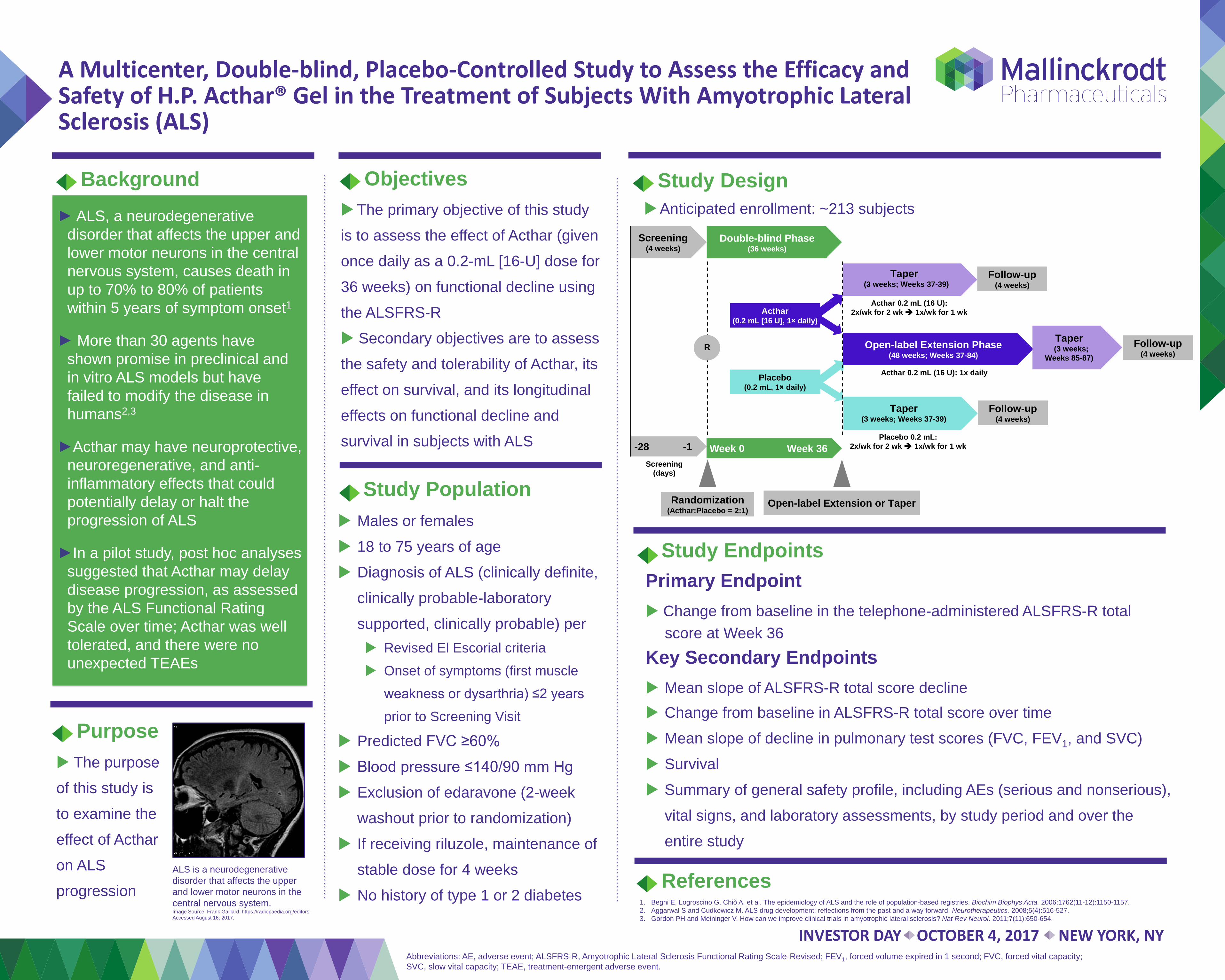
Slope estimates for weeks 8 and 36 also demonstrated that there was significantly less of a decline in ALSFRS total score in RCI-treated patients than in placebo-treated controls (Figure 2)
● Week 8 slopes: RCI, 0.003; placebo, −0.034 (slope difference, 0.036 [95% CI: 0.009, 0.063]; p=0.009; Figure 2A)
● Week 36 slopes: RCI, −0.022; placebo, −0.030 (slope difference, 0.008 [95% CI: 0.000, 0.015]; p=0.048; Figure 2B)
Figure 2. Slope Estimates for Change From Baseline in ALSFRS Total Score
A. Week 8a
B. Week 36a
Abbreviations: ALSFRS, Amyotrophic Lateral Sclerosis Functional Rating Scale; PRO-ACT, Pooled Resource Open-Access Amyotrophic Lateral Sclerosis Clinical Trials.a The PRO-ACT control group is treated as the reference group. The intercepts of regression lines are based on the average baseline ALSFRS total score.
PRO-ACT Prediction Algorithm Analysis The actual 9-month rate of ALSFRS decline was slower than the predicted rate
(Table 3)
Table 3. Comparison of Actual Observed and Predicted Slopes of 9-Month ALSFRS Total Score Decline
Parameter Statistic
Actual Predicted
P-ValuePilot Study(n=21)
PRO-ACT Algorithm
(n=21)
BaselineMean±SD −0.51±0.57 −0.75±0.26
0.087Median −0.36 −0.79Range (−2.21, 0.35) (−1.20, −0.29)
Abbreviations: ALSFRS, Amyotrophic Lateral Sclerosis Functional Rating Scale; PRO-ACT, Pooled Resource Open-Access Amyotrophic Lateral Sclerosis Clinical Trials; SD, standard deviation.
We conducted a post hoc matched case-control analysis using historical placebo-treated controls from the PRO-ACT database and a second post hoc analysis using an award-winning prediction algorithm for ALS progression based on PRO-ACT data
Patients who received RCI in the pilot study had a significantly slower decline in ALSFRS total score than matched PRO-ACT placebo-treated controls
The actual observed slope of ALSFRS total score in the patients from the pilot study declined at a slower rate than predicted by the PRO-ACT algorithm
Findings from these 2 post hoc exploratory analyses suggest the potential for RCI to slow the rate of functional decline in patients with ALS
These findings provide a rationale for the ongoing larger controlled study of RCI efficacy in the treatment of ALS (ClinicalTrials.gov identifier: NCT03068754)
Summary and Conclusions
Matched Case-Control Analysis Historical controls were obtained from the placebo groups of 16 phase 2 and 3
studies (n≥80 each) conducted between 1990 and 2010 and 1 large observational study (n=8635) in PRO-ACT
A review of available literature was used to identify 7 variables associated with disease progression:
1. ALSFRS total score2. Time from symptom onset to enrollment (≥18 months; <18 months)3. Gender (male; female)4. Age at symptom onset (≥40 years; <40 years)5. Site of onset (limb; bulbar; limb and bulbar)6. Body mass index (≥18.5 kg/m2; <18.5 kg/m2)7. Baseline creatinine level (≥53.04 µmol/L; <53.04 µmol/L)
Patients treated with RCI in the study were matched with up to 3 PRO-ACT controls using all identified variables
Mean changes in ALSFRS total score from baseline at weeks 8 and 36 were compared among the RCI and control groups using a linear mixed-effects model with repeated measures, with treatment, time, and treatment-by-time interaction as fixed effects; baseline ALSFRS total score was adjusted in the mixed-effects model
p-values <0.05 were considered statistically significant
PRO-ACT Prediction Algorithm Analysis The actual observed slope for ALS progression after 36 weeks of RCI treatment in
the pilot study was calculated as follows:
We adapted an award-winning algorithm from the DREAM Phil Bowen ALS Prize4Life Challenge13 that was developed using PRO-ACT data to derive a predicted slope for ALS progression at week 36 using 50 baseline features including patient demographics, disease characteristics, treatment, and laboratory test results
● The algorithm was modified to use random forests and only the features that were available from the pilot study
● The modified algorithm was cross-validated using a subset of PRO-ACT data, with the root mean square error of the modified algorithm (0.517) demonstrating a better performance than that of the original algorithm (0.559)
Mean values of the actual observed and PRO-ACT algorithm−predicted slopes for the 21 patients who completed the pilot study through week 36 were compared using a 2-sided paired t test
p-values <0.05 were considered statistically significant
(ALSFRS [week 36] – ALSFRS [baseline])(month [week 36 visit – baseline visit])
References1. Andersen PM, Borasio GD, Dengler R, et al. EFNS task force on management of amyotrophic
lateral sclerosis: guidelines for diagnosing and clinical care of patients and relatives. Eur J Neurol. 2005;12(12):921-938.
2. Miller RG, Jackson CE, Kasarskis EJ, et al. Practice parameter update: the care of the patient with amyotrophic lateral sclerosis: multidisciplinary care, symptom management, and cognitive/behavioral impairment (an evidence-based review): report of the Quality Standards Subcommittee of the American Academy of Neurology. Neurology. 2009;73(15):1227-1233.
3. US Food and Drug Administration. FDA Approves Drug to Treat ALS. https://www.fda.gov/NewsEvents/Newsroom/PressAnnouncements/ucm557102.htm. Published May 5, 2017. Accessed September 13, 2017.
4. Berry JD, Cudkowicz ME. New considerations in the design of clinical trials for amyotrophic lateral sclerosis. Clin Investig (Lond). 2011;1(10):1375-1389.
5. Aggarwal S, Cudkowicz M. ALS drug development: reflections from the past and a way forward. Neurotherapeutics. 2008;5(4):516-527.
6. The Writing Group; on behalf of the Edaravone (MCI-186) ALS 19 Study Group. Safety and efficacy of edaravone in well defined patients with amyotrophic lateral sclerosis: a randomised, double-blind, placebo-controlled trial. Lancet Neurol. 2017;16(7):505-512.
7. Starowicz K, Przewlocka B. The role of melanocortins and their receptors in inflammatory processes, nerve regeneration and nociception. Life Sci. 2003;73(7):823-847.
8. Ahmed TJ, Montero-Melendez T, Perretti M, Pitzalis C. Curbing inflammation through endogenous pathways: focus on melanocortin peptides. Int J Inflam. 2013;2013:985815.
9. Caruso C, Carniglia L, Durand D, Scimonelli TN, Lasaga M. Astrocytes: new targets of melanocortin 4 receptor actions. J Mol Endocrinol. 2013;51(2):R33-R50.
10.Strand FL, Kung TT. ACTH accelerates recovery of neuromuscular function following crushing of peripheral nerve. Peptides. 1980;1(2):135-138.
11.Bakker LA, Schroder CD, van Es MA, Westers P, Visser-Meily JMA, van den Berg LH. Assessment of the factorial validity and reliability of the ALSFRS-R: a revision of its measurement model. J Neurol. 2017;264(7):1413-1420.
12.Neurological Clinical Research Institute (NCRI) at Massachusetts General Hospital. PRO-ACT. https://nctu.partners.org/ProACT. Accessed September 12, 2017.
13.Küffner R, Zach N, Norel R, et al. Crowdsourced analysis of clinical trial data to predict amyotrophic lateral sclerosis progression. Nat Biotechnol. 2015;33(1):51-57.
Acknowledgment and Funding Professional writing and editorial support was provided by Stefanie Dorlas, BMath,
BEd, of MedLogix Communications, LLC, Schaumburg, Illinois, under the direction of the authors and was funded by Mallinckrodt ARD, Inc.
Figure 1. Pilot Study Design
Abbreviations: R, randomization; RCI, repository corticotropin injection; SC, subcutaneous.
Treatment Period(8 weeks)
Open-label Extension(28 weeks; optional)
Week 0 Week 8
Randomization(4 RCI dosing
groups = 1:1:1:1)
Open-label Extension or Taper
R
RCI SCGroup 1: Group 2:80 U 2x/wk 24 U 1x/d
Group 3: Group 4:56 U 2x/wk 16 U 1x/d Follow-up
(1 week)
Taper(3 weeks)
Follow-up(1 week)
Week 36
Taper(3 weeks)
Time Point Statistic Pilot Study(RCI; n=43)
Matched PRO-ACT Controlsa
(placebo; n=106)
Baseline
n 43 106Mean±SD 27.8±5.55 27.2±6.31Median 28.0 28.0Range (16.0, 37.0) (13.0, 37.0)
Week 8
n 37 53Mean±SD 28.0±5.41 26.5±7.42Median 28.0 28.0Range (13.0, 37.0) (12.0, 38.0)
Change from baseline at week 8
Mean±SD −0.4±1.88 −2.0±3.32LS mean±SE −0.5±0.32 −0.9±0.20LS Mean difference±SE 0.4±0.3895% CI −0.4, 1.1p-Value 0.327
Week 36
n 21 89Mean±SD 24.1±8.11 20.9±9.10Median 25.0 22.0Range (7.0, 36.0) (1.0, 38.0)
Change from baseline at week 36
Mean±SD −4.3±4.71 −6.6±5.57LS Mean±SE −2.2±0.55 −3.6±0.32LS Mean difference±SE 1.4±0.6495% CI 0.2, 2.7p-Value 0.025
Abbreviations: ALSFRS, Amyotrophic Lateral Sclerosis Functional Rating Scale; CI, confidence interval; LS, least squares; PRO-ACT, Pooled Resource Open-Access Amyotrophic Lateral Sclerosis Clinical Trials; RCI, repository corticotropin injection; SD, standarddeviation; SE, standard error. a The PRO-ACT control group is treated as the reference group.
-15
-10
-5
0
5
10
0 10 20 30 40 50 60 70
ALS
FRS
Tot
al S
core
- C
hang
e fro
m B
asel
ine
Treatment Group RCI Control
Study Day
RCI Slope = 0.002531Control Slope = -0.033715
RCI vs. Control Slope = 0.036247 (p-value: 0.0087)
-30
-25
-20
-15
-10
-5
0
5
10
15
0 100 200 300
ALS
FRS
Tot
al S
core
- C
hang
e fro
m B
asel
ine
Treatment Group RCI
RCI Slope = -0.02206Control Slope = -0.029741
RCI vs. Control Slope = 0.00768 (p-value: 0.0482)
Study Day
Control
Investor Day_ALS.indd 1 10/2/17 7:34 AM

Mal
linck
rod
t, t
he
“M”
bra
nd
mar
k, t
he
Mal
linck
rod
t P
har
mac
euti
cals
logo
an
d o
ther
bra
nd
s ar
e tr
adem
arks
of
a M
allin
ckro
dt
com
pan
y. ©
20
14
Mal
linck
rod
t.
Follow-up Period
(Weeks 50-54/56)
TaperActhar 80 U SC 3x/week
Treatment of Proteinuria Due to Treatment-Resistant or Treatment-Intolerant Idiopathic Focal Segmental Glomerulosclerosis (FSGS): A 2-Part Prospective Study of H.P. Acthar® Gel (PODOCYTE)
Primary Endpoint
u Proportion of subjects who achieve CR or PR of proteinuria at Week 24
Study Endpoints
No
Remission
► Primary FSGS is a major cause of
idiopathic nephrotic syndrome and is
the most common primary glomerular
disorder causing end-stage renal
disease in the United States1
► Primary FSGS is a progressive
disorder; ~50% of affected patients
develop end-stage renal disease over
a period of 5 to 8 years2
► Current treatments for primary FSGS
are effective in <50% of patients and
are associated with significant side
effects3,4
► Acthar is approved to induce a
diuresis or remission of proteinuria in
nephrotic syndrome without uremia,
the idiopathic type, or that due to
lupus erythematosus
► Remission of proteinuria (complete
or partial) in FSGS is associated with
an improved renal survival rate5
► Data from a recently published case
series suggested that 29% of patients
with steroid-resistant or steroid-
dependent primary FSGS achieved
complete or partial remission of
proteinuria after treatment with Acthar6
Key Secondary Endpoints
u Time to first relapse in subjects with CR or PR at Week 24 during the
Randomized Maintenance Period
u Proportion of subjects
u Who maintain remission of proteinuria at Week 50
u With remission at Week 24 who experience relapse during the Randomized
Maintenance Period
u Change in eGFR, total cholesterol, and uPCR from Week 24 to Week 50 in
subjects with remission at Week 24
Background
u The primary objective of this study
is to confirm the efficacy of Acthar in
inducing remission of proteinuria in
subjects with primary FSGS who are
resistant to, or intolerant of, at least
1 previous immunosuppressive
therapy such as corticosteroids or
CNIs
u Secondary objectives are to
confirm the safety and tolerability of
Acthar and to evaluate its efficacy in
maintaining remission of proteinuria
Objectives Study Design
Study Population
u Adult subjects (≥18 years old)
u Primary FSGS diagnosis
confirmed by renal biopsy
u Nephrotic
u uPCR >3.0 mg/mg
u eGFR >30 mL/min/1.73m2
u Intolerance to or failure to achieve
complete or partial remission with
≥1 previous immunosuppressant
u Treatment with an ACEi/ARB ≥4
weeks before Screening
u Blood pressure ≤150/90 mm Hg
Acthar 80 U SC 2x/week
Follow-up
Placebo 2x/week
Remission
Achieved
If remission is achieved, subjects are randomized
to Acthar vs placebo for maintenance
If remission is not achieved, subjects have the option
of entering an OLE period
Follow-up
INVESTOR DAY OCTOBER 4, 2017 NEW YORK, NYAbbreviations: ACEi, angiotensin-converting enzyme inhibitor; ARB, angiotensin receptor blocker; CNI, calcineurin inhibitors; CR, complete remission; eGFR, estimated glomerular filtration rate;
OLE, open-label extension; PR, partial remission; SC, subcutaneous; uPCR, urine protein-to-creatinine ratio.
Taper Screening Acthar 80 U SC 3x/week
Screening(Days -63 to -3)
Open-label Treatment(Weeks 0-23)
Taper(Weeks 24-25)
Randomized Maintenance Treatment or OLE(Weeks 26-49)
Purposeu The purpose of this study is to
provide nephrologists with additional
clinical evidence regarding the efficacy
and safety of Acthar in subjects with
treatment-resistant or treatment-
intolerant FSGS
uAnticipated enrollment: ~236 subjects
References1. Malaga-Diequez L, Bouhassira D, Gipson D, Trachtman H. Novel therapies for FSGS: preclinical and clinical studies. Adv Chronic Kidney Dis. 2015;22(2):e1-6.
2. Korbet SM. Clinical picture and outcome of primary focal segmental glomerulosclerosis. Nephrol Dial Transplant. 1999;14(suppl 3):68-73.
3. Bose B, Cattran D, Toronto Glomerulonephritis Registry. Glomerular diseases: FSGS. Clin J Am Soc Nephrol. 2014;9(3):626-632.
4. Cattran DC, Alexopoulos E, Heering P, et al. Cyclosporin in idiopathic glomerular disease associated with the nephrotic syndrome: workshop recommendations. Kidney Int.
2007;72(12):1429-1447.
5. Troyanov S, Wall CA, Miller JA, Scholey JW, Cattran DC, Toronto Glomerulonephritis Registry Group. Focal and segmental glomerulosclerosis: definition and relevance of a partial
remission. J Am Soc Nephrol. 2005;16(4):1061-1068.
6. Hogan J, Bomback AS, Mehta K, et al. Treatment of idiopathic FSGS with adrenocorticotropic hormone gel. Clin J Am Soc Nephrol. 2013;8(12):2072-2081.

Mal
linck
rodt
, the
“M”
bran
d m
ark,
the
Mal
linck
rodt
Pha
rmac
eutic
als l
ogo
and
othe
r bra
nds a
re tr
adem
arks
of a
Mal
linck
rodt
com
pany
. © 2
017
Mal
linck
rodt
.
During the last several years, there has been tremendous expansion in the range of agents available to treat
multiple sclerosis (MS)1,2
● Several disease-modifying therapies (DMTs) are currently available, and several more are under investigation
DMTs reduce the occurrence of MS relapses, slow disability worsening, and decrease activity on
magnetic resonance imaging
Despite these advances in treatment, many patients with MS experience relapses
High-dose corticosteroid therapy (eg, with methylprednisolone) is the mainstay of acute treatment of MS
relapses3,4
● Results from randomized, double-blind clinical trials suggest that 19% to 35% of patients may not adequately
respond to this therapy5,6
For patients who do not respond to or are unable to tolerate high-dose corticosteroids, options for acute treatment
of relapses are limited
Incomplete recovery from MS relapses may contribute to accrual of disability, highlighting the importance of
effective relapse treatment4,7,8
Repository corticotropin injection (RCI; H.P. Acthar Gel) contains a porcine-derived analogue of
adrenocorticotropic hormone (ACTH) approved by the US Food and Drug Administration for treatment of MS
relapses in adults9
● Anti-inflammatory and immunomodulatory effects of ACTH in MS historically were attributed solely to its ability
to stimulate endogenous cortisol, but more recent evidence suggests that corticosteroid-independent
melanocortin receptor–mediated activity may contribute10
Study objectives
● Characterize the population of patients who receive RCI for MS relapses
● Identify treatment patterns, MS relapse recovery, and safety outcomes
This interim report summarizes data collected through October 27, 2016
A Prospective Observational Registry of H.P. Acthar® Gel for the Treatment of Multiple Sclerosis Relapse
Bryan R. Due, PhD1; Patricia K. Coyle, MD2; Patrice M. Becker, MD3; and Timothy Vollmer, MD4
1Clinical Sciences, Mallinckrodt Pharmaceuticals, Hampton, NJ; 2Stony Brook University, Stony Brook, NY; 3Science & Technology, Mallinckrodt Pharmaceuticals, Hampton, NJ; 4Department of Neurology, University of Colorado Denver, Aurora, CO
Presented at the 2017 Annual Meeting of the Consortium of Multiple Sclerosis Centers ● New Orleans, Louisiana ● May 24-27, 2017
RT01
Introduction
Methods Study Design
Ongoing multicenter, prospective, 24-month, observational registry study
Target enrollment: 260 patients at up to 60 sites (ie, neurology practices in the United States that treat adult
patients with MS)
Enrollment and Data Collection
Potentially eligible patients are recruited during routine care visits at the study sites
● Those who meet the study eligibility criteria (Table 1) and provide informed consent are enrolled
Each patient will be followed up for a minimum of 6 months and a maximum of 24 months
Data will be abstracted from patient medical records at predefined time points (Figure 1)
RCI will be obtained via the usual commercial channels for prescription medications
While receiving RCI, patients will record data on daily RCI use in electronic diaries (Figure 1)
Patients will also complete the following self-report instruments at the times specified in Figure 1
● 29-item Multiple Sclerosis Impact Scale, version 1 (MSIS-29v1)
● 6-question Work Productivity and Activity Impairment questionnaire for multiple sclerosis (WPAI:MS)
● 5-question healthcare resource utilization (HRU) questionnaire
The clinician assessments below will be administered at the times depicted in Figure 1
● Expanded Disability Status Scale (EDSS) and Functional System Score (FSS)
● Clinical Global Impression of Improvement (CGI-I) scale
Because patients may have >1 MS exacerbation during the follow-up period, the exacerbation at study enrollment
is defined as the index exacerbation, and subsequent exacerbations are defined as relapses ● Relapses that occur during the study will be followed up as specified for the index exacerbation (ie, for
6 months) or until the study ends (if the latter occurs before 6 months have elapsed) (Figures 1 and 2)
Results Patient Characteristics and Medication Use at Enrollment
As of October 27, 2016, 45 patients had enrolled in the study and provided data
Patient characteristics and medication use at enrollment are shown in Tables 2 and 3, respectively
23 patients (51%) had a history of insufficient treatment response to, intolerance of, or intravenous access
problems with high-dose corticosteroid therapy
Subject AE Term Considered Serious
A
Nausea No
Vomiting No
Headache No
B UTI Yes
C Trigeminal neuralgia No
D UTI No
Acute sinusitis No
E Asthenia Yes
F MS relapse Yes
Abbreviations: AE, adverse event; MS, multiple sclerosis; SAE, serious adverse event; UTI, urinary tract infection.
Conclusions Data from this ongoing study will expand current understanding of RCI use for the treatment of MS relapses and
will provide information regarding
● Characteristics of patients treated with RCI
● MS relapse treatment patterns
● Treatment response and MS relapse recovery
● RCI safety
● Characteristics of patients who experience additional MS relapses (ie, following the index exacerbation)
during the study period
The data collected to date suggest that RCI is typically dosed using a regimen of 80 U/d for a period of 5 days
● Additional data on RCI dosing and therapeutic response collected during the remainder of the study could be
used to explore possible clinical implications for the treatment of MS relapses
Study enrollment is anticipated to conclude by the end of 2017
References 1. Comi G, Radaelli M, Soelberg Sørensen P. Evolving concepts in the treatment of relapsing multiple sclerosis. Lancet.
2017;389(10076):1347-1356.
2. Doshi A, Chataway J. Multiple sclerosis, a treatable disease. Clin Med (Lond). 2016;16(suppl 6):s53-s59.
3. Berkovich R. Treatment of acute relapses in multiple sclerosis. Neurotherapeutics. 2013;10(1):97-105.
4. Kalincik T. Multiple sclerosis relapses: epidemiology, outcomes and management. A systematic review. Neuroepidemiology. 2015;44(4):199-214.
5. Le Page E, Veillard D, Laplaud DA, et al. Oral versus intravenous high-dose methylprednisolone for treatment of relapses in patients with
multiple sclerosis (COPOUSEP): a randomised, controlled, double-blind, non-inferiority trial. Lancet. 2015;386(9997):974-981.
6. Sellebjerg F, Frederiksen JL, Nielsen PM, Olesen J. Double-blind, randomized, placebo-controlled study of oral, high-dose
methylprednisolone in attacks of MS. Neurology. 1998;51(2):529-534.
7. Goodin DS, Reder AT, Bermel RA, et al. Relapses in multiple sclerosis: relationship to disability. Mult Scler Relat Disord. 2016;6:10-20.
8. Lublin FD, Baier M, Cutter G. Effect of relapses on development of residual deficit in multiple sclerosis. Neurology. 2003;61(11):1528-1532.
9. H.P. Acthar Gel [package insert]. Hazelwood, MO: Mallinckrodt Pharmaceuticals; 2015.
10. Arnason BG, Berkovich R, Catania A, Lisak RP, Zaidi M. Mechanisms of action of adrenocorticotropic hormone and other melanocortins
relevant to the clinical management of patients with multiple sclerosis. Mult Scler. 2013;19(2):130-136.
11. Polman CH, Reingold SC, Banwell B, et al. Diagnostic criteria for multiple sclerosis: 2010 revisions to the McDonald criteria. Ann Neurol. 2011;69(2):292-302.
Acknowledgments and Funding Professional writing and editorial support was provided by Elizabeth Barton, MS, of MedLogix Communications, LLC, Schaumburg, Illinois,
under the direction of the authors. United BioSource Corporation® was the contract research organization for this study. Funding was provided
by Mallinckrodt Pharmaceuticals.
RCI Use
Data on RCI use have been collected for 31 patients and are summarized in Table 4
Safety
9 adverse events (AEs), including 3 serious adverse events (SAEs), have been reported (Table 5)
All SAEs were considered not related or unlikely related to RCI, and all patients recovered
Inclusion Age ≥18 years
Clinically definite relapsing form of MS according to McDonald criteria (2010 revision)11
Acute MS exacerbation as determined by treating clinician
Planning to initiate RCI therapy for acute MS exacerbation
Exclusion Diagnosis of progressive MS
Requirement for concomitant corticosteroid therapy
Receiving experimental drug therapy
History (within 5 years) of scleroderma, systemic fungal infections, ocular herpes simplex, or cancer
Recent surgery or a history (within 6 months) or presence of a peptic ulcer, congestive heart failure, or sensitivity to
proteins of porcine origin
Pregnancy, breastfeeding, or (if woman of childbearing potential) unwillingness to use appropriate contraception
Abbreviations: MS, multiple sclerosis; RCI, repository corticotropin injection.
Table 1: Key Inclusion and Exclusion Criteria
Figure 1. Patient Enrollment and Data Collection Overviewa,b
a Patients who experience post-enrollment relapses while the study is ongoing will be followed up for the relapse as specified for the index
exacerbation (ie, for 6 months) or until study close (if the latter occurs before 6 months have elapsed). Index exacerbation is defined as the MS
exacerbation at study enrollment. b SAEs will be reported within 24 hours of identification, even when outside of usual care visits.
Abbreviations: MS, multiple sclerosis; SAE, serious adverse event.
Figure 2. Data Collection Schematic With Case Examples
Abbreviations: AE, adverse event; CGI-I, Clinical Global Impression of Improvement; EDSS, Expanded Disability Status Scale; FPFV, first patient
first visit; HRU, healthcare resource utilization; LPFV, last patient first visit; LPLV, last patient last visit; MSIS-29v1, 29-item Multiple Sclerosis
Impact Scale, version 1; WPAI:MS, Work Productivity and Activity Impairment questionnaire for multiple sclerosis.
Table 2. Patient Characteristics at Enrollment
Characteristic Initial Screening (N=45)
Age,a mean (SD), y 50.2 (10.7)
Gender, No. (%)
Male 5 (11)
Female 31 (69)
Missing 9 (20)
Race, No. (%)
Black/African American 4 (9)
White 29 (64)
Hispanic 1 (2)
No information/missing 11 (24)
EDSS score,b,c mean (SD) 4.4 (1.9)
Previous treatments for MS, No. (%)
Methylprednisolone 13 (29)
RCI 11 (24)
IV steroids (unspecified) 2 (4)
Prednisone 2 (4)
Glatiramer acetate 1 (2)
Teriflunomide 1 (2)
None 1 (2)
Unknown 1 (2)
No information 12 (27)
MSIS-29v1 physical section score,d,e mean (SD) 65.4 (19.4) a Data were available for 36 patients. b Data were available for 27 patients. c Rated on a scale from 0 (normal neurologic
exam) to 10 (death due to MS). d Data were available for 35 patients. e Scored on a scale from 0 to 100, with 100
representing the worst possible score.
Abbreviations: EDSS, Expanded Disability Status Scale; IV, intravenous; MSIS-29v1, 29-item Multiple Sclerosis Impact
Scale, version 1; RCI, repository corticotropin injection; SD, standard deviation.
Table 3. Summary of Medication Use at Enrollment (N=45)
Characteristic No. (%)
DMT use
Yes 32 (71)
No 5 (11)
Missing 8 (18)
Specific DMT usea
Dimethyl fumarate 15 (33)
Glatiramer acetate 9 (20)
Natalizumab 8 (18)
Alemtuzumab 5 (11)
Teriflunomide 5 (11)
Fingolimod 4 (9)
Interferon β-1a 2 (4)
Other concomitant medication/supplement usea,b
Cholecalciferol 10 (22)
Ergocalciferol 8 (18)
Baclofen 7 (16)
Fampridine 6 (13)
Gabapentin 6 (13)
Cyanocobalamin 5 (11)
Multivitamins 5 (11)
Amantadine 3 (7)
Levothyroxine sodium 3 (7)
Topiramate 3 (7)
a Some patients were receiving >1 medication at time of enrollment. b Only medications used by ≥5% of patients are listed. Abbreviations: DMT, disease-modifying therapy; RCI, repository corticotropin injection.
Median (IQR)
No. of doses per patient 5.0 (5.0)
Strength per dose, U 80 (0)
No. of days doseda 5.0 (5.0)
Total dose per day, U 80 (0) a RCI dosing was on 5 consecutive days for 22 patients (71%).
Note: RCI was injected subcutaneously in all patients who specified the mode of administration.
Abbreviations: IQR, interquartile range; RCI, repository corticotropin injection.
Table 4. Summary of RCI Use (n=31)
Table 5. Summary of AEs
Mal
linck
rod
t, t
he
“M”
bra
nd
mar
k, t
he
Mal
linck
rod
t P
har
mac
euti
cals
logo
an
d o
ther
bra
nd
s ar
e tr
adem
arks
of
a M
allin
ckro
dt
com
pan
y. ©
20
14
Mal
linck
rod
t.
Study Population
Adult males or females with RRMS
Onset of relapse ≤25 days before the
Baseline visit
Treatment with corticosteroids within
10 days of relapse symptom onset
IVMP (1 g/day) OR
Oral prednisone (1250 mg/day) OR
Oral methylprednisolone
(1000 mg/day)
Failure to improve by at least 1 point
in 1 or more FSS functions 14 days
after the first dose of corticosteroids
An EDSS score of 3.5 to 6.5 at the
Baseline visit
► MS is a chronic neurodegenerative
disease characterized by
demyelination within the CNS
► According to the Multiple Sclerosis
Foundation, between 300,000 and
400,000 people in the United States
and ~2.5 million worldwide are
estimated to have MS; MS is more
common in women than men (3:1
ratio) and is most commonly
diagnosed between 20 and 40 years of
age, although it can develop in young
children, teenagers, and older adults1
► The most common form of MS is
RRMS, in which patients experience
episodes of worsening neurological
function followed by periods of partial
or complete recovery
►MS relapses are treated with high-
dose corticosteroids or ACTH for
patients who do not respond to or
tolerate corticosteroids
► In randomized trials, 20%-35% of
patients treated with corticosteroids do
not have significant improvements in
MS relapses or related symptoms2,3
► Acthar is approved for treating MS
exacerbations and is an option for
patients who do not respond to
standard of care treatment with high-
dose corticosteroids4-6
INVESTOR DAY OCTOBER 4, 2017 NEW YORK, NY
A Multicenter, Randomized, Double-blind, Placebo-Controlled Parallel-Group, Pilot Study to Assess the Efficacy and Safety of H.P. Acthar® Gel in Subjects With Relapsing-Remitting Multiple Sclerosis (RRMS)
Primary EndpointsEfficacy
Response rate based on the EDSS on Day 42 for each treatment group
Safety
Summary of safety profile, including AEs (serious and nonserious), vital signs,
and laboratory assessments, by study period and over the entire study
Study Endpoints
Background
PurposeThe purpose of this study is to
determine the efficacy and safety of
Acthar in subjects with RRMS who have
not responded adequately to treatment
with high-dose corticosteroids
The primary objectives of this study
are to generate an estimate of the
response rate for Acthar and assess its
safety and tolerability in subjects with
RRMS who have not responded to
high-dose IVMP, oral prednisone, or
oral methylprednisolone
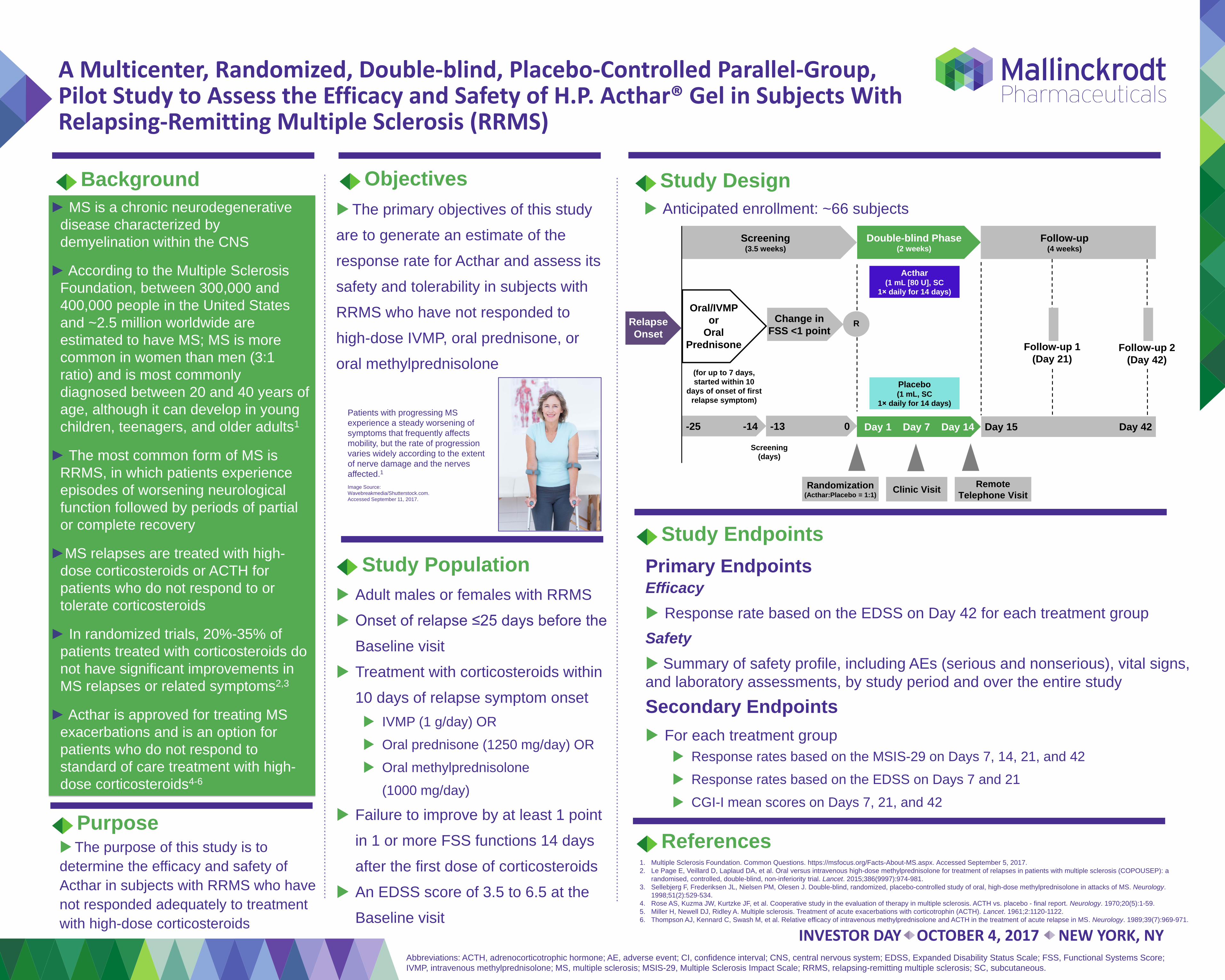
Objectives Study Design
Abbreviations: ACTH, adrenocorticotrophic hormone; AE, adverse event; CI, confidence interval; CNS, central nervous system; EDSS, Expanded Disability Status Scale; FSS, Functional Systems Score;
IVMP, intravenous methylprednisolone; MS, multiple sclerosis; MSIS-29, Multiple Sclerosis Impact Scale; RRMS, relapsing-remitting multiple sclerosis; SC, subcutaneous.
Secondary Endpoints
For each treatment group
Response rates based on the MSIS-29 on Days 7, 14, 21, and 42
Response rates based on the EDSS on Days 7 and 21
CGI-I mean scores on Days 7, 21, and 42
Patients with progressing MS
experience a steady worsening of
symptoms that frequently affects
mobility, but the rate of progression
varies widely according to the extent
of nerve damage and the nerves
affected.1
Image Source:
Wavebreakmedia/Shutterstock.com.
Accessed September 11, 2017.
Screening(3.5 weeks)
Double-blind Phase(2 weeks)
-25 -14
Screening
(days)
Day 1 Day 7 Day 14
Relapse
Onset
-13 0
Oral/IVMP
or
Oral
Prednisone
(for up to 7 days,
started within 10
days of onset of first
relapse symptom)
RChange in
FSS <1 point
Acthar(1 mL [80 U], SC
1× daily for 14 days)
Placebo(1 mL, SC
1× daily for 14 days)
Follow-up(4 weeks)
Randomization(Acthar:Placebo = 1:1)
Day 15 Day 42
Follow-up 1
(Day 21) Follow-up 2
(Day 42)
Clinic VisitRemote
Telephone Visit
References1. Multiple Sclerosis Foundation. Common Questions. https://msfocus.org/Facts-About-MS.aspx. Accessed September 5, 2017.
2. Le Page E, Veillard D, Laplaud DA, et al. Oral versus intravenous high-dose methylprednisolone for treatment of relapses in patients with multiple sclerosis (COPOUSEP): a
randomised, controlled, double-blind, non-inferiority trial. Lancet. 2015;386(9997):974-981.
3. Sellebjerg F, Frederiksen JL, Nielsen PM, Olesen J. Double-blind, randomized, placebo-controlled study of oral, high-dose methylprednisolone in attacks of MS. Neurology.
1998;51(2):529-534.
4. Rose AS, Kuzma JW, Kurtzke JF, et al. Cooperative study in the evaluation of therapy in multiple sclerosis. ACTH vs. placebo - final report. Neurology. 1970;20(5):1-59.
5. Miller H, Newell DJ, Ridley A. Multiple sclerosis. Treatment of acute exacerbations with corticotrophin (ACTH). Lancet. 1961;2:1120-1122.
6. Thompson AJ, Kennard C, Swash M, et al. Relative efficacy of intravenous methylprednisolone and ACTH in the treatment of acute relapse in MS. Neurology. 1989;39(7):969-971.
Anticipated enrollment: ~66 subjects

Mal
linck
rodt
, the
“M”
bran
d m
ark,
the
Mal
linck
rodt
Pha
rmac
eutic
als l
ogo
and
othe
r bra
nds a
re tr
adem
arks
of a
Mal
linck
rodt
com
pany
. © 2
015
Mal
linck
rodt
.
METHODS
Treatment with repository corticotropin injection reduces the progression of experimental autoimmune uveitis in rats Dale Wright, Ben Zweifel, Chris Bollinger, Kyle Hayes & Rick Fitch Mallinckrodt Autoimmune & Rare Disease Inc., Hampton, NJ
Purpose: Previous studies have suggested that
melanocortin receptor (MCR) agonists play a role in
regulating the progression and resolution of experimental
autoimmune uveitis (EAU). Repository corticotropin
injection (RCI: H.P. Acthar® Gel) is a complex formulation
containing a porcine ACTH analogue. ACTH is a
melanocortin peptide that binds to the 5 known MCRs.
Because RCI is an FDA-approved treatment for certain
inflammatory ocular disorders, the aim of this study was to
investigate the effects of RCI on a preclinical model of
EAU.
Method: Lewis rats were immunized with
interphotoreceptor retinoid binding protein (IRBP) peptide
(1177-1191) in complete Freud’s adjuvant. Inflammation
was observed under a dissection microscope on days 4, 7,
11 and 14 post immunization and disease was clinically
scored (as described in Figure 1) on a scale of 0-4 based
on their anterior clinical disease. Animals were
subcutaneously dosed with RCI (10, 40, or 400 IU/kg),
Placebo gel (5mL/kg) or Prednisolone (0.1, 1, or 5 mg/kg)
every other day starting on the first day of the study. On
day 14, whole eyes were collected, processed and
sections were stained with hematoxylin & eosin and
scored.
Results: Clinical assessment within the anterior chamber
of the eye performed in a blinded manner, demonstrated
that RCI administered at 40 or 400 IU/kg significantly
reduced the ocular clinical disease score on day 14
compared to placebo (0.93 ± 0.18 and 0.85 ± 0.17 versus
1.98 ± 0.22, respectively), (p≤ 0.01). In contrast, prednisolone marginally reduced the clinical disease
score, at the doses tested, with only the 1 mg/kg dose
having significance (1.05 ± 0.18; p ≤ 0.05). In addition, the clinical findings for RCI were supported by the histological
data, showing protection to the retinal architecture with a
reduction in inflammation at all 3 doses evaluated.
Conclusions: The treatment of EAU with RCI resulted in
the suppression of the ocular clinical score and
inflammation reducing retinal damage. These data are the
first to explore the effects of RCI in a preclinical model of
experimental autoimmune uveitis.
FIGURE 2: HISTOPATHOLOGY SCORE Following the clinical evaluations on Day 14, animals were euthanized and
eyes processed for histology. A blinded grader analyzed the slides and
scored utilizing the scoring system below. All concentrations of RCI tested
provided significant protection (p>0.01, one-way ANOVA Dunnett’s test
versus placebo control group) to the retina. The high dose prednisolone
also significantly reduced the scores compared to the placebo group
(p>0.05, one-way ANOVA Dunnett’s test). Representative histopathology
photos
SUMMARY ABSTARCT
FIGURE 1: Clinical Evaluations: Animals were observed under a dissection microscope and
scored on a scale of 0-4 based on their anterior clinical disease. Representative images of the
anterior chamber were taken at the time of clinical evaluations (A). Treatment with RCI
significantly reduced the ocular clinical score (mean ± SEM) on Days 11 (B) and 14 (C) for the
mid and high doses. Treatment with prednisolone showed a trend in the clinical score
reduction, with significance at the 5 mg/kg dose on day 11 and for the 1 mg/kg dose on day 14.
(*p < 0.05; ***p < 0.01; ***p < 0.001 ****p < 0.0001) one-way ANOVA Dunnett’s test.
IRBP-induced uveitis was successfully induced in Lewis
rats.
Disease control animals showed clinical signs of EAU, with
increased redness, neovascularization, and haziness in the
anterior chamber.
Treatment with RCI at 40 and 400 IU/kg significantly
alleviated clinical signs on Days 11 and 14.
Prednisolone tested at three different concentrations
showed a trend in reduced symptoms; however, only the
high dose (5 mg/kg) on Day 11 and the mid dose (1 mg/kg)
on Day 14 scored significantly less than placebo.
Histopathology scores supported the clinical findings.
All three doses of RCI tested provided protection to the
retinal architecture in addition to a reduction in the ocular
inflammation.
Prednisolone at 5 mg/kg) also significantly reduce the
retinal damage and ocular inflammation versus the
placebo.
Melanocortin receptors are uniquely expressed within the
eye.
Using quantitative-PCR, expression of Mc1r and Mc5r was
seen in the eye. The level of mRNA for Mc2r showed weak
expression.
Utilizing in situ hybridization, Mc1r and Mc5r in the retina.
RESULTS
Table 1. Melanocortin receptor expression in rat ocular tissue.
+, mRNA expression CT ≤ 31; (+) weak mRNA expression, 32 ≤ CT < 34; no mRNA expression, CT value ≥ 34.
Mc1r Mc2r Mc3r Mc4r Mc5r
+ (+) - - +
Placebo Control RCI 10 IU/kg RCI 40 IU/kg RCI 400 IU/kg
Prednisolone 0.1mg/kg Prednisolone 1.0 mg/kg Prednisolone 5.0 mg/kg FTY-720 0.3 mg/kg
Histology Scoring Scale
0: No disease, normal retinal architecture.
0.5: Trace. <1/4 Mild inflammatory cell infiltration of the retina with or
without photoreceptor damage.
1: ≥1/4 Mild inflammation and/or photoreceptor outer segment damage
2: ≥1/4 Mild to moderate inflammation and/or lesion extending to the outer nuclear layer.
3: ≥1/4 Moderate to marked inflammation and/or lesion extending to the inner nuclear layer.
4: ≥1/4 Severe inflammation and/or full-thickness retinal damage.
Clinical Scoring Scale
0-0.5: No disease; eye is translucent. Some blood vessels in the iris may be dilated.
1: Engorged blood vessels in iris; abnormal pupil contraction (or dilation).
2: Slight haziness to the anterior chamber.
3: Moderately opaque anterior chamber, but pupil still visible.
4: Opaque anterior chamber and obscured pupil
Treatment
Group
20 eyes/group
Table 2: Individual scoring of the
histopathology evaluations.
Significant values (p>0.05) are
highlighted in green for reduced
inflammatory damage compared
to placebo control. Significant
difference, as measured by the
board certified pathologist, are
highlighted in green (p ≤ 0.05, one-way ANOVA Dunnett’s test)
OD OS
Placebo 2.43 ± 1.30 2.40 ± 1.15
RCI 10 IU/kg 1.15 ± 0.71 1.43 ± 1.01
RCI 40 IU/kg 1.25 ± 0.75 1.50 ± 0.84
RCI 400 IU/kg 1.00 ± 0.53 1.05 ± 0.44
Pred 0.1 mg/kg 1.70 ± 0.89 1.75 ± 0.68
Pred 1.0 mg/kg 1.70 ± 0.89 1.65 ± 1.08
Pred 5.0 mg/kg 1.25 ± 0.82 1.20 ± 0.67
FTY720 0.3
mg/kg 0.75 ± 1.28 0.58 ± 0.79
A
C B
In situ hybridization was performed using the
Advanced Cell Diagnostics RNAScope ISH platform.
Custom designed ISH probe to rat Mc1r and Mc5r
mRNA were use on formalin fixed, paraffin
embedded sections. Images of retinal expression,
focused within the outer nuclear layer with some
minor expression in the inner layer.
negative control
Mc5r
Mc1r
Studies were performed by Ophthalmic Research Associates Inc.,
Andover MA
CT = cycle time
Placebo Control
Score: 2.5
RCI 10 IU/kg
Score: 1.5
RCI 40 IU/kg
Score: 1.5
RCI 400 IU/kg
Score: 1.0
Prednisolone 0.1mg/kg
Score: 2.0
Prednisolone 1.0 mg/kg
Score: 1.5
Prednisolone 5.0 mg/kg
Score: 1.5
FTY-720 0.3 mg/kg
Score: 0.5
CONCULSION
We show RCI (H.P. Acthar® Gel) can reduces the progression
of IRBP-induced uveitis. Additional studies will help elucidate
RCI’s mechanism(s) of action for immune suppression in
uveitis. However, these data suggest RCI's potential anti-
inflammatory effect in uveitis.
Pla
ceb
o
RC
I 10 IU
/kg
RC
I 40 IU
/kg
RC
I 400 IU
/kg
Pre
dn
iso
lon
e 0
.1 m
g/k
g
Pre
dn
iso
lon
e 1
mg
/kg
Pre
dn
iso
lon
e 5
mg
/kg
FT
Y-7
20 0
.3 m
g/k
g
0 .0
0 .5
1 .0
1 .5
2 .0
2 .5
3 .0
C lin ic a l S c o re D a y 1 1
Cli
nic
al
Sc
ore
± S
EM
**** ****
*
****
Pla
ceb
o
RC
I 10 IU
/kg
RC
I 40 IU
/kg
RC
I 400 IU
/kg
Pre
dn
iso
lon
e 0
.1 m
g/k
g
Pre
dn
iso
lon
e 1
mg
/kg
Pre
dn
iso
lon
e 5
mg
/kg
FT
Y-7
20 0
.3 m
g/k
g
0 .0
0 .5
1 .0
1 .5
2 .0
2 .5
C lin ic a l S c o re D a y 1 4
Cli
nic
al
Sc
ore
± S
EM
**** ***
*
Mal
linck
rodt
, the
“M”
bran
d m
ark,
the
Mal
linck
rodt
Pha
rmac
eutic
als l
ogo
and
othe
r bra
nds a
re tr
adem
arks
of a
Mal
linck
rodt
com
pany
. © 2
015
Mal
linck
rodt
.
METHODS
Suppression of acute uveitis following treatment with repository corticotropin injection Dale Wright, Ben Zweifel, Luke Oh, Prabha Sharma, Chris Bollinger, Kyle Hayes & Rick Fitch Mallinckrodt ARD Inc., Hampton, NJ
Repository corticotropin injection (RCI: H.P. Acthar® Gel) contains a
purified porcine pituitary ACTH analogue, and is an FDA-approved
treatment for several inflammatory eye diseases. ACTH binds to all 5
known melanocortin receptors and may suppress inflammation by
steroid-dependent and independent pathways. Endotoxin-induced
uveitis (EIU) in rodents is a useful experimental model to investigate
mechanism of action and pharmacological efficacy of potential
treatments. This study was conducted to investigate the potential anti-
inflammatory benefit of RCI in an acute rat model of EIU. EIU is
characterized by clinically relevant signs of inflammation, including
elevated inflammatory cytokines and cellular inflammation in the
anterior and vitreous chambers. Rats (16/group) were treated with
dexamethasone (Dex), placebo, or RCI at 160 IU/kg, 400 IU/kg or 800
IU/kg following EIU induction. Eyes were clinically examined at pre-
challenge, 6-8, 24, and 48 hours post challenge using the Combined
Draize and McDonald – Shadduck Scoring System. We show that RCI treatment significantly reduced ocular inflammation
and inflammatory cytokines in an EIU model of acute uveitis. The
mechanism of action of RCI may involve more than the induction of
corticosteroids, and will be explored further in future studies. MATERIAL & METHODS
Induction of Endotoxin-induced Uveitis. Female Sprague Dawley
rats were administered a single subcutaneous injection on Day 0 and
Day 1 of Dexamethasone (Dex) at 2 mg/kg (Group 3), Placebo gel
(Group 4), or RCI gel at 160 IU/kg (Group 5), 400 IU/kg (Group 6) or
800 IU/kg (Group 7). The non-treated group was used as a control for
disease induction (Group 2). EIU was induced by footpad injection with
100 µL of lipopolysaccharide (LPS) at 10 mg/mL. Clinical evaluation of
animals was conducted using slit−lamp and scored according to the
Combined Draize and McDonald-Shadduck Scoring System and the
Ocular Posterior Segment Scoring Scale. Animals were euthanatized
24 and 48 hours (8/group/time) after disease induction, one eye/animal
was collected, fixed and paraffin-embedded. Five sagittal sections for
each eye were stained with hematoxylin and eosin and assessed by a
board certified Pathologist for inflammatory cell infiltration, hemorrhage,
necrosis, congestion, edema using a scale from 0 to 5 as follows: 0=
normal, 1 = minimal, 2 = mild, 3 = moderate, 4 = marked, or 5 =
severe. Scores were combined to give a total inflammatory score for
each section.
Tissue cytokine expression. To evaluate cytokine expression, protein
extracts were isolated from Rat Retina/Uveal Tract of the other eye.
Tissue lysates were assayed for IL-1α, IL-6, MCP-1, MIP-2 and TNF-α
using Millipore's Milliplex Rat Cytokine/Chemokine Magnetic Bead
Panel (EMD Millipore; RECYTMAG-65K) on the Luminex 100 platform.
LPS-Induced TNF-α Production. RCI or vehicle was administered
subcutaneously at a volume of 5 ml/kg. LPS (Escherichia coli serotype
0111:B4; Sigma-Aldrich) was administered 1.0 h after compound
injection at a dose of 300 ug/rat in a volume of 0.5 ml. Blood was
collected in serum separator tubes via cardiac puncture 90 min after
LPS injection, a time point corresponding to maximal TNF-α production.
FIGURE 5: Reduction in inflammatory cytokines MIP-2, IL-1α, and TNFα
following treatment with RCI. Eyes were collected at 18 hours. Tissue
lysates were tested for IL-1α, IL-6, MCP1, MIP2, and TNFα. Only MIP2
(A) and IL-1α (B) showed a response to LPS challenge at 18 hours, and
both were dose-dependently decreased by RCI. To examine the effects of
RCI on TNFα (C) production, Sprague Dawley rats were treated with RCI
(10, 20, 40, 80 U/kg) and challenged with LPS. RCI significantly reduced
serum TNFα levels. (p ≤ 0.01, one-way ANOVA Dunnett’s test versus
disease control group)
SUMMARY INTRODUCTION
FIGURE 2. RCI treatment reduced the ocular inflammation score in the
Endotoxin-induced Uveitis model, (A) 18 hour and (B )48 hour post-LPS
injection. Symbols represent individual animal scores with mean +/-
SEM. RCI at 160 U/kg significantly reduced ocular inflammation at 48
hours whereas 400 U/kg and 800 U/kg (****p < 0.0001, one-way ANOVA
Dunnett’s test) significantly inhibited ocular inflammation at all time
points evaluated compared to Disease control animals.
Acute uveitis was successfully induced in Sprague Dawley rats. It was
manifested in the anterior chamber of the eye, peaked at 18-24 hours and
maintained out to 48 hours. Treatment with repository corticotropin injection
significantly reduced ocular inflammation in a dose-dependent manner.
RCI at 400 U/kg showed comparable level of reduction in clinical ocular
symptoms to the positive control dexamethasone while treatment at 800
U/kg was shown to be even more efficacious. Pro-inflammatory
chemokines and cytokines are thought to have a role in the recruitment of
inflammatory cells and pathogenesis in uveitis. Cytokines such as IL-6 and
TNF- α have been implicated the various clinical subtypes of uveitis, with
aqueous humor levels correlating with disease severity (1,2). IL-1α and
MIP2 were increased following LPS-induced uveitis. Treatment with RCI
shows a dramatic and dose responsive reduction on the levels in the
retina/uveal tract. Furthermore, in an LPS-induced TNF model, RCI
significantly reduced TNFα production. The expression of Mc1r and Mc5r
was show in the retina using quantitative PCR and in situ hybridization. In
this study, RCI at 160, 400, and 800 U/kg showed dose-dependent
suppression of the ocular inflammation and inflammatory cytokines induced
in an experimental uveitis rat model. Additional studies are needed to
elucidate RCI’s mechanism(s) of action for immune suppression in uveitis.
However, these data support the use of RCI in ophthalmic diseases.
REFERENCES
1. Rosenbaum JT, McDevitt RO, Guss RB, Egbert PR. (1980) Endotoxin-
induce uveitis in rats as a model for human disease. Nature 286:611-
613.
2. Ooi KG, Galatowicz G, Calder VL, Lightman SL. (2006) Cytokines and
chemokines in uveitis: is there a correlation with clinical phenotype? Clin
Med Res 4:294-309.
FIGURE 3: Treatment with RCI decreases the summed histopathology
score 48 hour post LPS. Endotoxin-induced Uveitis leads to an acute
inflammatory response composed largely of neutrophils and macrophages.
This leads to an inflammatory response that is observed predominantly in
the anterior chamber segment of the eye. (p ≤ 0.01, one-way ANOVA
Dunnett’s test versus disease control group)
FIGURE 4: HISTOPATHOLOGY IMAGES. Sagittal sections were
stained with hematoxylin and eosin and assessed for inflammation and
edema. Ocular inflammation at 48 hours showed an increase in
neutrophils and macrophage in the anterior chamber, ciliary body and
retina. Treatment with RCI dose-dependently reduced inflammation,
edema, and the presence of proteinaceous fluid. A, Group 1; B, Group 2;
C, Group 3; D, Group 4l; E, Group 5; F, Group 6; G, Group 7
A
F E D
C B
G
RESULTS
Treatment Group Ocular Clinical
Score (mean ±
sem)
IL-1α (pg/ml)
(mean ± sem)
MIP2 (pg/ml)
(mean ± sem)
Group 1
(Naïve) 0 37 ± 8.5* 62 ± 6.0*
Group 2
(Disease Control) 15.5 ± 1.2 207 ± 69 240 ± 47
Group 3
(Dex 2 mg/kg) 2.6 ± 0.5* 54 ± 11* 92 ± 10*
Group 4
(Placebo) 18.4 ± 1.6 135 ± 33 175 ± 28
Group 5
(RCI 160 IU/kg) 9.4 ± 1.0* 130 ± 35 158 ± 43
Group 6
(RCI 400 IU/kg) 2.0 ± 0.3* 96 ± 20 114 ± 18*
Group 7
(RCI 800 IU/kg) 0.9 ± 0.2* 51 ± 8.3* 126 ± 32*
* = p ≤ 0.05 in a one-way ANOVA Dunnett’s test versus disease control group
A
C
B
negative control
Mc5r
Mc1r
Figure 1. In situ hybridization was performed using the Advanced Cell
Diagnostics RNAScope ISH platform. Custom designed ISH probes to
rat Mc1r and Mcr mRNA were used on formalin fixed, paraffin
embedded sections. Images of retinal expression, focused within the
outer nuclear layer with some minor expression in the inner layer.
Table 1. Melanocortin receptor expression in rat ocular tissue. +,
mRNA expression CT ≤ 31; (+) weak mRNA expression, 32 ≤ CT < 34; no mRNA expression, CT value ≥ 34.
Mc1r Mc2r Mc3r Mc4r Mc5r
+ (+) - - +
CT = cycle time
To
tal
Oc
cu
lar I
nfl
am
ma
tory
Sc
ore
(m
ea
n+
SE
M)
Naiv
e
Dis
ease C
on
tro
l
Dex 2
mg
/kg
Pla
ceb
o G
el
RC
I 160 U
/kg
RC
I 400 U
/kg
RC
I 800 U
/kg
0
1 0
2 0
3 0
****
****
****
To
tal
Oc
cu
lar I
nfl
am
ma
tory
Sc
ore
(m
ea
n+
SE
M)
Naiv
e
Dis
ease C
on
tro
l
Dex 2
mg
/kg
Pla
ceb
o G
el
RC
I 160 U
/kg
RC
I 400 U
/kg
RC
I 800 U
/kg
0
1 0
2 0
3 0
****
****
********
A B
Su
mm
ed
His
top
ath
Sc
ore
s
Naiv
e
Dis
ease C
on
tro
l
Dex 2
mg
/kg
Pla
ceb
o G
el
RC
I 160 U
/kg
RC
I 400 U
/kg
RC
I 800 U
/kg
0
5
1 0
1 5
**
**
****
********
LP
S C
on
tro
l
Pla
c e bo 1 0 2 0 4 0 8 0
0
1 0 0 0
2 0 0 0
3 0 0 0
4 0 0 0
5 0 0 0
L P S In d u c e d T N F α
TN
Fα
(p
g/m
l)
R C I IU //k g )
** ** **
Acknowledgement: EIU was performed by Toxikon Corporation Bedford, MA

Mal
linck
rodt
, the
“M”
bran
d m
ark,
the
Mal
linck
rodt
Pha
rmac
eutic
als l
ogo
and
othe
r bra
nds a
re tr
adem
arks
of a
Mal
linck
rodt
com
pany
. © 2
014
Mal
linck
rodt
.
► RA is an autoimmune disorder
characterized by chronic inflammation,
articular erosions, and periarticular
bone loss; prevalence is estimated at
0.5%-1% of the adult population in
developed countries, with an annual
incidence rate of 5-50 new cases per
100,0001
►The goal of treatment is focused on
achieving remission (absence of
inflammatory disease); low disease
activity is an acceptable alternative
► Disease-modifying antirheumatic
drugs (DMARDs) and corticosteroids
are commonly used to manage RA,
but 28%-58% of patients do not
achieve a minimal 20% improvement
in ACR criteria (ACR20)2; those
patients who achieve improvement can
experience a waning in response3
► Acthar is approved as an adjunctive
therapy for short-term administration
(to tide the patient over an
exacerbation) in RA (selected cases
may require low-dose maintenance
therapy). An open-label single-center
study suggested that 12 weeks of
Acthar was an effective add-on therapy
for patients with active RA refractory to
at least 3 therapeutic agents with
different mechanisms of action4
INVESTOR DAY OCTOBER 4, 2017 NEW YORK, NY
A Multicenter, 2-Part Study to Assess the Efficacy and Safety of H.P. Acthar® Gel in Subjects With Rheumatoid Arthritis (RA) With Persistently Active Disease
Primary Endpoint Proportion of subjects with DAS28-ESR ˂3.2 at Week 12
Study Endpoints
Background
The primary objective of this study is
to assess the efficacy of Acthar given as
a 1-mL (80 U) dose 2x/week for
12 weeks as determined by DAS28-ESR
in subjects with persistently active RA
Secondary objectives are to assess
the safety and tolerability of Acthar as
well as its efficacy to maintain LDA (in
subjects who have achieved LDA after
12 weeks of treatment)
Objectives Study Design
Study Population
Diagnosis of RA screening
Per 2010 ACR/EULAR classification
Persistently active disease
DAS28-ESR ˃3.2 (at screening and
baseline)
Corticosteroid, MTX, DMARD use for
≥12 weeks prior to screening* Stable prednisone dose (5-10 mg) for
≥4 weeks prior to screening
MTX ≤20 mg per week + 1 allowed
biologic or nonbiologic DMARD OR
1 allowed biologic DMARD
*All starting dose levels must remain stable through the study duration.
Abbreviations: ACR, American College of Rheumatology; ACR20, 20% improvement in ACR criteria; ACTH, adrenocorticotropic hormone; AE, adverse event; CDAI, Clinical Disease Activity Index;
DAS28-ESR, Disease Activity Score with 28 joint count and ESR; DMARD, disease-modifying antirheumatic drug; ESR, erythrocyte sedimentation rate; EULAR, European League Against Rheumatism;
LDA, low disease activity; MTX, methotrexate; RA, rheumatoid arthritis.
Secondary Endpoints
Proportion of subjects
Who maintained DAS28-ESR ˂3.2 for Weeks 12-24
With CDAI ≤10 at Week 12
Who meet criteria for ACR20 at Week 12
Time to disease activity flare for Weeks 12-24
Summary of general safety profile (including the below) by study period and
the entire study
AEs (serious and nonserious) Vital signs
Laboratory assessments
Purpose The purpose of this study is to
confirm the efficacy of Acthar for the
management of RA in patients who
have persistent disease activity, with
secondary evaluation of the potential
benefit after LDA is achieved
Screening (4 weeks)
Double-blind Phase (12 weeks)
-28 -1
Screening
(days)
Week 13 Week 24
Week 12:
Randomization (Acthar:Placebo = 1:1)
Week 28:
Follow-up
R
Acthar (1 mL [80 U], 2×/week)
Placebo (1 mL, 2×/week)
Follow-up (4 weeks)
Follow-up (4 weeks)
Week 1 Week 12
Open-label Treatment (12 weeks)
Acthar (1 mL [80 U], 2×/week)
LDA
No
LDA Follow-up
(4 weeks)
References 1. Scott DL, Wolfe F, Huizinga TW. Rheumatoid arthritis. Lancet. 2010;376(9746):1094-1108.
2. Redlich K, Schett G, Steiner G, et al. Rheumatoid arthritis therapy after tumor necrosis factor and interleukin-1 blockade. Arthritis Rheum. 2003;48(12):3308-3319.
3. Finckh A, Simard JF, Gabay C, et al. Evidence for differential acquired drug resistance to anti-tumour necrosis factor agents in rheumatoid arthritis. Ann Rheum Dis. 2006;65(6):746-752.
4. Gillis T, Crane M, Hinkle C, Wei N. Repository corticotropin injection as adjunctive therapy in patients with rheumatoid arthritis who have failed previous therapies with at least
three different modes of action. Open Access Rheumatol. 2017;9:131-138.
Anticipated enrollment: ~232 subjects
Week 0 (Baseline Visit)
Week 4 Week 8 Week 16 Week 20 Screening Visit Week 24
Follow-up
Mal
linck
rod
t, t
he
“M”
bra
nd
mar
k, t
he
Mal
linck
rod
t P
har
mac
euti
cals
logo
an
d o
ther
bra
nd
s ar
e tr
adem
arks
of
a M
allin
ckro
dt
com
pan
y. ©
20
14
Mal
linck
rod
t.
u Proportion of responders as assessed by Systemic Lupus Erythematosus
Responder Index (SRI) at Week 16
Secondary Endpoints
Primary Endpoint
► SLE is a chronic, autoimmune disease
that results in widespread inflammation
and tissue damage to affected areas,
including the joints, skin, brain, lungs,
kidneys, and blood vessels;1 ~1.5 million
Americans and millions more worldwide
have SLE2
► One-third of SLE-related deaths in the
United States occur in patients younger
than 45 years3 despite declining
mortality rates due to improvements in
treatment and medical care
► A number of medications are used in
the treatment of SLE, including NSAIDs,
antimalarials, glucocorticoids, and
immunosuppressive agents; the primary
goal of treatment with these medications
is to control or halt the inflammatory
process while minimizing side effects
► Acthar is approved by the FDA for use
during an exacerbation or as
maintenance therapy in select cases of
SLE
► Results from a recent randomized,
double-blind, placebo-controlled pilot
study as well as a single-center open-
label investigation suggested that Acthar
was an effective treatment alternative
for reducing several measures of
disease activity in patients with
moderately active SLE4,5
INVESTOR DAY OCTOBER 4, 2017 NEW YORK, NY
A Multicenter, Randomized, Double-blind, Placebo-Controlled Study to Assess the Efficacy and Safety of H.P. Acthar® Gel in Subjects With Persistently Active Systemic Lupus Erythematosus (SLE) Despite Moderate-Dose Corticosteroids
Study Endpoints
Background
uThe primary objective of this study is
to determine the ability of Acthar to
reduce SLE activity (as measured by
SRI) in subjects requiring moderate-
dose corticosteroids for persistently
active disease
Objective Study Design
Study Population
u Diagnosis of SLE according to ACR
revised criteria
u Active SLE as demonstrated by
SLEDAI-2K score (arthritis and/or
rash)*
u Moderate to severe arthritis and/or
rash by BILAG-2004*
u Documented history or screening
result of positive ANA, ENA, or anti-
ds-DNA
u Corticosteroid use for ≥8 weeks prior
to screening
u Stable dose (7.5-30 mg) for
≥4 weeks prior to screening
* Must be present at Screening and Randomization Visits
Abbreviations: ACTH, adrenocorticotropic hormone; ANA, antinuclear antibody; BILAG-2004, British Isles Lupus Assessment Group:2004; CLASI, Cutaneous Lupus Erythematosus Disease Area and Severity Index; ds, double strand; ENA,
extractable nuclear antigens; FDA, Food and Drug Administration; hSLEDAI, Hybrid Systemic Lupus Erythematosus Disease Activity Index; IFA, immunofluorescent assay; NSAID, nonsteroidal anti-inflammatory drug; PD, pharmacodynamics;
PGA, Physician’s Global Assessment; QOD, every other day; SLE, systemic lupus erythematosus; SLEDAI-2K, Systemic Lupus Erythematosus Disease Activity Index-2000; SRI, Systemic Lupus Erythematosus Responder Index.
Purposeu The purpose of this study is provide
additional data to support the efficacy
and safety of Acthar in SLE and to
further explore the PD, potential
pharmacoeconomic, and steroid-sparing
effects of Acthar
Screening(4 weeks)
-28 -1
Screening
(days) Week 13 Week 24
Randomization(Acthar:Placebo = 1:1)
Follow-up
R
Acthar(1 mL [80 U] QOD for 4 weeks;
then 2×/week for 20 weeks)
Placebo(1 mL QOD for 4 weeks; then
2×/week for 20 weeks)
Follow-up(4 weeks [± 5 days])
Week 1 Week 16 Week 24
References1. Centers for Disease Control and Prevention. Lupus Basic Fact Sheet. https://www.cdc.gov/lupus/basics/index.html. Accessed August 30, 2017.
2. Lupus Research Alliance. About Lupus. http://www.lupusny.org/about-lupus. Accessed August 30, 2017.
3. Centers for Disease Control and Prevention. Fact Sheet – Lupus. https://www.cdc.gov/media/pressrel/fs020503.htm. Accessed August 31, 2017.
4. Furie R, Mitrane M, Zhao E, Das M, Li D, Becker PM. Efficacy and tolerability of repository corticotropin injection in patients with persistently active SLE: results of a phase 4,
randomised, controlled pilot study. Lupus Sci Med. 2016;3(1):e000180.
5. Fiechtner JJ, Montroy T. Treatment of moderately to severely active systemic lupus erythematosus with adrenocorticotropic hormone: a single-site, open-label trial. Lupus.
2014;23(9):905-912.
Stable steroid dose(7.5 to 30 mg)
Taper
Primary endpoint
assessment
Follow-up(4 weeks [± 5 days])
Follow-up(4 weeks [± 5 days])
u Time to first response as assessed by SRI
u Change from baseline over time (Weeks 0 to 16) in the following disease activity
measures
u SLEDAI-2K uTotal BILAG-2004
u PGA u CLASI activity score (CLASI activity score at baseline)
u 28-Joint Count (tender and swollen; tender and swollen joints at baseline)
u Proportion with decrease ≥4 points from baseline in SLEDAI-2K (Weeks 0 to 16)
u Summary of safety measures including
u Vital signs u AEs (serious and non-serious)
u Laboratory assessments
SLE can cause rashes
or sores that appear
on sun-exposed areas
of skin.Image Source:
Sand M, Sand D, Thrandorf C, Paech
V, Altmeyer P, Bechara FG.
https://commons.wikimedia.org/wiki/Fi
le:Lupus_pernio_01.jpg. Accessed
September 1, 2017.
u Anticipated enrollment: ~162 subjects

IntroductionCorticotropin (adrenocorticotropin, or ACTH) is the
principal regulator of production of glucocorticoid
hormones from the adrenal cortex. The discovery of
anti-inflammatory properties of such glucocorticoids led
directly to the use of ACTH preparations as therapeutic
agents to augment endogenous glucocorticoid
production; HP Acthar® Gel (repository corticotropin
injection, RCI) is FDA approved for the treatment of
certain inflammatory and autoimmune diseases. Both
clinical observation (1,2) and experimental evidence (3)
have, however, suggested that augmentation of
endogenously produced glucocorticoid levels might not
be the operative mechanism underlying all RCI effects.
We used RNA-Seq methods to define the effects of RCI
on human B lymphocytes at the molecular level and to
compare the changes in gene expression resulting from
RCI treatment with those resulting from glucocorticoid
exposure.
Results
ConclusionsThese experiments identify specific mRNAs that
are modulated by RCI treatment during B cell
activation by IL4 and CD40 ligand in vitro.
• Our findings confirm that RCI exerts effects
directly on mRNA expression in human B cells
under glucocorticoid-free conditions.
• We found only negligible overlap between the
sets of human B cell mRNAs whose levels were
modulated by RCI action and those that were
modulated by glucocorticoid action.
• We show that specific biologic pathways of B
cell function are significantly altered by RCI
treatment, and that these pathways are distinct
from any modulated by the action of
glucocorticoids.
Repository corticotropin injection exerts direct acute effects on human B cell gene expression distinct from the actions of glucocorticoids
Nancy J Olsen1, Ann Benko1,Carl McAloose1, Teresa Sunyer2, Patrice Becker2, William J Kovacs1
1Penn State MS Hershey Medical Center, Hershey PA USA 2Mallinckrodt ARD Inc., Hampton NJ USA
MethodsCell preparation and culture: Peripheral blood B cells
from healthy volunteers were isolated with magnetic
CD19 MicroBeads (Miltenyi Biotec). Cells were plated at
0.5-1.0 x 10⁶/ml and stimulated with 10 ng/ml IL-4 and 2
µg/ml recombinant human CD40L (R&D Systems). Cells
were also treated with either: (A) RCI (Mallinckrodt ARD,
Inc.) at concentrations of 0.124 µM, 1.24 µM or 2.49 µM,
(B) placebo gel identical to RCI but lacking active drug
(Mallinckrodt ARD, Inc.), (C) dexamethasone (Dex) at 10
nM concentration, or (D) ethanol vehicle control for Dex.
Cells were harvested at 19-24 hours.
RNA isolation, library preparation and sequencing: RNA
was isolated from cells (RNeasy Mini Kit; QIAGEN),
quantitated (Nanodrop 2000c spectrophotometer), and
quality was determined (Agilent 2100 BioAnalyzer). A
barcoded cDNA library from each sample was prepared
using the TruSeq Stranded Total RNA with Ribo-Zero
Gold Library Prep Kit (Illumina). Libraries were pooled,
diluted, denatured, and loaded onto TruSeq SR v3 flow
cells on an Illumina HiSeq 2500 for sequencing.
Quality control, mapping, quantification of RNA-Seq
reads: Illumina CASAVA pipeline v1.8 was used to
extract de-multiplexed sequencing reads and FastQC
was used to validate raw sequence data. After alignment
to the reference genome using TopHat (v 2.0.9) the read
counts were calculated with HTSeq.
Differential gene expression analysis: RUVSeq R
package v3.1 with edgeR was used to identify
differentially expressed genes, comparing RCI with
placebo gel and Dex with its own vehicle control.
Analysis of Interactions and Overrepresentation of
Functional Pathways among RCI- and Dex-regulated
mRNAs
STRING (Search Tool for the Retrieval of Interacting
Genes/Proteins) was used to analyze potential
relationships among the mRNAs modulated by RCI or
Dex (3). Interactions were quantitated using STRING by
combining the probabilities from the different evidence
channels and corrected for the probability of randomly
observing an interaction. Evidence channels include
experimental data from BIND, DIP, HPRD, IntAct, MINT,
and PID and curated data from BioCyc, Gene Ontology
(GO), Kyoto Encyclopedia of Genes and Genomes
(KEGG), and Reactome. Overrepresentation of gene
products in specific GO and KEGG pathways was
assessed using STRING.
References1. Baram, TZ, et al., 1996. High-dose corticotropin (ACTH) versus
prednisone for infantile spasms: a prospective, randomized, blinded study.
Pediatrics 97:375.
2. Berkovich, R, et al., 2014 Mechanisms of action of ACTH in the
management of relapsing forms of multiple sclerosis. Ther Adv Neurol Disord7:83.3. Olsen, NJ, et al., 2015. Direct effects of HP Acthar Gel on human B
lymphocyte activation in vitro. Arthritis Res. Ther. 17: 300.
4. Szklarczyk, D, et al., 2017. The STRING database in 2017: quality-
controlled protein-protein association networks, made broadly accessible.
Nucl Acids Res. 45:D362
Figure 1: Dose-dependent effects on gene expression
in IL4/CD40L-activated human B cells resulting from
treatment with RCI or Dex. Data are shown as
expression relative to levels observed in cells treated
with respective placebo. A) Transcripts up regulated
by RCI or Dex at concentrations as shown. (B.)
Transcripts downregulated by RCI or Dex at
concentrations as shown.
Figure 2. Dose-response relationships for mRNAs
modulated by RCI in IL4/CD40 ligand-activated human B
lymphocytes. Mean ± SEM for three RNA-Seq expts.
A) Expression relative to the level observed in placebo
gel–treated cells for mRNA transcripts that were
increased by treatment with RCI doses of 0.124 μM,
1.24 μM, and 2.49 μM.
B) Expression relative to level in placebo gel–treated
cells for mRNA transcripts that were decreased by
treatment with 1.24 μM and 2.49 μM RCI.
Figure 3: STRING network analysis of mRNAs
upregulated by RCI or Dex in IL4/CD40L-activated
B cells cultured for 19-24 hrs. Shown are (1) the
total number of transcripts upregulated, (2) the
number of interactions among the encoded proteins
identified by STRING from experimental and
curated databases, (3) the number of GO Biologic
Process Pathways found to be significantly over-
represented in the regulated gene set, and (4) the
number of KEGG pathways found to be
significantly over-represented in the regulated gene
set. Only one mRNA, encoding glutathione-S-
transferase µ3 (GSTM3), was upregulated in both
RCI- and Dex-treated B cells.
Figure 4: STRING network analysis of mRNAs
downregulated by RCI or Dex in IL4/CD40L-
activated human B cells in culture at 19-24 hours.
Data are presented as in Figure 3. Only three
mRNAs (MACROD2, PARM1, and TNFSF11) were
downregulated in both RCI- and Dex-treated B
cells.
AcknowledgementsSupported by Mallinckrodt Pharmaceuticals. Phlebotomy assistance of
Jamie Carter LPN and support of Penn State Hershey Genome Science
Facilities, Yuka Imamura Kawasawa, PhD, Director, are appreciated.
115 Transcripts
88 interactions
(34 expected; p = 1.2 x 10⁻¹⁴)
13 Gene Ontology
Biologic Process Pathways
3 KEGG Pathways
55 Transcripts
17 interactions
(7 expected; p = 8.3 x 10⁻⁴)
0 GO Biologic Process
Pathways
0 KEGG Pathways
1 Transcript
UP-REGULATED BY RCI
UP-REGULATED BY DEX
81 Transcripts
47 interactions
(25 expected; p = 5.21 x 10⁻⁵)
79 Gene Ontology
Biologic Process Pathways
25 KEGG Pathways
18 Transcripts
2 interactions
(1 expected;
p = 0.187)
0 GO Biologic
Process Pathways
0 KEGG Pathways
3 Transcripts
DOWN-REGULATED BY RCI
DOWN-REGULATED
BY DEX

Figure 1. Specialist Visits
Sarcoidosis is a multisystem inflammatory disorder characterized
by granulomas (clumps of immune system cells), usually in the
lungs but sometimes in other organs.
► Patients with sarcoidosis have a lower quality of life and a
higher risk of comorbidities (e.g., lung disease, skin disorders,
and cardiac complications).
► This study estimated the health care resource utilization and
costs (direct health care costs and indirect work-loss costs) of
sarcoidosis to commercial payers.
Study cohort: 7,119 patients with sarcoidosis and 7,119 matched patients in the control cohort
► After matching, the pairs of sarcoidosis and control patients had similar demographics, comorbidities, and health care utilization during the
baseline period.
► Compared with controls, sarcoidosis patients had higher rates of essential (primary) hypertension (35.3% vs. 32.0%), interstitial lung disease
(20.8% vs 2.5%), and uveitis (5.1% vs 0.4%).
Health care resource utilization:
► Sarcoidosis patients had 4.2 more medical visits than matched controls during the outcome period, a 22% difference.
- Each additional visit was associated with $1,236 in additional costs.
► Sarcoidosis patients had 37% more inpatient admissions, 15% more emergency department visits, and 22% more outpatient/physician office
visits than controls.
► Sarcoidosis patients were significantly more likely than controls to visit specialists (Figure 1).
BACKGROUND/PURPOSE RESULTS RESULTS
► Sarcoidosis patients had 41% higher medical costs and 30% higher work-loss costs in the outcome period than matched controls (Figure 3).
Data source
► De-identified health care utilization records from OptumHealth
Care Solutions, LLC, for more than 19.1 million beneficiaries
with commercial insurance from 84 Fortune 500 companies
Definitions
► Study period: January 1, 1998, to March 31, 2015
► Index date:
- Sarcoidosis patients: Date of earliest sarcoidosis diagnosis
- Control group: Date of a randomly selected medical claim
► Baseline period: 12 months before index date
► Outcome period: 12 months after index date
Sample
► Sarcoidosis group: Patients with at least one diagnosis of
sarcoidosis during the study period
► Control group: Patients with no sarcoidosis diagnosis during
the study period
► Both groups: Aged 18–64 on the index date
► Matching: Each patient in the sarcoidosis cohort matched to a
patient in the control cohort with the same Charlson
Comorbidity Index (composite measure of health status),
availability of work-loss data, and propensity score
Outcomes measured
► Health care resource use and costs (assessed from the payer
perspective)
► Indirect work-loss costs (estimated for patients with disability
information available)
► Sarcoidosis-related clinical characteristics
METHODS
Cost differential
Commercial payers incurred $19,714 in total annual health care costs per sarcoidosis patient during the outcome period, $5,190 (36%) more than
controls (Figure 2).
► Over 50% of the additional costs were due to outpatient/physician office visits.
- Annual cost for outpatient/ physician office visits for sarcoidosis patients: $9,050
- Annual cost for control patients: $6,296
► Nearly 40% of the cost differential was due to inpatient admissions; each additional admission was associated with $18,980 in additional costs.
Figure 3. Annual Medical and Work-Loss Costs Per Patient
► Sarcoidosis patients had significantly more healthcare resource utilization compared with matched controls
► The excess resource utilization among sarcoidosis patients resulted in a 36% increase in annual healthcare costs compared with matched controls
► Sarcoidosis patients in the US impose a total direct medical burden of $1.3 to $8.7 billion (in 2015$) to commercial insurers, amounting to $0.3 to $2.3 billion in
excess costs over matched controls
Disability and medically related absenteeism
► Sarcoidosis patients had 4.6 more days of work loss than matched controls (15.9
days vs. 11.3 days), 2.9 more disability days (7.5 vs. 4.6), and 1.8 more days of
medically related absenteeism (8.4 vs. 6.7).
► Sarcoidosis patients had $3,288 in annual work-loss costs, $761 (30%) more
than controls ($2,527).
38.3%
29.3% 28.7%
15.8% 13.4% 5.1%
15.2% 19.7%
13.7%
3.2% 0%
10%
20%
30%
40%
Pulmonologist Ophthalmologist Cardiologist Gastroenteroenterologist Rheumatologist% o
f patients
with a
t le
ast 1 v
isit
*P < 0.001
Sarcoidosis Patients Control Patients
Figure 4. Direct and Incremental Medical Burden
Total direct medical burden:
Total per-patient healthcare cost among
sarcoidosis patients
$19,714 per sarcoidosis patient
Prevalence of sarcoidosis in the US
48–330 sarcoidosis cases
per 100,000 persons
Number of adults in the US covered by
commercial insurance
133.9 million persons
Total estimated direct medical cost
burden of sarcoidosis in the US
$1.3 –$8.7 billion
Incremental direct medical burden:
Incremental per-patient healthcare cost
(sarcoidosis minus control)
$5,190 per sarcoidosis patient
Prevalence of sarcoidosis in the US
48–330 sarcoidosis cases
per 100,000 persons
Number of adults in the US covered by
commercial insurance
133.9 million persons
Total incremental direct medical cost
burden of sarcoidosis in the US
$0.3 –$2.3 billion
Clinical Outcomes
► Sarcoidosis patients had significantly higher rates of comorbidities and
prescription drug use during the outcome period than matched controls.
Direct and indirect burden of sarcoidosis:
► Sarcoidosis patients impose a total direct medical cost burden of up to $8.7
billion on commercial payers, or up to $2.3 billion in excess costs compared with
matched controls (Figure 4).
$0k $2k $4k $6k $8k $10k $12k $14k $16k $18k $20k
Control Patients
Sarcoidosis Patients
*All comparisons significant at P = 0.001
Outpatient Visits Inpatient Admissions Emergency Department Other Visits Prescription Drugs
46% 32% 3% 5% 14%
Figure 2. Total Annual Health Care Costs* Per Sarcoidosis Patient
43% 31% 3% 6% 17% Incremental cost
$5,190
8.4 6.7
7.5
4.6
0
2
4
6
8
10
12
14
16
Sarcoidosis Patients Control Patients
Work
-loss d
ays
*Difference in work-loss days = 4.6 days (P = 0.001)
Work-loss days*
Medically Related Absenteeism Disability Days
2,316 1,900
971
627
$0
$1,000
$2,000
$3,000
$4,000
Sarcoidosis Patients Control Patients
Work
-loss c
osts
**Difference in work-loss costs = $761 (P < 0.001)
Work-loss costs**
Medically Related Absenteeism Disability Days
SUMMARY
$1.3 –$8.7 billion
$0.3 –$2.3 billion
Health Care Resource Utilization and Costs for Sarcoidosis in a Commercially Insured US Population
J. Bradford Rice,a Alan White,a Andrea Lopez,a Alexandra Conway,a Aneesha Wagh,a Winnie W. Nelson,b Michael Philbin,b and George J. Wanb
aAnalysis Group; bMallinckrodt Pharmaceuticals, Health Economics & Outcomes Research
Podium presentation at The Second International Conference on Respiratory and Pulmonary Medicine. October 17-19, 2016. Chicago, IL.
Journal publication: Rice JB, White A, Lopez A, Conway A, Wagh A, Nelson WW, Philbin M, Wan G. Economic burden of sarcoidosis in a commercially-insured population in the United States. J Med Econ. 2017 Jul 21:1-8. doi: 10.1080/13696998.2017.1351371. [Epub ahead of print]
Burden of Non-infectious Inflammatory Eye Diseases: A Systematic Literature ReviewRice JB,1 White AG,1 Scarpati LM,1 Philbin MJ,2 Wan GJ,2 Nelson WW2
1 Analysis Group, Inc., 111 Huntington Avenue, 14th Floor, Boston, MA 02199 2 Mallinckrodt Pharmaceuticals, 53 Frontage Road, Hampton, NJ 08827-9001
Presented at the AMCP Managed Care & Specialty Pharmacy Annual Meeting, Denver, Colorado, March 27–30, 2017. This study was sponsored by Mallinckrodt Pharmaceuticals.
BACKGROUND & STUDY OBJECTIVE
XNon-infectious inflammatory eye diseases (IEDs), such as uveitis, are rare conditions with estimated incidence rates of 24.9–52.4 cases per 100,000 patient-years and annual prevalence rates of 58.0–115.3 per 100,000 patient-years1,2
X10% of the over 65 population who are registered as legally blind can attribute their vision loss to IEDs and their complications3
XThe primary goal of the treatment of IEDs is to prevent permanent vision loss
XOther treatment goals include relieving ocular pain, eliminating ocular inflammation or identifying the source of inflammation, preventing the formation of synechiae, and managing intraocular pressure4
XBecause of the breadth of treatment options and research in recent years, the objective of this study was to conduct a systematic literature review on the burden of non-infectious inflammatory eye diseases, with a specific focus on clinical and economic outcomes to better capture the implications of IEDs and the variety of treatment options utilized
METHODS
Figure 1: Study approach
Papers identified through Ovid
Papers included and summarized
Papers included after title/abstract screening
Papers included after full text screening
Identification
Screening
Included
Reasons for exclusion
– Population not of interest
– Focus other than non-infectious inflammatory eye diseases
– Inappropriate study design
– Inappropriate publication type
– Outcomes not of interest
– Not English
NOTE: [1] The “populations not of interest” which were excluded were the pediatric population and patients with infectious eye diseases.
Literature Databases XThe Ovid search platform (Wolters Kluwer) was used to search medical and scientific databases for papers of interest
– The Ovid platform is a commonly-used search platform for systematic literature reviews, and includes search capabilities across a range of journal articles and other publications, including those not indexed by MEDLINE
XDatabases accessed through Ovid platform:
– Ovid MEDLINE In-Process & Other Non-Indexed Citations, Ovid MEDLINE Daily, and Ovid MEDLINE
– Embase
– Cochrane Central Register of Controlled Trials
– Cochrane Database of Systematic Reviews
– Cochrane Methodology Register
– Health Technology Assessment
– NHS Economic Evaluation Database
RESULTS (CONT.)
Treatment Options for Non-infectious Inflammatory Eye Diseases
XSystemic treatments (immunomodulators, biologics, and/or oral corticosteroids) are generally considered effective at treating inflammation associated with IEDs5
XCorticosteroids are typically used as the first-line drug of choice, however, IED patients treated with corticosteroids had a significantly higher level of diagnoses of glaucoma, cystoid macular degeneration, and retinal detachments following initiation of treatment and the literature suggests that patients are not appropriately treated6
XPatients whose inflammation is not controlled with corticosteroids, often turn to immunosuppressants as a “steroid-sparing” therapy. These drugs have been shown to be effective at controlling inflammation, but many ophthalmologists are reluctant to use immunosuppressants due to other perceived risks (especially in younger patients), including potentially increased cancer risk and safety concerns during pregnancy and the postpartum period7
XThere is a general consensus against using biologics as a first-line treatment,8 due to high costs ($13,000–$20,000/year) and a perceived lack of efficacy and safety evidence9
XFinally, implants represent a technological advancement relative to systemic treatments, but give rise to numerous additional considerations
– Increased rates of adverse events relative to systemic steroids in some patients (including cataracts, elevated intraocular pressure, glaucoma)11
– Large upfront costs, but potentially lower maintenance costs over time, as projected by an economic model, despite higher rates of adverse events10
– Given the increased risk of adverse events, patients with implants require “close monitoring” which can increase the costs of outpatient visits11
– Adverse events associated with implants included costly (surgical) procedures/treatments13
Clinical Outcomes: Common Adverse Events
Figure 5: Common adverse events
Outcomes / adverse event # of papers Prevalence range
Glaucoma/elevated intraocular pressure 31 3%–65%
Cataracts 17 2%–91%
Retinal detachment 6 11%
Endophthalmitis 5 1%–5%
Hypotony 4 2%
Hypertension 4 Not reported
Hyperglycemia 2 25%
Ocular hypertension 2 12%–28%
Decrease in visual acuity 2 Not reported
Arthralgia 2 Not reported
X IED treatment, in particular corticosteroids, is associated with numerous adverse events (AEs), with frequently-cited AEs including glaucoma, elevated intraocular pressure, cataracts, and retinal detachment
XThe large variation in the risk of the above adverse effects is attributable to the differences across papers in study populations, therapies, and study designs
CONCLUSIONS & IMPLICATIONS
X Inflammatory eye diseases (IED) are rare conditions that threaten a patient’s vision
XThe literature confirms that there is a considerable economic burden associated IEDs
– Health care cost of IED patients 3 to 8 times higher than an average patient
– Inpatient admissions were 83% higher for corticosteroid treatment than biologics
X IED treatment, in particular corticosteroids, had many of the same adverse ocular outcomes that treatments are supposed to prevent
XAE rates of different corticosteroid administrations may differ tremendously
– 4 out of 5 steroid implant patients had cataract surgery after 2 years; 3 times the risk of systemic steroids
– 1 out of 4 steroid implant patients had intraocular pressure-lowering surgery after 2 years; 8 times the risk of systemic steroids
XDespite ocular AEs, long-term corticosteroid use remains common, indicating that providers may not be intensifying therapy appropriately
METHODS (CONT.)
Literature Identification CriteriaFigure 2: Ovid search criteria
Non-infectious IED Clinical trialsClinical, medical, or economic burden
At least one of the following:“inflammatory ophthalmic disease” “inflammatory ocular disease” “inflammatory eye disease” “uveitis” “keratitis”“iritis” “iridocyclitis” “choroiditis”“optic neuritis”“chorioretinitis”“anterior segment inflammation”
Ovid publication type is“uveitis adj4 trial” “clinical trial”“clinical trial, Phase I” “clinical trial, Phase II” “clinical trial, Phase III” “clinical trial, Phase IV” OR “clinical trial” in title/abstract
“complications” “clinical outcomes” “economic?” “burden” “resource utilization”“cost$” lost productivity” “disability” “medically-related absenteeism” “quality of life”“QALY?”“pharmacoeconomic?” “price$”
AND at least one of the following:“non-infectious” “autoimmune”
“cost-effectiveness analysis”
Keywords:
XPaper titles/abstracts were searched to identify those with at least one keyword for non-infectious inflammatory eye disease AND represents clinical trial OR includes relevant keywords for clinical or economic outcomes
Additional criteria:
XPapers were further restricted
– Date range: 2011–2016 for non-clinical trials; 2009–2016 for clinical trials (expanded date range to include the MUST trial)
– Non-conference abstracts only
– Adult humans only (i.e., papers were restricted to human subjects only and excluded if they had any of the following keyword prefixes: “pediatr$”, “infant$”, “child$”, or “juvenile$,” where $ represents a wild card)
Review Process
Reviewers:
XTitles and abstracts resulting from the literature identification criteria were assigned to two reviewers
XReviewers worked independently and were given detailed instructions on the inclusion criteria; reviewers confirmed the initial inclusion criteria
XUpon further review of the full text, certain papers were excluded from further consideration (see below)
XReviewers extracted information from papers based on pre-determined templates in Excel
XA project manager reviewed the extractions and worked to resolve inconsistencies, if any
Inclusion criteria:
XFocus of the paper on non-infectious inflammatory eye diseases
XAppropriate study designs (exclude papers with N < 40)
XPapers other than non-systematic reviews without expert opinions, conference abstracts, or abstracts published without an accompanying full-text article
XFocus of paper on clinical outcomes, economic outcomes, or medical resources utilization
XEnglish only
REFERENCES
1. Acharya NR et al., JAMA Ophthalmology, 2013.2. Gritz DC and Wong IG, Ophthalmology, 2004.3. McCluskey et al., BMJ, 2000.4. American Optometric Association, 1994.5. Gui et al., Ocular Immunology and Inflammation, 2015. 6. Chu et al., Journal of Ophthalmic Inflammation and
Infection, 2013.7. Shaw, American Academy of Ophthalmology, 2011.
8. Heo et al., Expert Opinion on Biological Therapy, 2012.9. Kheir et al., Expert Review of Ophthalmology, 2016.10. Sugar et al., Ophthalmology, 2014.11. Parekh et al., JAMA Ophthalmology, 2015.12. Thorne et al., Acta Ophthalmologica, 2016. 13. Kempen et al., Ophthalmology 201114. Yu T et al., Investigative Ophthalmology and Visual
Science, 2015.
RESULTS
Summary Statistics XOf the 39 papers extracted, the study designs were as follow: 21 clinical trials, 7 retrospective database analyses, 6 literature reviews, 3 chart reviews, and 2 patient surveys
Figure 3: Paper counts
No. of papers remaining
Unique results from Ovid search 288
Exclusion criteria
Population not of-interest 54
Focus other than non-infectious inflammatory eye diseases 58
Inappropriate study design 53
Inappropriate publication type 78
Outcomes not of interest 6
Non-English 0
Final count of papers extracted 39
NOTE: Papers were excluded sequentially following these criteria. The above counts reflect both the title/abstract screen and the full-text screen.
Figure 4: Types of treatment options examined for non-infectious inflammatory eye diseases
8
9
13
15
Biologics
Implants
Immunosuppresants
Steroids
NOTES: [1] Counts reflect papers meeting inclusion criteria following both title/abstract review and full-text review.[2] Papers reviewing multiple treatment options are included multiple times in the counts above. The “steroids” category includes both systemic and injected steroids. [3] Examples of immunosuppressants studied in the final selection of papers include azathioprine, cyclosporine-A, and cyclophosphamide; examples of biologics include infliximab and adalimumab.
Key Themes
General research focus
of included articles
XCost and comparative effectiveness of newer treatments for the various types of IEDs
XClinical outcomes among patients with IEDs generally, as well as IED patients receiving specific therapies
XTreatment options assessed include implants and systemic treatments (steroids, immunosuppressants, biologics)
Economic findings
XThe majority of economic papers assess the cost or comparative effectiveness of treatment options, as well as outcomes among patients with IEDs following specific treatments
XVery few papers assess the economic burden of IEDs broadly; three papers assessing the economic impact of IEDs find significant costs to the payers
Clinical findings
XStudies with steroid therapies report numerous negative clinical findings
XNon-steroid therapies may be associated with improvements in ocular comorbidities, which may result in lower health care costs and reductions in health care resource use
RESULTS (CONT.)
Economic Outcomes
Figure 6: IED Economic Outcomes
Economic Outcomes
Among privately insured patients, average cost for non-infectious uveitis patients was between $13,728 to $32,268 in 2009 dollars (or, $16,302 to $38,318 in 2016 dollars), or 3.1 to 8.3 times the costs of an average patient6
Inpatient admissions were 83% higher for patients receiving corticosteroid treatment than those receiving therapy with biologics, 44 vs. 24 admissions per month per 1000 patients6
Healthcare costs for patients with non-infectious intermediate, posterior or panuveitis are 3.5 to 5.1 times those of matched controls without the conditions12
Implants up-front cost was $22,700 to $43,100 during first 6 months), compared to $7,700 to $8,100 with systemic therapies10
Implants were associated with increased rates of adverse ocular events compared to systemic corticosteroids, including cataract surgery (80% vs. 31%) and intraocular pressure-lowering surgery (26% vs. 4%)13
XThe general consensus among these papers was that, within relatively narrowly-defined disease areas, the adverse effects associated with chronic steroid use may be costly
– These findings have been documented for selected disease areas (e.g., panuveitis, posterior uveitis6), yet the literature is silent on the economic burden of non-infectious inflammatory eye diseases more broadly

Effects of Repository Corticotropin Injection on Medication Use in Patients with Rheumatologic Conditions: A Claims Data Study
Gihyun Myung, MD1, Winnie W. Nelson, PharmD, MS, MBA2, Maureen A. McMahon, MD1
1Division of Rheumatology/Department of Internal Medicine, University of California-Los Angeles, Los Angeles, CA, 2Health Economics & Outcomes Research, Mallinckrodt Pharmaceuticals, Hampton, NJ
American College of Rheumatology Annual Meeting, November 11-16, 2016, Washington, DC
► Background/Purpose: Repository corticotropin injection (RCI) may
produce anti-inflammatory and immune-modulatory effects. This study
examined the demographics of patients who used RCI and the trends in
medication use, specifically prednisone, after RCI initiation.
► Methods: This retrospective analysis of the Symphony Health
Solutions Patient Transactional Dataset from 2008 to 2015 included
patients with at least 1 claim for RA, SLE, or DM/PM, and any use of
RCI. Patients with claims for non-rheumatologic conditions that may
also be treated by RCI, namely, multiple sclerosis and proteinuria, were
excluded. Demographics, patterns of RCI use, and concomitant
medications (corticosteroids [CS], biologics, NSAIDs, and DMARDs)
were reported. Patients were followed for concomitant medication use
from 2 years prior to and 1 year after RCI initiation. Paired two-tailed t-
tests were used to calculate the p values for the use of each drug class
before/after RCI initiation.
► Results: Out of 2.7 million rheumatologic patients in the database
over 6 years, there were 2,749 patients who used RCI – 1,269 RA
patients, 874 SLE patients, and 606 with DM/PM (Table 1). SLE patients
were younger than RA and DM/PM patients, and most of the patients
were female for all 3 conditions. The majority of patients received 80U
of RCI twice weekly. The study identified 504 RA, 322 SLE, and 222
DM/PM patients with sufficient follow up time to evaluate concomitant
medication use. For all 3 conditions, the proportions of patients who
used any CS were significantly lower after RCI initiation: reduced from
67% pre-index to 54% post-index for RA, from 73% to 58% for SLE, and
from 76% to 58% for DM/PM (p < 0.05 for all comparisons, Figure 1).
Proportions of patients on biologics and DMARDs were also
significantly lowered after RCI initiation. In Figure 2, among patients
who had taken CS consistently 24 weeks before RCI initiation, dose
reductions were statistically significant for RA (28%), and trended lower
without statistical significance for SLE (25%) and DM/PM (25%).
Limitations of the retrospective analysis include uncertainties in
diagnosis, medication use, and factors influencing medication changes.
► Conclusion: This claims-based study of patients with RA, SLE, and
DM/PM indicated that RCI use may be associated with significant
reductions in CS requirements.
Study Design and Data Source
► This study used data from the Symphony Health Solutions claims
database, which captures health events in 17 out of every 20 persons in
the U.S. with any insurance types, including Medicare and Medicaid.
► Pediatric and adult patients newly initiated on RCI were included in
the analysis if they had at least one claim for the following International
Classification of Disease, Ninth Revision (ICD-9) diagnosis codes:
- RA (ICD-9 code 714.0, 714.30, 714.31, 714.32, 714.33)
- SLE (ICD-9 code 710.0)
- DM (ICD-9 code 710.3), and PM (ICD-9 code 710.4)
► Patient characteristics were reported for the overall study population
► A subset of patients were followed longitudinally to study medication
use patterns. Those with insurance claims two years prior to the first
RCI use and one year after the last RCI use were included.
Statistical Analysis
► Paired two-tailed t-tests examined the use of CS, biologics, NSAIDs,
and DMARDs during 6 months before and 6 months after RCI initiation.
The mean prednisone dose pre-RCI and post-RCI was compared
among patients who received prednisone prior to RCI initiation.
2280
ABSTRACT
► RCI works by stimulating the adrenal cortex to secrete cortisol,
corticosterone, and aldosterone1. Additionally, it has been shown that
RCI binds to and activates all five known melanocortin receptors
(MCRs)2. Thus, RCI may produce anti-inflammatory and immuno-
modulatory effects by directly activating MCRs.
► The purpose of this study was to analyze prescription use patterns in
patients with rheumatologic conditions and to examine the trends of
concomitant medication use, especially prednisone, after RCI
administration.
BACKGROUND/PURPOSE
METHODS
CONCLUSION
DISCLOSURE
► This claims-based study indicates that RCI use might reduce the use of
prednisone, DMARDs, and biologics. Further prospective study is needed
to determine the impact of such reductions.
► From the overall population, there was a subset of 504 RA, 322 SLE,
and 222 DM/PM patients evaluated for the use of biologics, NSAIDs,
DMARDs, and CS before and after RCI use.
► The proportion of patients on prednisone after RCI use decreased
from 67% of patients to 54% for RA, 73% to 58% for SLE, and 76% to
58% for DM/PM. The declines were significant for all three disease
groups, as indicated by p value <0.05 (Figure 1).
► The use of NSAIDs, DMARDs, and biologics decreased significantly
in RA and SLE patients. The use of DMARDs decreased significantly in
DM/PM patients (Figure 1).
1. Gettig J, Cummings JP, and Matuszewski K. H.P. Acthar Gel and Cosyntropin Review. Pharmacy and Therapeutics 2009;34:250-257
2. Gong R. Leveraging melanocortin pathways to treat glomerular diseases. Adv Chronic Kidney Dis. 2014;21(2):134–151
REFERENCES
Table 1. Patient Characteristics of the Overall Population
Patient Characteristics RA SLE DM/PM
Number of patients on RCI 1,269 874 606
Age, Mean (Years) 59.1 48.1 55.5
Female 78% 89% 70%
RCI dose of 80U twice weekly 58% 57% 68%
RCI dose of 200U weekly 12% 15% 16%
RCI duration, Mean (Days) 115.7 129.2 157.1
RESULTS
Figure 1. Medication Use Before and After Repository Corticotropin Injection (RCI) Initiation
67% 54%
73% 58%
76% 58%
58%
47%
50%
37%
65%
57%
26%
22%
31%
25%
19%
14%
14%
9%
1%
2%
RA, 6 monthsbefore RCI
RA, 6 monthsafter RCI
SLE, 6 monthsbefore RCI
SLE, 6 monthsafter RCI
DM/PM, 6 monthsbefore RCI
DM/PM, 6 monthsafter RCI
Corticosteroids DMARD NSAID Biologics
0
5
10
15
20
- Q8 - Q7 - Q6 - Q5 - Q4 - Q3 - Q2 - Q1 + Q1 + Q2 + Q3 + Q4CS
Do
se
(m
ea
n m
g/d
ays
of
su
pp
ly)
Time
RA SLE DM/PM
RA SLE DM/PM
Number of patients 201 131 121
CS Dose 1Q Pre-RCI, Mean ± 95% CI 15.86 ± 1.71 17.39 ± 2.27 17.31 ± 2.18
CS Dose 1Q Post-RCI, Mean ± 95% CI 11.47 ± 1.63 13.05 ± 2.27 12.98 ± 2.34
Figure 2. Corticosteroid (CS) Dose Trends in Patients who had 24 weeks of Consistent Use before Repository Corticotropin
Injection (RCI) Initiation
RCI
Initiation
RESULTS
► Among RA patients who had taken prednisone consistently 24
weeks before RCI use, the mean prednisone dose significantly
decreased by 28% at 12 weeks (or 1 quarter) after RCI initiation (15.86
± 1.71 mg per day to 11.47 ± 1.63mg per day) (Figure 2).
► In SLE and DM/PM, the mean prednisone dose trended lower with
25% reductions but without statistical significance (Figure 2). * *
*
* *
*
* *
*
*
* denotes p<0.05. Sum of column >100% because patients received multiple medications concomitantly
LIMITATIONS
► Factors such as co-morbidities and disease activities might have
influenced medication changes.
► Diagnoses and medication use were derived from information in
outpatient, institutional, and pharmacy claims. As a result, no
information was available on the diagnostic certainty or disease activity.
► No information was available on clinical consequences of decreases
in concomitant medication use after RCI administration.
► Further prospective study will be needed to address disease
activities during and after RCI use and to determine clinical impact of
reduction in prednisone use.
ACKNOWLEDGEMENT
► The authors would like to thank Elaine Böing, HEOR Associate, for
assistance with poster preparation.
► Study funding was provided by Mallinckrodt Pharmaceuticals. Data
Analysis was performed by Mallinckrodt Pharmaceuticals.

Mal
linck
rod
t, t
he
“M”
bra
nd
mar
k, t
he
Mal
linck
rod
t P
har
mac
euti
cals
logo
an
d o
ther
bra
nd
s ar
e tr
adem
arks
of
a M
allin
ckro
dt
com
pan
y. ©
20
14
Mal
linck
rod
t.
► DMD is an X chromosome–linked
disease that causes progressive
muscle loss and leads to symptomatic
cardiomyopathy and respiratory
failure, which both contribute to the
associated mortality1,2
► Corticosteroids (mainly prednisone
and deflazacort) are the only
medications to date that have
demonstrated broad efficacy on a
variety of functional endpoints in
randomized, placebo-controlled
clinical studies of patients with DMD3-5
► MNK-1411 is a long-acting
formulation of cosyntropin acetate, a
synthetic 24–amino acid
adrenocorticotropic hormone
(ACTH1-24) analogue
► It is hypothesized that MNK-1411
may delay DMD progression by
activating melanocortin receptors to
reduce inflammation and attenuate
muscle damage and by stimulating
endogenous cortisol release6-9
► In a single-center, open-label,
PK/PD, and safety study in healthy
adult volunteers, IM and SC doses of
MNK-1411 were well tolerated, and
adverse events were consistent with
the safety profile of long-acting
cosyntropin products marketed
outside the United States
A Multicenter, Randomized, Parallel-Group, Double-blind, Multiple-Dose, Placebo-Controlled Study to Assess the Efficacy and Safety of MNK-1411 in Male Subjects 4 to 8 Years of Age With Duchenne Muscular Dystrophy (DMD)
Primary Endpoint
u Change from baseline in the 10-meter walk/run test at Week 24
Study Endpoints
Background
Secondary Endpoints
u Change from baseline in North Star Ambulatory Assessment, 4-stair climb,
time to stand from supine, and quantitative muscle testing at Week 24
u General safety profile, including adverse events, vital signs,
immunogenicity, and laboratory assessments over the entire study
uThe primary objective of this study is
to determine the effect of MNK-1411 on
motor function in subjects with DMD
u Secondary objectives include
assessing the effect of MNK-1411 on
additional measures of motor function
and muscle strength and determining
the safety and tolerability of MNK-1411
in patients with DMD
Objectives Study Design
Study Population
u Males, 4 to 8 years of age (inclusive)
u DMD diagnosis confirmed by
u Complete dystrophin deficiency
u Identifiable DMD gene mutation
u Complete dystrophin gene
sequencing consistent with DMD
u Clinical profile
Abbreviations: IM, intramuscular; PD, pharmacodynamics; PK, pharmacokinetics; SC, subcutaneous; TEAE, treatment-emergent adverse event.
*All subjects will have a follow-up visit at 28 (±7) days after their last dose of study drug. Subjects who complete the study and do not enter the
open-label extension will have their follow-up visit at approximately Week 28. Subjects who complete the open-label extension will have their follow-up
visit at approximately Week 52.
Screening(4 weeks)
Double-Blind Phase(24 weeks)
Open-label Extension Phase(Weeks 24-48)
-28 -1
Screening
(days)
Week 0 Week 24
Randomization(MNK-1411:Placebo = 2:2:1:1)
Open-label Extension or Follow-up
R
MNK-1411(High dose SC 2× per week)
Placebo(High dose, volume matched,
SC 2× per week)
*Follow-up(Week 52 [± 7days])
MNK-1411(Low dose SC 2× per week)
Placebo(Low dose, volume matched,
SC 2× per week)
*Follow-up(Week 28 [± 7 days])
*Follow-up(Week 28 [± 7 days])
INVESTOR DAY OCTOBER 4, 2017 NEW YORK, NY
Image Source: EasyStand.
https://www.flickr.com/photos/easystand/4921292690/in/photostream/.
Accessed August 21, 2017.
Subjects remain on dose from the double-blind phase
References1. Bushby K, Finkel R, Birnkrant DJ, et al. Diagnosis and management of Duchenne muscular dystrophy, part 1: diagnosis, and pharmacological and psychosocial management.
Lancet Neurol. 2010;9(1):77-93.
2. McDonald CM, Henricson EK, Abresch RT, et al. The 6-minute walk test and other clinical endpoints in Duchenne muscular dystrophy: reliability, concurrent validity, and minimal
clinically important differences from a multicenter study. Muscle Nerve. 2013;48(3):357-368.
3. Fenichel GM, Florence JM, Pestronk A, et al. Long-term benefit from prednisone therapy in Duchenne muscular dystrophy. Neurology. 1991;41(12):1874-1877.
4. Griggs RC, Moxley RT 3rd, Mendell FR, et al. Prednisone in Duchenne dystrophy. A randomized, controlled trial defining the time course and dose response. Clinical Investigation
of Duchenne Dystrophy Group. Arch Neurol. 1991;48(4):383-388.
5. Mendell JR, Moxley RT, Griggs RC, et al. Randomized, double-blind six-month trial of prednisone in Duchenne's muscular dystrophy. N Engl J Med. 1989;320(24):1592-1597.
6. Catania A, Gatti S, Colombo G, Lipton JM. Targeting melanocortin receptors as a novel strategy to control inflammation. Pharmacol Rev. 2004;56(1):1-29.
7. Lam CW, Perretti M, Getting SJ. Melanocortin receptor signaling in RAW264.7 macrophage cell line. Peptides. 2006;27(2):404-412.
8. Cossu G, Cusella-De Angelis MG, Senni MI, et al. Adrenocorticotropin is a specific mitogen for mammalian myogenic cells. Dev Biol. 1989;131(2):331-336.
9. Xia Y, Wikberg JE. Localization of ACTH receptor mRNA by in situ hybridization in mouse adrenal gland. Cell Tissue Res. 1996;286(1):63-68.
PurposeuThe purpose of this study is to
evaluate the effects of MNK-1411 in
patients with DMD
u Anticipated enrollment: ~132 subjects
Recommended