1
Transcriptomic analysis of dystonia-associated genes reveals functional
convergence within specific cell types and shared neurobiology with psychiatric
disorders
Niccolò E. Mencacci1*, Regina Reynolds2*, Sonia Garcia Ruiz2, Jana Vandrovcova3, Paola
Forabosco4, UK Brain Expression Consortium, International Parkinson’s Disease Genomics
Consortium, Michael E. Weale5, Kailash P. Bhatia6, John Hardy2,3,7,8, Juan A Botía3,9*, Mina
Ryten2,5*
1. Department of Neurology, Northwestern University Feinberg School of Medicine,
Chicago, IL, 60611, USA
2. Department of Neurodegenerative Disease, UCL Queen Square Institute of Neurology,
Queen Square, London WC1N 3BG, UK.
3. Reta Lila Weston Research Laboratories, Department of Molecular Neuroscience,
University College London (UCL) Institute of Neurology, London, UK
4. Istituto di Ricerca Genetica e Biomedica, Cittadella Universitaria di Cagliari, 09042,
Monserrato, Sardinia, Italy
5. Department of Medical & Molecular Genetics, King's College London, Guy's Hospital,
London, UK
6. Department of Clinical and Movement Neurosciences, University College London Queen
Square Institute of Neurology, London, UK.
7. UK Dementia Research Institute at UCL and Department of Neurodegenerative Disease,
UCL Institute of Neurology, University College London, London, UK.
8. Institute for Advanced Study, The Hong Kong University of Science and Technology,
Hong Kong SAR, China
9. Department of Information and Communications Engineering, University of Murcia, Spain
*These authors contributed equally to this manuscript.
.CC-BY-NC-ND 4.0 International licenseauthor/funder. It is made available under aThe copyright holder for this preprint (which was not peer-reviewed) is the. https://doi.org/10.1101/2020.01.31.928978doi: bioRxiv preprint
2
Correspondence to:
Niccolò E. Mencacci, Department of Neurology, Northwestern University Feinberg School of
Medicine, Chicago, IL, US. E-mail: [email protected]
Mina Ryten, Department of Neurodegenerative Disease, University College London (UCL)
Institute of
Neurology, London, UK. E-mail: [email protected]
.CC-BY-NC-ND 4.0 International licenseauthor/funder. It is made available under aThe copyright holder for this preprint (which was not peer-reviewed) is the. https://doi.org/10.1101/2020.01.31.928978doi: bioRxiv preprint

3
Abstract
Dystonia is a neurological disorder characterized by sustained or intermittent muscle
contractions causing abnormal movements and postures, often occurring in absence of any
structural brain abnormality. Psychiatric comorbidities, including anxiety, depression,
obsessive-compulsive disorder and schizophrenia, are frequent in dystonia patients. While
mutations in a fast-growing number of genes have been linked to Mendelian forms of
dystonia, the cellular, anatomical, and molecular basis remains unknown for most genetic
forms of dystonia, as does its genetic and biological relationship to neuropsychiatric
disorders. Here we applied an unbiased systems-biology approach to explore the cellular
specificity of all currently known dystonia-associated genes, predict their functional
relationships, and test whether dystonia and neuropsychiatric disorders share a genetic
relationship. To determine the cellular specificity of dystonia-associated genes in the brain,
single-nuclear transcriptomic data derived from mouse brain was used together with
expression-weighted cell-type enrichment. To identify functional relationships amongst
dystonia-associated genes, we determined the enrichment of these genes in co-expression
networks constructed from ten human brain regions. Stratified linkage-disequilibrium score
regression was used to test whether co-expression modules enriched for dystonia-associated
genes significantly contribute to the heritability of anxiety, major depressive disorder,
obsessive-compulsive disorder, schizophrenia, and Parkinson’s disease. Dystonia-associated
genes were significantly enriched in adult nigral dopaminergic neurons and striatal medium
spiny neurons. Furthermore, four of the 220 gene co-expression modules tested were
significantly enriched for the dystonia-associated genes. The identified modules were derived
from the substantia nigra, putamen, frontal cortex, and white matter, and were all
significantly enriched for genes associated with synaptic function. Finally, we demonstrated
significant enrichments of the heritability of depression, obsessive-compulsive disorder and
schizophrenia, but not anxiety and Parkinson’s disease, within the putamen and white matter
modules. In conclusion, multiple dystonia-associated genes interact and contribute to
pathogenesis likely through dysregulation of synaptic signalling in striatal medium spiny
neurons, adult nigral dopaminergic neurons and frontal cortical neurons. Furthermore, the
enrichment of the heritability of psychiatric disorders in the co-expression modules enriched
for dystonia-associated genes indicates that psychiatric symptoms associated with dystonia
are likely to be intrinsic to its pathophysiology.
.CC-BY-NC-ND 4.0 International licenseauthor/funder. It is made available under aThe copyright holder for this preprint (which was not peer-reviewed) is the. https://doi.org/10.1101/2020.01.31.928978doi: bioRxiv preprint
4
Keywords
Dystonia; network analysis; medium-spiny neurons; transcriptomic analysis; synaptic
transmission.
Abbreviations
MSNs, Medium-spiny neurons; EWCE, Expression-weighted cell-type enrichment;
WGCNA, Weighted Gene Co-Expression Analysis; LDSC, linkage disequilibrium score
regression
Running title
Transcriptomic Analysis of Dystonia Genes
.CC-BY-NC-ND 4.0 International licenseauthor/funder. It is made available under aThe copyright holder for this preprint (which was not peer-reviewed) is the. https://doi.org/10.1101/2020.01.31.928978doi: bioRxiv preprint
5
Introduction
The term dystonia defines a heterogeneous family of hyperkinetic movement disorders
unified by their clinical manifestations, which include sustained or intermittent muscle
contractions causing abnormal, often repetitive, movements, postures, or both (Albanese et
al., 2013). While dystonia can occur as a consequence of both focal and degenerative brain
lesions, most commonly affecting the basal ganglia (Bhatia and Marsden, 1994) or
cerebellum (Batla et al., 2015), the majority of patients with dystonia have normal
neuroimaging findings and pathological studies have consistently shown the absence of even
subtle structural abnormalities (Sharma, 2019). Thus, like epilepsy, dystonia has a dual
nature, presenting both as the symptom of specific brain lesions and as a discrete disease
entity.
The etiology of dystonia occurring in the absence of structural abnormalities is unknown in
most cases. Pathogenic mutations in a fast-growing number of genes have been linked to
Mendelian forms of “idiopathic dystonia” and appear to be an important cause of dystonia,
especially in cases with pediatric onset of symptoms and/or strong family history (Balint et
al., 2018). Clinically, mutations in these dystonia-associated genes (from hereafter termed
DYT genes) produce a range of phenotypes, including isolated (dystonia occurring alone),
combined (dystonia associated with myoclonus or parkinsonism) and paroxysmal forms of
the condition (intermittent attacks of dystonia with normal interictal neurological
examination) (Marras et al., 2016; Lohmann and Klein, 2017; Zhang et al., 2019), with a
subset of genes capable of producing multiple clinical presentations (Friedman et al., 2016;
Carecchio et al., 2017; Zech et al., 2017; Balint et al., 2019).
Genetic discoveries played a key role in conclusively pushing dystonia into the realm of
neurological disorders after decades of controversy during which dystonia was considered by
many to be a psychiatric condition (Lesser and Fahn, 1978). Still, dystonia not only affects
motor function, but presents with additional psychiatric symptoms in 50-90% of patients with
dystonia, including both patients with sporadic and monogenic forms (Stamelou et al., 2012;
Peall et al., 2013; Zurowski et al., 2013; Peall et al., 2015; Conte et al., 2016). These
symptoms range from depression and anxiety (Lencer et al., 2009; Fabbrini et al., 2010;
Steinlechner et al., 2017) to obsessive compulsive disorder (Voon et al., 2010; Barahona-
Correa et al., 2011) and psychosis (Brashear et al., 2012; Vijiaratnam et al., 2018; Timmers
.CC-BY-NC-ND 4.0 International licenseauthor/funder. It is made available under aThe copyright holder for this preprint (which was not peer-reviewed) is the. https://doi.org/10.1101/2020.01.31.928978doi: bioRxiv preprint
6
et al., 2019), with depression and anxiety disorders being the most common (Smit et al.,
2016; Berman et al., 2017). Psychiatric co-morbidities often precede the onset of motor
symptoms (Moraru et al., 2002; Lencer et al., 2009) and have a profound impact on dystonia
patients’ quality of life (Smit et al., 2016; Eggink et al., 2019).
These observations raise the question of whether psychiatric symptoms are intrinsic to the
neurobiology of dystonia, a question that is hard to address as the physiological and
pathogenic roles of DYT genes are known only for a minority of genes. For instance, it is
well established that the genes associated with forms of dystonia responsive to dopamine
replacement (i.e. DOPA-responsive dystonias) are all involved in the synthesis and
metabolism of dopamine in nigral dopaminergic neurons (Ng et al., 2015; Ribot et al., 2019).
However, little is known about the biological function of many of the other DYT genes and
how they contribute to disease pathogenesis.
The recent progress in our understanding of the genetic architecture of both dystonia and
neuropsychiatric diseases, together with the increased availability of brain-related functional
genomic annotations, offers a unique opportunity to robustly examine the relationship
between these conditions. More specifically, whole-exome sequencing efforts have resulted
in the identification of several novel monogenic causes of dystonia (described above), and
genome wide association studies (GWAS) for schizophrenia (Pardinas et al., 2018),
obsessive-compulsive disorder (IOCDF-GC and OCGAS, 2018), anxiety (Otowa et al.,
2016), and major depressive disorder (Wray et al., 2018) have provided an increasingly long
list of risk loci.
Thus, in this study we applied a comprehensive systems biology approach to explore the
following unanswered questions: (i) Which brain cells are most relevant to the pathogenesis
of monogenic dystonias? (ii) Do DYT genes interact and coalesce in shared cellular and
molecular pathways? And if yes, in which brain regions and/or cells does this happen? (iii) Is
there a genetic relationship between dystonia and neuropsychiatric disorders suggesting a
shared neurobiological basis? Setting aside the DYT genes contributing to forms of DOPA-
responsive dystonia, our analyses suggest that multiple dystonia-genes interact and play a
fundamental role in the pathogenesis of dystonia likely through dysregulation of synaptic
signalling in striatal medium spiny neurons (MSNs) and frontal cortex pyramidal neurons.
.CC-BY-NC-ND 4.0 International licenseauthor/funder. It is made available under aThe copyright holder for this preprint (which was not peer-reviewed) is the. https://doi.org/10.1101/2020.01.31.928978doi: bioRxiv preprint

7
Furthermore, we show that DYT genes enrich within co-expression modules that also enrich
for the heritability of neuropsychiatric disorders, suggesting that the psychiatric symptoms
associated with dystonia are intrinsic to its pathophysiology.
.CC-BY-NC-ND 4.0 International licenseauthor/funder. It is made available under aThe copyright holder for this preprint (which was not peer-reviewed) is the. https://doi.org/10.1101/2020.01.31.928978doi: bioRxiv preprint
8
Methods
Definition of a list of “idiopathic dystonia”-related genes
The Movement Disorders Society (MDS) has recently provided a list of the established genes
clinically associated with prominent dystonia (Marras et al., 2016). From this list, we
included only those genes that when mutated cause prominent dystonia in the absence of
imaging or neuropathological evidence of structural or degenerative abnormalities (as
reported by at least two independent groups). For this reason, all DYT genes systematically
associated with a radiological or pathological phenotype of basal ganglia injury (i.e.
mitochondrial, lysosomal storage, or metabolic disorders), damage due to metal accumulation
(i.e. iron, manganese, copper and calcium) or overt neurodegeneration (e.g. TAF1 and other
genes associated with degenerative complex dystonia-parkinsonism) were excluded.
Furthermore, we included all DYT genes regardless of their most commonly associated
clinical phenotype (i.e. isolated, combined and paroxysmal dystonia). To ensure our list of
DYT genes was up to date, we complemented the MDS list with the additional confirmed
isolated and combined dystonia genes (ANO3 and KMT2B) included in the most recently
published review on dystonia (Balint et al., 2018). Furthermore, we included four additional
genes for which a established pathogenic role in dystonia has only been recently
demonstrated, namely KCTD17 (Mencacci et al., 2015; Graziola et al., 2019; Marce-Grau et
al., 2019), HPCA (Charlesworth et al., 2015; Atasu et al., 2018), SLC18A2 (Rilstone et al.,
2013; Rath et al., 2017), DNAJC12 (Anikster et al., 2017; Veenma et al., 2018). As for
paroxysmal dystonias, we included the confirmed genes listed in the most recent review on
the topic (Zhang et al., 2019). The confirmed DYT genes included in the analysis are listed in
Table 1. Unconfirmed DYT genes (i.e. CIZ1, COL6A3, RELN) were not included in the
analysis.
Expression-weighted cell-type enrichment (EWCE)
EWCE (see URLs) was used to determine whether DYT genes have higher expression within
particular brain-related cell types than would be expected by chance (Skene and Grant, 2016).
As our input we used 1) the list of DYT genes as defined above and 2) specificity values
calculated by Skene et al for level 1 cell types from the Karolinska single-cell RNA-
sequencing superset, which includes cell types from the neocortex, hippocampus,
hypothalamus, striatum and midbrain (see URLs) (Skene et al., 2018). EWCE with the target
list was run with 100,000 bootstrap replicates, which were sampled from a background list of
.CC-BY-NC-ND 4.0 International licenseauthor/funder. It is made available under aThe copyright holder for this preprint (which was not peer-reviewed) is the. https://doi.org/10.1101/2020.01.31.928978doi: bioRxiv preprint
9
genes that excluded all genes without a 1:1 mouse:human ortholog. We additionally
controlled for transcript length and GC-content biases by selecting bootstrap lists with
comparable properties to the target list. Data are displayed as standard deviations from the
mean, and any values < 0, which reflect a depletion of expression, are displayed as 0. P-
values were corrected for multiple testing using the Benjamini-Hochberg method over all cell
types.
Weighted Gene Co-Expression Analysis (WGCNA)
We generated Gene Co-expression Networks (GCN) for CNS tissue-specific transcriptomic
data generated by the UK Brain Expression Consortium (Forabosco et al., 2013) and
Genotype-Tissue Expression Consortium (2015) (Version 6; www.gtexportal.org). In total,
57 gene-level expression datasets across an equal number of tissues were used with the
weighted gene co-expression network analysis (WGCNA) R package with k-means
adjustment to generate tissue-specific networks (Langfelder and Horvath, 2008; Langfelder et
al., 2008; Botia et al., 2017). For each tissue, a “signed” GCN was constructed by creating a
signed Topological Overlap Measure (TOM) matrix based on Pearson correlation. Gene
modules were created by hierarchical clustering based on a 1-TOM dissimilarity matrix. The
results of the initial hierarchical clustering were post-processed using the k-means clustering
search method with 30 iterations. All co-expression networks are available online via the
CoExp website (https://snca.atica.um.es/coexp/Run/Catalog/) or CoExpNets package
(https://github.com/juanbot/CoExpNets) to enable use with third-party software.
To assess module preservation within and between datasets we performed a preservation
analysis based on WGCNA using the Z summary statistic to evaluate preservation
(Langfelder et al., 2011). As per Langfelder et al., if Z summary statistic > 10, there is strong
evidence of module preservation; if 2 < Z summary statistic < 10, there is weak to moderate
evidence of preservations; and finally, if Z summary statistic < 2, there is no evidence of
preservation.
Gene modules were functionally annotated with gProfileR R package using Gene Ontology
(GO) database without Electronic Inferred Annotations (EIA) and accounting for multiple
testing with gSCS (Reimand et al., 2007). Additional functional annotations for modules of
interest were generated using the web server SynGO (https://www.syngoportal.org/), which
provides an expert-curated resource for synapse function and gene enrichment analysis
(Koopmans et al., 2019). All gene set enrichment analyses were performed using default
.CC-BY-NC-ND 4.0 International licenseauthor/funder. It is made available under aThe copyright holder for this preprint (which was not peer-reviewed) is the. https://doi.org/10.1101/2020.01.31.928978doi: bioRxiv preprint
10
settings, with no annotation filters applied and a minimum gene count of three for ontology
terms to be included in the overrepresentation analysis.
Stratified linkage disequilibrium score regression (LDSC)
Stratified LDSC (see URLs) (Finucane et al., 2015) was used to test whether co-expression
modules enriched for DYT genes significantly contributed to the common SNP heritability of
four neuropsychiatric disorders (anxiety, major depressive disorder; obsessive compulsive
disorder; and schizophrenia) and one neurodegenerative disorder (Parkinson’s disease)
(Table 2). Co-expression modules from UKBEC were downloaded using the CoExpNets
package (see URLs). As previously performed, modules were filtered to include only genes
with module membership ≥ 0.5 (Reynolds et al., 2019). Furthermore, gene coordinates were
extended by 100kb upstream and downstream of their transcription start and end site, in order
to capture regulatory elements that might contribute to disease heritability (Finucane et al.,
2018).
All annotations were constructed in a binary format (1 if the SNP was present within the
annotation and 0 if not), using all SNPs with a minor allele frequency > 5%. Annotations
were then added individually to the baseline model of 53 annotations provided by Finucane et
al. (version 1.1, see URLs), comprising genome-wide annotations reflecting genetic
architecture. HapMap Project Phase 3 (HapMap3) (Altshuler et al., 2010) SNPs and 1000
Genomes Project (Abecasis et al., 2012) Phase 3 European population SNPs were used for
the regression and LD reference panels, respectively. The MHC region was excluded from all
analyses due to the complex and long-range LD patterns in this region. For all stratified
LDSC analyses, we report a one-tailed p-value (coefficient p-value) based on the coefficient
z-score outputted by stratified LDSC. A one-tailed test was used as we were only interested
in annotation categories with a significantly positive contribution to trait heritability,
conditional upon the baseline model. The significance threshold was set to 0.05 divided by
the number of modules for each co-expression network (four for UKBEC) and the number of
GWAS runs.
.CC-BY-NC-ND 4.0 International licenseauthor/funder. It is made available under aThe copyright holder for this preprint (which was not peer-reviewed) is the. https://doi.org/10.1101/2020.01.31.928978doi: bioRxiv preprint
11
Results
Dystonia genes are highly and specifically expressed in midbrain dopaminergic and striatal
medium spiny neurons
In the first instance, we wanted to study the cellular specificity of DYT genes, with the aim of
identifying the cell types most likely involved in primary pathology. Using EWCE analysis
and single-cell gene expression profiling of the mouse brain (Karolinska Institute brain
superset from the Linnarsson group), we demonstrated that DYT genes are significantly
enriched in two cell types, namely adult nigral dopaminergic neurons (FDR-adjusted p-value
< 0.00001) and striatal MSNs (FDR-adjusted p-value = 0.00156; Fig. 1A; Supplementary
Table 1).
The enrichment within adult nigral dopaminergic neurons was largely driven by genes
responsible for forms of dystonia responsive to dopaminergic therapies (i.e. DOPA-
responsive dystonias) (Ng et al., 2015). In fact, we noted that SLC6A3, SLC18A2, TH, DDC,
and GCH1, had the highest specificity values in adult nigral dopaminergic neurons (Fig. 1B;
Supplementary Table 1).
ANO3, ADCY5, GNAL, and KCTD17 were the genes with the highest specificity values in
striatal MSNs. Amongst these, ADCY5 and GNAL had specificity values more than 4-fold
higher in MSNs than in other cell-types, indicating almost exclusive expression in this cell
type (Supplementary Fig. 1; Supplementary Table 1). While some of the genes driving
this enrichment have an established function in MSNs (i.e. GNAL and ADCY5 are involved in
striatal dopaminergic and adenosinergic post-receptor signaling) (Goodchild et al., 2013),
ANO3 and KCTD17 have not been studied in this context and their physiological role in
MSNs is currently unknown.
Gene co-expression network analysis identifies dystonia-enriched modules
Commonalities in the cellular specificity of DYT genes suggested the possibility that a subset
of DYT genes may be functionally related. However, EWCE analysis was based on cell-
specific gene expression data derived from mouse and did not consider correlations in gene
expression. Co-expression networks have proven to be an efficient means of identifying
hidden functional relationships between genes of interest (Oldham et al., 2008; Kelley et al.,
2018). Thus, to further explore the possibility that a subset of DYT genes are functionally
related, we tested for the enrichment of DYT genes within co-expression networks generated
from ten regions of the human brain (cerebellar cortex, frontal cortex, hippocampus, inferior
.CC-BY-NC-ND 4.0 International licenseauthor/funder. It is made available under aThe copyright holder for this preprint (which was not peer-reviewed) is the. https://doi.org/10.1101/2020.01.31.928978doi: bioRxiv preprint
12
olivary nucleus, occipital cortex, putamen, substantia nigra, temporal cortex, thalamus and
intralobular white matter) using transcriptomic data provided by the UK Brain Expression
Consortium (UKBEC) (www.rytenlab.com/coexp/Run/Catalog/) (Ramasamy et al., 2014).
Amongst the 220 gene co-expression modules tested, we identified four modules in which
DYT genes were significantly enriched (FDR-adjusted p-value < 0.05; Supplementary
Table 2). These four modules derived from the substantia nigra (“cyan” module), putamen
(“cyan” module), frontal cortex (“lightyellow” module) and white matter (“blue” module)
networks. No enrichment of DYT genes was observed in any of the other brain regions. To
replicate these findings, we tested for enrichment of DYT genes in co-expression networks
generated using transcriptomic data from the Genotype Tissue Expression Project (GTEx);
only co-expression networks derived from frontal cortex, putamen and substantia nigra were
used. Amongst the co-expression modules tested, DYT genes enriched in three modules:
frontal cortex “turquoise” module, putamen “blue” module and substantia nigra
“darkorange2” module.
The overlap of DYT genes with the substantia nigra “cyan” module was driven by seven
DYT genes (GCH1, TH, SLC6A3, DNAJC12, DDC, SLC18A2, PTS; FDR-adjusted p-value =
0.0409) (Fig. 2A). This finding was replicated using GTEx-derived co-expression networks,
with a significant enrichment of DYT genes in the substantia nigra “darkorange2” module
(FDR-adjusted p-value = 0.005) driven by an overlapping set of six genes, namely GCH1,
TH, SLC6A3, DDC, SLC18A2 and PTS. Importantly, mutations in all DYT genes enriched in
these modules cause DOPA-responsive dystonias and are well known to be functionally
related to dopamine synthesis and/or metabolism.
The most significant enrichment of DYT genes was detected in a putamen gene co-
expression module. Of the 28 DYT genes tested, eight overlapped with the “cyan” module in
this tissue (ADCY5, ANO3, KCTD17, HPCA, PRRT2, SCN8A, KCNA1, CACNA1A; FDR-
adjusted p-value = 0.0001) (Fig. 2B). This module included established cellular markers of
MSNs, including DRD1, DRD2, ADORA2A and PPP1R1B, indicating the module captures
the expression signature of these neurons. Again, this finding was supported by replication in
GTEx-derived co-expression networks (putamen “blue” module; FDR-adjusted p-value =
0.026), with the enrichment driven by an overlapping set of six DYT genes (ADCY5,
KCTD17, HPCA, PRRT2, and CACNA1A).
.CC-BY-NC-ND 4.0 International licenseauthor/funder. It is made available under aThe copyright holder for this preprint (which was not peer-reviewed) is the. https://doi.org/10.1101/2020.01.31.928978doi: bioRxiv preprint

13
Significant enrichment of DYT genes was also detected in the white matter “blue” module
(overlap of eight genes, PRRT2, PNKD, SCN8A, KCNA1, ATP1A3, ANO3, KCTD17, HPCA;
FDR-adjusted p-value = 0.0203) and the “light-yellow” frontal cortex module (overlap of six
genes, ANO3, GNAL, KCTD17, HPCA, KCNMA1, CACNA1A; FDR-adjusted p-value =
0.0203). Both modules were predicted to be expression signatures of cortical pyramidal
neurons. Where analysis was possible due to data availability (white matter tissue is not
available in GTEx), we noted that once again the findings in frontal cortex were replicated in
GTEx-derived co-expression networks (frontal cortex “turquoise” module; p-value =0.0289).
Functional annotation showed that all four UKBEC dystonia-linked modules were
significantly enriched for genes associated with neuronal synaptic transmission (putamen
“cyan” module, corrected p-value = 3.44 x10⁻⁸; white matter “blue” module p-value = 3.4 x
10-52; frontal cortex “lightyellow” module, p-value = 9.82 x 10⁻⁷; substantia nigra cyan
module, p-value = 0.0247). Furthermore, in the case of both the “blue” and “lightyellow”
modules we found significant enrichment of terms relating to neurodevelopment, namely
neuron projection development (white matter “blue” module, p-value = 4.96 x 10⁻²⁶) and
nervous system development (frontal cortex “lightyellow” module, nervous system
development, p-value = 1.14 x 10⁻⁵). Finally, in case of the “cyan” putamen module, the most
over-represented terms related to metal ion transmembrane transport, indicating an
enrichment of genes coding for ion channels.
To investigate the significance of these modules further, we sought to identify whether the
enrichment of genes relating to synaptic transmission was driven by genes involved in post-
synaptic as compared to pre-synaptic structures. To address this question, we checked for
enrichment of genes that have been reliably associated with a specific synaptic structure
using the recently released SynGO database (Koopmans et al., 2019). This demonstrated
significant enrichment of genes associated with both presynaptic and postsynaptic structures
amongst those contained within all four modules of interest (namely the substantia nigra
“cyan”, putamen “cyan”, frontal cortex “lightyellow” and white matter “blue” modules).
However, whereas the substantia nigra “cyan” and white matter “blue” modules were more
significantly enriched for genes associated with presynaptic structures, the putamen “cyan”
and frontal cortex “lightyellow” modules showed more significant enrichments for genes
associated with postsynaptic structures (Fig. 3; Supplementary Table 3).
.CC-BY-NC-ND 4.0 International licenseauthor/funder. It is made available under aThe copyright holder for this preprint (which was not peer-reviewed) is the. https://doi.org/10.1101/2020.01.31.928978doi: bioRxiv preprint
14
Dystonia-linked modules in the putamen and substantia nigra represent expression signatures
which are unique to these tissues
We noted high overlaps amongst the DYT genes that enrich in putamen, frontal cortex and
white matter co-expression modules, with three DYT genes appearing in all three of the
relevant modules (ANO3, HPCA and KCTD17), and a further four DYT genes appearing in at
least two of these modules (KCNA1, CACNA1A, PRRT2, and SCN8A). As expected, given
their well-defined role in nigral dopamine synthesis, the genes clustering in the substantia
nigra module were found to enrich exclusively in that tissue. We extended this analysis by
calculating module preservation statistics to formally assess how similar the four dystonia-
linked modules were to all co-expression modules identified in the UKBEC dataset.
Interestingly, we found that while the white matter “blue” and frontal cortex “lightyellow”
modules showed strong evidence of preservation in other brain regions, the substantia nigra
and putamen “cyan” modules showed weak evidence of preservation (white matter “blue”
module, median Z summary statistic = 56.71; frontal cortex “lightyellow” module, median Z
summary statistic = 12.35; putamen “cyan” module, mean Z summary statistic = 5.93;
substantia nigra “cyan” module, median Z summary statistic = 3.95) (Fig. 4, Supplementary
Table 4). This indicates that the dystonia-linked putamen and substantia nigra co-expression
modules represent expression signatures specific to these brain tissues. Furthermore, these
results suggest that while the DYT genes contained within these modules may be widely
expressed, they have gene-gene interactions which are tissue-specific in nature.
Heritability of major-depressive disorder, obsessive-compulsive disorder and schizophrenia is
significantly enriched in dystonia-linked modules
The identification of four UKBEC-derived co-expression modules enriched for DYT genes
(substantia nigra “cyan”, putamen “cyan”, frontal cortex “lightyellow” and white matter
“blue”) provided an opportunity to investigate commonalities in the underlying genetic
architecture of dystonia and a range of neuropsychiatric disorders noted to have a high
prevalence amongst individuals with the condition. Using stratified LDSC we tested whether
genes assigned to the modules with high confidence (based on a module membership of >
0.5) significantly contributed to the common SNP heritability of anxiety, major-depressive
disorder, obsessive-compulsive disorder, and schizophrenia. Given that in recent years
dystonia has been viewed primarily as a movement disorder with significant phenotypic
.CC-BY-NC-ND 4.0 International licenseauthor/funder. It is made available under aThe copyright holder for this preprint (which was not peer-reviewed) is the. https://doi.org/10.1101/2020.01.31.928978doi: bioRxiv preprint
15
overlap with Parkinson disease (Shetty et al., 2019), we extended our analysis to include this
condition.
Stratified LDSC demonstrated significant enrichments of obsessive-compulsive disorder and
schizophrenia heritability in the putamen “cyan” (obsessive-compulsive disorder, co-efficient
p-value = 0.0019; schizophrenia, co-efficient p-value = 0.000047) and a significant
enrichment of major-depressive disorder and schizophrenia heritability in the white matter
“blue” module (major-depressive disorder, co-efficient p-value = 0.00039; schizophrenia, co-
efficient p-value = 9.14 x 10⁻¹⁰; Fig. 5, Supplementary Table 5). While no significant
enrichment in heritability was observed for genes contained within the substantia nigra
“cyan” module for any of the diseases we investigated, including Parkinson disease, the
frontal cortex “lightyellow” module was nominally enriched for the heritability of major
depressive disorder (co-efficient p-value = 0.018).
.CC-BY-NC-ND 4.0 International licenseauthor/funder. It is made available under aThe copyright holder for this preprint (which was not peer-reviewed) is the. https://doi.org/10.1101/2020.01.31.928978doi: bioRxiv preprint
16
Discussion
While there has been remarkable progress in our understanding of the genetic structure of
dystonia, the anatomical, cellular, and molecular basis remains unknown for most genetic
forms of dystonia, as does its genetic and biological relationship to neuropsychiatric
disorders. Using a systems biology approach leveraging our current understanding of the
genetic basis of dystonia and neuropsychiatric disease, we show that: (i) the expression of the
currently known DYT genes is significantly enriched in adult nigral dopaminergic neurons
and striatal MSNs; (ii) multiple DYT genes are highly co-expressed with each other in
multiple brain regions relevant to dystonia functional neuroanatomy, including the substantia
nigra, putamen, frontal cortex and white matter; (iii) and finally, there is evidence of a genetic
relationship between dystonia and complex neuropsychiatric diseases.
We found that the enrichment of DYT genes in adult nigral dopaminergic neurons detected
using EWCE analysis was largely driven by genes that are involved in various steps of
dopamine synthesis and metabolism in nigral dopaminergic neurons and when mutated are
responsible for DOPA-responsive dystonias (Ribot et al., 2019). Similarly, WGCNA-based
analyses showed that the same genes clustered together in a single co-expression module
specific to the substantia nigra (“cyan” module). These results are consistent with
expectation, thus highlighting the reliability of our hypothesis-free approach in identifying
disease-relevant cell types and tissue-specific gene-gene interactions amongst DYT genes.
With this in mind, we note that none of the “non-DOPA-responsive” DYT genes were highly
specific to dopaminergic neurons, nor did we find that any co-clustered with the DOPA-
responsive DYT genes in the substantia nigra “cyan” co-expression module. Together, these
results confirm the growing view that DOPA-responsive dystonias should be considered a
distinct subgroup of dystonia from both a clinical and biological perspective. Furthermore,
our data indicate that non-DOPA-responsive DYT genes are unlikely to contribute to
dystonia through dysfunction of dopamine metabolism in dopaminergic neurons.
Other neuronal cell types highlighted by our EWCE- and WGCNA-based analyses include
MSNs, which constitute 95% of the cellular population of the putamen (Gerfen and Surmeier,
2011). EWCE analysis showed that four DYT genes, namely ADCY5, GNAL, ANO3, and
KCTD17, had the highest specificity of expression in MSNs, suggesting they were the DYT
genes driving the enrichment in MSNs. Importantly, WGCNA results went beyond simply
suggesting a role for individual DYT genes within MSNs and indicated a functional
.CC-BY-NC-ND 4.0 International licenseauthor/funder. It is made available under aThe copyright holder for this preprint (which was not peer-reviewed) is the. https://doi.org/10.1101/2020.01.31.928978doi: bioRxiv preprint
17
interaction of multiple DYT genes in a single convergent pathway. Indeed, we observed eight
DYT genes, including three out of the four DYT genes with the highest specificity in MSNs
(ADCY5, ANO3, and KCTD17) and five others that were not highlighted by EWCE analysis
(CACNA1A, SCN8A, KCNA1, PRRT2, HPCA), to be co-expressed in the putamen “cyan”
module (the top module for DYT gene enrichment across all tested co-expression modules).
This co-expression module also contained several established MSN-specific expression
markers, including: DRD1, DRD2 and ADORA2A, which encode the striatal dopamine and
adenosine receptors; and PPP1R1B, which encodes DARPP-32, the universal marker of
MSNs and part of the signalling cascade downstream of dopaminergic and adenosinergic
receptor activation (Fienberg et al., 1998). In summary, these results support a functional
interaction and biological convergence of multiple DYT genes in MSNs. This biological
convergence is noteworthy, given that the function of several DYT genes found clustering in
the putamen “cyan” co-expression module (e.g. KCTD17, HPCA, ANO3) is poorly
characterized, especially in the context of MSN biology.
To better understand the biological function of DYT genes, all dystonia-linked co-expression
modules were functionally annotated using Gene Ontology terms. This functional annotation
revealed that all four dystonia-linked co-expression modules were enriched for genes
associated with synaptic function (in particular, genes associated with presynaptic and
postsynaptic structures), suggesting that DYT-genes found within these modules are involved
too in synaptic function. Notably, in the putamen-specific “cyan” co-expression module the
enrichment for genes associated with postsynaptic structures yield a lower p-value than that
for genes associated with presynaptic structures. Given the central role of MSNs in receiving
and gating synaptic inputs from cortical and thalamic glutamatergic neurons, this suggests
that disruption of postsynaptic function in MSNs through mutations in multiple DYT genes
may be important in dystonia pathogenesis. In support of this hypothesis are the following
observations: (i) abnormal plasticity and loss of synaptic downscaling at cortico-striatal
synapses has been shown to be a dystonia endophenotype shared by different genetic animal
models of dystonia (Martella et al., 2014; Calabresi et al., 2016; Maltese et al., 2017;
Zakirova et al., 2018; Yu-Taeger et al., 2019); (ii) ADCY5 and GNAL, two DYT genes, form
part of the signalling transduction machinery in response to stimulation of dopaminergic and
adenosinergic signaling in MSNs (Herve, 2011; Goodchild et al., 2013); (iii) and finally,
Insomniac and nca (Drosophila homologues of KCTD17 and HPCA, respectively, both of
which are DYT genes in the putamen “cyan” module) have been found to modulate sleep in
.CC-BY-NC-ND 4.0 International licenseauthor/funder. It is made available under aThe copyright holder for this preprint (which was not peer-reviewed) is the. https://doi.org/10.1101/2020.01.31.928978doi: bioRxiv preprint
18
Drosophila through disruption of dopaminergic post-synaptic signalling (Pfeiffenberger and
Allada, 2012; Chen et al., 2019; Kikuma et al., 2019).
These findings have significant clinical implications, particularly for classification of
dystonias. Currently, genetic forms of dystonia are primarily classified based on their clinical
presentation. However, there is significant variability and pleiotropy in the clinical
presentation of DYT mutation carriers (Table 1), which makes the exact assignment of a
DYT mutation to a specific class of dystonia a hard task. Additionally, it is debatable whether
grouping patients based on their clinical presentation correctly mirrors the underlying
neuroanatomical or biological substrates of different types of dystonia. Importantly, the
phenotypes associated with mutations in the DYT genes enriched in the putamen “cyan”
module belonged to different clinical subtypes of dystonia, namely isolated dystonia (ANO3
and HPCA), combined dystonia (KCTD17 and ADCY5), and paroxysmal dystonia (KCNA1,
CACNA1A, PRRT2 and SCN8A). Furthermore, the putamen “cyan” module contained several
other genes linked to monogenic hyperkinetic movement disorders, including genes
associated with inherited forms of chorea (PDE2A, GPR88, HTT, VPS13A, JPH3) and for
dyskinetic epileptic encephalopathies (GNB1, UNC13A, GRIN1, STX1B, KNCQ2, and
CACNA1B), strongly suggesting that similar neuroanatomical and biological substrates
underlie different clinical subtypes of monogenic dystonias and other hyperkinetic movement
disorders. This finding is highly consistent with the growing appreciation that many
neurogenetic disorders are characterised by genetic pleiotropy and variable expressivity
(Warman Chardon et al., 2015), and indicates that the current system of dystonia
classification based on clinical presentation may not reflect the molecular structure of the
disease.
Clinically, variable expressivity may also extend to the neuropsychiatric symptoms often
observed in individuals with dystonia. In support of this, we showed that the co-expression
modules enriched for DYT genes also enriched for the heritability of several neuropsychiatric
disorders, including major-depressive disorder, obsessive-compulsive disorder and
schizophrenia. These results also reinforce the concept that neuropsychiatric disorders
commonly observed in dystonic patients, such as major-depressive disorder and obsessive-
compulsive disorder, are intrinsic to the neurobiology of dystonia. More specifically, these
findings suggest that psychiatric symptoms are not merely a reaction to the disability arising
.CC-BY-NC-ND 4.0 International licenseauthor/funder. It is made available under aThe copyright holder for this preprint (which was not peer-reviewed) is the. https://doi.org/10.1101/2020.01.31.928978doi: bioRxiv preprint
19
from dystonia, but rather, the underlying molecular pathophysiology of dystonia increases a
patient’s risk of developing psychiatric symptoms.
Notably, integration of GWAS-identified risk variants for obsessive-compulsive disorder and
schizophrenia together with recent transcriptomic analyses have implicated MSNs in the
neurobiology of these neuropsychiatric diseases (Skene et al., 2018; Yilmaz et al., 2018). We
too observed an enrichment of obsessive-compulsive disorder and schizophrenia heritability
in the MSN-related “cyan” putamen module, suggesting that dysfunction of MSN synaptic
activity, resulting from different types of genetic insult, may represent an overlap in the
biology of dystonia and these neuropsychiatric conditions. Similarly, glutamatergic
pyramidal neurons have also been shown to be central to the aetiology of schizophrenia, and
the “blue” white-matter module, which enriched for markers of pyramidal neurons, also
enriched for DYT genes and schizophrenia heritability (Skene et al., 2018). Conversely, the
enrichment of major-depressive disorder heritability was observed only for the frontal cortex
“lightyellow” and “blue” white matter modules but not in the “cyan” putamen module. Given
the known role of frontal and prefrontal cortex circuits in the pathogenesis of major-
depressive disorder and other mood disorders (Price and Drevets, 2012), these results suggest
that synaptic dysfunction induced by dysregulation of dystonia genes and their interactors in
these particular brain regions might underpin the high risk and occurrence of depression in
dystonia patients. These findings appear to support the concept that the same genetic
disruption could operate across multiple brain regions and produce different clinical effects
(i.e. dystonia vs predisposition to psychiatric symptoms), depending on the tissue-specific
gene-gene interactions present.
Limitations and Conclusions
While our analysis highlights the contribution of certain brain regions, cell-types and
biological functions in dystonia pathogenesis, we recognise its limitations. First and foremost,
this study depends on the quality and completeness of the genetic data we use. As we can
only analyse known DYT genes to identify cell types and co-expression modules of interest,
our findings could change with the identification of additional disease-associated genes. This
limitation might explain our failure to detect an enrichment of DYT genes in other dystonia-
associated brain regions (e.g. cerebellum). Similarly, limitations in GWAS power (in
particular, for anxiety and obsessive-compulsive disorder) limit our ability to assess
enrichments in heritability within specific gene sets. Secondly, this study is limited by the
.CC-BY-NC-ND 4.0 International licenseauthor/funder. It is made available under aThe copyright holder for this preprint (which was not peer-reviewed) is the. https://doi.org/10.1101/2020.01.31.928978doi: bioRxiv preprint
20
availability of high quality, region and cell-specific gene expression data. Due to its
completeness, we use mouse-derived cellular profiles to assess cell specificity of gene
expression of the DYT genes, but we appreciate there may be species differences. In addition,
the co-expression networks analysed are generated through the analysis of a set of adult-
derived brain regions, limiting the spatial resolution, and more importantly, preventing the
analysis of potentially critical developmental windows. Hence, we are unable to analyse DYT
genes which are likely to generate dystonia through a fundamental role during brain
development, such as TOR1A, THAP1 and KMT2B (Vasudevan et al., 2006; Zhao et al.,
2013; Faundes et al., 2018). Finally, given that we do not have access to significant quantities
of brain transcriptomic data from individuals with genetic forms of dystonia, we assume that
DYT genes operate through a loss of their normal function rather than through novel gains of
function.
In conclusion, this work enabled the unbiased identification of brain region-specific modules
of biologically related genes and provided insights on cell-specific molecular signatures
relevant to dystonia. We find that multiple DYT genes are functionally related in the adult
human brain and likely contribute to modulation of synaptic signalling in striatal MSNs, adult
dopaminergic neurons and frontal cortical neurons. While the exact mechanism of each
individual DYT gene’s participation in this physiological process remains unknown, these
results demonstrate a functional convergence of DYT genes linked to different phenotypic
presentations and apparently unrelated cellular processes. These results bear significance for
the treatment of dystonia, as future therapeutic approaches may target shared
pathophysiological abnormalities, as opposed to symptoms. Finally, we demonstrate a genetic
relationship between dystonia and several neuropsychiatric disorders, suggesting that
disruption of genetic networks linked to dystonia pathogenesis in discrete brain regions may
represent the neurobiological basis for the phenotypic overlap between dystonia and
neuropsychiatric disorders.
.CC-BY-NC-ND 4.0 International licenseauthor/funder. It is made available under aThe copyright holder for this preprint (which was not peer-reviewed) is the. https://doi.org/10.1101/2020.01.31.928978doi: bioRxiv preprint

21
Funding
N.E.M. is supported by a Parkinson’s foundation grant. R.H.R. was supported through the
award of a Leonard Wolfson Doctoral Training Fellowship in Neurodegeneration. J.H. was
supported through the UK Medical Research Council (MRC) and by the UK Dementia
Research Institute. M.R. was supported by the MRC through the award of a Tenure-track
Clinician Scientist Fellowship (MR/N008324/1).
Competing interests
M.E.W. is an employee of Genomics plc, a genomics based healthcare company. His
involvement in the conduct of this research was solely in his former capacity as a Reader in
Statistical Genetics at King’s College London. All other authors report no competing
interests.
Appendix
UK Brain Expression Consortium (UKBEC) members and affiliations
Juan A Botía (Department of Molecular Neuroscience, University College London (UCL)
Institute of Neurology, London, UK; Department of Information and Communications
Engineering, University of Murcia, Spain), Karishma D'Sa (Department of Molecular
Neuroscience, University College London (UCL) Institute of Neurology, London, UK;
Department of Medical & Molecular Genetics, King's College London, Guy's Hospital,
London, UK), Paola Forabosco (Istituto di Ricerca Genetica e Biomedica, Cittadella
Universitaria di Cagliari, 09042, Monserrato, Sardinia, Italy), Sebastian Guelfi (Department
of Molecular Neuroscience, University College London (UCL) Institute of Neurology,
London, UK), John Hardy (Department of Molecular Neuroscience, University College
London (UCL) Institute of Neurology, London, UK), Jana Vandrovcova (Department of
Molecular Neuroscience, University College London (UCL) Institute of Neurology, London,
UK), Chris-Ann Mackenzie (Department of Neuropathology, MRC Sudden Death Brain
Bank Project, University of Edinburgh, Edinburgh, UK), Adaikalavan Ramasamy
(Department of Molecular Neuroscience, University College London (UCL) Institute of
Neurology, London, UK; Jenner Institute, University of Oxford, Oxford, UK), Mina Ryten
.CC-BY-NC-ND 4.0 International licenseauthor/funder. It is made available under aThe copyright holder for this preprint (which was not peer-reviewed) is the. https://doi.org/10.1101/2020.01.31.928978doi: bioRxiv preprint
22
(Department of Molecular Neuroscience, University College London (UCL) Institute of
Neurology, London, UK; Department of Medical & Molecular Genetics, King's College
London, Guy's Hospital, London, UK), Colin Smith (Department of Neuropathology, MRC
Sudden Death Brain Bank Project, University of Edinburgh, Edinburgh, UK), Daniah
Trabzuni (Department of Molecular Neuroscience, University College London (UCL)
Institute of Neurology, London, UK; Department of Genetics, King Faisal Specialist Hospital
and Research Centre, Riyadh, Saudi Arabia), Michael E Weale (Department of Medical &
Molecular Genetics, King's College London, Guy's Hospital, London, UK)
IPDGC consortium members and affiliations
United Kingdom: Alastair J Noyce (Preventive Neurology Unit, Wolfson Institute of
Preventive Medicine, QMUL, London, UK and Department of Molecular Neuroscience,
UCL, London, UK), Rauan Kaiyrzhanov MD (Department of Molecular Neuroscience, UCL
Institute of Neurology, London, UK), Ben Middlehurst (Institute of Translational Medicine,
University of Liverpool, Liverpool, UK), Demis A Kia BSc (UCL Genetics Institute; and
Department of Molecular Neuroscience, UCL Institute of Neurology, London, UK), Manuela
Tan BA (Psych) (Department of Clinical Neuroscience, University College London, London,
UK), Henry Houlden MD (Department of Molecular Neuroscience, UCL Institute of
Neurology, London, UK), Huw R Morris PhD FRCP (Department of Clinical Neuroscience,
University College London, London, UK), Helene Plun-Favreau PhD (Department of
Molecular Neuroscience, UCL Institute of Neurology, London, UK), Peter Holmans PhD
(Biostatistics & Bioinformatics Unit, Institute of Psychological Medicine and Clinical
Neuroscience, MRC Centre for Neuropsychiatric Genetics & Genomics, Cardiff, UK), John
Hardy PhD (Department of Molecular Neuroscience, UCL Institute of Neurology, London,
UK), Daniah Trabzuni PhD (Department of Molecular Neuroscience, UCL Institute of
Neurology, London, UK; Department of Genetics, King Faisal Specialist Hospital and
Research Centre, Riyadh, 11211 Saudi Arabia), Jose Bras PhD (UK Dementia Research
Institute at UCL and Department of Molecular Neuroscience, UCL Institute of Neurology,
London, UK), John Quinn PhD (Institute of Translational Medicine, University of Liverpool,
Liverpool, UK), Kin Y Mok FRCP PhD (Department of Molecular Neuroscience, UCL
Institute of Neurology, London, UK; Division of Life Science, Hong Kong University of
Science and Technology, Hong Kong SAR, China), Kerri J. Kinghorn MBBChir PhD MRCP
.CC-BY-NC-ND 4.0 International licenseauthor/funder. It is made available under aThe copyright holder for this preprint (which was not peer-reviewed) is the. https://doi.org/10.1101/2020.01.31.928978doi: bioRxiv preprint
23
(Institute of Healthy Ageing, University College London, London, UK), Kimberley
Billingsley (Institute of Translational Medicine, University of Liverpool, Liverpool, UK),
Nicholas W Wood PhD FRCP (UCL Genetics Institute; and Department of Molecular
Neuroscience, UCL Institute of Neurology, London, UK), Patrick Lewis PhD (University of
Reading, Reading, UK), Rita Guerreiro PhD (UK Dementia Research Institute at UCL and
Department of Molecular Neuroscience, UCL Institute of Neurology, London, UK), Ruth
Lovering PhD (University College London, London, UK), Lea R’Bibo (Department of
Molecular Neuroscience, UCL Institute of Neurology, London, UK), Claudia Manzoni PhD
(University of Reading, Reading, UK), Mie Rizig PhD (Department of Molecular
Neuroscience, UCL Institute of Neurology, London, UK), Mina Ryten (MBBS PhD MRCP,
Department of Molecular Neuroscience, UCL Institute of Neurology, London, UK),
Sebastian Guelfi (BSc, Department of Molecular Neuroscience, UCL Institute of Neurology,
London, UK), Valentina Escott-Price PhD (MRC Centre for Neuropsychiatric Genetics and
Genomics, Dementia Research Institute, Cardiff University School of Medicine, Cardiff,
UK), Viorica Chelban (Department of Molecular Neuroscience, UCL Institute of Neurology,
London, UK), Thomas Foltynie (UCL Institute of Neurology, London, UK) MRCP PhD,
Nigel Williams PhD (MRC Centre for Neuropsychiatric Genetics and Genomics, Cardiff,
UK), Chingiz Shashakin PhD (Department of Molecular Neuroscience, Institute of
Neurology, UCL, London, UK), Nazira Zharkinbekova (Department of Molecular
Neuroscience, Institute of Neurology, UCL, London, UK) Elena Zholdybayeva PhD
(Department of Molecular Neuroscience, Institute of Neurology, UCL, London, UK), Akbota
Aitkulova PhD (Department of Molecular Neuroscience, Institute of Neurology, UCL,
London, UK), Kirsten Harvey MD PhD (UCL School of Pharmacy, UK).
France: Alexis Brice MD (Institut du Cerveau et de la Moelle épinière, ICM, Inserm U 1127,
CNRS, UMR 7225, Sorbonne Universités, UPMC University Paris 06, UMR S 1127, AP-HP,
Pitié-Salpêtrière Hospital, Paris, France), Fabrice Danjou MD, PhD (Institut du Cerveau et de
la Moelle épinière, ICM, Inserm U 1127, CNRS, UMR 7225, Sorbonne Universités, UPMC
University Paris 06, UMR S 1127, AP-HP, Pitié-Salpêtrière Hospital, Paris, France), Suzanne
Lesage PhD (Institut du Cerveau et de la Moelle épinière, ICM, Inserm U 1127, CNRS, UMR
7225, Sorbonne Universités, UPMC University Paris 06, UMR S 1127, AP-HP, Pitié-
Salpêtrière Hospital, Paris, France), Jean-Christophe Corvol MD, PhD (Institut du Cerveau et
de la Moelle épinière, ICM, Inserm U 1127, CNRS, UMR 7225, Sorbonne Universités,
UPMC University Paris 06, UMR S 1127, Centre d’Investigation Clinique Pitié
.CC-BY-NC-ND 4.0 International licenseauthor/funder. It is made available under aThe copyright holder for this preprint (which was not peer-reviewed) is the. https://doi.org/10.1101/2020.01.31.928978doi: bioRxiv preprint
24
Neurosciences CIC-1422, AP-HP, Pitié-Salpêtrière Hospital, Paris, France), Maria Martinez
PhD (INSERM UMR 1220; and Paul Sabatier University, Toulouse, France),
Germany: Anamika Giri M.Sc. (Department for Neurodegenerative Diseases, Hertie
Institute for Clinical Brain Research, University of Tübingen, and DZNE, German Center for
Neurodegenerative Diseases, Tübingen, Germany), Claudia Schulte (Department for
Neurodegenerative Diseases, Hertie Institute for Clinical Brain Research, University of
Tübingen, and DZNE, German Center for Neurodegenerative Diseases, Tübingen, Germany),
Kathrin Brockmann MD (Department for Neurodegenerative Diseases, Hertie Institute for
Clinical Brain Research, University of Tübingen, and DZNE, German Center for
Neurodegenerative Diseases, Tübingen, Germany), Javier Simón-Sánchez PhD (Department
for Neurodegenerative Diseases, Hertie Institute for Clinical Brain Research, University of
Tübingen, and DZNE, German Center for Neurodegenerative Diseases, Tübingen, Germany),
Peter Heutink PhD (DZNE, German Center for Neurodegenerative Diseases and Department
for Neurodegenerative Diseases, Hertie Institute for Clinical Brain Research, University of
Tübingen, Tübingen, Germany), Patrizia Rizzu PhD (DZNE, German Center for
Neurodegenerative Diseases), Manu Sharma PhD (Centre for Genetic Epidemiology, Institute
for Clinical Epidemiology and Applied Biometry, University of Tubingen, Germany),
Thomas Gasser MD(Department for Neurodegenerative Diseases, Hertie Institute for Clinical
Brain Research, and DZNE, German Center for Neurodegenerative Diseases, Tübingen,
Germany),
United States of America: Aude Nicolas (Laboratory of Neurogenetics, National Institute on
Aging, Bethesda, MD, USA), Mark R Cookson PhD (Laboratory of Neurogenetics, National
Institute on Aging, Bethesda, USA), Sara Bandres-Ciga PhD (Laboratory of Neurogenetics,
National Institute on Aging, Bethesda, MD, USA), Cornelis Blauwendraat PhD (National
Institute on Aging and National Institute of Neurological Disorders and Stroke, USA), David
W. Craig (Department of Translational Genomics, Keck School of Medicine, University of
Southern California, Los Angeles, USA), Faraz Faghri MS (Laboratory of Neurogenetics,
National Institute on Aging, Bethesda, USA; Department of Computer Science, University of
Illinois at Urbana-Champaign, Urbana, IL, USA), J Raphael Gibbs PhD (Laboratory of
Neurogenetics, National Institute on Aging, National Institutes of Health, Bethesda, MD,
USA), Dena G. Hernandez PhD (Laboratory of Neurogenetics, National Institute on Aging,
Bethesda, MD, USA), Kendall Van Keuren-Jensen (Neurogenomics Division, TGen,
Phoenix, AZ USA), Joshua M. Shulman MD, PhD (Departments of Neurology,
.CC-BY-NC-ND 4.0 International licenseauthor/funder. It is made available under aThe copyright holder for this preprint (which was not peer-reviewed) is the. https://doi.org/10.1101/2020.01.31.928978doi: bioRxiv preprint

25
Neuroscience, and Molecular & Human Genetics, Baylor College of Medicine, Houston,
Texas, USA; Jan and Dan Duncan Neurological Research Institute, Texas Children’s
Hospital, Houston, Texas, USA), Hampton L. Leonard (Laboratory of Neurogenetics,
National Institute on Aging, Bethesda, MD, USA), Mike A. Nalls PhD (Laboratory of
Neurogenetics, National Institute on Aging, Bethesda, USA; CEO/Consultant Data Tecnica
International, Glen Echo, MD, USA), Laurie Robak MD, PhD (Baylor College of Medicine,
Houston, Texas, USA), Steven Lubbe PhD (Ken and Ruth Davee Department of Neurology,
Northwestern University Feinberg School of Medicine, Chicago, IL, USA), Steven
Finkbeiner (Departments of Neurology and Physiology, University of California, San
Francisco; Gladstone Institute of Neurological Disease; Taube/Koret Center for
Neurodegenerative Disease Research, San Francisco, CA, USA), Niccolo E. Mencacci MD,
PhD (Northwestern University Feinberg School of Medicine, Chicago, IL, USA), Codrin
Lungu (National Institutes of Health Division of Clinical Research, NINDS, National
Institutes of Health, Bethesda, MD, USA), Andrew B Singleton PhD (Laboratory of
Neurogenetics, National Institute on Aging, Bethesda, MD, USA), Sonja W. Scholz MD,
PhD (Neurodegenerative Diseases Research Unit, National Institute of Neurological
Disorders and Stroke, Bethesda, MD, USA), Xylena Reed PhD (Laboratory of
Neurogenetics, National Institute on Aging, Bethesda, MD, USA). Kendall Van Keuren-
Jensen (Neurogenomics Division, TGen, Phoenix, AZ, USA).
Canada: Ziv Gan-Or MD, PhD, (Montreal Neurological Institute and Hospital, Department
of Neurology & Neurosurgery, Department of Human Genetics, McGill University,
Montréal, QC, H3A 0G4, Canada), Guy A. Rouleau MD, PhD, (Montreal Neurological
Institute and Hospital, Department of Neurology & Neurosurgery, Department of Human
Genetics, McGill University, Montréal, QC, H3A 0G4, Canada)
The Netherlands: Jacobus J van Hilten (Department of Neurology, Leiden University
Medical Center, Leiden, Netherlands), Johan Marinus (Department of Neurology, Leiden
University Medical Center, Leiden, Netherlands)
Spain: Astrid D. Adarmes-Gómez (Instituto de Biomedicina de Sevilla (IBiS), Hospital
Universitario Virgen del Rocío/CSIC/Universidad de Sevilla, Seville), Miquel Aguilar
(Fundació Docència i Recerca Mútua de Terrassa and Movement Disorders Unit, Department
of Neurology, University Hospital Mutua de Terrassa, Terrassa, Barcelona.),Ignacio Alvarez
(Fundació Docència i Recerca Mútua de Terrassa and Movement Disorders Unit, Department
of Neurology, University Hospital Mutua de Terrassa, Terrassa, Barcelona.),Victoria Alvarez
.CC-BY-NC-ND 4.0 International licenseauthor/funder. It is made available under aThe copyright holder for this preprint (which was not peer-reviewed) is the. https://doi.org/10.1101/2020.01.31.928978doi: bioRxiv preprint
26
(Hospital Universitario Central de Asturias, Oviedo), Francisco Javier Barrero (Hospital
Universitario Parque Tecnologico de la Salud, Granada), Jesús Alberto Bergareche Yarza
(Instituto de Investigación Sanitaria Biodonostia, San Sebastián), Inmaculada Bernal-Bernal
(Instituto de Biomedicina de Sevilla (IBiS), Hospital Universitario Virgen del
Rocío/CSIC/Universidad de Sevilla, Seville), Marta Blazquez (Hospital Universitario Central
de Asturias, Oviedo), Marta Bonilla-Toribio (Instituto de Biomedicina de Sevilla (IBiS),
Hospital Universitario Virgen del Rocío/CSIC/Universidad de Sevilla, Seville), Juan A. Botía
PhD, (Universidad de Murcia, Murcia), María Teresa Boungiorno (Fundació Docència i
Recerca Mútua de Terrassa and Movement Disorders Unit, Department of Neurology,
University Hospital Mutua de Terrassa, Terrassa, Barcelona.) Dolores Buiza-Rueda (Instituto
de Biomedicina de Sevilla (IBiS), Hospital Universitario Virgen del Rocío/CSIC/Universidad
de Sevilla, Seville), Ana Cámara (Hospital Clinic de Barcelona), Maria Carcel (Fundació
Docència i Recerca Mútua de Terrassa, Barcelona and Department of Neurology, Movement
Disorders Unit, Hospital Universitari Mutua de Terrassa, Barcelona), Fátima Carrillo
(Instituto de Biomedicina de Sevilla (IBiS), Hospital Universitario Virgen del
Rocío/CSIC/Universidad de Sevilla, Seville), Mario Carrión-Claro (Instituto de Biomedicina
de Sevilla (IBiS), Hospital Universitario Virgen del Rocío/CSIC/Universidad de Sevilla,
Seville), Debora Cerdan (Hospital General de Segovia, Segovia), Jordi Clarimón PhD,
(Memory Unit, Department of Neurology, IIB Sant Pau, Hospital de la Santa Creu i Sant Pau,
Universitat Autònoma de Barcelona and Centro de Investigación Biomédica en Red en
Enfermedades Neurodegenerativas (CIBERNED), Madrid),Yaroslau Compta (Hospital Clinic
de Barcelona), Monica Diez-Fairen (Fundació Docència i Recerca Mútua de Terrassa and
Movement Disorders Unit, Department of Neurology, University Hospital Mutua de
Terrassa, Terrassa, Barcelona.), Oriol Dols-Icardo PhD, (Memory Unit, Department of
Neurology, IIB Sant Pau, Hospital de la Santa Creu i Sant Pau, Universitat Autònoma de
Barcelona, Barcelona, and Centro de Investigación Biomédica en Red en Enfermedades
Neurodegenerativas (CIBERNED), Madrid), Jacinto Duarte (Hospital General de Segovia,
Segovia), Raquel Duran (Centro de Investigacion Biomedica, Universidad de Granada,
Granada), Francisco Escamilla-Sevilla (Hospital Universitario Virgen de las Nieves, Instituto
de Investigación Biosanitaria de Granada, Granada), Mario Ezquerra (Hospital Clinic de
Barcelona),Manel Fernández (Hospital Clinic de Barcelona), Rubén Fernández-Santiago
(Hospital Clinic de Barcelona), Ciara Garcia (Hospital Universitario Central de Asturias,
Oviedo), Pedro García-Ruiz (Instituto de Investigación Sanitaria Fundación Jiménez Díaz,
.CC-BY-NC-ND 4.0 International licenseauthor/funder. It is made available under aThe copyright holder for this preprint (which was not peer-reviewed) is the. https://doi.org/10.1101/2020.01.31.928978doi: bioRxiv preprint
27
Madrid), Pilar Gómez-Garre (Instituto de Biomedicina de Sevilla (IBiS), Hospital
Universitario Virgen del Rocío/CSIC/Universidad de Sevilla, Seville), Maria Jose Gomez
Heredia (Hospital Universitario Virgen de la Victoria, Malaga), Isabel Gonzalez-Aramburu
(Hospital Universitario Marqués de Valdecilla-IDIVAL, Santander) ,Ana Gorostidi Pagola
(Instituto de Investigación Sanitaria Biodonostia, San Sebastián), Janet Hoenicka (Institut de
Recerca Sant Joan de Déu, Barcelona), Jon Infante (Hospital Universitario Marqués de
Valdecilla-IDIVAL and University of Cantabria, Santander, and Centro de Investigación
Biomédica en Red en Enfermedades Neurodegenerativas (CIBERNED), Silvia Jesús
(Instituto de Biomedicina de Sevilla (IBiS), Hospital Universitario Virgen del
Rocío/CSIC/Universidad de Sevilla, Seville), Adriano Jimenez-Escrig (Hospital Universitario
Ramón y Cajal, Madrid), Jaime Kulisevsky MD, PhD,(Movement Disorders Unit,
Department of Neurology, IIB Sant Pau, Hospital de la Santa Creu i Sant Pau, Universitat
Autònoma de Barcelona, Barcelona, and Centro de Investigación Biomédica en Red en
Enfermedades Neurodegenerativas (CIBERNED)), Miguel A. Labrador-Espinosa (Instituto
de Biomedicina de Sevilla (IBiS), Hospital Universitario Virgen del Rocío/CSIC/Universidad
de Sevilla, Seville), Jose Luis Lopez-Sendon (Hospital Universitario Ramón y Cajal,
Madrid), Adolfo López de Munain Arregui (Instituto de Investigación Sanitaria Biodonostia,
San Sebastián), Daniel Macias (Instituto de Biomedicina de Sevilla (IBiS), Hospital
Universitario Virgen del Rocío/CSIC/Universidad de Sevilla, Seville), Juan Marín MD,
(Movement Disorders Unit, Department of Neurology, IIB Sant Pau, Hospital de la Santa
Creu i Sant Pau, Universitat Autònoma de Barcelona, Barcelona, and Centro de Investigación
Biomédica en Red en Enfermedades Neurodegenerativas (CIBERNED)), Maria Jose Marti
(Hospital Clinic Barcelona), Juan Carlos Martínez-Castrillo (Instituto Ramón y Cajal de
Investigación Sanitaria, Hospital Universitario Ramón y Cajal, Madrid), Carlota Méndez-del-
Barrio (Instituto de Biomedicina de Sevilla (IBiS), Hospital Universitario Virgen del
Rocío/CSIC/Universidad de Sevilla, Seville), Manuel Menéndez González (Hospital
Universitario Central de Asturias, Oviedo), Adolfo Mínguez (Hospital Universitario Virgen
de las Nieves, Granada, Instituto de Investigación Biosanitaria de Granada), Pablo Mir
(Instituto de Biomedicina de Sevilla (IBiS), Hospital Universitario Virgen del
Rocío/CSIC/Universidad de Sevilla, Seville), Elisabet Mondragon Rezola (Instituto de
Investigación Sanitaria Biodonostia, San Sebastián), Esteban Muñoz (Hospital Clinic
Barcelona), Javier Pagonabarraga MD, PhD,(Movement Disorders Unit, Department of
Neurology, IIB Sant Pau, Hospital de la Santa Creu i Sant Pau, Universitat Autònoma de
.CC-BY-NC-ND 4.0 International licenseauthor/funder. It is made available under aThe copyright holder for this preprint (which was not peer-reviewed) is the. https://doi.org/10.1101/2020.01.31.928978doi: bioRxiv preprint
28
Barcelona, Barcelona, and Centro de Investigación Biomédica en Red en Enfermedades
Neurodegenerativas (CIBERNED)), Pau Pastor (Fundació Docència i Recerca Mútua de
Terrassa and Movement Disorders Unit, Department of Neurology, University Hospital
Mutua de Terrassa, Terrassa, Barcelona.), Francisco Perez Errazquin (Hospital Universitario
Virgen de la Victoria, Malaga), Teresa Periñán-Tocino (Instituto de Biomedicina de Sevilla
(IBiS), Hospital Universitario Virgen del Rocío/CSIC/Universidad de Sevilla, Seville), Javier
Ruiz-Martínez (Hospital Universitario Donostia, Instituto de Investigación Sanitaria
Biodonostia, San Sebastián), Clara Ruz (Centro de Investigacion Biomedica, Universidad de
Granada, Granada), Antonio Sanchez Rodriguez (Hospital Universitario Marqués de
Valdecilla-IDIVAL, Santander), María Sierra (Hospital Universitario Marqués de Valdecilla-
IDIVAL, Santander), Esther Suarez-Sanmartin (Hospital Universitario Central de Asturias,
Oviedo), Cesar Tabernero (Hospital General de Segovia, Segovia), Juan Pablo Tartari
(Fundació Docència i Recerca Mútua de Terrassa and Movement Disorders Unit, Department
of Neurology, University Hospital Mutua de Terrassa, Terrassa, Barcelona), Cristina Tejera-
Parrado (Instituto de Biomedicina de Sevilla (IBiS), Hospital Universitario Virgen del
Rocío/CSIC/Universidad de Sevilla, Seville), Eduard Tolosa (Hospital Clinic Barcelona),
Francesc Valldeoriola (Hospital Clinic Barcelona), Laura Vargas-González (Instituto de
Biomedicina de Sevilla (IBiS), Hospital Universitario Virgen del Rocío/CSIC/Universidad de
Sevilla, Seville), Lydia Vela (Hospital Universitario Fundación Alcorcón, Madrid), Francisco
Vives (Centro de Investigacion Biomedica, Universidad de Granada, Granada).
Austria: Alexander Zimprich MD (Department of Neurology, Medical University of Vienna,
Austria)
Norway: Lasse Pihlstrom MD, PhD (Department of Neurology, Oslo University Hospital,
Oslo, Norway)
Estonia: Pille Taba MD, PhD, (Department of Neurology and Neurosurgery, University of
Tartu, Tartu, Estonia)
Australia: Sulev Koks MD, PhD, (Centre for Molecular Medicine and Innovative
Therapeutics, Murdoch University, Murdoch, 6150, Perth, Western Australia; The Perron
Institute for Neurological and Translational Science, Nedlands, 6009, Perth, Western
Australia)
.CC-BY-NC-ND 4.0 International licenseauthor/funder. It is made available under aThe copyright holder for this preprint (which was not peer-reviewed) is the. https://doi.org/10.1101/2020.01.31.928978doi: bioRxiv preprint
29
References
Abecasis GR, Auton A, Brooks LD, DePristo MA, Durbin RM, Handsaker RE, et al. An
integrated map of genetic variation from 1,092 human genomes. Nature 2012; 491(7422): 56-
65.
Albanese A, Bhatia K, Bressman SB, Delong MR, Fahn S, Fung VS, et al. Phenomenology
and classification of dystonia: a consensus update. Movement disorders : official journal of
the Movement Disorder Society 2013; 28(7): 863-73.
Altshuler DM, Gibbs RA, Peltonen L, Altshuler DM, Gibbs RA, Peltonen L, et al. Integrating
common and rare genetic variation in diverse human populations. Nature 2010; 467(7311):
52-8.
Anikster Y, Haack TB, Vilboux T, Pode-Shakked B, Thony B, Shen N, et al. Biallelic
Mutations in DNAJC12 Cause Hyperphenylalaninemia, Dystonia, and Intellectual Disability.
American journal of human genetics 2017; 100(2): 257-66.
Atasu B, Hanagasi H, Bilgic B, Pak M, Erginel-Unaltuna N, Hauser AK, et al. HPCA
confirmed as a genetic cause of DYT2-like dystonia phenotype. Movement disorders :
official journal of the Movement Disorder Society 2018; 33(8): 1354-8.
Balint B, Mencacci NE, Valente EM, Pisani A, Rothwell J, Jankovic J, et al. Dystonia.
Nature reviews Disease primers 2018; 4(1): 25.
Balint B, Stephen CD, Udani V, Sankhla CS, Barad NH, Lang AE, et al. Paroxysmal
Asymmetric Dystonic Arm Posturing-A Less Recognized but Characteristic Manifestation of
ATP1A3-related disease. Movement disorders clinical practice 2019; 6(4): 312-5.
Barahona-Correa B, Bugalho P, Guimaraes J, Xavier M. Obsessive-compulsive symptoms in
primary focal dystonia: a controlled study. Movement disorders : official journal of the
Movement Disorder Society 2011; 26(12): 2274-8.
Batla A, Sanchez MC, Erro R, Ganos C, Stamelou M, Balint B, et al. The role of cerebellum
in patients with late onset cervical/segmental dystonia?--evidence from the clinic.
Parkinsonism & related disorders 2015; 21(11): 1317-22.
Berman BD, Junker J, Shelton E, Sillau SH, Jinnah HA, Perlmutter JS, et al. Psychiatric
associations of adult-onset focal dystonia phenotypes. Journal of neurology, neurosurgery,
and psychiatry 2017; 88(7): 595-602.
Bhatia KP, Marsden CD. The behavioural and motor consequences of focal lesions of the
basal ganglia in man. Brain : a journal of neurology 1994; 117 ( Pt 4): 859-76.
.CC-BY-NC-ND 4.0 International licenseauthor/funder. It is made available under aThe copyright holder for this preprint (which was not peer-reviewed) is the. https://doi.org/10.1101/2020.01.31.928978doi: bioRxiv preprint
30
Botia JA, Vandrovcova J, Forabosco P, Guelfi S, D'Sa K, Hardy J, et al. An additional k-
means clustering step improves the biological features of WGCNA gene co-expression
networks. BMC systems biology 2017; 11(1): 47.
Brashear A, Cook JF, Hill DF, Amponsah A, Snively BM, Light L, et al. Psychiatric
disorders in rapid-onset dystonia-parkinsonism. Neurology 2012; 79(11): 1168-73.
Calabresi P, Pisani A, Rothwell J, Ghiglieri V, Obeso JA, Picconi B. Hyperkinetic disorders
and loss of synaptic downscaling. Nature neuroscience 2016; 19(7): 868-75.
Carecchio M, Mencacci NE, Iodice A, Pons R, Panteghini C, Zorzi G, et al. ADCY5-related
movement disorders: Frequency, disease course and phenotypic variability in a cohort of
paediatric patients. Parkinsonism & related disorders 2017; 41: 37-43.
Charlesworth G, Angelova PR, Bartolome-Robledo F, Ryten M, Trabzuni D, Stamelou M, et
al. Mutations in HPCA cause autosomal-recessive primary isolated dystonia. American
journal of human genetics 2015; 96(4): 657-65.
Chen KF, Lowe S, Lamaze A, Kratschmer P, Jepson J. Neurocalcin regulates nighttime sleep
and arousal in Drosophila. eLife 2019; 8.
Consortium TG. Human genomics. The Genotype-Tissue Expression (GTEx) pilot analysis:
multitissue gene regulation in humans. Science (New York, NY) 2015; 348(6235): 648-60.
Conte A, Berardelli I, Ferrazzano G, Pasquini M, Berardelli A, Fabbrini G. Non-motor
symptoms in patients with adult-onset focal dystonia: Sensory and psychiatric disturbances.
Parkinsonism & related disorders 2016; 22 Suppl 1: S111-4.
Eggink H, Coenen MA, de Jong R, Toonen RF, Eissens MH, Veenstra WS, et al. Motor and
non-motor determinants of health-related quality of life in young dystonia patients.
Parkinsonism & related disorders 2019; 58: 50-5.
Fabbrini G, Berardelli I, Moretti G, Pasquini M, Bloise M, Colosimo C, et al. Psychiatric
disorders in adult-onset focal dystonia: a case-control study. Movement disorders : official
journal of the Movement Disorder Society 2010; 25(4): 459-65.
Faundes V, Newman WG, Bernardini L, Canham N, Clayton-Smith J, Dallapiccola B, et al.
Histone Lysine Methylases and Demethylases in the Landscape of Human Developmental
Disorders. American journal of human genetics 2018; 102(1): 175-87.
Fienberg AA, Hiroi N, Mermelstein PG, Song W, Snyder GL, Nishi A, et al. DARPP-32:
regulator of the efficacy of dopaminergic neurotransmission. Science (New York, NY) 1998;
281(5378): 838-42.
.CC-BY-NC-ND 4.0 International licenseauthor/funder. It is made available under aThe copyright holder for this preprint (which was not peer-reviewed) is the. https://doi.org/10.1101/2020.01.31.928978doi: bioRxiv preprint
31
Finucane HK, Bulik-Sullivan B, Gusev A, Trynka G, Reshef Y, Loh PR, et al. Partitioning
heritability by functional annotation using genome-wide association summary statistics.
Nature genetics 2015; 47(11): 1228-35.
Finucane HK, Reshef YA, Anttila V, Slowikowski K, Gusev A, Byrnes A, et al. Heritability
enrichment of specifically expressed genes identifies disease-relevant tissues and cell types.
Nature genetics 2018; 50(4): 621-9.
Forabosco P, Ramasamy A, Trabzuni D, Walker R, Smith C, Bras J, et al. Insights into
TREM2 biology by network analysis of human brain gene expression data. Neurobiology of
aging 2013; 34(12): 2699-714.
Friedman JR, Meneret A, Chen DH, Trouillard O, Vidailhet M, Raskind WH, et al. ADCY5
mutation carriers display pleiotropic paroxysmal day and nighttime dyskinesias. Movement
disorders : official journal of the Movement Disorder Society 2016; 31(1): 147-8.
Gerfen CR, Surmeier DJ. Modulation of striatal projection systems by dopamine. Annual
review of neuroscience 2011; 34: 441-66.
Goodchild RE, Grundmann K, Pisani A. New genetic insights highlight 'old' ideas on motor
dysfunction in dystonia. Trends in neurosciences 2013; 36(12): 717-25.
Graziola F, Stregapede F, Travaglini L, Garone G, Verardo M, Bosco L, et al. A novel
KCTD17 mutation is associated with childhood early-onset hyperkinetic movement disorder.
Parkinsonism & related disorders 2019; 61: 4-6.
Herve D. Identification of a specific assembly of the g protein golf as a critical and regulated
module of dopamine and adenosine-activated cAMP pathways in the striatum. Frontiers in
neuroanatomy 2011; 5: 48.
International Obsessive Compulsive Disorder Foundation Genetics Collaborative (IOCDF-
GC) and OCD Collaborative Genetics Association Studies (OCGAS). Revealing the complex
genetic architecture of obsessive-compulsive disorder using meta-analysis. Molecular
psychiatry 2018; 23(5): 1181-8.
Kelley KW, Nakao-Inoue H, Molofsky AV, Oldham MC. Variation among intact tissue
samples reveals the core transcriptional features of human CNS cell classes. Nature
neuroscience 2018; 21(9): 1171-84.
Kikuma K, Li X, Perry S, Li Q, Goel P, Chen C, et al. Cul3 and insomniac are required for
rapid ubiquitination of postsynaptic targets and retrograde homeostatic signaling. Nature
communications 2019; 10(1): 2998.
.CC-BY-NC-ND 4.0 International licenseauthor/funder. It is made available under aThe copyright holder for this preprint (which was not peer-reviewed) is the. https://doi.org/10.1101/2020.01.31.928978doi: bioRxiv preprint

32
Koopmans F, van Nierop P, Andres-Alonso M, Byrnes A, Cijsouw T, Coba MP, et al.
SynGO: An Evidence-Based, Expert-Curated Knowledge Base for the Synapse. Neuron
2019; 103(2): 217-34.e4.
Langfelder P, Horvath S. WGCNA: an R package for weighted correlation network analysis.
BMC bioinformatics 2008; 9: 559.
Langfelder P, Luo R, Oldham MC, Horvath S. Is my network module preserved and
reproducible? PLoS computational biology 2011; 7(1): e1001057.
Langfelder P, Zhang B, Horvath S. Defining clusters from a hierarchical cluster tree: the
Dynamic Tree Cut package for R. Bioinformatics (Oxford, England) 2008; 24(5): 719-20.
Lencer R, Steinlechner S, Stahlberg J, Rehling H, Orth M, Baeumer T, et al. Primary focal
dystonia: evidence for distinct neuropsychiatric and personality profiles. Journal of
neurology, neurosurgery, and psychiatry 2009; 80(10): 1176-9.
Lesser RP, Fahn S. Dystonia: a disorder often misdiagnosed as a conversion reaction. The
American journal of psychiatry 1978; 135(3): 349-52.
Lohmann K, Klein C. Update on the Genetics of Dystonia. Current neurology and
neuroscience reports 2017; 17(3): 26.
Maltese M, Martella G, Imbriani P, Schuermans J, Billion K, Sciamanna G, et al. Abnormal
striatal plasticity in a DYT11/SGCE myoclonus dystonia mouse model is reversed by
adenosine A2A receptor inhibition. Neurobiology of disease 2017; 108: 128-39.
Marce-Grau A, Correa M, Vanegas MI, Munoz-Ruiz T, Ferrer-Aparicio S, Baide H, et al.
Childhood onset progressive myoclonic dystonia due to a de novo KCTD17 splicing
mutation. Parkinsonism & related disorders 2019; 61: 7-9.
Marras C, Lang A, van de Warrenburg BP, Sue CM, Tabrizi SJ, Bertram L, et al.
Nomenclature of genetic movement disorders: Recommendations of the international
Parkinson and movement disorder society task force. Movement disorders : official journal of
the Movement Disorder Society 2016; 31(4): 436-57.
Martella G, Maltese M, Nistico R, Schirinzi T, Madeo G, Sciamanna G, et al. Regional
specificity of synaptic plasticity deficits in a knock-in mouse model of DYT1 dystonia.
Neurobiology of disease 2014; 65: 124-32.
Mencacci NE, Rubio-Agusti I, Zdebik A, Asmus F, Ludtmann MH, Ryten M, et al. A
missense mutation in KCTD17 causes autosomal dominant myoclonus-dystonia. American
journal of human genetics 2015; 96(6): 938-47.
.CC-BY-NC-ND 4.0 International licenseauthor/funder. It is made available under aThe copyright holder for this preprint (which was not peer-reviewed) is the. https://doi.org/10.1101/2020.01.31.928978doi: bioRxiv preprint
33
Moraru E, Schnider P, Wimmer A, Wenzel T, Birner P, Griengl H, et al. Relation between
depression and anxiety in dystonic patients: implications for clinical management.
Depression and anxiety 2002; 16(3): 100-3.
Nalls MA, Blauwendraat C, Vallerga CL, Heilbron K, Bandres-Ciga S, Chang D, et al.
Identification of novel risk loci, causal insights, and heritable risk for Parkinson's disease: a
meta-analysis of genome-wide association studies. The Lancet Neurology 2019; 18(12):
1091-102.
Ng J, Papandreou A, Heales SJ, Kurian MA. Monoamine neurotransmitter disorders--clinical
advances and future perspectives. Nature reviews Neurology 2015; 11(10): 567-84.
Oldham MC, Konopka G, Iwamoto K, Langfelder P, Kato T, Horvath S, et al. Functional
organization of the transcriptome in human brain. Nature neuroscience 2008; 11(11): 1271-
82.
Otowa T, Hek K, Lee M, Byrne EM, Mirza SS, Nivard MG, et al. Meta-analysis of genome-
wide association studies of anxiety disorders. Molecular psychiatry 2016; 21(10): 1391-9.
Pardinas AF, Holmans P, Pocklington AJ, Escott-Price V, Ripke S, Carrera N, et al. Common
schizophrenia alleles are enriched in mutation-intolerant genes and in regions under strong
background selection. Nature genetics 2018; 50(3): 381-9.
Peall KJ, Kuiper A, de Koning TJ, Tijssen MA. Non-motor symptoms in genetically defined
dystonia: Homogenous groups require systematic assessment. Parkinsonism & related
disorders 2015; 21(9): 1031-40.
Peall KJ, Smith DJ, Kurian MA, Wardle M, Waite AJ, Hedderly T, et al. SGCE mutations
cause psychiatric disorders: clinical and genetic characterization. Brain : a journal of
neurology 2013; 136(Pt 1): 294-303.
Pfeiffenberger C, Allada R. Cul3 and the BTB adaptor insomniac are key regulators of sleep
homeostasis and a dopamine arousal pathway in Drosophila. PLoS genetics 2012; 8(10):
e1003003.
Price JL, Drevets WC. Neural circuits underlying the pathophysiology of mood disorders.
Trends in cognitive sciences 2012; 16(1): 61-71.
Ramasamy A, Trabzuni D, Guelfi S, Varghese V, Smith C, Walker R, et al. Genetic
variability in the regulation of gene expression in ten regions of the human brain. Nature
neuroscience 2014; 17(10): 1418-28.
.CC-BY-NC-ND 4.0 International licenseauthor/funder. It is made available under aThe copyright holder for this preprint (which was not peer-reviewed) is the. https://doi.org/10.1101/2020.01.31.928978doi: bioRxiv preprint
34
Rath M, Korenke GC, Najm J, Hoffmann GF, Hagendorff A, Strom TM, et al. Exome
sequencing results in identification and treatment of brain dopamine-serotonin vesicular
transport disease. Journal of the neurological sciences 2017; 379: 296-7.
Reimand J, Kull M, Peterson H, Hansen J, Vilo J. g:Profiler--a web-based toolset for
functional profiling of gene lists from large-scale experiments. Nucleic acids research 2007;
35(Web Server issue): W193-200.
Reynolds RH, Botia J, Nalls MA, Hardy J, Gagliano Taliun SA, Ryten M. Moving beyond
neurons: the role of cell type-specific gene regulation in Parkinson's disease heritability. NPJ
Parkinson's disease 2019; 5: 6.
Ribot B, Aupy J, Vidailhet M, Mazere J, Pisani A, Bezard E, et al. Dystonia and dopamine:
From phenomenology to pathophysiology. Progress in neurobiology 2019: 101678.
Rilstone JJ, Alkhater RA, Minassian BA. Brain dopamine-serotonin vesicular transport
disease and its treatment. The New England journal of medicine 2013; 368(6): 543-50.
Sharma N. Neuropathology of Dystonia. Tremor and other hyperkinetic movements (New
York, NY) 2019; 9: 569.
Shetty AS, Bhatia KP, Lang AE. Dystonia and Parkinson's disease: What is the relationship?
Neurobiology of disease 2019; 132: 104462.
Skene NG, Bryois J, Bakken TE, Breen G, Crowley JJ, Gaspar HA, et al. Genetic
identification of brain cell types underlying schizophrenia. Nature genetics 2018; 50(6): 825-
33.
Skene NG, Grant SG. Identification of Vulnerable Cell Types in Major Brain Disorders
Using Single Cell Transcriptomes and Expression Weighted Cell Type Enrichment. Frontiers
in neuroscience 2016; 10: 16.
Smit M, Kuiper A, Han V, Jiawan VC, Douma G, van Harten B, et al. Psychiatric co-
morbidity is highly prevalent in idiopathic cervical dystonia and significantly influences
health-related quality of life: Results of a controlled study. Parkinsonism & related disorders
2016; 30: 7-12.
Stamelou M, Edwards MJ, Hallett M, Bhatia KP. The non-motor syndrome of primary
dystonia: clinical and pathophysiological implications. Brain : a journal of neurology 2012;
135(Pt 6): 1668-81.
Steinlechner S, Hagenah J, Rumpf HJ, Meyer C, John U, Baumer T, et al. Associations of
specific psychiatric disorders with isolated focal dystonia, and monogenic and idiopathic
Parkinson's disease. Journal of neurology 2017; 264(6): 1076-84.
.CC-BY-NC-ND 4.0 International licenseauthor/funder. It is made available under aThe copyright holder for this preprint (which was not peer-reviewed) is the. https://doi.org/10.1101/2020.01.31.928978doi: bioRxiv preprint

35
Timmers ER, Smit M, Kuiper A, Bartels AL, van der Veen S, van der Stouwe AMM, et al.
Myoclonus-dystonia: Distinctive motor and non-motor phenotype from other dystonia
syndromes. Parkinsonism & related disorders 2019; 69: 85-90.
Vasudevan A, Breakefield XO, Bhide PG. Developmental patterns of torsinA and torsinB
expression. Brain research 2006; 1073-1074: 139-45.
Veenma D, Cordeiro D, Sondheimer N, Mercimek-Andrews S. DNAJC12-associated
developmental delay, movement disorder, and mild hyperphenylalaninemia identified by
whole-exome sequencing re-analysis. European journal of human genetics : EJHG 2018;
26(12): 1867-70.
Vijiaratnam N, Newby R, Kempster PA. Depression and psychosis in ADCY5-related
dyskinesia-part of the phenotypic spectrum? Journal of clinical neuroscience : official journal
of the Neurosurgical Society of Australasia 2018; 57: 167-8.
Voon V, Butler TR, Ekanayake V, Gallea C, Ameli R, Murphy DL, et al. Psychiatric
symptoms associated with focal hand dystonia. Movement disorders : official journal of the
Movement Disorder Society 2010; 25(13): 2249-52.
Warman Chardon J, Beaulieu C, Hartley T, Boycott KM, Dyment DA. Axons to Exons: the
Molecular Diagnosis of Rare Neurological Diseases by Next-Generation Sequencing. Current
neurology and neuroscience reports 2015; 15(9): 64.
Wray NR, Ripke S, Mattheisen M, Trzaskowski M, Byrne EM, Abdellaoui A, et al. Genome-
wide association analyses identify 44 risk variants and refine the genetic architecture of major
depression. Nature genetics 2018; 50(5): 668-81.
Yilmaz Z, Halvorsen M, Bryois J, Yu D, Thornton LM, Zerwas S, et al. Examination of the
shared genetic basis of anorexia nervosa and obsessive-compulsive disorder. Molecular
psychiatry 2018.
Yu-Taeger L, Ott T, Bonsi P, Tomczak C, Wassouf Z, Martella G, et al. Impaired dopamine-
and adenosine-mediated signaling and plasticity in a novel rodent model for DYT25 dystonia.
Neurobiology of disease 2019: 104634.
Zakirova Z, Fanutza T, Bonet J, Readhead B, Zhang W, Yi Z, et al. Mutations in
THAP1/DYT6 reveal that diverse dystonia genes disrupt similar neuronal pathways and
functions. PLoS genetics 2018; 14(1): e1007169.
Zech M, Boesch S, Jochim A, Weber S, Meindl T, Schormair B, et al. Clinical exome
sequencing in early-onset generalized dystonia and large-scale resequencing follow-up.
.CC-BY-NC-ND 4.0 International licenseauthor/funder. It is made available under aThe copyright holder for this preprint (which was not peer-reviewed) is the. https://doi.org/10.1101/2020.01.31.928978doi: bioRxiv preprint
36
Movement disorders : official journal of the Movement Disorder Society 2017; 32(4): 549-
59.
Zhang XJ, Xu ZY, Wu YC, Tan EK. Paroxysmal movement disorders: Recent advances and
proposal of a classification system. Parkinsonism & related disorders 2019; 59: 131-9.
Zhao Y, Xiao J, Gong S, Clara JA, Ledoux MS. Neural expression of the transcription factor
THAP1 during development in rat. Neuroscience 2013; 231: 282-95.
Zurowski M, McDonald WM, Fox S, Marsh L. Psychiatric comorbidities in dystonia:
emerging concepts. Movement disorders : official journal of the Movement Disorder Society
2013; 28(7): 914-20.
.CC-BY-NC-ND 4.0 International licenseauthor/funder. It is made available under aThe copyright holder for this preprint (which was not peer-reviewed) is the. https://doi.org/10.1101/2020.01.31.928978doi: bioRxiv preprint
37
Figures
Figure 1. Dystonia-associated genes are highly expressed in dopaminergic neurons and
medium spiny neurons. (A) Enrichment of dystonia-associated genes in level 1 cell types
from the Karolinska superset was determined using EWCE. Standard deviations from the
mean indicate the distance of the mean expression of the target list from the mean expression
of the bootstrap replicates. Asterisks denote significance at p < 0.05 after correcting for
multiple testing with the Benjamini-Hochberg method over all level 1 cell types. Numerical
results are reported in Supplementary Table 1. (B) Plot of specificity values for all dystonia-
associated genes within adult nigral dopaminergic neurons and medium spiny neurons (level
1 cell types from the Karolinska single-cell RNA-sequencing superset). Specificity values
were derived from Skene et al. (Skene et al., 2018) who calculated specificity by dividing the
mean expression of a gene in one cell type by the mean expression in all cell types. In other
words, specificity is the proportion of a gene’s total expression attributable to one cell type,
with a value of 0 meaning a gene is not expressed in that cell type and a value of 1 meaning
that a gene is only expressed in that cell type. In both plots, cell types are coloured by the
overall class they belong to (e.g. astrocyte, neuron, oligodendrocyte, etc.).
.CC-BY-NC-ND 4.0 International licenseauthor/funder. It is made available under aThe copyright holder for this preprint (which was not peer-reviewed) is the. https://doi.org/10.1101/2020.01.31.928978doi: bioRxiv preprint
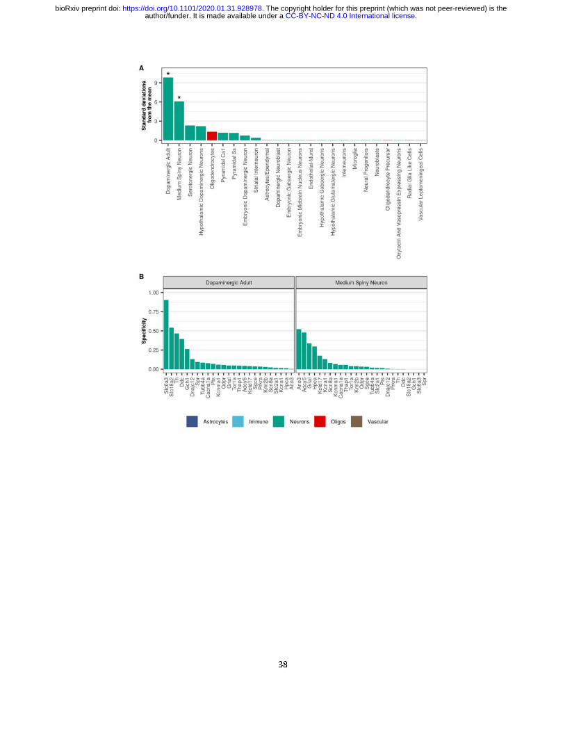
38
.CC-BY-NC-ND 4.0 International licenseauthor/funder. It is made available under aThe copyright holder for this preprint (which was not peer-reviewed) is the. https://doi.org/10.1101/2020.01.31.928978doi: bioRxiv preprint
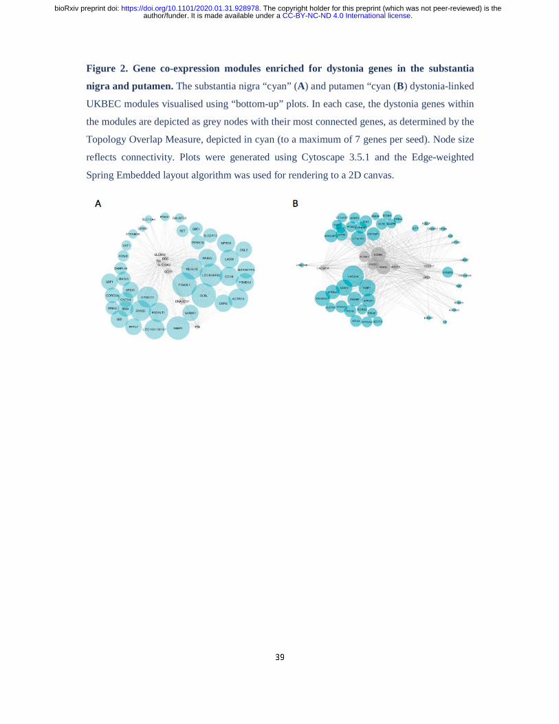
39
Figure 2. Gene co-expression modules enriched for dystonia genes in the substantia
nigra and putamen. The substantia nigra “cyan” (A) and putamen “cyan (B) dystonia-linked
UKBEC modules visualised using “bottom-up” plots. In each case, the dystonia genes within
the modules are depicted as grey nodes with their most connected genes, as determined by the
Topology Overlap Measure, depicted in cyan (to a maximum of 7 genes per seed). Node size
reflects connectivity. Plots were generated using Cytoscape 3.5.1 and the Edge-weighted
Spring Embedded layout algorithm was used for rendering to a 2D canvas.
.CC-BY-NC-ND 4.0 International licenseauthor/funder. It is made available under aThe copyright holder for this preprint (which was not peer-reviewed) is the. https://doi.org/10.1101/2020.01.31.928978doi: bioRxiv preprint
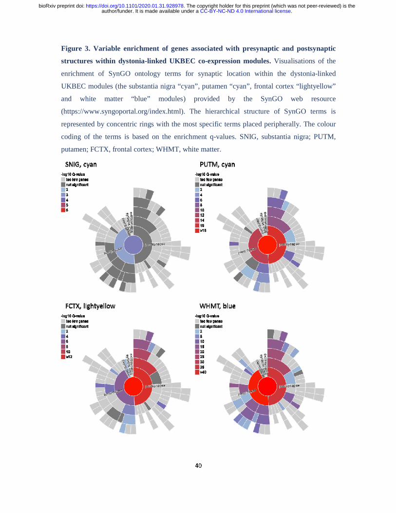
40
Figure 3. Variable enrichment of genes associated with presynaptic and postsynaptic
structures within dystonia-linked UKBEC co-expression modules. Visualisations of the
enrichment of SynGO ontology terms for synaptic location within the dystonia-linked
UKBEC modules (the substantia nigra “cyan”, putamen “cyan”, frontal cortex “lightyellow”
and white matter “blue” modules) provided by the SynGO web resource
(https://www.syngoportal.org/index.html). The hierarchical structure of SynGO terms is
represented by concentric rings with the most specific terms placed peripherally. The colour
coding of the terms is based on the enrichment q-values. SNIG, substantia nigra; PUTM,
putamen; FCTX, frontal cortex; WHMT, white matter.
.CC-BY-NC-ND 4.0 International licenseauthor/funder. It is made available under aThe copyright holder for this preprint (which was not peer-reviewed) is the. https://doi.org/10.1101/2020.01.31.928978doi: bioRxiv preprint
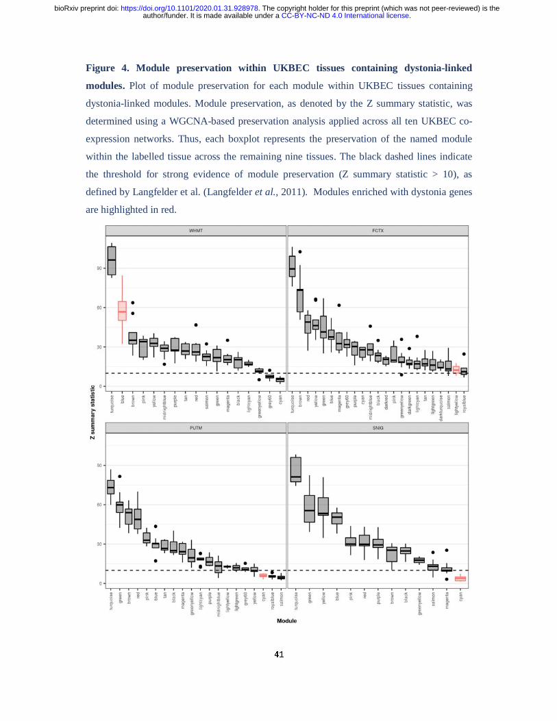
41
Figure 4. Module preservation within UKBEC tissues containing dystonia-linked
modules. Plot of module preservation for each module within UKBEC tissues containing
dystonia-linked modules. Module preservation, as denoted by the Z summary statistic, was
determined using a WGCNA-based preservation analysis applied across all ten UKBEC co-
expression networks. Thus, each boxplot represents the preservation of the named module
within the labelled tissue across the remaining nine tissues. The black dashed lines indicate
the threshold for strong evidence of module preservation (Z summary statistic > 10), as
defined by Langfelder et al. (Langfelder et al., 2011). Modules enriched with dystonia genes
are highlighted in red.
.CC-BY-NC-ND 4.0 International licenseauthor/funder. It is made available under aThe copyright holder for this preprint (which was not peer-reviewed) is the. https://doi.org/10.1101/2020.01.31.928978doi: bioRxiv preprint
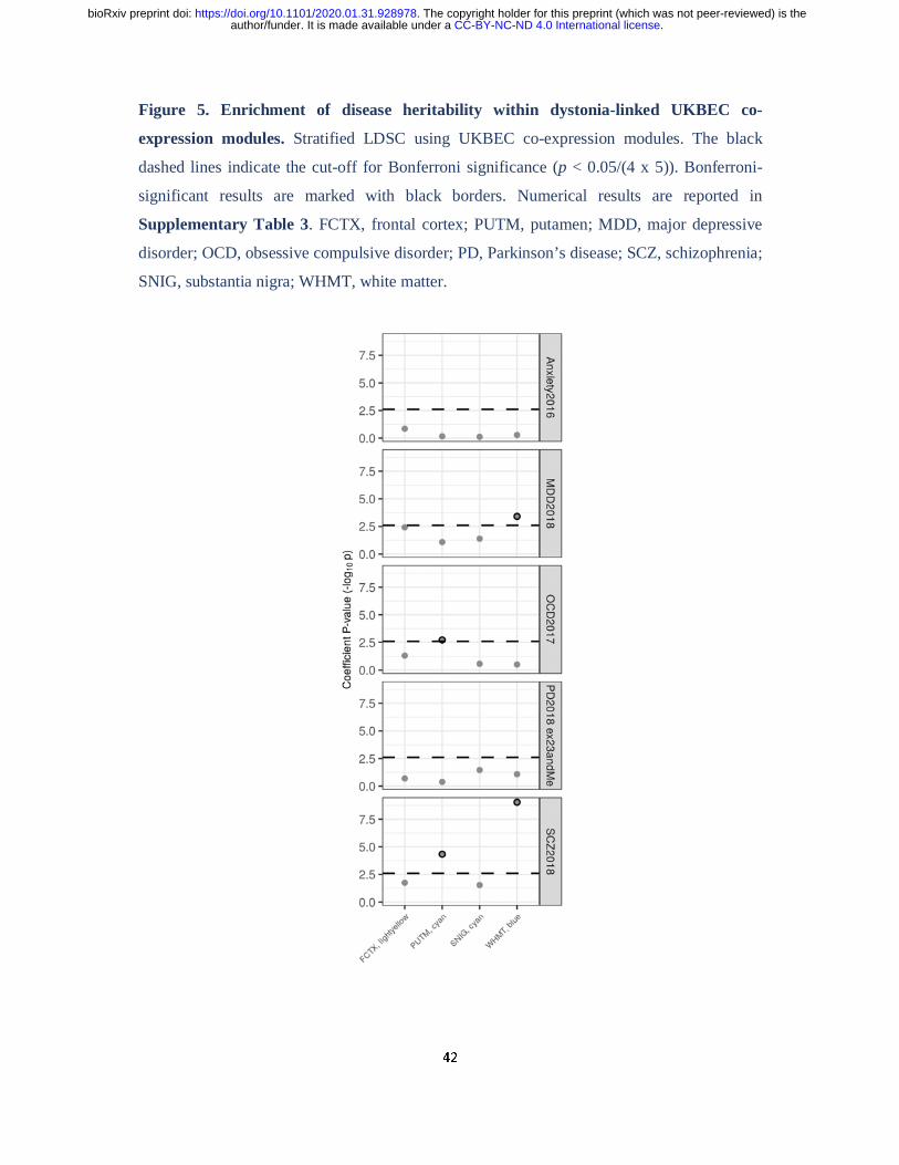
42
Figure 5. Enrichment of disease heritability within dystonia-linked UKBEC co-
expression modules. Stratified LDSC using UKBEC co-expression modules. The black
dashed lines indicate the cut-off for Bonferroni significance (p < 0.05/(4 x 5)). Bonferroni-
significant results are marked with black borders. Numerical results are reported in
Supplementary Table 3. FCTX, frontal cortex; PUTM, putamen; MDD, major depressive
disorder; OCD, obsessive compulsive disorder; PD, Parkinson’s disease; SCZ, schizophrenia;
SNIG, substantia nigra; WHMT, white matter.
.CC-BY-NC-ND 4.0 International licenseauthor/funder. It is made available under aThe copyright holder for this preprint (which was not peer-reviewed) is the. https://doi.org/10.1101/2020.01.31.928978doi: bioRxiv preprint
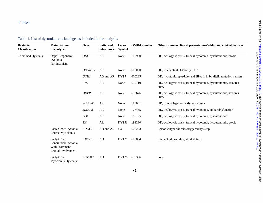
43
Tables
Table 1. List of dystonia-associated genes included in the analysis.
Dystonia Classification
Main Dystonic Phenotype
Gene Pattern of inheritance
Locus Symbol
OMIM number Other common clinical presentations/additional clinical features
Combined Dystonia Dopa-Responsive Dystonia-Parkinsonism
DDC AR None 107930 DD, oculogyric crisis, truncal hypotonia, dysautonomia, ptosis
DNAJC12 AR None 606060 DD, Intellectual Disability, HPA
GCH1 AD and AR DYT5 600225 DD, hypotonia, spasticity and HPA in in bi-allelic mutation carriers
PTS AR None 612719 DD, oculogyric crisis, truncal hypotonia, dysautonomia, seizures, HPA
QDPR AR None 612676 DD, oculogyric crisis, truncal hypotonia, dysautonomia, seizures, HPA
SLC18A2 AR None 193001 DD, truncal hypotonia, dysautonomia
SLC6A3 AR None 126455 DD, oculogyric crisis, truncal hypotonia, bulbar dysfunction
SPR AR None 182125 DD, oculogyric crisis, truncal hypotonia, dysautonomia
TH AR DYT5b 191290 DD, oculogyric crisis, truncal hypotonia, dysautonomia, ptosis
Early-Onset Dystonia-Chorea-Myoclonus
ADCY5 AD and AR n/a 600293 Episodic hyperkinesias triggered by sleep
Early-Onset Generalized Dystonia With Prominent Cranial Involvement
KMT2B AD DYT28 606834 Intellectual disability, short stature
Early-Onset Myoclonus-Dystonia
KCTD17 AD DYT26 616386 none
.C
C-B
Y-N
C-N
D 4.0 International license
author/funder. It is made available under a
The copyright holder for this preprint (w
hich was not peer-review
ed) is the.
https://doi.org/10.1101/2020.01.31.928978doi:
bioRxiv preprint
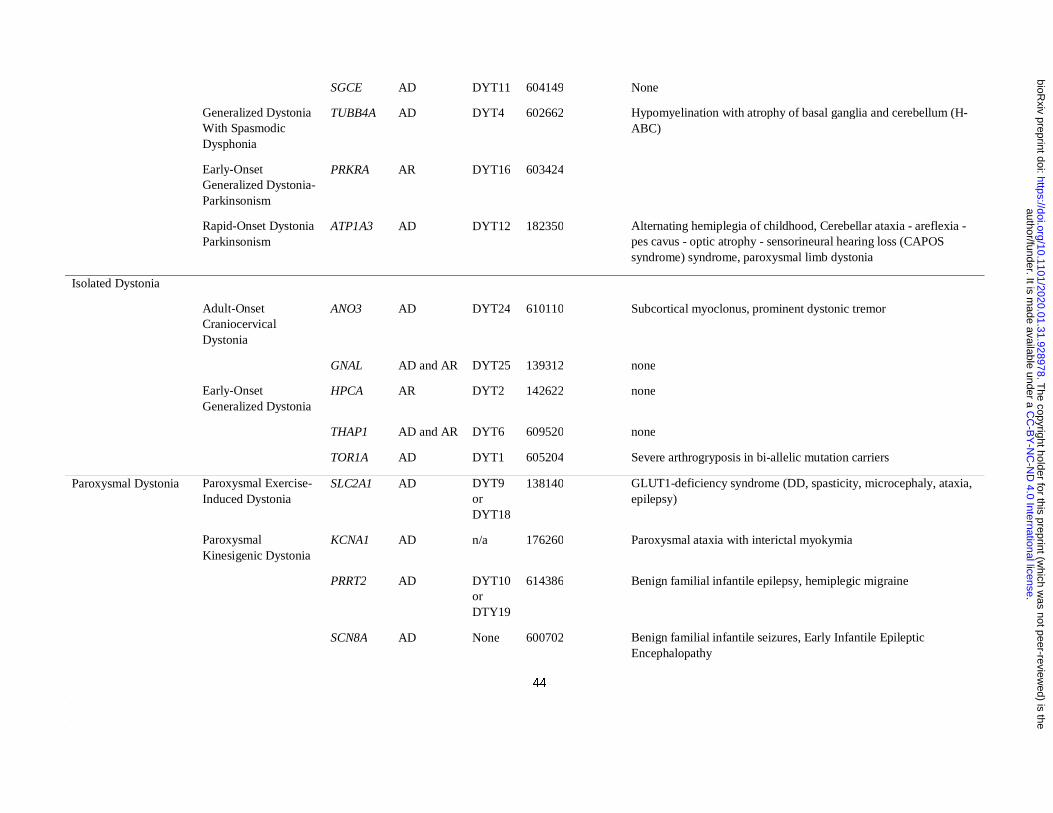
44
SGCE AD DYT11 604149 None
Generalized Dystonia With Spasmodic Dysphonia
TUBB4A AD DYT4 602662 Hypomyelination with atrophy of basal ganglia and cerebellum (H-ABC)
Early-Onset Generalized Dystonia-Parkinsonism
PRKRA AR DYT16 603424
Rapid-Onset Dystonia Parkinsonism
ATP1A3 AD DYT12 182350 Alternating hemiplegia of childhood, Cerebellar ataxia - areflexia - pes cavus - optic atrophy - sensorineural hearing loss (CAPOS syndrome) syndrome, paroxysmal limb dystonia
Isolated Dystonia
Adult-Onset Craniocervical Dystonia
ANO3 AD DYT24 610110 Subcortical myoclonus, prominent dystonic tremor
GNAL AD and AR DYT25 139312 none
Early-Onset Generalized Dystonia
HPCA AR DYT2 142622 none
THAP1 AD and AR DYT6 609520 none
TOR1A AD DYT1 605204 Severe arthrogryposis in bi-allelic mutation carriers
Paroxysmal Dystonia Paroxysmal Exercise-Induced Dystonia
SLC2A1 AD DYT9 or DYT18
138140 GLUT1-deficiency syndrome (DD, spasticity, microcephaly, ataxia, epilepsy)
Paroxysmal Kinesigenic Dystonia
KCNA1 AD n/a 176260 Paroxysmal ataxia with interictal myokymia
PRRT2 AD DYT10 or DTY19
614386 Benign familial infantile epilepsy, hemiplegic migraine
SCN8A AD None 600702 Benign familial infantile seizures, Early Infantile Epileptic Encephalopathy
.C
C-B
Y-N
C-N
D 4.0 International license
author/funder. It is made available under a
The copyright holder for this preprint (w
hich was not peer-review
ed) is the.
https://doi.org/10.1101/2020.01.31.928978doi:
bioRxiv preprint
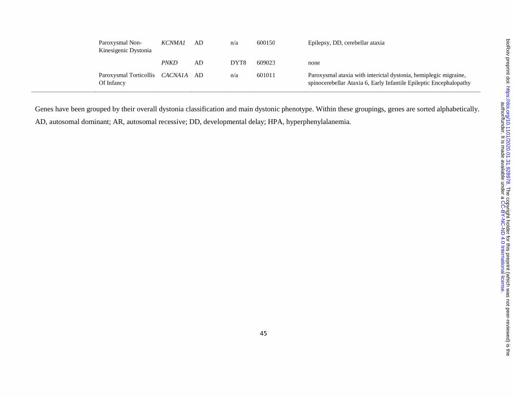
45
Paroxysmal Non-Kinesigenic Dystonia
KCNMA1 AD n/a 600150 Epilepsy, DD, cerebellar ataxia
PNKD AD DYT8 609023 none
Paroxysmal Torticollis Of Infancy
CACNA1A AD n/a 601011 Paroxysmal ataxia with interictal dystonia, hemiplegic migraine, spinocerebellar Ataxia 6, Early Infantile Epileptic Encephalopathy
Genes have been grouped by their overall dystonia classification and main dystonic phenotype. Within these groupings, genes are sorted alphabetically.
AD, autosomal dominant; AR, autosomal recessive; DD, developmental delay; HPA, hyperphenylalanemia.
.C
C-B
Y-N
C-N
D 4.0 International license
author/funder. It is made available under a
The copyright holder for this preprint (w
hich was not peer-review
ed) is the.
https://doi.org/10.1101/2020.01.31.928978doi:
bioRxiv preprint
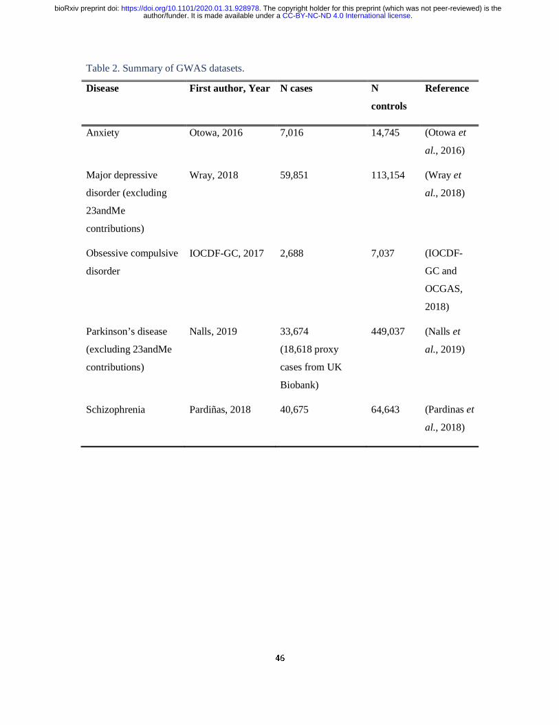
46
Table 2. Summary of GWAS datasets.
Disease First author, Year N cases N
controls
Reference
Anxiety Otowa, 2016 7,016 14,745 (Otowa et
al., 2016)
Major depressive
disorder (excluding
23andMe
contributions)
Wray, 2018 59,851 113,154 (Wray et
al., 2018)
Obsessive compulsive
disorder
IOCDF-GC, 2017 2,688 7,037 (IOCDF-
GC and
OCGAS,
2018)
Parkinson’s disease
(excluding 23andMe
contributions)
Nalls, 2019 33,674
(18,618 proxy
cases from UK
Biobank)
449,037 (Nalls et
al., 2019)
Schizophrenia Pardiñas, 2018 40,675 64,643 (Pardinas et
al., 2018)
.CC-BY-NC-ND 4.0 International licenseauthor/funder. It is made available under aThe copyright holder for this preprint (which was not peer-reviewed) is the. https://doi.org/10.1101/2020.01.31.928978doi: bioRxiv preprint
47
Description of Supplementary Data
Supplementary Tables
Supplementary Table 1. Cell-specific enrichments p-values of dystonia genes calculated
using EWCE
Supplementary Table 2. Enrichment p-values of dystonia genes within gene co-expression
modules generated using brain transcriptomic data from the UK Brain Expression
Consortium and the GTEx consortium
Supplementary Table 3. Enrichment p-values of SynGO annotations within the substantia
nigra “cyan”, putamen “cyan”, frontal cortex “lightyellow” and white matter “blue” modules.
Supplementary Table 4. Module preservation across four UKBEC tissues containing
dystonia-linked modules.
Supplementary Table 5. Results of stratified LDSC analysis across all gene co-expression
modules of interest and a range of neuropsychiatric and neurodegenerative disorders
Supplementary Figures
Supplementary Figure 1. Cell-specific profiles of dystonia-associated genes.
Plots of specificity values for all dystonia-associated genes within level 1 cell types from the
Karolinska single-cell RNA-sequencing superset. Specificity values were derived from Skene
et al.2 who calculated specificity by dividing the mean expression of a gene in one cell type
by the mean expression in all cell types. In other words, specificity is the proportion of a
gene’s total expression attributable to one cell type, with a value of 0 meaning a gene is not
expressed in that cell type and a value of 1 meaning that a gene is only expressed in that cell
.CC-BY-NC-ND 4.0 International licenseauthor/funder. It is made available under aThe copyright holder for this preprint (which was not peer-reviewed) is the. https://doi.org/10.1101/2020.01.31.928978doi: bioRxiv preprint

48
type. For each gene, the top 3 cell types, as ranked by specificity, are marked with black
borders.
.CC-BY-NC-ND 4.0 International licenseauthor/funder. It is made available under aThe copyright holder for this preprint (which was not peer-reviewed) is the. https://doi.org/10.1101/2020.01.31.928978doi: bioRxiv preprint
Recommended