62 Early-Life Experiences: Enduring Behavioral,Neurological, and Endocrinological ConsequencesR D Romeo, Barnard College, New York, NY, USA
A C Tang, University of New Mexico, Albuquerque, NM, USA
R M Sullivan, Nathan Kline University and New York University Langone Medical Center, Orangeburg, NY, USA
� 2009 Elsevier Inc. All rights reserved.
Chapter Outline
62.1 Introduction 1976
62.2 Hypothalamic–Pituitary–Adrenal Axis 1976
62.2.1 Development of the HPA Axis 1977
62.2.1.1 Neonatal development of the HPA axis 1977
62.2.1.2 Pubertal development of the HPA axis 1978
62.3 Neonatal Experiences and Enduring Behavioral, Neurological,
and Endocrinological Consequences 1980
62.3.1 Individual Differences in the Development of the HPA Axis and
Neonatal Experience 1980
62.3.2 Neonatal Handling 1981
62.3.2.1 Neonatal handling and behavior 1981
62.3.2.2 Neonatal handling and brain 1983
62.3.2.3 Neonatal handling and endocrine function 1984
62.3.3 Neonatal Novelty Exposure 1984
62.3.3.1 Neonatal novelty exposure and behavior 1985
62.3.3.2 Neonatal novelty exposure and brain 1987
62.3.3.3 Neonatal novelty exposure and endocrine function 1987
62.3.3.4 Neonatal novelty exposure and maternal influence 1989
62.3.4 Maternal Deprivation 1989
62.3.4.1 Maternal deprivation and behavior 1990
62.3.4.2 Maternal deprivation and brain 1990
62.3.4.3 Maternal deprivation and endocrine function 1991
62.3.5 Pain, Fear Conditioning, and Context of Early-Life Adversity 1992
62.3.5.1 Odor–shock conditioning and behavior 1992
62.3.5.2 Odor–shock conditioning and brain 1993
62.3.5.3 Odor–shock conditioning and endocrine function 1993
62.3.6 Functional Consequences of Early-Life Experiences 1993
62.4 Pubertal Experiences and Enduring Behavioral and Endocrine Consequences 1994
62.4.1 Pubertal Experience and Behavior 1994
62.4.2 Pubertal Experience and Brain 1995
62.4.3 Pubertal Experience and Endocrine Function 1995
62.5 Adolescence as a Period of Intervention to Mitigate Early Developmental Insults 1996
62.5.1 Reversals of Perinatal Insults through Pubertal Environmental Enrichment 1996
62.5.2 Mitigation of Perinatal Brain Damage through Pubertal Environmental Enrichment 1996
62.6 Conclusions 1997
References 1997
1975

1976 Early-Life Experiences
Glossaryadrenocorticotropic hormone (ACTH) A peptide
hormone released from the anterior
pituitary gland that mediates the production
and secretion of hormones of the adrenal
cortex.
arginine vasopressin (AVP) A peptide hormone
that participates in the release of
stress-related hormones, such as ACTH.
corticosterone (CORT) A steroid hormone
released by the adrenal cortex in response to
stress.
corticotropin-releasing hormone (CRH)
A peptide hormone that participates in the
release of stress-related hormones, such as
ACTH. This hormone is also referred to as
corticotropin-releasing factor (CRF).
glucocorticoid receptor (GR) A low-affinity
steroid hormone receptor for the corticoid
steroids, such as CORT.
hypothalamic–pituitary–adrenal axis (HPA axis)
The major neuroendocrine axis that
mediates the hormonal stress response.
mineralocorticoid receptor (MR) A high-affinity
steroid hormone receptor for the corticoid
steroids, such as CORT.
paraventricular nucleus of the hypothalamus
(PVN) A nucleus in the hypothalamus that contains
CRH and AVP neurosecretory cells that
regulate the release of stress-related
hormones.
62.1 Introduction
Though individuals function in the present, we carrywith us previous experiences that can fundamen-tally change how we respond physiologically andbehaviorally to internal and external challenges.Since the seminal work of Weininger (1954) on gen-tling and Levine (1957) on infantile experiences inrats, an ever-growing body of literature has indictedthat experiences early in development can have long-lasting effects on an individual’s physiological andbehavioral potentials. In fact, some of these effects ofearly experience are so enduring that they can betransgenerational (Denenberg and Rosenberg, 1967).The purpose of this chapter is to highlight some recentstudies regarding how experiences neonatally and/orpubertally can modulate later adult functioning.
Specifically, we emphasize the role of early experi-ence such as neonatal handling, novelty exposure,maternal deprivation, and odor–shock conditioningon immediate and long-term emotionality and cogni-tive abilities. As the neonatal period is not the onlydevelopmental stage when individuals are susceptibleto both positive and negative influences, we also dis-cuss how exposure to stressors during adolescencemodifies later stress responsiveness and emotionalbehavior. We conclude by briefly describing someprovocative experiments which indicate that experi-ences during puberty can offset or mitigate develop-mental insults that occur perinatally. These dataindicate that at least some enduring consequences ofearly-life experience remain malleable, evenwell intoadolescence and adulthood.
The hormones released during stressful eventsappear to be a common thread in how early experi-ences during the neonatal or pubertal stage of develop-ment affect an individual’s immediate and long-termphysiological and behavioral function. Thus, we beginthis chapter by briefly examining the hypothalamic–pituitary–adrenal (HPA) axis, the major neuroendo-crine axis mediating the hormonal stress response(Herman and Cullinan, 1997; Herman et al., 2003).Below, we discuss the components that comprise thisaxis and its neonatal and pubertal maturation.
62.2 Hypothalamic–Pituitary–Adrenal Axis
The release of stress-related hormones by the HPAaxis is driven by a cascade of signals beginning withthe release of the neuropeptides corticotropin-releasinghormone (CRH) and arginine vasopressin (AVP) fromthe paraventricular nucleus of the hypothalamus(PVN). CRH and AVP are released into the hypophy-seal-portal system, which bring about the release ofadrenocorticotropic hormone (ACTH) from theanterior pituitary. ACTH then stimulates the produc-tion and secretion of glucocorticoids (i.e., cortisol inprimates and corticosterone (CORT) in many rodentspecies) from the cortex of the adrenal gland. Thehormones secreted by the HPA axis control theirown secretion through a neuroendocrine negativefeedback loop. That is, glucocorticoids feed back onthe PVN and extrahypothalamic sites (e.g., pituitary,hippocampus, and prefrontal cortex (PFC)) to inhibitthe further release of hypothalamic CRH and AVP(Herman et al., 2003; Figure 1).

Paraventricular nucleus of the hypothalamus(PVN)
CRH/AVP
ACTH
CORT
(–)
(–)
(+)
(+)
AP
CM
Hypothalamus
Pituitary
Adrenal
Figure 1 A diagram of the HPA axis. ACTH,
adrenocorticotropin hormone; AP, anterior pituitary; AVP,
arginine vasopressin; C, cortex; CORT, corticosterone;CRH, corticotropin-releasing hormone; M, medulla; PVN,
paraventricular nucleus of the hypothalamus; (þ), positive
drive; (–), negative feedback.
Early-Life Experiences 1977
Two receptors (the mineralocorticoid receptor(MR) and the glucocorticoid receptor (GR)) mediatethe actions of glucocorticoids in the central nervoussystem. These steroid receptors are found in relativelyhigh concentrations throughout the neural–pituitarynetwork that controls both negative feedback andactivation of the HPA axis (Sapolsky et al., 2000). Thehigh-affinity MR is typically saturated at basal gluco-corticoid levels, while the low-affinity GR is primarilyoccupied only when elevated concentrations of gluco-corticoids are present (deKloet et al., 1998).Thus, whenglucocorticoid levels rise in response to stressors, thenegative feedback on the HPA axis is primarilymediated by the GR (de Kloet et al., 1998). However,the MR also appears to play a role in glucocorticoid-mediated negative feedback under mildly stressful con-ditions (Pace and Spencer, 2005).
The hormonal stress response is essential to sur-vival as it allows an organism to cope with the inter-nal and external demands imposed by a challengingevent. This response attempts to restore the organismto homeostasis, a process termed allostasis (McEwenand Stellar, 1993). However, prolonged or morechronic exposures to stress and stress-induced hor-mones can lead to allostatic overload, resulting in anumber of negative effects, particularly in regard toneurobiological and emotional function (Herbert
et al., 2006; McEwen, 2003, 2004; McEwen andStellar, 1993; Sapolsky, 1999; van Praag, 2004).
Both the magnitude and duration of the hormonalstress response change dramatically throughout anorganism’s life span. For instance, neonatal animalsshow reduced stress reactivity in response to stressorsthat typically elicit robust stress responses in adults(Sapolsky and Meaney, 1986). The reduced stressreactivity experienced by neonates has been positedto protect the developing organism from the negativeinfluences of stress hormones (Sapolsky and Meaney,1986). Conversely, aged animals show heightened andmore prolonged stress responses compared to youngeradults (Sapolsky, 1999). This has been proposed tocontribute to the decline in neurophysiological andcognitive function observed during aging (Sapolsky,1999; Sapolsky et al., 1985). Thus, parameters thatchange the responsiveness of the HPA axis, such asdevelopment, may have profound consequences onwhether stressors lead to adaptive or maladaptiveresponses. The next section briefly describes some ofthe major changes that occur in HPA function duringneonatal and pubertal maturation.
62.2.1 Development of the HPA Axis
62.2.1.1 Neonatal development ofthe HPA axis
For the first 2 weeks of life, basal plasma CORTconcentrations are relatively low (Henning, 1978). Inrats, these basal levels of CORT begin to increasearound postnatal day (PND)15 in both males andfemales, and peak around PND24 (Henning, 1978;Meaney et al., 1985c). As alluded to above, the neona-tal stage of development is marked by a particularlystriking change in HPA reactivity, that is, the ability ofstressors to evoke CORT secretion in neonatal rats(PND1–14) is greatly reduced compared to that inadults (Butte et al., 1973; Cote and Yasumura, 1975;Guillet and Michaelson, 1978; Guillet et al., 1980;Levine et al., 1967). This period of reduced HPAresponsiveness during neonatal development hasbeen termed the stress hyporesponsive period (SHRP).
It appears that all levels of the HPA axis areinvolved in mediating the SHRP (Sapolsky andMeaney, 1986). At the level of the hypothalamus,CRH levels in the neonatal PVN are lower comparedto those found in adults (Walker et al., 1986a). At thelevel of the pituitary, CRH-induced ACTH release isreduced (Walker et al., 1986a), in part due toincreases in CORT-induced negative feedback onthe neonatal pituitary gland (Walker et al., 1986b).
Basal
Stress
600
Prepubertal maleAdult male
*
*400
Cor
ticos
tero
ne (n
gm
l–1)
200
00 30 60 120
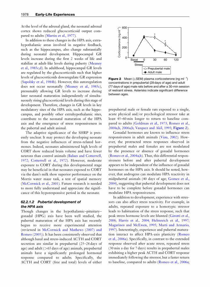
Figure 2 Mean (�SEM) plasma corticosterone (ng ml�1)
concentrations in prepubertal (28days of age) and adult
(77 days of age) male rats before and after a 30-min sessionof restraint stress. Asterisks indicate significant difference
between ages.
1978 Early-Life Experiences
At the level of the adrenal gland, the neonatal adrenalcortex shows reduced glucocorticoid output com-pared to adults (Martin et al., 1977).
In addition to these changes in the HPA axis, extra-hypothalamic areas involved in negative feedback,such as the hippocampus, also change substantiallyduring neonatal development. Hippocampal GRlevels increase during the first 2 weeks of life andstabilize at adult-like levels during puberty (Meaneyet al., 1985c,d). In adulthood, hippocampal GR levelsare regulated by the glucocorticoids such that higherlevels of glucocorticoids downregulate GR expression(Sapolsky et al., 1984b). However, this autoregulationdoes not occur neonatally (Meaney et al., 1985c),presumably allowing GR levels to increase duringlater neonatal maturation independently of simulta-neously rising glucocorticoid levels during this stage ofdevelopment. Therefore, changes in GR levels in keymodulatory sites of the HPA axis, such as the hippo-campus, and possibly other extrahypothalamic sites,contribute to the neonatal maturation of the HPAaxis and the emergence of stress responsiveness inthe pubertal and adult animal.
The adaptive significance of the SHRP is pres-ently unclear. It may protect the developing neonatefrom the negative influences of stress-related hor-mones. Indeed, neonates administered high levels ofCORT show reduced brain volume and have fewerneurons than control animals (Balazs and Cotterrell,1972; Cotterrell et al., 1972). However, moderateexposure to CORT during this developmental stagemay be beneficial in that neonates exposed to CORTvia the dam’s milk show superior performance on theMorris water maze task, a test of spatial memory(McCormick et al., 2001). Future research is neededto more fully understand and appreciate the signifi-cance of this hyporesponsive period in the neonate.
62.2.1.2 Pubertal development of
the HPA axis
Though changes in the hypothalamic–pituitary–gonadal (HPG) axis have been well studied, thepubertal maturation of the HPA axis has recentlybegun to receive more experimental attention(reviewed in McCormick and Mathews (2007) andRomeo (2005)). It has been consistently observed thatalthough basal and stress-induced ACTH and CORTsecretion are similar in prepubertal (25–28 days ofage) and adult (>65 days of age) animals, prepubertalanimals have a significantly prolonged hormonalresponse compared to adults. Specifically, theACTH and CORT (free and total) levels of either
prepubertal male or female rats exposed to a single,acute physical and/or psychological stressor take atleast 45–60min longer to return to baseline com-pared to adults (Goldman et al., 1973; Romeo et al.,2004a,b, 2006a,b; Vazquez and Akil, 1993; Figure 2).
Gonadal hormones are known to influence stressresponsiveness in adult animals (Viau, 2002). How-ever, the protracted stress responses observed inprepubertal males and females are not modulatedby the presence or absence of gonadal hormones(Romeo et al., 2004a,b). Thus, this differential respon-siveness before and after pubertal developmentappears to be independent of the influence of gonadalhormones on the HPA axis. It should be noted, how-ever, that androgens can modulate HPA reactivity inmidpubertal animals (40 days of age; Gomez et al.,2004), suggesting that pubertal development does nothave to be complete before gonadal hormones canmodulate HPA responsiveness.
In addition to development, experience with stres-sors can also affect stress reactivity. For example, inadults, repeated exposure to a homotypic stressorleads to habituation of the stress response, such thatpeak stress hormone levels are blunted (Girotti et al.,2006; Harris et al., 2004; Helmreich et al., 1997;Magarinos and McEwen, 1995; Marti and Armario,1997). Interestingly, experience and pubertal matura-tion interact to affect HPA-axis plasticity (Romeoet al., 2006a). Specifically, in contrast to the extendedresponse observed after acute stress, repeated stress(30min a day for 7 days) results in prepubertal malesexhibiting a higher peak ACTH and CORT responseimmediately following the stressor, but a faster returnto baseline, compared to adults (Romeo et al., 2006a;

Acute stress Chronic stress600
400
200
Cor
ticos
tero
ne (n
gm
l–1)
0
Stress Stress
Basal
*
*
*
0 45 Basal 0 45
PrepubertalAdult
Figure 3 Mean (� SEM) plasma corticosterone (ng ml�1) concentrations in prepubertal (28 days of age) and adult (77 days
of age) male rats exposed to a single (acute stress) or a daily 30-min session of restraint stress for 7 days (chronic stress).
Asterisks indicate significant difference between ages.
Early-Life Experiences 1979
Figure 3). Midpubertal rats (40 days of age) exposedto a repeated homotypic stressor begin to show adiminished HPA response similar to adults (Gomezet al., 2002). Therefore, it appears that within theadolescent window of development the HPA axisbegins to react to homotypic stressors in a similarmanner as adults. The physiological and neuro-behavioral implications of this differential stress reac-tivity in adolescent and adult animals remain largelyunknown.
It is unclear what mechanisms contribute to thepubertal change in stress responsiveness. Volumetricanalyses of the PVN have not revealed any grossanatomical differences between the pubertal andadult PVN. Specifically, estimated volume, somalarea, and cell number in both the magnocellularand parvocellular aspects of the PVN are similar inprepubertal and adult animals (Romeo et al., 2007).Furthermore, peripheral injection of the retrogradetracer fluoro-gold reveals similar numbers of anteriorpituitary projecting neurosecretory neurons in theparvocellular region of the PVN in both prepubertaland adult males (Romeo et al., 2007).
Despite these similarities, it is interesting to notethat CRH cells in the PVN show greater activation,as indexed by Fos immunohistochemistry, in pre-pubertal compared to adult animals in response toacute or repeated restraint stress (Romeo et al., 2006a).There are also developmental changes in CRHmRNA expression in the PVN such that prepubertalmales have greater basal CRH expression than adults(Romeo et al., 2007). Though baseline levels of CRHmRNA are higher in juvenile animals, social stressorsor restraint leads to increases in CRH expression in
both the pubertal and adult PVN (McCormick et al.,2006; Romeo et al., 2007; Viau et al., 2005). Together, itappears that the basal and stress-induced regulation ofCRH cells in the PVNmay contribute to the differen-tial HPA reactivity exhibited by adolescent and adultanimals. However, it will be important to continue toinvestigate the role that possible differential releaseof CRH, sensitivity of the anterior pituitary to CRH(and other secretagogs such AVP), and/or the sensi-tivity of the adrenal cortex to ACTHmay play in theseage-dependent differential responses.
Given the importance of the hippocampus onglucocorticoid-mediated negative feedback on theHPA axis (Herman et al., 2003; Sapolsky et al.,1984a), a few studies have assessed both baselineand stress-induced changes in hippocampal GRexpression in adolescent animals. Meaney et al.(1985c) have shown a slight decrease in GR concen-trations in hippocampal homogenates in midpubertal(i.e., 35 days) compared to adult (90–120 days) males,while mRNA studies have found no difference inhippocampal GR levels between prepubertal, mid-pubertal, or adult animals (Romeo et al., 2008;Vazquez, 1998). Moreover, acute stress (30min ofrestraint) decreases GR mRNA in the hippocampalformation in both prepubertal and adult males(Romeo et al., 2008). In general, these data indicatethat there are more similarities than differences inGR expression in the pubertal and adult hippo-campus, and suggest hippocampal GR-mediated neg-ative feedback contributes little to this robust changein HPA responsiveness exhibited by adolescent ani-mals. However, GR-mediated negative feedback onthe HPA axis can be mediated at other neural loci

1980 Early-Life Experiences
such as the medial prefrontal cortex (mPFC) and alsoat the pituitary gland (Herman and Cullinan, 1997;Herman et al., 2003). It is also becoming apparentthat, at least in adulthood, MRs play a role in gluco-corticoid-mediated negative feedback under mildlystressful conditions (Pace and Spencer, 2005).Clearly, future studies will need to explore pubertalchanges in GR and MR content and their regulationand function in the entire neural–pituitary networkthat mediates HPA reactivity.
62.3 Neonatal Experiences andEnduring Behavioral, Neurological,and Endocrinological Consequences
62.3.1 Individual Differences inthe Development of the HPA Axis andNeonatal Experience
One of the most enduring findings concerning thedevelopment of the HPA axis is its modifiability orplasticity by a diverse range of environmental variablesacross different stages of development. This plasticityprovides a source of individual differences in the regu-lation of theHPA stress response, which, in turn, modu-lates a wide range of psychological and physiologicalfunctions. Much progress has been made in terms ofcharacterizing and documenting the specific experi-mental conditions or experiences that create specificbehavioral, neural, and endocrinological changes. How-ever, utilizing recent methodological developments inneuroscience and behavioral sciences, the relationshipamong different experiential manipulations remainsto be understood, inconsistent findings remain to beexplained, and a unifying framework for integratingthese diverse findings remains to be developed.
Two historically significant ideas (neonatalhandling and maternal separation/deprivation para-digms) appeared to be responsible for the develop-ment of the two most popular early-experiencemanipulations. The first is the construct of stressintroduced by Selye (1936). With the publication ofhis seminal work in Nature, he introduced what mighthave been the antecedent to a class of experimentalparadigms in which the effects of stressful events werecharacterized in rodents. In an attempt to study effectsof stress inflicted by electrical shock, Levine (1960)accidentally discovered something rather surprising,in that the bodily and behavioral responses of therats to shocks, which also entail a certain amount ofexperimenter handling, were no different from thatof the rats who were only handled but received no
shock (handled group). Furthermore, in comparison tothe control rats that just stayed in their home environ-ment (referred to as nonhandled rats), both groupsshowed reduced emotionality and a less sluggishHPA response to stressful events. This accidental dis-covery has led to the creation of a large body ofliterature built upon the manipulation referred to asneonatal handling (Daly, 1973; Denenberg, 1964, 1978;Levine, 1957; Meaney et al., 1988).
The second is the idea that early mother–infantexperiences influence adult outcomes. Althoughinitially considered radical, convergence of datafrom both clinical observations and basic researchgradually led to its acceptance. Specifically, astuteclinical observations identified a link between dis-turbed maternal care or maternal separation anddisturbed emotional and cognitive functioning thatbegan in infancy and lasted into adolescence (Bowlby,1951). Concurrent work by Harlow and Harlow(1965) on infant monkeys separated from their motherappeared to mirror the strong emotional and physicalstunting of orphaned infants. These data provided thefoundation for early-life-experience-related researchwith the unifying clinical and basic research themethat maternal carewas critical for the normal develop-ment of infants. Because infant separation from themother could be clearly defined and measured, itbecame an important variable for manipulatingearly-life experience (Bowlby, 1951, 1969, 1982).
Over the past five decades, these two lines ofresearch each independently revealed informationthat advanced our understanding of their very oftenopposite effects on HPA development and their func-tional consequences. Each research paradigm has itsdistinct advantages and faces its unique theoreticaland practical challenges. The experimental procedurein neonatal handling involves assigning an entire litter,or rat family, to the handled and nonhandled condi-tion, and is relatively easy to carry out. It produces aset of effects on behavior, brain, and the HPA axis thatare generally considered desirable (Levine, 1960; butsee recent discussions of effect on reproductivebehavior (Greisen et al., 2005). The handling litera-ture also faces major conceptual and interpretationaldifficulties that were initially pointed out over threedecades ago by Daly (1973). Using this procedure, itwas impossible to tease apart several distinct, yetconfounding factors, which differed between thehandled and nonhandled pups. With the exceptionof the effects on HPA function and measures of emo-tionality, handling effects on a variety of brain andbehavioral functions could not be replicated.
Early-Life Experiences 1981
Since then, research in neonatal handlingappeared to have passed its golden days, but wasrevived by the elegant work of Meaney et al. (1988)showing parallel changes in hippocampal-dependentspatial learning, hippocampal cell counts, and a vari-ety of HPA-related measures. This work has ledto new approaches in the understanding of howmaternal care contributes to emotional and neuro-endocrine development (Liu et al., 1997). This workalso motivated research efforts to directly addressthe difficult methodological issues raised in a criti-cal review by Daly (1973). Specifically, an experi-mental paradigm was developed using a split-litter(within-family) design to better isolate the knownconfounding factors in neonatal handling (seeSection 62. 3.3; Tang, 2001 ).
Another major line of research of maternal sepa-ration/deprivation involves exposing an entire litterof pups to relatively prolonged periods of pup–damseparation (>3 h). Different from the neonatalhandling manipulation, maternal separation pro-duces a set of effects that are generally considerednegative, with a relatively clear correspondence toreal-world scenarios in child development. Conse-quently, practical implications of findings associatedwith this paradigm can be easily discussed. Today,while the rodent early-experience research continuesto rely on the maternal separation/deprivation para-digm, understanding the relatively subtler caregiverbehaviors and contrasting quantity and quality ofmaternal care have become increasingly important.This direction of research differs from the earliermaternal separation/deprivation work in theirpotential implications. While the maternal depriva-tion/separation literature provided mainly character-ization of functional deficits, the maternal careliterature offered insights into potential beneficial,or at least opposing, effects from those induced bymaternal deprivation or separation.
The interest in the role of maternal care inearly-experience effects on development has beenat least as old as the neonatal handling literature.The most notable hypothesis is the maternal media-tion hypothesis (for review, see Macri and Wurbel(2006)). In its most extreme form, this hypothesisstates that the effects of neonatal handling have noth-ing to do with handling-induced activation of thepup’s HPA axis, but solely mediated by a handlingeffect on maternal behavior, which in turn resulted inchanges in offspring development. Since then, manyhave categorized and quantified a variety of maternalbehaviors and conducted carefully controlled studies
in which the influence of maternal care on behavior,brain, and neuroendocrine function was determined(Meaney, 2001).
In the past decade, while some continue to focus onidentifying more molecular markers within the brainthat correlate with natural variations in the amountof specific maternal behaviors, such as licking andgrooming (Weaver et al., 2004), others have shownthat early stimulation via handling can affect theoffspring’s brain and HPA axis in the absence of mater-nal behaviors (Denenberg, 1999). Additional situationshave also been noted where effects of neonatal han-dling treatment, and other early-stimulation manipu-lations, could not be explained solely by increasedmaternal care (Macri et al., 2004; Macri and Wurbel,2006; Tang et al., 2006), and in some situations wherean increase in maternal care was negatively associatedwith offspring development (Macri et al., 2004). Theserecent observations suggest that a new theory of earlyexperience other than maternal mediation is needed toprovide a unifying explanation for how early experi-ence and maternal influence jointly shape offspringbehavioral, neural, and endocrine development.
Following the two historical traditions of neonatalhandling and maternal deprivation, we discuss recentfindings from studies using the neonatal noveltyexposure and the odor-shocking conditioning para-digms to support an alternative theory of maternalmodulation. Instead of affecting offspring develop-ment through maternal behavior, early stimulationexerts direct, independent effects on the offspring.Maternal variables modulate these direct stimulationeffects, thereby jointly shaping offspring development.
62.3.2 Neonatal Handling
Behavioral, brain, and HPA changes as a result ofneonatal handling have been reviewed throughoutits history of the past half century. For comprehensivereviews, we would like to refer the readers to severalmanuscripts from various periods (Daly, 1973;Denenberg, 1964; Macri and Wurbel, 2006; Meaneyet al., 1996). Here, instead of reiterating previouslydocumented findings, we present the neonatalhandling paradigm from a methodological point ofview in order to present recent progress and to arriveat a modern interpretation of the neonatal handlingliterature.
62.3.2.1 Neonatal handling and behavior
Neonatal handling, also known as postnatal handling,is an early-life behavioral manipulation with several

1982 Early-Life Experiences
defining features. First, it is a procedure carried outon all pups from an entire litter from a single birth.Specifically, a litter of pups are assigned to either theexperimental group (handled) or the control group(nonhandled). This feature determines that the litteris the unit of experimental manipulation and theresults of such a manipulation provide informationon between-litter differences in the offspring, but notwithin-litter differences (i.e., between-sibling differ-ences). This feature further implies that pups withineach litter might be highly correlated due to the factthat they share the same dam and home/housingenvironment prior to weaning. Thus, to draw statisti-cally sound conclusions, one needs to use litter as theunit of analysis; an exception to this rule, however,can be made if one fails to find sufficient strongwithin-litter correlation (equivalent to a nonsignifi-cant litter effect). Many handling studies do not useappropriate statistical treatment of the data, and thismight explain why some handling effects cannot bereliably reproduced.
Second, neonatal handling is a procedure involv-ing multiple distinct procedural differences, includ-ing differences in experimenter handling, absence ofthe dam, absence of litter siblings, and experience of arelatively novel nonhome environment. One conse-quence of having these multiple factors is that it isdifficult, if not impossible, to pinpoint the precisecause of any observed difference between the experi-mental and control groups. In the case of a handlingeffect on spatial learning, improved learning isequally likely a result of maternal separation, touch-ing by experimenters, or experiencing a relativelynovel or unfamiliar environment, thus making itdifficult to determine how to use these research find-ings to optimize the early-life environment to facili-tate cognitive development. It would be a nontrivialmistake if one implements an intervention programby separating the infants from their mothers, or byhaving physical contact with strangers, when the truecause of improved learning is the early experience ofnovelty. This state of confusion is reflected by the factthat some researchers still use the phrase maternalseparation to refer to the same handling manipula-tion. This implies a causal relation between maternalabsence and the observed differences between theso-called handled and nonhandled rats, even thoughcarefully controlled experiments have demonstra-ted that exposure to a novel environment (Benettiet al., 2007) and tactile stimulation ( Jutapakdeegulet al., 2003) can both contribute to developmentaldifferences.
In terms of outcome assessment from this earlyenvironmental manipulation, the most reliablebehavioral finding is a change in the activity of theoffspring in an open field test (Denenberg, 1969). Thespecific parameters of the open field tests vary a greatdeal, including the number of days, number of trials,and the duration of trials. Regardless of these varia-tions, the handling procedure appears to reliablyproduce some differences in open field activity. Onthe other hand, the interpretation of open field activ-ity is less clear. Initially, an open field was used as afear- or anxiety-inducing stimulus and it was rea-soned that animals that were highly emotionallyreactive to this novelty would show different levelsof activity compared to animals showing relativelylower emotional reactivity. Multivariate analysis sug-gests that this activity measure may reflect severalunderlying psychological dimensions. Given thismultiplicity of interpretation, open field activitymay be better viewed as a hallmark measure to indi-cate the presence of a neonatal handling effect than asthe measure for a single clearly defined psychologicalconstruct.
In addition to the open field test, a number ofbehavioral paradigms have been used to examine arange of psychological functions during the past fivedecades, with most of the explorations concentratedin the 1950s to 1970s (Daly, 1973). Although theseexplorations utilized a variety of behavioral protocolsdesigned to measure learning and memory and socialfunctions, the conclusions reached by reviews at theend of that era were not optimistic. The effects ofneonatal handling on these learning and memorymeasures and on social-function measures werenot replicated, and competing hypotheses existedconcerning what was the critical underlying variablethat controlled the direction and magnitude of theso-called handling effects. Unfortunately, this criti-cism was never seriously rebutted and perhapsthrough both collective forgetting and introductionof new investigative tools, the difficulties in interpre-tation were put aside.
The introduction of the Morris water task in theearly 1980s (Morris et al., 1982) added a new behav-ioral assessment tool for detecting handling-inducedchanges in learning and memory. Combined with across-sectional investigation of aging effects andmultileveled analyses across receptors, cell counts,and basal and evoked stress hormone concentrations,Meaney et al. (1988) rekindled the interest in theneonatal handling paradigm by demonstrating anage-specific handling effect on spatial learning near

Early-Life Experiences 1983
the end of life. Today, at the level of behavior, limita-tions on the number of replications available, range offunctional assessment, and repeated measures acrossdevelopmental stages within a given individual re-main a concern. These limitations need to be over-come before findings from this type of approach canprovide any insight into our understanding of humancognitive and social development. Further, theseshortcomings bear critical relevance to our interpre-tation of brain and neuroendocrine changes asso-ciated with measures of behavior.
62.3.2.2 Neonatal handling and brain
Although the handling procedure involves multiplecomponents, one common feature of these compo-nents is that they all increase the novelty or reducethe familiarity of the pups’ environment. The physicalhandling, the time spent in a nonhome environment,and the absence of the familiar dam and siblings can allcontribute to a reduction in environmental familiarityor a relative increase in environmental novelty. As themost robust effects of neonatal handling have beenchanges in behaviorally measured emotional responsesto novelty, we focus the following review on keybrain structures that are known to increase responseto novelty.
We first consider the locus ceruleus (LC), the brainstructure that contains norepinephrine (NE) neuronsand generates the first neuromodulatory responses tonovelty in the environment (McEwen and Sapolsky,1995). The activation of these neurons leads to anincrease in NE input to cortical and subcortical struc-tures throughout the brain, which in turn directlyaffects neuronal excitability as well as synaptic plastic-ity. Gamma-aminobutyric acid-A (GABA-A) receptorlevels in the LC (Caldji et al., 2000) and NE outputfrom LC to the PVN (Liu et al., 2000) are decreased bythe neonatal handling procedure, whereas NE auto-receptors in the LC are increased by handling treat-ment (Liu et al., 2000). This means that the LC ofhandled rats may be more capable of providing localfeedback control of its own activity via increased pres-ence of autoreceptors, and possibly a reduced globalinhibition via reduction in GABA-A receptors.Although it is difficult to determine the significanceof reports of handling-induced reductions in the vol-ume and neuronal number in LC (Lucion et al., 2003),it does confirm that the LC may be an importantneural substrate supporting the behavioral expressionsof neonatal handling.
Aside from its role in processing spatial and emo-tional information, the hippocampus is an important
structure for detecting novelty, such that novelobjects or a novel context are known to increasehippocampal activation (Knight and Nakada, 1998;Kumaran and Maguire, 2007; Nyberg, 2005; Parkin,1997). Interestingly, much of the investigation on thehandling effects on the hippocampus have not dealtwith detection or acute responses to novelty. Instead,the most replicated finding across several stages ofdevelopment is an increase in GR function, long afterthe neonatal handling has occurred (Meaney andAitken, 1985; Meaney et al., 1985a,b, 1988, 1989;O’Donnell et al., 1994). This increase in hippocampalGR function is hypothesized to support a greaternegative feedback control of the HPA axis via theinhibitory effects of GR binding on hippocampalneuronal excitability (Meaney et al., 1989). Whilechanges in hippocampal cell counts were foundbetween the handled and nonhandled rats, this dif-ference was only present later in life (Meaney et al.,1988), and thus, could not explain functional differ-ences between the handled and nonhandled rats priorto aging and senescence. An interesting exceptionto these data is the failure to generalize from theLong–Evans hooded rats to the Lewis rats (Durandet al., 1998). The latter is characterized by its highanxiety and hyporesponsive HPA axis (Durand et al.,1998). For this strain, no increases in hippocampalGR were induced by neonatal handling. Though thisstrain-specific effect was interpreted as suggestingan unspecified role of genetics (Durand et al., 1998),it is possible that the handling effects on the hippo-campus depend on the existing level of circulatingCORT, which is different between these two strains.
As regions of the frontal cortex form direct con-nections with the hippocampus, it is not surprisingthat the frontal cortex also shows handling-inducedchanges. Earlier studies showed a concurrent obser-vation of increased GR function in the frontal cortexand the hippocampus (Meaney et al., 1985b). Thisincrease may allow the frontal cortex to participate infurther feedback control via interaction within thefrontal-limbic circuit (Herman et al., 2003). Morerecently, basal single-unit activity in the mPFC ofanesthetized rats was significantly increased by thehandling treatment (Stevenson et al., 2008). Thischange in background activity may support differen-tial patterns of frontal activity in response to novelty.In the dorsal anterior cingulate cortex, a regioninvolved in the perception and regulation of emo-tions, neonatal handling treatment increased den-dritic spine density in layer III cortical neurons,suggesting a change in synaptic function (Helmeke
1984 Early-Life Experiences
et al., 2001). Although relatively few studies haveexamined and replicated handling effects on frontallobe function, these few studies offer convergingevidence that neonatal handling-induced changeswithin the frontal cortex occur at multiple levels,such as receptor and synaptic density and singleneuron activity.
Given the role of amygdala in emotional proces-sing, it was somewhat surprising that initial reportsregarding handling-induced changes in GR were notobserved in the amygdala of adult rats (Meaney et al.,1985b). Later studies using different techniquesrevealed that GR expression was reduced in the cen-tral nucleus of the amygdala (CeA) in the handledpups as early as PND9, in comparison to the controls(Fenoglio et al., 2004). This change in GR precededan increase in GR expression in the hippocampus atPND23 (Avishai-Eliner et al., 2001). It is important tonote that the direction of change in GR expression isopposite in the amygdala and hippocampus. There-fore, the causes of handling-induced GR up- ordownregulation cannot be explained by changes inthe circulating CORT concentration alone.
Furthermore, handling-induced changes in amyg-dala are not restricted to those that are directlyaffected by hormones of the endocrine system. Instead,it appears to involve all major neuromodulatory sys-tems. Handled rats showed reduced levels of themRNA for the gamma 2 subunit of the GABA-Areceptor complex in the amygdaloid nuclei (Caldjiet al., 2000), and decreased levels of 5-hydroxytrypta-mime (5-HT), 5-hydroxyindoleacetic acid (5-HIAA),dopamine (DA), and noradrenaline (NA; Arbroeliusand Eklund, 2007). These observations suggest thatamygdalar function in the handled rats may be affectedvia changes in multiple neuromodulatory systems. Toaccount for both up- and downregulation of GR in theamygdala and the hippocampus, to relate changesacross multiple neuromodulatory systems, and morebroadly, to explain these handling effects across multi-ple brain regions, a computational theory that takesinto consideration the dynamic interaction betweencomponents of the stress circuit is needed.
62.3.2.3 Neonatal handling and endocrine
function
The effects of neonatal handling on endocrine func-tion have received much experimental attention. Ourdiscussion focuses on CORT, while acknowledgingthat many studies have examined effects on CRHand ACTH secretion and various neuroendocrinemanipulations of the HPA axis for the purpose of
determining where within the axis handling-inducedchanges occurred.
Early studies using repeated electric shocks as astressor showed that rats that experienced neonatalhandling showed a faster rise and faster recovery tobaseline in the evoked CORT response comparedto control rats (Levine, 1960). This careful descrip-tion of the stress-induced temporal response profileof CORT has been preserved in some, but not all,later studies. This inconsistent practice is reflected inthe use of phrases in some studies, such as ‘‘handlingdecreasing corticosterone’’ or ‘‘handling facilitatingcorticosterone recovery’’ without referencing whenthe measure was taken relative to the presence of thestressor. The most robust and perhaps most oftenreplicated finding through the past five decades hasbeen the faster return of CORT to baseline, while theeffects of handling on basal levels of CORT, the rateof initial rise and peak CORT levels appeared to beinconsistent, or at least lack consistent replication.
This distinction between CORTmeasures obtainedat different times relative to a stressor is critical, asCORTcan play different roles prior to, upon the onset,and during the short or long presence of a stressor.While it may be considered adaptive to mount a fastinitial response to a stressor whose level of threat iscurrently unknown, it would be maladaptive to sustaina high level of CORToutput when the stressor is laterjudged of little threat (McEwen, 2007). At the level ofneuronal activation, the initial fast rise will mainlyincrease neuronal excitability via the MRs, while asustained high level of CORT output will reduceneuronal excitability via the GRs (de Kloet et al.,1999; Joels et al., 2006). Hence, the shape of theCORT response profile may determine the balanceof excitation and inhibition within neural circuitsaffecting functions supported by regions of the braincontaining MRs and GRs, particularly the hippo-campus and frontal cortex (McEwen et al., 1968). Tounderstand how early stimulation induced changes inendocrine function can be linked to functional changesat the level of behavior, it is critical to make necessarymethodological improvement such that the effects onbasal CORT levels and the initial rise of CORT canbe reliably obtained and replicated.
62.3.3 Neonatal Novelty Exposure
In studies that use the handling procedure, briefmaternal separation, maternal stress, experimenterhanding, and experience of a relatively novel non-home physical environment may all contribute to the
Early-Life Experiences 1985
effects of the neonatal handling treatment. It is possi-ble to view all these components as environmentalmanipulations that increase novelty, surprise, oruncertainty of the pups’ environment, or decreasethe environmental familiarity in comparison to thebackground familiar home environment. In thissense, it is possible to view these different compo-nents simply as different cases of increased novelty.If we consider a change in environmental novelty orfamiliarity as the essence of the neonatal handlingmanipulation, then it might be possible to induce thehallmark changes in emotional reactivity and neuro-endocrine function by manipulating environmentalnovelty via the exposure to a novel physical environ-ment alone. To do so, the procedure of neonatalnovelty exposure was introduced (Tang, 2001).
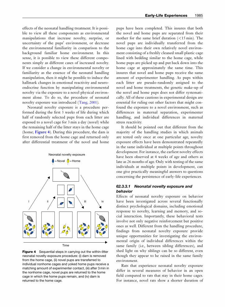
Neonatal novelty exposure is a procedure per-formed during the first 3 weeks of life during whichhalf of randomly selected pups from each litter areexposed to a novel cage for 3min a day (novel) whilethe remaining half of the litter stays in the home cage(home; Figure 4). During this procedure, the dam isfirst removed from the home cage and returned onlyafter differential treatment of the novel and home
Neonatal novelty exposure
= Novel
i ii iii
Time
iv
= Home
Figure 4 Sequential steps in carrying out the within-litterneonatal novelty exposure procedure: (i) dam is removed
from the home cage, (ii) novel pups are transferred to
individual nonhome cages and yoked home pups receive a
matching amount of experimenter contact, (iii) after 3min inthe nonhome cage, novel pups are returned to the home
cage in which the home pups remain, and (iv) dam is
returned to the home cage.
pups have been completed. This insures that boththe novel and home pups are separated from theirmother for the same brief duration (<15min). Thenovel pups are individually transferred from thehome cage into their own relatively novel environ-ment consisting of a freshly cleaned small plastic cagelined with bedding similar to the home cage, whilehome pups are picked up and put back down into thehome cage at approximately the same time. Thisinsures that novel and home pups receive the sameamount of experimenter handling. As pups withineach litter are pseudo-randomly assigned to thenovel and home treatments, the genetic make-up ofthe novel and home pups does not differ systemati-cally. All of these cautions in experimental design areessential for ruling out other factors that might con-found the exposure to a novel environment, such asdifferences in maternal separation, experimenterhandling, and individual differences in maternalstress reactivity.
It should be pointed out that different from themajority of the handling studies in which animalsare tested only once at one particular age, noveltyexposure effects have been demonstrated repeatedlyin the same individual at multiple points throughoutdevelopment. For instance, the earliest novelty effectshave been observed at 4 weeks of age and others aslate as 26 months of age. Only with testing of the sameindividuals at multiple points in development, canone give practically meaningful answers to questionsconcerning the persistence of early-life experiences.
62.3.3.1 Neonatal novelty exposure and
behavior
Effects of neonatal novelty exposure on behaviorhave been investigated across several functionallydistinct psychological domains, including emotionalresponse to novelty, learning and memory, and so-cial interaction. Importantly, these behavioral testsinvolve not only negative reinforcement but positiveones as well. Different from the handling procedure,findings from neonatal novelty exposure provideunique opportunities for investigating the environ-mental origin of individual differences within thesame family (i.e., between sibling differences), andshed light on why siblings can be so different, eventhough they appear to be raised in the same familyenvironment.
Rats that experience neonatal novelty exposurediffer in several measures of behavior in an openfield compared to rats that stay in their home cages.For instance, novel rats show a shorter duration of

1986 Early-Life Experiences
initial freezing upon entering the open field (Tang,2001), and their activity levels show a greater initialincrease across two brief (20 s) trials of exposure(Reeb et al., 2007). Both observations suggest thatnovel rats are faster to recover from the initial behav-ioral inhibition induced by the unfamiliar environ-ment. When exposed to an odor that the rats havenever experienced before, novel rats show shorterapproach latencies and higher frequencies of explo-ration, again suggesting that novel rats are faster inrecovering from behavioral inhibition induced by anovel odor (Yang et al., 2008).
As emotional states are known to influence cogni-tive processes, one may predict differential perfor-mance in learning and memory tasks that inevitablyinvolve varying degrees of novelty. When tested in aworking memory version of the Morris water mazetask, in which the rats must locate a hidden platform inthe water, novel rats show significantly greater workingmemory (Reeb et al., 2007; Tang, 2001; Tang et al.,2006). Specifically, this is indexed by a greater reduc-tion in the time to locate the hidden platform afteronly a single-trial exposure to the location of thehidden platform (Reeb et al., 2007; Tang, 2001; Tanget al., 2006). The effects of novelty exposure on work-ing memory can be eliminated by increased familiarityvia repeated swim trials involving identical platformlocation and reinstated by an increase in novelty bytesting the rats again after a prolonged period of delay(e.g., several months; Tang, 2001).
In contrast to learning in the Morris water task,which involves cold water as a negative reinforcer,learning and memory in an odor discrimination-learning task that involves sweets as positive reinfor-cers also differ between the novel and home rats(Tang, 2001). Distinct from the findings from nega-tive reinforcement learning, novel and home ratsshow similar performance in their initial learning ofthe discrimination task in which they learn that oneof the two odors is associated with a sweet reward(Tang, 2001). It is only during a retention testconducted 6 days after the last day of training thatnovel rats show complete retention of the task (butthe home rats show significant forgetting). Further-more, when these rats are tested for reversal learning,which clearly introduces surprise to the testing situ-ation, novel rats are faster than home rats at acquiringthe new stimulus–reward relationship (i.e., reversingto the previously nonrewarding odor; Tang et al.,unpublished observation).
In the domain of social functions, the effectsof neonatal novelty exposure have been found in
aggressive behaviors, social competition, social rec-ognition memory, and social engagement. Althoughbiting occurred infrequently during free dyadic inter-actions between a novel and home rat, the home ratsbit the novel rats twice as often as the novel rats bitthe home rats (Reeb and Tang, unpublished observa-tion). This observation is consistent with the inter-pretation that home rats are more likely to perceivethreats than novel rats. When meeting another rat24 h after their initial interaction, novel rats show agreater habituation than the home rats in the fre-quency of social investigation, and this habituationis blocked among the novel rats only by inserting ameeting with another stranger rat between the initialinteraction and an interaction taking place 24 h later.These findings suggest that novel rats have bettermemory of the previous-encounter conspecific thanthe home rats (Tang et al., 2003a). It is possible that thehome rats are more fearful during their initial socialinteraction and that this fear contributes to theirimpaired 24-h memory for the previously encoun-tered conspecific.
Similar to the acquisition of the odor–reward asso-ciation, learning to retrieve a chocolate reward in atesting cage does not differ between the novel andhome rats. Only when placed in a competitive situa-tion, where only one of the two rats could gain access toa reward (chocolate drops), novel rats win the compe-tition more frequently than the home rats (Tang et al.,2006). When the rats are tested in competition withoutfood deprivation, thus with lower levels of stress ingeneral, this greater competitive ability is presentonly during the initial encounters (first day) anddisappears during the later encounters (second day).The presence of this novelty effect when the testingsituation is novel may once again reflect a difference inperceived threat between the novel and home ratswhen facing the surprise of seeing another rat.
During free interaction, a greater proportion ofnovel rats’ social initiations are reciprocated by thehome rats, suggesting that the novel rats are moreable to engage the home rats. Furthermore, this differ-ential ability in social engagement is found only uponthe pair’s initial encounter and disappears at the secondsession of interaction 5min later, and can be reinstatedby priming the rats with a surprise event – spending2min in a plastic bottle immediately before socialinteraction (Tang et al., unpublished observation).
Together, these findings demonstrate the impactof early environmental differences in novelty on awide range of psychological functions and reveal amodified response to novelty and surprise as the key

Early-Life Experiences 1987
psychological mechanisms mediating the behavioraldifferences later in life. It is important to note thatthese different psychological functions are assessed atages from as early as immediately after weaning(4weeks of age) to the end of life (26months ofage). Thus, even though the differential treatment dur-ing the neonatal novelty exposure only consisted ofapproximately 1 h of total difference, the impactof this early-environmental experience appears to belifelong.
62.3.3.2 Neonatal novelty exposure and brain
Effects of neonatal novelty exposure on the brainhave been examined at the level of functional brainasymmetry, hippocampal gross anatomy, and synapticplasticity. It has been long known that early stimula-tion, such as the handling procedure, induces changeswithin the brain asymmetrically (Denenberg, 1981).Specifically, in the nonhandled rats, left and rightcortical damage produce similar effects on openfield activity, while handled rats show differentialchanges in response to left and right cortical damage(Denenberg, 1978). Given this asymmetric effect, theexploration of neonatal novelty effect on brain devel-opment has been characterized by separate measuresfor the left and right brain, whereas majority of earlystimulation studies do not differentiate left and rightmeasures.
Consistent with this finding of an asymmetricalearly stimulation effect, novel and home rats arefound to differ in their handedness, with the novelrats showing a left shift in their paw preference in areaching task (Tang and Verstynen, 2002). This leftshift is consistently observed across consecutive daysand across several months of delay, thus suggestinga persistent increase in right cerebral dominance.Supporting this interpretation is another reliableobservation from a second form of functional brainasymmetry measure, the turning asymmetry duringspontaneous exploration in a novel environment.Upon initially entering a novel environment, novelrats have a greater right turn bias than the home rats(Tang et al., 2003b). As turning toward the rightinvolves stronger pushing by the left front limb, thisdifference in asymmetry also indicates an increase ofright cerebral dominance (Tang and Reeb, 2004). Incontrast to the functional asymmetry in handedness,this turning asymmetry is transient, appearing to bedependent upon the novelty of the situation. Forinstance, it shows a clear modulation by time, withthe greatest novelty effect found during the firstminutes of spontaneous exploration (Tang et al.,
unpublished observation). Together, these findingsoffer clear evidence for a modification of brain asym-metry as a result of neonatal novelty exposure.
As the hippocampus plays a critical role in learningand memory (Eichenbaum, 1997) and in regulatingHPA output (Herman et al., 2003; Sapolsky et al.,1984a), effects of neonatal novelty exposure on theanatomy and function of this structure have been inves-tigated. Although there are no overall differences inhippocampal volume, the novel and home rats differ intheir patterns of volumetric asymmetry with the novelrats showing a relatively greater right hippocampusthan the home rats by 1% of the total hippocampalvolume (Verstynen et al., 2001). Although 1% seems asmall effect, computationally it may make a significantdifference in dynamics of the neural networks, givingrise to functional asymmetry (Reggia et al., 1998).
Neonatal novelty exposure effects on synapticplasticity have been examined in the CA1 region ofthe hippocampus. Long-term potentiation (LTP), themost extensively studied form of synaptic plasticity,differs between novel and home rats. Regardless ofthe side of hippocampus, neonatal novelty exposureleads to enhanced LTP, while synaptic transmissionprior to LTP induction shows no significant differ-ence (Tang and Zou, 2002). When the data are fur-ther analyzed, taking into consideration the side ofthe hippocampus, evidence of asymmetric noveltyexposure effects are found. This early-life stimula-tion effect is characterized by two forms of selectivity:a selectivity for the right hippocampus and a selec-tivity for LTP (Tang et al., 2008).
As these asymmetric changes within the brain allinvolve an increased dominance of right-side func-tion, it begs the question of how these asymmetriceffects are achieved and what evolutionary signifi-cance this asymmetric environment may have. Asmeasures of asymmetry have been shown to havepredictive power for measures of memory (Tanget al., 2003a; Tang and Reeb, 2004) and the asymme-try in synaptic plasticity is selective for LTP, one mayspeculate that early experience is critical for thedevelopment of normal functional lateralization, oravoidance of abnormal functional lateralization. Itshould be noted that abnormal functional lateraliza-tion has been associated with a range of psycho-pathologies (Davidson, 2003).
62.3.3.3 Neonatal novelty exposure and
endocrine function
The investigation of neonatal novelty exposure onendocrine function has focused on how circulating

Control
Home
Novel
7–8 months
Application of CORT
120
100
80
60
40
20
00 10 20 30
Home
Novel
40 50
Time (min)P
opsp
ike
(%)
1 mV5 ms( a )
( b )
CORT Washout
Figure 5 Bath application of 100-nM CORT results in
greater inhibition of population spikes in slices from novelthan in home rats. (a) Examples of population spikes before,
during and after 20min of CORT perfusion in novel and
home slices. (b) Time course of CORT effect demonstrates
the significantly greater reduction of population spikeamplitude in novel slices.
1988 Early-Life Experiences
CORT, both basal and evoked, relate to other func-tional measures, and to novelty of the situation inwhich behavioral measures are obtained. Thisapproach differs from that used in most handlingstudies where CORT measures are obtained underconditions of shock or restraint that may not allowgeneralization to learning situations where the maxi-mum level and temporal characteristics of HPA acti-vation are rather different.
The most surprising finding is that no effectof neonatal novelty exposure is found on socialinteraction-evoked CORT responses obtained shortlyafter social competition, even though the behavioralmeasures differ between the novel and home rats(Akers et al., unpublished observation). This meansthat if circulating CORT somehow contributes tobehavioral differences observed in these tasks, thenthe behavioral effects cannot be mediated by differen-tial concentrations of CORTalone. Interestingly, noveland home rats are found to differ in the plasticity ofthe evoked CORTresponse to social competition, withonly the novel rats showing habituation of CORTresponses across 2 days of repeated social competitionagainst the same competitor. This means novel andhome rats differ in their ability to downregulate theirHPA output according to recent experience.
Similarly, no effect of neonatal novelty exposure isfound on evoked CORT after animals have becomehighly familiarized with the Morris water task. Incontrast, seemingly small deviations from the testingroutine, such as an unexpected visit to another roomwhere the animal spends a few minutes in an openfield, are able to produce differential levels of circu-lating CORT between the novel and home rats. Spe-cifically, we have found that the novel rats show agreater sensitivity to this surprise manipulation(Tang et al., unpublished observation). This meansthat the HPA axis of novel rats is not only better atdownregulating its responses when the testing situa-tion has become familiar, but also better at respond-ing to changes in their environment.
The hippocampus provides the major environmen-tally related driving force for the HPA axis. Thus,clues about how novel rats might achieve better detec-tion of environmental novelty and downregulation ofHPA output as a result of environmental familiaritymay be obtained by examining electrophysiologicaldata of population spikes recorded in the CA1 of thehippocampus (Zou et al., 2001; Figure 5). Afterthe onset of perfusion of the slice by stress levels ofCORT, the amplitude of the population spike from
hippocampal slices from novel rats shows a small ini-tial, brief increase, followed within a few minutes bya large decrease. Conversely, the slices from homerats show relatively little change in response toCORT perfusion. This differential effect suggeststhat novel and home may achieve different levels ofself-regulation via differential levels of functionalGRs. Additional pharmacological experiments furtherreveal that LTP of the population spikes among thenovel and home rats is also differentially modulated bystress levels of CORT after the short-term effect onneuronal excitability is washed out. These findingsserve to explain how it is possible that the same con-centration of circulating CORT could support differ-ential effects on the function of the circuit, hencemediating differential effects on behavioral measuresdiscussed earlier.
One of the possible consequences of this enhancedself-regulation of CORT secretion is that novel ratsmight be able to maintain a lower basal level ofCORT than home rats. This was confirmed by the

Early-Life Experiences 1989
finding that at 16months of age novel rats havelower basal CORT than home rats (Tang et al.,2003a). Most interestingly, even though this measureis temporally remote from behavioral measures ofsocial recognition memory, lower basal CORT retro-actively predicted 24-h recognition memory 8monthsearlier (Tang et al., 2003a).
62.3.3.4 Neonatal novelty exposure and
maternal influence
These neonatal novelty exposure-induced changes inbehavioral, neural, and endocrinological functionsprovide unequivocal evidence that as little as 1 h oftotal difference in early life can induce a wide rangeof long-lasting effects on development. The transientnature of this environmental manipulation forms asharp contrast to the omnipresence of the motherto the developing offspring. It begs the question ofwhat role the mother plays in relation to these earlystimulation effects. Specifically, does the motherdiscriminate between her novel and home pups, andto whom does she show preferential care?
Common wisdom would suggest that novel ratswould receive preferential maternal care. However,data from studies of multiple cohorts of rats do notsupport this idea. The dams either show no preferencein their care toward the novel and home pups or showpreferential care toward the home pups (Tang et al.,2006). In other words, neonatal novelty exposure treat-ments lead to enhanced functionality in the novel ratsdespite the fact that the mothers show preferentialcare toward the home rats. Instead, the state of thedam’s stress response system (e.g., her circulatingCORT) can set the stage for differential responses ofpups to neonatal novelty exposure. Individual differ-ences in the mother’s HPA function can thus lead todifferential neonatal novelty exposure effects. Thishypothesis has been confirmed by a positive correla-tion between maternal-evoked CORT response to1-min swim stress and novelty effects on offspringcognitive and emotional measures and a complemen-tary negative correlation between maternal basalCORT and novelty effects (Reeb et al., 2007).
These findings not only shed light on how earlystimulation via neonatal novelty exposure interactswith maternal influence, but also suggest alternativeinterpretations of the handling experiments origi-nally thought to provide clear evidence for support-ing the maternal mediation hypothesis (Liu et al.,1997). In response to the stress of handling and sepa-ration from her pups, the dams of the handled pups
not only increase their care-giving behavior uponreunion, but also more than likely demonstrate ele-vated circulating CORT levels. A dam’s circulatingCORT can affect the pups’ circulating CORT levelsvia her milk supply (Catalani et al., 2000; Macri et al.,2007; Meerlo et al., 2001). Thus, it is possible that theincrease in maternal care is only an epiphenomenonand it is handling-induced maternal stress that pro-vides the pups with low doses of CORT exposureearly in development, which in turn shapes thepups’ HPA development.
62.3.4 Maternal Deprivation
In this section, we shift our discussion to the impactof more prolonged phases of separation from the damand the resulting behavioral, neural, and endocrino-logical changes in the offspring. Specifically, we dis-cuss maternal deprivation and maternal separationparadigms that remove pups from the nest for rela-tively extended periods of time (ranging fromapproximately 3 to 24 h) either once or multipletimes. These paradigms are thought to model infantor childhood neglect. Within a couple of hours ofseparation, this procedure usually activates the stressaxis with increases in CORTand ACTH. Also, robustbehavioral responses are evident in the pup. Maternaldeprivation has been one of the more prolific proce-dures in the early-life experience literature and theimpact of this paradigm on our understanding ofearly-life effects has been critically important. How-ever, due to variability in maternal deprivation pro-cedures between labs, considerable variability inresults exists in this literature preventing simplisticstatements concerning effects of maternal depriva-tion. For example, due to pups’ reliance on behavioralthermoregulation and insufficient thermogenesis, aminor variation of 1–2 �C in surface or ambient tem-perature can produce a hyperthermia/hypothermiathat can greatly alter organ function, including theinfant brain, which is uniquely dependent upon bodytemperature in pups (Kleitman and Satinoff, 1982;Sullivan and Leon, 1988).
The unique role of sensory stimuli in controllingpup behavior, brain, and physiology also contributesto the importance of minor procedural difference inmaternal deprivation. Indeed, sensory stimuli main-tain pups at homeostasis in myriad systems, withdifferent stimuli and its patterning controlling spe-cific systems and referred to as hidden regulators(Hofer, 1995). Thus, the maternal deprivation

1990 Early-Life Experiences
procedure can be viewed as removal of sensory stimuli,with minor variations between labs removing moreor less of these sensory stimuli normally providedby the mother, siblings, and the nest. For example,tactile stimulation increases growth hormone, warmthincreases NE, maternal odor increases behavioralactivity, and cold increases CORT (Hofer, 1973;Kuhn and Schanberg, 1998). Together, these datasuggest very specific and minor changes in experi-mental protocols that can produce a diversity ofbehavioral and physiological responses in pups thatdo not facilitate fine-grain interpretations of this liter-ature. However, this literature clearly illustrates thatearly-life separation from the mother produces robustbrain changes in specific neural loci and specificmodifications in endocrine control that are related tolong-term changes in emotion and cognition that arereviewed below.
Recent literature has questioned whether the crit-ical factor in maternal separation experiments is theseparation itself or the mother’s response to the pupsat reunion. The extent of changes in maternal behav-ior induced by maternal separation is variable acrossparadigms and laboratories and likely contribute tothe varied outcomes reported. For instance, sepa-rated, cold pups in the same room as the motherwill likely produce different results than a laboratorythat keeps separated pups thermoneutral in a roomfree of maternal odors. Thus, it is likely that bothpups’ response to separation and the mother’sresponse to reunion contribute to the short- andlong-term effects of maternal deprivation. Regardlessof the relative contribution of dam versus pup to theobserved changes, this literature strongly supportsthe importance of early-life experience on later-lifebehavior, brain, and endocrine responses.
62.3.4.1 Maternal deprivation and behavior
The rat pups’ immediate behavioral response tomater-nal separation remains fairly consistent throughoutearly life and produces increased behavioral activityand vocalizations, including ultrasonic vocalization(Hofer et al., 2001). Interestingly, these immediateresponses can be greatly attenuated if pups areprovided with adequate warmth and maternal odor(Hofer and Shair, 1978; Sokoloff and Blumberg, 1997).Within approximately an hour, this response changesto hypoactivity, although the immediate responseremains more persistent as pups mature (Hofer andShair, 1991). Separation from the mother also altersthe pups’ future responses to the mother at reunion
(hyperresponsiveness) and subsequent separation(increased ultrasonic vocalization), suggesting mater-nal separation has profound chronic effects on pups’behavior during the preweanling period (Hofer andShair, 1978).
The long-term effects of maternal separationappear to produce an animal that is more behavior-ally responsive to stressful situations (Andersen et al.,1999; Kosten et al., 2005). However, these animals arealso thought to exhibit generalized cognitive andcontextual fear-learning impairments (Bean et al.,2002; Kosten et al., 2005, 2006), anhedonia (Matthewsand Robbins, 2003), decreased or increased foodintake based on the context (McIntosh et al., 1999;Penke et al., 2001), and susceptibility to drugand alcohol abuse (Cirulli and Alleva, 2003). Thelong-term effects of maternal deprivation appear toalter maternal care, which is then transmitted non-genomically through the next generations (Fleminget al., 2002). In summary, maternal deprivation isconsidered an early-life stressor that produces ananimal behaviorally equipped for a stressful adultlife and alters specific and global behaviors such asmaternal care, food intake, and metabolism.
62.3.4.2 Maternal deprivation and brain
Maternal deprivation produces ubiquitous changes inthe brain, although the reciprocally interacting limbicsystem and stress axis have received particularlyintense assessment. A comprehensive review of thematernal deprivation effects on the brain is beyondthe scope of this chapter, although we attempt to high-light the major findings of this area related to stress.Overall, the brain of infant rats with or without mater-nal deprivation shows a unique response to stress, withthe time course and intensity of the neural responsediverging from that documented in adults.
Presentation of a stressor to infant rats producesvery rapid changes in CRH mRNA in the PVN innondeprived pups (Dent et al., 2000a). Interestingly,this response in maternally deprived pups is signifi-cantly reduced (Dent et al., 2000a). The inability toidentify this period of hyperresponsiveness in previ-ous studies appears to be due to a pup’s very rapidonset of the stress response compared to adults. Thus,while the adrenal gland shows an SHRP, the centralcomponents (i.e., brain) of the HPA axis are respon-sive, though hyporesponsive with maternal depriva-tion (Suchecki et al., 1993).
While infant maternal deprivation causes adecrease in PVN CRH in infancy, it shows a marked

Early-Life Experiences 1991
increase in adulthood (Plotsky and Meaney, 1993).Indeed, neural changes due to maternal deprivationcan be seen throughout the brain in cells containingCRH and GR, especially in areas that integratethe endocrine and behavioral responses to stress.Specifically, compared to nondeprived animals, adultanimals that were maternally deprived show a reduc-tion in CRH receptor-binding density in the ante-rior pituitary but increases in CRH receptorbinding/immunoreactivity in the raphe nucleus,parabrachial nucleus, amygdala, and bed nucleus ofthe stria terminalis (BNST). GRs are also modulatedfollowing maternal deprivation. Specifically, mater-nal deprivation is associated with decreased GR inthe hippocampus later in life, which suggests mater-nally deprived pups have impaired HPA negativefeedback. Furthermore, the LC, which controlsmuch of the brain’s NE and potentiates the HPAaxis, is also modified and associated with a down-regulation of NE receptors (Ladd et al., 2000).
The short-(infant) and long-term (adult) effects ofinfant maternal deprivation diverge. For example,maternal deprivation has been shown to induce anupregulation of brain-derived neurotrophic factor(BDNF) expression in hippocampus and PFC inpups, although in adulthood there is reduction inPFC BDNF expression (Roceri et al., 2004). Finally,connectivity between brain areas appears disruptedby maternal deprivation. For example, compared tonondeprived pups, maternally separated pups showaltered responses of the BNSTand PVN to amygdalastimulation (Sanchez et al., 1995), though synapticchanges and dendritic-branching modifications alsocontribute to these changes (Braun et al., 2000). Over-all, these data suggest that the neural effects of mater-nal deprivation occur throughout the brain andinclude anatomical and synaptic changes withinand between brain areas. Importantly, maternaldeprivation induces short-term effects in infancyand long-term effects in adulthood that do not alwayscorrespond. It should also be noted that consider-able divergence in adult outcome following maternaldeprivation appears in the literature suggesting thatthe onset and duration of the maternal deprivation isa critical variable in both the immediate response andthe long-term response (Muneoka et al., 1994).
In summary, maternal deprivation alters the HPAaxis and brain areas that integrate and control HPAregulation both in infancy and adulthood. Impor-tantly, the infant peripheral hyporesponsiveness isassociated with neural hyperresponsiveness. While
the causal mechanisms connecting specific stimuli(or the absence thereof ) and neural response tomaternal deprivation in infant and adult neural con-sequences still need to be assessed, there is consider-able convergence between the maternal deprivationliterature and the clinical literature on early-lifeneglect.
62.3.4.3 Maternal deprivation and
endocrine function
Removing pups from the mother for a prolongedperiod of time has immediate and long-term effectson a pup’s endocrine system, although this effectchanges depending on the age of manipulation andage of assessment. Overall, the literature indicatesprolonged separation from the mother overrides theSHRP and pups show an elevation in baseline CORT,which is potentiated by presentation of a stressor.Thus, while the mechanism is unclear, maternalseparation appears to activate the normally quiescentHPA axis to permit a CORT response. In a compre-hensive study, pups were separated from the motherfor times ranging from 15min to 24 h and the responseto stress (CORT and ACTH) assessed at differentdevelopmental ages (Dent et al., 2000b). Overall, pupsshow a CORT response that became more robust withlength of separation from the mother, as well as age. Itshould also be noted that during early life, normalCORTand ACTH responses can be elicited in neona-tal pups injected with an endotoxin, indicating a func-tioning HPA axis in pups that are stressor specific(Dallman, 2000; Dent et al., 1999; Stanton et al., 1987;Suchecki et al., 1993; Walker et al., 1991; Witek-Janusek, 1988). Thus, the pituitary–adrenocorticalsystem of the neonatal rat is responsive to stressthroughout development in a time-dependent andstressor-specific fashion.
The classic long-term impact of maternal depri-vation emerges around weaning and continues intoadulthood. For example, weanling-aged pups thatwere separated from their mothers for only onematernal deprivation session at 3 days of age showedheightened ACTH and CORT, although not if thesame manipulation was done at 7 days of age. Onthe other hand, pups maternally deprived at 11 daysof age show an elevated CORT response at weaningbut by periadolescence the stress-induced CORTresponse is attenuated compared to nondeprivedcontrols (Suchecki and Tufik, 1997). Finally, sex dif-ferences in response to maternal deprivation occur atboth adolescence and adulthood (Rees et al., 2006).
1992 Early-Life Experiences
These studies suggest that the response to early-lifematernal deprivation is a complex phenomenon, withthe causal mechanisms still a mystery.
62.3.5 Pain, Fear Conditioning, andContext of Early-Life Adversity
Early-life adverse experience alters adult emotional-ity and cognitive function in clinical populations andhas been modeled in infant rodents with moderatelypainful electric shock. Early-life experience withshock, which does not increase a pup’s CORT levelsuntil PND10, appears to activate a pup’s nociceptivesystems and elicit pain-related behavioral responses(Barr, 1995; Collier and Bolles, 1980; Emerich et al.,1985; Fitzgerald, 2005; Stehouwer and Campbell,1978). Overall, repeated shock in early life appearsto heighten adult emotionality/anxiety and has alsobeen shown to both attenuate and enhance learning,depending on the task. Previous work on infantadverse experiences using shock suggests that unpre-dictable shock produces greater changes in adultemotionality than predictable shock (Bell andDenenberg, 1962; Denenberg, 1963; Denenberg andBell, 1960; Henderson, 1965) and varies from theeffect typically observed in adults (Shors et al.,1990; Weiss, 1970).
Central processing of pain in infants appears todiverge from adults, as indicated by the ontogeny offear conditioning using pairings of odors and 0.5-mAelectric shock to either the tail or hindlimb. As avehicle to understand infant processing of pain, thereare several advantages to using the olfactory learningparadigm of fear conditioning. Specifically, the adultfear-conditioning circuit is relatively well defined andpermits the direct comparison of the maturing infantbrain to the adult circuit (Debiec and LeDoux, 2004;Fanselow and Gale, 2003; Funk and Amir, 2000;Schettino and Otto, 2001; Sevelinges et al., 2007). Inaddition, the predictable versus unpredictable natureof stimuli are inherent in the fear-conditioning para-digm’s use of the experimental (paired odor and shock)and control (unpaired odor and shock) conditioninggroups. Finally, pups must learn the odor used forattachment to the mother, with a wide range of stimulifunctioning as a reward to support odor-preferencelearning, including painful 0.5-mA shock (Camp andRudy, 1988; Sullivan et al., 2000).
While odor-approach/preference learning in earlylife appears paradoxical, this limitation on aversivelearning is seen in species other than rats and isimportant for attachment. For example, during
imprinting in chicks, shock enhances followingbehavior of the surrogate, although an aversion occursif presented just hours after the sensitive periodcloses (Hess, 1962; Salzen, 1970). These learning lim-itations have also been documented in infant dogsthat continue to approach a human attendant thatshocks or mishandles the puppies (Rajecki et al.,1978), as well as nonhuman primates that continueto approach a caregiver that handles them roughly(Harlow and Harlow, 1965; Maestripieri et al., 1999;Sanchez et al., 2001). In young rat pups, this limita-tion on learning also extends to inhibitory condition-ing and passive avoidance (Blozovski and Cudennec,1980; Collier et al., 1979; Myslivecek, 1997; Stehouwerand Campbell, 1978).
We suggest that this model of infant odor–shockconditioning in infancy may be useful as a paradigmto better understand how early-life abuse can supportand maintain attachment. Furthermore, we suggestthat in understanding the unique ways in which theinfant brain processes painful stimuli, such as shock,we may gain insight into the enduring effects ofearly-life pain.
62.3.5.1 Odor–shock conditioning and
behavior
During a sensitive period in early life (until PND10),an odor paired with a painful stimulus (0.5-mA tail-or footshock, tail pinch, or mother handling pupsroughly) results in pups approaching that odorwhen it is subsequently encountered (Camp andRudy, 1988; Haroutunian and Campbell, 1979;Moriceau et al., 2004, 2006; Moriceau and Sullivan,2004; Roth and Sullivan, 2001, 2005; Spear, 1978;Sullivan, 2003; Sullivan et al., 2000). This is in sharpcontrast to odor–shock conditioning in adults(an auditory stimulus can also be used), which isreferred to as fear conditioning, and produces learnedfreezing responses and odor avoidance in adults withthe amygdala being a critical site for plasticity (Daviset al., 2003; Debiec and LeDoux, 2004; Fanselow andGale, 2003; Fanselow and Poulos, 2005; Hess et al.,1997; LeDoux, 2003; Rosenkranz and Grace, 2002;Sananes and Campbell, 1989; Schettino and Otto,2001; Sevelinges et al., 2007). The failure of pups toavoid an odor previously paired with pain occursdespite a functional nociceptive system (Barr, 1995;Emerich et al., 1985; Fitzgerald, 2005; Stehouwer andCampbell, 1978).
Considering the necessity of pups learning a pref-erence to their mother’s odor for nipple attachment,and other related attachment behaviors, it is certainly
Early-Life Experiences 1993
beneficial for pups not to learn an aversion to theirmother’s odor or inhibit approach responses to nestodors. Perhaps this attenuated avoidance learningensures that pups continue to only approach/followthe caregiver (Hofer, 1981; Hofer and Sullivan, 2001).In addition, this odor–0.5-mA shock paradigm supportslearning an odor that appears to function similar to thematernal odor, since it supports nipple attachment(Sullivan et al., unpublished observation).
This early-life attachment odor is retained intoadulthood where it alters emotion and cognition. Spe-cifically, the odor paired with pain to produce theattachment odor can attenuate adult fear condition-ing as well as attenuate amygdala neural activity, asmeasured by 2-deoxyglucose (2-DG) autoradiography,that supports this learning (Sevelinges et al., 2007).It should be noted that pups that receive unpairedpresentation of the odor–shock do not learn about theodor in infancy, nor does the odor alter adult fearconditioning. However, these unpaired pups exhibitanxiety-like behaviors in adulthood that are not exhib-ited by paired odor–shock pups (Tyler et al., 2007).
62.3.5.2 Odor–shock conditioning and brain
Fear conditioning requires the amygdala, althoughmany additional brain areas are involved in thiscomplex neural circuit (Fanselow and Gale, 2003;Fanselow and LeDoux, 1999; Herzog and Otto,1997; Maren, 2003; McGaugh et al., 1999; Pape andStork, 2003; Pare et al., 2004; Rosenkranz and Grace,2002; Sananes and Campbell, 1989; Schettino andOtto, 2001; Sevelinges et al., 2004; Walker andDavis, 1997). The importance of the amygdala inearly-life fear conditioning is further suggested bythe concurrent emergence of fear conditioning andamygdala plasticity (Sullivan et al., 2000). We havedata suggesting the inability of pups to learn thisearly-life version of fear conditioning may be dueto the amygdala’s failure to participate in early-lifelearning. Specifically, during the sensitive period,when odor–shock produces an odor preference, theolfactory bulb and anterior piriform olfactory cortexshow activation during learning, while the amygdaladoes not appear to participate, as indicated by 2-DGor c-Fos expression (Moriceau and Sullivan, 2006;Moriceau et al., 2006; Roth and Sullivan, 2005). Incontrast, similar conditioning in postsensitive periodpups (PND12) that learn odor aversion and freezingshows activation of the posterior piriform cortex,basolateral, lateral, and cortical amygdalar nuclei(Moriceau and Sullivan, 2006; Moriceau et al., 2006;Sullivan et al., 2000). Importantly, temporary
suppression of the pup’s postsensitive period amyg-dala during fear conditioning prevents learning(Moriceau and Sullivan, 2006).
62.3.5.3 Odor–shock conditioning and
endocrine function
CORT infused either systemically or directly intothe amygdala during odor–0.5-mA shock condition-ing results in an odor aversion in sensitive-periodpups, overriding the pups low CORT levels dur-ing the SHRP and permitting amygdala plasticity(Moriceau et al., 2004, 2006; Moriceau and Sullivan,2004, 2006). A similar role of CORThas been demon-strated during imprinting in ducklings, with lowCORT levels important for following the surrogate.
In pups, CORT levels can be modulated by theenvironment with early-life stress causing a pre-mature increase in CORT levels (Levine, 1962, 1994),or through CORT received through their mother’smilk (Yeh, 1984). In addition, the mother modulates apup’s CORT concentration based on the level of hermaternal care, with decreases in maternal care causingan increase in the pup’s CORT levels (Stanton et al.,1987; Suchecki et al., 1993). Indeed, the attenuation ofthe shock-induced CORT release can be blocked bymaternal presence and pup attachment learning toodor–shock conditioning is reinstated (Moriceau et al.,2006). The important role of maternal suppression ofshock-induced CORT release in pups odor aversionlearning was verified by systemic and intraamygdalaCORT infusions, which permitted pups to learn odoraversions in the presence of the mother. Similar socialbuffering of CORT release by social cues has beenfound in other paradigms and species in both infancyand adulthood (Carter and Keverne, 2002; Carter et al.,2003; Dallman, 2000; Deschamps et al., 2003; DeVries,2002; DeVries et al., 2003; Hennessy, 1984; Hennessyet al., 2006; Wiedenmayer et al., 2003).
62.3.6 Functional Consequences ofEarly-Life Experiences
Together, there is remarkable convergence acrossthese various early-life experience paradigms withlong-term effects seen in the HPA axis and brainareas with strong reciprocal interactions over theHPA axis, such as the LC, hippocampus, amygdala,and PFC. However, it is likely multiple neonatalmechanisms mediate these long-term effects, includ-ing changes in pups HPA axis and related brain areasin response to early-life manipulations, as well asthrough modifications of maternal care.

1994 Early-Life Experiences
62.4 Pubertal Experiences andEnduring Behavioral and EndocrineConsequences
As reviewed above, there is a vast body of literaturethat indicates neonatal development is a crucial stageof maturation that, if disturbed, modified or altered,results in changes in the future behavioral, neuro-logical, and endocrinological function of an individ-ual (Gutman and Nemeroff, 2002; Heim andNemeroff, 2001; Maccari et al., 2003; Zhang et al.,2006). However, the neonatal period is not the onlydevelopmental stage when individuals are susceptibleto both positive and negative influences. Whilepuberty has long been regarded as a period of rapidphysical growth and reproductive maturation, morerecently it is also regarded as an additional criticalperiod of neurobehavioral developmental vulnerabil-ities (Andersen, 2003; Dahl, 2004; Kleinert, 2007;Patton and Viner, 2007; Romeo, 2003; Sisk and Foster,2004; Spear, 2000). For instance, it is well establishedthat adolescence is marked by increases in the mor-bidity of various psychological disorders such as anx-iety, depression, and schizophrenia (Conger andPetersen, 1984; Costello et al., 2003; Masten, 1987;Patel et al., 2007). The mechanisms mediatingthe pubertal increase in these disorders are presentlyunknown.
Recent studies in adolescent boys and girls haveindicated that pubertal exposure to stress may be aparticularly relevant environmental factor that contri-butes to an individual’s vulnerability to variouspsychopathologies later in life (Grant et al., 2003,2004; Turner and Lloyd, 2004). However, it is stillunclear how exposure to stress during puberty maylead to various psychological dysfunctions (Romeoan d Mc Ewe n, 20 06 ). As described in Section 62.2.1.2,in response to acute or repeated stress, pubertal ani-mals demonstrate a more prolonged or higher peakACTH and CORT response, respectively, comparedto adults (Romeo et al., 2006a). Thus, it is possible thatthis differential responsiveness may contribute to theincrease in stress-related psychopathologies duringadolescence. In fact, the differential hormonal responsein adolescent compared to adult animals may be com-pounded in the pubertal individual by three additionalfactors. First, plasma levels of CORT-binding globulin(CBG), a protein that binds CORTmaking it unavail-able to target tissues such as the brain (Breuner andOrchinik, 2002), are lower in pubertal than adult ani-mals (Romeo et al., 2006a; Smith andHammond, 1991).
Second, the prepubertal brainmay bemore sensitive tostress-related hormones, as experiments have shownan equivalent dose of CORT increases hippocampalN-methyl-D-aspartate (NMDA) receptor subunitexpression (e.g., NR2A and NR2B) to a greater degreein prepubertal than adult males (Lee et al., 2003).Finally, brain regions that continue to mature duringadolescence (e.g., hippocampus, amygdala, and PFC),are also the most sensitive to CORTand are extremelyimportant in modulating emotionality (McEwen,2005). Thus, upon experiencing a similar stressor, theimmature, and possibly more sensitive, pubertal brainis differentially exposed to unbound, bioavailableCORT compared to the adult brain. In the next sec-tions, we briefly review the growing body of literaturethat supports the assertion that chronic stress duringadolescence may be particularly detrimental to boththe immediate and future neurobehavioral functionof an organism.
62.4.1 Pubertal Experience and Behavior
Long-term effects of stress on emotionality are notedin rats and mice exposed to a variety of stress para-digms during puberty. Specifically, anxiety- anddepressive-like behaviors are greater in adult animalsthat experience stress during adolescence comparedto unstressed controls (Avital et al., 2006; Leussis andAndersen, 2008; Maslova et al., 2002a,b; Pohl et al.,2007; Schmidt et al., 2007; Stone and Quartermain,1998; Tsoory et al., 2007; Vidal et al., 2007; Wrightet al., 2008). Unfortunately, it remains unclear whetheror not changes in adult emotionality after adolescentstress are dependent on the stressors occurring duringthe adolescent window of development. That is, wouldsimilar behavioral changes ensue if exposure to thesestressors occurred in adulthood?
Recent data from Avital and Richter-Levin (2004)have shown that the timing of the stress exposure isimportant. In particular, adolescent male rats tran-siently exposed to elevated platform stress (EPS)exhibit higher levels of anxiety-like behavior whentested as adults, while adults exposed to same tran-sient EPS treatment do not show any changes in theiranxiety levels (Avital and Richter-Levin, 2004). Wehave also recently shown that exposure to mild physi-cal (1 h or restraint stress every other day) and/orsocial stress (social isolation) during adolescenceleads to increases in depressive-like behaviors andelevated basal levels of plasma CORT in adulthood(Romeo et al., unpublished observation). Moreover,
Early-Life Experiences 1995
adolescent animals that experience both types ofstressors show the greatest increase in depressive-like behaviors, suggesting an additive effect of thestressors on subsequent behavior. It is importantto note that when adult animals experience thesesame mild physical and/or social stressors for thesame length of time, no changes in depressive-likebehavior or basal HPA function are observed (Romeoet al., unpublished observation). These data stronglyargue that, similar to the perinatal period of develop-ment, adolescence is a particularly sensitive period tostress, especially in regards to emotional behavior andmodulation of the HPA axis.
Though it remains unclear what mechanismsmediate these changes in adult behavior after expo-sure to stressors during adolescence, it is possiblethat stress-induced disruptions of developmentallyimportant social interactions occur. For instance,play behavior during adolescent development is inte-gral for the emergence of proper social behaviors inadulthood (Vanderschuren et al., 1997). It has beenreported that stressors such as food deprivation (Siviyand Panksepp, 1985), footshock (Wood et al., 1995),predator odor (Siviy et al., 2006), intense lightingconditions (Vanderschuren et al., 1995), or restraint(Romeo et al., 2006b) inhibit this developmentallyimportant social behavior. Therefore, the stress-induced reduction of play during the adolescentperiod may contribute, at least in part, to the behav-ioral disturbances observed in adult animals afterexperiencing pubertal stress.
62.4.2 Pubertal Experience and Brain
Although accumulating data indicate that stress dur-ing adolescence may be particularly disruptive inthe context of emotional development, the neuralsubstrates that mediate these changes are not wellunderstood. In fact, studies investigating the struc-tural alterations of the brain induced by pubertalstress are sorely lacking. An elegant study by Isgoret al. (2004) demonstrated that chronic variablestress (CVS) during adolescence leads to long-termchanges in hippocampal structure and function. Morespecifically, they found that compared to nonstressedcontrols, animals exposed to CVS during adolescencehad significantly smaller hippocampal volumes,lower hippocampal GR expression, a hyperrespon-sive HPA axis to later stressors, and reduced perfor-mance on a hippocampal-dependent water maze task(Isgor et al., 2004). Interestingly, these effects ofCVS experienced during adolescence were noted
30 days after the stressors had been terminated(Isgor et al., 2004). It is important to note that effectsof chronic stress on hippocampal structure in adultsreturn to prestress levels within 10–21 days (McEwenand Margarinos, 1997). Thus, the study by Isgor et al.(2004) suggests stress-induced structural remode-ling of the hippocampus during adolescence islonger lasting, and perhaps permanent, compared tostress-induced remodeling in adulthood.
Future studies will need to investigate what rami-fications adolescent stress exposure may have on otherbrain regions that continue to develop during adoles-cence, that are highly sensitive to glucocorticoids andintimately involved in emotional processing, such asthe mPFC and amygdala (McEwen, 2005; Romeo andMcEwen, 2006). It should be noted that recent studieshave demonstrated stress-induced changes in synap-togenesis and DA receptor levels in adolescent PFC,which may be associated with the behavioral dysfunc-tion associated with pubertal stress (Leussis andAndersen, 2008; Leussis et al., 2008; Wright et al.,2008).
62.4.3 Pubertal Experience and EndocrineFunction
Relatively few studies have investigated the effects ofchronic stress during adolescence on later stress reac-tivity in adulthood. As mentioned above, we havefound that animals exposed to restraint stressand/or social isolation during puberty demonstrateelevated levels of basal CORT in adulthood (Romeoet al., unpublished observation). Furthermore, Isgoret al. (2004) have found that animals exposed to CVSduring puberty demonstrate increased stress-inducedHPA responsiveness in adulthood. Specifically, ani-mals stressed during adolescence demonstrate a sig-nificantly more prolonged stress-induced CORTresponse compared to animals not exposed to puber-tal stress (Isgor et al., 2004). It was also found thatanimals exposed to stress during puberty showdecreased hippocampal GR expression in adulthood,compared to their unstressed counterparts (Isgoret al., 2004), suggesting decreased hippocampalGR-mediated negative feedback on the HPA axis.Not all experiments have found changes in adultHPA function following adolescent stress. Forinstance, experiments that employed chronic unpre-dictable stress, such as restraint or footshock, duringpuberty were unable to find any changes in adultHPA function (Maslova et al., 2002a; McCormicket al., 2005; Overmier and Murison, 1991).

1996 Early-Life Experiences
Taken together, it appears that chronic stress dur-ing adolescence may lead to subtle changes in HPAfunction. However, compared to the reliable andlong-term effects of neonatal stress on later HPAreactivity, the existing literature does not supportsuch a consistent, robust change in adult HPA func-tion following adolescent stress. Though it is unclearwhy perturbations of neonatal compared to thepubertal HPA axis may lead to more dramaticchanges in later stress responsiveness, it is possiblethat the constituent neuroendocrine parts of the HPAaxis are more malleable during the neonatal period,and thus, more sensitive to perturbations. Futurestudies will need to compare the relative sensitivitiesof the developing neonatal and pubertal HPA axes,and also assess possible cumulative effects of bothneonatal and pubertal stress exposure on later HPAfunction in adulthood.
62.5 Adolescence as a Period ofIntervention to Mitigate EarlyDevelopmental Insults
Adolescence is clearly marked by profound changesin an individual’s nervous system, physiology, andbehavior (Andersen, 2003; Dahl, 2004; Romeo, 2003,2005; Romeo et al., 2002; Spear, 2000, 2007).Although this may render an individual especiallyvulnerable to harm during this period, it may alsoallow for interventions to mitigate earlier or con-current emotional and/or physical trauma (Andersen,2003; Dahl, 2004). Recent research has explored theability of environmental enrichment during adoles-cence to ameliorate the negative influences of earlierperinatal insults. Environmental interventions may bemore attractive than pharmacological interventions tocombat adolescent stress-induced neurobehavioraldysfunction, as factors such as cost and unwantedside effects would likely be less with environmentalinterventions (Raz, 2006; Whittington et al., 2004).Next, we discuss some promising data regarding therole of environmental enrichment in reducing effects ofperinatal stress, and even neonatal brain damage.
62.5.1 Reversals of Perinatal Insultsthrough Pubertal EnvironmentalEnrichment
Animals derived from stressful pregnancies showincreases in anxiety-related behaviors and HPA
reactivity and depressed play behavior later in life(Laviola et al., 2004; Morley-Fletcher et al., 2003;Richardson et al., 2006). However, prenatally stressedanimals raised in an enriched environment (e.g.,larger housing, toys, and running wheel) during ado-lescence do not show these physiological and behav-ioral changes compared to prenatally stressedoffspring raised under standard laboratory conditions(Laviola et al., 2004; Morley-Fletcher et al., 2003). Inaddition to prenatal stress, postnatal stress in the formof suboptimal maternal care and maternal separationlead to increased HPA reactivity and emotionalityand reduced cognitive function in adulthood(Meaney, 2001; Pryce et al., 2005). Similar to theabove-mentioned studies, animals exposed to post-natal stress, but raised in enriched environmentsduring puberty, show less HPA reactivity and emo-tionality and greater cognitive abilities compared totheir postnatally stressed counterparts that wereraised in normal laboratory environments (Bredyet al., 2003, 2004; Francis et al., 2002). Taken together,these studies clearly demonstrate that the pubertalperiod of development can serve as a time for inter-ventions to reduce or reverse the adverse effectsaccumulated from earlier developmental insults.The question still remains as to what aspects of theenvironment during the pubertal enrichment expo-sure contribute to the changes in later physiology andbehavior. Furthermore, it is unclear whether pubertalindividuals are equally sensitive to these environ-mental interventions throughout the entire adoles-cence period, or whether more circumscribed timesduring puberty (early-, mid-, or late puberty) wouldbe adequate.
62.5.2 Mitigation of Perinatal BrainDamage through Pubertal EnvironmentalEnrichment
A dramatic example of puberty as a window of oppor-tunity to diminish the impact of earlier developmen-tal insults comes from the classic work of Twiggs et al.(1978). In this study, prepubertal males were housedin social isolation (solitary) or in groups (social) andthen given lesions of the medial preoptic nucleus ofthe anterior hypothalamus (MPN), an area of thebrain critical for the display of male mating behavior(Heimer and Larsson, 1966/1967). Although MPNlesions in adulthood lead to the irreversible elimina-tion of male sexual behavior, males receiving a lesionprior to puberty are able to show copulatory beha-viors upon reaching adulthood (Twiggs et al., 1978).

Early-Life Experiences 1997
Interestingly, however, only the animals raised in thesocial groups during adolescence demonstrate behav-ioral reversal of the effects of MPN lesions (Twiggset al., 1978). These data indicate that pubertal devel-opment and social environment can interact todiminish, or even reverse, prior brain damage.
62.6 Conclusions
In conclusion, it is clear that early-life experiencescan have many enduring neurobehavioral conse-quences. Accumulating evidence highlights theimportance of the HPA axis on this often complexinteraction of behavior, brain, and endocrine func-tion. For instance, the HPA axis is programmable viamultiple environmental sources and across multipledevelopmental stages. Early in development, thismodification is jointly programmed by stressors(e.g., handling, novelty, and prolonged lack of mater-nal care) and stress diminishers (e.g., maternal pres-ence and increases in the stability, quantity, andquality of parental care) present simultaneously inthe environment. However, it is important to notethat in addition to the neonatal period of develop-ment, the HPA axis remains plastic and modifiableduring later stages of maturation, including adoles-cence and adulthood. The fact that environmentalenrichment during adolescence can offset or mitigateearlier developmental insults, such as neonatal stress,attests to this continued experience-dependent mod-ulation of neurobehavioral and endocrine potentials.These studies indicate that past experiences may notpermanently alter the function of an organism, butinstead may set the stage for future plastic responses.Though many lines of research have begun to bearfruit regarding how early experiences influence thephysiology and behavior of an organism throughoutthe life span, it is important to keep in mind thatmuch work needs to be done. Given the importanceof past experiences on the future health and develop-ment of an individual, a greater appreciation andunderstanding of early-life experiences on enduringbehavioral, neurological, and endocrinological con-sequences remain vital.
References
Andersen SL (2003) Trajectories of brain development: Point ofvulnerability or window of opportunity. Neuroscience andBiobehavioral Reviews 27: 3–18.
Andersen SL, Lyss PJ, Dumont NL, and Teicher MH (1999)Enduring neurochemical effects of early maternal separation
on limbic structures. Annals of the New York Academy ofSciences 877: 756–759.
Arbroelius L and Eklund MB (2007) Both long and brief maternalseparation precedes persistent changes in tissue levels ofbrain monoamines in middle-aged female rats.Neuroscience 145: 738–750.
Avishai-Eliner S, Eghbal-Ahmadi M, Tabachnik E, Brunson KL,and Baram TZ (2001) Down-regulation of hypothalamiccorticotropin-releasing hormone messenger ribonucleic acid(mRNA) precedes early-life experience-induced changes inhippocampal glucocorticoid receptor mRNA. Endocrinology142: 89–97.
Avital A, Ram E, Maayan R, Weizman A, and Richter-Levin G(2006) Effects of early-life stress on behavior andneurosteroid levels in the rat hypothalamus and entorhinalcortex. Brain Research Bulletin 68: 419–424.
Avital A and Richter-Levin G (2004) Exposure of juvenile stressexacerbates the behavioural consequences of exposure tostress in the adult rat. International Journal ofNeuropsychopharmacology 8: 1–11.
Balazs R and Cotterrell M (1972) Effects of hormonal state oncell number and functional maturation of the brain. Nature236: 348–350.
Barr GA (1995) Ontogeny of nociception and antinociception.NIDA Research Monograph 158: 172–201.
Bean ML, Cole MA, Spencer RL, and Rudy JW (2002) Neonatalhandling enhances contextual fear conditioning and alterscorticosterone stress response in young rats. Hormones andBehavior 41: 33–40.
Bell RW and Denenberg VH (1962) The interrelationships ofshock and critical periods in infancy as they affect adultlearning and activity. Animal Behaviour 11: 21–27.
Benetti F, Andrade de Araujo P, Sanvitto GL, and Lucion AB(2007) Effects of neonatal novelty exposure on sexualbehavior, fear, and stress-response in adult rats.Developmental Psychobiology 49: 258–264.
Blozovski D and Cudennec A (1980) Passive avoidancelearning in the young rat. Developmental Psychobiology 13:512–518.
Bowlby J (1951) Maternal Care and Mental Health. New York:Basic Books.
Bowlby J (1969) Attachment and Loss. New York: Basic Books.Bowlby J (1982) Attachment and loss: Retrospect and
prospect. American Journal of Orthopsychiatry 52: 664–678.Braun K, Lange E, Metzger M, and Poeggel G (2000) Maternal
separation followed by early social deprivation affects thedevelopment of monoaminergic fiber systems in the medialprefrontal cortex of Octodon degus. Neuroscience 95:309–318.
Bredy TW, Humpartzoomian RA, Cain DP, and Meaney MJ(2003) Partial reversal of the effect of maternal care oncognitive function through environmental enrichment.Neuroscience 118: 571–576.
Bredy TW, Zhang TY, Grant RJ, Diorio J, and Meaney MJ (2004)Peripubertal environmental enrichment reverses the effectsof maternal care on hippocampal development andglutamate receptor subunit expression. European Journal ofNeuroscience 20: 1355–1362.
Breuner CW and Orchinik M (2002) Plasma binding proteins asmediators of corticosteroid action in vertebrates. Journal ofEndocrinology 175: 99–112.
Butte JC, Kakihana R, Farnham ML, and Noble EP (1973) Therelationship between brain and plasma corticosterone stressresponse in developing rats. Endocrinology 92: 1775–1779.
Caldji C, Francis D, Sharma S, Plotsky PM, and Meaney MJ(2000) The effects of early rearing environment on thedevelopment of GABAA and central benzodiazepine receptorlevels and novelty-induced fearfulness in the rat.Neuropsychopharmacology 22: 219–229.

1998 Early-Life Experiences
Camp LL and Rudy JW (1988) Changes in the categorization ofappetitive and aversive events during postnataldevelopment of the rat. Developmental Psychobiology 2:25–42.
Carter CS and Keverne EB (2002) The neurobiology of socialaffiliation and pair bonding. In: Pfaff D, Arnold A, Etgen A,Fahrbach S, and Rubin R (eds.) Hormones, Brain, andBehavior, pp. 299–337. San Diego, CA: Academic Press.
Carter CS, Williams JR, Witt DM, and Insel TR (2003) Oxytocinand social bonding. Annals of the New York Academy ofSciences 652: 204–211.
Catalani A, Casolini P, Saccianoce S, Patacchioli FR,Spinozzi P, and Angelucci L (2000) Maternal corticosteroneduring lactation permanently affects brain corticosteronereceptors, stress response and behaviour in rat progeny.Neuroscience 100: 319–325.
Cirulli F and Alleva BE (2003) Early disruption of themother–infant relationship: Effects of brain plasticity andimplications for psychopathology. Neuroscience andBiobehavioral Reviews 27: 73–82.
Collier AC and Bolles RC (1980) The ontogenesis of defensivereactions to shock in preweanling rats. DevelopmentalPsychobiology 13: 141–150.
Collier AC, Mast J, Meyer DR, and Jacobs CE (1979)Approach–avoidance conflict in preweanling rats:A developmental study. Animal Learning and Behavior 7:514–520.
Conger J and Petersen A (1984) Adolescence and Youth:Psychological Development in a Changing World. New York:Harper and Row.
Costello EJ, Mustillo S, Erkanli A, Keeler G, and Angold A (2003)Prevalence and development of psychiatric disorders inchildhood and adolescence. Archives of General Psychiatry60: 837–844.
Cote TE and Yasumura S (1975) Effect of ACTH and histaminestress on serum corticosterone and adrenal cyclic AMPlevels in immature rats. Endocrinology 96: 1044–1047.
Cotterrell M, Balazs R, and Johnson AL (1972) Effects ofcorticosteroids on the biochemical maturation of rat brain:Postnatal cell formation. Journal of Neurochemistry 19:2151–2167.
Dahl RE (2004) Adolescent brain development: A period ofvulnerabilities and opportunities. Annals of the New YorkAcademy of Sciences 1021: 1–22.
Dallman MF (2000) Moments in time – the neonatal rathypothalamic–pituitary–adrenal axis. Endocrinology 141:1590–1592.
Daly M (1973) Early stimulation of rodents: A critical review ofpresent interpretations. British Journal of Psychology 64:435–460.
Davidson RJ (2003) Affective neuroscience andpsychophysiology: Toward a synthesis. Psychophysiology40: 655–665.
Davis M, Walker DL, and Myers KM (2003) Role of the amygdalain fear extinction measured with potentiated startle. Annalsof the New York Academy of Sciences 985: 218–232.
de Kloet ER, Oitzl MS, and Joels M (1999) Stress and cognition:Are corticosteroids good or bad guys? Trends inNeuroscience 22: 422–426.
de Kloet ER, Vreugdenhil E, Oitzl MS, and Joels M (1998) Braincorticosteroid receptor balance in health and disease.Endocrine Reviews 19: 269–301.
Debiec J and LeDoux JE (2004) Disruption of reconsolidationbut not consolidation of auditory fear conditioning bynoradrenergic blockade in the amygdala. Neuroscience 129:267–272.
Denenberg V (1964) Critical periods, stimulus input, andemotional reactivity: A theory of infantile stimulation.Psychological Reviews 71: 335–351.
Denenberg VH (1963) Early experience and emotionaldevelopment. Scientific American 208: 138–146.
Denenberg VH (1969) Open-field behavior in the rat: What doesit mean? Annals of the New York Academy of Sciences 159:852–859.
Denenberg VH (1978) Infantile stimulation induces brainlateralization in rats. Science 201: 1150–1152.
Denenberg VH (1981) Hemispheric laterality in animals and theeffects of early experience. Behavioral and Brain Sciences 4:1–49.
Denenberg VH (1999) Commentary: Is maternal stimulation themediator of the handling effect in infancy? DevelopmentalPsychobiology 34: 1–3.
Denenberg VH and Bell RW (1960) Critical periods for theeffects of infantile experience on adult learning. Science 131:227–228.
Denenberg VH and Rosenberg KM (1967) Nongenetictransmission of information. Nature 216: 549–550.
Dent GW, Okimoto DK, Smith MA, and Levine S (2000a) Stressinduced alterations in corticotropin-releasing hormone andvasopressin gene expression in the paraventricular nucleusduring ontogeny. Neuroendocrinology 71: 333–342.
Dent GW, Smith MA, and Levine S (1999) The ontogeny of theneuroendocrine response to endotoxin. DevelopmentalBrain Research 117: 21–29.
Dent GW, Smith MA, and Levine S (2000b) Rapid induction ofcorticotropin-releasing hormone gene transcription in theparaventricular nucleus of the developing rat. Endocrinology141: 1593–1598.
Deschamps S, Woodside B, and Walker CD (2003) Pups’presence eliminates the stress hyporesponsiveness of earlylactating females to a psychological stress representing athreat to pups. Journal of Neuroendocrinology 15: 486–497.
DeVries AC (2002) Interaction among social environment, thehypothalamic–pituitary–adrenal axis, and behavior.Hormones and Behavior 41: 405–413.
DeVries AC, Glasper ER, and Detillion CE (2003) Socialmodulation of stress responses. Physiology and Behavior 79:399–407.
Durand M, Sarrieau A, Aguerre S, Mormede P, and Chaouloff F(1998) Differential effects of neonatal handling on anxiety,corticosterone response to stress and hippocampalglucocorticoid and serotonin (5-HT)2A receptors in Lewisrats. Psychoneuroendocrinology 23: 323–335.
Eichenbaum H (1997) Declarative memory: Insights fromcognitive neurobiology. Annual Reviews of Neuroscience 48:547–572.
Emerich DF, Scalzo FM, Enters EK, Spear N, and Spear L (1985)Effects of 6-hydroxydopamine-induced catecholaminedepletion on shock-precipitated wall climbing of infant ratpups. Developmental Psychobiology 18: 215–227.
Fanselow MS and Gale GD (2003) The amygdala, fear andmemory. Annals of the New York Academy of Sciences 985:125–134.
Fanselow MS and LeDoux JE (1999) Why we think plasticityunderlying Pavlovian fear conditioning occurs in thebasolateral amygdala. Neuron 23: 229–232.
Fanselow MS and Poulos AM (2005) The neuroscience ofmammalian associative learning. Annual Review ofPsychology 56: 207–234.
Fenoglio KA, Brunson KL, Avishai-Eliner S, Chen Y, andBaram TZ (2004) Region-specific onset of handling-inducedchanges in corticotropin-releasing factor and glucocorticoidreceptor expression. Endocrinology 145: 2702–2706.
Fitzgerald M (2005) The development of nociceptive circuits.Nature Reviews Neuroscience 6: 507–520.
Fleming AS, Kraemer GW, Gonzalez A, Lovic V, Rees S, andMelo A (2002) Mothering begets mothering: Thetransmission of behavior and its neurobiology across

Early-Life Experiences 1999
generations. Pharmacology, Biochemistry and Behavior 73:61–75.
Francis DD, Diorio J, Plotsky PM, and Meaney MJ (2002)Environmental enrichment reverses the effects of maternalseparation on stress reactivity. Journal of Neuroscience 22:7840–7843.
Funk D and Amir S (2000) Enhanced Fos expression within theprimary olfactory and limbic pathway induced by an aversiveconditioned odor stimulus. Neuroscience Letters 98:405–406.
Girotti M, Pace TW, Gaylord RI, Rubin BA, Herman JP, andSpencer RL (2006) Habituation to repeated restraint stress isassociated with lack of stress-induced c-fos expression inprimary sensory processing areas of the rat brain.Neuroscience 138: 1067–1081.
Goldman L, Winget C, Hollingshead GW, and Levine S (1973)Postweaning development of negative feedback in thepituitary–adrenal system of the rat. Neuroendocrinology 12:199–211.
Gomez F, Houshyar H, and Dallman MF (2002) Markedregulatory shifts in gonadal, adrenal, and metabolic systemresponses to repeated restraint stress occur within a 3-weekperiod in prepubertal male rats. Endocrinology 143:2852–2862.
Gomez F, Manalo S, and Dallman MF (2004) Androgen-sensitivechanges in regulation of restraint-induced adrenocorticotropinsecretion between early and late puberty in male rats.Endocrinology 145: 59–70.
Grant KE, Compas BE, Stuchlmacher AF, Thurn AE,McMahon SD, and Halpert JA (2003) Stressors and child andadolescent psychopathology: Moving from markers tomechanisms of risk. Psychological Bulletin 129: 447–466.
Grant KE, Compas BE, Thurm AE, McMahon SD, andGipson PY (2004) Stressors and child and adolescentpsychopathology: Measurement issues and prospectiveeffects. Journal of Clinical Child and Adolescent Psychology33: 412–425.
Greisen MH, Bolwig TG, Husum H, Nedergaard P, andWortwein G (2005) Maternal separation affects male ratcopulatory behaviour and hypothalamic corticotropinreleasing factor in concert. Behavioral Brain Research 158:367–375.
Guillet R and Michaelson SM (1978) Corticotropinresponsiveness in the neonatal rat. Neuroendocrinology 27:119–125.
Guillet R, Saffran M, and Michaelson SM (1980) Pituitary–adrenal response in neonatal rats. Endocrinology 106:991–994.
Gutman DA and Nemeroff CB (2002) Neurobiology of early lifestress: Rodent studies. Seminars in Clinical Neuropsychiatry7: 89–95.
Harlow HF and Harlow MK (1965) The affectionalsystems. In: Schrier A (ed.) Behavior of NonhumanPrimates, pp. 287–334. New York: Academic Press.
Haroutunian V and Campbell BA (1979) Emergence ofinteroceptive and exteroceptive control of behavior in rats.Science 205: 927–929.
Harris RBS, Gu H, Mitchell TD, Endale L, RussoM, and Ryan DH(2004) Increased glucocorticoid response to a novel stress inrats that have been restrained. Physiology and Behavior 81:557–568.
Heim C and Nemeroff CB (2001) The role of childhood traumain the neurobiology of mood and anxiety disorders:Preclinical and clinical studies. Biological Psychiatry 49:1023–1039.
Heimer L and Larsson K (1966/67) Impairment ofmating behavior on male rats following lesions of thepreoptic-anterior hypothalamic continuum. Brain Research3: 248–263.
Helmeke C, Poeggel G, and Braun K (2001) Differentialemotional experience induces elevated spine densities onbasal dendrites of pyramidal neurons in the anteriorcingulate cortex of Octodon degus. Neuroscience 104:927–931.
Helmreich DL, Morano MI, Akil H, and Watson SJ (1997)Correlation between changes in stress-inducedcorticosterone secretion and GR mRNA levels. Stress 2:101–112.
Henderson ND (1965) Acquisition and retention of conditionedfear during different stages in the development of mice.Journal of Comparative Psychology 59: 439–442.
Hennessy MB (1984) Presence of companion moderatesarousal in monkeys with restricted social experience.Physiology and Behavior 33: 693–698.
Hennessy MB, Hornschuh G, Kaiser S, and Sachser N (2006)Cortisol responses and social buffering: A study throughoutthe life span. Hormones and Behavior 49: 383–390.
Henning SJ (1978) Plasma concentrations of total and freecorticosterone during neonatal development in the rat.American Journal of Physiology 235: E451–E456.
Herbert J, Goodyer I, Grossman AB, et al. (2006) Docorticosteroids damage the brain? Journal ofNeuroendocrinology 18: 393–411.
Herman JP and Cullinan WE (1997) Neurocircuitry of stress:Central control of the hypothalamo-pituitary–adrenocorticalaxis. Trends in Neuroscience 20: 78–84.
Herman JP, Figueiredo H, Mueller NK, Ulrich-Lai Y,Ostander MM, Choi DC, and Cullinan WE (2003) Centralmechanisms of stress integration: Hierarchical circuitrycontrolling hypothalamic–pituitary–adrenocorticalresponsiveness. Frontiers in Neuroendocrinology 24:151–180.
Herzog C and Otto T (1997) Odor-guided fear conditioning inrats. Part 2: Lesions of the anterior perirhinal cortex disruptfear conditioning to the explicit conditioned stimulus but notto the training context. Behavioral Neuroscience 111:1265–1272.
Hess EH (1962) Ethology: An approach to the complete analysisof behavior. In: Brown R (ed.) New Directions in Psychology,pp. 157–266. New York: Holt Rinehart and Winston.
Hess US, Gall CM, Granger R, and Lynch G (1997) Differentialpatterns of c-fos mRNA expression in amygdala duringsuccessive stages of odor discrimination learning. Learningand Memory 4: 262–283.
Hofer MA (1973) Studies on how early maternal separationproduces behavioral change in young rats. PsychosomaticMedicine 37: 245–264.
Hofer MA (1981) Parental contributions to the development oftheir offspring. In: Gubernick DJ and Klopfer PD (eds.)Parental Care in Mammals, pp. 77–115. New York: Plenum.
Hofer MA (1995) Hidden regulators: Implications for anew understanding of attachment, separation, and loss.In: Goldberg S, Muir R, and Kerr J (eds.) AttachmentTheory: Social, Developmental, and Clinical Perspectives,pp. 203–230. Hillsdale, NJ: Analytic Press.
Hofer MA and Shair HN (1978) Ultrasonic vocalization duringsocial interactions and isolation distress in 2 week old rats.Developmental Psychobiology 11: 495–501.
Hofer MA and Shair HN (1991) Trigeminal and olfactorypathways mediating isolation distress and companioncomfort responses in rat pups. Behavioral Neuroscience105: 699–706.
Hofer MA, Shair HN, Masmela JR, and Brunelli SA (2001)Developmental effects of selective breeding for an infantiletrait: The rat pup ultrasonic isolation call. DevelopmentalPsychobiology 39: 231–246.
Hofer MA and Sullivan RM (2001) Towards a neurobiology ofattachment. In: Nelson CA and Luciana M (eds.)

2000 Early-Life Experiences
Developmental Cognitive Neuroscience, pp. 599–616.Cumberland, RI: MIT Press.
Isgor C, Kabbaj M, Akil H, and Watson SJ (2004) Delayedeffects of chronic variable stress during peripubertal-juvenileperiod of hippocampal morphology and on cognitive andstress axis function in rats. Hippocampus 14: 636–648.
Joels M, Pu Z, Wiegert O, Oitzl MS, and Krugers HJ (2006)Learning under stress: How does it work? Trends inCognitive Science 10: 152–158.
Jutapakdeegul N, Casalotti SO, Govitrapong P, andKotchabhakdi N (2003) Postnatal touch stimulation acutelyalters corticosterone levels and glucocorticoid receptor geneexpression in the neonatal rat. Developmental Neuroscience25: 26–33.
Kleinert S (2007) Adolescent health: An opportunity not to bemissed. Lancet 369: 1057–1058.
Kleitman N and Satinoff E (1982) Thermoregulatory behavior inrat pups from birth to weaning. Physiology and Behavior 29:537–541.
Knight RT and Nakada T (1998) Cortico-limbic circuits andnovelty: A review of EEG and blood flow data. Reviews inNeuroscience 9: 57–70.
Kosten TA, Lee HJ, and Kim JJ (2006) Early life stress impairsfear conditioning in adult male and female rats. BrainResearch 1087: 142–150.
Kosten TA, Miserendino MJ, Bombace JC, Lee HJ, and Kim JJ(2005) Sex-selective effects of neonatal isolation on fearconditioning and foot shock sensitivity. Behavioral BrainResearch 157: 235–244.
Kuhn CM and Schanberg SM (1998) Responses to maternalseparation: Mechanisms and mediators. InternationalJournal of Developmental Neuroscience 16: 261–270.
Kumaran D and Maguire EA (2007) Which computationalmechanisms operate in the hippocampus during noveltydetection? Hippocampus 17: 735–748.
Ladd CO, Huot RL, Thrivikraman KV, Nemeroff CB, Meaney MJ,and Plotsky PM (2000) Long-term behavioral andneuroendocrine adaptations to adverse early experience.Progress in Brain Research 122: 81–103.
Laviola G, Rea M, Morley-Fletcher S, et al. (2004) Beneficialeffects of enriched environment on adolescent rats fromstressed pregnancies. European Journal of Neuroscience20: 1655–1664.
LeDoux JE (2003) The emotional brain, fear and the amygdala.Cellular and Molecular Neurobiology 23: 727–738.
Lee PR, Brandy D, and Koenig JI (2003) Corticosterone altersN-methyl-D-aspartate receptor subunit mRNA expressionbefore puberty. Molecular Brain Research 115: 55–62.
Leussis MP and Andersen SL (2008) Is adolescence a sensitiveperiod for depression? Behavioral and neuroanatomicalfindings form a social stress model. Synapse 62: 22–30.
Leussis MP, Lawson K, Stone K, and Andersen SL (2008) Theenduring effects of an adolescent social stressor on synapticdensity. Part II: Poststress reversal of synaptic loss in thecortex by adinazolam and MK-801. Synapse 62: 185–192.
Levine S (1957) Infantile experience and resistance tophysiological stress. Science 126: 405–406.
Levine S (1960) Stimulation in infancy. Scientific American 202:81–86.
Levine S (1962) Plasma-free corticosteroid response to electricshock in rats stimulated in infancy. Science 135: 795–796.
Levine S (1994) The ontogeny of the hypothalamic–pituitary–adrenal axis: The influence of maternal factors. Annals of theNew York Academy of Sciences 746: 275–293.
Levine S, Glick D, and Nakane PK (1967) Adrenal and plasmacorticosterone and vitamin A in rat adrenal glands duringpostnatal development. Endocrinology 80: 910–914.
Liu D, Caldji C, Sharma S, Plotsky PM, and Meaney MJ (2000)Influence of neonatal rearing conditions on stress-induced
adrenocorticotropin responses and norepinephrine releasein the hypothalamic paraventricular nucleus. Journal ofNeuroendocrinology 12: 5–12.
Liu D, Diorio J, Tannenbaum B, et al. (1997) Maternalcare, hippocampal glucocorticoid receptors, andhypothalamic–pituitary–adrenal responses to stress.Science 277: 1659–1662.
Lucion AB, Pereira FM, Winkelman EC, Sanvitto GL, andAnselmo-Franci JA (2003) Neonatal handling reduces thenumber of cells in the locus coeruleus of rats. BehavioralNeuroscience 117: 894–903.
Maccari S, Darnaudery M, Morley-Fletcher S, Zuena AR,Cinque C, and Van Reeth O (2003) Prenatal stress andlong-term consequences: Implications of glucocorticoidhormones. Neuroscience and Biobehavioral Reviews 27:119–127.
Macri S, Mason GJ, and Wurbel H (2004) Dissociation in theeffects of neonatal maternal separations on maternal careand the offspring’s HPA and fear responses in rats.European Journal of Neuroscience 20: 1017–1024.
Macri S, Pasquali P, Bonsignore LT, Pieretti S, Cirulli F,Chiarotti F, and Laviola G (2007) Moderate neonatal stressdecreases within-group variation in behavioral, immune andHPA responses in adult mice. PLoS ONE 2: 1–9.
Macri S and Wurbel H (2006) Developmental plasticity of HPAand fear responses in rats: A critical review of the maternalmediation hypothesis. Hormones and Behavior 50: 667–680.
Maestripieri D, Tomaszycki M, and Carroll KA (1999)Consistency and change in the behavior of rhesus macaqueabusive mothers with successive infants. DevelopmentalPsychobiology 34: 29–35.
Magarinos AM and McEwen BS (1995) Stress-induced atrophyof apical dendrites of hippocampal CA3c neurons:Involvement of glucocorticoid secretion and excitatoryamino acid receptors. Neuroscience 69: 89–98.
Maren S (2003) The amygdala, synaptic plasticity, and fearmemory. Annals of the New York Academy of Sciences 985:106–113.
Marti O and Armario A (1997) Influence of regularity ofexposure to chronic stress on the pattern of habituation ofpituitary–adrenal hormones, prolactin and glucose. Stress 1:179–189.
Martin CE, Cake MH, Hartmann PE, and Cook IF (1977)Relationship between foetal corticosteroids, maternalprogesterone, and parturition in the rat. ActaEndocrinologica 100: 167–176.
Maslova LN, Bulygina VV, and Markel AL (2002a) Chronicstress during prepubertal development: Immediate andlong-lasting effects of arterial blood pressure andanxiety-related behavior. Psychoneuroendocrinlogy 27:549–561.
Maslova LN, Bulygina VV, and Popova NK (2002b) Immediateand long-lasting effects of chronic stress in the prepubertalage on the startle reflex. Physiology and Behavior 75:217–225.
Masten A (1987) Toward a developmental psychopathology ofearly adolescence. In: Levin M and McArnarny E (eds.) EarlyAdolescent Transitions, pp. 261–278. Lexington, KY: Heath.
Matthews K and Robbins TW (2003) Early experience as adeterminant of adult behavioural responses to reward: Theeffects of repeated maternal separation in the rat.Neuroscience and Biobehavioral Reviews 27: 45–55.
McCormick CM and Mathews IZ (2007) HPA function inadolescence: Role of sex hormones in its regulation and theenduring consequences of exposure to stressors.Pharmacology, Biochemistry, and Behavior 86: 220–233.
McCormick CM, Merrick A, Secen J, and Helmreich DL (2006)Social instability in adolescence alters the central andperipheral hypothalamic–pituitary–adrenal responses to a

Early-Life Experiences 2001
repeated homotypic stressor in male and female rats.Journal of Neuroendocrinology 19: 116–126.
McCormick CM, Rioux T, Fisher R, Lang K, MacLaury K, andTeillon SM (2001) Effects of neonatal corticosteronetreatment on maze performance and HPA axis in juvenilerats. Physiology and Behavior 74: 371–379.
McCormick CM, Robarts D, Kopeikina K, and Kelsey JE (2005)Long-lasting sex- and age-specific effects of social stressorson corticosterone responses to restraint and on locomotorresponses to psychostimulants in rats. Hormones andBehavior 48: 64–74.
McEwen BS (2003) Mood disorders and allostatic load.Biological Psychiatry 54: 200–207.
McEwen BS (2004) Protection and damage from acute andchronic stress: Allostasis and allostatic overland andrelevance to the pathophysiology of psychiatric disorders.Annals of the New York Academy of Sciences 1032: 1–7.
McEwen BS (2005) Glucocorticoids, depression, andmood disorders: Structural remodeling in the brain.Metabolism – Clinical and Experimental 54: 20–23.
McEwen BS (2007) Physiology and neurobiology of stress andadaptation: Central role of the brain. Physiological Reviews87: 873–904.
McEwen BS and Margarinos AM (1997) Stress effects onmorphology and function of the hippocampus. Annals of theNew York Academy of Sciences 821: 271–284.
McEwen BS and Sapolsky RM (1995) Stress and cognitivefunction. Current Opinion in Neurobiology 5: 205–216.
McEwen BS and Stellar E (1993) Stress and the individual:Mechanisms leading to disease. Archives of InternalMedicine 153: 2093–2101.
McEwen BS, Weiss JM, and Schwartz LS (1968) Selectiveretention of corticosterone by limbic structures in rat brain.Nature 220: 911–912.
McGaugh JL, Roozendaal B, and Cahill L (1999) Modulation ofmemory storage by stress hormones and the amygdaloidcomplex. In: Gazzaniga M (ed.) Cognitive Neuroscience, pp.1081–1098. Cambridge, MA: MIT Press.
McIntosh J, Anisman H, and Merali Z (1999) Short- and long-periods of neonatal maternal separation differentially affectanxiety and feeding in adult rats: Gender-dependent effects.Developmental Brain Research 113: 97–106.
Meaney MJ (2001) Maternal care, gene expression, and thetransmission of individual differences in stress reactivityacross generations. Annual Review of Neuroscience 24:1161–1192.
Meaney MJ and Aitken DH (1985) The effects of early postnatalhandling on hippocampal glucocorticoid receptorconcentrations: Temporal parameters. Brain Research 354:301–304.
Meaney MJ, Aitken DH, Bodnoff SR, Iny LJ, and Sapolsky RM(1985a) The effects of postnatal handling on the developmentof the glucocorticoid receptor systems and stress recovery inthe rat. Progress in Neuropsychopharmacology andBiological Psychiatry 9: 731–734.
Meaney MJ, Aitken DH, Bodnoff SR, Iny LJ, Tatarewicz JE, andSapolsky RM (1985b) Early postnatal handling altersglucocorticoid receptor concentrations in selected brainregions. Behavioral Neuroscience 99: 765–770.
Meaney MJ, Aitken DH, van Berkel C, Bhatnagar S, andSapolsky RM (1988) Effect of neonatal handling onage–related impairments associated with the hippocampus.Science 239: 766–768.
MeaneyMJ, Aitken DH, Viau V, Sharma S, and Sarrieau A (1989)Neonatal handling alters adrenocortical negative feedbacksensitivity and hippocampal type II glucocorticoid receptorbinding in the rat. Neuroendocrinology 50: 597–604.
Meaney MJ, Diorio J, Francis D, et al. (1996) Earlyenvironmental regulation of forebrain glucocorticoid
receptor gene expression: Implications for adrenocorticalresponses to stress. Developmental Neuroscience 18:49–72.
Meaney MJ, Sapolsky RM, and McEwen BS (1985c) Thedevelopment of the glucocorticoid receptor system in the ratlimbic brain. I: Ontogeny and autoregulation. DevelopmentalBrain Research 18: 159–164.
Meaney MJ, Sapolsky RM, and McEwen BS (1985d) Thedevelopment of the glucocorticoid receptor system in the ratlimbic brain. II: An autoradiographic study. DevelopmentalBrain Research 18: 165–168.
Meerlo P, Horvath KM, Luiten PGM, Angelucci L, Catalani A,and Koolhaas JM (2001) Increased maternal corticosteronelevels in rats: Effects on brain 5-HT1A receptors andbehavioral coping with stress in adult offspring. BehavioralNeuroscience 115: 1111–1117.
Moriceau S, Roth TL, Okotoghaide T, and Sullivan RM (2004)Corticosterone controls the developmental emergence offear and amygdala function to predator odors in infant ratpups. International Journal of Developmental Neuroscience22: 415–422.
Moriceau S and Sullivan RM (2004) Corticosterone influenceson mammalian neonatal sensitive-period learning.Behavioral Neuroscience 118: 274–281.
Moriceau S and Sullivan RM (2006) Maternal presence serves toswitch between learning attraction and fear in infancy.Nature Neuroscience 9: 1004–1006.
Moriceau S, Wilson DA, Levine S, and Sullivan RM (2006) Dualcircuitry for odor–shock conditioning during infancy:Corticosterone switches between fear and attraction viaamygdala. Journal of Neuroscience 26: 6737–6748.
Morley-Fletcher S, Rea M, Maccari S, and Laviola G (2003)Environmental enrichment during adolescence reverses theeffects of prenatal stress on play behaviour and HPA axisreactivity in rats. European Journal of Neuroscience 18:3367–3374.
Morris RGM, Garrud P, Rawlins JNP, and O’Keefe J (1982)Place navigation impaired in rats with hippocampal lesions.Nature 297: 681–683.
Muneoka KM, Mikuni T, Ogawa K, Kitera K, and Takahashi K(1994) Periodic maternal deprivation-induced potentiation ofthe negative feedback sensitivity to glucocorticoids to inhibitstress-induced adrenocortical response persists throughoutthe animal’s life-span. Neuroscience Letters 168: 89–92.
Myslivecek J (1997) Inhibitory learning and memory in newbornrats. Progress in Neurobiology 53: 399–430.
Nyberg L (2005) Any novelty in hippocampal formation andmemory? Current Opinion in Neurology 18: 424–428.
O’Donnell D, Larocque S, Seckl JR, and Meaney MJ (1994)Postnatal handling alters glucocorticoid, but notmineralocorticoid messenger RNA expression in thehippocampus of adult rats. Molecular Brain Research 26:242–248.
Overmier JB and Murison R (1991) Juvenile and adult footshockto restraint modulate later adult gastric pathophysiologicalreactions to restraint stresses in rats. BehavioralNeuroscience 105: 246–252.
Pace TWW and Spencer RL (2005) Disruption ofmineralocorticoid receptor function increases corticosteroneresponding to a mild, but not moderate, psychologicalstressor. American Journal of Physiology 288: E1082–E1088.
Pape HC and Stork O (2003) Genes and mechanisms in theamygdala involved in the formation of fear memory. Annalsof the New York Academy of Sciences 985: 92–105.
Pare D, Quirk GL, and LeDoux JE (2004) New vistas onamygdala networks in conditioned fear. Journal ofNeurophysiology 92: 1–9.
Parkin AJ (1997) Human memory: Novelty, association and thebrain. Current Biology 7: R768–R769.

2002 Early-Life Experiences
Patel V, Flisher AJ, Hetrick S, and McGorry P (2007) Mentalhealth of young people: A global public-health challenge.Lancet 369: 1302–1313.
Patton GC and Viner R (2007) Pubertal transitions in health.Lancet 369: 1130–1139.
Penke Z, Felszeghy K, Fernette B, Sage D, Nyakas C, andBurlet A (2001) Postnatal maternal deprivation produceslong-lasting modifications of the stress response, feedingand stress-related behavior in the rat. European Journal ofNeuroscience 14: 747–755.
Plotsky PM and Meaney MJ (1993) Early, postnatal experiencealters hypothalamic corticotropin-releasing factor (CRF)mRNA, median eminence CRF content and stress-inducedrelease in adult rats. Molecular Brain Research 18: 195–200.
Pohl J, Olmstead MC, Wynne-Edwards KE, Harkness K, andMenard JL (2007) Repeated exposure to stress across thechildhood-adolescent period alters rats’ anxiety- anddepression-like behaviors in adulthood: The importance ofstressor type and gender. Behavioral Neuroscience 121:462–474.
Pryce CR, Ruedi-Bettschen D, Dettling AC, Weston A,Russig H, Ferger B, and Feldon J (2005) Long-term effects ofearly-life environmental manipulations in rodents andprimates: Potential animal models in depression research.Neuroscience Biobehavioral Review 29: 649–674.
Rajecki DW, Lamb ME, and Obmascher P (1978) Towards ageneral theory of infantile attachment: A comparative reviewof aspects of the social bond. Behavioral Brain Science 3:417–464.
Raz A (2006) Perspectives on the efficacy of antidepressantsfor child and adolescent depression. PLoS Medicine 3:35–41.
Reeb BC, Romeo RD, McEwen BS, and Tang AC (2007)Explaining early stimulation effect: Maternal modulation asan alternative to maternal mediation. Society for Research inChild Development, Abstract.
Rees SL, Steiner M, and Fleming AS (2006) Early deprivation,but not maternal separation, attenuates rise incorticosterone levels after exposure to a novel environmentin both juvenile and adult female rats. Behavioral BrainResearch 175: 383–391.
Reggia JA, Goodall S, and Shkuro Y (1998) Computationalstudies of lateralization of phoneme sequence generation.Neural Computation 10: 1277–1297.
Richardson HN, Zorrilla EP, Mandyam CD, and Rivier CL (2006)Exposure to repetitive versus varied stress during prenataldevelopment generates two distinct anxiogenic andneuroendocrine profiles in adulthood. Endocrinology 147:2506–2517.
Roceri M, Cirulli F, Pessina C, Peretto P, Racagni G, andRiva MA (2004) Postnatal repeated maternal deprivationproduces age-dependent changes of brain-derivedneurotrophic factor expression in selected rat brain regions.Biological Psychiatry 55: 708–714.
Romeo RD (2003) Puberty: A period of both organizational andactivational effects of steroid hormones on neurobehavioraldevelopment. Journal of Neuroendocrinology 15:1185–1192.
Romeo RD (2005) Neuroendocrine and behavioral developmentduring puberty: A tale of two axes. Vitamins and Hormones71: 1–25.
Romeo RD, Ali FS, Karatsoreos IN, Bellani R, Chhua N,Vernov M, and McEwen BS (2008) Glucocorticoid receptormRNA expression in the hippocampal formation of male ratsbefore and after pubertal development in response to acuteand repeated stress. Neuroendocrinology 87: 160–167.
Romeo RD, Bellani R, Karatsoreos IN, Chhua N, Vernov M,Conrad CD, and McEwen BS (2006a) Stress history andpubertal development interact to shape hypothalamic
pituitary adrenal axis plasticity. Endocrinology 147:1664–1674.
Romeo RD, Karatsoreos IN, Jasnow AM, and McEwen BS(2007) Age- and stress-induced changes incorticotropin-releasing hormone mRNA expression in theparaventricular nucleus of the hypothalamus.Neuroendocrinology 85: 199–206.
Romeo RD, Karatsoreos IN, and McEwen BS (2006b) Pubertalmaturation and time of day differentially affect behavioraland neuroendocrine responses following an acute stressor.Hormones and Behavior 50: 463–468.
Romeo RD, Lee SJ, Chhua N, McPherson CR, and McEwen BS(2004a) Testosterone cannot activate an adult-like stressresponse in prepubertal male rats. Neuroendocrinology 79:125–132.
Romeo RD, Lee SJ, and McEwen BS (2004b) Differential stressreactivity in intact and ovariectomized prepubertal and adultfemale rats. Neuroendocrinology 80: 387–393.
Romeo RD and McEwen BS (2006) Stress and the adolescentbrain. Annals of the New York Academy of Sciences 1094:202–214.
Romeo RD, Richardson HN, and Sisk CL (2002) Puberty andthe maturation of the male brain and sexual behavior:Recasting a behavioral potential. NeuroscienceBiobehavioral Reviews 26: 379–389.
Rosenkranz JA and Grace AA (2002) Dopamine-mediatedmodulation of odour-evoked amygdala potentials duringPavlovian conditioning. Nature 417: 282–287.
Roth TL and Sullivan RM (2001) Endogenous opioids and theirrole in odor preference acquisition and consolidationfollowing odor–shock conditioning in infant rats.Developmental Psychobiology 39: 188–198.
Roth TL and Sullivan RM (2005) Memory of early maltreatment:Neonatal behavioral and neural correlates of maternalmaltreatment within the context of classical conditioning.Biological Psychiatry 57: 823–831.
Salzen EA (1970) Imprinting and environmental learning. In:Aronson LR, Tobach E, Lehrman DS, and Rosenblatt JS(eds.) Development and Evolution of Behavior, pp. 158–178.San Francisco, CA: W. H. Freeman.
Sananes CB and Campbell BA (1989) Role of the centralnucleus of the amygdala in olfactory heart rate conditioning.Behavioral Neuroscience 103: 519–525.
Sanchez MM, Aguado F, Sanchez-Toscano F, and Saphier D(1995) Effects of prolonged social isolation on responses ofneurons in the bed nucleus of the stria terminalis, preopticarea, and hypothalamic paraventricular nucleus tostimulation of the medial amygdala.Psychoneuroendorinology 20: 525–541.
Sanchez MM, Ladd CO, and Plotsky PM (2001) Earlyadverse experience as a developmental risk factor forlater psychopathology: Evidence from rodent andprimate models. Developmental Psychopathology 13:419–449.
Sapolsky RM (1999) Glucocorticoids, stress, and their adverseneurological effects: Relevance to aging. ExperimentalGerontology 34: 721–732.
Sapolsky RM, Krey L, and McEwen BS (1984a) Glucocorticoidsensitive hippocampal neurons are involved in terminatingthe adrenocortical stress response. Proceedings of theNational Academy of Sciences of the United States ofAmerica 81: 6174–6177.
Sapolsky RM, Krey L, and McEwen BS (1984b) Stressdown-regulates corticosterone receptors in a site-specificmanner. Endocrinology 114: 287–292.
Sapolsky RM, Krey LC, and McEwen BS (1985) Prolongedglucocorticoid exposure reduces hippocampal neuronnumber: Implications for aging. Journal of Neuroscience 5:1222–1227.

Early-Life Experiences 2003
Sapolsky RM and Meaney MJ (1986) Maturation of theadrenocortical stress response: Neuroendocrine controlmechanisms and the stress hyporesponsive period. BrainResearch Reviews 396: 64–76.
Sapolsky RM, Romero LM, and Munck AU (2000) How doglucocorticoids influence stress responses? Integratingpermissive, suppressive, stimulatory, and preparativeactions. Endocrine Reviews 21: 55–89.
Schettino LF and Otto T (2001) Patterns of Fos expression in theamygdala and ventral perirhinal cortex induced by training inan olfactory fear-conditioning paradigm. BehavioralNeuroscience 115: 1257–1272.
Schmidt MV, Sterlemann V, Ganea K, et al. (2007) Persistentneuroendocrine and behavioral effects of a novel,etiologically relevant mouse paradigm for chronic socialstress during adolescence. Psychoneuroendocrinology 32:417–429.
Selye H (1936) A syndrome produced by diverse nocuousagents. Nature 138: 32–35.
Sevelinges Y, Gervais R, Messaoudi B, Granjon L, andMouly AM (2004) Olfactory fear conditioning induces fieldpotential potentiation in rat olfactory cortex and amygdala.Learning and Memory 11: 761–769.
Sevelinges Y, Moriceau S, Holman P, et al. (2007) Enduringeffects of infant memories: Infant odor–shock conditioningattenuates amygdala activity and adult fear conditioning.Biological Psychiatry 62: 1070–1079.
Shors TJ, Foy MR, Levine S, and Thompson RF (1990)Unpredictable and uncontrollable stress impairs neuronalplasticity in the rat hippocampus. Brain Research Bulletin 24:663–667.
Sisk CL and Foster DL (2004) The neural basis of puberty andadolescence. Nature Neuroscience 7: 1040–1047.
Siviy SM, Harrison KA, and McGregor IS (2006) Fear, riskassessment, and playfulness in the juvenile rat. BehavioralNeuroscience 120: 49–59.
Siviy SM and Panksepp J (1985) Energy balance and play injuvenile rats. Physiology and Behavior 35: 435–441.
Smith CL and Hammond GL (1991) Ontogeny ofcorticosterone-binding globulin biosynthesis in the rat.Endocrinology 128: 983–988.
Sokoloff G and Blumberg MS (1997) Thermogenic, respiratory,and ultrasonic responses to week-old rats across thetransition from moderate to extreme cold exposure.Developmental Psychobiology 30: 181–194.
Spear NE (1978) Processing Memories: Forgetting andRetention. Hillsdale, NJ: Erlbaum.
Spear LP (2000) The adolescent brain and age-relatedbehavioral manifestations. Neuroscience and BiobehavioralReview 24: 417–463.
Spear L (2007) The developing brain and adolescent-typicalbehavior patterns. In: Romer D and Walker EF (eds.)Adolescent Psychopathology and the Developing Brain,pp. 9–30. Oxford: Oxford University Press.
Stanton ME, Wallstrom J, and Levine S (1987) Maternal contactinhibits pituitary adrenal stress responses in preweanlingrats. Developmental Psychobiology 20: 131–145.
Stehouwer DJ and Campbell BA (1978) Habituation of theforelimb-withdrawal response in neonatal rats. Journal ofExperimental Psychology: Animal Behavior Processes 4:104–119.
Stevenson CW, Marsden CA, and Mason R (2008) Early lifestress causes FG-7142-induced corticolimbic dysfunction inadulthood. Brain Research 1193: 43–50.
Stone EA and Quartermain D (1998) Greater behavioral effectsof stress in immature as compared to mature male mice.Physiology and Behavior 63: 143–145.
Suchecki D, Mozaffarian D, Gross G, Rosenfeld P, and Levine S(1993) Effects of maternal deprivation on the ACTH
stress response in the infant rat. Neuroendocrinology 57:204–212.
Suchecki D and Tufik S (1997) Long-term effects of maternaldeprivation on the corticosterone response to stress in rats.American Journal of Physiology 273: R1332–R1338.
Sullivan RM (2003) Developing a sense of safety: Theneurobiology of neonatal attachment. Annals of the NewYork Academy of Sciences 1008: 122–132.
Sullivan RM, Landers M, Yeaman B, andWilson DA (2000) Goodmemories of bad events in infancy. Nature 407: 38–39.
Sullivan RM and LeonM (1988) Physical stimulation reduces thebrain temperature of infant rats. DevelopmentalPsychobiology 21: 237–250.
Tang AC (2001) Neonatal exposure to novel environmentenhances hippocampal-dependent memory function duringinfancy and adulthood. Learning and Memory 8: 257–264.
Tang AC, Akers KG, Reeb BC, Romeo RD, and McEwen BS(2006) Programming social, cognitive, and neuroendocrinedevelopment by early exposure to novelty. Proceedings ofthe National Academy of Sciences of the United States ofAmerica 103: 15716–15721.
Tang AC, Nakazawa M, and Reeb BC (2003a) Neonatal noveltyexposure affects sex differences in open field disinhibition.NeuroReport 14: 1553–1556.
Tang AC and Reeb BC (2004) Neonatal novelty exposure,dynamics of brain asymmetry, and social recognitionmemory. Developmental Psychobiology 44: 84–93.
Tang AC, Reeb BC, Romeo RD, and McEwen BS (2003b)Modification of social memory, hypothalamic–pituitary–adrenal axis, and brain asymmetry by neonatal noveltyexposure. Journal of Neuroscience 23: 8254–8360.
Tang AC and Verstynen T (2002) Early life environmentmodulates ‘handedness’ in rats. Behavioral Brain Research131: 1–7.
Tang AC and Zou B (2002) Neonatal exposure to noveltyenhances long-term potentiation in CA1 of the rathippocampus. Hippocampus 12: 398–404.
Tang AC, Zou B, Reeb BC, and Connor JA (2008) An epigeneticinduction of a right-shift in hippocampal asymmetry:Selectivity for short- and long-term potentiation by notpost-tetanic potentiation. Hippocampus 18: 5–10.
Tsoory M, Cohen H, and Richter-Levin G (2007) Juvenile stressinduces a predisposition to either anxiety or depressive-likesymptoms following stress in adulthood. EuropeanNeuropsychopharmacology 17: 245–256.
Turner RJ and Lloyd DA (2004) Stress burden and the lifetimeincidence of psychiatric disorder in young adults. Archives ofGeneral Psychiatry 61: 481–488.
Twiggs DG, Popolow HB, and Gerall AA (1978) Medial preopticlesions and male sexual behavior: Age and environmentalinteractions. Science 200: 1414–1415.
Tyler K, Sullivan RM, Moriceau S, and Greenwood-VanMeerveld B (2007) Long-term colonic hypersensitivityin adult rats induced by neonatal unpredictable vs.predictable shock. Neurogastroenterological Motility 19:761–768.
van Praag HM (2004) Can stress cause depression? Progress inNeuropsychopharmacology and Biological Psychiatry 28:891–907.
Vanderschuren LJ, Niesink RJ, Spruijt BM, and Van Ree JM(1995) Influence of environmental factors on social playbehavior of juvenile rats. Physiology and Behavior 58:119–123.
Vanderschuren LJMJ, Niesink RJM, and Van Ree JM (1997) Theneurobiology of social play behavior in rats. Neuroscienceand Biobehavioral Reviews 21: 309–326.
Vazquez DM (1998) Stress and the developinglimbic-hypothalamic–pituitary–adrenal axis.Psychoneuroendocrinology 23: 663–700.

2004 Early-Life Experiences
Vazquez DM and Akil H (1993) Pituitary–adrenal responseto ether vapor in the weanling animal: Characterization ofthe inhibitory effect of glucocorticoids onadrenocorticotropin secretion. Pediatric Research 34:646–653.
Verstynen T, Tierney R, Urbanski T, and Tang A (2001) Neonatalnovelty exposure modulates hippocampal volumetricasymmetry in the rat. NeuroReport 12: 3019–3022.
Viau V (2002) Functional cross-talk between thehypothalamic–pituitary–gonadal and adrenal axes. Journalof Neuroendocrinology 14: 506–513.
Viau V, Bingham B, Davis J, Lee P, and Wong M (2005) Genderand puberty interact on the stress-induced activation ofparvocellular neurosecretory neurons andcorticotropin-releasing hormone messengerribonucleic acid expression in the rat. Endocrinology146: 137–146.
Vidal J, de Bie J, Granneman RA, Wallinga AE, Koolhaas JM,and Buwalda B (2007) Social stress during adolescence inWistar rats induces social anxiety in adulthood withoutaffecting brain monoaminergic content and activity.Physiology and Behavior 92: 824–830.
Walker C-D, Perrin M, Vale W, and Rivier C (1986a) Ontogeny ofthe stress response in the rat: Role of the pituitary and thehypothalamus. Endocrinology 118: 1445–1451.
Walker C-D, Sapolsky RM, Meaney MJ, Vale WW, and Rivier CL(1986b) Increased pituitary sensitivity to glucocorticoidfeedback during the stress nonresponsive period in theneonatal rat. Endocrinology 119: 1816–1821.
Walker CD, Scribner KA, Cascio CS, and Dallman MF (1991)The pituitary–adrenocortical system of neonatal rats isresponsive to stress throughout development in atime-dependent and stressor-specific fashion.Endocrinology 128: 1385–1395.
Walker DL and Davis M (1997) Double dissociation between theinvolvement of the bed nucleus of the stria terminalis and thecentral nucleus of the amygdala in startle increasesproduced by conditioned versus unconditioned fear. Journalof Neuroscience 17: 9375–9383.
Weaver ICG, Cervoni N, Champagne FA, et al. (2004) Epigeneticprogramming by maternal behavior. Nature Neuroscience 7:847–854.
Weininger O (1954) Physiological damage under emotionalstress as a function of early experience. Science 119:285–286.
Weiss JM (1970) Somatic effects of predictable andunpredictable shock. Psychosomatic Medicine 32: 397–408.
Whittington CJ, Kendall T, Fonagy P, Cottrell D, Cotgrove A,and Boddington E (2004) Selective serotonin reuptakeinhibitors in childhood depression: Systematic reviewof published versus unpublished data. Lancet 363:1341–1345.
Wiedenmayer CP, Magarinos AM, McEwen BS, and Barr GA(2003) Mother lowers glucocorticoid levels of preweanlingrats after acute threat. Annals of the New York Academy ofSciences 1008: 304–307.
Witek-Janusek L (1988) Pituitary–adrenal response to bacterialendotoxin in developing rats. American Journal of Physiology255: E525–E530.
Wood RD, Molina VA, Wagner JM, and Spear LP (1995) Playbehavior and stress responsivity in periadolescent offspringexposed prenatally to cocaine. Pharmacology, Biochemistry,and Behavior 52: 367–374.
Wright LD, Hebert KE, and Perrot-Sinal TS (2008)Periadolescent stress exposure exerts long-term effects onadult stress responding and expression of prefrontaldopamine receptors in male and female rats.Psychoneuroendocrinology 33: 130–142.
Yang Z, Plakio A, and Tang AC (2008) Early experiencepermanently reduces behavioral inhibition towards novelty.In: 21st Annual Convention, Association for PsychologicalScience. Chicago, IL.
Yeh KY (1984) Corticosterone concentration in the serum of milkof lactating rats: Parallel changes after induced stress.Endocrinology 115: 1364–1370.
Zhang TY, Bagot R, Parent C, et al. (2006) Maternalprogramming of defensive responses throughsustained effects on gene expression. Biological Psychiatry73: 72–89.
Zou B, Golarai G, Connor JA, and Tang AC (2001) Neonatalexposure to a novel environment enhances the effectsof corticosterone on neuronal excitability andplasticity in adult hippocampus. Developmental BrainResearch 130: 1–7.

Biographical Sketch
Russell D. Romeo, PhD, is currently an assistant professor at Barnard College of Columbia University in the Department of
Psychology and Neuroscience and Behavior Program. His research focuses on how gonadal and adrenal hormonesinfluence the pubertal maturation of the nervous system and behavior. He is also interested in how adverse experiencesearly in development may change behavioral, emotional, and physiological potentials in adulthood.
Akaysha C. Tang, PhD, is currently an associate professor at University of New Mexico in the Department of Psychology
and Department of Neuroscience. Her research focuses on how early-life experience affects social, emotional, andcognitive development throughout the life span. Her research involves analysis from the level of synapses to level ofbehavior. She is also interested in application of advanced signal processing tools for noninvasive task-dependent measures
of human brain development in relation to social, emotional, and cognitive functions.

Regina M. Sullivan, PhD, is currently a member of the Emotional Brain Institute with positions as a research scientist at theNathan Kline Institute and research professor of Child and Adolescent Psychiatry at the New York University LangoneSchool of Medicine. Her research focuses on the neurobiology of infant attachment within normal and abusive mother–
infant attachments. She is interested in identifying the infant’s unique neural pathway and unique processing of stimuli forclues to enduring effects of early-life experiences and pathways to pathology.
Recommended