Hand Therapy Review CourseUC Irvine Medical Center
Orange, CAFebruary 24‐26, 2017
Shoulder & Elbow Anatomy
Tambra Marik, OTD/L, CHT
Marik HTRC ASHT 2017 1
Objectives
• Integrate functional movement to the shoulder anatomy and relate movement to muscular recruitment
• Identify key boney landmarks and muscular locations
• Organize muscle groups for learning
• Utilize anatomy to think like a clinician
Marik HTRC ASHT 2017 2
Agenda
• Why do hand therapy practitioners need to learn the shoulder girdle and elbow anatomy?
• Osteology
• Joints
• Kinesiology
• Ligaments
• Glenohumeral & Elbow: static & dynamic stabilizers
• Muscular & Peripheral Nerve Innervations
Marik HTRC ASHT 2017 3
Centennial Vision Statement
“We envision that occupational therapy is a powerful, widely recognized, science-driven, and evidence-based profession with a globally connected and diverse workforce meeting society’s occupational needs.”(AOTA, 2006)
Marik HTRC ASHT 2017 4
Conceptual FrameworkOutcome for Shoulder Pathology
Figure retrieved from: Goldhahn et al 2014
Person Factors
Shoulder Impingement
i.e. ROM, strength, pain
i.e. unable to lift items shoulder level, unable to play water polo
i.e. leisure and home roles, inability to work
i.e. compensation i.e. comorbidities, personal demands
Marik HTRC ASHT 2017 5
I. Shoulder Girdle Osteology
6Marik HTRC ASHT 2017
Shoulder GirdleOsteology
Wikipedia shoulder girdle from:http://upload.wikimedia.org/wikipedia/commons/thumb/4/49/Human_arm_bones_diagram.svg/683px‐Human_arm_bones_diagram.svg.png
7Marik HTRC ASHT 2017
CLAVICLE
Left Clavicle
Trapezius
Sternocleidomastiod
Pectoralis Major Deltoid
Subclavius
AcromionArticulation/lateral
Sternal Articulation/medial
Caudal side(top)
Cephal side(bottom)
Clavicle Wikipedia from http://upload.wikimedia.org/wikipedia/commons/f/f0/Gray201.png 8Marik HTRC ASHT 2017
SCAPULA
Scapula Wikipedica from http://upload.wikimedia.org/wikipedia/commons/thumb/e/ed/Scapula_ant_numbered.png/476px‐Scapula_ant_numbered.png
9
Volar
Dorsal
Glenoid fossa
Acromion
Coracoid process
Superior angle
Lateral border
Medial border
Inferior angle
Acromion
Supraspinatus fossa
Infraspinatus fossa
Superior angle
Inferior angle
Medial borderLateral border
Subscapularis fossa
Marik HTRC ASHT 2017
Humerus
Intertubercle Groove
Humerus, Wikipedia fromhttp://upload.wikimedia.org/wikipedia/commons/thumb/e/ee/HumerusBack.png/311px‐HumerusBack.png
Spiral Groove
Humerus Wikipedia fromhttp://upload.wikimedia.org/wikipedia/commons/thumb/e/e2/Gray818.png/280px‐Gray818.png
10Marik HTRC ASHT 2017
II. Shoulder GirdleJoints
11Marik HTRC ASHT 2017
Joints of the Shoulder Girdle
• Scapulothoracic
• Sternoclavicular
• Acromioclavicular
• Glenohumeral
12http://upload.wikimedia.org/wikipedia/commons/thumb/0/02/Scapula_‐_posterior_view2.png/600px‐Scapula_‐_posterior_view2.png
http://upload.wikimedia.org/wikipedia/commons/thumb/8/82/Clavicle_‐_anterior_view.png/250px‐Clavicle_‐_anterior_view.png
SaddleJoint
Ball & Socket Joint
PlaneSynovialJoint
Marik HTRC ASHT 2017

GLENOHUMERAL STABILIZERS
STATIC
• Ligaments/Capsule
• Geometry of GH joint
• Glenoid Labrum
• Negative intra‐articular pressures
DYNAMIC
• Muscles
– Scapular Stabilizers
– Rotator Cuff Muscles
– Long head of the biceps
13
Finnoff J, Doucette S, Hicken G. Glenohumeral instability and dislocation. Phys Med and Rebab Clinics of N Amer, 2004;15:575‐605.Marik HTRC ASHT 2017
III. Shoulder GirdleLigaments
14Marik HTRC ASHT 2017
Ligaments of the Shoulder Girdle
• Sternoclavicular (SC)
• Acromio‐clavicular (AC)
• Coracoclavicular
• Coracoacomion
• Coracohumeral ligament (CHL)
• Glenohumeral ligament (superior, middle, inferior)15
http://upload.wikimedia.org/wikipedia/commons/3/3b/Gray326.png
Marik HTRC ASHT 2017
Sternoclavicular Ligaments
16Marik HTRC ASHT 2017
– Anterior/Posterior
– Interclavicular
17
http://upload.wikimedia.org/wikipedia/commons/3/3b/Gray326.png
Ligaments of the Sternoclavicular Joint
Wikipedia acromioclavicular joint from http://upload.wikimedia.org/wikipedia/commons/3/3b/Gray326.png
Marik HTRC ASHT 2017
Static Stabilizers of Clavicle & Scapula
• Acromio‐clavicular– Acromio‐clavicular ligament
• Coracoacromial– Coracoacromion ligament
• Coracoclavicular– Conoid
– Trapzoid ligament
18
Wikipedia acromioclavicular joint from http://upload.wikimedia.org/wikipedia/commons/3/3b/Gray326.png
Wikipedia acromioclavicular joint from http://upload.wikimedia.org/wikipedia/commo/3/3b/Gray326.pngMarik HTRC ASHT 2017

Glenohumeral Ligaments
19Marik HTRC ASHT 2017
Glenohumeral Static Stabilizers
• Ligaments
• Capsule
• Labrum
• Boney Architect
http://www.ithaca.edu/faculty/lahr/LE2000/UE_Ind_Study_99/shoulder/edited/glenoid_fossa.jpeg
20Marik HTRC ASHT 2017
Ligaments of the Glenohumeral Joint/(SGHL, MGHL, IGHL)
• Coracohumeral ligament (CHL)
• Superior glenohumeral ligament (SGHL)
• Middle glenohumeral ligament (MGHL)
• Inferior glenohumeral ligament (IGHL)
• Posterior band glenohumeral ligament
http://upload.wikimedia.org/wikipedia/commons/3/3b/Gray326.png
Axillary Pouch
SGHL
MGHL
IGHL
21
CHL
Marik HTRC ASHT 2017
Multidirectional Instability (MDI)Inferior translation restrained by
coracohumeral and superior glenohumeral ligaments
Positive Test:Visible sulcus sign.
(Hawkins & Mohtadi 1991)
Inferior translation,November 2010. Courtesy Blaydes Chun
Marik HTRC ASHT 2017 22
0 to 30 degrees
45 to 60 degrees
90 degrees
Glide antero-medial direction
Anterior translation, November 2010. Courtesty Blaydes Chun.
Anterior translation, November 2010. Courtesty Blaydes Chun.
Anterior translation, November 2010. Courtesty Blaydes Chun.
Shoulder capsule, ORIF from:http://www.eorif.com/Shoulderarm/Shoulder%20anat/Images/Shoulder‐ligaments.jpg
Marik HTRC ASHT 2017 23
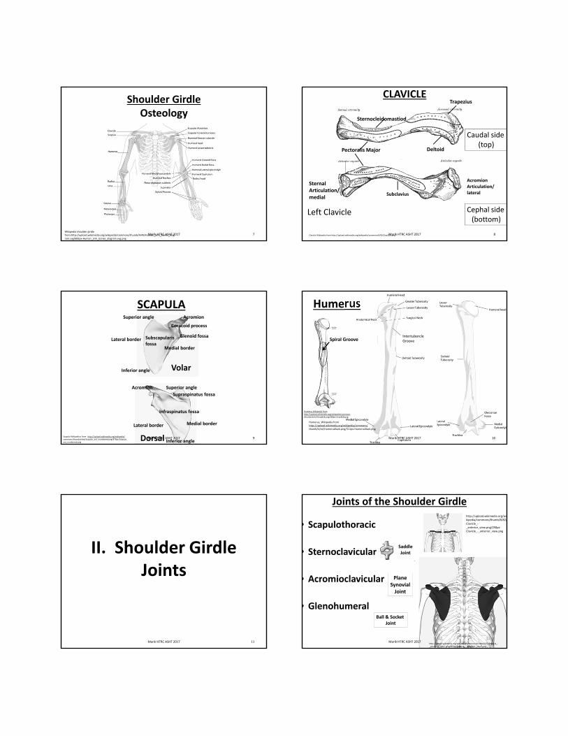
IV. KINESIOLOGYPRECISE TIMING
Clavicle Force Couples
Scapular Force Couples
Glenohumeral Force Couples
Marik HTRC ASHT 2017 24

SCAPULOHUMERAL RHYTHM
Illustration retrieved on 12/28/14 from: http://www.geocities.ws/ptexas9/angles.jpg
3 Phases of Motion (Scapula contribution varies, Scibek & Carcia, 2012)
Phase I 0º to 60º
Phase II 60º to 120º
Phase III 120º to 180º
90º sagittal plane flexion30º scapula and 60º glenohumeral 25Marik HTRC ASHT 2017
Framework for Teaching2° of humeral motion1° degree scapula motion
Retrieved from:http://www.zimmer.com/web/enUS/images/products/joints/shoulders/bigliani_flatlow_shoulder2.jpg on 02/22/09
Video Movement Patterns
Marik HTRC ASHT 2017 26
3 Dimensional Motions of Scapula
27
Retrieved on 06/07/09: http://www.flzine.com/wp-content/uploads/2009/03/rotator.gif
Upward Rotation
Posterior Tilt
From Int. Rot.
Towards Ext. Rot.
Marik HTRC ASHT 2017
AXIOSCAPULAR MUSCLESSCAPULAR FORCE COUPLE MUSCLES
• Serratus Anterior
• Trapezius (upper, middle, lower)
28Scapula force couples, Nov. 12, 2012 courtesy of Kathy Wilson
Marik HTRC ASHT 2017
29
Upper Trapezius (UT) and
Serratus Anterior (SA)provide
rotatory force early needed for
Upward Rotation.
Picture adapted from: Donatelli, R. (1997).
Scapulo‐thoracic MotionPhase 1 & 2
Marik HTRC ASHT 2017
Phase I & II0‐60°60° to 120°
30
Picture adapted from: Donatelli, R. (1997).
Marik HTRC ASHT 2017
Phase III120‐180°
3 Dimensional Motions of the Clavicle
31
Elevate
RetractPosterior Rotate
Photo retrieved from: http://upload.wikimedia.org/wikipedia/commons/8/82/Clavicle_‐_anterior_view.png
Mechanical coupling at acromioclavicular (AC) joint and sternoclavicular (SC) joint
Marik HTRC ASHT 2017
SCAPULAR MOTIONRelative to the
Acromio‐clavicular Joint
Phase I: SC elevation and AC posterior tilt = 16°scapula upward rotation
Phase II & III: SC elevation & posterior rotation = 2°scapula posterior tilt
Phase II & III:SC retraction and AC internal rotation = 2° scapula external rotation.
32
Illustration from: Ludewig P, Braman J. Shoulder impingement: Biomechanical considerations in rehabilitation. Manual Ther, 2011;16:33‐39
Marik HTRC ASHT 2017
Teece, R., Lunden, J., Lloyd, A., Kaiser, A., Cieminski, C., Ludewig, P. (2008). Three‐dimensional acromioclavicular jointmotions during elevation of the arm. Jour of Orthop Sports Phys Ther, 38: 181‐190.
33
Three Dimensional Motions of the Glenohumeral Joint
Superior / Inferior Glide
Retrieved on 2/21/10 from:http://www.ithaca.edu/faculty/lahr/LE2000/UE_Ind_Study_99/shoulder/edited/glenoid_fossa.jpeg
Spins, Rolls & Glides
External RotationLudewig et al 2009
Marik HTRC ASHT 2017 34
Glenohumeral Motions: Anterior and Posterior Balance Forces
Phase I (0°‐60°):0.7 to 2.7mm of anterior translationPhase II (60°‐120°):0‐1.5mm of posterior translationPhase III (120°‐180°): 4.5mm posterior translationLudewig & Cook, 2002.rieved on 08/02/09 from:
http://www.bosshin.com/owners_manual_instability/
Marik HTRC ASHT 2017
SCAPULOHUMERAL MUSCLESGLENOHUMERAL FORCE COUPLE
MUSCLES
35Rotator cuff, Dreamstime from: http://www.dreamstime.com/stock‐illustration‐shoulder‐anatomy‐medically‐accurate‐illustration‐image57248638#res13544998">The Shoulder Anatomy Photo</a>
Deltoid. Wikipedia from: https://upload.wikimedia.org/wikipedia/commons/9/93/Deltoideus.png
S.I.T.SSupraspinatusInfraspinatusTeres minorSubscapularis
Deltoids
Marik HTRC ASHT 2017 36
DELTOID AND SUPRASPINATUS
Donatelli, R. (1997). Physical Therapy of shoulder. Philadelphia, PA: Chruchill Livingstone
Retrieved and adapted on 06/07/09 from:http://img.tfd.com/dorland/thumbs/bursa_subacromialis.jpg
Marik HTRC ASHT 2017
SCAPULOHUMERAL RHYTHM
First Phase 0 to 60 Degrees GH ForcesSetting Phase

37
The deltoid provides a shearing force between 60 to 100 degrees of arm elevation and the rotator cuff provides a compressive force.
SCAPULOHUMERAL RHYTHMSecond Phase GH 60 to 120 Degrees
Retrieved and adapted on 06/08/09 from:http://www.chiroandosteo.com/content/13/1/20 Marik HTRC ASHT 2017 38
2nd Phase 60 to 120 DegreesBalance Forces Needed Between Subscapularis and Infraspinatus to Keep the Glenohumeral
Joint Centered
Albert, M. (2008). Evaluation and Treatment of Shoulder Biomechanics .
Ratio of posteriorto anterior forces
60% to 70%.ER/IR ratio ~ 66%
Marik HTRC ASHT 2017
39
SCAPULOHUMERAL RHYTHMFinal GH Phase 120 to 180 Degrees
• The humerus must disengage from the scapula!
Retrieved from:http://www.zimmer.com/web/enUS/images/products/
joints/shoulders/bigliani_flatlow_shoulder2.jpg on 02/22/09
Marik HTRC ASHT 2017 40
Scapulothoracic joint moves ~ 60 degrees
Glenohumeral joint moves ~ 120 degrees
Total Motion ~ 180 degrees
GH 120
ST 60Retrieved on 06/08/09 adaptedFrom:http://www.goodbyepain.biz/images/gallery_3.jpg
Total 180
SH Rhythm1 degree scapularmovement for 2 degrees humeral
movement
Marik HTRC ASHT 2017
Video SH Rhythm
Marik HTRC ASHT 2017 41 Marik HTRC ASHT 2017 42
Movement Pattern Video
Marik HTRC ASHT 2017 43
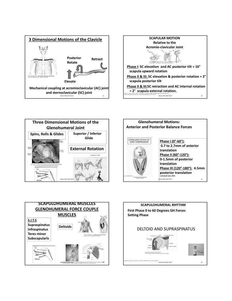
V. MUSCLES OF THE SHOULDER GIRDLE
44Marik HTRC ASHT 2017
SHOULDER GIRDLE MUSCLE ANATOMY
AxioscapularOriginate: Axio‐skeletonInsert: Scapula
ScapulohumeralOriginate: ScapulaInsert: Humerus
Axiohumeral:Originate: Axio‐skeletonInsert: Humerus
Wikapedia glenohumeral muscles from:http://upload.wikimedia.org/wikipedia/commons/thumb/6/6c/Pectoralis_major.png/250px‐Pectoralis_major.png
45Marik HTRC ASHT 2017
Axio‐scapularAnterior
• Serratus Anterior (originates anterior/inserts posterior)
• Pectoralis Minor
Posterior
• Trapezius
• Rhomboids (Minor & Major)
• Levator Scapulae
46Marik HTRC ASHT 2017
Anterior
Serratus Anterior
Pectoralis Minor
Wikapedia: Pectoralis Minor from http://openphysio.co.za/images/thumb/5/5d/Levator_scapulae.jpg/150px‐Levator_scapulae.jpg
Wikapedia: Serratus Anterior from http://openphysio.co.za/images/thumb/5/5d/Levator_scapulae.jpg/150px‐Levator_scapulae.jpg
Axioscapular Anterior
Marik HTRC ASHT 2017 47
Serratus Anterior
Origin: First 8 to 9 ribs
Insertion: Medial border of scapula
Innervation: Long thoracic nerve (C5, C6, C7)
Cadaver photo courtesy of Ithaca University,http://ect.downstate.edu/courseware/haonline/imgs/00000/0000/500/586.jpg. Courtesy of Dr. Stephan Lahr
Wika pedia: pectoralis minor from http://upload.wikimedia.org/wikipedia/commons/c/c9/Gray1215.png
Marik HTRC ASHT 2017
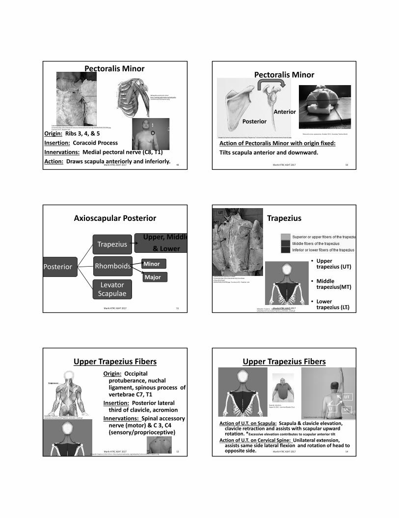
Pectoralis Minor
Origin: Ribs 3, 4, & 5
Insertion: Coracoid Process
Innervations: Medial pectoral nerve (C8, T1)
Action: Draws scapula anteriorly and inferiorly.
Wikapedia pectoralis minor:http://upload.wikimedia.org/wikipedia/commons/f/f2/Gray411.png
49
Cadaver photo courtesy of Ithaca University,http://ect.downstate.edu/courseware/haonline/imgs/00000/0000/500/586.jpg. Courtesy of Dr. Stephan Lahr
Marik HTRC ASHT 2017
Pectoralis Minor
Action of Pectoralis Minor with origin fixed:
Tilts scapula anterior and downward.
Anterior
Posterior
50
Images by human anatomy basics from http://hippie.nu/~unicorn/tut/img/basics/humananatomy/scapula.jpeg
Pectoralis minor assessment, October 2012. Courtesty Tambra Marik
Marik HTRC ASHT 2017
Posterior
Trapezius
Rhomboids
Levator Scapulae
Upper, Middle
& Lower
Minor
Major
Axioscapular Posterior
Marik HTRC ASHT 2017 51
Trapezius
• Upper trapezius (UT)
• Middle trapezius(MT)
• Lower trapezius (LT)Wikipedia Trapezius: http://upload.wikimedia.org/
wikipedia/commons/a/aa/Trapezius_animation_small2.gif
UT
MT
LT
52
Cadaver photo courtesy of Ithaca University,http://ect.downstate.edu/courseware/haonline/imgs/00000/0000/500/586.jpg. Courtesy of Dr. Stephan Lahr
Marik HTRC ASHT 2017
Upper Trapezius Fibers
Origin: Occipital protuberance, nuchal ligament, spinous process of vertebrae C7, T1
Insertion: Posterior lateral third of clavicle, acromion
Innervations: Spinal accessory nerve (motor) & C 3, C4 (sensory/proprioceptive)
Wikipedia trapezius 2/10/13 from: http://upload.wikimedia.org/wikipedia/commons/6/60/Trapezius.png
Wikipedia Trapezius: http://upload.wikimedia.org/wikipedia/commons/a/aa/Trapezius_animation_small2.gif
Marik HTRC ASHT 2017 53
Upper Trapezius Fibers
Action of U.T. on Scapula: Scapula & clavicle elevation, clavicle retraction and assists with scapular upward rotation. *Excessive elevation contributes to scapular anterior tilt
Action of U.T. on Cervical Spine: Unilateral extension, assists same side lateral flexion and rotation of head to opposite side. 54
Scapular elevation, August 8, 2012. Courtesy Blaydes Chun
Scapula force couple, October 2012. Courtesy Kathy Wilson
Marik HTRC ASHT 2017

Middle Trapezius FibersOrigin: Spinous process
C7 – T3
Insertion: Medial acromion process and spine of scapula
Innervation: Spinal accessory nerve (motor) & cervical spinal nerves (motor & sensory) C 3, C4
Wikapedia trapezius from 2/10/13 http://upload.wikimedia.org/wikipedia/commons/2/2d/Trapezius_Gray409.PNG
55Marik HTRC ASHT 2017
Lower Trapezius FibersOrigin: Spinous process T4 to T12
Insertion: Inferior portion of spine of scapula
Innervations: Spinal accessory nerve (motor) & cervical spinal nerves (motor & sensory)
C 3, C4
Photos retrieved on 2/10/13 from: http://upload.wikimedia.org/wikipedia/commons/6/60/Trapezius.png
Spinal Accessory nerve injury from:http://electrodiagnosis.net/NewsResearch/EandRNewsletter/Vol_4_Issue_2_Differential%20Diagnosis
%20and%20Treatment%20of%20Winging%20Scapul
Marik HTRC ASHT 2017
Middle &Lower Trapezius Fibers
Action of LT: Scapular depression and adduction, assists with scapula upward rotation and assists with scapular external rotation.
Wikipedia trapezius from 2/10/13 from:http://upload.wikimedia.org/wikipedia/commons/2/2d/Trapezius_Gray409.PNG
57
Scapula force couple, October 2012. Courtesy Kathy Wilson
Marik HTRC ASHT 2017
Rhomboid major/minor
58
Innervations: Dorsal scapular nerveC4, C5
Wikepedia rhomboids from: http://upload.wikimedia.org/wikipedia/commons/7/70/Rhomboid_muscles_animation_small.gif
Kendall et al 2005
Cadaver photo courtesy of Ithaca University,http://ect.downstate.edu/courseware/haonline/imgs/00000/0000/500/586.jpg. Courtesy of Dr. Stephan Lahr
R. Minor
R. Major
Action: Scapular adduction/retraction, elevation, downward rotation and resist lateral translation of serratus anterior
Marik HTRC ASHT 2017
Levator Scapulae (L.S.)Action of Levator Scapulae on Scapula:
Scapular elevation and assists with scapula downward rotation
Action Levator Scapulae on Cervical: Same side cervical flexion and rotation. Assists with cervical extension
Innervations: Dorsal scapular nerve (C5) and ventral primary rami of C3 and C4
Cadaver photo courtesy of Ithaca University,http://ect.downstate.edu/courseware/haonline/imgs/00000/0000/500/586.jpg. Courtesy of Dr. Stephan Lahr
Wikipedia levator scapulae on 2/10/13 from:http://upload.wikimedia.org/wikipedia/commons/thumb/2/2c/Muscle_%C3%A9l%C3%A9vateur_de_la_scapula.png/636px‐Muscle_%C3%A9l%C3%A9vateur_de_la_scapula.png
L.S.
59Marik HTRC ASHT 2017
Scapulohumeral Muscles
60
Anterior
Anterior & Middle Deltoids
Subscapularis
Corcobrachialis
Posterior
Posterior & Middle Deltoids
Supraspinatus
Infraspinatus
Teres minor
Teres major
Wikipedia posterior cuffi from:from:http://images.google.com/imgres?imgurl=http://upload.wikimedia.org/wikipedi
Wikipedia anterior deltoid, corcobrachialis, subscapularis from : http://openphysio.co.za/index.php?title=Muscles_that_move_the_shoulder_girdle
Marik HTRC ASHT 2017
DELTOID MUSCLE
• Anterior deltoids
• Middle deltoids
Image cadaver deltoidsIthaca College Physical Therapy DepartmentGross Anatomy – Joints website:http://www.ithaca.edu/faculty/lahr/LE2000/LE_index.html“. Courtesy Stephan Lahr
61Marik HTRC ASHT 2017
Action of Anterior Deltoid Fibers:
Flex shoulder and contribute to medial/internal rotation when supine
Action of Middle Deltoid Fibers: Shoulder abduction
Innervations: Axillary nerve (C5, C6)
Axillary nerve injury from: https://www.google.com/search?site=&tbm=isch&source=hp&biw=1280&bih=687&q=axillary+nerve+injury&oq=axillary+nerve+injury&gs_l=img.3..0j0i24l9.1584.4971.0.5338.21.14.0.7.7.0.109.1173.13j1.14.0....0...1ac.1.64.img..0.21.1217.Yc_hh87ohmU#imgrc=i2ACRTjOZA3U5M%3A
Marik, T. Actions of anterior deltoid, August 2012. Courtesy Tambra Marik
Wikapedia deltoids from:http://openphysio.co.za/index.php?title=Muscles_that_move_the_shoulder_girdle
Marik HTRC ASHT 2017
Scapulohumeral Anterior Muscles
• Anterior/Middle Deltoid
• Subscapularis
• Coracobrachialis
Subscapularis
Wikapedia subscapularis from: http://upload.wikimedia.org/wikipedia/commons/f/f2/Gray411.png63
Wikapedia subscapularis from:http://upload.wikimedia.org/wikipedia/commons/f/f2/Gray411.png
Marik HTRC ASHT 2017
Subscapularis (located anterior)
Action of Subscapularis: Contributes to medial/internal rotation of the shoulder and compression of the humeral head in the glenoid fossa during joint movements
Innervation: Upper and lower subscapular nerve (C6, C7)
Image retrieved from http://upload.wikimedia.org/wikipedia/commons/thumb/6/61/Subscapularis_muscle_frontal.png/250px‐Subscapularis_muscle_frontal.png
64Marik HTRC ASHT 2017
Coracobrachialis(located anterior)
Action of Coracobrachialis: Flexes and adducts the shoulder
Innervation: Musculocutaneous (C6, C7)
Wikapedia coracobrachialis from: http://upload.wikimedia.org/wikipedia/commons/thumb/6/61/Subscapularis_muscle_frontal.png/250px‐Subscapularis_muscle_frontal.png
Coracobrachialis
65Marik HTRC ASHT 2017
Scapulohumeral Posterior Muscles
• Posterior/Middle Deltoid
• Supraspinatus
• Infraspinatus
• Teres minor
• Teres major
Image retrieved from: http://upload.wikimedia.org/wikipedia/commons/f/f2/Gray411.png
Image provided by the Ithaca College Physical Therapy DepartmentGross Anatomy – Joints website:http://www.ithaca.edu/faculty/lahr/LE2000/LE_index.html“
Courtesy Stephan Lahr.
Supra.
Infra.
T. Minor
T. Major
Post. Deltoid
66Marik HTRC ASHT 2017

Posterior Deltoid
Action of Posterior Deltoid Fibers: Shoulder extension and contributes to lateral/external rotation in the prone position
Innervations: Axillary nerve (C5, C6)
67
Marik, T. Actions of anterior deltoid, August 2012. Courtesy Tambra Marik
Posterior deltoid, Wikipedia from https://upload.wikimedia.org/wikipedia/commons/thumb/9/93/Deltoideus.png/250px‐Deltoideus.png
Marik HTRC ASHT 2017
Supraspinatus
Action of Supraspinatus: Abducts and externally/laterally rotates the humerus. Compression/stabilization of humeral head in the glenoid fossa during shoulder movements
Innervation: Suprascapular nerve (C4, C5, C6)
Wikapedia suparspinatus from: http://upload.wikimedia.org/wikipedia/commons/f/f2/Gray411.png
AbductionER
68
Marik, T. Supraspinatus actions, August 2012. Courtesy Tambra Marik.
Marik HTRC ASHT 2017
Infraspinatus
Action of Infraspinatus: External rotation of the shoulder. Compression/stabilization of humeral head in glenoid fossa during shoulder movements.
Innervation: Suprascapular nerve (C4, C5, C6)
Wikapedia infraspinatus from http://upload.wikimedia.org/wikipedia/commons/f/f2/Gray411.png
ER
69
Marik, T. External rotation, August 2012. Courtesy Tambra MarikImage retrieved from: https://encrypted‐tbn3.google.com/images?q=tbn:ANd9GcRbiPKCRKCFS4HfJrg42Jv3hTzf7jsQtGY708tf20xgvvxfld2q
Marik HTRC ASHT 2017
Teres Minor
Action of Teres Minor: External rotation of the shoulder. Compression/stabilization of humeral head in glenoid fossa during shoulder movements.
Innervation: Axillary Nerve (C5, C6)
Image retrieved from: http://upload.wikimedia.org/wikipedia/commons/f/f2/Gray411.png
ER
70
Marik, T. External rotation, August 2012. Courtesy Tambra Marik
Marik HTRC ASHT 2017
Palpation: Infraspinatus & Teres Minor
71
Infra. & Teres Minor
Marik HTRC ASHT 2017
Teres Major
Action of Teres Major: Medial/internal rotation of the humerus and extends humerus.
Innervation: Lower subscapular nerve (C5, C6, C7)
Wikapedia teres major Image retrieved from: http://upload.wikimedia.org/wikipedia/commons/f/f2/Gray411.png
IR
Ext.
72
Marik, T. Teres major actions, August, 2012. Courtesy Tambra Marik
Image retrieved from: http://upload.
wikimedia.org/wikipedia/commons/f/f2/Gray411.png
Marik HTRC ASHT 2017

Rotator Cuff (S.I.T.S.)
• Supraspinatus
• Infraspinatus
• Teres Minor
• Subscapularis
Image retrieved and modified from:Retrieved on 06/07/09from:http://www.bartleby.com/107/Images/small/image410.jpg
S.I.T.S.Compress/concavity to stabilize and center the humeral head during shoulder motions.
Image provided by the Ithaca College Physical Therapy DepartmentGross Anatomy – Joints website:http://www.ithaca.edu/faculty/lahr/LE2000/LE_index.html Courtesy Stephan Lahr
73Marik HTRC ASHT 2017
Axiohumeral Muscles
74
Anterior
Pectoralis Major
Posterior
Latissimus
Dorsi
Wikipediat pectoralis major from: http://images.google.com/imgres?imgurl=http://upload.wikimedia.org/wikipedi
Wikipedia latissimus dorsi from:from:http://images.google.com/imgres?imgurl=http://upload.wikimedia.org/wikipedi
Marik HTRC ASHT 2017
Pectoralis Major Middle (Clavicular) & Lower (Sternal) Fibers
Innervation: Lateral pectoral nerve and medial pectoral nerve (C7, C8 & T1)
Cadaver mage retrieved from:http://ect.downstate.edu/courseware/haonline/simgs/00000/0000/500/575.jpgDrawing pect. major retrieve from:from:http://images.google.com/imgres?imgurl=http://upload.wikimedia.org/wikipedi
75
Cadaver photo courtesy of Ithaca University,http://ect.downstate.edu/courseware/haonline/imgs/00000/0000/500/586.jpg. Courtesy of Dr. Stephan Lahr
Wikipedia pectoralis major from:http://images.google.com/Imgres?imgurl=http://upload.wikimedia.org/wikipedi
Marik HTRC ASHT 2017
Pectoralis MajorAction of Clavicular Head (upper fibers): Horizontal adduction, medial/internal rotation and forward flexion of the humerus
Actions of Sternal Head (lower fibers): Oblique adduction towards opposite hip, medial rotation, and depresses the shoulder girdle
Combined Actions of Sternal & Clavicular Head: Humeral flexion, horizontal adduction, and medial rotation
Marik, T. Pectoralis major muscle actions, August 2012. Courtesy: Tambra Marik
Horz. Add.
76Marik HTRC ASHT 2017
77
Pectoralis Major
• Short muscle fiber exists when the extended arm does not drop down to the mat.
Kendall, F.(2005). MUSCLES TESTING AND FUNCTION WITH POSTURE AND PAIN. Baltimore, MD: Lippincott Williams & Wilkins.
Sternal fibers assess with horizontal abduction at 145°
Marik, T. Assess pectoralis major clavicular fibers,December, 2009. Courtesy Tambra Marik
Marik HTRC ASHT 2017 78
Assess Length of Pectoralis MajorAssess Length of Pectoralis Major
• Short muscle fibers exist when the arm can not drop down to the mat.
Kendall, F.(2005). MUSCLES TESTING AND FUNCTION WITH POSTURE AND PAIN. Baltimore, MD: Lippincott Williams & Wilkins.
Clavicular Fibers assess with horizontal abduction at 90°
Marik, T. Assess pectoralis major clavicular fibers,December, 2009. Courtesy Tambra Marik
Marik HTRC ASHT 2017
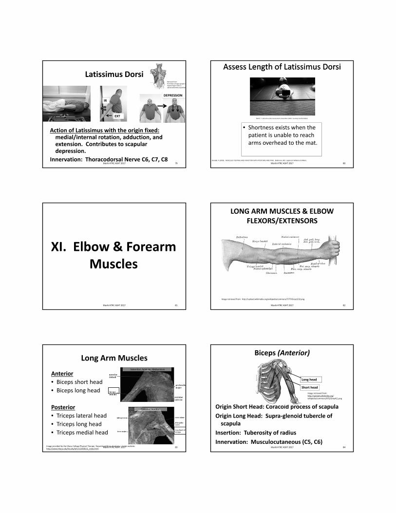
Latissimus Dorsi
Action of Latissimus with the origin fixed:medial/internal rotation, adduction, and extension. Contributes to scapular depression.
Innervation: Thoracodorsal Nerve C6, C7, C8
IR
EXT
DEPRESSION
79
Retrieved from: from:http://images.google.coimgres?imgurl=http://upload.wikimedia.org/wikiped
Marik HTRC ASHT 2017 80
Assess Length of Latissimus DorsiAssess Length of Latissimus Dorsi
• Shortness exists when the patient is unable to reach arms overhead to the mat.
Kendall, F.(2005). MUSCLES TESTING AND FUNCTION WITH POSTURE AND PAIN. Baltimore, MD: Lippincott Williams & Wilkins.
Marik, T. Latissimus Dorsi assessment, December 2009. Courtesy Tambra Marik
Marik HTRC ASHT 2017
XI. Elbow & Forearm Muscles
81Marik HTRC ASHT 2017
LONG ARM MUSCLES & ELBOW FLEXORS/EXTENSORS
Image retrieved from: http://upload.wikimedia.org/wikipedia/commons/7/77/Gray1232.png
82Marik HTRC ASHT 2017
Long Arm Muscles
Anterior
• Biceps short head
• Biceps long head
Posterior
• Triceps lateral head
• Triceps long head
• Triceps medial head
Image provided by the Ithaca College Physical Therapy DepartmentGross Anatomy – Joints website:http://www.ithaca.edu/faculty/lahr/LE2000/LE_index.html
83Marik HTRC ASHT 2017
Biceps (Anterior)
Origin Short Head: Coracoid process of scapula
Origin Long Head: Supra‐glenoid tubercle of scapula
Insertion: Tuberosity of radius
Innervation: Musculocutaneous (C5, C6)
Short head
Long head
Image retrieved from:http://upload.wikimedia.org/wikipedia/commons/f/f2/Gray411.png
84Marik HTRC ASHT 2017

Biceps
Action of both Head of Biceps: Elbow flexion and supination
Action of Short Head of Biceps: Assists with shoulder adduction
Action of Long Head of the Biceps: Assists with shoulder abduction.
Image retrieved from:http://upload.wikimedia.org/wikipedia/commons/f/f2/Gray411.png
SUPINATION
ELBOW FLEXION
85Marik HTRC ASHT 2017
Triceps (Posterior)
Origin Long Head: Infraglenoid tubercle of scapulaOrigin Lateral Head: Lateral posterior humerus & lateral
intermuscular septumOrigin Medial Head: Medial posterior humerus and medial
intermuscular septum
Insertion: Posterior OlecranonInnervation: Radial (C6, C7, C8)
Image retrieved from:http://upload.wikimedia.org/wikipedia/commons/f/f2/Gray411.png
Long Head Lateral Head Lateral Head
86Marik HTRC ASHT 2017
Triceps
Action of Triceps: Extends elbow
Action of Long Head of Triceps: Contributes to shoulder extension and shoulder adduction.
Image retrieved from:http://upload.wikimedia.org/wikipedia/commons/f/f2/Gray411.png
87Marik HTRC ASHT 2017
Elbow Extensors: Anconeus (Posterior)
Photo retrieved from: http://upload.wikimedia.org/wikipedia/commons/thumb/d/d1/Slide1TTTT.JPG/800px‐Slide1TTTT.JPG
88
Assists with elbow extension
Marik HTRC ASHT 2017
Range of Motion
Normal Arc: Elbow 0º to 140º; forearm 0º to 85º
Functional Arc: 30º to 130º (Bryce & Armstrong, 2008)
Marik HTRC ASHT 2017 89
Elbow Flexors & ExtensorsElbow Flexors (anterior)
• Biceps Brachii (short & long heads)
• Brachialis
• Brachioradialis
Elbow Extensors (posterior)
• Triceps, (long, lateral & medial heads)
• AnconeusImage provided by the Ithaca College Physical Therapy DepartmentGross Anatomy – Joints website:http://www.ithaca.edu/faculty/lahr/LE2000/LE_index.html
90Marik HTRC ASHT 2017

Elbow Flexors: Biceps (Anterior)
Origin Short Head: Coracoid process of scapula
Origin Long Head: Supra‐glenoid tubercle of scapula
Insertion: Tuberosity of radius
Innervation: Musculocutaneous (C5, C6)
Short head
Long head
Image retrieved from:http://upload.wikimedia.org/wikipedia/commons/f/f2/Gray411.png
Image retrieved from:http://upload.wikimedia.org/wikipedia/commons/f/f2/Gray411.png
91Marik HTRC ASHT 2017
Elbow Flexors: Biceps
Action of both Head of Biceps: Elbow flexion and supination
Action of Short Head of Biceps: Assists with shoulder adduction
Action of Long Head of the Biceps: Assists with shoulder abduction.
Image retrieved from:http://upload.wikimedia.org/wikipedia/commons/f/f2/Gray411.png
SUPINATION
ELBOW FLEXION
92Marik HTRC ASHT 2017
Elbow Flexors: Brachialis (Anterior)
Origin: Distal half of anterior humerusInsertion: Coronoid process and tuberosity of the ulnaInnervation: Musculcutaneous & small branch from radial nerve (C5, C6)
Image retrieved from: http://upload.wikimedia.org/wikipedia/commons/thumb/3/33/Slide6yyy.JPG/800px‐Slide6yyy.JPG
93Marik HTRC ASHT 2017
Elbow Flexors: Brachialis
Action: Flexes the elbow
Image retrieved from: http://upload.wikimedia.org/wikipedia/commons/thumb/3/33/Slide6yyy.JPG/800px‐Slide6yyy.JPG
94Marik HTRC ASHT 2017
Elbow Flexors: Brachioradialis (Anterior)
Origin: Lateral supracondylar ridge of the humerus
Insertion: Lateral side of styloid process
Innervation: Radial Nerve (C5, C6)
Image retrieved from: http://upload.wikimedia.org/wikipedia/commons/0/09/Brachioradialis.png
Cadaver retrieved From: http://ect.downstate.edu/courseware/haonline/imgs/00000/0000/700/734.jpg
95Marik HTRC ASHT 2017
Elbow Flexors: Brachioradialis
Action: Flexes the elbow. Assists with forearm rotation when there is resistance to elbow flexion.
Image retrieved from: http://upload.wikimedia.org/wikipedia/commons/thumb/3/33/Slide6yyy.JPG/800px‐Slide6yyy.JPG
96Marik HTRC ASHT 2017

Elbow Extensors:Triceps (Posterior)
Origin Long Head: Infraglenoid tubercle of scapulaOrigin Lateral Head: Lateral posterior humerus & lateral
intermuscular septumOrigin Medial Head: Medial posterior humerus and medial
intermuscular septum
Insertion: Posterior OlecranonInnervation: Radial (C6, C7, C8)
Image retrieved from:http://upload.wikimedia.org/wikipedia/commons/f/f2/Gray411.png
Long Head Lateral Head Lateral Head
97Marik HTRC ASHT 2017
Elbow Extensors: Triceps (Posterior)
Action of Triceps: Extends elbow
Action of Long Head of Triceps: Contributes to shoulder extension and shoulder adduction.
Image retrieved from:http://upload.wikimedia.org/wikipedia/commons/f/f2/Gray411.png
98Marik HTRC ASHT 2017
Elbow Extensor: Anconeus (Posterior)
Origin: Posterior lateral epicondyle
Insertion: Olecranon and upper posterior ulna
Innervation: Radial Nerve (C7, C8)
Images retrieved from: http://upload.wikimedia.org/wikipedia/commons/thumb/d/d1/Slide1TTTT.JPG/800px‐Slide1TTTT.JPG
99Marik HTRC ASHT 2017
Elbow Extensor: Anconeus (Posterior)
Action: Contributes to elbow extension
Images retrieved from: http://upload.wikimedia.org/wikipedia/commons/thumb/d/d1/Slide1TTTT.JPG/800px‐Slide1TTTT.JPG
100Marik HTRC ASHT 2017
Forearm Rotation Muscles
Supinators & Pronators
101Marik HTRC ASHT 2017
Forearm Supinator: Supinator
Origin: Lateral epicondyle, radial collateral ligament, annular ligament, and supinator crest of ulnaInsertion: Lateral 1/3 of radius covering anterior and posteriorInnervation: Radial Nerve (C5, C6)
Images retrieved from: http://upload.wikimedia.org/wikipedia/commons/c/c6/Gray420.png
102Marik HTRC ASHT 2017

Forearm Supinator: Supinator Muscle
Action: Supinates the forearm
Images retrieved from: http://upload.wikimedia.org/wikipedia/commons/thumb/d/d1/Slide1TTTT.JPG/800px‐Slide1TTTT.JPG
Palm Up
103Marik HTRC ASHT 2017
Forearm Pronators: Pronator Teres
Origin: Proximal medial epicondyle and common flexor tendonInsertion: Mid‐lateral surface of the radiusInnervation: Median Nerve (C6, C7)
Image retrieved from: http://upload.wikimedia.org/wikipedia/commons/d/de/Muscles_of_forearm.jpg
104Marik HTRC ASHT 2017
Forearm Pronators: Pronator Teres
Action: Pronates the forearm and assists with elbow flexion
Image retrieved from: http://upload.wikimedia.org/wikipedia/commons/d/de/Muscles_of_forearm.jpg
Palm Down
105Marik HTRC ASHT 2017
Forearm Pronators: Pronator Quadratus Quadratus
Origin: Anterior distal ulna
Insertion: Anterior distal radius
Innervation: Median Nerve (C7, C8, T1)
Images retrieved from: http://upload.wikimedia.org/wikipedia/commons/thumb/b/b4/Slide2VVVV.JPG/800px‐Slide2VVVV.JPG
106Marik HTRC ASHT 2017
Forearm Pronators: Pronator Quadratus
Action: Forearm pronation
Images retrieved from: http://upload.wikimedia.org/wikipedia/commons/thumb/b/b4/Slide2VVVV.JPG/800px‐Slide2VVVV.JPG
Palm Down
107Marik HTRC ASHT 2017
XII. Important Soft Tissue Regions of the Elbow
108Marik HTRC ASHT 2017

IMPORTANT SOFT TISSUE AREAS
• Potential nerve compression sites of arm, elbow & forearm
– Ulnar Nerve
– Median Nerve
– Radial Nerve
http://upload.wikimedia.org/wikipedia/commons/7/75/Gray413_color.png
109Marik HTRC ASHT 2017
Potential Soft Tissue Sites of Ulnar Nerve Compression
Ulnar Nerve Compression Sites:
• Arcade of Struthers
• Medial intramuscular septum
• Condylar groove
• Cubital tunnel retinaculum
• Deep flexor pronator aponeurosis
.
UlnarNerve
Proximal to ElbowUlnar Nerve
Arcade Struthers
Medial Intermuscular
Septum
Arcade StruthersArcade Struthers
Ulnar Nerve
Image from: http://upload.wikimedia.org/wikipedia/commons/e/e7/Gray528.png 110Marik HTRC ASHT 2017
Special Tests: Medial ElbowCubital Tunnel Syndrome
TinelsSensitivity:54% to 70%
Modified Shoulder Internal Rotation TestSensitivity: 87% (5 second)Specificity: 97% Ochi et al 2012
Scratch CollapseSensitivity: 69%Spec.: 99%
Marik HTRC ASHT 2017 111
Potential Soft Tissue Sites of Median Nerve Compression
• Median Nerve (Pronator Syndrome)
• Anterior Interosseous Nerve (branch of median nerve)
http://upload.wikimedia.org/wikipedia/commons/thumb/a/a7/Nerves_of_the_left_upper_extremity.gif/314px‐Nerves_of_the_left_upper_extremity.gif
112Marik HTRC ASHT 2017
Potential Soft Tissue Sites of Median Nerve Compression
• Median Nerve (Pronator Syndrome)
Possible Compression Sites• Pronator teres(most common site)
• Arch for flexor digitorum superficialis (FDS)
• Ligament of Struthers(located at supracondylar process)
• Lacertus fibrosis (bicipital aponeurous)
http://upload.wikimedia.org/wikipedia/commons/thumb/a/a7/Nerves_of_the_left_upper_extremity.gif/314px‐Nerves_of_the_left_upper_extr
FDS
Pronator Teres
113Marik HTRC ASHT 2017
Potential Soft Tissue Sites of Median Nerve Compression
• Anterior Interosseous Nerve (branch of median nerve)
Possible Compression Sites
• Fibrous bands from deep PT
• Flexor digitorum superficialis arch
• Gantzer muscle (FPL accessory)
• Flexor carpi radialis
• Palmaris longus
http://upload.wikimedia.org/wikipedia/commons/thumb/a/a7/Nerves_of_the_left_upper_extremity.gif/314px‐Nerves_of_the_left_upper_extremity.gif
PronatorTeres
114Marik HTRC ASHT 2017

Pronator Syndrome
Anterior Interosseous Syn.
Proximal forearm pain volarly
Proximal forearm pain volarly
Numbness/tingling volar index, long and ring
Sensory distubances absent
Thumb, index, and long finger weakness
Thumb IP weakness and FDP weakness index
Symptoms reproduced with palpation pronator
No symptoms with palpation of pronator
115Marik HTRC ASHT 2017
Potential Soft Tissue Sites of Radial Nerve Compression
• Intermuscular Septum (spiral groove b/w triceps & brachiallis)
• Edge for ECRB• Radio‐capitellar joint due to arthrosis (b/w brach. & BR)
• Ligament of Frosche• Between 2 heads of supinator
• Exit under brachioradiallis
Brachialis
Triceps
Radial Nerve
ECRB
http://upload.wikimedia.org/wikipedia/commons/7/75/Gray413_color.pngRetrieved on 4/3/11 from:http://upload.wikimedia.org/wikipedia/commons/c/c6/Gray420.png
Supinator
Radial Nerve
116Marik HTRC ASHT 2017
Radial Nerve Signs/Symptoms• Compression proximal elbow present with weakness of wrist and digit extensors
• Compression distal to elbow can present with weak finger ext. and normal wrist ext. strength (PIN)
• Compression proximal ligament Froshe can present with sensation disturbance.
Retrieved on 4/3/11 from:http://upload.wikimedia.org/wikipedia/commons/c/c6/Gray420.png
117Marik HTRC ASHT 2017
THANK YOU!
118Marik HTRC ASHT 2017
REFERENCES1. Ahmad Z, Siddiqui N, Malik SS, Abdus‐Samee M, Tytherweigh‐Strong G, Ruston M. (2013). Lateral epicondylitis. A review
of pathology and management. Bone & Joint Surgery;95(9):1158‐1164.2. Bryce CD, ArmstrHariri S, Safran MR. (2010). Ulnar collateral ligament injury in overhead athlete. Clin Sports
Med.;29(4):619e644. 3. Chong AD. Anatomy and biomechanics of the elbow. Orthop Clin North Am. 2008;39(2):141e154.4. Devereaux, M. W., & ElMaraghy, A. W. (2013). Improving the Rapid and Reliable Diagnosis of Complete Distal Biceps Tendon
Rupture A Nuanced Approach to the Clinical Examination. The American journal of sports medicine, 0363546513493383.5. Hart D, Carmichael S. Biomechanics of the shoulder. Jour of Ortho and Sports Phys Ther, 1985;6(4):229‐234.6. Hegedus, E. J., Goode, A. P., Cook, C. E., Michener, L., Myer, C. A., Myer, D. M., & Wright, A. A. (2012). Which physical
examination tests provide clinicians with the most value when examining the shoulder? Update of a systematic review with meta‐analysis of individual tests. British journal of sports medicine, bjsports‐2012.
7. Hutchinson RL, Rayan G. (2011). Diagnosis of cubital tunnel syndrome. Journal of Hand Surgery Am.;36(9):1519e1521.8. Jia, X., Petersen, S. A., Khosravi, A. H., Almareddi, V., Pannirselvam, V., & McFarland, E. G. (2009). Examination of the
shoulder: the past, the present, and the future. The Journal of Bone & Joint Surgery, 91(Supplement_6), 10‐18.9. Jobe C, Phipatanakul W, Coen M. Gross Anatomy of the Shoulder. In: Rockwook C, Matsen F, Wirth M, Lippitt S. The
Shoulder, 4th ed, Philadelphia, PA: Saunders Elsevier; 2009:33‐100.10. Kendall, F.(2005). MUSCLES TESTING AND FUNCTION WITH POSTURE AND PAIN. Baltimore, MD: Lippincott Williams &
Wilkins.11. Koo J, Szabo R. Compression neuropathies of the median nerve. JASHS, 2004;4(3):156‐175.12. Ludewig P, Cook T, Nawoczenski D. Three Dimensional Scapular Orientation and Muscle Activity at Selected Positions of
Humeral Elevation. JOSPT, 1996;24(2):57‐65. 13. Ludewig et al (2009). Motion of the Shoulder Complex during Multiplanar Humeral Elevation. Jour Bone & Joint Surg,
91:378‐8914. Nordin M, Frankel V. Basic Biomechanics of the Musculoskeletal System. 3rd ed, Baltimore, MD:Lippincott Williams &
Watkins;2001. 15. Ochi K, Horiuchi Y, Tenabe A, Waseda M, Kuneko Y, Koyanagi T. (2012) Shoulder internal rotation elbow flexion test for
diagnosing cubital tunnel syndrome. Journal of Shoulder Elbow Surgery;21(6):777e781.16. O’Driscoll SW, Lawton RL, Smith AM. (2005). The “moving valgus stress test” for medial collateral ligamnt tears of the eblwo.
American Journal of Sports Medicine;33(2):231‐239.17. Palastanga N, Field D, Soames R. The Upper Limb. Anatomy and Human Movement: Structure and Function. 4th ed.
Woburn, MA: Butterworth‐Heinnemann, 2002:41‐109.18. Regan W, Lapner PC.(2006). Prospective evaluation of two diagnostic apprehension signs for posterolateral instability of the
elbow. Journal Shoulder Elbow Surgery;15(3):344e346.19. Tosti R, Jennings J, Sewards JM. (2013). Lateral epicondylitis of the elbow. American Journal of Medicine;126(4):357.
119Marik HTRC ASHT 2017
SPECIAL THANKS FOR PHOTOS AND ILLUSTRATIONS PROVIDED BY:
Dr. Stephen Lahr
Ithaca CollegeDepartment of Physical TherapyHuman Anatomy Review Site
and
Suny Downstate University Medical Center
120Marik HTRC ASHT 2017

Conclusion
• Preparatory activities to reach occupation‐base goals
• Think function
• Organize muscular groups for learning
• Think like a clinician
Marik HTRC ASHT 2017 121
THANK YOU
AMAZING VARIETIES OF FUNCTION OF
THE SHOULDERS &
ELBOWS
Marik HTRC ASHT 2017 122
References• Ahmad Z, Siddiqui N, Malik SS, Abdus‐Samee M, Tytherweigh‐Strong G, Ruston M. (2013). Lateral epicondylitis. A review of pathology and
management. Bone & Joint Surgery;95(9):1158‐1164.• American Occupational Therapy Association (2006). AOTA’s Centennial Vision. Retrieved
from:http://www.aota.org/~/media/Corporate/Files/AboutAOTA/Centennial/Background/Vision1.ashx• Bryce CD, ArmstrHariri S, Safran MR. (2010). Ulnar collateral ligament injury in overhead athlete. Clin Sports Med.;29(4):619e644. • Chong AD. Anatomy and biomechanics of the elbow. Orthop Clin North Am. 2008;39(2):141e154.• Devereaux, M. W., & ElMaraghy, A. W. (2013). Improving the Rapid and Reliable Diagnosis of Complete Distal Biceps Tendon Rupture A Nuanced
Approach to the Clinical Examination. The American journal of sports medicine, 0363546513493383.• Hart D, Carmichael S. Biomechanics of the shoulder. Jour of Ortho and Sports Phys Ther, 1985;6(4):229‐234.• Hegedus, E. J., Goode, A. P., Cook, C. E., Michener, L., Myer, C. A., Myer, D. M., & Wright, A. A. (2012). Which physical examination tests provide
clinicians with the most value when examining the shoulder? Update of a systematic review with meta‐analysis of individual tests. Britishjournal of sports medicine, bjsports‐2012.
• Hutchinson RL, Rayan G. (2011). Diagnosis of cubital tunnel syndrome. Journal of Hand Surgery Am.;36(9):1519e1521.• Jia, X., Petersen, S. A., Khosravi, A. H., Almareddi, V., Pannirselvam, V., & McFarland, E. G. (2009). Examination of the shoulder: the past, the present,
and the future. The Journal of Bone & Joint Surgery, 91(Supplement_6), 10‐18.• Jobe C, Phipatanakul W, Coen M. Gross Anatomy of the Shoulder. In: Rockwook C, Matsen F, Wirth M, Lippitt S. The Shoulder, 4th ed, Philadelphia, PA:
Saunders Elsevier; 2009:33‐100.• Kendall, F.(2005). MUSCLES TESTING AND FUNCTION WITH POSTURE AND PAIN. Baltimore, MD: Lippincott Williams & Wilkins.• Koo J, Szabo R. Compression neuropathies of the median nerve. JASHS, 2004;4(3):156‐175.• Ludewig P, Cook T, Nawoczenski D. Three Dimensional Scapular Orientation and Muscle Activity at Selected Positions of Humeral Elevation. JOSPT,
1996;24(2):57‐65. • Ludewig et al (2009). Motion of the Shoulder Complex during Multiplanar Humeral Elevation. Jour Bone & Joint Surg, 91:378‐89• Nordin M, Frankel V. Basic Biomechanics of the Musculoskeletal System. 3rd ed, Baltimore, MD:Lippincott Williams & Watkins;2001. • Ochi K, Horiuchi Y, Tenabe A, Waseda M, Kuneko Y, Koyanagi T. (2012) Shoulder internal rotation elbow flexion test for diagnosing cubital tunnel
syndrome. Journal of Shoulder Elbow Surgery;21(6):777e781.• O’Driscoll SW, Lawton RL, Smith AM. (2005). The “moving valgus stress test” for medial collateral ligamnt tears of the eblwo. American Journal of Sports
Medicine;33(2):231‐239.• Palastanga N, Field D, Soames R. The Upper Limb. Anatomy and Human Movement: Structure and Function. 4th ed. Woburn, MA: Butterworth‐Heinnemann, 2002:41
109.• Regan W, Lapner PC.(2006). Prospective evaluation of two diagnostic apprehension signs for posterolateral instability of the elbow. Journal Shoulder Elbow
Surgery;15(3):344e346.• Scibek, J. S., & Carcia, C. R. (2012). Assessment of scapulohumeral rhythm for scapular plane shoulder elevation using a modified digital inclinometer. World journal of orthopedics, 3(6), 87
• Teece, R., Lunden, J., Lloyd, A., Kaiser, A., Cieminski, C., Ludewig, P. (2008). Three‐dimensional acromioclavicular joinmotions during elevation of the arm. Jour of Orthop Sports Phys Ther, 38: 181‐190.
• Tosti R, Jennings J, Sewards JM. (2013). Lateral epicondylitis of the elbow. American Journal of Medicine;126(4):357.
• Illustration retrieved on 12/28/14 from: http://www.geocities.ws/ptexas9/angles.jpg
Marik HTRC ASHT 2017 123
Photo References• Clavicle movement Moon OUHSC from http://images.google.com/imgres?imgurl=http://moon.ouhsc.• Clavicle Wikipedia from http://upload.wikimedia.org/wikipedia/commons/f/f0/Gray201.png• Humerus, Wikipedia from http://upload.wikimedia.org/wikipedia/commons/thumb/e/ee/HumerusBack.png/311px‐HumerusBack.png• Chun, B., 2010. Inferior translation. Courtesy Blaydes Chun.• Chun, B., 2010. Anterior translations. Courtesty Blaydes Chun.• Hicker, R. (Photographer). (2011). Moss rain forest [Photograph]. Courtesy of Ronald Hicker. • Lahr, S. (2013). Cadaver photos. Courtesy Dr. Stephan Lahr.• Lower trapezius from http://www.exrx.net/Graphics/SerratusAnteriorPull.gif• Marik, T., 2012. Pectoralis minor assessment. Courtesy Tambra Marik.• Pectoral girdle: http://upload.wikimedia.org/wikipedia/commons/
thumb/2/22/Pectoral_girdle_front_diagram.svg/250px‐Pectoral_girdle_front_diagram.svg.png• Pectoral girdle: http://upload.wikimedia.org/wikipedia/commons/
thumb/2/22/Pectoral_girdle_front_diagram.svg/250px‐Pectoral_girdle_front_diagram.svg.pn• Pectoralis minor Wikapedia pectoralis minor from http://upload.wikimedia.org/wikipedia/commons/f/f2/Gray411.png• Rotator cuff, Dreamstime from: http://www.dreamstime.com/stock‐illustration‐shoulder‐anatomy‐medically‐accurate‐illustration‐
image57248638#res13544998">The Shoulder Anatomy Photo</a>• Scapula: http://hippie.nu/~unicorn/tut/img/basics/humananatomy/scapula.jpeg• Scapula upward rotation from http://www.flzine.com/wp-content/uploads/2009/03/rotator.gif• Scapula Wikipedica from http://upload.wikimedia.org/wikipedia/• commons/thumb/e/ed/Scapula_ant_numbered.png/476px‐Scapula_• ant_numbered.png• Scapulojhumral rhythm geocities from: http://www.geocities.ws/ptexas9/angles.jpg• Shoulder capsule, ORIF from: http://www.eorif.com/Shoulderarm/Shoulder%20anat/Images/Shoulder‐ligaments.jpg• Shoulder girdle from http://upload.wikimedia.org/wikipedia/commons/thumb/4/49/Human_arm_bones_diagram.svg/683px‐
Human_arm_bones_diagram.svg.png• Superior scapula rib vector (2009). From:http://www.exrx.net/
• Images/SuperiorScapulaRibVectors.gif• Wilson, K. (2012). Shoulder flexion. Courtesy of Kathy Wilson.• Wilson, K. (2012). Scapula force couples. Courtesy of Kathy Wilson• Wilson, K., (2012) .Scapula upward rotation. Courtesy of Kathy Wilson.
Marik HTRC ASHT 2017 124
Photo References• Anterior deltoid Wikapedia from http://openphysio.co.za/index.php?title=Muscles_that_move_the_shoulder_girdle
• Clavicle Wikipedia from http://upload.wikimedia.org/wikipedia/commons/f/f0/Gray201.png• Humerus, Wikipedia from http://upload.wikimedia.org/wikipedia/commons/thumb/e/ee/HumerusBack.png/311px‐HumerusBack.png• Chun, B., 2010. Inferior translation. Courtesy Blaydes Chun.• Chun, B., 2010. Anterior translations. Courtesty Blaydes Chun.• Glenohumeral muscles, Wikapedia from: http://upload.wikimedia.org/wikipedia/commons/thumb/6/6c/Pectoralis_major.png/250px‐Pectoralis_major.png• Hicker, R. (Photographer). (2011). Moss rain forest [Photograph]. Courtesy of Ronald Hicker. • Lahr, S. (2013). Cadaver photos. Courtesy Dr. Stephan Lahr.• Levator scapula from Wikipedia: fromhttp:// openphysio.co.za/images/thumb/5/5d/Levator_scapulae.jpg/150px‐Levatorscapulae.jpg• Lower trapezius from http://www.exrx.net/Graphics/SerratusAnteriorPull.gif• MaRIK, T., 2009. Latissimus Dorsi assessment. Courtesy Tambra Marik.• Marik, T., 2009. Pectoralis Major assessment. Courtesy Tambra Marik.• Marik, T., 2012. Pectoralis minor assessment. Courtesy Tambra Marik.• Marik, T. Pectoralis major muscle actions, August 2012. Courtesy: Tambra Marik• Pectoral girdle: http://upload.wikimedia.org/wikipedia/commons/
thumb/2/22/Pectoral_girdle_front_diagram.svg/250px‐Pectoral_girdle_front_diagram.svg.png• Pectoral girdle: http://upload.wikimedia.org/wikipedia/commons/thumb/2/22/Pectoral_girdle_front_diagram.svg/250pxPectoral_girdle_front_diagram.svg.pn• Pectoralis minor Wikapedia pectoralis minor from http://upload.wikimedia.org/wikipedia/commons/f/f2/Gray411.pn• Posterior cuff muscles Wikapedia from: from:http://images.google.com/• imgres?imgurl=http://• upload.wikimedia.org/wikipedi
• Scapula: http://hippie.nu/~unicorn/tut/img/basics/humananatomy/scapula.jpeg• Scapula upward rotation from http://www.flzine.com/wp-content/uploads/2009/03/rotator.gif• Scapula Wikipedica from http://upload.wikimedia.org/wikipedia/• commons/thumb/e/ed/Scapula_ant_numbered.png/476px‐Scapula_• ant_numbered.png• Shoulder girdle from http://upload.wikimedia.org/wikipedia/commons/thumb/4/49/Human_arm_bones_diagram.svg/683px‐Human_arm_bones_diagram.svg.png• Superior scapula rib vector (2009). From:http://www.exrx.net/
• Images/SuperiorScapulaRibVectors.gif• Wilson, K. (2012). Shoulder flexion. Courtesy of Kathy Wilson.• Wilson, K. (2012). Scapula force couples. Courtesy of Kathy Wilson• Wilson, K., (2012) .Scapula upward rotation. Courtesy of Kathy Wilson.
Marik HTRC ASHT 2017 125
Recommended