8/4/2019 1. Review Exam Material (a)
http://slidepdf.com/reader/full/1-review-exam-material-a 1/13
©© 2007 McGraw 2007 McGraw --Hill Higher Education. All rights reserved.Hill Higher Education. All rights reserved.
Course IntroductionCourse Introduction
•• Kinesiology (Kinesiology (kinein kinein = to move = to move ))
– – Study of human movementStudy of human movement
•• Foundation in 3 major areas of study:Foundation in 3 major areas of study:
1.1. MechanicsMechanics == biomechanicsbiomechanics
2.2. AnatomyAnatomy == musculoskeletalmusculoskeletal anatomyanatomy
3.3. PhysiologyPhysiology == neuromuscularneuromuscular physiologyphysiology
©© 2007 McGraw 2007 McGraw --Hill Higher Education. All rights reserved.Hill Higher Education. All rights reserved.
Course IntroductionCourse Introduction
•• KHP 3352 course divided:KHP 3352 course divided:
– – Applied AnatomyApplied Anatomy
•• Muscles identified by origin, insertion, fascicleMuscles identified by origin, insertion, fasciclearrangement, resultant joint angles, andarrangement, resultant joint angles, and
functional applicationfunctional application
– – BiomechanicsBiomechanics
•• Forces affecting movement and theirForces affecting movement and theirmathematical descriptionmathematical description
©© 2007 McGraw 2007 McGraw --Hill Higher Education. All rights reserved.Hill Higher Education. All rights reserved.
Musculoskeletal SystemMusculoskeletal System
•• Arrangement of bones, joints, andArrangement of bones, joints, and
musclesmuscles
•• Muscles apply force only byMuscles apply force only by
shorteningshortening
©© 2007 McGraw 2007 McGraw --Hill Higher Education. All rights reserved.Hill Higher Education. All rights reserved.
Skeletal FunctionsSkeletal Functions
1.1. ProtectionProtection (skull = brain, sternum = heart, ribs = lungs)(skull = brain, sternum = heart, ribs = lungs)
2.2. SupportSupport to maintain postureto maintain posture
3.3. MovementMovement by serving as points ofby serving as points of
attachment for musclesattachment for muscles (177/206 for movement)(177/206 for movement)
4.4. MineralMineral storagestorage such as calcium &such as calcium &
phosphorusphosphorus
5.5. HemopoiesisHemopoiesis – – process of blood cellprocess of blood cell
formation in the red bone marrowformation in the red bone marrow•• Vertebral bodies, femur, humerus, ribs, sternumVertebral bodies, femur, humerus, ribs, sternum
©© 2007 McGraw 2007 McGraw --Hill Higher Education. All rights reserved.Hill Higher Education. All rights reserved.
BONESBONESOsteologyOsteology
•• 206 bones206 bones
– – Axial skeletonAxial skeleton
•• 80 bones80 bones
– – AppendicularAppendicular•• 126 bones126 bones
•• Occasional variationsOccasional variations
©© 2007 McGraw 2007 McGraw --Hill Higher Education. All rights reserved.Hill Higher Education. All rights reserved.
Skeletal SystemSkeletal System
See Fig. 1.7 (pg. 10)
8/4/2019 1. Review Exam Material (a)
http://slidepdf.com/reader/full/1-review-exam-material-a 2/13
©© 2007 McGraw 2007 McGraw --Hill Higher Education. All rights reserved.Hill Higher Education. All rights reserved.
Axial Skeleton (80) Axial Skeleton (80)•• SkullSkull
– – Cranium (8 bones)Cranium (8 bones)
– – Face (14 bones)Face (14 bones)
•• HyoidHyoid – – UU--shaped, tongue attachmentshaped, tongue attachment
– – No articulation with other bonesNo articulation with other bones
•• SternumSternum – – ManubriumManubrium (articulates w/ clavicle and 1(articulates w/ clavicle and 1stst rib)rib)
– – Body (articulates w/ next 9 ribs)Body (articulates w/ next 9 ribs)
– – XiphoidXiphoid process (lowerprocess (lower cartilagenouscartilagenous part; ossified in adults)part; ossified in adults)
•• Ribs (12 pairs)Ribs (12 pairs) – – With VC and sternum form thoraxWith VC and sternum form thorax
– – Head of each rib = articulate with vertebraeHead of each rib = articulate with vertebrae
– – 11--10 attach directly/indirectly to sternum, 11+12 = floating10 attach directly/indirectly to sternum, 11+12 = floating
•• Vertebral Column (24 + sacrum + coccyx)Vertebral Column (24 + sacrum + coccyx) – – 7 cervical7 cervical
– – 12 thoracic12 thoracic
– – 5 lumbar5 lumbar
– – Sacrum = 5 fused vertebrae, Coccyx = 4Sacrum = 5 fused vertebrae, Coccyx = 4--5 fused vertebrae5 fused vertebrae©© 2007 McGraw 2007 McGraw --Hill Higher Education. All rights reserved.Hill Higher Education. All rights reserved.
Appendicular Skeleton (126) Appendicular Skeleton (126)•• Upper appendagesUpper appendages
– – Shoulder girdle (clavicle and scapula)Shoulder girdle (clavicle and scapula)
– – HumerusHumerus
– – UlnaUlna
– – RadiusRadius
– – Hand:Hand: Carpal bones at wrist (8)Carpal bones at wrist (8)
Metacarpals (heads form knuckles)Metacarpals (heads form knuckles)
PhalangesPhalanges
•• Lower appendagesLower appendages
– – Pelvic girdlePelvic girdle
– – Femur (longest and heaviest)Femur (longest and heaviest)
– – Patella (largestPatella (largest ““sesamoidsesamoid”” bone)bone)
– – Tibia (medial of lower leg bones; weight bearing)Tibia (medial of lower leg bones; weight bearing)
– – Fibula (lateral)Fibula (lateral)
– – Foot:Foot: Tarsals, metatarsal, phalangesTarsals, metatarsal, phalanges
(2(2--way arch support via tendons)way arch support via tendons)
©© 2007 McGraw 2007 McGraw --Hill Higher Education. All rights reserved.Hill Higher Education. All rights reserved.
Types of bones Types of bones
•• Long bones:Long bones: humerus, fibulahumerus, fibula
•• Short bones:Short bones: carpals, tarsalscarpals, tarsals
•• Flat bones:Flat bones: skull, scapulaskull, scapula
•• Irregular bones:Irregular bones: pelvis, vertebrae,pelvis, vertebrae, ossiclesossicles
•• SesamoidSesamoid bones:bones: patellapatella
©© 2007 McGraw 2007 McGraw --Hill Higher Education. All rights reserved.Hill Higher Education. All rights reserved.
Long BonesLong Bones
– – Long cylindrical shaft withLong cylindrical shaft with
relatively wide, protruding endsrelatively wide, protruding ends
– – Shaft containsShaft contains medullarymedullary canalcanal
– – Examples:Examples:
•• PhalangesPhalanges
•• Metatarsals + metacarpalsMetatarsals + metacarpals
•• Femur, tibia, fibulaFemur, tibia, fibula
•• Humerus, radius, ulnaHumerus, radius, ulna
©© 2007 McGraw 2007 McGraw --Hill Higher Education. All rights reserved.Hill Higher Education. All rights reserved.
Short BonesShort Bones
– – Small, cubeSmall, cube--shaped, solid bonesshaped, solid bones
– – Usually largeUsually large articulararticular surfaces insurfaces in
order to articulate with more thanorder to articulate with more thanone boneone bone
– – Examples:Examples:
•• Carpals and tarsalsCarpals and tarsals
©© 2007 McGraw 2007 McGraw --Hill Higher Education. All rights reserved.Hill Higher Education. All rights reserved.
Flat BonesFlat Bones
– – Usually curved surface + varyUsually curved surface + vary
from thickfrom thick (site of tendon attachment)(site of tendon attachment) toto
very thinvery thin – – PlatePlate--likelike
– – Examples:Examples:
•• Cranial bonesCranial bones
•• RibsRibs
•• SternumSternum
•• ScapulaScapula

8/4/2019 1. Review Exam Material (a)
http://slidepdf.com/reader/full/1-review-exam-material-a 3/13
©© 2007 McGraw 2007 McGraw --Hill Higher Education. All rights reserved.Hill Higher Education. All rights reserved.
Irregular andIrregular and SesamoidSesamoid BonesBones
•• IrregularIrregular
– – Include bonesInclude bones
throughout entirethroughout entire
spinespine
•• SesamoidSesamoid
– – PatellaPatella
©© 2007 McGraw 2007 McGraw --Hill Higher Education. All rights reserved.Hill Higher Education. All rights reserved.
Typical Bony Features Typical Bony Features
•• DiaphysisDiaphysisLong cylindrical shaftLong cylindrical shaft
•• EpiphysisEpiphysisEnds of long bones formed from spongy boneEnds of long bones formed from spongy bone
•• EpiphysealEpiphyseal plateplate (growth plate)(growth plate)Thin cartilage plate separatingThin cartilage plate separating diaphysisdiaphysis ++epiphysesepiphyses
©© 2007 McGraw 2007 McGraw --Hill Higher Education. All rights reserved.Hill Higher Education. All rights reserved.
Typical Bony Features Typical Bony Features
•• CortexCortexDense compact bone forming wall ofDense compact bone forming wall of diaphysisdiaphysis
•• PeriosteumPeriosteumFibrous membrane covering outer surface ofFibrous membrane covering outer surface ofdiaphysisdiaphysis – – Contains osteoblasts +Contains osteoblasts + osteocytesosteocytes
•• EndosteumEndosteumFibrous membrane lining the inside of cortexFibrous membrane lining the inside of cortex – – ContainsContains ostoeclastsostoeclasts
•• MedullaryMedullary (marrow) cavity(marrow) cavityContains yellow or fatty marrowContains yellow or fatty marrow
©© 2007 McGraw 2007 McGraw --Hill Higher Education. All rights reserved.Hill Higher Education. All rights reserved.
Typical Bony Features Typical Bony Features
•• ArticularArticular (hyaline) cartilage(hyaline) cartilage
Covering the epiphysis to provideCovering the epiphysis to provide
cushioning + reduce frictioncushioning + reduce friction
©© 2007 McGraw 2007 McGraw --Hill Higher Education. All rights reserved.Hill Higher Education. All rights reserved.
Bone PropertiesBone Properties
•• Bone size + shape are influenced byBone size + shape are influenced by directiondirection
++ magnitudemagnitude of forces applied to themof forces applied to them
•• BonesBones reshapereshape themselves based uponthemselves based uponstresses placed upon themstresses placed upon them
•• Bone mass increases over time withBone mass increases over time with
increased stressincreased stress
– – ““WolffWolff’’s Laws Law””
©© 2007 McGraw 2007 McGraw --Hill Higher Education. All rights reserved.Hill Higher Education. All rights reserved.
Bone PropertiesBone Properties
•• OsteogenesisOsteogenesis
– – OsteoblastsOsteoblasts form bone on matrixform bone on matrix•• In response to stressIn response to stress
– – OsteoclastsOsteoclasts reabsorb bonereabsorb bone•• In the absence of stressIn the absence of stress
•• Bone typesBone types – – CompactCompact
DDense outer boneense outer bone
– – CancellousCancellous
Open, spongy looking inner boneOpen, spongy looking inner bone
Allow bone to be strong, yet lightAllow bone to be strong, yet light

8/4/2019 1. Review Exam Material (a)
http://slidepdf.com/reader/full/1-review-exam-material-a 4/13
©© 2007 McGraw 2007 McGraw --Hill Higher Education. All rights reserved.Hill Higher Education. All rights reserved.
Bone MarkingsBone Markings
•• ProjectionsProjections
Sites ofSites of attachmentattachment
(Ligaments, muscles, tendons)(Ligaments, muscles, tendons)
– – TuberosityTuberosity
– – CrestCrest
– – TrochanterTrochanter
– – LineLine
– – TubercleTubercle
– – EpicondyleEpicondyle
– – SpineSpine
©© 2007 McGraw 2007 McGraw --Hill Higher Education. All rights reserved.Hill Higher Education. All rights reserved.
Bone MarkingsBone Markings
•• ProjectionsProjections
Help formHelp form joints joints
– – HeadHead
– – FacetFacet
– – CondyleCondyle
©© 2007 McGraw 2007 McGraw --Hill Higher Education. All rights reserved.Hill Higher Education. All rights reserved.
Bone MarkingsBone Markings
•• Cavities (depressions)Cavities (depressions)
Including openings + groovesIncluding openings + grooves
– – ForamenForamen
– – FossaFossa
– – FoveaFovea
– – MeatusMeatus
– – SinusSinus
– – SulcusSulcus (groove)(groove)
Table 1.4 Manual
©© 2007 McGraw 2007 McGraw --Hill Higher Education. All rights reserved.Hill Higher Education. All rights reserved.
END OF BONESEND OF BONES
ARTICULATIONS NEXT TIME
©© 2007 McGraw 2007 McGraw --Hill Higher Education. All rights reserved.Hill Higher Education. All rights reserved.
ARTICULATIONS ARTICULATIONS
•• Structure and function of joints intimately relatedStructure and function of joints intimately related
•• Movements permitted at a joint largelyMovements permitted at a joint largely
determined by:determined by: – – Configuration of bones forming the articulationConfiguration of bones forming the articulation
– – Reinforcing ligaments surrounding jointReinforcing ligaments surrounding joint
©© 2007 McGraw 2007 McGraw --Hill Higher Education. All rights reserved.Hill Higher Education. All rights reserved.
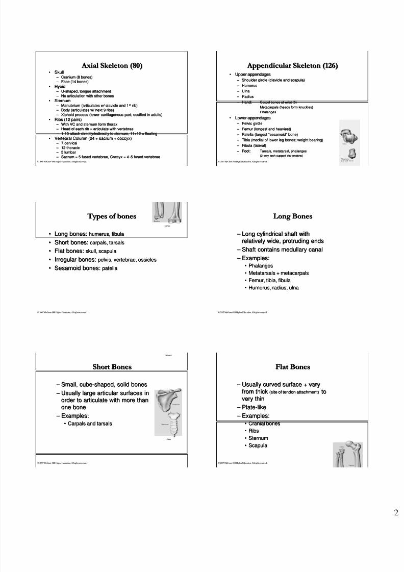
Classification of JointsClassification of Joints
•• ArticulationArticulation
Connection of bones at a joint usually to allowConnection of bones at a joint usually to allow
movement between surfaces of bonesmovement between surfaces of bones•• 3 major classifications according to3 major classifications according to
structure + movement characteristicsstructure + movement characteristics1.1. SynarthrodialSynarthrodial
2.2. AmphiarthrodialAmphiarthrodial
3.3. DiarthrodialDiarthrodial
8/4/2019 1. Review Exam Material (a)
http://slidepdf.com/reader/full/1-review-exam-material-a 5/13
©© 2007 McGraw 2007 McGraw --Hill Higher Education. All rights reserved.Hill Higher Education. All rights reserved.
Classification of JointsClassification of Joints
ArthrodialCondyloidalEnarthrodialGinglymus
SellarTrochoidal
----------Diarthrodial
-----Symphysis
SynchondrosisSyndesmosis
Amphiarthrodial
----------
Gomphosis
SutureSynarthrodial
Functionalclassification
SynovialCartilagenousFibrous
Structural classification
©© 2007 McGraw 2007 McGraw --Hill Higher Education. All rights reserved.Hill Higher Education. All rights reserved.
SynarthrodialSynarthrodial
•• ImmovableImmovable
•• No motionNo motion
– – Suture: skull suturesSuture: skull sutures – – GomphosisGomphosis: teeth fitting into: teeth fitting into
mandible or maxillamandible or maxilla
©© 2007 McGraw 2007 McGraw --Hill Higher Education. All rights reserved.Hill Higher Education. All rights reserved.
Amphiarthrodial Amphiarthrodial
•• Slightly movableSlightly movable
•• Slight motionSlight motionA.A. SyndesmosisSyndesmosis
B.B. SymphysisSymphysis
C.C. SynchrondosisSynchrondosis
©© 2007 McGraw 2007 McGraw --Hill Higher Education. All rights reserved.Hill Higher Education. All rights reserved.
Amphiarthrodial Amphiarthrodial A. A. SyndesmosisSyndesmosis
•• Bones joined together by a strongBones joined together by a strong
ligamentligament oror interosseusinterosseus membranemembrane
•
•
M i n i m a l m o v e m e n t b e t w e e n t h e
M i n i m a l m o v e m e n t b e t w e e n t h e
b o n e s
b o n e s
•• Bones may or may not touch eachBones may or may not touch each
other at the actual jointother at the actual joint
– – CoracoclavicularCoracoclavicular joint joint
– – DistalDistal tibiofibulartibiofibular joint joint
©© 2007 McGraw 2007 McGraw --Hill Higher Education. All rights reserved.Hill Higher Education. All rights reserved.
Amphiarthrodial AmphiarthrodialB.B. SymphysisSymphysis
•• Joint separated byJoint separated by fibrocartilagefibrocartilage
padpad
•• Very slight movement betweenVery slight movement betweenbonesbones
– – SymphysisSymphysis PubisPubis
– – IntervertebralIntervertebral discsdiscs
©© 2007 McGraw 2007 McGraw --Hill Higher Education. All rights reserved.Hill Higher Education. All rights reserved.
Amphiarthrodial AmphiarthrodialC.C. SynchrondosisSynchrondosis
•
•
S e p a r a t e d b y
S e p a r a t e d b y
h y a l i n e c a r t i l a g e
h y a l i n e c a r t i l a g e
•
•
V e r y s l i g h t m o v e m e n t b e t w e e n t h e
V e r y s l i g h t m o v e m e n t b e t w e e n t h e
b o n e s
b o n e s
–
–
C o s t o c h o n d r a l
C o s t o c h o n d r a l
j o i n t s
j o i n t s
( r i b s w i t h t h e
( r i b s w i t h t h e
s t e r n u m
s t e r n u m
)
)

8/4/2019 1. Review Exam Material (a)
http://slidepdf.com/reader/full/1-review-exam-material-a 6/13
©© 2007 McGraw 2007 McGraw --Hill Higher Education. All rights reserved.Hill Higher Education. All rights reserved.
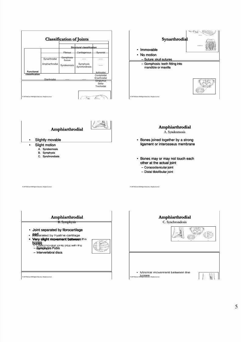
DiarthrodialDiarthrodial Joints Joints
•• AllAll synovial jointssynovial joints
•• Freely movableFreely movable
– – Joint capsuleJoint capsule – – Synovial membraneSynovial membrane
– – Synovial fluidSynovial fluid
(lubricant)(lubricant)
©© 2007 McGraw 2007 McGraw --Hill Higher Education. All rights reserved.Hill Higher Education. All rights reserved.
DiarthrodialDiarthrodial Joints Joints
©© 2007 McGraw 2007 McGraw --Hill Higher Education. All rights reserved.Hill Higher Education. All rights reserved.
DiarthrodialDiarthrodial Joints Joints
©© 2007 McGraw 2007 McGraw --Hill Higher Education. All rights reserved.Hill Higher Education. All rights reserved.
DiarthrodialDiarthrodial Joints Joints
•• ArticularArticular (hyaline)(hyaline) cartilagecartilage coverscovers articulararticularsurface of bones inside joint cavitysurface of bones inside joint cavity – – Absorbs shockAbsorbs shock
– – Protects the boneProtects the bone
•• Absorbs synovial fluid during jointAbsorbs synovial fluid during joint unloadingunloading
•• Secretes synovial fluid during jointSecretes synovial fluid during joint loadingloading
©© 2007 McGraw 2007 McGraw --Hill Higher Education. All rights reserved.Hill Higher Education. All rights reserved.
DiarthrodialDiarthrodial Joints Joints
•• Six typesSix types
•• Each has a different type of bonyEach has a different type of bony
arrangement:arrangement:
4.4. CondyloidCondyloid
5.5. EnarthrodialEnarthrodial
6.6. SellarSellar
1.1. ArthrodialArthrodial
2.2. GinglymusGinglymus
3.3. TrochoidTrochoid
©© 2007 McGraw 2007 McGraw --Hill Higher Education. All rights reserved.Hill Higher Education. All rights reserved.
DiarthrodialDiarthrodial Joints Joints1.1. Arthrodial Arthrodial (Gliding)(Gliding)
•• 2 flat bony surfaces butt against each other2 flat bony surfaces butt against each other
•• Small amount of sliding motion possible inSmall amount of sliding motion possible in
any 1 joint articulationany 1 joint articulation•• NonaxialNonaxial
8/4/2019 1. Review Exam Material (a)
http://slidepdf.com/reader/full/1-review-exam-material-a 7/13
©© 2007 McGraw 2007 McGraw --Hill Higher Education. All rights reserved.Hill Higher Education. All rights reserved.
•• ExamplesExamples
– – Vertebral facets (spinal column)Vertebral facets (spinal column)
– – IntercarpalIntercarpal ++ intertarsalintertarsal joints joints
DiarthrodialDiarthrodial Joints Joints1.1. Arthrodial Arthrodial (Gliding)(Gliding)
©© 2007 McGraw 2007 McGraw --Hill Higher Education. All rights reserved.Hill Higher Education. All rights reserved.
DiarthrodialDiarthrodial Joints Joints2.2. GinglymusGinglymus (Hinge)(Hinge)
•• ArticularArticular surfaces allow motion insurfaces allow motion in
only one planeonly one plane
– – UniaxialUniaxial•• Flexion and extensionFlexion and extension
•• ExamplesExamples
– – ElbowElbow
– – KneeKnee
– – TalocruralTalocrural (ankle)(ankle)
©© 2007 McGraw 2007 McGraw --Hill Higher Education. All rights reserved.Hill Higher Education. All rights reserved.
DiarthrodialDiarthrodial Joints Joints3.3. Trochoid Trochoid (Pivot)(Pivot)
•• Rotational movement along longRotational movement along long
axisaxis
– – UniaxialUniaxial
•• ExamplesExamples
– – AtlantoaxialAtlantoaxial joint joint
– – RadioRadio--ulnar jointulnar joint
©© 2007 McGraw 2007 McGraw --Hill Higher Education. All rights reserved.Hill Higher Education. All rights reserved.
DiarthrodialDiarthrodial Joints Joints4.4. CondyloidCondyloid (Ellipsoid)(Ellipsoid)
•• One bone with an oval concave surfaceOne bone with an oval concave surface
received by another bone with an oval convexreceived by another bone with an oval convex
surfacesurface
– – BiaxialBiaxial ball & socket type jointball & socket type joint
•• Flexion, extension, abduction, adduction,Flexion, extension, abduction, adduction,
circumductioncircumduction
©© 2007 McGraw 2007 McGraw --Hill Higher Education. All rights reserved.Hill Higher Education. All rights reserved.
Diarthrodial JointsDiarthrodial Joints4.4. CondyloidCondyloid (Ellipsoid)(Ellipsoid)
•• ExamplesExamples
– – 2nd2nd--5th5th metacarpophalangealmetacarpophalangeal (knuckle) joints(knuckle) joints
– – Wrist articulation (carpals + radius)Wrist articulation (carpals + radius)
©© 2007 McGraw 2007 McGraw --Hill Higher Education. All rights reserved.Hill Higher Education. All rights reserved.
DiarthrodialDiarthrodial Joints Joints5.5. EnarthrodialEnarthrodial
•• Bony rounded head fitting into aBony rounded head fitting into aconcaveconcave articulararticular surfacesurface – – MultiaxialMultiaxial oror triaxialtriaxial ball & socketball & socket
•• ExamplesExamples
– – Hip jointHip joint
– – Shoulder jointShoulder joint

8/4/2019 1. Review Exam Material (a)
http://slidepdf.com/reader/full/1-review-exam-material-a 8/13
©© 2007 McGraw 2007 McGraw --Hill Higher Education. All rights reserved.Hill Higher Education. All rights reserved.
•• 2 reciprocally concave & convex2 reciprocally concave & convex articulararticularsurfacessurfaces
– – UniqueUnique triaxialtriaxial joint joint
•• Only exampleOnly example – – 11stst carpometacarpalcarpometacarpal joint at joint at thumbthumb
DiarthrodialDiarthrodial Joints Joints6.6. SellarSellar (Saddle)(Saddle)
©© 2007 McGraw 2007 McGraw --Hill Higher Education. All rights reserved.Hill Higher Education. All rights reserved.
Joint Stability Joint Stability
•• Function of joints = provide means of movingFunction of joints = provide means of moving
•• Secondary function = provide stability withoutSecondary function = provide stability without
interfering with the desired motionsinterfering with the desired motions
– – All joints do not have the same degree of stabilityAll joints do not have the same degree of stability
•• EmersonEmerson’’s Laws Law:: ““For everything that is given,For everything that is given,
something is takensomething is taken””
– – Movement is gained at the expense of stabilityMovement is gained at the expense of stability
©© 2007 McGraw 2007 McGraw --Hill Higher Education. All rights reserved.Hill Higher Education. All rights reserved.
Joint Stability Joint Stability
•• Resistance to displacementResistance to displacement
•• 5 factors responsible for stability:5 factors responsible for stability:1.1. Bony structureBony structure
2.2. LigamentousLigamentous arrangementarrangement
3.3. Muscle tensionMuscle tension
4.4. FasciaFascia
5.5. Atmospheric pressureAtmospheric pressure
©© 2007 McGraw 2007 McGraw --Hill Higher Education. All rights reserved.Hill Higher Education. All rights reserved.
Shape of Bony StructureShape of Bony Structure
•• May refer to kind of joint:May refer to kind of joint:
– – Hinge,Hinge, condyloidcondyloid, pivot, or ball, pivot, or ball--andand--socketsocket
•• Or specific characteristics of a joint:Or specific characteristics of a joint:
– – Depth of socketDepth of socket
More stable,More stable,less mobileless mobile
More mobile,More mobile,less stableless stable
©© 2007 McGraw 2007 McGraw --Hill Higher Education. All rights reserved.Hill Higher Education. All rights reserved.
Ligamentous ArrangementsLigamentous Arrangements
•• Ligaments are strong, flexible, stressLigaments are strong, flexible, stress--
resistant, somewhat elastic, fibrous tissuesresistant, somewhat elastic, fibrous tissues
formed into bands or cordsformed into bands or cords•• Help maintain relationship of bonesHelp maintain relationship of bones
– – Check movement at normal limits of jointCheck movement at normal limits of joint
– – Resist movements for which joint not constructedResist movements for which joint not constructed
•• Stretch when subject to prolonged stressStretch when subject to prolonged stress
– – Once stretched, function is affectedOnce stretched, function is affected
©© 2007 McGraw 2007 McGraw --Hill Higher Education. All rights reserved.Hill Higher Education. All rights reserved.
Muscular ArrangementMuscular Arrangement
•• Muscles spanning jointsMuscles spanning joints
aid in stabilityaid in stability•• Especially when bonyEspecially when bony
structure contributes littlestructure contributes little
to stabilityto stability
•• ExampleExample
– – Rotator cuffRotator cuff Fig 5.13
Muscles acting to stabilize theMuscles acting to stabilize the
shouldershoulder
8/4/2019 1. Review Exam Material (a)
http://slidepdf.com/reader/full/1-review-exam-material-a 9/13
©© 2007 McGraw 2007 McGraw --Hill Higher Education. All rights reserved.Hill Higher Education. All rights reserved.
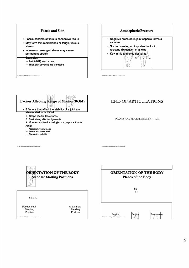
Fascia and SkinFascia and Skin
•• Fascia consists of fibrous connective tissueFascia consists of fibrous connective tissue
•• May form thin membranes or tough, fibrousMay form thin membranes or tough, fibrous
sheetssheets•• Intense or prolonged stress may causeIntense or prolonged stress may cause
permanent stretchpermanent stretch
•• ExamplesExamples
– – IliotibialIliotibial (IT) tract or band(IT) tract or band
– – Thick skin covering the knee jointThick skin covering the knee joint
©© 2007 McGraw 2007 McGraw --Hill Higher Education. All rights reserved.Hill Higher Education. All rights reserved.
Atmospheric Pressure Atmospheric Pressure
•• Negative pressure in joint capsule forms aNegative pressure in joint capsule forms a
vacuumvacuum
•• Suction created an important factor inSuction created an important factor inresisting dislocation of a jointresisting dislocation of a joint
•• Key in hip and shoulder jointsKey in hip and shoulder joints
©© 2007 McGraw 2007 McGraw --Hill Higher Education. All rights reserved.Hill Higher Education. All rights reserved.
Factors Affecting Range of Motion (ROM)Factors Affecting Range of Motion (ROM)
•• 3 factors that affect the stability of a joint are3 factors that affect the stability of a joint arealso related to its ROM:also related to its ROM:
1.1. Shape of articular surfacesShape of articular surfaces
2. Restraining effect of ligaments2. Restraining effect of ligaments
3. Muscles and tendons (single most important factor)3. Muscles and tendons (single most important factor)
Also:Also: – – Apposition of bulky tissueApposition of bulky tissue
– – Gender and fitness levelGender and fitness level
– – Disease (i.e. arthritis)Disease (i.e. arthritis)
©© 2007 McGraw 2007 McGraw --Hill Higher Education. All rights reserved.Hill Higher Education. All rights reserved.
END OF ARTICULATIONSEND OF ARTICULATIONS
PLANES AND MOVEMENTS NEXT TIME
©© 2007 McGraw 2007 McGraw --Hill Higher Education. All rights reserved.Hill Higher Education. All rights reserved.
ORIENTATION OF THE BODY ORIENTATION OF THE BODY
Standard Starting PositionsStandard Starting Positions
Fig 2.10
FundamentalStandingPosition
AnatomicalStandingPosition
©© 2007 McGraw 2007 McGraw --Hill Higher Education. All rights reserved.Hill Higher Education. All rights reserved.
ORIENTATION OF THE BODY ORIENTATION OF THE BODY
Planes of the BodyPlanes of the Body
Fig
2.8
Sagittal Frontal Transverse

8/4/2019 1. Review Exam Material (a)
http://slidepdf.com/reader/full/1-review-exam-material-a 10/13
©© 2007 McGraw 2007 McGraw --Hill Higher Education. All rights reserved.Hill Higher Education. All rights reserved.
Axes of motion Axes of motion
•• Motion through a plane revolves around anMotion through a plane revolves around an
axisaxis•• Axis is at right angles to the plane of motionAxis is at right angles to the plane of motion
©© 2007 McGraw 2007 McGraw --Hill Higher Education. All rights reserved.Hill Higher Education. All rights reserved.
Axes of motion Axes of motion
• Lateral axis
•• Movements in sagittal planeMovements in sagittal plane
– – SitSit--upup
©© 2007 McGraw 2007 McGraw --Hill Higher Education. All rights reserved.Hill Higher Education. All rights reserved.
Axes of motion Axes of motion
•• AnteroposteriorAnteroposterior axisaxis
•• Movements in frontal planeMovements in frontal plane
– – Jumping JacksJumping Jacks
©© 2007 McGraw 2007 McGraw --Hill Higher Education. All rights reserved.Hill Higher Education. All rights reserved.
Axes of motion Axes of motion
•• VerticalVertical axisaxis
•• Movements inMovements in
horizontal/transverse planehorizontal/transverse plane
– – Spinal rotation to left or rightSpinal rotation to left or right
©© 2007 McGraw 2007 McGraw --Hill Higher Education. All rights reserved.Hill Higher Education. All rights reserved.
Fundamental MovementsFundamental Movements
•• AngularAngular movements:movements: change in angle between boneschange in angle between bones
– – AbdAbductionuction (lateral movement from midline;(lateral movement from midline; frontalfrontal))
– – AddAdductionuction (movement toward midline;(movement toward midline; frontalfrontal))
•• Horizontal abduction/adduction;Horizontal abduction/adduction; horizontalhorizontal
– – FlexionFlexion (bending movement decreasing angle;(bending movement decreasing angle; sagittalsagittal))
– – ExtensionExtension (straightening movement increasing angle;(straightening movement increasing angle; sagittalsagittal))
•• Hyperextension = extension beyond anatomical positionHyperextension = extension beyond anatomical position
©© 2007 McGraw 2007 McGraw --Hill Higher Education. All rights reserved.Hill Higher Education. All rights reserved.
Fundamental MovementsFundamental Movements
•• CircularCircular movements:movements: combination of planescombination of planes
– – CircumductionCircumduction (circular movement describing a cone)(circular movement describing a cone)•• e.g. arm circles (true only in ball and socket)e.g. arm circles (true only in ball and socket)
– – RotationRotation (pivoting on bone(pivoting on bone’’s long axis)s long axis)
•• External or lateral (away from midline)External or lateral (away from midline)
•• Internal or medial (towards midline)Internal or medial (towards midline)

8/4/2019 1. Review Exam Material (a)
http://slidepdf.com/reader/full/1-review-exam-material-a 11/13
©© 2007 McGraw 2007 McGraw --Hill Higher Education. All rights reserved.Hill Higher Education. All rights reserved.
Fundamental MovementsFundamental Movements
•• SpecialSpecial MovementsMovements
– – Supination/ Supination/ pronationpronation:: rotational movement specific torotational movement specific toforearmforearm
•• Supination = external rotation; results in palm up (anatomicalSupination = external rotation; results in palm up (anatomical
position)position)
•• Pronation = internal rotation; results in palm downPronation = internal rotation; results in palm down
– – Eversion/InversionEversion/Inversion:: specific to footspecific to foot
•• Eversion = sole of foot turned outward or laterallyEversion = sole of foot turned outward or laterally
•• Inversion = sole of foot turned inward of mediallyInversion = sole of foot turned inward of medially
((notnot pigeonpigeon--toed)toed)
©© 2007 McGraw 2007 McGraw --Hill Higher Education. All rights reserved.Hill Higher Education. All rights reserved.
Fundamental MovementsFundamental Movements
•• SpecialSpecial MovementsMovements……
– – Elevation/depressionElevation/depression•• Elevation = superior movement of shoulder girdle; shruggingElevation = superior movement of shoulder girdle; shrugging
shouldersshoulders
•• Depression = inferior movement of shoulder girdleDepression = inferior movement of shoulder girdle
– – Protraction/retractionProtraction/retraction•• Protraction = anterior movement of shoulder girdle away fromProtraction = anterior movement of shoulder girdle away from
spine; rounding of shoulders (spine; rounding of shoulders (abdabduction of scapula)uction of scapula)
•• Retraction = posterior movement of shoulder girdle towardsRetraction = posterior movement of shoulder girdle towards
spine; squaring shoulders (spine; squaring shoulders (addadduction of scapula)uction of scapula)
©© 2007 McGraw 2007 McGraw --Hill Higher Education. All rights reserved.Hill Higher Education. All rights reserved.
InIn--class worksheet:class worksheet:
•• PLANES AND AXES OF MOVEMENTPLANES AND AXES OF MOVEMENT
Anatomical directional terminology review onAnatomical directional terminology review on
next set of slidesnext set of slides……
©© 2007 McGraw 2007 McGraw --Hill Higher Education. All rights reserved.Hill Higher Education. All rights reserved.
Anatomical directional terminology Anatomical directional terminology
From Van De GraaffKM: Human anatomy , ed
6, New York, 2002, McGraw-Hill
©© 2007 McGraw 2007 McGraw --Hill Higher Education. All rights reserved.Hill Higher Education. All rights reserved.
Anatomical directional terminology Anatomical directional terminology
•• AnteriorAnterior
– – in front or in the frontin front or in the front
partpart
•• PosteriorPosterior – – behind or in the rearbehind or in the rear
partpart
©© 2007 McGraw 2007 McGraw --Hill Higher Education. All rights reserved.Hill Higher Education. All rights reserved.
Anatomical directional terminology Anatomical directional terminology
•• InferiorInferior (infra)(infra)
– – below in relation to another structure; caudalbelow in relation to another structure; caudal
•• SuperiorSuperior (supra)(supra)
– –
above in relation to another structure; cephalicabove in relation to another structure; cephal ic
•• DistalDistal
– – situated away from center or midline of body;situated away from center or midline of body;away from the point of originaway from the point of origin
•• ProximalProximal
– – nearest the trunk or point of originnearest the trunk or point of origin
•• LateralLateral
– – on or to the side; outside, farther from theon or to the side; outside, farther from themedian ormedian or midsagittalmidsagittal planeplane
•• MedialMedial
– – relating to the middle or center; nearer to therelating to the middle or center; nearer to themedial ormedial or midsagittalmidsagittal planeplane

8/4/2019 1. Review Exam Material (a)
http://slidepdf.com/reader/full/1-review-exam-material-a 12/13
©© 2007 McGraw 2007 McGraw --Hill Higher Education. All rights reserved.Hill Higher Education. All rights reserved.
Anatomical directional terminology Anatomical directional terminology
•• DeepDeep
Beneath or below the surface; used to describeBeneath or below the surface; used to describe
relative depth or location of muscles or tissuerelative depth or location of muscles or tissue
•• SuperficialSuperficial
Near the surface; used to describe relative depth orNear the surface; used to describe relative depth or
location of muscles or tissuelocation of muscles or tissue
©© 2007 McGraw 2007 McGraw --Hill Higher Education. All rights reserved.Hill Higher Education. All rights reserved.
Anatomical directional terminology Anatomical directional terminology
•• ProneProne
Body lying face down; lying on stomachBody lying face down; lying on stomach
•• SupineSupine
Body lying face up; lying on backBody lying face up; lying on back
©© 2007 McGraw 2007 McGraw --Hill Higher Education. All rights reserved.Hill Higher Education. All rights reserved.
Anatomical directional terminology Anatomical directional terminology
•• DorsalDorsal
Relating to the back; being or located near, on,Relating to the back; being or located near, on,
or toward the back, posterior part, or upperor toward the back, posterior part, or upper
surfacesurface
•• VentralVentral
Relating to the belly or abdomen, on or towardRelating to the belly or abdomen, on or toward
the front, anterior partthe front, anterior part
©© 2007 McGraw 2007 McGraw --Hill Higher Education. All rights reserved.Hill Higher Education. All rights reserved.
Anatomical directional terminology Anatomical directional terminology
•• VolarVolar
Relating to palm of the hand or sole of the footRelating to palm of the hand or sole of the foot
•• PlantarPlantar
Relating to the sole or undersurface of the footRelating to the sole or undersurface of the foot
©© 2007 McGraw 2007 McGraw --Hill Higher Education. All rights reserved.Hill Higher Education. All rights reserved.
Body RegionsBody Regions
©© 2007 McGraw 2007 McGraw --Hill Higher Education. All rights reserved.Hill Higher Education. All rights reserved.
Body regionsBody regions
•• AxialAxial
– – Head (cephalic)Head (cephalic)
•• Cranial and facialCranial and facial – – Neck (cervical)Neck (cervical)
– – TrunkTrunk
•• Thorax (thoracic)Thorax (thoracic)
•• Back (dorsal)Back (dorsal)
•• Abdomen (abdominal)Abdomen (abdominal)
•• Pelvis (pelvic)Pelvis (pelvic)
8/4/2019 1. Review Exam Material (a)
http://slidepdf.com/reader/full/1-review-exam-material-a 13/13
©© 2007 McGraw 2007 McGraw --Hill Higher Education. All rights reserved.Hill Higher Education. All rights reserved.
Body regionsBody regions
•• AppendicularAppendicular
– – Upper limbsUpper limbs
•• Shoulder (Shoulder (acromialacromial ))
•• Arm (brachial)Arm (brachial)
•• Forearm (Forearm (antebrachialantebrachial))
•• Hand (Hand (manusmanus))
– – Lower limbsLower limbs
•• Thigh (femoral)Thigh (femoral)
•• LegLeg
•• Foot (pedal)Foot (pedal)
Recommended