Embed Size (px)
Citation preview

ZOOTAXAISSN 1175-5326 (print edition)
ISSN 1175-5334 (online edition)Copyright © 2010 · Magnolia Press
Zootaxa 2395: 17–33 (2010) www.mapress.com/zootaxa/ Article
Diopatra (Annelida: Onuphidae) diversity in European waters with the description of Diopatra micrura, new species
ADÍLIA PIRES1, HANNELORE PAXTON2, VICTOR QUINTINO1 & ANA MARIA RODRIGUES1,3
1CESAM, Department of Biology, University of Aveiro, 3810–193 Aveiro, Portugal2Department of Biological Sciences, Macquarie University, Sydney, NSW 2109, Australia3Corresponding author. E-mail: [email protected]
Abstract
This study describes a new species of the genus Diopatra Audouin and Milne-Edwards, 1833. Diopatra micrura sp. nov., was found on the western and the southern coast of Portugal and can be distinguished from other Diopatra species by a characteristic striped colour pattern of the antennae and palps. Other diagnostic morphological characteristics include ventral parapodial lobes, crescentic nuchal organs, ceratophores with 12–15 rings, and subacicular hooks from chaetigers 8–13. This species was found mainly in fine or very fine sand with variable fines content, from the intertidal region up to 50 meters depth.
Molecular studies of mitochondrial DNA genes 16S rDNA and COI confirmed the distinction of D. micrura sp. nov., from other European Diopatra species. The percentage of nucleotides divergence between the new species and D. neapolitana and D. marocensis was respectively 16% and 17% for COI and 12% and 15% for 16S. The nucleotide sequence for the 16S gene was always the same in all specimens of D. micrura and two haplotypes were found for the COI gene. The discovery of D. micrura sp. nov., brings the number of Diopatra species known from Portugal to three and from Europe to four; a key to the four species is provided.
Key words: Taxonomy, striped antennae, 16S rDNA, COI, distribution, Portugal
Introduction
The genus Diopatra Audouin and Milne-Edwards, 1833 includes about 50 species distributed around the world (Budaeva & Fauchald 2008). These onuphid polychaetes are common in intertidal and shallow subtidal areas of all major oceans although better represented in warmer waters (Paxton 1986). The genus is characterised by the presence of peristomial cirri and spiralled branchiae (Paxton 1986).
Diopatra neapolitana Delle Chiaje, 1841 was until very recently the only recognised species of Diopatrain European waters. Recent studies revealed the presence of Diopatra marocensis Paxton et al., 1995 in Portugal (Rodrigues et al. 2009) and a species reported as Diopatra sp. A from Arcachon to Dunquerque, France, by Berke et al. (2010).
The present paper reports the discovery of another species, Diopatra micrura, sp. nov., increasing to three the number of Diopatra species known from Portugal and to four the number of European species.
Besides the morphological description, this study also uses a molecular approach to confirm the distinction of D. micrura sp. nov. from D. neapolitana, D. marocensis, and Diopatra sp. from Arcachon Bay, by characterising two mitochondrial DNA genes, 16S rDNA (16S ribosomal RNA gene) and COI (cytochrome c oxidase subunit I) (Halanych & Janosik 2006). It also presents the distribution of D. micrura sp. nov., along the Portuguese coast together with the sediment type and depth of occurrence.
Accepted by P. Hutchings: 21 Jan. 2010; published:10 Mar. 2010 17

Material and methods
Sampling. On the western coast of Portugal, specimens of D. micrura were collected in Ria de Aveiro, near the mouth and in intertidal areas, on the adjacent shelf area, on the shelf off Nazaré and in Guia, off the Tagus Estuary; on the southern coast, specimens were collected near the Guadiana river mouth (Fig. 1, Table 1).
FIGURE 1. Sampling areas where Diopatra micrura sp. nov., was found: A—Aveiro (shelf and Ria); B— coastal shelf off Nazaré; C—Guia, coastal shelf off Tagus estuary; D—coastal shelf off Guadiana estuary
PIRES ET AL.18 · Zootaxa 2395 © 2010 Magnolia Press

TABLE 1. List of sites where Diopatra micrura sp. nov. was sampled. AS—Shelf off Aveiro; NS—Nazaré; RA—Ria de Aveiro; GS—Shelf off Guadiana River; TS—Shelf off Tagus Estuary; SVT - Total Volatile Solids (organic matter).
Site Latitude (ºN) Longitude (ºW) Depth (m) Date Sediment type Fines content (%) SVT (%)
TS1 38° 39' 45.840" 9° 25' 40.440" 40 Mar-94 Silty fine sand 9.03 2.71
TS1A 38° 39' 45.840" 9° 25' 40.440" 40 Apr-01 Silty fine sand 9.03 2.71
TS1B 38° 39' 45.840" 9° 25' 40.440" 40 Oct-03 Silty fine sand 9.03 2.71
TS2 38° 40' 29.340" 9° 27' 59.580" 40 Jan-97 Clean fine sand 3.4 1.43
TS3 38° 40' 33.600" 9° 28' 11.640" 40 Jan-97 Clean fine sand 4.47 1.41
TS3A 38° 40' 33.600" 9° 28' 11.640" 40 Oct-01 Clean fine sand 4.47 1.41
TS3B 38° 40' 33.600" 9° 28' 11.640" 40 Oct-03 Clean fine sand 4.47 1.41
TS4 38° 40' 20.820" 9° 28' 7.440" 45 Jan-97 Clean fine sand 3.52 1.12
TS4A 38° 40' 20.820" 9° 28' 7.440" 45 Oct-01 Clean fine sand 3.52 1.12
TS4B 38° 40' 20.820" 9° 28' 7.440" 45 Oct-03 Clean fine sand 3.52 1.12
TS5 38° 40' 25.080" 9° 27' 50.640" 40 Jan-97 Clean fine sand 3.36 1.39
TS5A 38° 40' 25.080" 9° 27' 50.640" 40 Apr-97 Clean fine sand 3.36 1.39
TS6 38° 40' 37.620" 9° 28' 22.080" 40 Jan-97 Clean fine sand 5.32 1.49
TS6A 38° 40' 37.620" 9° 28' 22.080" 40 Oct-98 Clean fine sand 5.32 1.49
TS6B 38° 40' 37.620" 9° 28' 22.080" 40 Apr-01 Clean fine sand 5.32 1.49
TS7 38° 40' 51.060" 9° 29' 0.180" 40 Jan-97 Silty medium sand 10.06 1.41
TS7A 38° 40' 51.060" 9° 29' 0.180" 40 Oct-97 Silty medium sand 10.06 1.41
TS8 38° 41' 20.220" 9° 29' 51.600" 40 Jan-97 Clean coarse sand 0.81 1.28
TS8A 38° 41' 20.220" 9° 29' 51.600" 40 Oct-02 Clean coarse sand 0.81 1.28
TS9 38° 40' 1.560" 9° 29' 41.640" 34 Jan-97 Clean fine sand 1.57 1.4
TS10 38° 40' 37.620" 9° 27' 53.940" 38 Apr-97 Clean fine sand 2.07 1.30
TS10A 38° 40' 37.620" 9° 27' 53.940" 38 Oct-02 Clean fine sand 2.07 1.30
TS11 38° 40' 20.100" 9° 28' 27.120" 45 Apr-97 Silty fine sand 5.08 1.45
TS12 38° 40' 16.920" 9° 27' 38.460" 40 Apr-97 Clean fine sand 3.71 1.34
TS12A 38° 40' 16.920" 9° 27' 38.460" 40 Oct-01 Clean fine sand 3.71 1.34
TS13 38° 40' 38.820" 9° 27' 27.540" 26 Apr-97 Clean fine sand 3.88 1.61
TS14 38° 40' 2.640" 9° 27' 0.480" 40 Apr-97 Silty fine sand 5.20 1.50
TS14A 38° 40' 2.640" 9° 27' 0.480" 40 Oct-99 Silty fine sand 5.20 1.50
TS14B 38° 40' 2.640" 9° 27' 0.480" 40 Oct-03 Silty fine sand 5.20 1.50
TS14C 38° 40' 2.640" 9° 27' 0.480" 40 Jan-06 Silty fine sand 5.20 1.50
TS15 38° 40' 56.100" 9° 28' 8.400" 26 Oct-99 Clean fine sand 3.87 1.28
TS15A 38° 40' 56.100" 9° 28' 8.400" 26 Oct-02 Clean fine sand 3.87 1.28
TS16 38° 40' 51.060" 9° 29' 0.180" 40 Apr-01 Silty medium sand 10.06 1.41
TS17 38° 39' 53.640" 9° 28' 29.940" 60 Oct-01 Silty very fine sand 14.29 2.21
TS18 38° 39' 51.060" 9° 27' 16.500" 50 Oct-02 Silty very fine sand 24.87 3.39
TS19 38° 40' 15.109" 9° 26' 30.275" 35 Oct-02 Clean fine sand 2.83 1.2
TS20 38° 40' 4.881" 9° 24' 9.258" 27 Oct-02 Clean fine sand 4.50 1.6
TS21 38° 39' 49.183" 9° 23' 43.095" 26 Oct-02 Silty very fine sand 11.12 1.6
TS22 38° 40' 6.180" 9° 27' 23.880" 40 Oct-03 Clean fine sand 4.10 1.84
TS23 38° 39' 49.200" 9° 23' 43.080" 26 Oct-03 Silty fine sand 11.05 2.52
TS23A 38° 39' 49.200" 9° 23' 43.080" 26 Oct-04 Silty fine sand 11.05 2.52
TS23B 38° 39' 49.200" 9° 23' 43.080" 26 Oct-07 Silty fine sand 11.05 2.52
to be continued.
Zootaxa 2395 © 2010 Magnolia Press · 19DIOPATRA DIVERSITY IN EUROPEAN WATERS

In Ria de Aveiro, the sediment was collected with a shovel, digging about 30 cm depth, and the Diopatratubes were gently removed from the sediment. In the laboratory, the animals were pushed out from the tube and examined alive. Specimens from the other localities were obtained from earlier collecting trips samplings (Table 1) and were re-examined for taxonomic confirmation. In those localities the sediment was collected with grabs, either a 0.1 m2 Smith-McIntyre (shelf off Aveiro, Guia and Nazaré) or a 0.05 m2 Ponar (southern coast). The samples were washed through a 1 mm mesh sieve and fixed with 4% formalin neutralized with borax. All organisms collected were sorted and identified under a stereomicroscope and then transferred for long-term storage in 70% ethanol.
Eight Diopatra micrura specimens from Guia, five from Ria de Aveiro and two Diopatra sp. specimens from Arcachon Bay, France, were collected for genetic studies and preserved in ethanol (96%).
Morphological characterisation and data analysis. In the laboratory, 88 adult specimens of D. micrura(five from Ria de Aveiro, nine from the shelf off Aveiro, three from the shelf off Nazaré and 71 from Guia, off the Tagus Estuary) and eight specimens of Diopatra sp. from France (seven from Arcachon Bay and one from Marennes Oléron, about 150 km North of Arcachon), were examined for morphological studies. Fixed specimens were measured for the width of the 10th chaetiger (without parapodia), the length of antennae and palps. Total length of complete specimens was measured. The numbers of chaetigers in complete specimens, of rings on the ceratophores, of whorls of the branchiae and of teeth in the pectinate chaetae were counted. The first chaetiger with subacicular hooks and the last chaetiger with branchiae were recorded. The colour pattern and the form of the prostomium of the species were described in live and preserved specimens. The terminology used for the prostomial appendages followed Paxton (1998). The relationship between the width of the 10th chaetiger, taken as a measure of the specimen size, and other morphological descriptors was analysed through linear regression analysis.
The morphological data of Diopatra micrura sp. nov., and of the Diopatra specimens obtained from France were compared to the data presented by Rodrigues et al. (2009), for D. neapolitana and D. marocensis. Using the data recorded for the width of the 10th chaetiger, the number of rings in the ceratophores, the number
TABLE 1. (continued.)
Site Latitude (ºN) Longitude (ºW) Depth (m) Date Sediment type Fines content (%) SVT (%)
TS23C 38° 39' 49.200" 9° 23' 43.080" 26 Oct-08 Silty fine sand 11.05 2.52
TS23D 38° 39' 49.200" 9° 23' 43.080" 26 Sep-09 Silty fine sand 11.05 2.52
TS24 38° 39' 54.600" 9° 26' 14.340" 40 Oct-04 Silty fine sand 5.33 1.73
TS24A 38° 39' 54.600" 9° 26' 14.340" 40 Jan-06 Silty fine sand 5.33 1.73
TS25 38° 40' 7.440" 9° 27' 48.420" 45 Jan-06 Clean fine sand 4.56 1.54
TS26 38° 40' 33.720" 9° 28' 45.900" 45 Oct-07 Silty fine sand 5.82 1.60
AS1 40° 40' 50.637" 8° 46' 45.085" 15 Dec-02 Clean very fine sand 1.6 0.7
AS2 40° 41' 11.718" 8° 46' 32.926" 15 Dec-02 Clean very fine sand 1.61 0.84
AS3 40° 41' 4.855" 8° 47' 8.918" 18 Dec-02 Clean very fine sand 1.88 0.97
AS4 40° 40' 38.579" 8° 46' 26.623" 13 Dec-02 Clean very fine sand 1.67 0.92
AS5 40° 41' 0.196" 8° 46' 15.844" 13 Dec-02 Clean very fine sand 1.34 0.72
AS6 40° 41' 56.762" 8° 46' 6.409" 15 Dec-02 Clean very fine sand 1.18 0.71
GS1 37° 10' 21.119" 7° 28' 2.219" 12 May-07 Silty fine sand 5.03 -
GS2 37° 8' 52.440" 7° 24' 59.281" 10 May-07 Very silty very fine sand
46.14 -
NS 39° 50' 43.901" 9° 6' 40.799" 37.2 Apr-08 Silty fine sand 6.65 0.94
RA1 40° 38' 28.896" 8° 44' 0.276" Intertidal Oct-08 Silty fine sand 24.7 4.3
RA2 40° 38' 28.896" 8° 44' 0.276" Intertidal Mar-09 Silty fine sand 24.7 4.3
RA3 40° 38' 28.896" 8° 44' 0.276" Intertidal Sep-09 Silty fine sand 24.7 4.3
RA4 40° 38' 28.896" 8° 44' 0.276" Intertidal Nov-09 Silty fine sand 24.7 4.3
PIRES ET AL.20 · Zootaxa 2395 © 2010 Magnolia Press

of whorls in the branchiae, the number of teeth in the pectinate chaetae, the first chaetiger to show subacicular hooks and the presence-absence of ventral lobe in the parapodia 5–20, a data matrix was constructed using as many specimens per species as possible (41 D. micrura sp. nov., 35 D. neapolitana, 35 D. marocensis and 8 Diopatra sp. specimens obtained in France). Following normalisation of the variables, the morphological data matrix was submitted to classification, using Un-weighted Pair Group Mean Average upon the Euclidean distance matrix between specimens, and ordination, using Principal Components Analysis, with the software PRIMER v6 (Clarke & Gorley 2006).
The holotype and five paratypes of the new species were deposited in the Museu Nacional de História Natural, Lisbon (MNHN), five paratypes in the Museo Nacional de Ciencias Naturales, Madrid (MNCNM), and six paratypes in the Australian Museum, Sydney (AM). The remaining specimens (including specimens used for DNA sequencing) are kept at the Departamento de Biologia, Universidade de Aveiro, CESAM, Campus Universitario de Santiago, Aveiro (UA). The specimens for scanning electron microscopy (SEM) viewing were fixed in formalin. The specimens were dehydrated in graded ethanol series and critical point dried in a Bal-Tec CPD-030 critical point dryer, using liquid CO2. After drying, the specimens were sputter coated with gold: palladium alloy 60:40 in a Polaron sputter coating system. SEM micrographs were taken in a Hitachi SU-70 scanning microscope.
Grain-size analysis. Sediment grain-size was analysed in the sites where D. micrura was collected, by wet and dry sieving following Quintino et al. (1989). The sediment was classified according to the median value (P50), following the Wentworth scale (Doeglas 1968): very fine sand (median between 0.063–0.125 mm); fine sand (0.125–0.250 mm); medium sand (0.250–0.500 mm) or coarse sand (0.500–1 mm). The final sediment classification adopted the description “clean“, “silty” or “very silty”, when fines (particles with diameter below 0.063 mm) were below 5%, from 5% to 25%, and from 25% to 50%, respectively, of the total sediment, dry weight (Quintino et al. 1989). Sediments with more than 50% fines were classified as mud.
Genetic characterisation and data analysis. Total genomic DNA was extracted with the DNeasy Blood & Tissue Kit (Qiagen), following the manufacturer protocol. Purified DNA was aliquoted in TE buffer (10 mM Tris-HCl, 1 mM EDTA, pH 8.0) and stored at -20 ºC, until required.
About 500 bp part of the mitochondrial 16S rDNA and about 700 bp of cytocrome c oxidase subunit I (COI) were amplified by PCR. Amplification of the 16S rDNA gene was performed using the 16SarL and 16SbrH primers from Palumbi et al. (1991). The COI gene fragment was amplified using the LCO 1490 and HCO 2198 primers of Folmer et al. (1994).
Each PCR was performed in a final volume of 50 µl containing 10–100 ng of genomic DNA, 1 µM of each primers, 1x PCR buffer, 1.5 mM MgCl2, 0.2 mM of each dNTP (Promega) and 0.5 U Taq DNA polymerase (Promega). Amplification occurred on an MJ Mini Thermal-Cycler (Citomed). The thermal cycling parameters were: initial denaturation at 94 ºC, 3 min., followed by 34 cycles of denaturation at 94 ºC, 1 min.; primer-specific annealing temperature (49 ºC for 16S and 45 ºC for COI), 30 sec.; extension at 72 ºC, 2 min. and final extension at 72 ºC for 5 min. The amplification products were visualised, after agarose gel electrophoresis and ethidium bromide staining.
The DNA sequences determinations of 16S/COI PCR-amplified were commercially performed (STAB Vida, Portugal). The nucleotide sequence of each fragment was determined on both strands of PCR products from two independent reactions. The DNA and deduced amino acid sequence alignments were made with the Biological Sequence Alignment Editor (BioEdit v7.0.0, free software by Tom Hall, Department of Microbiology, North Carolina State University, USA). The sequences obtained for D. micrura sp. nov., and Diopatra sp. from Arcachon were compared to D. neapolitana and D. marocensis, obtained in a previous study (Rodrigues et al. 2009). The DNA sequences were analysed with Genetyx-WIN v5.1 (Software Development, Tokyo), to determine the divergence percentage between the various Diopatra species, for both genes. These sequences were also analysed together in a single dataset, separately for each gene, withMarphysa sanguinea (Montagu 1813) as the outgroup. The dataset sequences were aligned in MEGA v4 (Tamura et al. 2007) with CLUSTALW, using the default alignment settings. The phylogenetic analysis were conducted using MEGA v4 (Tamura et al. 2007) by applying Neighbor Joining (NJ). To verify the robustness of the internal nodes of NJ trees, bootstrap analysis was carried out using 1,000 pseudo replicates.
Zootaxa 2395 © 2010 Magnolia Press · 21DIOPATRA DIVERSITY IN EUROPEAN WATERS

Results
Diopatra micrura, sp nov.Figs. 1–8; Tables 1–3
Type material. Holotype: MNHN MB29-000166, Sta. RA4 (Nov-09) (incomplete specimen, 51 mm long (61 chaetigers), 3.3 mm wide).
Paratypes: MNHN MB29-000167, Sta NS (1); MNHN MB29-000171, Sta.RA1 (1); MNHN MB29-000168, Sta TS14B (1); MNHN MB29-000170, Sta TS23C (1); MNHN MB29-000169, Sta TS24 (1); MNCN 16.01/11627, Sta. TS1A (1); MNCN 16.01/11628, Sta.TS3A (1); MNCN 16.01/11629, Sta. TS4A (1); MNCN 16.01/11630, Sta. TS22 (1); MNCN 16.01/11631, Sta. TS25 (1); AM W36251, Sta. NS (1); AM W36252 Sta. TS13 (1); AM W36253 Sta. TS23 (2); AM W36254, Sta. TS23C (1); AM W36255, Sta. RA2 (1); DBUA-01140.01, Sta. TS1B (1); DBUA-01141.01, Sta. TS12 (1); DBUA-01142.01, Sta. TS12A (1); DBUA-01143.01, Sta. TS14C (1.); DBUA-01144.01, Sta TS23A (1).
Etymology. The striped antennae of the new species evoke the pattern of the coral snakes Micrurus spp., hence the name Diopatra micrura, sp. nov.
Morphological description. Length of complete preserved specimens from 1.7 to 7.8 cm, number of chaetigers from 70 to 97; width of 10th chaetiger from 0.6 to 4.5 mm without parapodia. Some incomplete specimens regenerating anterior end of body (paratype AM W36252); one specimen posterior end.
Overall colour of living specimens greenish dorsally, cream ventrally. Antennostyles and palpostyles with very characteristic transverse brown bands, 4–8 on antennae and 2–4 on palps (Figs 2A–C, 3A). Frontal lips whitish with brown pigment at base and ceratophores with brown rings (Fig 2B). Prostomium with brown pigment; area of nuchal grooves paler (Figs 2B, C). Peristomium with brown pigment (Fig. 2C, 3A), peristomial cirri cream. Additionally, anterior 10–15 chaetigers with small iridescent white spots (Fig. 2C) and following chaetigers with iridescent transverse white line (Fig. 2A). Laterally, from chaetigers 1–4 to 13–23 two brown patches, one on each side (Figs 2C, 3A). Branchiae green, parapodia cream (Fig. 2D); dorsal cirri with iridescent white spots.
In preserved individuals, the body is cream with two brown patches laterally on each segment up to chaetigers 13–23 (Fig. 3A). Lack of coloration in middle of each chaetiger forming “white” line along body (Fig. 3A). Brown pigmentation of antennae, palps, ceratophores, prostomium, frontal lips and peristomium noticed in living specimens still present.
Prostomium anteriorly rounded with subulate frontal lips (Figs 4A, B). Ceratophores of antennae and palps with proximal rings and longer distal ring, holotype with 14 proximal rings, other specimens with 12–15 rings (Figs 2A–C, 3A, 4A). Antennostyles relatively long, tapering to distal end, ending in fine point; in holotype laterals reaching chaetiger 9, median reaching chaetiger 5, in other specimens 6–13 and 4–10 respectively; palpostyles shorter, reaching chaetiger 2 in holotype, 2–4 in other specimens. Length of antennae quite variable, apparently unrelated to size of specimens (based on width of 10th chaetiger) (Fig. 5A). Sensory buds present on antennostyles and palpostyles forming 12–14 irregular longitudinal rows (Fig. 4C). Sensory buds slightly raised, with pores forming circles (Fig. 4D). In addition, randomly distributed sensory buds on ceratophores, frontal lips, upper lips, prostomium, peristomial cirrus, peristomium and branchiae (Figs 4G, H). Nuchal grooves crescentic (Fig. 4E). Peristomium as long as first chaetiger, bearing pair of peristomial cirri, about twice as long as peristomium (Fig. 3A).
First four modified parapodia (chaetiger 1 to 4) projecting laterally and slightly anteriorly, slightly longer than following non-modified, laterally projecting parapodia (Figs 4B, F). Prechaetal lobes rounded, present up to chaetigers 7–11, postchaetal lobes subulate (Fig. 4F), becoming gradually smaller towards posterior region but still distinct till end of body. Ventral lobes present on chaetiger 5 to 14–20, subulate to ovate (Fig. 2D); most distinct on chaetigers 6–15, then shifting more dorsally, forming new prechaetal lip by chaetiger 20–25. Dorsal cirri subulate, becoming more slender posteriorly; ventral cirri cirriform on first 4 chaetigers. Spiralled branchiae from chaetiger 4 in holotype, chaetigers 4 or 5 in paratypes, best developed from chaetigers 6 to 9 with 8–14 whorls, reaching to prostomium when anteriorly extended (Figs 2A, C); decreasing gradually
PIRES ET AL.22 · Zootaxa 2395 © 2010 Magnolia Press
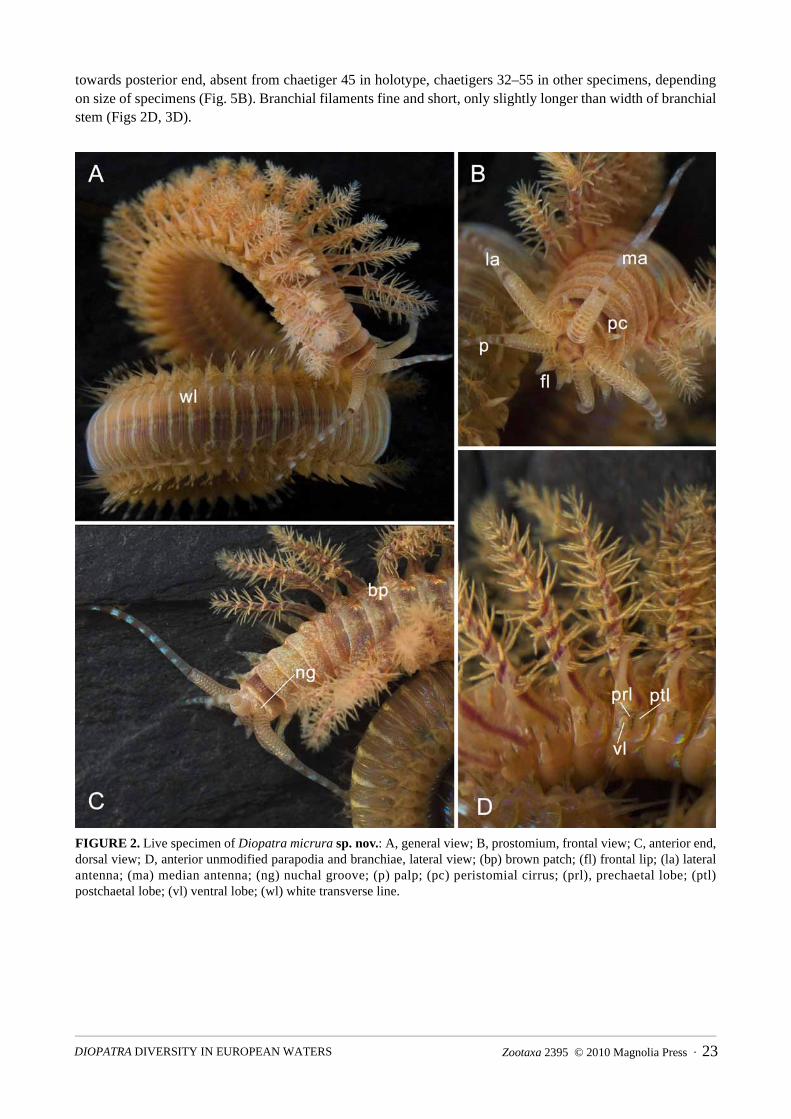
towards posterior end, absent from chaetiger 45 in holotype, chaetigers 32–55 in other specimens, depending on size of specimens (Fig. 5B). Branchial filaments fine and short, only slightly longer than width of branchial stem (Figs 2D, 3D).
FIGURE 2. Live specimen of Diopatra micrura sp. nov.: A, general view; B, prostomium, frontal view; C, anterior end, dorsal view; D, anterior unmodified parapodia and branchiae, lateral view; (bp) brown patch; (fl) frontal lip; (la) lateral antenna; (ma) median antenna; (ng) nuchal groove; (p) palp; (pc) peristomial cirrus; (prl), prechaetal lobe; (ptl) postchaetal lobe; (vl) ventral lobe; (wl) white transverse line.
Zootaxa 2395 © 2010 Magnolia Press · 23DIOPATRA DIVERSITY IN EUROPEAN WATERS

FIGURE 3. Diopatra micrura sp. nov.: A, anterior end, dorsal view; B, maxillary apparatus, dorsal view; C, mandibles, ventral view; D, parapodium of chaetiger 6, anterior view; E, parapodium of chaetiger 1, anterior view; F, pectinate chaeta; G, limbate chaeta; H, pseudocompound hook; I, subacicular hook; (dc) dorsal cirrus; (prl) prechaetal lobe; (ptl) postchaetal lobe; (vl) ventral lobe; (vc) ventral cirrus.
PIRES ET AL.24 · Zootaxa 2395 © 2010 Magnolia Press
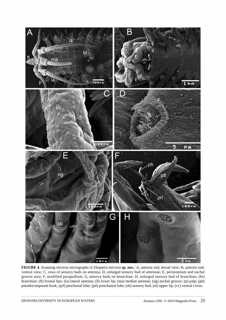
FIGURE 4. Scanning electron micrographs of Diopatra micrura sp. nov.: A, anterior end, dorsal view; B, anterior end, ventral view; C, rows of sensory buds on antenna; D, enlarged sensory bud of antennae; E, peristomium and nuchal groove area; F, modified parapodium; G, sensory buds on branchiae; H, enlarged sensory bud of branchiae; (br) branchiae; (fl) frontal lips; (la) lateral antenna; (ll) lower lip; (ma) median antenna; (ng) nuchal groove; (p) palp; (ph) pseudocompound hook, (prl) prechaetal lobe; (ptl) postchaetal lobe; (sb) sensory bud; (ul) upper lip; (vc) ventral cirrus.
Zootaxa 2395 © 2010 Magnolia Press · 25DIOPATRA DIVERSITY IN EUROPEAN WATERS

FIGURE 5. Diopatra micrura sp. nov.: A, relationship between body width (chaetiger 10, without parapodia) and length of lateral and median antennae. B, relationship between body width (chaetiger 10, without parapodia) and chaetigers of last branchiae and first subacicular hooks.
Modified parapodia with 1–2 slender upper limbate chaetae and 5–6 bidentate pseudocompound hooks (Fig. 3E). Hooks with moderately long pointed hoods (Figs 3H, 6A) and two rows of small spines along their shafts (Figs 3H, 6B). Remaining parapodia with limbate and pectinate chaetae (Figs 3D, 6C, D). Pectinate chaetae flat, with 5–10 long teeth, ending in slender tips (Figs 3F, 6E); limbate chaetae with narrow serrated wings, overall spiny (Figs 3G, 6F). Starting from chaetiger 11 in holotype, chaetigers 8–13 in other specimens, lower limbate chaetae replaced by 2 thick bidentate subacicular hooks with translucent guards (Fig. 3I). Slope of the regression line of start of subacicular hooks very close to nil, indicating a non-significant relationship to size of specimens (Fig. 5B).
Pygidium with two pairs of anal cirri; dorsal pair about as long as the last six chaetigers, ventral pair about as long as the two last chaetigers.
PIRES ET AL.26 · Zootaxa 2395 © 2010 Magnolia Press

Mandibles (Fig. 3C) weakly sclerotised, with slender shafts and strongly calcified cutting plates. Maxillae moderately sclerotised (Fig. 3B). Maxillary formula (based on 9 paratypes): Mx I= 1+1; MxII = 8–10 + 8–11; Mx III = 8–11 + 0; Mx IV = 5–8 + 7–11; Mx V = 1 + 1.
Tube characteristic of genus, cylindrical with soft inner secreted layer and outer layer of debris, fragments of sea grass, algae and shells.
FIGURE 6. Scanning electron micrographs of Diopatra micrura sp. nov.: A, distal ends of pseudocompound hooded hooks; B, spines of pseudocompound hooded hook; C, parapodium of chaetiger 9; D, pectinate and limbate chaetae of parapodium 13; E, pectinate chaeta; F, spines of limbate chaetae; (s) spines; (ls) limbate chaeta; (ps) pectinate chaeta; (ptl) postchaetal lobe; (vl) ventral lobe.
Zootaxa 2395 © 2010 Magnolia Press · 27DIOPATRA DIVERSITY IN EUROPEAN WATERS

TABLE 2. Intraspecific variability of the most important morphological characters of Diopatra micrura sp. nov. (SD = Standard deviation, N = number of individuals observed).
Remarks. The intraspecific variability of the major morphological characters of Diopatra micrura is summarised in Table 2 and the comparison with other European Diopatra species in Table 3.
The multivariate analysis of the morphological data is shown in Figure 7. The groups of individuals belonging to the various species form distinct clusters (Fig. 7, upper graph), represented by well isolated clouds of points in the ordination diagram (Fig. 7, lower graph). The PCA axis 1 and 2 comprehend 91.3% of the total variance. Diopatra neapolitana opposes D. marocensis on the ordination axis 1, with D. micruraoccupying a transition position, on the positive pole of axis 2. Most of the Diopatra specimens from France form a distinct cluster, isolated in the negative pole of axis 2 but closer to D. marocensis. This cluster includes five specimens from Arcachon Bay and a single specimen from Marennes Oléron (Fig. 7). Nevertheless, two specimens from the Arcachon Bay are plotted together with the cluster of Diopatra neapolitana, indicating the coexistence of at the least two species in this Bay. The morphological descriptors most strongly correlated with PCA axis 1 were the number of branchiae whorls (r = - 0.92), the number of rings in the ceratophores (r = - 0.90) and the presence-absence of ventral lobe in the parapodia 5–20 (r = - 0.85). The chaetiger where the subacicular hooks start (r = - 0.72), the width of the 10th chaetiger (r = - 0.70) and the number of teeth in the pectinate chaetae (r = - 0.53), were the variables strongly correlated with PCA axis 2, the latter especially related to the Diopatra sp. individuals from Arcachon Bay (Fig. 7).
Distribution and habitat. Diopatra micrura occurs along the western and southern Portuguese coast. Specimens were collected in Ria de Aveiro, near the mouth, intertidally and on the adjacent shelf area (A), on the shelf off Nazaré (B), in Guia, off the Tagus Estuary (C), and near the Guadiana river mouth (D) (Fig. 1).
The species seems to have a preference for fine sand and shallow waters. In the shelf area off the Tagus estuary, it was found in 22 of the 30 sites comprising the annual monitoring program of this area, carried out since March 1994. Diopatra micrura has been found in every annual sampling campaign, in sites ranging from 40 to 60 metres depth, on fine and very fine sand, with fines content up to 25% of total sediment. In the Aveiro shelf, D. micrura was found in 8 of 22 sites sampled in 2002, always close to 15 metres depth and in fine and very fine sand with less than 5% fines content. In the Nazaré shelf, the species was found at 37 metres depth, on fine sand with 7% fines. On the southern coast, it was found in fine and very fine sand with less than 5% fines, ranging from 4 to 10 metres depth. Finally, in Ria de Aveiro the species was found in the intertidal region, together with D. marocensis and D. neapolitana, in very fine sand with close to 25% fines content.
Character Range Mean SD N
Length, complete preserved specimens (cm) 1.7–7.8 5.4 3.25 3
Number of chaetigers, complete specimens 70–97 86.0 14.18 3
Width of 10th chaetiger without parapodia (mm) 0.6–4.5 1.90 0.78 77
Lateral antennophores (number of rings) 12–15 14.42 0.75 85
Median antennophore (number of rings) 12–15 13,46 0,91 83
Palpophores (number of rings) 12–15 13.36 0.93 87
Lateral antennae (reaching chaetiger) 6–13 9.64 1.71 66
Median antenna (reaching chaetiger) 4–10 8.25 1.43 67
Palps (reaching chaetiger) 2–4 2.26 0.48 72
Peristomial cirrus/peristomium (length ratio) 1.5–2.8 1.84 0.30 63
First branchiae (chaetiger) 4–5 4.54 0.5 89
Last branchiae (chaetiger) 32–55 42.82 8.34 18
Branchial whorls (maximum number) 8–14 10.92 1.79 75
First subacicular hooks (chaetiger) 8–13 11.24 1.08 57
Last prechaetal lobes (chaetiger) 7–11 9.36 1.16 42
Last ventral lobes (chaetiger) 14–20 16.83 1.80 37
Pectinate chaetae (number of teeth) 5–10 7.00 0.98 45
PIRES ET AL.28 · Zootaxa 2395 © 2010 Magnolia Press

Zootaxa 2395 © 2010 Magnolia Press · 29DIOPATRA DIVERSITY IN EUROPEAN WATERS

FIGURE 7. Classification (upper graph) and ordination (lower graph) analysis of specimens of European Diopatraspecies, according to morphological descriptors. Most of the specimens obtained in France (Arcachon Bay and Marennes Oléron) form an isolated cluster, corresponding to a fourth species, but also include individuals belonging to D. neapolitana.
PIRES ET AL.30 · Zootaxa 2395 © 2010 Magnolia Press

FIGURE 8. Phylogenetic analysis of the data set containing the 16S and COI sequences of Diopatra species. Numbers near the nodes indicate the percent bootstrap values. The branch length indicator represents 0.02 substitutions per site.
Genetic analysis. A 702-bp COI fragment and a 525-bp 16S fragment were successfully obtained from 14 individuals of D. micrura. COI and 16S nucleotide sequences from D. micrura sp. nov., were deposited at EMBL database, under the accession numbers: 16S – GQ456163 and COI – GQ456161 and GQ456162.
For the 16S gene, all individuals displayed identical nucleotide sequence but in the case of the COI gene, one individual from Ria de Aveiro presented a base alteration, at position 276, where a nucleotide adenine was replaced by a thymine (ATA to TTA), corresponding to an amino acid alteration (methionine to leucine). All specimens sampled on the shelf off the Tagus Estuary shared the same nucleotide sequence.
The percentage of nucleotides divergence of the 16S and COI genes between D. micrura and D.marocensis was 15% and 17%, respectively (nucleotide substitution). For D. micrura and D. neapolitana, the divergence was 16% for COI and 12% for 16S. For COI, deduced amino acid sequence comparison between the species revealed that D. micrura differs from D. marocensis in six amino acids and from D. neapolitana in two amino acids, for one haplotype, and in three for the other, revealing 2.59% and 1.08% of divergence, respectively. The majority of the differences in nucleotides between D. micrura and those two species occurred on the third position of the codon and therefore corresponded to silent alterations.
Comparing COI and 16S genes of Diopatra sp. from Arcachon Bay with D. neapolitana, D. marocensisand D. micrura, the percentage of nucleotides divergence varied between 17% and 19% in the case of the COI and between 16% and 19%, for 16S gene (nucleotide substitution). The phylogenetic analysis from both genes (Fig. 8) separates the Diopatra species into four clades.
Discussion
The new species, Diopatra micrura, was found on the western and the southern coast of Portugal, in fine or very fine sand with less than 30% of fines content, from the intertidal region up to 50 metres depth. Diopatra micrura coexists with other Diopatra species, namely D. neapolitana and D. marocensis but it is much less common and was never recorded in densities as high as those of the other two species. This study also showed the coexistence between D. neapolitana and Diopatra sp. in the Bay of Arcachon, from intertidal specimens collected in 2009, contrary to the opinion expressed by Berke et al. (2010) who set the Northern limit distribution of D. neapolitana on the Spanish French border.
The presence of D. micrura off the Tagus Estuary, on the western coast of Portugal, can be traced back as far as 1994, where the species has been regularly recorded in monitoring samples taken yearly. In that same coastal area, D. marocensis has shown an increase in density and distributional area over the last five years (Rodrigues et al. 2009), but this has not, so far, excluded D. micrura. This study shows that D. micrura can be
Zootaxa 2395 © 2010 Magnolia Press · 31DIOPATRA DIVERSITY IN EUROPEAN WATERS

distinguished from its European congeners by morphological and genetic characteristics and proposes a key to the European species of Diopatra.
Diopatra micrura is most closely related to D. neapolitana, a species with which it occurs sympatrically in Ria de Aveiro. Both species possess ventral lobes on parapodia 5–20. These lobes have only been observed in D. monroviensis Augener, 1918 from West Africa and in D. aciculata Knox & Cameron, 1971 from Australia. The latter is morphologically very similar to D. neapolitana but shows some genetic isolation as was discussed by Rodrigues et al. (2009). Diopatra micrura can easily be distinguished from the other species by its striped antennostyles and palpostyles, crescentic rather than rounded nuchal grooves, much smaller adult size and more anterior start of the subacicular hooks. Furthermore, there are differences in the construction of the tubes. The characteristic tubes of D. monroviensis have a thick outer layer of sand with even thicker ridges every centimetre or so, while those of the other three species lack the ridges and have also some fragments of seagrass, algae and shells attached.
In the molecular studies of the 16S and COI genes, all individuals of D. micrura displayed an identical nucleotide sequence for the 16S gene but, in the case of the COI gene, one individual from Ria de Aveiro presented a base alteration at position 276. This corresponded to a replacement of adenine by thymine, and an amino acid alteration occurred. However as these amino acids belong to the same chemical group (Stryer 1999) the sequences are translated in proteins of the same family that will have the same function.
The phylogenetic relationship analysis of the European Diopatra species revealed four clades, representing four distinct species of Diopatra, emphasising the validity of D. micrura as a distinct species. The mitochondrial genes, COI and 16S rRNA, are considered conserved genes, but the relative nucleotide divergence that we obtained between the four Diopatra species – averaging 17.5% and 15.4% respectively - isusual among different species of polychaetes. In fact, in the case of 16S rRNA, in the dorvilleid genusOphryotrocha the mean sequence divergence is 12% (Dahlgren et al. 2001); in the syllid genus Autolytus it is about 21% (Nygren & Sundberg 2003) and within the Palola genus the mean divergence is 12.4% (Schulze 2006). In the case of COI, sequence divergence in the terebellid genus was 20% for two Loimia species and 19% for two Amphitrite species (Schulze 2006) and for the Palola genus, the mean divergence is 14.5% (Schulze 2006).
The multivariate analysis of morphological descriptors showed a very good separation between the groups of individuals from different species and allows similar conclusions regarding the validity of the four European species of Diopatra, for which the following key is proposed:
1 Antennae with transverse brown bands; parapodia 5–20 with ventral lobes; 12–16 rings on ceratophores ................ 2- Antennae without transverse brown bands; ventral lobes absent; 6–11 rings on ceratophores ................................... 32 Antennae with 4–8 transverse brown bands (Fig. 2A–C), small species, up to 10 cm long, 4.5 mm wide; subacicular
hooks starting from chaetiger 8–13 ............................................................................................... D. micrura, sp. nov.- Antennae with single median brown band; large species, up to 40 cm long, 9 mm wide; subacicular hooks starting
from chaetiger 19–25 .............................................................................................................................. D. neapolitana3 Dorsum with mid-dorsal brown patch, forming line along anterior part of body; nuchal grooves crescentic; parapo-
dia with single postchaetal lobes; pectinate chaetae with 11–20 teeth .................................................. D. marocensis- Dorsum without pigment; nuchal grooves rounded; parapodia 1–5 with double postchaetal lobes; pectinate chaetae
with 25–32 teeth......................................................................................................... Diopatra sp. from Arcachon Bay
Acknowledgements
Adília Pires benefited from a Ph.D. grant (SFRH/BD/28509/2006) given by the Portuguese FCT (Fundação para a Ciência e Tecnologia). This work also benefited from the sampling campaigns of the project ACOSHELF (POCI/MAR/56441/2004; PPCDT/MAR/56441/2004) and by CESAM own research funds. The Diopatra micrura data in the Guia area were collected under the project “Programa de Monitorização Ambiental do Emissário Submarino e da ETAR da Guia do Sistema Multimunicipal de Saneamento da Costa do Estoril”, funded by SANEST, S.A.. SEM observations and micrographs where done on RNME-Pole of Universidade de Aveiro, project: REDE/1509/RME/2005, FCT. The D. micrura drawings (Fig. 3) were made
PIRES ET AL.32 · Zootaxa 2395 © 2010 Magnolia Press

by Joana Oliveira; Nicolas Lavesque, from Station Marine d’Arcachon, was kind enough to send specimens from France; António Calado and Marta Ferro (UA), helped in the preparation of specimens for SEM; Rui Marques, Aldiro Pereira and our colleagues from the laboratory helped with the sampling.
References
Audouin, J.V. & Milne-Edwards, H. (1833) Classification des Annélides, et description de celles qui habitent les côtes de la France. Annales des Sciences Naturelle, Paris, 28, 187–247.
Augener, H. (1918) Polychaeta. In ‘Beiträge zur Kenntnis der Meeresfauna West-Afrikas’ (ed. W. Michaelsen). 2, 67–625. Friedrichsen & Co.,Hamburg.
Berke, S.K., Mahon, A.R., Lima, F.P., Halanych, K.M., Wethey, D.S. & Woodin, S.A. (2010) Range shifts and species diversity in marine ecosystem engineers: patterns and predictions for European sedimentary habitats. Global Ecology and Biogeography, 19, 223–232.
Budaeva, N. & Fauchald, K. (2008) Diopatra tuberculantennata, a new species of Onuphidae (Polychaeta) from Belize with a key to onuphids from the Caribbean Sea. Zootaxa, 1795, 29–45.
Clarke, K.R. & Gorley, R.N. (2006) PRIMER v6: User Manual/Tutorial. PRIMER-E, Plymouth, 190 pp.Dahlgren, T., Åkesson, B., Schander, K., Halanych, M. & Sundberg, P. (2001) Molecular phylogeny of the model annelid
Ophryotrocha. Biological Bulletin, 201,193–203.Delle Chiaje, S. (1841) Descrizione e Notomia degli Animali Invertebrati della Sicilia Citeriore osservati vivi negli anni
1822–1830. Tomo 3 Molluschi Acefali, Bracciopedi, Cirropedi, Crostacei, Anellosi. Stabilimento Tipografico di C. Batelli e Comp, Naples.
Doeglas, D.J. (1968) Grain-size indices, classification and environment. Sedimentology, 10, 83–100.Folmer, O., Black, M., Hoeh, W., Lutz, R. & Vrijenhoek, R. (1994) DNA primers for amplification of mitochondrial
cytocrome c oxidase subunit I from diverse metazoan invertebrates. Molecular Marine Biology and Biotechnology,3, 294–295.
Halanych, K.M. & Janosik, A.M. (2006) A review of molecular markers used for Annelid phylogenetics. Integrative and Comparative Biology, 46, 533–543.
Knox, GA. & Cameron, D.B. (1971) Port Phillip Survey 1957–1963. Victoria, Australia. part 2. No. 4. Polychaeta. Memoirs of the National Museum Victoria, 32, 21–42.
Montagu, G. (1813) Descriptions of several new or rare animals principally marine, found on the south coast of Devonshire. Transactions of the Linnean Society of London, series 1, 11, 18–21.
Nygren, A. & Sundberg, P. (2003) Phylogeny and evolution of reproductive modes in Autolytinae (Syllidae, Annelida). Molecular Phylogenetics and Evolution, 29, 235–249.
Palumbi, S., Martin, A., Romano, S., McMillan, W. O., Stice, L. & Grabowski, G. (1991) The simple fools guide to PCR, version II. University of Hawaii, Honolulu, 46 pp.
Paxton, H. (1986) Generic revision and relationships of the family Onuphidae (Annelida: Polychaeta). Records of the Australian Museum, 38, 1–74.
Paxton, H. (1998) The Diopatra chiliensis confusion – redescription of D. chiliensis (Polychaeta, Onuphidae) and implicated species. Zoologica Scripta, 27, 31–48.
Paxton, H., Fadlaoui, S.& Lechapt, J.P. (1995) Diopatra marocensis, a new brooding species of Onuphidae (Annelida: Polychaeta). Journal of Marine Biological Association of the U.K., 75, 949–955.
Quintino, V., Rodrigues, A.M., & Gentil, F. (1989) Assessment of macrozoobenthic communities in the lagoon of Óbidos, western coast of Portugal. Scientia Marina, 53, 645–654.
Rodrigues, A.M., Pires, A., Mendo, S. & Quintino, V. ( 2009) Diopatra neapolitana and D. marocensis from the Portuguese coast: morphological and genetic comparison. Estuarine, Coastal and Shelf Science, 85, 609–617.
Schulze, A. (2006) Phylogeny and genetic diversity of Palolo worms (Palola, Eunicidae) from the Tropical North Pacific and the Caribbean. Biological Bulletin, 210, 25–37.
Stryer, L. (1999) Biochemistry, W. H. Freeman, New York, 1064 pp.Tamura, K. Dudley, J., Nei, M. & Kumar, S. (2007). MEGA4: Molecular Evolutionary Genetics Analysis (MEGA)
software version 4.0. Molecular Biology and Evolution, 24, 1596–1599.
Zootaxa 2395 © 2010 Magnolia Press · 33DIOPATRA DIVERSITY IN EUROPEAN WATERS