Embed Size (px)
Citation preview
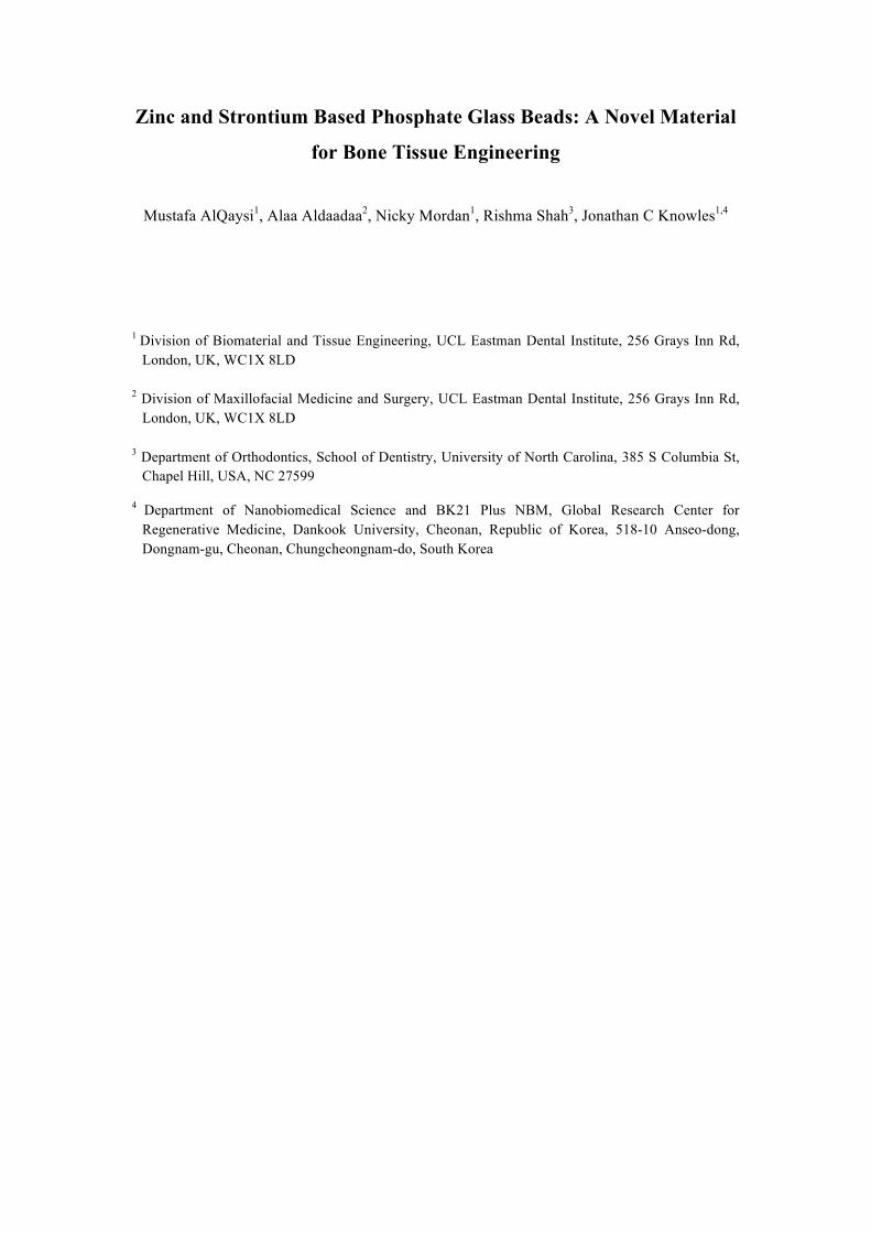
Zinc and Strontium Based Phosphate Glass Beads: A Novel Material
for Bone Tissue Engineering
Mustafa AlQaysi1, Alaa Aldaadaa2, Nicky Mordan1, Rishma Shah3, Jonathan C Knowles1,4
1 Division of Biomaterial and Tissue Engineering, UCL Eastman Dental Institute, 256 Grays Inn Rd,
London, UK, WC1X 8LD 2 Division of Maxillofacial Medicine and Surgery, UCL Eastman Dental Institute, 256 Grays Inn Rd,
London, UK, WC1X 8LD 3 Department of Orthodontics, School of Dentistry, University of North Carolina, 385 S Columbia St,
Chapel Hill, USA, NC 27599
4 Department of Nanobiomedical Science and BK21 Plus NBM, Global Research Center for Regenerative Medicine, Dankook University, Cheonan, Republic of Korea, 518-10 Anseo-dong, Dongnam-gu, Cheonan, Chungcheongnam-do, South Korea
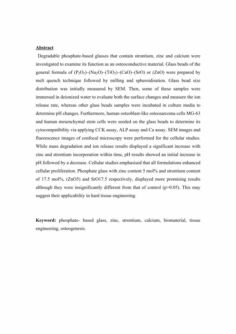
Abstract
Degradable phosphate-based glasses that contain strontium, zinc and calcium were
investigated to examine its function as an osteoconductive material. Glass beads of the
general formula of (P2O5)–(Na2O)–(TiO2)–(CaO)–(SrO) or (ZnO) were prepared by
melt quench technique followed by milling and spheroidisation. Glass bead size
distribution was initially measured by SEM. Then, some of these samples were
immersed in deionized water to evaluate both the surface changes and measure the ion
release rate, whereas other glass beads samples were incubated in culture media to
determine pH changes. Furthermore, human osteoblast-like osteosarcoma cells MG-63
and human mesenchymal stem cells were seeded on the glass beads to determine its
cytocompatibility via applying CCK assay, ALP assay and Ca assay. SEM images and
fluorescence images of confocal microscopy were performed for the cellular studies.
While mass degradation and ion release results displayed a significant increase with
zinc and strontium incorporation within time, pH results showed an initial increase in
pH followed by a decrease. Cellular studies emphasised that all formulations enhanced
cellular proliferation. Phosphate glass with zinc content 5 mol% and strontium content
of 17.5 mol%, (ZnO5) and SrO17.5 respectively, displayed more promising results
although they were insignificantly different from that of control (p>0.05). This may
suggest their applicability in hard tissue engineering.
Keyword: phosphate- based glass, zinc, strontium, calcium, biomaterial, tissue
engineering, osteogenesis.

Introduction
Nowadays the high rate of bone fractures, as a result of pathological bone diseases
and trauma, necessitates an effort to create a new generation of biomaterials for bone
tissue regeneration. In fact, there are various treatment options for bone defects, which
vary from conservative therapy to radical orthopedic surgeries. The latter may require
the use of bone grafts, mainly autogenous graft. This may associate with much
morbidity such as blood loss, pain and sepsis. This pushed to develop other options of
synthetic grafts to reduce such side effects (1). Phosphate-based glass is one of these
synthetic options that studied as osteoconductive biomaterials for bone treatment
application. This may be related to its degradable nature and ion releasing that may
have a positive role not only on bone growth but also on bone cell responses (2).
Various studies were carried out on different phosphate glass systems starting from the
basic tertiary glass and ending in the complex glass structure. Studies on the former
were limited only to the (P2O-CaO-Na2O) system and most of these were basically on
glass discs, and these studies showed that calcium oxide incorporation can decrease
glass degradation rate (3, 4). The more advanced glass systems (P2O5–SiO2–CaO–
MgO–Na2O–K2O) were investigated also where phosphate glass was produced in
highly porous form scaffold that was resembling the cancellous bone. These scaffolds
exhibited acceptable metabolic activity for hMSCs (5).Other attempts used different
technique in the production of multi metal oxides (P2O-CaO-Na2O-K2O) phosphate
glass scaffold. These produced scaffolds believed to have both the biodegradation and
bioactivity properties(6). However, all these varieties in glass composition may lead to
different glass degradation rates. Hence further attempts were carried out to produce a
glass composition with the optimum degradation rate. Multiple important studies were
performed for achieving this purpose by adding titanium dioxide to glass composition;
this resulted in lower degradation rates and better cytocompatibility (7, 8). The addition
of various proportions of both zinc and strontium to the tertiary phosphate glass was
also studied. It was found in previous study that compensating (0-10 mol %) of CaO
with ZnO did not have any significant effect on mass loss in comparison with >10 mol%
replacement of CaO of which it had a higher mass loss (9). As a result of this mass loss,
zinc release was enhanced giving rise to a decrease in biocompatibility of these
particular glasses (10). Other experiments aimed to determine the effect of inclusion of
strontium in phosphate glasses taking into consideration that strontium has a well-
known role in bone tissue growth and bone density enhancement (11). One of these

experiments investigated the physical and biological properties of different strontium
silicate glasses (SiO2)46.5–(P2O5)1–(Na2O)26.4–(CaO)(23.1-x) –(SrO)x (x= 0, 2.3, 11.5 or
23.1) (mol%) and revealed that increasing strontium may improve cell proliferation and
enhance the anabolic activity of alkaline phosphatase (ALP) in Saos-2 osteoblast like
cells. This may be due to the fact that strontium has a dual action of preventing
resorption of calcium phosphate by the osteoclasts cells and decreasing phosphatase
protein activity (12). These results were further confirmed by the addition of strontium
to borate–based glasses which enhanced both glass degradation and formation of an
apatite layer encouraging Saos-2 osteoblast like cells to adhere to the glass surface (13).
Another trial investigated the addition of strontium to phosphate based glass discs in
different percentages (P2O5)–(Na2O)–(CaO)–(SrO) and found that strontium additives
can lead to enhancement in degradation more than that of strontium free glass discs. It
can also give a slightly positive outcome in regards to cell culture (14). Later on, other
attempts were performed to investigate quaternary strontium phosphate glasses which
involved the development of four different compositions (P2O)50-(TiO2)3-(Na2O)17-
(CaO)(30-x)-(SrO)x (x=0,1,3,5) and showed that despite the rise of degradation rate as
strontium oxide increased, cell proliferation of MG63 cells was not affected and
showed no significant differences compared to SrO0 and SrO5 (15). Two recent studies
examined four different compositions of both strontium and zinc based phosphate
glasses and revealed that SrO17.5 and SrO35 were suitable for MG63 cells growth,
whereas ZnO15 showed a degree of cytotoxicity in comparison to both ZnO5 and
ZnO10 as both of them exhibited acceptable results of biocompatibility and metabolic
activity (16, 17). Although these have shown some cellular promising results of adding
zinc and strontium to phosphate glass, they were only performed on glass discs, as a
primary step toward creating scaffolds, as they did not have the full requirements of a
scaffold. The main aim of this study was to develop glass in a more useful format i.e.
glass beads, of previous compositions that had clinical potential in addition to further
assessment of these compositions aiming to apply phosphate glasses in clinical related
bone repair treatment.

Materials and methods
Glass preparation
Four different compositions of phosphate glass beads were developed to check their
cellular biocompatibility and the ability of cells to both penetrate through and adhere
to them. This was performed using the following precursors: phosphorus pentoxide
(P2O5 98%, VWR, Lutterworth, UK), sodium dihydrogen phosphate (NaH2PO4, 99%,
VWR), titanium dioxide (TiO2, 99%, VWR), calcium carbonate (CaCO3, 98.5%,
VWR), strontium carbonate (SrCO3, 99.9% Sigma-Aldrich Gillingham, UK) and zinc
oxide (ZnO, 99.95%, Sigma-Aldrich Gillingham, UK). The glass compositions were
spitted into zinc and strontium groups. While zinc group was 50 P2O5 - 10Na2O - 5TiO2
– (35-x) CaO – x ZnO (mol %) where x (zinc oxide) was 5% mol and 10% mol,
strontium was 50 P2O5 - 10Na2O - 5TiO2 – (35-x) CaO – x where x (strontium oxide)
was 17.5% mol and 35% mol. Electronic balance (Sartorius) was used for weighing
precursors powder (Table 1) followed by blending using Stomacher 400
blender/Seward. Following mixing of the precursors, the mixture was then placed into
a 200 ml volume Pt/10%Rh crucible type 71040 (Johnson Matthey, Royston, UK)
which was subsequently placed in a furnace (Carbolite) at 1350oC for four hours, then
the melted glass was quenched on metal plate. The quenched glass was broken into
small pieces then further milled by (MM 301 Mixer Mill, Retsch GmbH and Hope,
UK). Afterwards, glass beads were produced by flame spheriodisation apparatus
following the method performed previously by Lakhkar et al (8) and were collected in
4 different glass containers which were primarily visualised under light microscope to
confirm their production.
phosphate glass composition
Glass name and composition Amount (grams)
P2O5 Na2O TiO2 CaO Sr ZnO SrO17.5% P50Na10Ti5Ca17.5Sr17.5 56.8 24 4 17.5 25.8 0
SrO35% P50Na10Ti5Sr35 56.8 24 4 0 51.6 0
ZnO10% P50Na10Ti5Ca25Zn10 56.8 24 4 25 0 8.14
ZnO15% P50Na10Ti5Ca20Zn15 56.8 24 4 20 0 12.21
Table 1: phosphate glass beads composition

XRD.
The next step after glass preparation was evaluation of glass structure crystallinity by
using x-ray diffractometer XRD (D8 Advance Diffractometer,Bruker, Coventry, UK)
to ensure that our sample were not crystalised. Specimens of all glass powder were
positioned in a flat plate geometry, and Ni-filtered Cu Ka radiation was used. Data were
collected using a Lynx Eye detector with a step size of 0.02o over an angular range of
2θ=10–100o and a count time of 12 seconds.
Glass Beads distribution
About 50 mg was taken from each of the synthesized glass beads; this was distributed
on sticky dark tabs which were placed on an SEM stub for particle size measurement.
The powder was slightly blown by compressed air to ensure its retention on the dark
tabs. Following this, coating with gold and visualization under scanning electron
microscope (SEM) (Philips XL30 FEGSEM) was performed. Five SEM images were
chosen from various sites from stub (center, left, right, up and down), the diameter size
in microns for each bead was determined using Saturn software to measure the
frequency distribution of the produced glass beads .
Glass degradation
Glass degradation study was performed by incubating 200 mg of each composition
of glass beads in 2 ml of ultrapure 18M W/cm2 water at 37oC for (day 1, 4, 7, 14).
About 50 mg was taken out at each time interval to be visualized under SEM to assess
the degradation and the surface changes.
Ion release
Ion release study aimed to calculate the concentration of different ions as this might
be helpful in understanding the link between ions concentration and the cellular
response. This was done by using triplicates of 100 mg of each glass beads composition;
these triplicates were immersed in 1ml of ultrapure 18M W/cm2water and incubated at
37oC for four time points (1, 4, 7 and 14 days). At each time point, the de ionized water
was stored for ion release study and replaced with fresh for the next time point. Then
ion analysis was carried out for the anions (PO43-, P2O7
4-, P3O94-, P3O10
5-) and cations
(Na+, Ca2+, Zn2+,Sr2+) using the ion chromatography systems (ICS1000, ICS 2500,
Dionex, Thermo Scientific, Hemel Hempstead, UK). For the cation measurements all
the samples were filtered prior to measurement to remove the anions (OnGuard IIa,
Dionex). The ions concentration was calculated at each time point and accumulated to
the previous time point.
pH study
pH measurement was determined by using triplicates of 200 mg glass beads in 2ml
culture media in 24 well plates for (day 1, 4, 7, 14). The glass beads were incubated at
37oC for the whole time period and changed every two days to mimic the cell culture
study environment. At each point, the culture media pH was measured with an Orion
star A111 PH meter (Thermo scientific, Hemel Hempstead, UK) and then replaced by
fresh media for the next day point. Culture media alone was used as control for the
whole time course.
Cell Culture Studies
Both human osteoblast-like osteosarcoma cell line (MG63, European Collection of
Cell Cultures, Porton Down, UK) and human mesenchymal stem cells (hMSCs)
(passage 3) were used for cell studies in which they were incubated in standard
conditions (37°C, 95% air, 5% CO2, 95 % relative humidity) in Dulbecco’s modified
Eagle medium (DMEM, Gibco, Life Technologies, Paisley, UK). MG63 cells were
selected as they have been commonly used to establish the preliminary aspects of
biocompatibility for a wide range of phosphate glasses, whereas hMSCs are considered
the gold standard for such studies. By the time of reaching 80% confluence, cells were
trypisinzed to allow seeding onto glass beads. The glass beads were sterilised by
immersion in ethanol then dry heat at 180OC for 1 hour.
The seeding procedure was similar for both cell types which was performed firstly by
coating 100 mg of glass beads with bovine fibronectin in PBS (10 ug .mL-1) for one
hour to aid initial cell attachment and then these glass beads were placed in a 24
ultralow attachment well plate (Corning, USA) in which the plate bottom surface was
covered with beads completely. Later on, these glass beads were incubated in culture
media overnight at 37oC. The next day, the culture media was taken out and cell seeding
was performed on the glass beads according to the preferred seeding density in which

cells were left for 30 minutes in an incubator to allow cell attachment. After that the
glass beads were transferred into 6.5 mm inserts in 24 well plates.
Two types of culture media were used, (1) Dulbecco’s modified Eagle medium (Gibco,
Life Technologies, Paisley, UK) supplemented with 10% fetal bovine serum (Gibco)
and 1% penicillin/streptomycin (PAA Laboratories, GE Healthcare, Chalfont St. Giles,
UK) that was used for the MG63 cells study, while (2) osteogenic medium (OM) for
hMSCs studies and was prepared as the previous work (18) by using low glucose
Dulbecco’s modified eagle medium (DMEM) , supplemented with fetal bovine serum
, penicillin/streptomycin 1% , dexamethasone (0.1 µM), ascorbic acid 2-phosphate (0.2
mM), and glycerol 2-phosphate (10 mM; last three chemicals procured fromSigma–
Aldrich, UK). Both of these culture media were replaced by half every 3 days.
Commercial silica based glass microspheres (Polyscience Inc., USA) were used as a
control for all the cell culture studies.
CCK assay
Following the seeding of MG63 cells at a density of 3000 cells per trans-well insert
in 24 well plates, the 24 well tissue culture test plate was left in a 37°C/ 5% CO2
incubator for 1, 4 and 7 days. In parallel, cells were seeded in a second test plate at
different densities for calibration. At each time point, CCK8 (Cell Counting Kit 8,
Sigma-Aldrich) was added to each well in a 10% proportion of the culture media then
incubated for 3 hours. Afterwards, fluorescence measurement was performed for each
well plate in triplicate by using a plate reader (Infinite® M200, Tecan) at 450 nm
wavelengths.
Alkaline phosphatase Assay
hMSCs were used for this assay, which were seeded at a density of 25000 cells per
trans-well insert and incubated at 37°C/ 5% CO2 for 7 and 14 days. Subsequent dilutions
of ALP standard reagent were performed for calibration. In all time points, the culture
media was removed following the company protocol (Alkaline Phosphatase Assay,
SensoLyte® pNPP) followed by cells washing with 1X assay buffer and cells
permeabilising by Triton X-100. Afterwards, the glass beads were pipetted vigorously
for 1 minute to aid in cell permeabilisation, then cells were lysed further by two cycles
of freeze-thaw cycles (- 20oC for 20 min, followed by 37oC for 12 min) followed by

subsequent centrifugation at 4°C for 10 minutes at 2500 rpm Then, 50 𝜇𝑙 of the
supernatant was added to 50 𝜇𝑙 of pNPP in 96 well plate and kept for 4 hours in a
37°C/5% CO2 incubator. Finally, a stopping reagent was added into each well before
taking triplicate fluorescence measurements of each transwell using a plate reader
(Infinite® M200, Tecan) at 405 nm wavelengths.
Ca assay
Mesenchymal stem cells were used again for the Ca assay at a cell density of 25000
cell/ Trans-well and were incubated with the supplement of (OM) for 14 and 21 days.
At each time point, the culture media was taken out and the glass beads were washed
with phosphate buffered saline (PBS) three times then the cells were lysed by 1M HCl
and placed on a shaker for ≃ 40 minutes. After that, triplicates of 5ul of each trans well
aliquot was transferred to 96 well plate and about 200ul of prepared Ca working agent
was added to each triplicate then fluorescence measurement at wavelength 612 nm
(Infinite® M200, Tecan) was performed. Various gradual dilutions of Ca standard
(100uL- 0uL) were made for calibration following the protocol (QuantichromTM, Calcium
Assay Kit (DICA-500), Bioassay System). Other triplicates of 100 mg of each
composition were incubated alone without seeding cells to deduce the effect of glass
composition on the final results.
Cell imaging
For MG63 SEM imaging was performed for each time point by removing the culture
media then fixing cells initially in 3% glutaraldehyde followed by dehydration through
graded ethanol (50, 70, 90, and 100 %) then drying by hexamethyldisilazane (Aldrich,
UK). While for hMSCs, imaging was done by fluorescence microscopy using
phalloidin for cytoskeletal staining and propidium iodide for nucleus staining. The first
step of this procedure was cell fixation in 3.7% formaldehyde followed by cell
permeabilization by using 0.5% triton X-100 then finally cellular staining by both
phalloidin (Alexa Fluor® 488 Phalloidin, Sigma-Aldrich Gillingham, UK) and 4ug/
mL propidium iodide ( Propidium iodide, Sigma-Aldrich Gillingham, UK).
Statistical analysis

Cell data for both cell counting and metabolic activity measurements were
statistically assessed by Kruskal–Wallis where p<0.05 has been used as a significance
degree estimation.
Results
XRD
XRD spectra of the glasses showed a broad peak at 2θ values of ≃ 20–40°. This
emphasized the all the prepared glass samples were amorphous and were free
crystallinity as shown in figure1.
Figure1: XRD spectrum for glass
Glass beads distribution
The assessment of the different SEM images of the beads showed that the majority of
glass beads produced were between 63um-106um; therefore, these sizes were used for
the subsequent studies. Figure 2 demonstrates this as well as the picture of glass beads
under SEM .
050100150200250300350400
0
500
1000
1500
2000
2500
3000
10 30 50 70 90
intensity
(a.u.)
2θ(°)
ZnO5
ZnO10
SrO35SrO17.5

A B
Figure (2): A-SEM picture of glass beads B- frequency distribution of glass beads
Mass loss
As shown in (figure 3), the images taken to visualise the glass bead surface changes
showed that the amount of surface changes increased with time. These changes were at
their highest range on day 14 after immersion in deionized water in comparison with
images from the previous days beads immersion samples. Regarding day 1, there were
no clear surface changes in comparison with day 0. On day 7, there was a clear
difference and prominent wear on the glass beads surface in the zinc oxide groups
which revealed groups of pits formed while strontium oxide glass showed lower levels
of changes than the zinc oxide groups with the least change seen with SrO17.5.
However, on day 14 the glass degradation increased clearly with an increase in pits and
the presence of small cracks in the zinc oxide glass with the presence of more surface
wear in SrO35 and small pits in SrO17.5. These results showed that the ZnO10 glass
was the most vulnerable to surface changes followed by ZnO5 then SrO35 and finally
SrO17.5, which revealed the lowest level of surface changes.
0
5
10
15
20
25
30
0 10 20 30 40 50 60 70 80 90 100 110 120 130fr
eque
ncy(
per
cent
age%
)
Beads diameter (microns)
ZnO5
ZnO10
SrO17.5
SrO35

ZnO5 ZnO10 SrO17.5 SrO35
Day 0
Day 1
Day 7
Day14
Figure 3: SEM pictures for glass beads after incubation in de ionized water
showing the least degradation was with ZnO10 in day 14.
Ion release
Anion and cation release data revealed a gradual increase in the different ions released
for all the compositions over the whole time frame. While the trend of ion release for
the anions (P2O74-, P3O9
4-, P3O105-) was (ZnO10> ZnO5> SrO17.5> SrO35), it was for
PO43- as (ZnO5> ZnO 10> SrO17.5> SrO35). For all anions, SrO35 glass release of
anions was significantly lower compared to the other compositions. Over the whole
study, the anion release for both zinc containing glass compositions were close to each
other and were significantly higher in comparison to the ion release for the strontium
containing glasses (figure4).

Similarly, the cation release showed the same trend as the anions in which the zinc
based glass showed higher release of Na+ and Ca2+ compared to the strontium based
glasses. ZnO5 showed the maximum release of both Na+ and Ca2+; 106-ppm and 92-
ppm respectively, which was double the value of SrO17.5. The latter was followed by
SrO35 with the lowest level of release with less than 10 ppm for Na+ and lower than 5
ppm for Ca2+(figure 5 a&b).
Concerning zinc ion release, ZnO10 exhibited more Zn+2 ion release than ZnO5.
Despite Zn+ ions release was quite similar on day 1 at about 35 ppm, ZnO10 glass
clearly released more Zn+ ions over the time points after day 1 to stand at around 385
ppm after two weeks that was about 100 ppm/mg more than that of ZnO5 which ended
at 280 ppm on the same day point (figure 5 c).
Sr2+ ion release was around 3 ppm for both SrO17.5 and SrO35 on day 1. As the study
continued for the next time points, SrO35 tended to release more Sr 2+ ion than that of
SrO17.5 A linear trend was seen, to end around 33 ppm on day 14 which was
significantly higher than that of SrO17.5 at around 25 ppm (figure 5 d).
0
1000
2000
3000
4000
5000
6000
0 1 2 3 4 5 6 7 8 9 101112131415
Con
cent
ratio
n (P
Pm)
Time (days )
PO43- ZnO 5
ZnO 10SrO 17.5SrO 35
0100200300400500600700800900
1000
0 1 2 3 4 5 6 7 8 9 10 11 12 13 14 15
Con
cent
ratio
n (p
pm)
Time (days)
P2O74- ZnO 5
ZnO10SrO 17.5SrO 35
0200400600800
1000120014001600
0 1 2 3 4 5 6 7 8 9 10 11 12 13 14 15
conc
entra
tion
(ppm
)
Time (days)
P3O94- ZnO 5
ZnO10SrO 17.5SrO 35
0
200
400
600
800
1000
1200
0 1 2 3 4 5 6 7 8 9 10 11 12 13 14 15
conc
entra
tion
(PPm
)
Time (days)
P3O105-
ZnO 5 ZnO 10SrO 17.5SrO 35

Figure4: Anion release showing higher release of phosphates ions (PO43-, P2O7
4- ,
P3O94-, P3O10
5-) by zinc based glass in comparison to strontium glass.
Figure 5: Cation release (a) Na+ release, (b) Ca2+ release, (c) Zn+ release (d) Sr 2+
release
pH Measurement
pH data revealed a significant variation in pH level over the time course of the study.
pH readings were around 8.7 at the beginning of the study, however, it rose on day 1 to
reach around 9.2-9.3 for all groups. This rise was sustained at the same level until day
4 of which a slight decline was noticed for all the other time points in comparison to
the control, which remained stable for the whole experiment period except for the
ZnO10. Surprisingly, ZnO10 decreased significantly after day 4 and showed the lowest
pH level for the whole period until the end of the study finishing just slightly higher
than 8.2. These results were demonstrated in figure 6.
0
20
40
60
80
100
120
0 1 2 3 4 5 6 7 8 9 10 11 12 13 14 15
Con
cent
ratio
n (P
Pm)
Time (days)
(a)Na+ ZnO 5 ZnO 10SrO 17.5SrO 35
0
20
40
60
80
100
120
0 1 2 3 4 5 6 7 8 9 10 11 12 13 14 15
Con
cent
ratio
n (p
pm)
Time(days)
(b)Ca 2+ ZnO 5 ZnO 10SrO 17.5SrO 35
050
100150200250300350400450
0 1 2 3 4 5 6 7 8 9 10 11 12 13 14 15
Con
cent
ratio
n (p
pm)
Time (days)
(c)Zn2+ ZnO 5
ZnO 10
0
5
10
15
20
25
30
35
40
0 1 2 3 4 5 6 7 8 9 10 11 12 13 14 15
Con
cent
ratio
n (p
pm)
Time (days)
(d)Sr2+ SrO 17.5
SrO 35

Figure 6: pH level showing that ZnO10 was with the least pH level in comparison to
the control which remained steady.
Cell Assays
1- CCK Measurement
CCK data revealed that the number of cells increased gradually over time. Initially
cells were seeded as 3000 cells per trans-well. This number had reached 13000 cells on
day 1 for ZnO5 and SrO17.5. The former was with insignificantly higher than control
and ahead of both ZnO10 and SrO35 at around 7000 cells. On day 4, cells number in
both ZnO5 and SrO17.5 were around 34000 cells. This was significantly lower in
comparison to the control that was about 38000 cells. At the last time point, cellular
growth continued to increase and ended at around 40000 cells for ZnO5 and SrO17.5,
which was about 5000 less than that of the control.
In general, SrO35 and ZnO10 showed the lowest cell numbers for all three times points.
Whereas SrO17.5 and ZnO5 displayed more promising results in the same time frame.
Figure 7 shows the biocompatibility of different compositions in compare to control in
different time points.
8.2
8.4
8.6
8.8
9
9.2
9.4
0 1 2 3 4 5 6 7 8 9 10 11 12 13 14 15
pH
Time(days)
co
ZnO5
ZnO10
SrO17.5
SrO35

Figure 7: CCK assay for MG63 cells showing that both ZnO5 and SrO17.5 have good
biocompatibility in comparison with the control. ZnO5 and SrO35 displayed less
2-Alkaline phosphatase Measurement:
Figure 8 summarises the alkaline phosphatase results after one and two weeks. At day
7, all compositions showed an enzyme concentration that was similar to the control
(6.5ug/ trans-well) except ZnO5 and SrO35 which were statistically slightly higher than
control. On day 14, there was an increase in enzyme levels for all prepared glass types
with the highest levels being shown for glasses ZnO5 and SrO17.5 (17.5ug/ trans-well).
This was slightly less than the control (20ug/ trans-well). SrO35 displayed the lowest
enzyme levels (13ug/ trans-well).
0
10000
20000
30000
40000
50000
60000
day1 day4 day7
num
ber o
f cel
ls/tr
answ
ell
Time(days)
CCK essay
control
ZnO 5
ZnO 10
SrO 17.5
SrO 35
* *
* *
*

Figure 8: Alkaline phosphatase for hMSCs measured by ug/ trans-well, displaying an acceptable enzyme activity in ZnO5 and SrO17.5 in relation to the control.
3-Mineralization Measurement (Ca assay)
Figure 9 displays the Ca assay for day 14 and 21. At the first time point all glass
compositions seemed to stimulate cells to produce calcium at levels higher than that of
control. ZnO5 and SrO17.5 values were higher than that of ZnO10 and SrO35 while
the control showed the lowest concentration (2ug/ trans-well). Although the control
data showed a 4-fold increase on day 21 in compare to day 14, it showed the lowest
concentration compared to the zinc and strontium-based glasses. ZnO5 and Sr17.5
results were highly significant (28 ug/ transwell) than the other compositions and the
control.
0
5
10
15
20
25
day7 day14
Alk
alin
e ph
osph
atas
e con
c. (u
g/tra
nsw
ell)
Time (days)
control
ZnO 5
ZnO 10
SrO 17.5
SrO 35
* *
* *

Figure 9: Ca concentration for hMSCs measured by ug/ transwell for day 14 and 21, revealing that ZnO5 and SrO17.5 have the highest calcium concentration.
4- Cell imaging
SEM pictures of MG63 showed the ability of the cells to attach to the glass beads. Figure
10a displays the attachment of cells on strontium based glass day on day 1, while figure 10 b
shows the ability of MG63 to proliferate trying to make a continuous layer of cells among the
beads on day 7.
Figure 10: SEM pictures of MG63 on SrO17.5 strontium glass beads. a- day1, b- day7
Similarly, confocal images for hMSCs showed that these cells could attach to the beads and
even encapsulate these beads and take on their spherical morphology as shown in figure 11.
0
5
10
15
20
25
30
35
day 14 day21
Ca
conc
entra
tion
ug/tr
answ
ell
Time (days)
Control
ZnO5
ZnO10
SrO17.5
SrO35
* *
*
* *
* *
*
(a) (b)

Figure 11: Confocal images of hMSCs showing the cellular attachment on beads

Discussion :
The aim of this study was to determine the role of zinc and strontium in phosphate-
based glasses for hard tissue (bone) engineering application. According to previous
studies, calcium, zinc and strontium were proven to play a pivotal role in bone growth
and development (19-22). Various earlier studies were carried out within our
department assessing different properties of zinc and strontium phosphate glass. These
studies were performed by preparing different phosphate glass compositions that are
(P2O5-Na2O-CaO-TiO2 -(ZnO or SrO) based and showed that ZnO5, ZnO10, SrO17.5
and SrO35 had an acceptable physical, chemical and biological properties (16). This
was the main reason of this present study to focus on such glass compositions.
Furthermore, the other aim was to develop the glass in a form that may be more
accessible for cells to penetrate and colonise inside it rather than the use of glass discs
as performed in the previous studies. Hence, phosphate glasses in this study were
produced in the form of beads by using a spheriodisation method (8).
Qualitative assessment of bead degradation by imaging was adopted due to the
difficulty and unreliability of making quantitative mass loss measurements. The images
obtained gave us a general idea about the mechanical surface changes of glass beads
after immersion in deionized water. These images showed that there were no major
changes after day 1, however, as the incubation persisted for 1 week the erosion effect
started to appear. This was more noticeable in both compositions of zinc based glasses
and was in the form of pores and faint cracks which were more pronounced in ZnO10
phosphate glass in comparison to the less changed strontium compositions. Zinc
phosphate glass was affected further on day 14 as more deep cracks started to appear
with the formation of more pits. The strontium glasses, in turns, started to show few
pits with few surface changes on day 14.
In general, ZnO10 tended to be the most susceptible to degradation followed by ZnO5
and SrO35 followed finally by SrO17.5. These results were concurrent with previous
findings of mass loss trend. This can be explained by knowing that both zinc and
strontium attach to phosphate glass network via ionic bonds with oxygen. The Zn-O
bond has bond dissociation energy (284 Kj. mol-1) that is lower than that of the Sr-O
bond (454 Kj. mol-1). This makes the zinc phosphate glass more vulnerable to
hydrolysis as a result of its weaker bond strength. Moreover, our previous differential

thermal analysis findings confirmed the current results via the thermal variables (Tg,
Tc, Tm) which has followed the trend of (ZnO10 < ZnO5 < SrO35 < Sr 17.5) (16, 23).
The anion and cation release results for zinc phosphate glasses were concurrent with
the mass degradation data in which higher levels of phosphate ions occurred with the
more surface degraded glass. This can be explained by identifying the bond dissociation
energies for both CaO and ZnO; 383 Kj. mol-1 and 284 Kj. mol-1 respectively.
Consequently, when ZnO replace CaO, there are higher numbers of weaker bonds, and
hence more degradation and more ions are released. These results followed the pattern
of previous data (17). Conversely, anion and cation release from the strontium
containing samples did not follow the mass degradation pattern as the more degradable
SrO 35 phosphate glass released fewer ions than that of SrO 17.5. These surprising
results were actually similar to a previous study (16), the only exception was the Sr2+
ion release which was higher with SrO35 as it had double the amount of strontium than
that of SrO17.5. Actually, this may give us a justification to interpret such unexpected
results. Although SrO35 had more surface loss and higher degradation rate than
SrO17.5, the majority of the released ions were Sr2+ which has a molecular weight of
87 more than other ions such as Na+, Ca 2+ and P5- that have molecular weight of 22, 40
and 30 respectively.
Dulbecco Modified Eagles Medium (DMEM) with pH 8.4± 0.1 was used as an
immersion liquid for glass beads in pH studies to mimic the same condition for cell
culture studies. Data showed an irregular trend in which there was a rise on the first day
then a period of stability followed by a slow decline of pH level. The control and all
the compositions showed an initial increase in pH after one day which remained stable
until day 4 as a result of gas absorption effects (24). This rise in pH was higher for the
control group in comparison to the glass beads group. On day 7, however, there was a
gradual continuous decline in pH until day 14 for all glass beads. The control, however,
remained at the same level for the rest of the study period. Surprisingly, ZnO10 showed
the lowest pH change followed by SrO35, which was slightly less than the other two
groups. It appeared that the pH level was inversely related to the ion release, which was
higher with ZnO10. This may be as a result of the increase of phosphate ions release
that might form phosphoric acid in the solution and hence increase the culture media
acidity.

In general, cell culture studies displayed quite similar trends. In other words, the CCK
results displayed that there was less metabolic activity in ZnO10 glass beads group than
that in ZnO5 and SrO17.5 which were close to control. Furthermore, alkaline
phosphatase for hMSCs acted similarly after 1 and 2 weeks as ZnO5 and SrO17.5
exhibited insignificant difference of enzyme levels from control. On the other hand, Ca
assay demonstrated higher mineralisation rates in ZnO5 and SrO17.5 that were greater
than remaining groups of ZnO10 and SrO35, control group had the lowest
mineralization level.
Overall, cell studies gave an initial view about the role of zinc and strontium
concentrations in phosphate glass and showed that ZnO5 and SrO17.5 phosphate glass
beads have the most significantly positive effect on cells in compare to ZnO10 and
SrO35. The interpretation of these result depends mainly on the relation between the
released ions and their biological effect as shown by other studies (2). The current study
measurements showed that the maximum released concentration of calcium and sodium
among all glass compositions was about 95 ppm and 106 ppm respectively, which is
below the cytotoxic concentration suggested in previous studies (i.e. for Ca2+ =400
ppm, Na+= 220 ppm) (25, 26). Consequently, the release of Ca2+ and Na+ ions from
these glass systems should not have any harmful impact on cell function. Although
phosphate ions can play an important role in cell proliferation and metabolism, it was
difficult to investigate their actual effect due to the presence of high phosphate contents
ions in the medium thus they were not quantified in the medium. Regarding the
strontium-containing glass beads, it was found that its Sr2+ release was about 30 ppm
and 25ppm for SrO35 and SrO17.5 respectively. In a previous study, the optimum
concentration of SrCl2 to induce calcified matrix deposition was 5ug/ml, however, data
showed that concentration of 10 to 20 ug/ml could also stimulate ALP and matrix
deposition. There is a decline in the positive effect of Sr2+ions as the concentration
increase from 10 to 20 ug/ml. Bearing this in mind, the actual Sr2+ions concentration in
the cell culture study is around half that of the data shown in the ion release results
because of the frequent culture media change, so the real concentration of strontium in
culture media is probably about 15 ppm and 12.5 ppm for SrO35 and SrO17.5
respectively. This was within an acceptable range and following the same pattern of
cellular activity as discussed previously (27). Whereas for the zinc ion release, the
current results confirmed previous findings that showed substitution of calcium with
zinc by 10 mol% can result in unfavorable and cytotoxic effects. Hence, adding more
than 10 mol% ZnO may cause catastrophic effects as it can increase the release of
lactate dehydrogenase and induce oxidative stress (10, 28).
Conclusion
The current study showed that glass beads were successfully produced in different
compositions. It revealed also that ZnO5 and SrO17.5 phosphate glass beads exhibited
better results regarding cellular studies that were significantly better than ZnO10 and
SrO35 glass beads concluding that ZnO5 and SrO17.5 are more suitable for bone tissue
engineering. These results could be further studied in future to assess their impact on
bone tissue engineering aspects by performing other cell culture studies and use these
types of phosphate glass as scaffold materials for bone repair.

Refrences
1. DamienCJ,ParsonsJR.Bonegraftandbonegraftsubstitutes:Areviewofcurrenttechnologyandapplications.JournalofAppliedBiomaterials.1991;2(3):187-208.2. Lakhkar NJ, Lee I-H, Kim H-W, Salih V, Wall IB, Knowles JC. Bone formationcontrolled by biologically relevant inorganic ions: Role and controlled delivery fromphosphate-basedglasses.AdvancedDrugDeliveryReviews.2013;65(4):405-20.3. UoM,MizunoM,KubokiY,MakishimaA,WatariF.PropertiesandcytotoxicityofwatersolubleNa2O–CaO–P2O5glasses.Biomaterials.1998;19(24):2277-84.4. AhmedI,LewisM,OlsenI,KnowlesJC.Phosphateglassesfortissueengineering:Part 1. Processing and characterisation of a ternary-basedP(2)O(5)-CaO-Na(2)O glasssystem.Biomaterials.2004;25(3):491-9.5. Vitale-BrovaroneC,CiapettiG,LeonardiE,BaldiniN,BretcanuO,VernéE,etal.Resorbable Glass–Ceramic Phosphate-based Scaffolds for Bone Tissue Engineering:Synthesis, Properties, and In vitro Effects onHumanMarrow Stromal Cells. Journal ofbiomaterialsapplications.2010;26(4):465-89.6. BretcanuO,BainoF,VernéE,Vitale-BrovaroneC.Novelresorbableglass-ceramicscaffolds forhardtissueengineering:Fromtheparentphosphateglass to itsbone-likemacroporousderivatives.Journalofbiomaterialsapplications.2013;28(9):1287-303.7. Navarro M, Ginebra M-P, Clément J, Salvador M, Gloria A, Planell JA.PhysicochemicalDegradationofTitania-StabilizedSolublePhosphateGlassesforMedicalApplications.JournaloftheAmericanCeramicSociety.2003;86(8):1345-52.8. Lakhkar NJ, Park J-H, Mordan NJ, Salih V, Wall IB, Kim H-W, et al. Titaniumphosphate glass microspheres for bone tissue engineering. Acta Biomaterialia.2012;8(11):4181-90.9. SalihV,PatelA,KnowlesJC.Zinc-containingphosphate-basedglassesfortissueengineering.BiomedicalMaterials.2007;2(1):11-20.10. AbouNeelEA,O'DellLA,SmithME,KnowlesJC.Processing,characterisation,andbiocompatibility of zinc modified metaphosphate based glasses for biomedicalapplications.JournalofmaterialsscienceMaterialsinmedicine.2008;19(4):1669-79.11. HamdyNA.Strontiumranelateimprovesbonemicroarchitectureinosteoporosis.Rheumatology(Oxford,England).2009;48Suppl4:iv9-13.12. GentlemanE,FredholmYC,JellG,LotfibakhshaieshN,O'DonnellMD,HillRG,etal.Theeffectsofstrontium-substitutedbioactiveglassesonosteoblastsandosteoclasts invitro.Biomaterials.2010;31(14):3949-56.13. PanHB,ZhaoXL,ZhangX,ZhangKB,LiLC,LiZY,etal.Strontiumborateglass:potential biomaterial for bone regeneration. Journal of the Royal Society Interface.2010;7(48):1025-31.14. AbouNeelEA,ChrzanowskiW,PickupDM,O'DellLA,MordanNJ,NewportRJ,etal.Structureandpropertiesofstrontium-dopedphosphate-basedglasses.2008.15. LakhkarNJ,AbouNeelEA,SalihV,KnowlesJC.Strontiumoxidedopedquaternaryglasses: effect on structure, degradation and cytocompatibility. Journal of materialsscienceMaterialsinmedicine.2009;20(6):1339-46.16. AlQaysiM,WaltersNJ,ForoutanF,OwensGJ,KimHW,ShahR,etal.Strontium-and calcium-containing, titanium-stabilised phosphate-based glasses with prolongeddegradation for orthopaedic tissue engineering. Journal of biomaterials applications.2015;30(3):300-10.17. QaysiMA,PetrieA,ShahR,KnowlesJC.Degradationofzinccontainingphosphate-basedglassasamaterialfororthopedictissueengineering.JournalofMaterialsScience:MaterialsinMedicine.2016;27(10):1-11.18. de Girolamo L, Sartori MF, AlbisettiW, Brini AT. Osteogenic differentiation ofhumanadipose-derivedstemcells:comparisonoftwodifferentinductivemedia.Journaloftissueengineeringandregenerativemedicine.2007;1(2):154-7.
19. SeoH-J,ChoY-E,KimT,ShinH-I,Kwun I-S.Zincmay increasebone formationthrough stimulating cell proliferation, alkaline phosphatase activity and collagensynthesis in osteoblastic MC3T3-E1 cells. Nutrition Research and Practice.2010;4(5):356-61.20. YamaguchiM.Roleofnutritionalzincinthepreventionofosteoporosis.Molecularandcellularbiochemistry.2010;338(1-2):241-54.21. Bose S, Fielding G, Tarafder S, Bandyopadhyay A. Understanding of dopant-induced osteogenesis and angiogenesis in calcium phosphate ceramics. Trends inbiotechnology.2013;31(10):594-605.22. Romuald Mentaverri MB, Said Kamel and Patrice Fardellone. Potential Anti-Catabolic and Anabolic Properties of Strontium Ranelate. Current MolecularPharmacology.2012;5(189-194).23. CottrellTL.TheStrengthsofChemicalBonds.2nded.London:Butterworth;1958.24. Brauer DS. Bioactive Glasses-Structure and Properties. Angewandte Chemie-InternationalEdition.2015;54(14):4160-81.25. HallabNJ,VermesC,MessinaC,RoebuckKA,GlantTT,JacobsJJ.Concentration-andcomposition-dependenteffectsofmetalionsonhumanMG-63osteoblasts.JournalofBiomedicalMaterialsResearch.2002;60(3):420-33.26. MaenoS,NikiY,MatsumotoH,MoriokaH,YatabeT,FunayamaA,etal.Theeffectofcalciumionconcentrationonosteoblastviability,proliferationanddifferentiationinmonolayerand3Dculture.Biomaterials.2005;26(23):4847-55.27. LopaS,MercuriD,ColombiniA,DeContiG,SegattiF,ZagraL,etal.Orthopedicbioactive implants: Hydrogel enrichment ofmacroporous titanium for the delivery ofmesenchymalstemcellsandstrontium.JournalofbiomedicalmaterialsresearchPartA.2013;101(12):3396-403.28. Aina V, Perardi A, Bergandi L, Malavasi G, Menabue L, Morterra C, et al.Cytotoxicity of zinc-containing bioactive glasses in contact with human osteoblasts.Chemico-BiologicalInteractions.2007;167(3):207-18.