Embed Size (px)
Citation preview

BioOne sees sustainable scholarly publishing as an inherently collaborative enterprise connecting authors, nonprofitpublishers, academic institutions, research libraries, and research funders in the common goal of maximizing access tocritical research.
The Finding of Mediorhynchus gallinarum (Acanthocephala:Gigantorhynchidae) in Chickens from Indonesia, withExpanded Description Using SEMAuthor(s): Omar M. Amin , Richard A. Heckmann , Ana Sahara , and SetyoYudhantoSource: Comparative Parasitology, 80(1):39-46. 2013.Published By: The Helminthological Society of WashingtonDOI: http://dx.doi.org/10.1654/4562.1URL: http://www.bioone.org/doi/full/10.1654/4562.1
BioOne (www.bioone.org) is a nonprofit, online aggregation of core research in thebiological, ecological, and environmental sciences. BioOne provides a sustainable onlineplatform for over 170 journals and books published by nonprofit societies, associations,museums, institutions, and presses.
Your use of this PDF, the BioOne Web site, and all posted and associated contentindicates your acceptance of BioOne’s Terms of Use, available at www.bioone.org/page/terms_of_use.
Usage of BioOne content is strictly limited to personal, educational, and non-commercialuse. Commercial inquiries or rights and permissions requests should be directed to theindividual publisher as copyright holder.

The Finding of Mediorhynchus gallinarum (Acanthocephala:Gigantorhynchidae) in Chickens from Indonesia, with Expanded DescriptionUsing SEM
OMAR M. AMIN,1,5 RICHARD A. HECKMANN,2 ANA SAHARA,3 AND SETYO YUDHANTO4
1 Institute of Parasitic Diseases (IPD), 11445 E. Via Linda, #2-419, Scottsdale, Arizona 85259, U.S.A.
(e-mail: [email protected]),2 Department of Biology, Brigham Young University, Provo, Utah 84602, U.S.A. (e-mail: [email protected]),3 Department of Parasitology, Faculty of Veterinary Medicine, Gadjah Mada University, Yogyakarta, Indonesia
(e-mail: [email protected]), and4 Faculty of Veterinary Medicine, Gadah Mada University, Yogyakarta, Indonesia (e-mail: [email protected])
ABSTRACT: The original description of Mediorhynchus gallinarum (Bhalerao, 1937) and subsequent descriptions by other
observers were riddled with errors and misinterpretations. The present collection of many specimens of M. gallinarum from
chickens, Gallus gallus L., in Indonesia provided the opportunity to describe the Indonesian population, report the full range
of variation in morphometric characteristics, especially proboscis armature, correct a few misconceptions, and obtain
scanning electron microscopy documentation of previously unreported structures including features of the proboscis
and hooks, the epidermis, sensory pores, bursa, and egg topography. Additionally, Indonesia is a new locality record for
M. gallinarum.
KEY WORDS: Mediorhynchus gallinarum, Acanthocephala, chickens, Indonesia, description, Asian-African populations,
SEM.
The Asian and African distribution of Medio-rhynchus gallinarum (Bhalerao, 1937) is well
documented. The Asian material included the original
description from a single female as Leiperacanthusgallinarum by Bhalerao (1937) from India. That
description was marred by serious errors reviewed,
in part, by Van Cleave (1947). Bhalerao (1937)
assigned the genus to Palaeacanthocephala instead of
Archiacanthocephala, regarded the longitudinal ca-
nals of the lacunar system as lateral instead of dorsal
and ventral, interpreted proboscis hooks as in ‘‘eight
horizontal rows … each row containing 10 hooks,’’
thought that the proboscis receptacle was inserted at
the base and not at the middle of the proboscis,
misconstrued his ‘‘para-proboscideal sacs’’ as unique
structures of taxonomic importance that prompted
him to place his Leiperacanthus in a new family,
Leiperacanthidae, and interpreted the proboscis
receptacle as double-walled anteriorly. The ‘‘outer
wall’’ of the anterior ‘‘double-walled’’ proboscis
receptacle is actually a separate envelop of fibers
distinct from the single-walled receptacle but adjacent
to it, for the retraction of the proboscis. It was
properly interpreted by Lundstrom (1942) as an
‘‘outer cylinder’’ of longitudinal fibers. Tubangui and
Masilungan (1946) described M. gallinarum from
Manila also as L. gallinarum with ‘‘spines in the
anterior region of the body,’’ presumably referring to
the posterior proboscis, which he interpreted as
‘‘circular depression forming a sort of (spiny)
collar… separating anterior region from rest of
body,’’ referred to ‘‘four submedian proboscideal
sacs,’’ and mistook the anterior part of the proboscis
receptacle as double-walled. Petrochenko (1958)
placed M. gallinarum in Empodius Travassos, 1916,
also mistook the anterior part of the proboscis
receptacle as double-walled, and based his descrip-
tion on the account of Tubangui and Masilungan
(1946). Yamaguti (1954) described his specimens
from Celebes (now Sulawesi, an Indonesian prov-
ince) as Empodius sp., also mistook the anterior
proboscis receptacle as double-walled, and further
interpreted the posterior proboscis spines as emerging
from the neck. Nath and Pande (1963) described their
specimens from India and, like Bhalerao (1937), also
referred to ‘‘four para-proboscidal sacs,’’ and errone-
ously showed the posterior proboscis with 20 rows of
spines on one side each with 9 spines per row. Talbot
(1971) did not describe his specimens of M.gallinarum (except for figure 1 of a male) from
Papua and New Guinea, indicated that the ‘‘structure
of M. gallinarum has (already) been adequately
described from Indian specimens (Nath and Pande,
1963)’’ (implication of similarity), and predicted its
presence in Indonesia on the basis of its high5 Corresponding author.
Comp. Parasitol.80(1), 2013, pp. 39–46
39

prevalence in villages on the West Irian border and
on ‘‘the considerable interchange of people and
livestock which occurs between these border areas.’’
Schmidt and Kuntz (1977) reported, but did not
describe, M. gallinarum from Terabanon Concepcion
and Palawan Island, revised the genus Medio-rhynchus Van Cleave, 1916, provided a key to the
29 species known then, and noted 17 other species
‘‘of uncertain or no validity.’’ Humphrey (1979)
reported, but did not describe, M. gallinarum from
Papua New Guinea and showed higher prevalence of
worms from chickens raised in ‘‘extensive’’ terrain
and low lands with greater distribution of interme-
diate hosts.
The African reports included the only 2 descriptive
accounts of Harris (1973) and Junker and Boomker
(2006) from the coastal states of Kenya and South
Africa, respectively. Harris (1973) described Med-iorhynchus selengensis as a new species from a
galliform bird in Kenya that proved to be a junior
synonym of M. gallinarum (see Schmidt and Kuntz,
1977). In his description, Harris (1973) confused the
‘‘outer cylinder’’ of longitudinal fibers adjacent to the
proboscis receptacle as ‘‘a thick outer wall of circular
muscles.’’ Junker and Boomker (2006) provided a
detailed description of specimens from guinea fowl in
Kruger National Park, South Africa that, however,
included some inaccuracies such as the measurements
of hook length that included ‘‘their roots.’’ All other
reports from Africa were primarily ecological surveys
dealing with prevalence rates and host–parasite
relationships but not with morphology or taxonomy.
These included reports from elsewhere in South
Africa (Junker and Boomker, 2007; Davies et al.,
2008; Junker et al., 2008) and from the coastal state
of Somalia (Cancrini et al., 1988; Terregino et al.,
1999) as well as from the Central African state
of Berkina Faso (formerly Upper Volta) (Vercruysse
et al., 1985). Fabiyi (1972) reported ‘‘Empodiussegmentatus Marvel, 1902’’ from Guinea fowl in
Nigeria. This acanthocephalan is of questionable
identity and may be Empodisma segmentatusSouthwell and Macfie, 1925, which is probably
a Mediorhynchus different from Echinorhynchussegmentatus de Marvel, 1902, which may be M.gallinarum. Other African poultry examined from
Kenyan villages (Irungu et al., 2004) and from West
Africa in Nigeria (Fatihu et al., 1991) were negative
for M. gallinarum infections.
Although morphometric measurements and cor-
rectly interpreted morphological features in above
descriptive reports fell within the range of our
observations, some reports showed discrepancies that
will be noted in the following sections.
MATERIALS AND METHODS
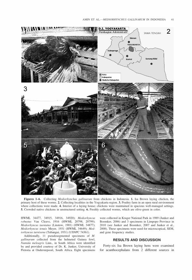
Forty-six Isa Brown laying hens ages 50–52 wk (Fig. 1)were examined for acanthocephalans from 2 different sourcesin Sleman district in Daerah Istimewa Yogyakarta, Indonesiafrom October 2010 to February 2011. The ‘‘Special Region’’(Province) of Daerah Istimewa Yogyakarta comprises 4districts and 1 city: Kulon progo District, Gunung kidulDistrict, Bantul District, Sleman District, and YogyakartaCity (Fig. 2). Twenty-six chickens were examined from alocal wet market and 20 other chickens were examined from apoultry farm in Kaliurang Sleman (107u159030 and107u299300E; 7u349510 and 7u479300S). The poultry farmwas situated in an open rural environment (Fig. 3) and thechickens were maintained in spacious well-managed settings(Fig. 4), unlike crowded native chickens in unstructuredsettings (Fig. 5) that were not used in this study.
Collected specimens were refrigerated in water for 2 duntil the proboscis was evaginated. Worms were puncturedwith a fine needle and subsequently stained in Mayer’s acidcarmine, destained in 4% hydrochloric acid in 70% ethanol,dehydrated in ascending concentrations of ethanol (24 hreach), and cleared in graduated concentrations of terpineolin 100% ethanol to 100% terpineol, then 50% terpineol in50% Canada balsam (24 hr each). Whole worms were thenmounted in Canada balsam. Measurements are presentedin micrometers, unless otherwise stated as range valuesfollowed by the mean in parentheses. Width measurementsrepresent maximum width. Trunk length does not includeproboscis, neck, or bursa. Voucher specimens were depositedin the University of Nebraska’s State Museum’s HaroldW. Manter Laboratory (HWML) collection no. HWML-49729 in Lincoln, Nebraska, USA.
For scanning electron microscopy (SEM) studies, 12specimens previously fixed in 70% ethanol were placed incritical-point drying baskets and dehydrated using ethanolseries of 95% and 100% for at least 10 min per soak followedby critical-point drying (Lee, 1992). Samples were mounted onSEM sample mounts, gold coated, and observed with ascanning electron microscope (XL30 ESEMFEG; FEI,Hillsboro, Oregon). Digital images of the structures wereobtained using digital imaging software attached to a computer.
Type or voucher cylindrical nonpseudosegmented Asianspecimens of M. gallinarum and 14 other species ofMediorhynchus from the HWML at Lincoln and the U.S.National Parasite Collection (USNPC) at Beltsville, Mary-land were examined for verification of the identity of ourspecimens and for comparative purposes. These specimensincluded Mediorhyncus conirostris Ward, 1966 (HWML34878); Mediorhyncus corcoracis Johnston and Edmonds,1950 (HWML 34649); Medirhyncus edmondsi Schmidt andKuntz, 1977 (USNPC 74356, 74358); Mediorhynchusemberizae (Rudolphi, 1819) (HWML 34507, 34508); M.gallinarum (USNPC 74360, HWML 34913, 34924, 34925);Mediorhyncus grandis Van Cleave, 1916 (HWML 30671,30676, 30695–30697); Mediorhyncus kuntzi Ward, 1960(HWML 34879); Mediorhyncus leptis Ward, 1966 (HWML34521); Mediorhyncus muritensis Lundstrom, 1942 (HWML34500); Mediorhyncus orientalis Belopolskaya, 1953(USNPC 74366, 74368, 74369, HWML 34748, 34906);Mediorhyncus papillosus Van Cleave, 1916 (USNPC 74359,
40 COMPARATIVE PARASITOLOGY, 80(1), JANUARY 2013
HWML 34477, 34915, 34916, 34920); Mediorhyncusrobustus Van Cleave, 1916 (HWML 20798, 20799);Mediorhyncus taeniatus (Linstow, 1901) (HWML 34877);Mediorhyncus tenuis Meyer, 1931 (HWML 34649); Med-iorhyncus turnixena (Tubangui, 1931) (USNPC 74361).
Additionally, 11 pseudosegmented specimens of M.gallinarum collected from the helmeted Guinea fowl,Numida meleagris Linn., in South Africa were identifiedby and provided courtesy of Dr. K. Junker, University ofPretoria at Onderstepoort, South Africa. Eight specimens
were collected in Kruger National Park in 1989 (Junker andBoomker, 2006) and 3 specimens in Limpopo Province in2010 (see Junker and Boomker, 2007 and Junker et al.,2008). These specimens were used for microscopical, SEM,and gene frequency studies.
RESULTS AND DISCUSSION
Forty-six Isa Brown laying hens were examined
for acanthocephalans from 2 different sources in
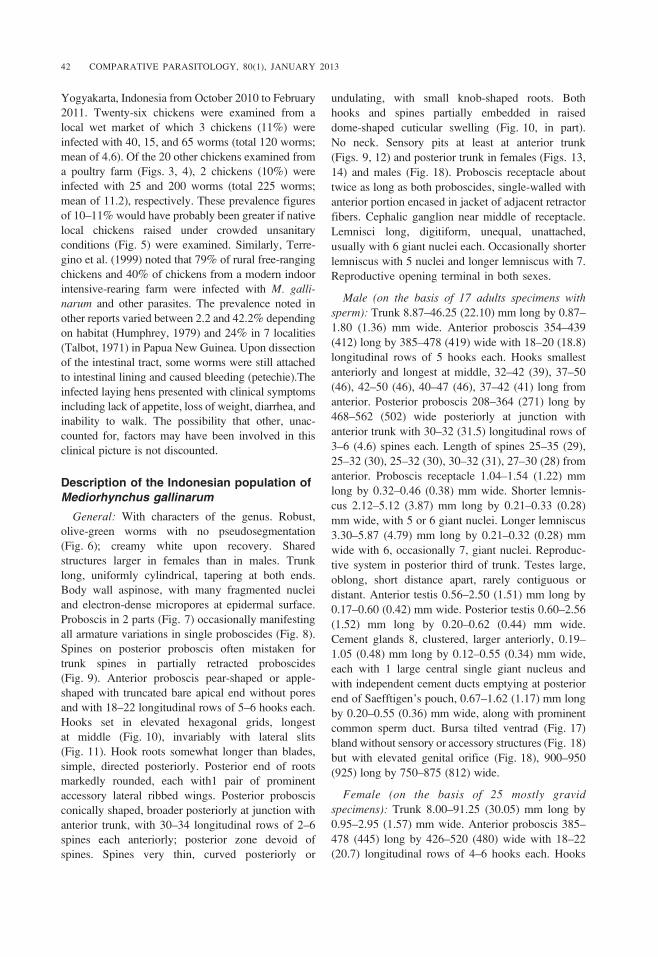
Figures 1–6. Collecting Mediorhynchus gallinarum from chickens in Indonesia. 1. Isa Brown laying chicken, theprimary host of these worms. 2. Collecting localities in the Yogyakarta region. 3. Poultry farm in an open rural environmentwhere collections were made. 4. Interior of a laying house; chickens were maintained in spacious well-managed settings.5. Crowded native chickens in unstructured setting. 6. Freshly collected worms, which are olive-green in color.
AMIN ET AL.—MEDIORHYNCHUS GALLINARUM IN INDONESIA 41
Yogyakarta, Indonesia from October 2010 to February
2011. Twenty-six chickens were examined from a
local wet market of which 3 chickens (11%) were
infected with 40, 15, and 65 worms (total 120 worms;
mean of 4.6). Of the 20 other chickens examined from
a poultry farm (Figs. 3, 4), 2 chickens (10%) were
infected with 25 and 200 worms (total 225 worms;
mean of 11.2), respectively. These prevalence figures
of 10–11% would have probably been greater if native
local chickens raised under crowded unsanitary
conditions (Fig. 5) were examined. Similarly, Terre-
gino et al. (1999) noted that 79% of rural free-ranging
chickens and 40% of chickens from a modern indoor
intensive-rearing farm were infected with M. galli-narum and other parasites. The prevalence noted in
other reports varied between 2.2 and 42.2% depending
on habitat (Humphrey, 1979) and 24% in 7 localities
(Talbot, 1971) in Papua New Guinea. Upon dissection
of the intestinal tract, some worms were still attached
to intestinal lining and caused bleeding (petechie).The
infected laying hens presented with clinical symptoms
including lack of appetite, loss of weight, diarrhea, and
inability to walk. The possibility that other, unac-
counted for, factors may have been involved in this
clinical picture is not discounted.
Description of the Indonesian population ofMediorhynchus gallinarum
General: With characters of the genus. Robust,
olive-green worms with no pseudosegmentation
(Fig. 6); creamy white upon recovery. Shared
structures larger in females than in males. Trunk
long, uniformly cylindrical, tapering at both ends.
Body wall aspinose, with many fragmented nuclei
and electron-dense micropores at epidermal surface.
Proboscis in 2 parts (Fig. 7) occasionally manifesting
all armature variations in single proboscides (Fig. 8).
Spines on posterior proboscis often mistaken for
trunk spines in partially retracted proboscides
(Fig. 9). Anterior proboscis pear-shaped or apple-
shaped with truncated bare apical end without pores
and with 18–22 longitudinal rows of 5–6 hooks each.
Hooks set in elevated hexagonal grids, longest
at middle (Fig. 10), invariably with lateral slits
(Fig. 11). Hook roots somewhat longer than blades,
simple, directed posteriorly. Posterior end of roots
markedly rounded, each with1 pair of prominent
accessory lateral ribbed wings. Posterior proboscis
conically shaped, broader posteriorly at junction with
anterior trunk, with 30–34 longitudinal rows of 2–6
spines each anteriorly; posterior zone devoid of
spines. Spines very thin, curved posteriorly or
undulating, with small knob-shaped roots. Both
hooks and spines partially embedded in raised
dome-shaped cuticular swelling (Fig. 10, in part).
No neck. Sensory pits at least at anterior trunk
(Figs. 9, 12) and posterior trunk in females (Figs. 13,
14) and males (Fig. 18). Proboscis receptacle about
twice as long as both proboscides, single-walled with
anterior portion encased in jacket of adjacent retractor
fibers. Cephalic ganglion near middle of receptacle.
Lemnisci long, digitiform, unequal, unattached,
usually with 6 giant nuclei each. Occasionally shorter
lemniscus with 5 nuclei and longer lemniscus with 7.
Reproductive opening terminal in both sexes.
Male (on the basis of 17 adults specimens withsperm): Trunk 8.87–46.25 (22.10) mm long by 0.87–
1.80 (1.36) mm wide. Anterior proboscis 354–439
(412) long by 385–478 (419) wide with 18–20 (18.8)
longitudinal rows of 5 hooks each. Hooks smallest
anteriorly and longest at middle, 32–42 (39), 37–50
(46), 42–50 (46), 40–47 (46), 37–42 (41) long from
anterior. Posterior proboscis 208–364 (271) long by
468–562 (502) wide posteriorly at junction with
anterior trunk with 30–32 (31.5) longitudinal rows of
3–6 (4.6) spines each. Length of spines 25–35 (29),
25–32 (30), 25–32 (30), 30–32 (31), 27–30 (28) from
anterior. Proboscis receptacle 1.04–1.54 (1.22) mm
long by 0.32–0.46 (0.38) mm wide. Shorter lemnis-
cus 2.12–5.12 (3.87) mm long by 0.21–0.33 (0.28)
mm wide, with 5 or 6 giant nuclei. Longer lemniscus
3.30–5.87 (4.79) mm long by 0.21–0.32 (0.28) mm
wide with 6, occasionally 7, giant nuclei. Reproduc-
tive system in posterior third of trunk. Testes large,
oblong, short distance apart, rarely contiguous or
distant. Anterior testis 0.56–2.50 (1.51) mm long by
0.17–0.60 (0.42) mm wide. Posterior testis 0.60–2.56
(1.52) mm long by 0.20–0.62 (0.44) mm wide.
Cement glands 8, clustered, larger anteriorly, 0.19–
1.05 (0.48) mm long by 0.12–0.55 (0.34) mm wide,
each with 1 large central single giant nucleus and
with independent cement ducts emptying at posterior
end of Saefftigen’s pouch, 0.67–1.62 (1.17) mm long
by 0.20–0.55 (0.36) mm wide, along with prominent
common sperm duct. Bursa tilted ventrad (Fig. 17)
bland without sensory or accessory structures (Fig. 18)
but with elevated genital orifice (Fig. 18), 900–950
(925) long by 750–875 (812) wide.
Female (on the basis of 25 mostly gravidspecimens): Trunk 8.00–91.25 (30.05) mm long by
0.95–2.95 (1.57) mm wide. Anterior proboscis 385–
478 (445) long by 426–520 (480) wide with 18–22
(20.7) longitudinal rows of 4–6 hooks each. Hooks
42 COMPARATIVE PARASITOLOGY, 80(1), JANUARY 2013

smallest anteriorly and longest at middle, 32–50 (42),
45–52 (50), 42–50 (49), 42–50 (47), 25–47 (39) long
from anterior. Posterior proboscis 177–385 (276)
long by 499–645 (579) wide posteriorly at junction
with anterior trunk with 30–34 (32) longitudinal rows
of 4–6 (4.8) spines each. Length of spines 27–35
(31), 27–37 (32), 22–42 (33), 25–37 (28), 22–32
(27) from anterior. Proboscis receptacle 1.00–1.72
Figures 7–12. Proboscis, hooks, and sensory pits of Mediorhynchus gallinarum. 7. The proboscis of a female wormshowing its division into anterior region and conically shaped posterior region; only the anterior part of the posteriorproboscis is armed with spines; the posterior unspiny part merges with the anterior trunk and is often confused with it. 8. Theproboscis of female worm showing the occasional presence of the full range of variation in the number of proboscis hooks of4–6 per row and of spines of 2–6 per row in the anterior and posterior proboscis, respectively, in individual worms. 9. Thepartial retraction of the proboscis in such worms led to the misinterpretation of spines of the posterior proboscis as trunkspines in some of the early descriptions. Note the sensory pit at the anterior trunk (upper left). 10. A middle hook set in araised hexagonal division of the proboscis. 11. A number of proboscis hooks showing the lateral grooves characteristic ofthat species. 12. Enlargement of the sensory pit shown in the anterior trunk of the specimen in Fig. 9.
AMIN ET AL.—MEDIORHYNCHUS GALLINARUM IN INDONESIA 43

(1.28) mm long by 0.34–0.54 mm wide. Shorter
lemniscus 2.60–6.50 (4.29) mm long by 0.19–0.29
(0.24) mm wide, with 5 or 6 (usually 6) giant nuclei.
Longer lemniscus 3.12–6.75 (4.76) mm long by
0.19–0.33 (0.25) mm wide with 6 giant nuclei.
Reproductive system short, in posterior 5% of trunk,
with prominent curvature of uterus and termina l
slit-shaped gonopore (Fig. 15). Eggs ovoid (Fig. 16),
47–57 (54) long by 24–32 (29) wide.
Morphological comparisons
Complete morphometric comparisons were not
possible because most reports, except for Junker and
Boomker (2006), lack a complete set of measurements.
Figures 13–18. Male and female structures. 13. The posterior end of a female specimen showing 3 sensory pits.14. Enlargement of the sensory pit area shown in Figure 13. 15. En face view of the terminal gonopore of a female showingits slit opening. 16. An egg. 17. Lateral view of a bursa showing its angle of articulation against posterior trunk. 18. A near-face view of a bursa showing its plain structure, muscular rim, and elevated genital orifice. Note sensory pore on top.
44 COMPARATIVE PARASITOLOGY, 80(1), JANUARY 2013
Only Bhalerao (1937) described his specimen’s color
as olive-green like ours but were creamy white upon
recovery like Talbot’s (1971) specimens. The size
and morphology of the trunk, anterior and posterior
proboscis, when properly interpreted, the proboscis
receptacle, lemnisci, uterus, and testes was comparable
in all collections, including ours, but markedly smaller
in the apparently younger specimens (males 8–11 mm
long, females 16–40 mm long) reported by Nath and
Pande (1963) from India. Nath and Pande (1963)
surprisingly reported and illustrated (Fig. 2) an
elaborate posterior proboscis with 20 rows of spines
on one side each with 9 spines and with no spineless
posterior zone, a gross exaggeration of the total of
30–34 rows of 3–6 spines each in our Indonesian
specimens that exhibited the widest range of variation
in 1 locality.
Important characters of taxonomic significance
include the proboscis armature and egg size and
morphology. Our Indonesian specimens exhibited the
widest range of variation from 1 locality in proboscis
armature of 18–22 longitudinal rows of 5–6 hooks
each on the anterior proboscis and 30–34 longitudinal
rows of 2–6 spines each on the posterior proboscis.
The usual armature reported was 20 rows of 5 hooks
each and 30 rows of 5–6 spines each but varied
between 18–22 rows of 4–5 hooks each and 25–32
rows of 2–6 spines each from different geographical
locations.
Our specimens from Indonesia, in addition, are
distinguished from others in all other locations by
having the smallest hooks (25–52 long) and egg
size (47–57 3 24–32), only comparable with the
specimens of Schmidt and Kuntz (1977) from
Terabanon Concepcion and Palawan Island that were
not described but examined and measured by us
(USNPC 74360, HWML 34913, 34924, 34925)
(hooks longest at middle: 40–50 long, eggs 50–62
3 25–30). Hook length reached 70 (Harris, 1973)
and 76 (Junker and Boomker, 2006) in African
specimens, 66 (Bhalerao, 1931) in an Indian female
specimen, 68 (Tubangui and Masilungan, 1946) in
specimens from the Philippines, and 70 (Yamaguti,
1954) in specimens from Celebes. Comparative
measurements of the markedly larger eggs were
available for specimens from Africa: 65–75 3 38–48
(Harris, 1973) and 70–86 3 43–52 (Junker and
Boomker, 2006) and from the Philippines: 64–68 3
40–43 (Tubangui and Masilungan, 1946 and Yama-
guti, 1963, respectively) and Japan. On the basis of
the taxonomically important characters of hook and
egg size alone, our specimens and those of Schmidt
and Kuntz (1977) appear to be more similar to one
another than either one to other specimens from
other Asian and African locations.
Distribution
The largest assemblage of Asian M. gallinarumpopulations was reported from a group of islands
between the Indian and the Pacific oceans in the
Philippines (Tubangui and Masilungan, 1946), Ter-
abanon Concepcion and Palawan Island (Schmidt and
Kuntz, 1977), Papua New Guinea (Talbot, 1971;
Humphrey, 1997), Celebes (Yamaguti, 1954), and
Indonesia (this paper) where the parasite appears to be
endemic in the domestic chicken and related birds. The
Indian collections from Muktesar and Mathura by
Bhalerao (1937) and Nath and Pande (1963),
respectively, in the landlocked northeastern state of
Uttar Pradesh suggest dispersal from Southeast Asia
through the Indo-Gangetic plain that spans most of
the state with movement of people and domestic
animals as Talbot (1971) proposed for the dispersal
of M. gallinarum from Papua and New Guinea to
Indonesia.
In Africa, M. gallinarum was also reported and
described from coastal Kenya in East Africa (Harris,
1973) and in South Africa (Junker and Boomker,
2006), which have no direct human–animal traffic
with the Asian-Pacific oceans’ islands. Present or past
routs of dispersal in this case are not known. The
ecology and host–parasite relationships of M. galli-narum were also reported from elsewhere in Africa
(Vercruysse et al., 1985; Cancrini et al, 1988,
Terregino et al., 1999; Junker and Boomker, 2007;
Davies, 2008; Junker et al., 2008) but the parasite
was absent in poultry examined in other Kenya
locations (Irungu et al., 2004) and in Nigeria, West
Africa (Fatihu et al., 1991).
Population differences
The possibility that the Asian and the African
populations of M. gallinarum represent 2 endemic
centers that may have evolved independently from
some hypothetical common ancestor may be sup-
ported on the basis of available morphological
evidence including the pseudosegmentation and the
presence of apical pores on the proboscis of the
African specimens. This is being explored in a project
using comparative gene sequence studies. The only
descriptive accounts from Africa are those of Harris
(1973) in Kenya and Junker and Boomker (2006)
in South Africa, who reported pseudosegmented
AMIN ET AL.—MEDIORHYNCHUS GALLINARUM IN INDONESIA 45

specimens. The African specimens from Burkina
Faso reported by Vercruysse et al. (1985) and
currently deposited at the British Museum of Natural
History were recently examined and reported to be
pseudosegmented (David Gibson, personal commu-
nication). The Asian specimens, like ours from
Indonesia, were not pseudosegmented. We examined
specimens from the Junker and Boomker (2006)
material for verification and future studies. Yamagu-
ti’s (1954) specimens from Celebes were reported to
be ‘‘corrugated transversely’’ but his figures 6 and 7
of the anterior and posterior portions of worms show
no segmentation. The ‘‘corrugated’’ state may have
been a state of contraction of the middle portion of
the trunk.
ACKNOWLEDGMENTS
We are grateful to Dr. Atif Naggar of Ain Shams
University, Cairo, Egypt, currently at Brigham
Young University, Provo, Utah, for his artful
preparation of the plates (Figs. 1–12). This project
was supported by an institutional grant from the
Institute of Parasitic Diseases to O.M.A.
LITERATURE CITED
Bhaleroa, G. D. 1937. On a remarkable Acanthocephalafrom a fowl in India. Proceedings of the ZoologicalSociety of London. Series B, Systematic and Morpho-logical 107:199–203.
Cancrini, G., A. Lori, R. Costantini, R. Romano, P.Lanfranchi, and M. A. Abdullatif. 1988. Elmintiintestinali in Acryllium vulturinum della Somalia.Parasitologia 30:34–46.
Davies, O. R., K. Junker, R. Jansen, T. M. Crowe, and J.Boomker. 2008. Age- and sex-based variation inhelminth infection of helmeted guineafowl (Numidameleagris) with comments on Swainson’s spurfowl(Pternistis swainsonii) and orange francolin (Sclerop-tila levaillantoides). South African Journal of WildlifeResearch 38:163–170.
Fabiyi, J. P. 1972. Studies on parasites of the grey-breastedhelmet Guineafowl (Numida Meleagridis galeataPallas) of the Vom area the the Benue State, Nigeria.Bulletin of the Epizootic Diseases of Africa 20:235–237.
Fatihu, M. Y., V. C. Ogbogu, C. O. Njoku, and D. I.Saror. 1991. Comparative studies of gastrointestinalhelminthes of poultry in Zaria, Nigeria. Revued’elevage et de Medecine Veterinaire des PaysTropicaux 44:175–177.
Harris, M. T. 1973. A new acanthocephalan from an EastAfrican galliform bird. Bulletin of the British Museum(Natural History) Zoology 24:455–461.
Humphrey, J. D. 1979. Helminths of the alimentary tract ofthe domestic fowl in Papua New Guinea. AustralianVeterinary Journal 55:205–207.
Irungo, L. W., R. N. Kimani, and S. M. Kisia. 2004.Helminth parasites in the intestinal of indigenouspoultry in parts of Kenya. Journal of South AfricanVeterinary Association 75:58–59.
Junker, K., and J. Boomker. 2006. Mediorhynchusgallinarum (Acanthocephala: Gigantorhynchidae) inhelmeted guineafowls, Numida meleagris, in theKruger National Park, South Africa. OnderstepoortJournal of Veterinary Research 73:283–292.
Junker, K., and J. Boomker. 2007. Helminths ofguineafowls in Limpopo Province, South Africa.Onderstepoort Journal of Veterinary Research 74:265–280.
Junker, K., L. Debusho, and J. Boomker. 2008. Thehelminth community of helmeted guineafowls, Numidameleagris (Linnaeus, 1758), in the north of LimpopoProvince, South Africa. Onderstepoort Journal ofVeterinary Research 75:225–235.
Lee, R. E. 1992. Scanning Electron Microscopy and X-RayMicroanalysis. Prentice Hall, Englewood Cliffs, NewJersey. 458 pp.
Lundstrom, A. 1942. Die Acanthocephalen Schwedens, mitAusnahme der fisch-Acanthocephalan von Susswasser-standorten. Lund. 238 pp.
Nath, D., and B. P. Pande. 1963. A note on theacanthocephalan infection of domestic fowl. IndianJournal of Helminthology 15:31–35.
Petrochenko V. I. 1958. Acanthocephala of Domestic andWild Animals. Vol. 2. Moscow, Izdatel’stvo AkademiiNauk SSSR. (Translated by Israel Program forScientific Translations, Jerusalem, 1971, 478 pp.)
Schmidt, G. D., and R. E. Kuntz. 1977. Revision ofMediorhynchus Van Cleave 1916 (Acanthocephala)with a key to species. Journal of Parasitology 63:500–507.
Talbot, N. T. 1971. An acanthocephalan parasite, Medior-hynchus gallinarum, of the domestic fowl in Papua andNew Guinea. Australian Veterinary Journal 47:334–336.
Terregino, C., E. Catelli, G. Poglayen, A. Tonelli, and O.I. Gadale. 1999. Prelimenary study of helminthes ofthe chicken digestive tract in Somalia. Revue d’Eleva-geet de Medicine Veterinaire des pays Tropicaux 52:107–112.
Tubangui, M. A., and V. A. Masilungan. 1946. On twoAcanthocephala from the Philippines. Journal ofParasitology 32:154–155.
Van Cleave, H. J. 1947. Thorny-headed worms (Acantho-cephala) as potential parasites of poultry. Proceedingsof the Helminthological Society of Washington 14:55–58.
Vercruysse, J., E. A. Harris, R. A. Bray, M. Nagalo, M.Pangui, and D. I. Gibson. 1985. A survey ofgastrointestinal helminthes of the common helmetGuinea fowl (Numida meleagris galeata) in BurkinaFaso. Avian Diseases 29:742–745.
Yamaguti, S. 1954. Parasitic worms from Celebes. Part 8.Acanthocephala. Acta Medicinae Okayama 8:406–413.
Yamaguti, S. 1963. Systema Helminthum, Acanthocephala.Vol. 5. Wiley Interscience, New York. 423 pp.
46 COMPARATIVE PARASITOLOGY, 80(1), JANUARY 2013