Embed Size (px)
Citation preview

4 f
~YTOTOXIC ALKALOIDS FROM
TAB.ER NltB110NTA NA JOHN'STONTI ------------------------/ by
MAIUO f'l. }iANGIN0-7'
'l'hesis submitted to the Graduate Faculty of the
Virginia Polytechnic Institute and State University
n partial fulfillment. of the requirements for the degreH of
MASTER OF SCIENCE
in
Chemistry
APPROVED:
---------------~----
D. G. I. Kingston, Chairman
·r---7---------M.A. Oqliaruso
May, 1974
Blacksburg, Virginia

ACKNOWLEDGEMENTS
The author of this thesis wishes to thank his research
advisor, , for his f.requent help and
valuable suggestions during the course of this project. Re
also extends his gratitu(ie to tbe other members of his
committee, and , for their interest in
this research and for providing some ideas for future study ..
The author is indebted to of the
Natural Products section, Drug Development Branch, Rational
Cancer Institute, and , of the u. s. Department of Agriculture, Beltsville, Md., for the supply
of l!.!!§£!!aemon t§:.!HL_.12hn§1Qa!!:. Financial support under
Public Health Service Research Grant from the
National Cancer Institute is gratefully acknowledged.
The author is also grateful to three of his fellow
graduate students, , , and
research.
people:
extractions
laboratory
, for 'their help and encouragement on this
Special thanks are in order for the following
for carrying out tbe initial
of the
assistance
plant
(in
material and for her va.luable
addition to keeping the author
informe.d of current events in the chemistry department); and
finally to for careful typing of
the manuscript and for arrangement of the tables and
f iqures.
ii

ACKNOWLEDGEMENTS
LIST OF FIGURES
LIST OF TABLES
.INTRODUCTION
HISTORICAL
A. The Biogenesis of Indole Alkaloids
page
ii
v
v
1
5
5
B. Tabern~~!i~Bftl A source of New Indole Alkaloids 10
RESULTS AID DISCOSSIOI
A. Isolation of Pure Constitu€nts
B. Spectral Data and Structural Characterization
of Isola tad Compounds
1. K139
2.. K117
3. K155 and K102
StnUU.RY
EXP ER I.MENTAL
A. General Inf o~mation
B. Fractionation I
1. Column Chromatography of F057
2. .Buff er Extraction of F064
l. Chromatography of P070
17
17
40
40
48
59
63
86
86
87
'11
91
93
4. Preparative Thin Layer Chromatography of P078 94
c. Fractionation II 99
iii

1. Column Chromatography of Fraction P057A 101
2. Cclunm Chromatography of Fraction F095 102
3. Column Chromatography of Fraction B 103
4. Column Chroma tog ra ph y of Fraction D 104
D. .Fractionation III 107
1. Chromatography of F124 on Alumina 110
2. Column Chromatography of F131 111
3, Column Chromatography of .Fraction F143 114
LIST OF REFERENCES 119
VITA 123
iv

page
1. Mass Spectrum of R117
2. Nuclear Magnet Resonance Spectrum of K155
3. Mass Spectrum 72
4. Nuclear ~aqnet Resonance Spectrum of K102
I. 21
II. PHRCEITAGE YIELDS OP ETHANOL FRACTIONS AND CRUDE
ALKALOIDAL FRACTIONS CORPARED TO WEIGHT OP
GROUND PL.IHfi'
III. BIOASSAY RESULTS ff!" l~THANOL Ff{ACTIONS AND CHU DE
.24
IV. BIOASSAY RESULTS PBO! GRADIENT ELUTIOI
CHROMATOGfi:A.PHY OF CRUDE ALKALOIDAL FRACTIONS
FROM %..?. _,HJ .ff l!~ X.Qt!l1 27
v. FRACTIONATION Or' F057 '1'0 OBTAI.N F078 29
VI. FRACTION/.\ TIO~~ OF f078 AND 1501.ATION OF K.102 30
VII. FR A CT IC NA 'I' I 0 N OE' F108 AND ISOLATION OF K125
VIII. FRACTIONATION OF P124 ANO ISOLATION OF K136
AND Kl39 35
IX. :r;~HACUO NATION OF .r' 143 AND ISCL.ATIO.N Cr K155 36
x. FRACTICNATION OF F095 AND ISOlATION OP K117 39
XI. PHOTON N.~.R. CHEMICAL SHIFT DATA FOB VOACA~INB
v

AllD K155 74
XII. PROTON CHEMICAL SHIFT DATA FOR K155 and iW12 79
XIII. PiAC'fIONA.'t'lON TREE OF !.:._!1.0J!!§IQ!Il FRACTION! 'l'.ION I 90
XIV. FRACTIONATION TREE OF l~liN~!QlU!
PlU\CT!ONA1'ION II 100
xv. FBACTIONl'fION THEE OF !.t._JOH]!§TON!l
P1UCTI05AT!tHl III 109
vi

One of the most exciting areas of natural products
research is their use in cancer chemotherapy. Several newly
discovered alkaloids have been employed successfully as
anti-tumor agents in man. For example, Vincaleukoblastinet
isolated in 1958, is one of the most effective drugs in the
treatment of Hodgkin's disease.2 Tvo new
alkaloids, thalicarpine3 and d-tetrandrine,• which are
inhibitors of intramuscular tumors are currently in clinical
testing for treatment of human neoplasms.s Ellipticine,6
the newest tumor inhibiting alkaloid is now ready for
clinical testing against human neoplasms.s
The discovery that a wide range of planti contain
constituents ~hich show reproducible anti-tumor activity has
led the National Institutes of Health (NIH) to initiate a
cancer chemotherapy program centered around the screening of
plants for anti-cancer activity. The Cancer Chemotherapy
National Service Center (CCNSC) of the National Institutes
of Health has joined vith the U.S. Department of Agriculture
to procure several thousand plant samples per year for
evaluation. These plants undergo fractionation studies which
are quided at each stage by biological screening assays. The
ultimate goal is the isolation of pure compounds which are
subjected to further screening if they show significant
activity in the initial testing system. The most promising
1

2
compounds isolated from the test plants are then subjected
to more extensive study in animals and finally undergo
clinical testinq in human subjects.
The major research goal of the individual laboratories
involved in the CCNSC program is not just the fractionation
and isolaticn of active plant constituents, but includes
also the ultiaate structure determination of the pure
compounds isolated from these plant extracts. Any promising
anti-cancer compounds will eventually undergo studies
related to their "biochemical mechanism of action," or will
be subjects of atteapted synthesis. Even the less proaising
anti-tumor agents may exhibit novel chemical or structural
features. For these reasons, the structural and
stereocheaical investigation of these pure coaponents is an
indispensable part of the cancer chemotherapy program.
As a result of new developments in physical methods of
analysis, the organic cheaist has available the tools to aid
in the structure determination of complex molecules. For
example, the use of aass spectroaetry allows the rapid
assignment of mclecular weight to unknown compounds even if
only a few milligrams of the component can be isolated. By
high resolution mass spectrometry, the exact empirical
formula of a compound can be determined, or at least, the
high resolution measurement can be used in conjunction with
the elemental analysis to determine the empirical

3
composition. Mass spectrometry also has another advantage as
a structural probe: many of the fragment ions observed by
this method have a molecular structure which is well
defined. From
the known
work done on other natural products and from
fragmentation patterns of siaple organic
the identity of fragment ions from unknown molecules,
compounds may be predicted vith a high level of confidence.
Evidence from nuclear magnetic resonance may be used in
conjunction with mass spectral data even when the structure
of an unknown natural product is too complex to be
determined directly by n.a.r. The data fro• n.m.r. can help
to deteraine the type of substitution in a molecule (e.g.
o-methyl or N-methyl groups), the number of methyl groups
present, and the number and type of aromatic protons.
Ultraviolet and infrared spectroscopy help the organic
chemist to analyze for the presence of particular functional
qroups; u.v. spectroscopy is especially helpful for
ascertaining the presence of various heterocyclic nitrogen
containing moieties in an unknown molecule.
X-ray diffraction is another physical method which has
great potential as a structural tool. The determination of
the absolute configuration of a complex molecule by
degradation reactions is a difficult and time-consuming
process especially when only small amounts of the unknown
compound are available. So X-ray diffraction is a very

4
promising technique, and has already been useful in
determining the absolute configuration of several
alkaloids.7,e
The application of the physical methods outlined here
is dynamically illustrated by research in the alkaloid
field. Since the isolation of vincaleukoblastine, some sixty
alkaloids from the !!n~.J!Q§~ plant species vere isolated
and identified before 1967. Fifty-five of these alkaloids
represented new compounds.9
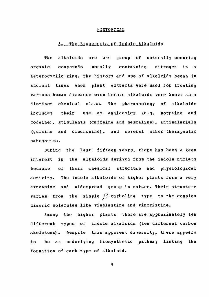
The alkaloids are one group of naturally occuring
organic com~cunds usually containing nitrogen in a
heterocyclic ring. The history and use of alkaloids began in
ancient times when plant extracts were used for treating
various human diseases even before alkaloids were known as a
distinct chemical class. The pharmacology of alkaloids
includes their use as analgesics (e.g. morphine and
codeine), stimulants (caffeine and mescaline), antimalarials
(quinine and cinchonine) , and several other therapeutic
cateqories.
During the last fifteen years, there has been a keen
interest in the alkaloids derived from the indole nucleus
because of their chemical structure and physiological
activity. The indole alkaloids of higher plants form a very
extensive and widespread group in nature. Their structure
varies from the simple ;9-carboline type to the complex
dimeric molecules like vinblastine and vincristine.
Among the higher plants there are approiiaately ten
different types of indole alkaloids (ten different carbon
skeletons). Despite this apparent diversity, there appears
to be an underlying biosynthetic pathway linking the
formation of each type of alkaloid.
5

6
The first observation linking thE biosynthetic pathway
is the fact that the tryptamine unit is repeatedly
encountered in indolc alkaloids of all types. For exdrnple,
tlte .:..;imp lest group of indole alkaloids incorporating
tryptamine dre the;9-ca~bolines ( j ) , which are synthesiz~d
al<lebydes10,11
by the condensation of tryptamine with
(Scheme I) :
NH2
0 11
+H-C-R--<
Scheme I
R
NH
(2)
The origin of the Strychnos type alkaloids ( l is
also explained by the pathway shown in Scheme I. This
Mannich co11densation reaction is considered to be the
stdrtinq point for biosynthesis of the more complex indole
alkaloids. The possibility that this reaction might he

7
"spontaneous" under the conditions present in the plant cell
has been a central feature in the biogenetic theory of
indole alkaloids.
suggested by two
This theory of spontaneous reaction is
observations. First, the alkaloids as a
group
them
amino
have an extensive diversity in structure which •akes
quite dissimilar to the basic metabolites like sugars,
acids, and fatty acids. Coupled with this is the lack
of any known metabolic function for the alkaloids. They are
not likely to be part of any enzymatic cycle related to
energy transfer
that alkaloids
or regulatory processes. It is more likely
are the endpoint of plant synthesis --
storage molecules synthesized from interaediates not
utilized in the regular aetabolic cycle. A reaction which
brinqs these cellular by-products together would be an
economical way for the cell to rid itself of molecules which
have no real metabolic utility. secondly, the intermediates
proposed in this spontaneous coupling are known to be
readily available in the plant cell. This is a likely
requirement for any reaction to occur without the aid of
enzymes.
In addition to the indolic portion of these alkaloids,
there is also a nine or ten carbon unit whose origin has
only been recently uncovered. According to the original
proposals, this nine or ten carbon unit is derived from an
aromatic amino acia,12 prephenic acid,13 or siaply by the

8
buildup of acetate or mevalonate units. Isotope labelling
experiments failed to confirm any of these proposals.
After these initial hypotheses were invalidated,
.several rE:searchers, notably Arigoni, Battersby,
and Scott,1~,1s laid out a well-documented pattern to
explain the biosynthesis of the non-indolic portion of these
complex alkaloids. The major achievement of their research
was the discovery that the ten carbon moiety has a terpenoid
origin an idea first suggested by Thomasl6 and
Wenkert.11 This ten carbon unit was shown to be derive<l
from geraniol via conversion to loganin and secologanint•
(Scheme II):
OH
.. ·Q-Glu:ose

9
The validity of this reaction scheme was demonstrated
by the experiments of Battersby et.a1.1a In
these experiments, the direct Mannich reaction of tryptamine
with secologanin was carried out and the result was the
formation of the,,8-carb.oline alkaloid isovincoside ( 1 ) or
the corresponding spiro-indolenine alkaloid ( ~ ) depending
on the pH of the reaction medium (Scheme III).
N H 0
II C-H
~H ·· .·Q-Glu
Scheme III
NH:?" .··~·o-Glu
In further experiments,19 secologanin tritiated at the
o-methyl carbon was prepared and fed to
plants. The three alkaloids ajmalicine, vindoline, and
catharanthine (each having a different carbon skeleton) were

10
isolated from the plants. The radioactivity was localized at
the a-methyl carbon in each alkaloid in almost 100~ yield,
indicating that secoloqanin was accepted as the precursor of
each indole alkaloid. In view of this experimental evidence,
the other varicus carbon skeletons of the coaplex indoles
are assumed to arise by simple rearrangements of the
secologanin structure during the biosynthesis of the
alkaloids. Kompis, ftosse, and Schmid have shown hov the
secoloqanin unit could be rearranged during biosynthesis to
yield five different basic skeletons which will explain the
origin of every type of indole alkaloid encountered in the
higher plants.20
One particular plant group, the family known as
!EQ£ynaceae, is a prolific source of indole alkaloids.
Within this family there are several genera which have been
well studied. The alkaloids derived from each genus seem to
be of a characteristic structural type. For example, most
alkaloids of Aspidospermine plants have the carbon
skeleton ( 2 ) ; consequently, alkaloids of this type are
known as !.§..Eidosperaine alkaloids even if they are
isolated from another genus. catharant~i~ alkaloids have
the skeleton ( & ), but this kind of alkaloid has been
isolated from plants of another genus as well.

1 1
(6)
The genus knovn as Tabernafil!lODt!!!!~ from the family
!E2£i~£~g is a particularly good source for alkaloids
having the iboga skeleton ( 1 ). The first such alkaloids
were isolated from the plant I~he£n~nthg_iboq~ by French
workers in 1901. 21,22 The interest in this plant ~as
spurred by the discovery that African natives used extracts
of Iah~.!! the __ ib.Q!I~ as a stimulant and an
hallucinogenic. 23 Ibogaine ( 1h ) was the first
crystalline alkaloid obtained from this plant.
Pharmacological investigation showed that this compound
initiated unusual excitatory effects on various
experimental animals,2•-26 and exhibited local anesthetic
properties as well. A later study showed that ibogaine was a
central nervous system stimulant with definite
anticonvulsant properties.27
Taylor and coworkers2•
study of I~hC£!!~,!ithg_ib..Qg~
the alkaloids ibogamine
repeated the chemotaxonomic
and isolated and characterized
1~ ), ibogaine ( 7b ) ,

12
tabernanthine ( 7c), and voacangine ( 1~ ).
7a Ibogamine, R =R =R =H 1 2 3
I!! Ibogaine, R =CH o, R =R =H l 3 2 3
7c Tabernanthine, R =CH o, R =R =H 2 3 l 3
7d Voacangine, R = CH o, R =H, R =COOCH 1 3 2 3 3
Subsequent work with Tabe£n~em2nt~n~ species yielded
new iboga alkaloids. The most interesting among these show a
rearranged iboga skeleton represented by the pseudoindoxyl
alkaloids rupicoline ( ~~ ) z9 and montanine ( ~h ) 2 9 and
by the indoxyl alkaloid crassanine ( 2 ). 30
Rupicoline, R=H Crassanine
8b Montanine, R=OH

13
Bupiccline and montanino are the predicted products
from the auto-oxidation of voacanginc during biosyuthesis in
the plant. However, the same workers could not isolate
More recent work shows that the ihoga skeleton is not
the only one encountered in species.
Burnell and Medina isolated from ~-R~Y£hQtrifoli~ three
alkaloids bearing the vobasine skeleton lQ and 11 ) , the
first isolation of such alkaloids from
lQ2. Affinine, Il=CH OH 11 Taberpsychine 2
lQ.Q l~p i-vob asi nic acid, R=COO El
Affininr~ ( 1Q~ was originally isolated from the
The corresponding acid,
epi-vobasinic acid lQQ ) , was found as a natural product
for the first time from a species,

14
although the compound was originally obtained as a
synthetic product.33 The most intere~ting alkaloid in this
group is the cyclic ether taberspychine ( 11 ) , whose
structure was determined mainly by a combination of data
from mass spectrometry and nuclear magnetic resonance
including spin decoupling experirnents.31
The important group of alkaloids having the
type skeleton has been isolated from only
one species of Cava, DaRocha, and
coworkers 34 isolated from the new
alkaloids (+)-minovinincine ( l~ ) and
(+)-8-oxominovincine, given the structure ( 11).
C==O. I CH3
(+) -Minovir.cine 12 (+)-8-0xominovincine
The final structure proof for ( 1J ) was obtained by
the permanganate oxidation of (+)-minovincine ( l:f. ) • The

15
Q-lactam ( 11 ) was obtained with physical and spectral
properties exactly identical to the natural product.
The same workers isolated only alkaloids with the
eLurnamine type skrolctou fz.:om the .spf'cies :L__r!g.;!:£12. • The
compounds (+)-vincamine 11 ) and (+)-apovincamine ( !~
were obtained in good yield but were not new alkaloids.3•
(~)
The fact that no iboga alkaloids were isolated from the
last three species mentioned ( T~
enhances t11E interest in the £ig1£1ii , and
.'.!\1£~ 11~£.!!!.Qll.t~.!2 £ species as a source of various alkaloidal
types anJ stimulates th~ controversy ov~r botanical
cl.:is.si fica tion cf the .T£E.Q!::B~fL!!!.2.D..!:~.!!£ species.
Consequently, an inve~tigation of the species
1'.<!Q.Q.!:!!~Q!!lO!l_!.2_.!}Q_jQ!!.!1~!2.!!ii was undertaken after initial
scr(?eniug by the NIH iuclica.tcd that this plant posscsseo
some anti-cancer activity. This investigation was directed
toward determining
reproJucible activity
what
and
part of the plant contained
ultimately to the isolation of

16
pure compounds for testing in the biological assay systems
developed by the NIH.

17
icinal agents. Bat the isolat
alkaloids in pure form is a relatively new devel nt
which began with the isolation of morphine in 1804.
These early isolations were tedious procedures which
consisted mainly
plant
of repeated extractions of thf:t er
by aqueous acids in the h
preparing crystallizable salts of in vidual
co~ponents. compounds vhich did not form salts readily
were seldcm nea in a form pure enough for
structural characterization ta be carried out.
The advent of uid chromatography in the early 1 0' s
offered the chemist a new method for the sei;aration
isolation of pure compounds. But it was not until a r
1945 that quid chromatography began to accelerate
work in the alkaloid field. Chro•atography offered two
main advant.a<ttS".s. :r~irst, components which could not iH~
crystalliz€d as salts from crude fractions could
obtained in a tor.u1 pure enough for crystallization as
neutral compounds and secondl7, components nt in
low concentration could now be separated and sub•itt
to structural characterization. Modern techn of

18
possible the separation of components present in very minute
concentration.
As mentioned above, the classical or phytochemical
approach to the study of plant constituents focuses on those
compounds which are most easily separated or crystallized.
The program initiated by the National Institutes of Health
differs from the classical approach in that the
fractionation and isolation studies are guided at every
stage by biclogical assays. This approach makes possible the
isolation of important minor constituents which could be
missed in the classical approach.
The bioassay tests employed by NIH and carried out at
various biomedical laboratories are designed to provide
initial screening of plant extracts to determine their
potential as anti-cancer agents.
The two most widely used test systems are the "survival
tumor" system and the in vitro cell culture.
In the "survival tumor" system, the evaluation of a
plant extract is done on the basis of its ability to
increase the lifetime of test animals implanted with a tumor
as compared to control animals which are implanted with the
same tumor but left untreated. The control animals have a
well defined life span after tumor implantation. The
survival bioassay used in this work is given the code
letters "PS," and the tumor is called the P388 lymphocytic

19
leukemia. The result of each test is reported as the ratio
of survival time of treated animals (T) to control animals
(C) expressed as a percentage (T/C)%. A minimal increase in
survival time of treated animals over control animals
resulting in a T/C > 125~ is necessary for
further testing.35
The cell culture employed in this work is
given the cede letters "KB," and the culture itself is the
human epidermoid carcinoma of the nasopharynx. Once again,
treated cultures are compared with control cultures during
evalution of the extract. The result of each test is
reported as the ED -- the dose that inhibits growth to 50~ 50
of control growth. For materials tested by weight (as in
this work), ED is expressed in •icrograms per ailliliter. 50
A result of ED < 20 is required for further testing.35 50
During the initial fractionation of a plant, the in
viyQ (animal tumor) testing system is used on the crude
extracts since ample amounts of material are usually
available. If the crude extracts show favorable !~_:tiVQ
activity, then the in vitro testing syste• is used for
bioassay during fractionation leading to isolation of the
anti-tuaor agent, since less material is required for the
test. Any pure material which shows favorable in vitrQ
activity is subjected to in vivQ testing when larger
amounts become available.
20
The starting point in any chemotaxonomic plant study is
the procurement of a suitable specimen. In this case, the
species Tabernaemontana johnstonii (a mediua-sized tree)
was collected in Kenya by the u.s. Department of
Agriculture. The plant was received as the dried roots,
stems, and bark which vere pulverized for fractionation. The
initial extraction was done on the ground plant using 95~
ethanol with all organic soluble material being collected.
This alcoholic extract was partitioned between concentrated
base (NH OH) and an immiscible organic solvent (ethyl •
acetate or chloroform) to remove acidic and phenolic
material. The basic components of the organic phase were
removed by partitioning with mineral acid (5-10~ H SO ) • 2 •
The new acid soluble fraction was then neutralized and
re-extracted with organic solvent to recover free tertiary
bases. A fractionation tree with typical weights is shown in
Table I. During the course of work on T. johnstonii , four
separate extractions of ground plant were required to obtain
sufficient material for isolation. Table II shows the amount
of starting qround plant in each extraction and the
percentage yield of the 95% ethanol fraction and the crude
alkaloidal fraction.

21
TABLE I TYPICAL FRACTIONATION TREE OF T. JOHNSTONII
2.5 kg Ground Plant I I Ethanol Extraction I
Ethanol Extract ( 150-2009)
J Ethyl Acetate I I NH OH Partition
--------------' . I Ethyl
Acetate Soluble
I I Extract 5-10~ H so
--------' 2 • I I I I v ' Ethyl Insoluble
Acetate Solid Soluble (10-40g) (50-60q)
I I v
CH Cl 3
Soluble (50-60g)
I I v
H SO 2 •
Soluble I I Neutralize NH OR
I I v
Insoluble Solid
(5-20g)
I Extract CHCl • '----- 3 I
' v CHCl
Insoluble
I Aqueous Soluble (1-10g)

22
TABLE II PERCENTAGE YIELDS OF ETHANOL FRACTIONS AND
CRUDE ALKALOIDAL FRACTIONS COMPARED TO WEIGHT OF GROUND PLANT
FI:'actionation I NumbeI:' I
I I
Weight of Ground Plant
I I
Weight of Ethanol
FI:'action (S of Ground
Plant
Weight of Alkaloid (I of Ground
Plant
I I I I I
--------- '-------'--- --------' I 2520 g
II 2800 g
III 2679 g
IV 2454 g
183 q (7. 3%)
105 g ( 3. 75~)
240 g (8.93)
120 g ( 4. 9%)
52.5 g (2.1%)
52.6 g ( 1. 9~)
31. 1 g (1. 16%)
67.0 g (2.7%)
23
At various points in the extraction of the ground
plant, aliquots of each new fraction were set aside for KB
and PS bioassay. The results of some of these bioassay tests
on the 95% ethanol fractions and the crude alkaloidal
fractions are shown in Table III.*
Several separation methods were attempted on the crude
alkaloidal fractions, and the initial methods are worthy of
some discussion. Direct chromatography of the crude
alkaloids on neutral alumina using a step-gradient was
attempted. The eluted fractions were subjected to thin layer
chromatography which shoved that separation of the crude
alkaloids was quite poor, and components of widely varying
Rf value were eluted in each fraction. Because of the number
of components and their considerable differences in
polarity, direct chromatography of the crude alkaloidal
fraction did not appear to be very promising.
* The numbering syste• used to identify fractions is as follows: fractions which are a mixture of components are prefixed with the letter "F" followed by a three digit nu•ber which increases in sequence as each new fraction is obtained. Thus, fraction "F043" is followed by fraction "F044." crystalline fractions are numbered in the same systea as the aixtures, except that they are prefixed with the letter "K." For exaaple, if a crystalline fraction is the next one obtained after "F082," then the crystalline fraction is labelled "K083."

24
TABLE III BIOASSAY BESDLTS OF ETHANOL FRACTIONS AND CRUDE
ALKALOIDAL FRACTIONS OF T. JOHHSTONII
Fractiona-1 KB Activity ti on I <µg/ml)
Number I I I ----------------- '----------· I I
I Ethanol I I Fraction (F053) I
I I _________ I I I Alkaloidal I Fraction (F057)
------'---
III
1 I Ethanol I Fraction ( - ) '---------' I Alkaloidal I Fraction (F124) I
------- '-----------' Ethanol I Fraction (F103)
IV '-----------' I Alkaloidal I Fraction (P108)
------' N.S. - Not Submitted for Assay
23
16
N. S.
18
26
5.5
PS Activity (T/C~)
142* 138**
142* 133••
N. S.
N.S.
171• 147••
104* 100••
* - A dose of 100 mg/kg body weight vas administered. ** - A dose of 50 mg/kg body weight was administered.

25
Buffer extraction was the second separation method
attempted on the crude alkaloids of T._j_Qh.~stonii • An
aliquot of the crude alkaloidal fraction was partitioned
between organic solvent and aqueous acids of increasing
acidity. When benzene was used as the organic phase with
buffers of pH 5, pH 4, pH 2.6, and finally 3.51 HCl, aost of
the alkaloids were extracted into the three buffer phases.
Thin layer chromatography of the aqueous extracts showed
that each buffer fraction contained six or aore components,
some of which were characterized by very similar Rf values.
When chloroform was used in place of benzene as the organic
phase, approximately one third of the crude alkaloid
remained in the organic phase. Thin layer chromatography
showed that the separation was not much improved over the
use of benzene as an organic phase.
Recent work on Tabernaemontana
the literature shows that buffer
species reported in
extraction was used
successfully for separating alkaloids after the initial use
of column chromatography on the crude alkaloidal fraction.
For example, Cava and coworkers subjected the crude
alkaloids of x~---~iedelii to chromatography on an
alumina column.3• The sample was eluted exhaustively in
succession with benzene, chloroform, chloroform-aethanol
(97:3), and neat methanol. In this way, four fractions of
relatively increasing polarity were obtained. The benzene
26
fraction was used in an acid extraction employing buffers of
pH 4.0, pH 2.6, and 1N HCl. A small amount of •aterial
insoluble in 1N HCl was also obtained. Pure alkaloids were
isolated dirEctly from the HCl insoluble material as well as
trom each acid fraction except the pH4.0 buffer. A slightly
modified version of Cava•a procedure was applied on the
crude alkaloids of T. johnstonii. The alkaloids were
eluted from an alumina column using in succession the
following neat solvents: benzene, chloroform, ethyl acetate,
n-propanol, and methanol. The five resulting fractions were
submitted for bioassay, and all five fractions shoved
promisinq KB activity. Table IV shows the KB activity of all
the fractions obtained by this alumina chromatography
elution method during the course of work on T. iohns~onii.
The significant observation from these activity data is that
the benzene and chloroform fractions consistently show high
KB activity. Por this reason, all subsequent vork vas
carried out on the benzene or chloroform fraction obtained
from alumina chromatography of the crude alkaloidal
fraction.

27
TABLE IV BIOASSAY RESULTS FROM GRADIENT ELUTION CHROMATOGRAPHY
OF CRUDE ALKALOIDAL FRACTIONS FROM T. ~OHHSTONII
crude I Benzene Chloro- I Ethyl n-Alkaloidl I fora I Acetate Propanol ------ '------' '------ -------
' I I Fractiont Fraction I FractionJ Fraction Fraction No. (Wt) I No. (it) I No. (Wt) I No. (Wt) No. (Wt) (KB Act. I [KB Act. I [KB Act. I (KB Act. [KB Act. µ_g/ml] I Jlq/ml] I j19/•1] I j19/•1] J.191•1] -----•-----'-----'--- -----1 I I
F057 I F064 I F065 I P066 F067 (20q) I (3.25g) I (8.23g) I (1.15g) (1.73g) (16] t [1.9] I [2.0] I [2.2) [2.5]
Methanol
Fraction No. (Wt) [ KB Act. jl<Jl•l]
F068 (2.51g) [ 2. 0]
----- '------ '-----'------ ----- ------1 I I I F057A I F094 I F095 I F096 F097
(27.4g) I (10.0g) I (4.20g) I (1.0g) (5.39) [N.S.] I [0.55] I (1.7] I [3.0] [2.8]
F098 (2.9g) [ 2. 7]
----' '------'----- -- -------' I I P108 I F126 I P127 I
(40.0q) I (16.6g) I (10.2g) I [5.5] I f2.7] I [1.85) I
P128 (0. Jg) (22.0J
F129 (5. 8g) [ 2. 8]
P130 (5.3g) [ 28. 0]
----'-----'-----'---- ----- ------' I I F124 I F131 I F132 I F133
(32.0q) I (12.4g) I (11.4g) I (0.89) [18] I [3.3] I [0.61] I (17.0]
---- '-------'-----'-----N.S. - Not submitted for assay.
F134 (3. 9g) [ 7. 1]
F135 (0.8g) [ 1. 8]

28
The benzene fraction labelled F064 was subjected to
buffer extracticn using aqueous acids of pH4.0, pH2.6, and
10% sulfuric acid. The pH2.6 fraction (Table V) has the
largest weight and contained the major coaponents, so it was
selected for chromatography on silica gel. This
chromatography yielded six fractions of which fraction F078
was chosen for preparative thin layer chroaatography.
Repeated chromatography on silica gel plates yielded 34 •g.
of an apparently homogeneous material (Table VI).
Recrystallization from methanol yielded 16 mg. of colorless 0
crystals with melting point of 220 (dee.). This material
was labelled K102 and showed a KB activity of 31 µ_g/al.
Further attempts at preparative thin layer chromatography
using fraction F078 or the other fractions from buffer
extraction of F064 failed to give any additional materials
which were capable of being recrystallized.

I Benzene I I F064 3.25g I
29
TABLE V FRACTIONATION OF F057 TO OBTAIN F078
I Chlora-l f 0£11
I F065 8.23g
F057 (20. Og) I I Column Chromatography I on Alumina '--------I Ethyl I n- I Methanol
I Acetate I Propanol I I F066 1. 15g
I I F067 F068 1.73g 2.51g
I Buff er Extraction I 3.0q I I I pH4. O I F069 0.40q
J I I pH2.6 I 10% H so I I 2 4
F070 0.90g 1.25q I I I Back Extraction I I pH 2.6 I ----'---, I I I
I I I F071 0.11g
I Buffer Soluble F070 <----0.33g 1.58g
Buffer Insoluble 0.59g
I I Coluan Chromatography I on Silica Gel
---------------' I I F077 F078 0.05g 0.23q
I F079 0.58g
Table of Fraction Activity
No. 'f.lq/ml)
P057 16 F065 2.0 F066 2.2 F067 2.5 F068 2.0 F069 2. 1 P070 17
I F080 0.07g
Ac ti vi ties Fraction
No.
F071 F078 P079 F079 P081 F082
I F081 0.035g
Activity 'f.19/•l)
31 18 18 25 24 29
I F082
0.035g

I I I A 37mg
30
TABLE VI FRACTIONATION OF F078 AND ISOLATION OF K102
I I I B 8mg
I I I E
F078 (205mg) I I Preparative T.L.C. I on Silica Gel I I
I I I I I I C D 73mg 9mg I I Preparative T.L.C. I on Silica Gel
-' I I I F
I I I G
1mg 34mg 10•g
Table of Activities
I I Recrystallization I f ro11 CH OH I 3
K102 16mg
Fraction No. KB Ac ti vi t y '19/•l)
F078 K102
18 31

31
Because thin layer chromatography is a slow method
which can handle only small amounts of material, another
method was needed for separating components of the benzene
fraction obtained from alumina chromatography of the crude
alkaloids. So direct column chromatography of the benzene
fraction on silica gel was attempted. Silica gel was chosen
as the adsorbent because the benzene fractions contained the
most nonpolar alkaloids, so an adsorbent with higher
selectivity for nonpolar components was needed. A small
scale chromatography using two grams of a benzene fraction
labelled P126 was performed (Table VII). Eleven fractions
were obtained after recombination of saaller fractions.
Recrystallization of the second fraction yielded 11 mg. of a
white crystalline material labelled K125, which was
different from K102. This material failed to give a positive
test with any alkaloidal spray reagents and was not
fluorescent under u.v. light. The results fro• KB bioassay
showed that K125 was inactive, having a KB value of over 100
µ_g/ml. Attempted crystallization or rechromatography of the
other fractions obtained from F126 failed to give any new
crystalline material. A large scale column chromatography
was next attempted on the benzene fraction labelled P131
which was obtained by alumina chromatography of the crude
alkaloids (Table VIII). Ten fractions were obtained after
recombination of smaller fractions. The least polar fraction

32
yielded a white crystalline material labelled K136 with the
same properties as K125; this material also gave negative
tests with alkaloidal spray reagents and shoved no
fluorescense under U.V. light. It activity in the KB
bioassay was also over 100 /.lg/ml. The fraction labelled K139
in Table III vas obtained by recrystallization from methanol
to yield approximately 120 mg. of crystalline material with 0
a melting point of 156-157 • This material gave positive
tests with alkaloidal spray reagents but shoved an Rf value
in T.L.C. different froa K102; the KB bioassay of K139
shoved it to be relatively inactive, with a value of 59
Because KB bioassay showed very promising activity in
fractions F142, F143, F144, and F145, immediate attention
was focused on these fractions. Thin layer chromatography
showed that fractions P142 and F143 contained four major
components all of which had Rf values between 0.35 and 0.55.
Fraction P143 was then used in column chroaatography on
silica gel. A total of six fractions were collected after
recombination of smaller fractions (Table IX). Fraction C
yielded a small amount of crystalline material with an Rf
value exactly like K102. When K102 and the material from
fraction c were spotted together on a T.L.c. plate, only one
spot was observed after development. Fraction E was
recrystallized from methanol to yield 105 mg. of a

33
0 crystalline material labelled K155 with melting point of 220
(dee.), which gave positive tests toward alkaloidal spray
reaqents. When K102 and K155 were mixed together for T.L.C.,
tvo spots were observed after development, indicating that
the compounds were different.

34
TABLE VII FRACTIONATION OF F108 AND ISOLATION OF K125
F108 I I Column Chromatography I on Alumina
-------~---------------'-------------' Benzene I Chloro- I Ethyl I I form I Acetate I I I F126 F127 F128 16.6g 10.2g O.Jg I I Column Chromatography I on Silica Gel I
J n-1 Propanol I F129 5.Bg
I l!ethanol I I P130 5.3q
'---------------------------------------1 I I I I I I I I I I I I I I I I I I I I I I I I I I I I I I I I A B C D E F G H I J K
I I Recrystallize I from Methanol I K125 11 mg
Table of Activities Fraction No. KB Activity </l<J/•l)
F108 F126 F127 F128 F129 F130 K125
5.5 2.7 1. 85 22 2.8 28
>100

35
TABLE VIII FRACTIONATION OF F124 AND ISOLATION OF K136 AND K139
F124 I I Column Chromatography I on Alumina
----------------------' I I I I Benzene I Chloro- I Ethyl I I form I Acetate I I I F131 F132 P133 12.4q 11.4g 0.8g I I I I I I
Column Chromatography on Silica Gel 12. lg
I I n-1 Propanol I F134 3.9g
I I Methanol I I P135 0.8g
I K136 0.12g
I I I K139 0.20g
I I I I I F137 F138 1.02g 0.39g
I
P140 P141 F142 F143 F144 F145 0.81g 3.53g 1.52g 1.63g 0.38g 2.13g
I Recrystallized I f roa Methanol I K139
Table of Activities Fraction No. KB Activity <µ_g/ml)
F124 F131 F132 F133 F134 F135 K136 F137 P138 K139 P140 F141 F142 F143 F144 F145
18 3.3 0.61 17 7.1 1.8 >100 17.8 >100 59 6.9 17.8 2.6 2.4 1.6 2.7

I I I A
36
TABLE IX FRACTIONATION OF F143 AND ISOLATION OP K155
I I I I I I B c
I
P143 I I 1 gra111 I Column Chromatography I on Silica Gel I
I I I I I I D E
I
I I I F
I I Recrystallized I I fro•
(K 102) (IC 155) 0.01 g
Table of Activities Fraction No. KB Activity VJ91•1)
F143 K155
2.4 19
Methanol

37
As vas mentioned earlier, the alumina chromatoqraphy of
the crude alkaloids using the five solvent gradient
consistently gave benzene and chloroform fractions with
substantial activity in the KB bioassay system. The work
described up to this point vas confined only to the benzene
tractions. Preliminary investigation of the chloroform
traction indicated that slightly different chromatographic
procedures would have to be employed in order to handle this
fraction. Because of the number of components and their
relative "closeness" in polarity, some new solvent systems
were needed for the chloroform fraction to improve the thin
layer sparation and make large scale chromatography
possible. Several solvent systems were investigated and the
two most effective were found to be ethanol-ethyl acetate
(1:4)
(1:4:5).
and ethanol-ethyl acetate-carbon tetrachloride
Thin layer chromatography of chloroform fraction F095
shoved that it contained four major components and a couple
of minor components as well. A highly fluorescent component
was observed in thin layer chromatography having an Rf value
of approximately 0.6 in CH OH CH Cl (1:9) solvent and 3 3
an Rf value of approximately 0.35 in CH CH OB 3 2
CH COCH CH eel (1:4) solvent. Sequential application 3 2 3
of these tvo sclvent systems in a large scale chromatography
appeared to be a possible vay of isolating one or aore
38
components. So column chromatography of fraction P095 on
silica gel was carried out starting with CH CB OH 3 z
CH COOCH CH ( 1: 4) as the eluant (Table X). Three 3 2 3
fractions were obtained of which fraction B contained most
of the fluorescent component. This fraction was subjected to
chromatography using the CH OH CH Cl (1: 19) eluant. 3 3
Four fractions were obtained (D through G), vith the
fluorescent component concentrated in fraction D. Fraction D
was then rechromatographed on silica gel using a less polar
eluant, namely CH OH - CHCl (1:49). This repeated 3 3
chromatography method seemed to work quite well since
fraction J vas obtained with the fluorescent component in
nearly homogeneous fora. A final chromatography on silica
gel usinq CH CH OH CH COOCH CH eel (1:4:5) 3 2 3 2 3 •
eluant yielded a completely homogeneous fraction (Table X).
Recrystallization of this fraction from methanol-ether gave
a slightly colored crystalline material with a melting point 0
of 179-181 , which was given the label K117. This material
gave positive tests toward alkaloidal spray reagents and
exhibited a st~onq, blue fluorescence under long wave u.v. liqht.

39
TABLE X FRACTIONATION OF F095 AND ISOLATION OF K117
F095 I I I I
3. 7g Chromatography on Silica Gel
I CH CH OH - CH COOCH CH
----------' 2 3 2
I I I B c A
0.56g 2.3g 0.7g I I I I
-------------' I I
Chromatography on Silica Gel CH OH - CHCl (1:9)
I I
(1: 4) 3
D E 0.70g 0.70g
F 0.55g
G 0.04g
I I Chromatoqraphy on I Silica Gel I CH OH - CHCl (1:49)
-------'---~----3 I I I H I J 0.23q 0.12g 0.27g
I I Column Chromatography I on Silica Gel CH CH OH
I K 0.03q
I 3 2
I CH COOCH CH - CCl (1:4:5) ____ I 3 2 3 •
I L 0.02q
I M 0.22g I I Recrystallization I from CH OH - (CH CH ) 0 I 3 3 2 2
K117 100mg
Table of Activities Fraction No. KB lctivi ty <µ_g/ral)
F095 K 117
1. 7 70

4, Fragment ions corresponding to the loss of 15 mass units, 29
mass units, and 59 units are al~o seen, signifying the loss
of a methyl group, an ethyl group, and a carbomethoxy group
tram the parent ion.
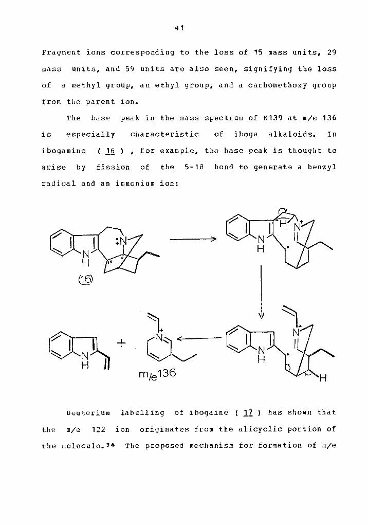
The base peak in the mass spectrum of K139 at m/e 136
is especially characteristic of iboga alkaloids. In
i bogamine ( 16 ) , for example, the base peak is thought to
arise by fission of the 5-18 bond to generate a benzyl
radical and an irnmonium ion:
+ N H
Deuterium labelling of ibogaine ( 12 ) has shown that
the m/e 122 ion originates from the alicyclic portion of
the rnolecule.36 The proposed mechanism for formation of m/e

42
122 requires the homolytic cleavage of the 7-8 bond to give
species ~ (Scheme IV):
(~ll ~j<
Scheme IV

43
The migration of hydrogen in species ! together with
the two bond fissions shown would give rise to the neutral
fragment ~ and the ion radical with m/e 123. Loss of one
hydrogen atom would give rise to the fully aromatized
pyridinium cation with m/e 122.
Using the information from the n.m.r. and the infrared
spectra of K139, the presence of a carbomethoxy group at
c-18 of the ibogamine skeleton could be proposed along vith
a methoxy sustituent in the benzene ring. The presence of
these groups would explain some of the additional peaks seen
in the mass spectrum of K139. For example, fission of the
7-8 bond in structure ( ~ ) would give ion £ (Scheme V).
Hydrogen migration from C-19 to the indole nucleus in
conjunction with the tvo bond fissions shovn, would lead to
ion ~ • This process is consistent vith deuterium
labelling results.3•

44
c
Scheme V
Loss of COOCH from ion D would give rise to the ion 3
with m/e 148. If the charge of the parent ion rests on the
indole nitrogen, then fission of the 7-8 bond in structure

45
1fl ) leads to thE ion with m/e 244:
CHO 3
8
The other impor-tant fr<qment in th£~ upper mass range of
K139 is at ~/e 283. Th~ probable mechanism for formation of
this iou is shown below, using the proposed structure
( .1£1 ) :

46
N H
COOCH3
This mechanism is consistent with the one proposed
for ibogaine,36 in which deuterium labelling showed that
the ion at m/e 225 included the indole nucleus as well as

47
carbon atoms C-18 and C-19, but not c-20.
The evidence presented thus far points to K139 being a
derivative of iboqamine l~ ) , with a carbomethoxy group
most likely at C-18 and a methoxy sustituent in the benzene
ring. Two known compounds tulfillinq these requirements are
voacangine ( 1.2~ ) and isovoacangine ( 12.h ) :
12!! Voacangine, R = OCH IR =H 1 3 2
1 9b Isovoacangine, R = H,H = OCH 1 2 3
The literature melting point of voacangine is
136-7 0
28 , and that of isovoacangine is given as
155-6° 37 A mixed melting point determination between K139
and an authentic sample of isovoacangine showed them to be
th~ same. The infcared spectrum of K139 and authentic
isovoacangine are identical in all respects.
Isovoacanqine was first obtained as the hydrochloride
These two genera are closely
related biogenetically with !~Q~rnae~on!~E~~

48
B. K117
This ccmpound was recrystallized from methanol-ether 0
and gave a melting point of 179-181 • The ultraviolet
spectrum shows absorption maxima at 315 na (logf=3.95), 242
nm (logf=3.95), and a shoulder at 226 na (loqf=3.91). This
type of ultraviolet spectrum is characteristic of the 2-acyl
indole group cf alkaloids.•o The infrared spectrum of K117
shows a broad band at 3330 cm-•, moderate bands at
3050 ca-•, 2940 c•-•, and 2850 cm-•. Strong absorption
bands are seen at 172 O cm- 1 (assigned to carbonyl
stretchinq of ester), 1540 cm-• (assigned to conjugated
carbonyl), 1445 c1.-• and 14 3 O c .- 1 , 1100 ca-1, and
730 cm-1 (assigned to 1,2 disubstituted benzene ring).
The n.m.r. spectrum of K117 shows a doublet at 1.1o(f corresponding to three protons and attributable to the
methyl doublet of an ethylidene group. A singlet integrating
for three protons is found at 2.64(f , and could be
attributed to an N - CH group. A one proton singlet is seen
at 2. 860 , 3
which may be the proton of a secondary amine.
Treatment with D o caused a reduction in the intensity of
this signal. A poo:ly resolved quartet at 5.3B(f is assigned
to the vinyl proton of an ethylidene group. Aro•atic protons
appear in the region 6.70-7.ao{f, which integrated for four
protons. A one proton singlet at 9.20Q is attributed to the

49
indole N-H proton.
Examination of the mass spectrum of a typical 2-acyl
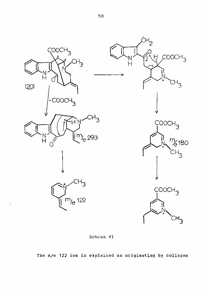
indole might be instructive. Vobasine ( 20 ), for example,
shows a molecular ion peak at m/e 352 and an M-59 peak at
m/e 293. The base peak is at m/e 180, and the only other
important peaks are at m/e 194 and m/e 122. 33 The origin
of the base peak in vobasine is explained in terms of
initial homolytic cleavage of the 5-6 bond just as in iboga
alkaloids. The resulting species ! (Scheme VI), can then
undergo ~cLafferty rearrangement to give the ion with •/e
180, which is probably capable of rearrangement to the
pyridinium ion ~ • The origin of the transferred hydrogen
from C-16 was verified by deuterium labelling.33
50
-COOCH3
l
Scheme VI
l + "cH
3
The m/e 122 ion is explained as originating by collapse

51
of the M-59 species at m/e 293 {Scheme VI).33
A third important peak in the spectrum of vobasine
appears at m/e 194. This has bePn explained in terms of
alpha-cleavage of the 3-14 bond in ion f to yield the ion
with m/e 194, which may be rearranged to the aromatic
pyridinium ion D :
N H
c
COOCH~ .._)
The mass spectrum of K117 (Figure 1) shows a molecular
ion at rn/e 338 and dn M-59 peak at m/e 279 as well as peaks
in the low mass range at m/e 180, m/e 166, and m/e 108. Each
of these ions is 14 mass units less than the important ions

52
of the vobasine spectrum as discussed previously. This
result could be explained if K117 were the N-demethyl
derivative ( 11 ) ot vobasine, since replacement of the
methyl group by hydrogen would explain the shift of all
peaks by 14 mass units without changing the exp~cted
fragmentation pattern of the vobasine skeleton. How€ver, the
configuration of the carbomethoxy group at C-16 of K117
( .f.1 ) is still in doubt.
ll R = COOCH , R = H, R = o, R = H l 3 2 3 "
££ R = COOCH , R = H, F = o, R = H l 3 2 3 •
23 H = COG CH , R = H, R = OH, R =H 1 3 2 3 " 21l n = H, R = COOC II , H = o, R = CH 1 z 3 3 " 3
Gorman and Sweeny reported the isolation of an alkaloid
called perivine to which they assigned the structure
( J.2 ) • 4 1 necause of a poorly resolved n.m.r. spectrum,
they converted ( l£ ) to its corresponding alcohol perivinol
( ~] ). The n.m.r. spectrum of perivinol showed a three
53
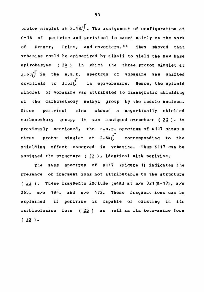
proton singlet at 2.4scf. The assignment of configuration at
C-16 of perivine and perivinol is based mainly on the work
of Renner, Prins, and covorkers.33 They shoved that
vobasine could be epimerized by alkali to yield the nev base
epivobasine
2.63{f in the
dovnfield to
24 in
n.m.r.
3.536'
which the three proton singlet at
spectrum of vobasine vas shifted
in epivobasine. Hence, the upfield
sinqlet of vobasine was attributed to diamagnetic shielding
of the carbcmethoxy methyl group by the indole nucleus.
Since perivinol also shoved a magnetically shielded
carbomethoxy group, it was assigned structure ( lJ ). As
previously mentioned, the n.m.r. spectrua of K117 shows a
three proton singlet at 2.64{f corresponding to the
shielding effect observed in vobasine. Thus K117 can be
assigned the structure ( 22 ), identical with perivine.
The mass spectrum of K117 (Figure 1) indicates the
presence of fragment ions not attributable to the structure
( 22 ). These fragments include ~eaks at m/e 321(~-17), m/e
265, m/e 184, and m/e 172. These fragment ions can be
explained if perivine is capable of existing in its
carbinolamine form ( l2 ) as well as its keto-amine fora
( 12 ).

- 54 -
0 (!) C\J " ~ ~ -
Q) 0
E E ::l L t) (l)
0 0. 00
(./) .--- ti)
Cf) <lS 2 ~
Q) L ::l CT>
LL 0
0 0 0 0 0 0 ~
0 C() (!) "\f" C\J ~
A.11SN31NI 3/\ll'Vl3C:!

55
In its carbinol-amine form, perivine would likely
exhibit some fraqmentation
spectrometer similar to those
akuammidine alkaloids ( ~1 ) .
reactions in
for ajmalicine
the mass
( 2 6 ) and

56
26 Ajmalicine l1 Akuammidine, R =COOCH 1 J
R -=CH OH 2 2
The fragmentation reactions are shown in Scheme VII:

57
-·OH
B)
Scheme VII

58
>
Scheme VII (Continued)
COOCH:3
OH
OH m1e 184

59
The first two fragmentation processes shown in Scheme
VII are typical of akuammidine alkaloids. In the first
process, the loss of an hydroxy group gives an M-17 peak in
analogy vi th the M-1 peak for loss of hydrogen in
akuammidine.•2 The second process in Scheme VII
corresponds to loss of the c-16 carbon bridge followed by
transfer of the C-18 hydrogen atom in a six-membered
transition state to yield the m/e 265 ion. This is a very
favorable process in akuammidine, where C-16 is
disubstituted. 42 The third framentation process in Scheme
VII is postulated as a retro-Diels-Alder fragmentation of
rinq c, followed by homolytic cleavage of the c c 14 15
bond to yield the conjugated ion with m/e 172. The
corresponding fragmentation process in ajmalicine gives rise
to an ion with m/e 156 which was shown by deuterium
labelling to contain the indole ring together with carbon
atoms 3, 6, and 14. 42 The last fragmentation process in
Scheme VII is postulated as originating from
retro-Diels-Alder fraqmentaticn of ring D followed by a
McLafferty type rearrangement to yield thejj-carboline ion
with m/e 184, although the rearrangement could conceivably
take place first.
c. K155 and K10~
Because of their similarity in physical properties and

spectral
toqether.
60
data, these two compounds will be discussed
The ultraviolet spectrum of the two compounds shows
that they both have the characteristic absorption of 2,3
disubstituted indoles. K102 has Amax at 244 nm (logf=4.73),
286 nm (logf=4.23), and 294 nm (logf=4.17); K 155 has Amax
at 2 2 9 nm (1ogf=4 • 7 1) , 2 8 6 nm ( 1 o g f = 4 • 2 2) , and 2 • 9 5 n 11
(loqf=4.20).
The infrared spectra of K102 and K155 are also quite
similar. In the high energy region, K102 shows maxima at
3390 cnr1, 2920 ca-1, and 2840 cm-t; in the same region,
K155 shows a broad absorption at 3500-3360 ca-1, and
moderate bands at 2950 cm-1 and 2870 ca-1. Both
compounds show intense carbonyl absorption: at 1730 cm-1
for K102 and at 1720 cm-1 for K155. In the fingerprint
region, K102 shows a broad band at 1460-1435 cm-1 whereas
K155 has a sharp •axima at 1460 cm-1 and a shoulder at
1430 cm-1.
Before discussing the n.m.r. spectra of K102 and K155,
initial information from mass spectrometry of the two
compounds should be considered. The mass spectrum of K102
shows the highest mass fragments at m/e 718 and •/e 704. The
mass spectrum of K155 gave exactly the sa•e highest mass
fragments, indicating that the two compounds are isomers.
Thus, the high molecular weight and u.v. spectra of K102 and

61
K155 indicate that they are more than likely bisindole
alkaloids "dimers" consisting of tvo distinct indole
units as in vinblastine and vincristine. Such alkaloids are
usually found in plants from which one or both of the
"monomeric" units have also been isolated.
The n.m.r. spectrum of K155 (Figure 2) gives some
information on the possible identity of one of the
structural moieties of this compound. In the high field
reqion, a doublet is observed at 1.66(f which integrates for
3 protons and can be assigned to the methyl signal of an
etbylidene group. The quartet at 5.3o(f corresponds to the
group. Three proton singlets
and 2.6o(f. The high field
vinyl proton of an ethylidene
are also observed at 2.44(f
signal (2.44cf ) has practically the same chemical shift as
the carbomethoxy singlets of perivino1•2 and vobasine.•s
The singlet at 2.6ocf in K155 is then an N-methyl group, and
has a chemical shift similar to that for vobasine.• 3 These
observations strongly suggest the presence of a
vobasine-like skeleton in K155, although the vobasine
fragment will not be present in the 2-acyl indole form since
only 2,3-disubstituted indole absorption is seen in the u.v. spectrum.

I I I I I I I I I I I I I I I I I I I I I I I I I -,--:-, I I I I I I I I I I I I I I I I I I I I
I 6
I 5
I 4
I I 3 2
Figure 2-Nuclear Magnetic Resonance spectrum d K155.
I Oppm
0\ N
63
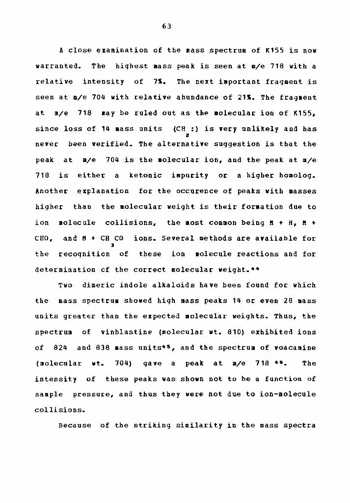
A close examination of the mass spectrum of K155 is now
warranted. The highest mass peak is seen at m/e 718 with a
relative intensity of 7%. The next important fragment is
seen at m/e 704 with relative abundance of 211. The fragment
at m/e 718 may be ruled out as the molecular ion of K155,
since loss of 14 mass units (CH :) is very unlikely and has 2
never been verified. The alternative suggestion is that the
peak at m/e 704 is the molecular ion, and the peak at m/e
718 is either a ketonic impurity or a higher homolog.
Another explanation for the occurence of peaks with masses
higher than the molecular weight is their for•ation due to
ion molecule collisions, the most common being M + H, M +
CHO, and M + CH co ions. Several methods are available for 3
the recoqnition of these ion molecule reactions and for
determination cf the correct molecular weight.••
Two dimeric indole alkaloids have been found for which
the mass spectrum shoved high mass peaks 14 or even 28 mass
units greater than the expected molecular weights. Thus, the
spectrum of vinblastine (molecular vt. 810) exhibited ions
of 824 and 838 mass units•s, and the spectrum of voacamine
(molecular wt. 704) gave a peak at m/e 718 ••. The
intensity of these peaks was shovn not to be a function of
sample pressure, and thus they were not due to ion-molecule
collisions.
Because of the striking similarity in the mass spectra

64
of voacamine and K155, they will be considered together in
the following discussion.
Thomas and Biemann investigated the origin of the high
mass peaks in the spectrum of voacaaine. • 7 In aqreement
with the hypothesis put forward by Buchi, Manning,
and Konti,•e the peak at K + 14 and some of the other high
mass peaks in the voacamine spectrum were found to be the
result of a sequence of thermal reactions occuring in the
mass spectrometer before ionization. The specific reaction
in question is an inter•olecular transfer of a aethyl group
to produce two nev species, vhich may undergo further
thermal reactions. The carboaethoxy group R of voacaaine I
( ~!.! ) was suggested as the source of the transferred
methyl group.•7

65
R. 1 (30) (_J1J
NH
~~ Vodcaruine, R = R = COOCH , R =H 1 2 3 3
29 Voacamine - d , R =COOCD , R =COOCH R =H , 3 1 3 2 3 3
30 Voacangine, R =COOCH 1 3
31 Voacangine - d , R = COO CD 3 l 3
To test this suggestion, Thomas and Diemann prepared
voacangin8 a and condens~d it with vobasinol to obtain
voacarnin2 d ( 22 ). 47 The mass spectrum of voacamine 3
- d showed a molecular ion at m/e 707 corresponding to the 3
exp2cted trideuterated compound. The new high mass peak of
724 (M+17) instead of 721 (11+14) indicated the specific

66
transfer of a CD group followed by Hofmann elimination of 3
H to yield the species with mass of 724.
The experimental data also shoved that the
transmethylation proceeded by transfer of a methyl group to
the alicyclic nitrogen of the vobasinol moiety. This
conclusion is based on the following observations. The peak
at m/e 676 in voacamine - d might at first be thought of 3
as arising from loss of OCD from the molecular ion at m/e 3
707, but this suggestion vas in direct contradiction with
the observation that m/e 676 is a pyrolysis product which
increases in intensity vith sample pressure.•7 Instead,
the peak at 11/e 676 arises from loss of M (CH ) (CD ) 3 3 2
following transmethylation and a second Hofmann eli•ination
on the species with mass of 724. All of these thermal
rearrangements are summarized in Scheme VIII:

676 <
67
-H· >
Schell'e VIII
CCXXJ~
(J_1)
t 724
CD3 \V
COOCH:3
(3_1)
~ith this understanding of the thermal transmethylation
reactions of voacamine, it is possible to explain the

68
identity of some of the high mass peaks in the spectrum of
voacamine. The ion at m/e 646 is an M-58 peak corresponding
to loss of the transferred CH group from the voacangine 3
moiety followed by loss of CO and protonation at the site 2
of the original carbomethoxy group. Ions at m/e 451 and a/e
464 are formed by fragaentation of the m/e 646 ion. These
last two fragmentations are typical of those already
described for vobasine and perivine. Fragments at m/e 522
and a/e 509 arise from the same vobasine-type fragmentation
except that electron impact occurs on the parent species
(M=704) instead of the ion at a/e 646. Some of these
fragmentation reactions are shown in Scheme IX:

69
-CH 3 >
coo
NH H
H
Scheme IX

70
The lower mass range of voacamine revealed the presence
of all the non-indolic fragments also seen in the mass
spectru11 of voacangine ( 30 ) , namely the ions at m/e 122,
11/e 124, 11/e 136, and m/e 14 8. The indole containing ions of
voacangine, namely m/e 160, m/e 184, 11/e 244, and 11/e 283
are absent in the spectrum of voacamine. So the connnecting
link between the voacangine monomer and the vobasinol
monomer must reside on the benzene ring or the indole
nitrogen of voacangine and not on the alicyclic portion of
voacangine.
The proof that the linkage between the voacangine unit
and the vobasinol unit resides on the benzene ring of
voacangine was provided by the following experiments.•e
Hexadeuteriovoacamine was prepared which gave one proton
singlets at 7.48()' and 1.1a{f, which could be attributed to
the indole N-H signals. The high field indole N-H signal
(7.480 ) disappeared on exchange in D o, while the low 2
field proton required acid catalysis. This result indicated
that the low field proton was probably hydrogen bonded and
this suggestion was enhanced by the synthesis of the model
compound 11 ), in which it was discovered that the indole
N-H proton was exchanged for deuterium only under acid
catalysis.
71
(3~
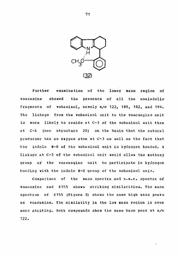
Further examination of the lower mass region of
voacamine showed the presence of all the nonindolic
fragments of vobasinol, namely m/e 122, 180, 182, and 194.
The linkage from the vobasinol unit to the voacangine unit
is more likely to reside at C-3 of the vobasinol unit than
at c-6 (see structure 28) on the basis that the natural
precursor has an oxygen atom at C-3 as well as the fact that
the indole N-H of the vobasinol unit is hydrogen bonded. A
linkage at c-3 of the vobasinol unit would allow the methoxy
qroup of the voacangine unit to participate in hydrogen
bonding with the indole N-H group of the vobasinol unit.
Comparison of the mass spectra and n.m.r. spectra of
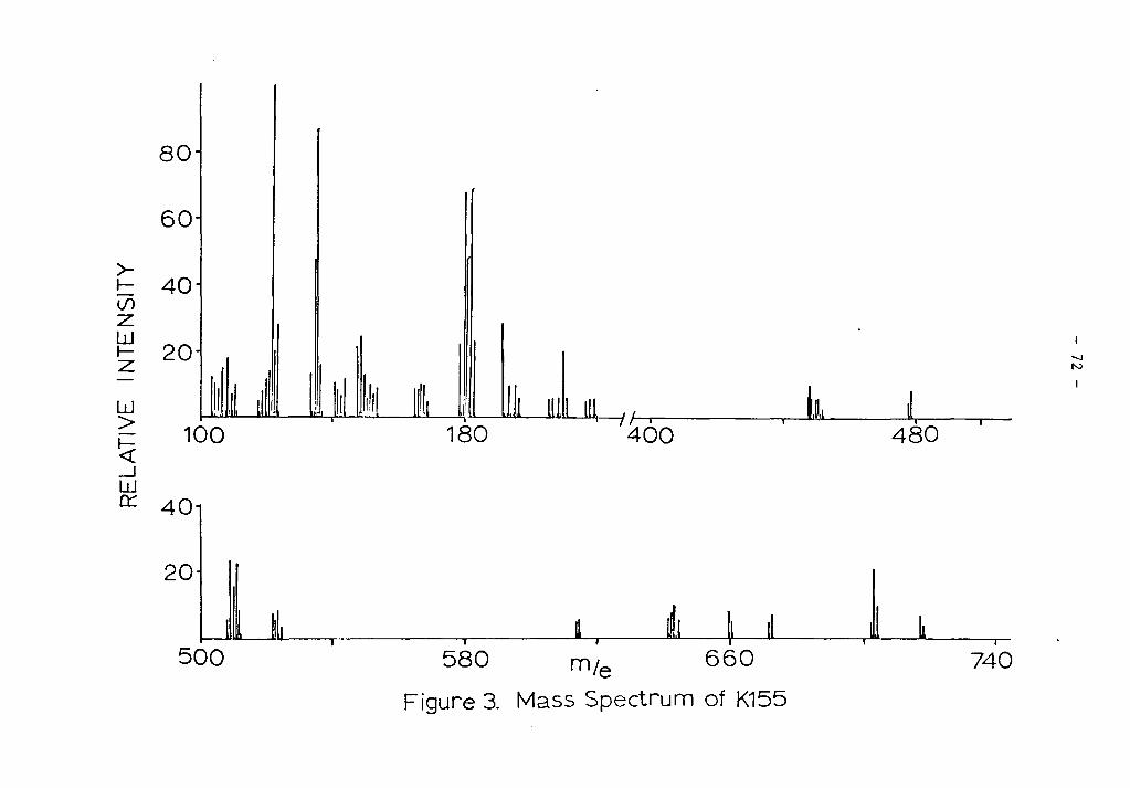
voacamine and K155 shows striking similarities. The mass
spectrum of K155 (Figure 3) shows the same high mass peaks
as voacamine. The similarity in the low mass region is even
more striking. Both compounds show the same base peak at n1/e
122.
80
60
~ 40 if) z ~ 20 z w > ~
100 _J w n::: 40
20
500
180 11 '"'" "''" Ill\ I / ,,~,\ ~ ' I I I I I
400 480
580 m1e 660 740
Figure 3. Mass Spectrum of K155
-...J N

73
The next most intense peaks are at m/e 136, 180, and 182 in
both spectra. The fragments at m/e 124, m/e 148, and m/e 194
are also in the same relative intensity in each spectra.
The n.m.r. spectra of voacaaineso and K155 are also
quite similar (Table XI). The assignaents of an ff-methyl
qroup, two carbomethoxy groups, and a methoxy group linked
to an aromatic ring can readily be aade for K155. The
aromatic region of K155 also integrates for six protons,
shovinq singlets at 6.786' and 6.94(f, a doublet at 7.060:
and downfield protons in the region 7.46{f to 1.10{). With the information obtained so far, it is tempting to
assign the structure of voacamine ( 28 ) to K155, but
investigation of the literature on dimeric alkaloids shows
that this assignment is not yet warranted.

74
TABLE XI PROTON N.ft.R. CHEMICAL SHIFT DATA POR
VOACAKINE 49 AND K155
Voacamine * Che11. Shift cd>, (No. of Protons), Assignment 1.66(doublet,3 protons),
C -= C - CH 3
2.44 (sing let, 3 protons) , COOCH 3
2.58 (singlet,3 protons) , N - CH
3.61 (singlet,3 protons) , COOCH 3
J.95(singlet,3 protons), 0 - CH 3
5.20 (quartet, 1 proton) , c -= c -
7.48(singlet,1 proton) ,indole N-H 7.78(singlet,1 proton) ,indole N-H
*Solvent: CDCl 3
3
ff
K155 * Chea. Shift 0} , (No. of Protons) 1.66(doublet,J protons)
2.44(singlet,3 protons)
2.60(singlet,3 protons)
3.66(singlet,3 protons)
3.92(singlet,3 protons)
5.30(quartet,1 proton)
6.78(singlet,1 proton)
6.94(singlet,1 protron)
7.06(doublet,2 protons)
7.46-7.70(2 protons)

75
Renner, Prins, and Stoll isolated from Cono_Eh.~£1119.!€!
Q.!!.£i~sim_~ a compound which was later found to be isoneric
with voacamine.37 This compound was also found to be a
dimeric alkaloid and was given the name conoduramine.
However, conoduramine was obtained from a plant which had
yielded isovoacangine but not voacangine. Since the n.m.r.
and infrared data of conoduramine and voacamine are so
similar, ccnoduramine was assigned the structure ( 11 ) , and
is the bisindole alkaloid with an isovoacangine moiety and
the vobasinol linkaqe located at the 5-position of thP
isovoacangine indcle ring.
COOCH3
11 Conoduramine
76
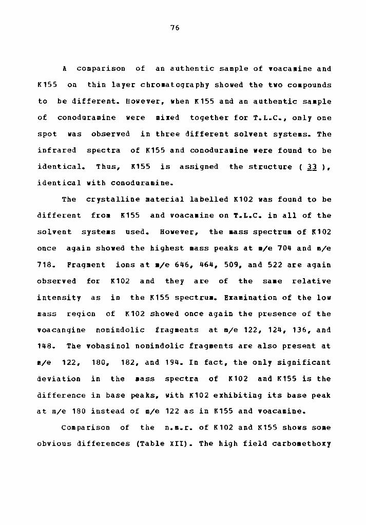
A comparison of an authentic sample of Yoacaaine and
K155 on thin layer chromatography shoved the two coapounds
to be different. However, when K155 and an authentic saaple
of conoduramine were mixed together for T.L.C., only one
spot was observed in three different solvent systems. The
inf rared spectra of K155 and conoduramine were found to be
identical. Thus, K155 is assigned the structure ( Jl ), identical with conoduramine.
The crystalline material labelled K102 was found to be
diffe~ent from K155 and voacaaine on T.L.C. in all of the
solvent systeas used. However, the aass spectrum of K102
once again shoved the highest mass peaks at m/e 704 and m/e
718. Fragment ions at a/e 646, 464, 509, and 522 are again
observed for K102 and they are of the saae relative
intensity as in the K155 spectrum. Examination of the low
mass region of K102 showed once again the presence of the
voacanqine nonindolic fragments at m/e 122, 124, 136, and
148. The vobasinol nonindolic fragments are also present at
m/e 122, 180, 182, and 194. In fact, the only significant
deviation in the mass spectra of K102 and K155 is the
difference in base peaks, with K102 exhibiting its base peak
at m/e 180 instead of m/e 122 as in K155 and voacamine.
Comparison of the n.m.r. of K102 and K155 shows some
obvious differences (Table XII). The high field carboaethoxy

77
signal of K102 appears at 2.s6cf whereas the high field
carbomethoxy signal of K155 appeared at 2.44{f. The same
relative shift is seen in the N-methyl group of K102 (2.646' instead of 2.6ocf as in K155) and in the aethoxy group of
K102 (3.98cJ instead of 3.92{f as in K155). The apparent low
field carbomethoxy signal of K102 appeared at 3.6acJand an
additional singlet appeared at 3.656. The integration for
these two singlets totaled to 3 protons. An additional
singlet also appeared in the n.m.r. spectrum of K102 at 3.08
J(Fiqure 4).
Thus, the evidence uncovered for K102 at this point
indicates that it is a bisindole alkaloid made up of a
vobasine moiety and either a voacangine or isovoacangine
moiety as in voacamine or conoduramine respectively. Further
investigation of the literature reveals that two additional
isomers of voacamine are known.

I I I I I I I I I I I I I I I I I I I I I I I I I I I I I I I I I I I I I I I I I I I I I I I I I I 'I
I 8
I 7 I
6 I 5
I 4
I 3
I 2
Figure 4-Nudear Magnetic Resonance spectrum of K102
r-1
I Oppm
-...J CXl

79
TABLE XII PROTON CHE~ICAL SHIFT DATA FOR K155 AND K102
K155 * Chem Shift J> , (No. of Protons),
Assignment 1.66(doublet,3 protons),
C = C - CH 3
2.44(sinqlet,3 protons) , COOCH 3
2.60(singlet,3 protons) , N - CH
3.66(singlet,3 protons), COOCH 3
3.92(singlet,3 protons) COOCH 3
3
5.30(quartet,1 proton), C = c - H
6.78(singlet,1 proton) ,aromatic-H
6.94(singlet,1 proton),aromatic-H
7.06(doublet,2 protons),aromatic-R
7.46-7.70(2 protons),indole N - H
•Solvent: CDCl 3
K102 * Chem Shift J>,
(No. of Protons) 1. 68 (doublet, 3 protons
2.56(singlet,3 protons)
2.64(singlet,3 protons) 3.08(singlet) 3. 65 (singlet) 3.68(singlet)
3.98(singlet,3 protons)
5.34(quartet,1 proton)
6.82(singlet,1 proton)
6.90(singlet,1 proton)
7.10(singlet,2 protons)
7.68(singlet,1 proton)
7.73(singlet,1 proton)

80
Renner reported the isolation of a bisindole alkaloid
from which vas given the
name voacamidine.so Chemical and spectral data indicated
that the co•pound was isoaeric with voacamine, but the
n.m.r. spectrum shoved that the coapounds vere different.
The m.m.r. spectrum of voacamidine shoved tvo shielded
methoxy groupings at 2.ssQ' and 3.oa{f .•9 The signal at
2.sa6 was assigned to the diaaagnetically shielded
carboaethoxy group of a vobasinol moiety in analogy vith
voacamine. But the signal at 3.oa6 was attributed to an
o-methyl qroup attached to the benzene ring of a voacangine
moiety. Consequently, voacamidine was assigned the structure
Clf!.), in which the aromatic aethoxy group of the
voacangine moiety is situated above the indole ring of the
vobasine
the C
fragment as a result of restricted rotation around
- c bond. The three proton singlet at 3. 08 6 was 3 11
attributed to this shielded o-aethyl group.
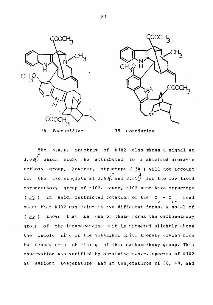
81
l!!. Voacamidine 35 Conodurine
The n.m.r. spectrum of K102 also shows a signal at
3.oe6 which might be attributed to a shielded aromatic
methox y group, however, structure ( l!! ) will not account
for the two singlets at 3. 68Q and 3. 656 for the low field
carbornethoxy grcup of K102. H2uce, K102 must have structure
( 35 ) in which restricted rotation of the C - c bond 3 1 ..
mHans that K102 can exist in two different forms. A model of
( J.2 ) shows that in cne of these for ms the carbometho xy
group of the isovoacangine uuit is situated slightly above
the indole ring of the vohasinol unit, thereby giving rise
to diamagnetic shielding of this carbomethoxy group. This
observation was verified by obtaining n.m.r. spectra of K102
at ambient tnmFerature and at temperatures of 30, 40, and

82
50° c in acetone - d solvent. The tvo singlets began to 6
coalesce as the temperature was increased, until only a
broad singlet was seen at 50 c. Prins, Stoll, and Renner isolated a second bisindole
alkaloid from ~Q.!!QEhariJ!gia dnrissima to which they gave
the naae conodurine.37 The structure ( 3 5 ) was later
assiqned to this alkaloid.•9 When an authentic sample of
conodurine and K102 were mixed together for T.L.c., only one
spot was observed after development in three different
solvent systems. The infrared spectra of K102 and authentic
conodurine were also found to be identical.
The pharmacological testing data on conodurine and
conoduramine is not very extensive, whereas voacaaine has
undergone quite detailed testing in the NIH anti-tumor
systems probably because of its dimeric structure, a feature
exhibited by some of the other anti-tumor alkaloids.
Voacamine was active in the W-256, S-180, and OS survival
tumor systems, but inactive in the PS system.s Conodurine
f ro11 T. iohnstonii was tested in the KB in vitro system
and qave a value of 31 j.1.9/•l, indicating that it was
moderately active. Conoduraaine from
tested in the KB system and gave a value of 19 µg/111
indicating that it vas active in this systea.
In this
plant species
The purpose
chdracterize
work, a phytochemical investigation of the
Igbefilgfil!!Qnta~g- johnstonii is described.
of the investigation vas to isolate and
the constituents of the plant for subsequent
testing in the anti-cancer bioassay system developed by the
National Institutes of Health.
Systematic fractionation of the plant in conjunction
with bioassay at each stage of fractionation has revealed
that the biological activity is concentrated in the
alkaloidal portion of the plant material. Purification of
the crude alkaloidal material by chromatography and buffer
extraction led to the isolation of four indole alkaloids,
two of which were shown to be dimeric in nature. A
nonalkaloidal material which was characterized as a
triterpene was also isolated from ~jQhnstQnii~
The two monomeric alkaloids were identified as
isovoacangine and perivine. Their identity was verified by
comparison with authentic samples. The two dimeric alkaloids
were shown by n.m.r. and mass spectrometry to be isomeric
with the known alkaloid voacamine. Subsequently, the two
alkaloids were identified as conoduramine and conodurine,
and these structural assignments were verified by comparison
with authentic samples.
Of the alkaloids characterized in this study, only
83

84
perivine had been tested previously in any anti-cancer
bioassay. Perivine was found to be inactive in each of the
following "survival tumor" systems: LE, Lt, P4, and WA.s
The result obtained during this work for the KB bioassay of
perivine shows that it is inactive in this system also. In
the only important pharmacological study involving
isovoacanqine, the compound was shown to cause a lovering in
blood pressure when administered intraveneously to the
guinea pig.s1 The current study shoved that isovoacangine
was also inactive in the KB bioassay.
Conodurine showed a KB activity of 31tJ_g/ml, indicating
only a moderate activity in the bioassay system.
Conoduramine shoved a KB activity of 19 J.19/•l, and thus is
within the threshold of activity established by the NIH.
After the KB bioassay data for these four alkaloids had been
obtained, a new in vitro PS bioassay was developed by NIH
as a counterpart to the normal "survival tumor" PS bioassay.
Each of the four alkaloids isolated in this work was
submitted for the new bioassay and the results obtained (in
/.lg/ml) were as follows:
Perivine
Isovoacangine
Conodurine
Conoduramine
20.0
18.0
29.0
20.0

85
These results indicate that at least some moderate PS
activity is possessed by each alkaloid listed above. The
combined KB and PS results for conodurine and conduraaine
are supported by the finding that the Geigy Pharaaceutical
Company has patented preparations of voacamine, voacaaidine,
conodurine, and conoduramine in tablet form for the
reduction of neoplasia growth.s2

EXPERIKENTAL
All inf rared spectra were recorded in KBr on a Beckman
IR-20AX infrared spectrophotometer or a Beckman Acculab 1
infrared spectrophotometer.
Ultraviolet spectra were run in 951 ethanol solution on
a Cary 14 u.v.-visible spectrophotometer.
Nuclear magnetic resonance spectra were obtained on a
JEOLCO JNM-PS-100 instrument using deuteriochloroform as the
solvent unless otherwise noted. Tetraaethylsilane (TMS) was
used as the internal standard.
The mass
Hitachi-Perkin-Elmer
ionization voltage
spectra were obtained on a
RMU-7 double beam spectrometer with an
of 70 e.v. unless otherwise noted.
Perfluorokerosene was used as the reference in counting the
spectra.
The data from carbon-hydrogen-nitrogen analysis was
obtained on a Perkin-Elmer 240 Elemental Analyzer.
ftel ting points (uncorrected) were obtained on a
Thomas-Hoover apparatus or a Ther•olyne heating stage with
microscope.
Solvents employed were reagent grade solvents
redistilled in glass except where large amounts of solvent
were required, in which case reagent grade solvents were
86

87
used directly.
The thin layer chromatography plates employed were of
two types. The first type were commercially prepared
Analtech T.L.C. plates precoated with silica gel GF with a
thickness of 250 microns. The second type were "homemade"
plates made of silica gel GF with a thickness of
approximately 300 microns. These plates were prepared on a
Shandon UNOPLAN mechanical spreader as an aqueous slurry.
Vizualization of T.L.C. plates was perforaed using a
Chromato-Vue CC-20 long and short wave viewing apparatus
manufactured by Ultra-violet Products, Inc. Further
vizualization vas done by spraying plates with either
iodoplatinate solution (5% H PtCl • 6 H O in aqueous KI) 2 6 2
or eerie ammoniu• sulfate solution [(NH ) Ce(SO ) 4 • • 4
2 H 0 in ortho-pbosphoric acid], both of which are very 2
sensitive to alkaloids.
~- Fractionation I
A total of 2.52 kg of dried plant was weighed out and
distributed evenly into six beakers. The aaterial was
stirred with cold 95i ethanol for 24 hours, decanted, and
stirred again with fresh ethanol for 4 hours. These ethanol
extracts were decanted again and fresh ethanol vas added
followed by ~arming on a hot plate with stirring for 4
additional hours. Dilute NH OH was added to each beaker 4

88
and the stirring was continued for another 4 hours.
The remaining solid plant material was filtered off and
discarded, and all the ethanol extracts were combined and
evaporated to 750 ml. A 1.83 g sample of this extract vas
set aside for bioassay and labelled P053. This extract was
added to a separatory funnel with 300 ml. of water and
shaken 3 times with 1-liter portions of ethyl acetate. The
ethyl acetate fractions were combined, while the remaining
aqueous layer was made basic with NH OH and put in a 4
liquid extractor. Exhaustive extraction with ethyl acetate
was continued for three days (changing ethyl acetate layer
each day). The final ethyl acetate extract vas colorless.
All of the ethyl acetate extracts were then combined
and evaporated to 900 al; a 1.69 g sample was taken out for
bioassay and labelled as P054. The ethyl acetate was then
extracted with six 500 al portions of 5% H so • The dark, 2 4
qummy material which foraed was separated, dissolved in
ethanol, freeze dried, and labelled F059.
The sulfuric acid fraction was extracted 3 times with
benzene, then neutralized to pH 7 with NH OH. The neutral 4
aqueous fraction was then extracted twice with chloroform,
made strongly basic (pH>10) and re-extracted 4 tiaes with
chlorofora. All chloroform extracts were combined, washed
with water, dried over cacl , and evaporated to dryness to 2
yield 52.5 g of crude alkaloidal material labelled P057. The

89
precipitate formed during the neutralization step was
separated from the basic solution after extraction and
labelled P058. The fractionation tree is shown in Table
XIII.

90
TABLE XIII FRACTICNATION TREE OF T. JOHNSTONII
FRACTIONATION I
2.52 kg Ground Plant I Ei:tract I 951 Ethanol I NH OH I Concentrate • I
Ethanol Extract (183g) F053
I Extract Ethyl I Acetate / NH OH
·~~~' ··~~-I Ethyl
Acetate Soluble
F054 I Ei:tract 5% H so
~~~~~--~'~--~----~-2 • I I I
Ethyl Solid H SO Acetate (39.3g) 2 • Soluble F059 Solnble (71.7q) I Wash with Benzene F056 I Neutralize to pH7
I Extract CHCl
I CH Cl
Soluble (52.5g)
F057
I 3 I !!ake to pH10 I Extract CHCl
~~-' 3 I
Solid (8. 35g)
POSS
Table of Activities
I CHCl
3
Insoluble
Fraction No. KB Ac ti vi ty ~g/111)
F053 F054 FOSS F056 F057 F058 F059
23 24
>100 >100
16 18 24
I Aqueous Soluble (13.39)
POSS

91
1. Column Chros~tography of F057
A 20 g sample of F057 was weighed out and dissolved in
a mixture of ether, chloroform, and methanol in a large
round-bottom flask. The volume vas reduced to about 140 ml
and 42 g of Woelm Alumina (Activity Grade III) vere added to
the flask. The aixture was reduced to dryness on a rotary
evaporator and the saaple vas scraped from the flask and
ground into s•all particles. This dry sample vas poured on
top of a glass column already containing about 250 g of
Woelm Alumina which was dry packed. The coluan was eluted in
order vith the following series of solvents: benzene,
chloroform, ethyl acetate, n-propanol, and methanol. About
700 ~l. of eluant vas used in each case. The eluted
fractions were evaporated and weighed to give the following
result:
Benzene 3.25
Chloroform 8.23
Ethyl Acetate 1. 15
N-Propanol 1. 73
Methanol 2. 51
h_ Buff er
Assay No.
F064
P065
P066
F067
F068
Extraction of F064
KB Activi~I ~·ll
1. 9
2.0
2.2
2.5
2.0
A 3g sample of the benzene fraction F064 was dissolved

92
in 50 ml of benzene. Buffers of pH 4.0 and pH 2.6 were
prepared fro• stock solutions of citric acid (0. 1 " C H 0 ) and sodium orthophosphate (0.2 Pl Na HPO
6 8 ., 2 • H20).$ The Mcilvaine procedure 53 vas used to make up .,
the buffers: 61.45 ml of 0.1 M citric acid and vas mixed
with enough 0.2 M sodiua phosphate to make 100 ml of pH 4.0
buffer; and 89.1 ml of 0.1 Pl citric acid vas mixed with
enough 0.2 Pl sodium phosphate to make 100 ml of pH 2.6
buffer.
The benzene solution of F064 vas first extracted with
pH 4.0 buffer. Some emulsion was foraed which vas broken by
addition of about 50 al of chlorofora vith stirring. The
aqueous layer was removed, and the remaining solvent layer
was re-extracted in order with pH 2.6 buffer and 101
H so . The organic layer which reaained after extraction 2 •
by 101 H SO was dried over ltgSO and evaporated to 2 • 4
dryness. Each of the aqueous extracts was made basic (pH
11-12) with 5% NH OH and extracted immediately vi th •
chloroform. The chloroform extracts were dried over MgSO •
and evaporated to dryness to give the following results:
Mate!:i~! Weight jg} Assay Mo. KB ActiviU
Extracted by pH 4.0 buffer 0.40 F069 2.1
Extracted by pH 2.6 buffer 1.25 F070 17
Extracted by 101 H SO 0.90 2 •
Acid Insoluble 0.11 F071 31

93
Thin layer chromatography of fraction F070 on silica
qel (CH CH OH CH COOCH CH CCl , 1:4:5) showed a 3 2 3 2 3 •
component near the solvent front and five other well-defined
component spots (short vave U.V.) with Rf values ranging
from 0.25 tc 0.70. The sulfuric acid fraction also shoved a
large component spot near the solvent fron on T.L.C., so
back extraction of the sulfuric acid fraction by pH 2.6
buffer was carried out. Two extractions with pH 2.6 buffer
f olloved by T.L.C. on silica gel shoved that some of the
front running component was being extracted into the pH 2.6
buffer. The extraction was continued until six extractions
with pH 2.6 buffer were completed. The aaterial obtained by
this extraction had a weight of 0.33 g and was added to the
previous F070 fraction to make a fraction weighing 1.58 g
(Table V).
3. Chroaatoqraphi of F070
A chromatographic column was prepared by slurry packing
160g of Woel• Silica Gel (Activity Grade III) onto a glass
column with dimensions 3 ca i.d. by 39 ca. The solvent for
slurry packing and for the subsequent elution was a mixture
by volume of CH CH OH
the same 3
solvent 2
mixture
CB COOCH CH 3
used 2 3
in the
CC! (1:4:5),
T.L.C. of this
fraction. A sample of P070 weighing 1.5 g vas dissolved in a
small amount of elating solvent and pipetted onto the

94
column. The eoltion was begun and fractions of approximately
25 ml were collected. Thirty-three fractions were collected.
and T.L.C. (CH OH - CHCl • 1:9) shoved that recombination 3 3
to make larger fractions vas warranted. The results of these
recombinations were as follows:
Fraction Weig!!! !J2proxi ~ate KB Activit.I No. jgl_ Rf Values Jµ.91•1}
P077 0.050 0.85 and 0.70 17
F078 0.230 0.85, o. 70, and 0.60 18
F079 0.58 o.a5. o. 10. 0.60, 0.55 18
F080 0.08 0.55, 0.45, and 0.40 25
F081 0.04 0.45 and 0.40 24
F082 0.04 0 - 0.15 29
4. Pre,Earati!~Ihin L~1er Chromatography of F078
Fraction FC78 was subjected to thin layer chromatogrphy
in several different solvent systems in addition to the two
already mentioned in the previous section. The solvent
system CH CH OH
most 3 2
effective
CH COOCH CH (1:4) was found to be the 3 2 3
from the standpoint of coaponent resolution
and minimization of tailing effects.
Preparative layer chromatography plates having a
thickness of 1000 microns and dimensions of 20 cm by 20 cm
were obtained from the Analtech Corporation. The sample to
be used on each ~late was dissolved in approximately 1 ml of

95
eluting solvent and applied using a Shandon mechanical
syringe applicator. Four individual runs vere required in
order to chromatograph 0.21 g of F078. The veights used in
each run were as follows: Run 1 (0. 061 g) ; Run 2 (0. 046 g) ;
Run 3 (0.051 g); Run 4 (0.052 g). Each plate was developed
in the CH CH OH CH COOCH CH system and the 3 2 3 2 3
development time was approximately 40 to 50 minutes. After
development, each plate vas vizualized by short wave u.v. liqht and divided into five segments having the following
approximate Rf values: Segment A, Rf = 0.90 - 1.0; Segment
B, Rf = 0.80 - 0.90; Segment c, Bf = 0.50 - 0.80; Segment D,
Rf = 0.10 - 0.50; Segaent E, Rf = o.o - 0.10. Each segment
vas then scraped individually from the plate, and the silica
gel was poured into methanol to extract the organic material
from the qel. The liquid was recovered after suction
filtration and each methanol extract vas evaporated to
dryness and weighed. Similar fractions from each run were
combined tc give the following results:
lf:~tiQ!!~~ Weigl!t {g) Descrietion
1-70-1 0.044 Mostly one component
1-70-2 0.013 f!ostly one component
1-70-3 0.090 Two components
1-70-4 o.oos Fluorescent material
1-70-5 0.009 Material left at origin

96
The fraction labelled 1-70-3 appeared to consist of two
components ~hich would be separable by careful preparative
T.L.C.; 70 mq of fraction 1-70-3 was weighed out and
dissolved in approxiaately 2 ml of the ethanol-ethyl acetate
(1:4) solvent. The sample was applied in equal proportion to
two Analtech Silica Gel G plates of 1000 aicrons thickness,
and the plates were developed in the saae solvent system.
Vizualization was done by short wave u.v. and three distinct
components appeared on each plate having approximate Rf
values of 0.9, 0.55, and 0.50. These components were taken
as segments A, B, and C respectively. Segaents B and C were
slightly overlapped, so the line of least concentration
between the two was chosen as the division point. The silica
gel was scraped from each segment and extracted with
methanol as before to yield the following fractions:
Fraction N£~
1-74-1
1-74-2
1-74-3
Weight_Jgl
0.002
0.034
0.010
Appearance
White, amorphous
White, crystalline
White, amorphous
Fraction 1-74-2 gave the appearance of being
crvstalline during evaporation of solvent. Recrystallization
from methanol yielded 16 mg of colorless rods with m.p.
above 210° with decomposition. Attempted recrystallization
of fractions 1-74-3, 1-70-1, and 1-70-2 yielded only

97
amorphous solids. The crystalline material from fractions
1-74-2 was labelled K102 and submitted immediately for KB
i.!L_!it£Q bioassay and for PS in vi tr2 assay when that
test became available. The KB activity of K102 was 31 /.lg/111
and the PS activity vas 29 µg/m.l. When a sample of
conodurine became a vailableS•, it was tested with K102 in
thin layer chromatography. Three plates with a thickness of
30 microns each were spotted with small amounts of K102,
pure conodurine, and a mixture of K102 and conodurine so
that a total of three sample spots appeared at the origin of
each plate. The plates were developed in the following
solvents: Plate A: ethanol-ethyl acetate (1:4), Plate B:
methanol-methylene chloride ( 1: 9) , Plate C:
methanol-acetonitrile (3:7). The developed plates were
vizualized by short vave u.v. Plate A showed all components
at an Rf value of 0.56. Plate B shoved all coaponents at an
Rf value of 0.69, and Plate C showed all coaponents at an Rf
value of 0.41. The saaple corresponding to the mixture of
K102 and conodurine appeared as only one component on each
plate both on viewing under u.v. light and after spraying
with eerie aaaoniu11 sulfate solution. The spectral data
recorded for K102 were as follows:
C H N 0 43 52 • 5
requires: C(73.26), H(7.44), N(7.95)
found: C(70.72), H(7.37), M(7.50)

98
C(71.03), H(7.41), N(7.85)
C(72.04), H(7.64), N(8.39)
Ultraviolet
Amax, (log ) : 228 nm (4.47), 275 nm (3.81), 298 na (3.93)
Infr~ed
v.a x: 33 80 cm-1, 2940 and 2920 (doublet) , 3850, 17 30, 162
1460, 1340, 1245, and 1230 (doublet), 11
1130, 1090, 1035, 1010. 850, 800, 740;
!~~.!h (See Table XII)
Mass ~Eectrua: m/e (Relative Intensity)
720 (2) 381(4) 144 (8)
719 (4) 352(5)M++ 136 (2LJ)
718(6) 336 ( 8) 124 (10)
704 (55) fll+ 309 (9) 122 (66)
689(4)M-15 295(4) 117 (9)
673(8) 257 (4) 44 (6)
646(5)M-58 208 (5) 42 (7)
574 (6) 194(23)
524 (28) 182(42)
522 (8) 181 (100)
511 (22) 180(100)
509 (22) 148(6)
393 (6) 145 ( 17)

99
A sample of ground plant weighing 2.8 kg was obtained
and exhaustive extraction was begun with 95% ethanol. The
procedure for this fractionation is exactly like the one
used in Fractionation I (page 90} , and the new fractionation
tree is shown in Table XIV. Because these fractions were
obtained by the same procedure used in a previous
extraction, they vere not submitted for bioassay.

100
TABLE XIV FRACTIONATION TREE OF T. JOHNSTON!!
FRACTIONATION II
2.8 kg Ground Plant I Extract with I 951 Ethanol / I
NH OH
" I Partial Evaporation I v
Ethanol Extract I I Extract Ethyl I Acetate I NH OH
I I
Ethyl Acetate Soluble
I
, ___ I I I
Solid 2.0 9 2-13-2
I Partial Evaporation I Extract SS H SO
_______ I 2 "
I I I I
Ethyl Solid Acetate (2.2g) Soluble F059A
I I
H SO 2 " Soluble
"
Aqueous Soluble
( 32. 8g) F056A
I Wash with Ethyl Acetate I Neutralize to pH7 I Extract with CHCl I Make to pH10 3
I Extract vith CHCl I 3
------------'----I I
CHCl 3
Soluble (52.6q)
F057A
I I
Solid (16. Sg) F058A
I I
CHCl 3
Insoluble

101
A saaple of the alkaloidal fraction F057A weighing 20.0
g was dissolved in a mixture of chloroform-benzene in a
large round-bottom flask. Approximately 60 g of Woelm
Alumina (Activity Grade III) was added and the mixture was
evaporated to dryness. The dry solid was scraped out of the
slask and ground up into smaller particles. A large glass
column was dry packed vith 160 g of fresh ioelm Alumina and
the dry sample was poured onto the column. An additional 50
g of fresh alumina vas added in order to provide a tight
packing for the sample. Elution was begun immediately with
neat benzene and continued in order with chloroform, ethyl
acetate, n-propanol, and methanol. Between 700 and 1000 ml
of each solvent was used in the elution. The five fractions
obtained were evaporated to dryness to give the following
result:
Eluting Solvent
Benzene
Chloroform
Ethyl Acetate
n-propanol
Methanol
Weigh! Jgl
10.0
4.2
0.7
1. 8
1.5
Assay No.
F094
F095
F096
F097
F098
0.55
1. 7
3.0
2.8
2.7

102
2~ Column Ch£Q~~1Q9~~hY of Fraction F095
The chloroform fraction F095 obtained in the previous
chromatography was set aside for thin layer chromatography.
T.L.C. on silica gel with development in CH OH CH Cl 3 6 3
(1:9) shoved four major components with Rf values between
approiimately 0.4 and 0.6. one of the components was highly
fluorescent under long waver u.v. irradiation with an Rf
value of
CH COOCH CH 3 2
components
approximately 0.55. T.L.C. in CH CH OH
CCl 3 •
3 2
(1:4:5) again shoved four major
after development, but the fluorescent component
nov showed an Rf value of approxiaately 0.40. T.L.C. in
CH CH OH CH COOCH CH (1:4) showed five components, 3 2 3 2 3
but soae tailing of polar components was observed.
A glass column with dimensions 4.3 cm i.d. by 65 cm was
dry packed with approximately 300 g of Baker Silica gel
(60-200 mesh) to which 4' water by weight had been added for
deactivation. The gel was packed dry in portions with
tapping. A solvent mixture of CH CH OH CH COOCH CH
( 1: 4) was run 3
through the column 2 3 2 3
for 12 hrs. to insure
complete equilibration of the gel. A 3.7 g sample of F095
dissolved in a small volume of the same solvent was pipetted
onto the top of the column. The elution was begun with the
CH CH OH CH COOCH CH solvent and 48 fractions were 3 2 3 2 3
obtained as follows:

103
CR CH OH - CH COOCH CH (1:4) 3 2 3 2 3
CH CH OH - CH COOCH CH (1: 1) 3 2
CH OH 3
CH OH 3
CH OH 3
CH OH 3
2 3
1-33 60 ml each
33-40 120 ml each
42 200 ml
43 400 Ill
44 200 111
45-48 250 1111 each
T.L.C. of the above fractions in CH CH OH 3 2
CH COOCH CH (1:4) indicated that some recombinations to 3 2 3
make larger fractions were warranted. These recombinations
gave the following three fractions (Table X):
A 0.56 0.9 and 0.85
B 2.3 0.95, 0.55 (fluorescent),
and 0.30
c 0.70 0.30, 0.20, and some very
polar material
A glass column with dimensions 2.5 cm i.d. by 85 cm was
slurry packed with 175 g of Woelm silica gel using
chloroform as the solvent. Fraction B (2.3 g) from the
previous chromatography vas dissolved in a small volume of

104
CH OH CH Cl (1:19) and pipetted onto the column. The 3 3
elution was begun with the same solvent and continued until
1 liter of eluant vas collected. Fractions of approximately
60 al volume were collected, and after T.L.C. appropriate
recombinations were made to give the following fractions
(Table X):
CH OH 3
CH OH 3
CH OH 3
CH OH 3
CH Cl
CH Cl
CH Cl
CHCl
(1:19) 3
(1:19) 3
( 1: 19) 3
(1: 19) 3
0.70
0.70
0.55
0.04
Fra.£!ion No.
D
E
F
G
o.as, o.65, o.s5
(Fluorescent)
0.55 (fluorescent)
and 0.40
0.55 (fluorescent)
and 0.4 and 0.3
Very polar aaterial
4. Column Chromato~g~Y of Fraction ~
The same column used in the preceeding chromatography
was stripped with 2 liters of •ethanol and then
re-equilibrated with 1 liter of methanol-chloroform (1:49)
mixture. The column was now a pale yellow color. Evaporation
of 700 ml of the CH OH CH Cl eluant yielded less than 3 3
5 mg of material. Fraction D (0.7 g) from the previous
chromatography was dissolved in the CH OH CH Cl ( 1: 4 9) 3 3
eluant and pipetted onto the colu•n. The elution was begun

105
and completed with the same eluant and 13 fractions were
collected and recombined as f ollovs (Table X) :
(1-7) K
(8-9) L
(10-20) K
Fraction
Weight jgl.
0.03
0.02
0.22
appeared
0.35 - o.so 0.30 (fluorescent)
0.30 (fluorescent)
to be homogeneous and was
crystallized from methanol-ether to yield 100 mg of slightly
colored crystals with ID. p. 135 0
145 (dee.).
Recrystallization of a small amount of this material yielded 0
14 mg of crystals vith a sharp melting point of 179 - 181
This crystalline material vas labelled K117, and small
amounts were submitted for KB in vit.[g and PS i~
bioassay. The results shoved a KB activity of 70 µ_g/ml and a
PS activity of 20 j19/ml. When an authentic sample of
perivine beca•e available,ss a mixed melting point between
this perivine and recrystallized K117 shoved no depression.
The two samples were also identical on T.L.C. in several
solvent systems. The analytical and spectral data obtained
for K117 were as follows:
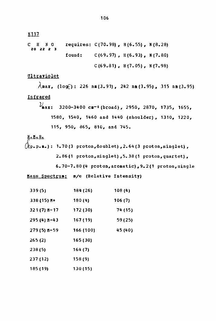
106
K111
c H N 0 requires: c (70. 98) , 8(6.55), N (8.28) 20 22 2 3
found: C (6 9. 97) I H(6.93), N(7.80)
C (69.81) I H(7.05), N(7.98)
Ul!rlliolet
Amax , ( 1 o g() : 2 2 6 nm ( 3. 9 1) , 2 4 2 n 11 ( 3 • 9 5) , 3 1 5 n 11 ( 3 • 9 5)
!!!f rared
V.ax: 3200-3400 ca-t(broad), 2950, 2870, 1735, 1655,
1580, 1540, 1460 and 1440 (shoulder), 1310, 1220,
115, 950, 865, 810, and 745.
N. K. R.
l'.fcp.p~.): 1.70(3 proton,doublet),2.64(3 proton,singlet),
2.86(1 proton,singlet) ,5.38(1 proton,quartet),
6.70-7.80(4 proton,aromatic),9.2(1 proton,single
Kass Seect~ua: m/e (Relative Intensity)
339(5) 184 (26) 108 (4)
338 (15) K+ 180(4) 106 (7)
321 (7) K-17 172 (30) 74 (15)
295 (4) M-43 167(19) 59 (25)
279 (5) ft-59 166(100) 45 (40)
265 (2) 165 (30)
238 (5) 164 (7)
237 (12) 158 (9)
185 ( 19) 130 (15)
107
~~-fractionation III
In order to obtain more crude alkaloidal material, a
third extraction of ground plant was necessary. A total of
2679g of ground plant was weighed out and evenly distributed
into five beakers. Stirring with 951 ethanol was carried out
tor 9 hours. The initial liquid extract vas decanted and
fresh ethanol vas added to the solid and stirring was
continued for another 4 hours. This liquid was decanted and
fresh ethanol was again added followed by four hours of
stirring at 50 c. The new extract was decanted and the solid
covered with fresh ethanol to which was added NH OH to •
bring the pH to 9-11. Stirring was continued for a final
four hours.
The combined ethanol extracts were concentrated on a
cyclone (steam) evaporator and then reduced to a volume of
about 500 ml on a rotary evaporator. Water was added to make
a total volume of one liter and this liquid was extracted
with three-1 liter portions of ethyl acetate. The reaaining
aqueous extract was then made basic (pH10) with NH OH and 4
placed in a liquid extractor for 60 hours during which time
fresh ethyl acetate vas added twice. After 60 hours, testing
of the aqueous layer with Mayer's reagent shoved that basic
material was still present at a concentration of 0.4 to 1.3
g/ml. The aqueous layer was adjusted to pH 7.0 and
extracted an additional 20 hours with ethyl acetate. The

108
insoluble emulsion which remained in the extractor was
filtered and evaporated to yield a fraction weighing 24.5 g
which vas labelled F122. The remaining aqueous extract was
freeze dried and yielded a weight of 31.9 g which was
labelled as fraction 3-9-1.
The ethyl acetate extracts were reduced to 1 liter and
extracted six times vith 500 ml portions of 51 sulfuric
acid. The remaining ethyl acetate was washed with fresh
water, dried oYer CaCl , filtered, and evaporated to yield 2
28.9 g of a fraction labelled F123.
The acid extract was adjusted to pH 7.0 with NH OH
and extracted tvice with 500 ml portions of ethyl acetate.
The remaining aqueous layer was brought to pH 10 and
extracted again with seven-500 ml portions of ethyl acetate.
A heavy, white emulsion which formed during one of the
extractions was filtered off and weighed to yield 123 g of a
material labelled 3-10-5. The ethyl acetate fractions were
combined, dried over cacl , and evaporated to give 37.1 g 2
of alkaloidal material labelled F124. The fractionation tree
is shown in Table XV.

109
TABLE XV FRACTIONATION TREE OF ~~OHNSTON!!
FRACTIONATION III
2.697 kg Ground Plant I Extract with I 95~ Ethanol I Concentrate I Extract with I 95~ Ethanol I NH OH
_____________ I " I I I I
Ethyl Solid Acetate (24.6 g) Soluble F122
I I Eztract 5% H so ________ 1 _______ 2 "
I I Ethyl H SO
Acetate z • Soluble Soluble (28.9q) I Neutralize to pH7
I I
Aqueous Soluble
(31.9 g)
3-9-1
F123 I Extract with ethyl acetate I Make to pH10
I CHCl
3
soluble (31.1g)
F124
I Extract with ethyl acetate
I Insoluble (123.1g) 3-10-5
'------ I Aqueous Soluble
Table of Activities Fraction No. KB Activity <µ_g/•l)
P122 F123 F124
21 31 18

110
A sam~le of 32 g of the crude alkaloidal fraction
labelled F124 was mixed with 80 g Woelm alumina (Activity
Grade III) in a 1-liter round-bottom blask. A mixture of
CH Cl CH OH was added to the flask and evaporated 3 3
slowly on a rotary evaporator to insure thorough absorption
of the alkaloidal •aterial onto the alumina. The resulting
solid was scraped out and ground up into small particles. A
glass column with dimensions 4 cm i.d. by 70 cm was packed
with about 280 g of the same grade of Woela alumina using a
slurry with benzene. The dry sample was poured over the
packed column and allowed to settle. The elution was begun
with neat benzene and continued until no color appeared in
the eluant. The solvents used in order were benzene (1500
ml) chlorofora (2500 ml), ethyl acetate (600 al), n-propanol
(1450 ml), and methanol (700 ml). Each fraction was
evaporated to dryness to give the following result:
Eluting We_!gh1 Solven!, ill
Benzene 12.4
Chloroform 11. 4
Ethyl Acetate 0.78
N-propanol 3.9
Methanol o. 8
F131
F132
F133
F134
F135
KB Activi!.I JµUA!!l
3.3
0.61
17
7.1
1. 8

1 1 1
A qlass column with dimensions 5 ca i.d. by 100 cm was
slurry packed with 700 g of Merck silica gel (70-325 mesh)
to which had been added 6% water by weight. Neat chloroform
was used as the packing solvent, and the sample was
dissolved in a saall volume of chloroform and pipetted onto
the coluan. The elution was begun vtih neat chloroform, and
fractions of approximately 50 al volume were collected. A
step-vise gradient was employed using the following
sol vents: neat chloroform, methanol-chloroform (1:49),
methanol-chloroform (4:46), and methanol-chloroform (3:22).
A total of 71 fractions were collected, and thin-layer
chromatography shoved that recombination into several larger
fractions was warranted. Ten fractions were obtained from
later recombination:
Eluting Weigk1 ,!ssay ,KB A£1i.!i1I SolVfil!1 jgl l!~ Jµl~1l
Neat CHCl 0.12 K136 >100 3
CH OB - CHCl ( 1: 4 9) 1.02 F137 17 3 3
CH OH - CHCl ( 1: 4 9) 0.39 F138 >100 3 3
CH OH - CHCl (1:49) 0.20 F139 59 3 3
CH OH - CHCl ( 1: 4 9) 0.81 F140 6.9 3 3
CH OH - CH Cl ( 1: 4 9) 3.63 F141 17 l 3
CH OH - CH Cl (1:49) 1. 52 F142 2.5 l 3
CH OH - CHCl (4:46) 1.63 F143 2.3 3 3

112
CH OH - CHCl (4:46) 0.38 F144 1. 6 3 3
CH OH - CHCl (3:22) 2.13 F145 2.0 3 3
Fraction K136 gave no visible components in T.L.c.
under short wave or long vave U.V. light, but appeared to be
crystalline after evaporation of solvent. This initial
crystalline material 0
showed a melting point of 171-175
Two further recrystallizations yielded white, flakey
crystals which 0 melted at 174-176 • Fraction K139 appeared
to be a very ho11ogeneous sample under T.L.C.
Recrystallization from aethanol yielded colorless rods with
m.p. 154-56°. A second recrystallization gave colorless rods 0
with m.p. 154-57 • The third recrystallization gave small 0
colorless plates with m.p. of 156-57 • A mixed melting point
determination between K139 and authentic isovoacangines6
shoved no depression. The T.L.C. and infrared spectra of the
two compounds were identical. The analytical and spectral
data for K136 and K139 were as follows:
C H 0 32 52 2
Infrared
requires: C(81.99), H(11.18)
found: C(82.06), H(11.62)
C(82.02), H(11.22)
v max: 2930 cm-1, 1740, 1370, 1255, 1030, and 985

113
Ma§§ s12ectrum: m/e (Relative Intensity)
470(2) 230 (3) 149(10) 110(23) 70(31b
469(6) 220 (22) 148(15) 108 (26) 68(12)
468 (16) M+ 219(100) 136 (25) 106 (17) 58 ( 10)
453 (5) M-15 204 ( 23) 135(12) 96 (28) 56 ( 19)
409(3)M-59 190 (28) 134 ( 20) 94 (23) 43 ( 17)
258 (4) 176(11) 124(17) 92 (9)
250(6) 162(14) 122 ( 24) 82(24)
244 (3) 150(11) 120 (22) 80 ( 10)
K.139
c H N 0 requires: C{71.71), H(7.66), N(7.60) 22 28 2 3
found: c (72. 03) , H(7.40), N(7.74}
Oltraviolet
. Amax, (loqf'): 228 n11(4.47), 275 nm (3.81), 298 na(3.93}
Infrared v max: 3350 cm-•, 2960-2840 (broad), 1730, 1635, 1470,
1350, 1250, 1160, 1080, 1040, 800, 730;
N. M. R.
~p.p.~: 0.9(3 proton, triplet), 3.7(3 proton,singlet},
3.8(3 proton,singlet), 6.80-7.40(3 proton,aromatic
7.70(1 proton,singlet).

114
Mass Spectrum: m/e (Re la ti ve Intensity)
369(13) 128 (6)
368(44)M+ 12 7 (7)
353(6)M-15 126(5)
3J9(4)M-59 12 5 ( 8)
309 (4) M-59 124 (54)
208(10) 123(11)
184(12)M++ 122(51)
160(21) 121 (10)
154 ( 15) 119 ( 8)
148(21) 108(11)
141 (8) 98 ( 9)
140(8) 94 ( 1 0)
137(18) 5 5 ( 10)
136 (100) 42(12)
135(19) 4 1 ( 13)
Fractions F142 through F145 from the previous
cbroillatography all showed promising KB activity {page 111),
so imm~<liatc attention was focused on these fractions.
Preliminary investigation of these fractions on T.L.C.
showGd that a solvent system of methanol-acetonitrile (1:4)
qave the best separation of sample components and the least

115
tailing on silica gel plates. Because it was the largest
traction by weight, F143 was selected for column
chromatography on silica gel. A glass column with dimensions
1.9 ca i.d. 50 cm was slurry packed with silica gel powder
(Baker 60 - 200 mesh) using neat chlorofor• as the packing
solvent. Approximately 50 g of silica gel was packed in this
manner. After the column was packed, the new solvent system
of methanol-acetonitrile (15:85) was added slowly to the
chloroform still remaining on the column. The new solvent
was then allowed to run onto the column until the column vas
completely saturated with the methanol-acetonitrile solvent.
one gram of F143 vas weighed out and dissolved in a small
volume of the methanol-acetonitrile solvent mixture. The
sample was added slovly to the top of the column and the
elution was begun with methanol-acetonitrile (15:85). A
total of 15 fractions were collected, evaporated, and
weighed to give the following result:
Eluti!l9 Solvent Weig!l.LJ.!fil. f,£actj.Qn NQ:.
CH OH - CH CN (15:85) 5 2-53-1 3 3
CH OH - CH CN (15:85) 7 2-53-2 3 3
CH OH CH CN (15:85) 4 2-53-3 3 3
CH OH - CH CN (15:85) 9 2-53-4 3 3
CH OH - CH CH (15:85) 15 2-53-5 3 3
CH OH CH CM (15:85) 59 2-53-6 3 3

116
CH OH - CH CN (15:85) 49 2-53-7 3 3
CH OH - CH CN ( 15: 85) 56 2-53-8 3 3
CH OH - CH CN ( 15: 85) 65 2-53-9 3 3
CH OH - CH CN (15:85) 63 2-53-10 3 3
CH OH - CH CN (15: 85) 73 2-53-11 3 3
CH OH - CH CN ( 15: 85) 72 2-53-12 3 3
CH OH - CH CN (15:85) 166 2-53-13 3 3
CH OH - CH CN ( 15: 85) 129 2-53-14 3 3
CH OH - CH CN (15:85) 45 2-53-15 3 3
CH OH - CH CN ( 15: 85) 53 2-53-16 3 3
Thin-layer chroma tograpby indicated that these
fractions could be recombined to make larger fractions. The
recombinations were made as follows:
Previous Nu•ber Nev Nuaber .AE.12rori11ate Rf Values
2-53-1 thru 2-53-5 2-54-1 0.9 and 0.76
2-53-6 thru 2-53-7 2-54-2 0.76 and o.72
2-53-10 thru 2-53-11 2-54-4 0.10, 0.40, and 0.20
2-53-12 thru 2-53-14 2-54-5 0.70 and 0.20 (major)
2-53-15 thru 2-53-16 2-54-6 0.70 and 0.20
Fraction 2-54-3 was recrystallized f roa aethanol to
yield approximately 30 ag of a colorless crystalline
material with T.L.C. characteristics exactly like K102 in
all solvent systems used. Fraction 2-54-5 consisted

117
predominantly of a component with an Rf value of
approximately 0.20; recrystallization of this yielded 105 mg
of a crude crystalline material. Further recrystallization
from methanol yielded 34 mg of colorless rods with melting 0
point above 220 and decomposition. This crystalline
material was labelled K155, and about 5 mg was submitted for
the KB assay and the PS in vitrg assay when
this test beca•e available. The KB activity of K155 was 19
µ_qfml and the PS activity was 20 g/ml. When a sample of
authentic conoduramine became available,s• it was tested
vi th K155 on silica gel thin-layer plates using the
followinq solvent systeas: Plate A: CH CH OH 3 2
CH COOCH CH (1:4); Plate B: CH OH 3
CR CN (3: 7) ; 3 2 3
Plate C: CH CH OH 3 2
plate was spotted
CH COOCH CR eel (1:4:5). Each 3 z 3 •
with samples of pure conoduramine, pure
K155, and a aixture of conoduramine and K155. The plates
were developed and then vizualized by short wave u.v. light
and spraying with eerie ammonium sulfate reagent. Plate A
shoved only single component spots with an Bf value of 0.44;
plate B shoved single plate component spots with an Rf value
of 0.20; and plate C shoved single component spots with an
Rf value of 0.30. Inf rared spectra of authentic conoduramine
and K155 also shoved the two compounds to be identical. The
following analytical and spectral data were obtained for
1<155:

C H N 0 43 52 • s
Ultraviolet
118
requires: C(73.26), H(7.44), N (7.95)
found: C(68.53), H(7.32), N(7.93)
Amax, (logf ) : 22 9 (4. 71} , 286 (4. 22) , 295 (4. 20)
Infrared
~ax: 3550-3200 cm-t (broad), 2960 and 2940 (doublet,
2860, 1730, 1640, 1470, 1445, 1345, 1325, 1230,
1210, 1165, 1090, 1020, 945, 850, 820, and 750.
lhM.R~ (See Table XI)
Mass SEectrua (See Figure 3)

119
LIST OP RBPERERCES
1) R.1.. Noble, c. T. Heer, and J.B. Cutts, l!!!H.ll!LN~!!....;.!Q£k A£!!d. Sci.L 16L 882-94, (1958}.
2) w. Shoier and A. Aisen.berg, £iH!~±: , 22.,;. 467, {1968) ..
3)
4)
!. Tomita, H. Purukava, Tetraltedrou Let,!;1 .c. 4-309,
S .. 'f. Lu, (1965) •
and s.a. Kupchan,
S.M. fi:upcha.n,, 4~P h~-~£1:1.L
N. Yokoyama, and e.s. 50.L 164, (1961).
'!.'hya9ari jan,
5) Dr. J.N. Hartwell, rson.al Communication.
6) s. Good.win, A.F. Smith, and E.C. Horning, ~:!!-l!.Qf_•_~hg~-'-.§.Q.£!..L J!_.1L 1903-8. (1959) •
7) J. ii. Moncrief a W. N. Lipscomb, i!.!.....;.!!.!E.~£hB!.:!..-§.Q£::.L !!L 4963, (1965).
9) lorbert leuss, B. Gorman, and I.S. Johnson, "Ratural Products in cancer Chemotherapy," Chapter x, pp .. 633 -702 of j'!gf!!Od§. __ !1!..-f2.!!£er ~§!tgI;9t!.L Vol .. III, Harris Bush (Erl .. ); Acar1emic Press, New York, 1967.
10) G. Hahn and H. Werner, All!!it..1. ~~~ 123, {1935).
11) J.B. Hendi:ickson and R. Silva,. ~;!!__l!!!!§!.h_~h!Z!~-22£~..t. 84, 4, 64]-50 (1962).
A.H. Battersby, T.G. Payne, D. 19§.~l 951-53.
_§~L 1.3.20,. 1956 ..
!!!L 98-102 (1962 > •
J.C. Byrne, B.s. Kapil, J.A. Bartin, Ariqoni, and E<:. Loew, ~!°!ft!._£Q!!H!Y.!!.&.L
15} .,IL.A. Quareshi,. and A.I. Scott, £ill_!;Q.!!H!!.!!!h .. .1. 945-50.
.12.2.11 544-53.

18) A.R. Battersby, Cb.ell! • .§QhL (C)
19) .Ibid.., 1187.
120
A.R. Burnett, and P.G. Parsons, 12.2.2.i. 1193-98.
20) I. Kompis, M .. Hesse, and H. Schmid, !;lQYQif!.L J!!L ( 1971) •
3
21) J. Dybowski and E .. Landrin, C 01!12!!!..-~!! d • L lJ.J.1 748 (1901).
22) A. Haller and E. Hecki?l, £Q1!!.E!~4~L 11J.;. 850 (1901).
11: 319 (190.5).
24) H. Lamb1~rt and E. Heckel, f.Q!l!..2!.!..-1:.§!!fl~ . .L
{1901).
26) M.C. Phisalix, {1091).
1J1i. 1235
1Q;.. 101
27) J.A. Schneider and E. B. Si99, Annals_Ne!_lQ!L!£!!.f!...!. ~gi .. .r.-2.L 3 (1957).
28) D.P. Dickel, c.L. Holden, R.C. Maxfield, L.E. Paszek, and W. I. Ta y.lor, !l.!!. _ _!me !:.!..-·-~}!gm. _ _liQ£.!..L f!Qi. 123 ( 1958).
,!1..c. 2265
30) M. P. Cava, Y. iatanabe, and K. Bessho, ~-·-Q:£.9..!.._£heJ!.!...z.. ll&.!!..c. 33, (8) I )350-2.,
49 --L
32) J.l. Weisbach, R.P. Raffauf, o.Biheiro, E. Macko, and B. Douglas, £~_f1H!!'.J!..!._~.£i.:.L 2b. 350 {1963).
33) o. Benner, D.A. Prins, A.L. Burlingame, and K. Biemann,
34}
1!~1Y.!......£J!j,~Act~.!...L !2.L 2186 ( 1963).
ff.P. Cava, S.S. Q£!h.,_Chfilb..i.
Tjoa, Q.I. Ahmed, and A.I. DaBocha, .11.L 3 , { 1 96 8 ) ..
121
35) Drug Research and Development Chemotherapy, Rational cancer Institute, Instruction 14: n Screening Data summary Interpretation,• {March 1972).
36} K. Biemann and M. Friedmann-spi.teller, ,4.!.._!!er.!..-fh§!.
37)
38)
.§.Q~,.t: !!J.L 4805 (1961) ..
o. Renner, D.A. Prins, and W.G. Stoll, Ac!;.~.!.L IJ2L 1572 (1959),,
ia tts, o. Collera, and A. Sandoval, 17 3-82 {l 8} •
39) R .. P. Cava, S.K. Talapatr:a, J.A. ieisbach, B. Douglas, R.P. Raffauf, and J.L. Beal, Igtr$!hedr:Q1[!~}::-LL {14), 931-3 5 (1965) ..
40) M. Gorman, R. lenss, N.J. cone, and J.A. Deyrup, ~~
!!!~....&heJ!.:._§.9£.!:..L !!~.L 1142 { 1960) ..
41) M .. Gormar~ and J. Sw.:H:my, ~~!.fahg~~!Lf.~t!..?.L (42), 31 (1064).
42) L.D. Ant(maccio, N.A.. Pereira, B. Gilbert, H. Vorhrueggen, e. Budzikiewica, J.B. iilson, L.J. Durham, and c .. Djerass.i, ~.:.-A~· .... ~~!!L~.!.L ft~L 2161 (1 ) •
43) o. Benner and D~A. Prins, (1961).
lh 209
44) rt. l~iemann, !!~§§_~~£Sro1.g_!£.I.t. !cGraw-a:ill Book co., Inc., 1.1., 1962, Chapter 3.
45) P. Bota11Hn:·, w. Mc~urr:ay, and K. Biemann, ~~-iJ!Ef:~_£h~fil~ So~ 86, 1439 (1964).
46) G. Buchi, R.E. Manning., and S.A. Monti, !l.!;._!!.!£f..!;._£!m!!.=.. 22.!h..L 85, 1893 (1963).
48) G. Buchi, R. E. Iia.nnin.g, and S .. A. Monti, .J.L_Am~f.L_Ch§!!.;!. ~~ ~2-L 4631 ( 1964).
49) o. Renner and H. Fritz, !..§!.!~t~!§dr.Q!L .. J!ft!h.t. 6, 2 -87 (1964).
13 468 ·(1957). --..L

122
51) G. Zetlar, E.. Lenschow., and ii. Prenger-Bernin.ghoff, lif!.U!lt~£l!!!!ieQ.@!!~~g§ _ _!,i~4tc--fh-Yllk01 -~_.._f2.1h<>l.tcL 26,Q.L (1), 26-4 9 ( 1968) •
52) Chemical Abstracts, !iaL Which Reduces Neoplasia lath. Appl. 6,. 514, 407 1
6,. 1964; e pp.
lo .. P 10434e: "A Preparation Growth." J .R. Geigy A.-G.; Bay 9, 1966; Swiss Appl, IOV
53) jgndbQQ!_£t_!"!i:2£h~!!i2!ilL_2~l~91!i!l!_gf!ll._ff!I£._~Ql~!!il.t: Biol.Qil.L 1st Edition; Herbert A. Sober {Ed.) ; The
Chemical Rubber Co.,. Cleveland, c 1968 ..
Sit) The sample was provided .by l?rof essor M. P. Cava ..
55} The sample was provided by Dr. Marvin Gorman ..
56} The sample was provided by Professor (J. Renner ..

CYTOTOXIC-ALKALOIDS FROft
TABE~!AEMQITANA_JONSTO!IJ
by
!ARIO MICHAEL !llGXNO
(ABSTRACT)
t§bernaemontana jobnstonii is a 11ediu11-sized tree
natiYe to Africa. Speciaens of the hardwood of the plant
were procured from the United States Department of
Agriculture in cooperation with the cancer chemotherapy
program initiated by the National Institutes of Health
(NIH). An investigation was undertaken to Yerify that the
plant possessed activity in the anti-tumor bioassay tests
conducted by the NIH. consequently, systeaatic fractionation
of the plant in conjunction with bioassay revealed that the
biological activity vas concentrated in the alkaloidal
portion of the plant material.
Purification of the crude
accomplished initially by a
alkaloidal material was
coabination of liquid
chromatography on alumina followed by buffer extraction, and
subsequently, by liquid chroaatoqraphy on alumina followed
by liquid chromatography on silica qel.
Four crystalline alkaloids were isolated during the
purification procedure. Two of these alkaloids,
isovoaca.nqine and peri vine, vere characterized by absorption

spectroscopy and mass spectrometry,. Their identity was
verified hy cowparison with authentic samples. The t~emaining
two alkaloids w~re shown by .nuclear magnetic resonancli: and
mass spectx:ometry to be isome.ric with the k11own bisird.ole
alkaloid voacaminie.. These two alkaloids were subsequently
identified
structural
as conodurine
assignments vere
authentic samples.
and conoduramiae, and their
verified by comparison vith
Bach of the isolated alkaloids was submitted for
bioassay .in the KB i.!LvitJ;Q and the PS i!L!.iif:2 test.
Only conodurine at1d condura11ine showed. KB activity with
values of 31 g/ml an<l 19 g/ml respectively .. All four
alkaloids .showed PS activity arid the following values ver:e
obtained: perivine (20 9/lll), isovoacangine (18 g/ml),
conodurine (29 g/ml), and conoduraaine (20 qjal).