Embed Size (px)
Citation preview

Regis University Regis University
ePublications at Regis University ePublications at Regis University
All Regis University Theses
Spring 2017
Yours, Mine and Ours: The Ethics of Using Human and Non-Yours, Mine and Ours: The Ethics of Using Human and Non-
Human Primate DNA in Genetic Research Human Primate DNA in Genetic Research
Brittany Truong
Follow this and additional works at: https://epublications.regis.edu/theses
Recommended Citation Recommended Citation Truong, Brittany, "Yours, Mine and Ours: The Ethics of Using Human and Non-Human Primate DNA in Genetic Research" (2017). All Regis University Theses. 822. https://epublications.regis.edu/theses/822
This Thesis - Open Access is brought to you for free and open access by ePublications at Regis University. It has been accepted for inclusion in All Regis University Theses by an authorized administrator of ePublications at Regis University. For more information, please contact [email protected].


YOURS, MINE, AND OURS: THE ETHICS OF USING HUMAN AND NON-
HUMAN PRIMATE DNA IN GENETIC RESEARCH
A thesis submitted to
Regis College
The Honors Program
in partial fulfillment of the requirements
for Graduation with Honors
by
Brittany Truong
May 2017

ii
Thesis written by
Brittany Truong
Approved by
__________________________________________________________________
Thesis Advisor
__________________________________________________________________
Thesis Advisor
__________________________________________________________________
Thesis Reader
Accepted by
__________________________________________________________________
Director, University Honors Program

iii

iv
Table of Contents
List of Figures ..................................................................................................................... v
List of Tables ..................................................................................................................... vi
Acknowledgements ........................................................................................................... vii
I. Introduction ..................................................................................................................... 1
II. Human DNA ownership rights and the progression of genetic research ....................... 9
III. Ethical considerations in non-human primate genetic research .................................. 17
IV. The effect of food availability on intraspecific interactions in mantled howler
monkeys (Alouatta palliata) in La Suerte, Costa Rica ..................................................... 28
V. Comparison of genetic structure between mantled howler (Alouatta palliata) and
white-faced capuchin (Cebus capucinus) monkeys in La Suerte, Costa Rica .................. 38
VI. Conclusion .................................................................................................................. 56
References ......................................................................................................................... 59
v
List of Figures
Figure 1: Alu polymorphism world map ............................................................................ 3
Figure 2: Average percentage of time A. palliata spent with nearest neighbor ............... 33
Figure 3: Average percentage of time A. palliata spent resting with nearest neighbor .... 34
Figure 4: Average percentage of time A. palliata spent feeding with nearest neighbor ... 35
Figure 5: Example of gel electrophoresis results .............................................................. 49
Figure 6: Example of Sanger sequencing electropherogram results ................................. 50

vi
List of Tables
Table 1: Ethogram for A. palliata .................................................................................... 32
Table 2: Mean heterozygosities and inbreeding coefficients for A. palliata and C.
capucinus .......................................................................................................................... 51

vii
Acknowledgements
First, I thank my thesis and research advisors, Dr. Marie-dominique Franco and
Dr. Amy Schreier, for their faith, trust, support, encouragement, and guidance on not only
this project, but also my other personal and academic endeavors. I am so honored to have
been part of their research team. They have pushed me out of my comfort zone, taught
me invaluable lessons and skills, challenged my thinking, and made me the scholar I am
today. I will always treasure the time I spent training in the laboratory with Dr. Franco,
the adventures I embarked on in the rainforest with Dr. Schreier, and the many laughs
and meaningful talks I shared with both.
I would like to thank my thesis reader, Dr. Becky Vartabedian, for her thought-
provoking questions and philosophical insight on this project. She opened my mind to
new perspectives and helped me relieve some of my stubbornness, which have helped me
grow as a person. I also thank my Honors Program advisors, Dr. Thomas Howe and Dr.
Catherine Kleier, for their tough love, encouragement, and words of wisdom throughout
my time at Regis University.
Thank you to my family and friends for their love, patience, understanding,
kindness, concern, and support through this long journey. I would not have made it
through with my sanity intact without them.
Finally, I would like to thank Regis University’s Biology Department for
laboratory space, materials, and equipment; Matt Barton, Nate Pryor, Heather Humphrey,
Jibin Abraham, and Sarah Seiwald for their assistance in the laboratory; La Suerte

viii
Biological Research Station for housing and an unforgettable field experience; and
TriBeta National Biological Honor Society and the Regis University Research and
Scholarship Council for funding.

1
I. Introduction
Growing up as a Vietnamese-American, I constantly wondered why I looked so
different from strangers at the park or at Safeway. I encountered people with light and
dark skin; blonde, red, and black hair; and blue, green, hazel, and brown eyes. In contrast,
everyone in my family had distinct Asian features – tan skin, dark brown hair, and small,
brown eyes. Even within my immediate family, nobody looked exactly alike, and in my
mind, this was beautiful and miraculous. I was told that all humans were created in God’s
own image and likeness and this was supposedly the reason for our uniqueness. This
answer never fully satisfied me, but I accepted it until the day I was first exposed to the
complex study of genetics.
I later learned that DNA is the essence of our existence; indeed, it is the substance
in our cells that distinguishes one individual from another. Approximately 99.9% of the
human genome is shared within the entire human population, and the other 0.1%
accounts for the genetic variation that is observed amongst individuals (National Human
Genome Research Institute, 2016). There are only four nucleotide bases that make up
DNA – adenine, thymine, cytosine, and guanine – and yet various sequences result in
billions of different and unique characteristics. Nucleotides resemble the English
alphabet. Twenty-six letters can be organized into a myriad of different words and
sentences, and in the same way, four nucleotides are pieced together as unique DNA
sequences. Together, they make one book, or genome, that is encrypted with information
specific to one person. Sequences can differ by a single nucleotide or in the number of

2
copies of a specific sequence. These variations are called DNA polymorphisms, or DNA
markers. Upon learning this, I quickly developed a passion for human genetics and
vowed that I would one day pursue a career in it. I wanted to have a better understanding
behind the mechanisms underlying our diversity.
In a genetics laboratory course during my sophomore year at Regis University, I
had the opportunity to study a specific polymorphism in my own DNA. I extracted DNA
from my cheek cells and used a process called polymerase chain reaction (PCR) to
amplify a specific sequence, called the Alu polymorphism, at the PV92 locus on
chromosome 16. This polymorphism is a 315 base pair long insertion that does not
encode any proteins (Mighell, Markham, & Robinson, 1997). There are more than a
million copies of it scattered throughout the genome. Many of these copies are considered
“fixed,” which means every person inherits a copy of the insertion at a specific locus. In
contrast, at other loci, the Alu polymorphism is “non-fixed,” and we can inherit zero, one,
or two copies of it from our parents. An individual can be homozygous for the sequence
(+/+), homozygous for its absence (-/-), or a heterozygote (+/-), depending on his/her
heritage. At the PV92 locus, the frequency of the allele varies in different human
populations across the world. For example, in France, the frequency is only 27.50%,
which is significantly lower than Japan’s frequency of 85.71% (Watkins et al., 2001).
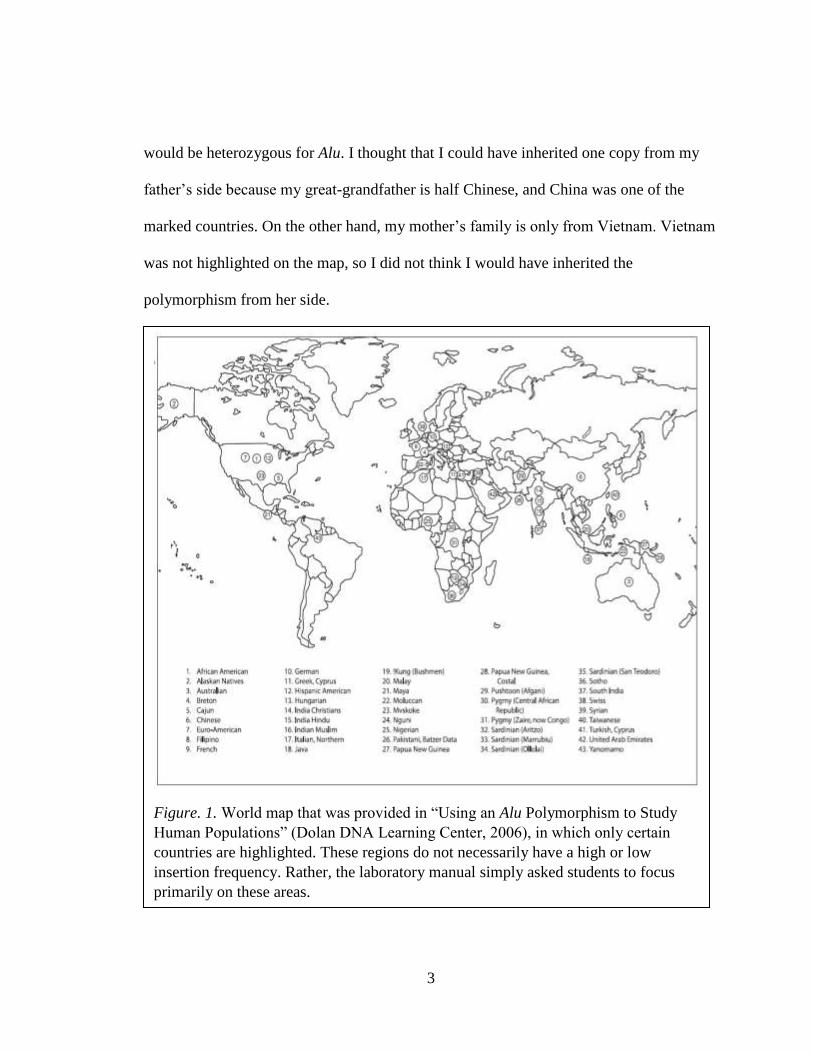
In this laboratory activity, we were asked to predict our own genotype. We were
given a blank world map, in which a small number of countries had been highlighted, and
my classmates and I incorrectly assumed that the featured regions were the only areas
where the Alu polymorphism was prevalent (Fig. 1). Based on this map, I predicted that I
3
would be heterozygous for Alu. I thought that I could have inherited one copy from my
father’s side because my great-grandfather is half Chinese, and China was one of the
marked countries. On the other hand, my mother’s family is only from Vietnam. Vietnam
was not highlighted on the map, so I did not think I would have inherited the
polymorphism from her side.
Figure. 1. World map that was provided in “Using an Alu Polymorphism to Study
Human Populations” (Dolan DNA Learning Center, 2006), in which only certain
countries are highlighted. These regions do not necessarily have a high or low
insertion frequency. Rather, the laboratory manual simply asked students to focus
primarily on these areas.

4
I found that I was homozygous (+/+) for the insertion. Still under the impression
that the map depicted Alu frequencies, this discovery surprised both me and my family.
For so many years, my mother’s father, my grandfather, had told me that all of our
ancestors originated from Vietnam, and he had always spoken with such assurance. The
results of this simple genetic test suggested otherwise, and it made me seriously question
my heritage.
I revisited the Alu polymorphism laboratory a few years later, only to realize that I
had made a mistake in my prediction and interpretation. Alu at PV92 actually has a
frequency of 87.50% in Vietnamese populations, not zero. In addition, China’s allele
frequency is 85.29% (Watkins et al., 2001). It was no wonder that I was homozygous for
the insertion. This new discovery reaffirmed what I had always known about my family
and my identity. Correcting the mistake did not significantly affect my life, but it was still
a relief to know that my grandfather was not mistaken. A small misinterpretation gave me
some emotional discomfort and had me living with the false impression that my mother’s
family was partially Chinese for years.
The full Alu polymorphism laboratory manual includes an “Informed Consent and
Disclosure” page, but I do not remember ever seeing this page in my genetics laboratory.
It states that students have the option to refrain from participating in the activity, and they
should be willingly giving up their DNA samples, knowing that the results could reveal
information about family relationships (Dolan DNA Learning Center, 2006). Had I
known this, I could have chosen not to participate in the laboratory activity. I could have
avoided the unexpected emotional discomfort I felt after seeing my results. In retrospect,

5
I probably would not have opted out of the activity because I loved extracting my own
DNA and my discomfort was minor. Imagine, though, if we had been analyzing a region
of my DNA that revealed something more significant, such as my likelihood of
developing breast cancer. This bit of information would have surely given me severe
emotional distress.
My experience has shown me the importance of informed consent when involving
humans in research. Donors should be aware of how their DNA is being used and the
potential risks. As a future genetic researcher, it is critical that I keep this in mind.
Researchers are not always clear about how they will use genetic samples in their studies,
which creates distrust in the general public (McGuire & Beskow, 2010). Many people are
not willing to participate in genetic research because they are afraid that their personal
information will be inappropriately shared with peers, employers, health insurance
companies, or health care providers, leading to genetic discrimination (Genetics Home
Reference, 2017). However, if we are to make any progress, we need DNA samples to
study. In turn, researchers may feel the need to commit DNA theft. They can steal
abandoned genetic samples, such as strands of hair left at the salon, and potentially
invade a person’s privacy rights (Joh, 2011). This then raises questions about DNA
ownership and whether humans have rights to their genetic material and information.
Humans are fortunate in that they have the ability to voice their opinions and fight
for the rights that they think they deserve. However, animals do not have this same
liberty. Non-human primates, in particular, are regularly used as models for studying
human genes and diseases because they are our closest relatives (NIH, 2007). Unlike

6
humans, they are not psychologically affected by the availability of genetic information,
and they are not worried that their personal information will fall into the wrong hands;
however, that does not mean we are free to extract, analyze, or modify their DNA as we
please. In addition to the few million base pairs that humans and non-human primates
share, we also share a capacity to suffer. Our shared sentience requires that we reevaluate
non-primates’ rights and avoid subjecting them to unnecessary suffering in genetic
research.
I became interested in human and non-human primates’ rights because of the
research I had been conducting with Dr. Marie-dominique Franco and Dr. Amy Schreier.
Since my junior year, I have been extracting DNA from and analyzing the genetic
structure of two monkey species, mantled howler (Alouatta palliata) and white-faced
capuchin (Cebus capucinus) monkeys, from La Suerte Biological Research Station
(LSBRS) in Costa Rica. I worked with fecal samples that Dr. Franco and Dr. Schreier had
collected during previous summers containing genetic material that was essentially stolen
from the monkeys without their consent. During the summer of 2016, I traveled to Costa
Rica to steal some more and then proceeded to extract information from the DNA once I
returned to Regis University. I did not feel guilty doing either. I was not hurting the
monkeys in any way. If anything, I was helping them by shedding light on how
deforestation was negatively affecting their genetic diversity.
Like so many other people, I previously held an anthropocentric perspective on
the world, and I valued human life over the lives of animals. I used to think that all
research involving animals was justified if it provided some sort of benefit to humans,

7
and animals’ rights were trivial compared to our own. I realize now, after working in the
genetics laboratory, living with and developing such a strong connection to the monkeys
at LSBRS, and writing my Honors Thesis, that humans and animals are so much more
connected than I had initially realized. Both humans and animals have rights that must be
respected when they are involved in any sort of research.
In Chapter II, I will discuss human DNA ownership and argue that while humans
do not have rights to their genetic material, they should have ownership over their genetic
information. Everyone has a right to decide whether or not he/she wants to know what
information is contained within his/her DNA and who has access to it. Genetic research
can move forward only if we protect individuals’ rights. Then in Chapter III, I will
discuss the ethical considerations of using non-human primates in genetic research. In
addition to sharing over 90% of our DNA (NIH, 2007), non-human primates also share
our capacity to suffer, and researchers should exercise the humane treatment principle
when involving them in their studies. In Chapter IV, I will present my findings from the
behavioral study I conducted in Costa Rica. I examined the effect of food availability on
social structure and intraspecific interactions in the A. palliata population at LSBRS. I
hypothesized that adult males and females would spend significantly more time with one
another as opposed to with individuals of the same sex. This trend was observed amongst
adult males, but there was no significant difference for females in the amount of time
spent near individuals of the same or opposite sex. Finally, I will report my research on
the genetic structure of A. palliata and C. capucinus at LSBRS. I rejected my first
hypothesis that neither species would be in Hardy-Weinberg Equilibrium. Although both

8
species are inbred, my prediction that habitat fragmentation would more negatively affect
C. capucinus compared to A. palliata is also rejected.

9
II. Human DNA ownership rights and the progression of
genetic research
The completion of the Human Genome Project in 2003 led to rapid advancements
in biotechnology, making it relatively quick, cheap, and easy to obtain and analyze
genetic information (National Institutes of Health [NIH], 2016c). The Human Genome
Project revealed the exact sequence of a person’s DNA, which was a major step towards
uncovering the function of specific genes and identifying mutations associated with
genetic diseases; however, there is still much to be learned. Researchers require a supply
of DNA samples to study, but many people are unwilling to donate samples of their
genetic material (Presidential Commission for the Study of Bioethical Issues, 2012).
They are uncomfortable with the thought of someone having direct access to their
personal genetic information. Yet, humans discard DNA samples, such as hair or saliva,
daily. Researchers could potentially steal and analyze abandoned DNA, but is it ethical to
do so? Do humans have ownership rights to their DNA?
In this chapter, I discuss human DNA ownership rights and the implications they
have for human genetic research. I argue that while humans do not own their genetic
material once it has been removed from their bodies, they do have ownership rights to
their genetic information. DNA contains very personal data, and we should decide who
has access to it. The high demand for genetic samples in research can be met if
researchers vow to respect our DNA ownership rights and are more transparent about
how they will use our DNA in studies.

10
DNA is the essence of our existence. It is a string of four nucleotide bases –
adenine, thymine, cytosine, and guanine – held together by a phosphate-sugar backbone
and packed in the nuclei of our cells. These nucleotide bases can be arranged in an
infinite number of ways. Just as the 26 letters of the alphabet are organized into words
and sentences, nucleotides are pieced together to form genes. Together, they make one
unique book, or genome, that is encrypted with information. Anyone can access a book in
a library and gather information from it. Likewise, anyone can access another person’s
genome by taking a few cell samples and extracting the personal information found
inside. Genetic researchers are particularly interested in studying these books to better
understand human nature, such as our evolutionary history, our genetic variability, and
the mechanisms of genetic diseases.
An entire genome is contained in the nucleus of each cell, and many people do not
realize that they leave traces of their DNA everywhere they go. Strands of hair left behind
at the salon or Starbucks coffee lids thrown in the trash have several copies of a person’s
genome that he/she has left behind. These are rather convenient sources of DNA for
researchers to inconspicuously take and analyze without concerning the “donor” (Joh,
2011). This is called DNA theft, and though it is not so common today, there have been
cases where researchers have profited from nonconsensual genetic sampling (Skloot,
2010).
Henrietta Lacks was an African-American woman who developed cervical cancer
at the age of 31. She was diagnosed and treated by the doctors at John Hopkins Hospital
in 1951. John Hopkins was known as a charity hospital and one of the only hospitals at

11
the time to treat African-Americans. Rebecca Skloot (2010), in her book The Immortal
Life of Henrietta Lacks, describes:
But first – though no one had told her that [the doctor] was collecting samples or
asked if she wanted to be a donor – [he] picked up a sharp knife and shaved two
dime-sized pieces of tissue from Henrietta’s cervix: one from her tumor, and one
from the healthy cervical tissue (p. 33).
This demonstrates how Lacks’ cells were essentially stolen from her body without her
knowledge or consent. Dr. George Gey and his research team immediately discovered
that Lacks’ cancerous cervix cells were immortal and could proliferate indefinitely with
ample nutrients and space. Dr. Gey was the first to develop an immortal human cell line,
which he called “HeLa” for the first two letters of the woman’s first and last name. Then,
in the 1950s, Microbiological Associates, a biological supply company, was the first to
commercialize HeLa cells and make a profit by selling them to other laboratories (Skloot,
2010). Since then, many other biotechnology companies have cultivated HeLa cells and
thrived off of them. The cell line is still currently used in research, and a single vial of
HeLa cells costs over $400 (American Type Culture Collection [ATCC], 2017).
The exploitation of HeLa cells begs the question of whether it was ethical for the
doctors to have actively taken tissue samples from Lacks without her knowledge and then
make a profit from it (Skloot, 2010). They committed DNA theft and invaded her genetic
privacy (Joh, 2011), but Skloot (2010) suggests that they did not do so out of ill will.
They only wanted to study human cancer cells, and during this time period, it was not
uncommon to take cells and overlook informed consent (Skloot, 2010). Moreover,

12
Tupasela (2011) argues that human samples, when it is no longer part of the person,
becomes human waste because it “has no use value for its holder” (p. 514). We have no
use for saliva left on a coffee mug or excised tissue samples. Aside from the fact that they
hold copies of our genome, these samples have no value to us. We also leave our DNA
everywhere, so it is not practical to initiate laws that criminalize DNA theft (Skene,
2005). Indeed, we do not have ownership rights over our genetic material after it has been
removed from our bodies.
It is natural to feel uneasy about this. The thought of researchers stealing our
DNA nonconsensually is unsettling; however, Skene (2005) points out that our
discomfort stems not from an attachment to the genetic material, but rather, the
information contained in it. Our DNA contains very personal data. Genes dictate our
physical traits, such as eye color, height, and skin tone, that can be directly observed, but
they can also reveal our likelihood of developing a fatal disease, which is more hidden. I
concede that we have ownership rights to our genetic information because it contains
sensitive data. Researchers must obtain consent from the donor before extracting the
information and sharing it with others (McGuire & Beskow, 2010).
When we participate in genetic research, we have a right to decide whether we
want to know the information contained in our DNA (Laurie, 1999; Andorno, 2004; Juth,
2014). Researchers often feel obligated to disclose significant findings to the person who
donated their DNA, especially if they discover that he/she has a life-threatening condition
(Hallowell, Hall, Alberg, & Zimmern, 2015). This is a sensitive situation. Finding out
that we are at risk for or have developed a genetic disease can harm us psychologically

13
and emotionally, by causing “unwanted changes in self-image, reduced autonomy, [or]
feelings of anxiety or depression” (Juth, 2014, p. 38). For example, patients who
discovered they carried mutations in the BRCA1 and/or BRCA2 genes experienced
elevated emotional distress thinking about possible future cancer treatments and their
relatives’ risks of developing cancer (Hamilton, Lobel, & Moyer, 2009). Some people
prefer to remain in blissful ignorance and think genetic information is an unnecessary
burden (Andorno, 2004; Herring & Foster, 2012). On the other hand, Takala (2001)
dictates that we should know what is contained in our DNA because it can help us make
more informed decisions about how we live. We can make simple lifestyle changes, such
as changing our diets to avoid heart disease, but we can also be more drastic, like
choosing not to have children so as not to pass on our condition and subject future
generations to pain (Juth, 2014).
With genetic research, there is also concern that researchers may inappropriately
disclose personal genetic information to other parties, such as employers, health care
providers, health insurance companies, etc., leading to genetic discrimination (Genetics
Home Reference, 2017). The National Partnership for Women & Families (2004) reports
several cases where individuals have been inexplicably fired from a job or denied health
insurance after individuals shared the results of their genetic tests. For example, a young
boy with Long QT Syndrome, a rare genetic disorder that causes fast, chaotic heartbeats,
was denied coverage under his father’s health insurance because of his condition. In
2008, the United States issued the Genetic Information Nondiscrimination Act (GINA) to
protect individuals from genetic biases. GINA prohibits health insurance companies from
14
using genetic testing to deny or underwrite coverage, and it prevents employers from
using it to hire or fire employees (NIH, 2016a). However, GINA does not apply to
companies that have less than 15 employees or to members of the United States Military.
According to the NIH (2016a), GINA sets a “floor of minimum protection” and state
legislators are responsible for issuing additional laws to protect individuals. Most states
have passed laws that then regulate access to and the disclosure of genetic information
(Presidential Commission for the Study of Bioethical Issues, 2012). Unfortunately,
despite all of these laws seeking to protect individuals’ DNA ownership rights, voluntary
human involvement in genetic research is still low (Saulsberry & Terry, 2013).
Perhaps people would be more willing to participate if researchers were more
transparent with their goals and the purpose of their experiments. Donors like to know
how their DNA is being used, and this information should be provided in the consent
forms they sign beforehand (Skloot, 2010). However, researchers often use broad blanket
statements in these forms to give them greater flexibility (McGuire & Beskow, 2010).
For example, in 2004, the Havasupai Native American Tribe filed a lawsuit against
researchers at Arizona State University for using Havasupai blood samples in projects
that tribe members had not agreed to (National Congress of American Indians, n.d.). The
original study was designed to look only at the prevalence of type II diabetes in this
community, but the samples were also used in schizophrenia and inbreeding studies
(Markow et al., 1993). Inbreeding, in particular, is a taboo subject amongst the Havasupai
people. Upon hearing that their samples were improperly used, they grew distressed and
took the case to court. The tribe received a $700,000 settlement in 2010, and Arizona

15
State University returned all of the Havasupai DNA samples they had collected (National
Congress of American Indians, n.d.). This demonstrates how important it is that
researchers are explicit about how they intend to use collected DNA samples. They
should not violate participants’ trust. McGuire and Beskow (2010) wisely point out,
though, that it is impossible to know upfront exactly how a sample will be used. Samples
can be put aside and stored in biobanks for years. When researchers are finally ready for
them, the original project that the donor agreed to participate in may have changed. It
may be a hassle to contact the donor and inform him/her of experimental changes, but
this step is crucial for maintaining trust between participants and researchers. Participants
should also be comforted by the fact that by federal law, they can withdraw their consent
to participate at any point, and researchers must stop using their samples (McGuire &
Beskow, 2010).
Although there are many potential risks that come with taking part in genetic
research, this should not discourage people from doing so. Rather, I think it is important
that we do opt to participate when given the option. By 2015, over 1,000 human genomes
were sequenced and made publicly available as part of the 1,000 Genomes Project
(International Genome Sample Resource, 2015a). Anyone seeking to better understand
human genetic variation can access these genomes. Researchers have already used this
data and identified new genetic markers linked to medical conditions, including celiac
disease, prostate cancer, glioma, type II diabetes, breast cancer, and diabetes (Zheng-
Bradley & Flicek, 2016). This is thanks to the many people who have donated their DNA
samples for genetic research. New data are currently being added to the public database

16
by the International Genome Sample Resource (IGSR) (International Sample Resource,
2015b). The IGSR is hoping to add samples from populations that were not part of the
original 1,000 Genomes Project. This will allow researchers to develop an even better
understanding of human genetic variation by including samples from across the globe;
however, this can only be done if people willingly donate their DNA.
I have worked with DNA samples in the genetics laboratory since my junior year
at Regis University, but I have never had to ask my research participants for their consent
to be part of my study. This is because my study subjects have been mantled howler
(Alouatta palliata) and white-faced capuchin (Cebus capucinus) monkeys who are not
able to offer any form of consent (See Chapter III and V). In turn, I have been free to
steal and analyze their DNA without worrying about genetic discrimination or causing
them psychological harm. As a future genetic researcher, I am most interested in studying
human diversity and the development of genetic diseases. I will soon be making the
transition from studying monkeys to humans, and I will need to adjust my research
practices accordingly. I recognize that I cannot force humans to participate in my future
experiments by stealing their fecal samples, as I did with the monkeys. I must encourage
them to do so by ensuring them that I will honor and respect their DNA ownership rights
and by being transparent with what my studies entail.

17
III. Ethical considerations in non-human primate genetic
research
In the previous chapter, I examined human DNA ownership rights and argued that
humans have ownership of their genetic information but not necessarily the material after
it has been removed from their bodies. Genetic studies involving humans are highly
regulated in order to protect human interests and prevent physical or psychological harm.
In contrast, the use of non-human primates in genetic research is still widely debated.
There are studies that focus on the animals themselves, namely their evolution, kinship
patterns, and diversity; however, because humans and non-human primates share over
90% of DNA, they are also useful models for studying human genes and diseases
(National Institutes of Health [NIH], 2007). They offer many benefits in genetic studies,
but it is important to keep in mind that humans and non-human primates share more than
a few million DNA base pairs – we also share a capacity to suffer. This mutual sentience
requires that we reevaluate how we conduct genetic research with non-human primates.
Can we justify animal suffering if it provides a significant benefit to human life?
In this chapter, I discuss the ethical considerations that must be assessed when
using non-human primates in genetic research. I argue that because both humans and
non-human primates have the ability to suffer, researchers must apply the humane
treatment principle when designing and conducting their studies. In other words, they
must ensure that non-human primates are not enduring any unnecessary suffering during
sample collection, sequence analysis, or genome modifications. As so, I have myself

18
implemented the humane treatment principle in my undergraduate research involving
monkeys, and I plan on upholding this mindset as I pursue my career in genetic research.
I first became interested in these issues after having worked with monkey feces
for over a year in the genetics laboratory at Regis University. Not all genetic research is
conducted for the sake of humans. Some studies are designed to learn more about the
animals themselves, namely their evolutionary history, kinship patterns, etc., which could
have important implications for their well-being and existence. My research project was a
conservation genetics project, part of larger study overseen by Dr. Marie-dominique
Franco and Dr. Amy Schreier, in which I analyzed the genetic structures of mantled
howler (Alouatta palliata) and white-faced capuchin (Cebus capucinus) monkeys from
La Suerte Biological Research Station (LSBRS) in Costa Rica. I traveled to LSBRS to
collect A. palliata and C. capucinus fecal samples in the summer of 2016, extracted DNA
from the feces, and determined both species’ genetic diversity by analyzing specific
microsatellites (See Chapter V). To put it simply, I stole genetic samples from monkeys
in Costa Rica and nonconsensually extracted information from their DNA. Although I
discouraged this type of behavior in my previous chapter, my actions felt justified.
Indeed, the primary goal of my research project was to determine if deforestation was
negatively affecting genetic diversity in A. palliata and C. capucinus by forcing them to
inbreed. I knew that my research was essentially helping the monkeys as my results will
provide insight into their adverse situation. Moreover, none of my methods, from the
sampling down to the genetic analysis, harmed the monkeys in any way.

19
Genetic samples can be obtained from animals noninvasively or invasively. In
noninvasive sampling, Pauli, Whiteman, Riley, and Middleton (2009) explain, “Animals
are unaware of sampling and, therefore, are unaffected by it…or animals are unrestrained
and do not exhibit a chronic or severe stress response or experience reduction in survival
or reproduction” (p. 350). For example, researchers can collect abandoned feces, hair,
feathers, egg shells, and snake skins and extract DNA from these samples for their studies
(Taberlet, Walts, & Lulkart, 1999). In contrast, invasive sampling consists of drawing
blood or scraping tissues. Noninvasive sampling is attractive to many researchers, like
myself, because it requires less equipment and minimizes physical pain. Moreover,
projects that use noninvasive sampling are more likely to be approved by the Institutional
Animal Care and Use Committee (IACUC). The purpose of the Committee is to
minimize harm inflicted on research animals. This ensures that animals are not subjected
to unnecessary suffering (Steneck, 1997).
Unfortunately, the quantity and quality of DNA extracted from noninvasive
samples are much poorer compared to invasive ones, making them more difficult to
analyze (Taberlet, Walts, & Lulkart, 1999). Dai, Lin, Fang, Zhou, and Chen (2015)
extracted DNA from Chinese egrets (Egretta eulophotes) and found that blood yielded
the highest DNA concentration (252.16 ± 17.05 ng/μL) when compared to plucked
feathers (182.49 ± 7.95 ng/μL), shed feathers (13.59 ± 2.10 ng/μL), and feces (9.77 ±
1.83 ng/μL). In addition, noninvasively collected DNA tends to degrade readily and
contains polymerase chain reaction (PCR) inhibitors, like melanin and keratin, which
interfere with DNA amplification (McDonald & Griffith, 2011). In contrast, nucleated

20
blood is purer and more concentrated. For blood, DNA extraction only requires 50 μL of
volume, and any remaining blood that is drawn can be preserved indefinitely. Should any
problems arise, researchers can repeat the experiment using the excess. Those that collect
samples noninvasively do not often have this same liberty, depending on how much of
the original sample they were able to collect and how much was needed for DNA
extraction. Researchers cannot make any mistakes in DNA extraction or amplification,
nor can they repeat their experiments to validate their results when they have a small
sample size. Rather than dealing with the many problems that come with analyzing
noninvasive samples, many researchers opt for the more convenient route and study DNA
collected invasively (McDonald & Griffith, 2011).
My own experience has shown me that it is incredibly difficult to work with and
analyze DNA that has been collected using noninvasive techniques. The feces I analyzed
had only small amounts of DNA. Extraction was difficult because the DNA was not
evenly distributed in the stool, and the samples were often watery. I spent my first year of
research trouble-shooting in the laboratory as I was unable to amplify DNA from C.
capucinus. I had been using the same extraction and PCR procedures for both the A.
palliata and C. capucinus samples. However, C. capucinus feces have even lower
concentrations of DNA than A. palliata, and the DNA is more sensitive to time and
temperature. Therefore, the samples had to be handled differently. After months of trial
and error, I deteriorated my supply of feces, but I finally perfected the C. capucinus
extraction and amplification protocols. Nevertheless, I knew that the DNA was fragile,
and I worried that it might degrade if it was not properly stored or if it was left at room

21
temperature for too long. If the DNA had degraded, I would not have been able to redo
some of the experiments because I exhausted my supply of original genetic material.
Therefore, I sympathize with fellow researchers who work with noninvasive genetic
samples.
Although I recognize how much easier it would have been to extract DNA from
and analyze purer genetic samples, like blood, I also maintain that using feces was the
more ethical route. Obtaining blood would have required capturing the monkeys,
necessitating professional help to dart them with drugs (usually ketamine solutions) that
temporarily immobilize them and cause them to fall from the trees (Jones & Bush, 1988).
However, darting is a dangerous technique. Wasserman, Chapman, Milton, Goldberg,
and Zigler (2013) found that darting adult red colobus monkeys (Procolobus
rufomitratus) with telazol and ketamine led to an acute increase in cortisol levels,
indicating physiological stress. Jones and Bush (1988) note that of the 27 redtail monkeys
(Cercopithecus ascanius) they darted, two died and one was severely injured. In addition,
because the understory of the forest was so thick, they could not predict where C.
ascanius would fall and waited until they hit the ground to capture them (Jones & Bush,
1988). This demonstrates how darting is a dangerous and difficult procedure that inflicts
unnecessary pain. Rather than risk harming A. palliata and C. capucinus, I chose to
follow them in the rainforest and wait for them to defecate instead.
My research ethics have been guided by the “humane treatment principle”
outlined by Gary L. Francione. He dictates, “[The] humane treatment principle…[is] the
view that because animals can suffer, we have a moral obligation that we owe directly to

22
animals not to impose unnecessary suffering on them” (Francione, 2004, p. 113). The
term “unnecessary” is key in Francione’s definition, but it constitutes a gray area. How
do we decide what is considered “unnecessary suffering,” especially in genetic research
involving non-human primates? There is a significant difference between simply
analyzing sequences for the sake of the monkeys, as I have done, and analyzing and
modifying the DNA to benefit humans. Cohen (2001) argues that we have a “duty” to use
animals in biomedical experiments to promote human health and save lives (p. 5). In
doing so, he implies that their suffering on our behalf is absolutely necessary. Because
they are our closest evolutionary relatives, non-human primates are regularly used as
models for studying human disease (Harding, 2013). Rhesus macaque (Macaca mulatta)
oocytes have been injected with viruses expressing exon 1 of the human huntingtin
(HTT) gene to model Huntington’s disease. The authors argue that M. mulatta models
more accurately mimic the neurological and behavioral mechanisms of Huntington’s
compared to rodent models (Yang et al., 2008). Researchers are drawn to non-human
primates because they are often better models for studying human physiology, behavior,
disease, and genetics (Harding, 2013). They do not see the experiments as “unnecessary
suffering” because the monkeys are essentially helping humans.
Though this may be true, it is important to keep in mind that humans and non-
human primates share more than physiological factors and homologous genes – we also
share a capacity to suffer. Indeed, Ferdowsian et al. (2011) found that when chimpanzees
(Pan troglodytes) were subjected to experimentation or experienced other traumatic
events, they exhibited behaviors similar to those observed in humans with post-traumatic

23
stress disorder (PTSD) and depression. They refused to engage in play or grooming, were
antagonistic towards peers, isolated themselves, and sat in a hunched, remote posture
(Ferdowsian et al., 2011). Given our shared sentience, Francione (2004) supplements his
humane treatment principle with the “principle of equal consideration,” which states that
we are required to “weigh our suffering in not using animals against animal interests in
avoiding suffering” (p. 121). Before conducting an experiment, we must consider the
costs and benefits to both parties, and the benefits to one must significantly outweigh the
costs to the other (Sandoe & Holtug, 1996). However, in a primarily anthropocentric
world as our own, we cannot help but prioritize our own needs above other animals’.
Francione (2004) has observed that even when animal and human interests have
an equal weight, we almost always disregard the animals’ interests in favor of our own.
For example, M. mulatta have been used as models to test the efficacy of vaccines against
Shigella dysenteriae 1 (SD1). SD1 causes Shigellosis exclusively in humans and non-
human primates, and they experience the same symptoms, namely lymphocytosis, acute
colitis, fevers, dehydration, and bloody diarrhea (Islam et al., 2014). However, only M.
mulatta, not humans, are used in the initial stages of vaccine tests, demonstrating how we
prioritize human interests above animals’. We tend to view animals as “property” in our
society, or “nothing more than things” (Francione, 2004, p. 108). This type of mindset is
alarming, as it gives researchers the false impression that they can use animals,
particularly non-human primates, as they please. Therefore, instead of seeing them as
“property,” we should try to remember that non-human primates are our relatives.

24
That is not to say that we should completely ban the use of non-human primates
in genetic research. Francione (2004) argues, “[We] ought to treat like cases alike unless
there is a good reason not to do so” (p. 121). “Like cases,” in my opinion, are the species
that are most closely related to us, namely the great apes. Orangutans, gorillas,
chimpanzees, and bonobos are our closest relatives, sharing between 97.0-98.8% of DNA
with us (NIH, 2011; Wall, 2013). They have cognitive abilities and are capable of using
tools (Chuecco, n.d.). In addition, P. troglodytes can communicate with humans through
American Sign Language (Rivas, 2005) and even grieve deceased group members (van
Leeuwen, Mulenga, Bodamer, & Cronin, 2016). These significant similarities render that
they be treated like humans with regard to genetic research. They should be protected
from experiments that inflict any physical, emotional, or psychological pain on them. The
Great Ape Project, created in 1994, is working towards establishing international
personhood rights to all of the great apes. Several countries, including Spain, New
Zealand, and Austria, have already banned biomedical research involving great apes
(Project R&R, 2017). In 2015, the United States banned the use of P. troglodytes in all
biomedical research. P. troglodytes individuals owned by the NIH are expected to be
reallocated to Chimp Haven, a federal sanctuary located in Louisiana, by 2025 – 10 years
after the ban was instated (NIH, 2016b).
As we move along the phylogenetic tree, our relatedness to other non-human
primates, such as monkeys including M. mulatta, A. palliata, and C. capucinus,
decreases. In turn, they are no longer considered “like cases.” In other words, our
physiologic and genetic similarities are enough to justify their use in some research, but

25
not enough to offer them limited human rights, like the great apes. For example, humans
share 98.8% of their DNA with P. troglodytes (Wall, 2013) and approximately 93% with
M. mulatta (NIH, 2007). In addition, the great apes behave more similarly to humans than
monkeys. Mark Mirror Tests were administered to P. troglodytes (Gallup, 1970), M.
mulatta (Gallup, Wallnau, & Suarez, 1980), and brown capuchins (Cebus apella) (Roma
et al., 2007), in which individuals were marked with a red mark on their heads and placed
in front of a mirror. Only P. troglodytes noticed and reached for the mark on themselves,
indicating that they are self-aware (Suddendorf & Butler, 2013).
Studying non-human primate DNA can, and has, provided incredible insight into
our own genes and evolutionary history. Primates are thus indispensable in genetic
research. Recent advancements in biotechnology, such as the development of the
CRISPR-Cas9 (clustered regularly interspaced short palindromic repeats) system, will
undoubtedly increase their involvement in genetic research. CRISPR-Cas9 takes
advantage of a mechanism utilized by bacteria to fight viral infections using a DNA-
cleaving enzyme called Cas9 to edit genomes (Jinek et al., 2013). Chen et al. (2015) have
mutated a dystrophin gene in M. mulatta to give them Duchenne muscular dystrophy
(DMD), a severe genetic disorder characterized by progressive muscle loss. The
genetically modified M. mulatta will allow researchers to study the pathology of the
disease and uncover a future cure for humans. The significant benefits the study could
provide justifies the research, though it pains me to think about M. mulatta being
subjected to pain for our sake.

26
I have struggled thinking about where we should draw the line on what is
considered morally ethical in genetic research involving non-human primates. It was not
until just recently that I began advocating in favor of the humane treatment principle and
the principle of ethical consideration. Before I traveled to Costa Rica and lived in the
jungle for a month, I believed any kind of research involving animals was morally
permissible if it provided any kind of benefit to human life because I value humans over
animals. The gap that once separated humans from animals has decreased after my time
with the monkeys and other wildlife in Costa Rica.
I developed a connection to them – a month in the rainforest will have that effect
on you. I studied their social behavior (See Chapter IV) and observed firsthand how
similar the monkeys were to us. Suddenly the line separating what is right and wrong for
animal experimentation has blurred. I cannot even imagine the devastation I would feel if
I heard about researchers genetically modifying A. palliata or C. capucinus. Before my
trip, I would not have been bothered by it. It is sad to think that it took a month of being
immersed in the rainforest for me to finally recognize that humans and animals,
particularly non-human primates, have a deeper connection than I had recently thought.
When we feel a connection to a particular animal, whether that is C. capucinus or simply
a pet dog, we are more likely to fight against their mistreatment. It is important to
recognize, however, that we are connected to all animals, not just our favorite ones. My
willingness to allow researchers to modify M. mulatta on our behalf, but not A. palliata
or C. capucinus, indicates that I am guilty of biases as well.

27
Of course, humans and animals share a right to live a life free from unnecessary
suffering (Singer, 1975). Therefore, researchers have a responsibility to follow the
humane treatment principle and the principle of ethical consideration when designing and
conducting genetic experiments. Thus far, it has been relatively easy for me because I am
simply looking at DNA sequences, not modifying it. Later in my career, I may be
presented with opportunities to conduct more invasive genetic studies, like genome
editing. I have already established a commitment to the humane treatment principle and
principle of equal consideration that will guide my ethical decisions as I pursue a career
in genetic research.
28
IV. The effect of food availability on intraspecific interactions
in mantled howler monkeys (Alouatta palliata) in La Suerte,
Costa Rica
Introduction
In the previous chapter, I discussed the ethical considerations that must be
acknowledged when conducting research with non-human primates. I argued that
researchers should not subject them to unnecessary suffering because they are sentient
creatures like us. These feelings developed after I spent a month in Costa Rica observing
mantled howler (Alouatta palliata) monkeys firsthand for this behavioral study.
Primate social systems are influenced by the environment, especially the
availability of resources. Female primates organize themselves around the availability of
food, and male relationships are structured around access to females (van Schaik, 1989).
When food is abundant and evenly dispersed, contest competition, or direct aggression,
between individuals is less likely to occur, and within group scramble competition, or the
exploitation of resources, is more common (Koenig, 2002). For example, leaves are an
abundant food source, so primates with folivorous diets typically have egalitarian social
structures with nonlinear dominance hierarchies.
A. palliata are arboreal New World primates with a frugivorous and folivorous
diet (Crockett & Eisenberg, 1987). Fruit is seasonal and not always readily available.
Even when A. palliata are able to find fruit, they supplement their meal with leaves,
indicating that they are more folivorous (Milton, 1981). Leaves are evenly distributed and

29
more accessible, so A. palliata are characterized as being non-aggressive and egalitarian
with limited contest competition (Wang & Milton, 2002). Moreover, A. palliata groups
are relatively large (>10 individuals) and contain multiple males and females (Clarke,
Zucker, & Scott, 1986). Even so, direct social interactions between individuals are not
common, and the easiest way to observe relationships in A. palliata groups is looking at
proximity to an individual’s nearest neighbor (Crockett & Eisenberg, 1987).
Males are not often in close proximity, but they are highly tolerant of one another.
They generally benefit from living in multi-male groups. With more males, there are
more individuals to help defend resources and protect other members (Bezanson, Garber,
Murphy & Premo, 2008). A. palliata groups also consist of nonlinear hierarchies with one
alpha, or dominant, male. The alpha male is one of the most vocal individuals and his
position is usually spatially centered. Alpha males are prioritized and have greater access
to food, but otherwise, all A. palliata males have equal access to estrous and receptive
females (Wang & Milton, 2002). Nevertheless, reproductive success decreases when
there are too many males in a single group (Ryan, Philip, Milton, & Getz, 2008). Males
also rarely show aggression towards one another. When agonistic events occur, A.
palliata males exhibit more “ritualized” behaviors, such as baring teeth or shaking
branches, that do not require a high amount of energy (Jones, 1980). In contrast, it is not
uncommon to see females close to one another. At Hacienda La Pacifica in Costa Rica,
Zucker and Clarke (1998) found that high-ranking females were seen together more often
than low-ranking ones, especially when one female had an infant. They also noted that
when a new male immigrated into a group, females banded together and stayed in closer

30
proximity to one another for added protection. However, because their food is readily
available, A. palliata females do not usually rely on one another for foraging and
generally have weaker affiliative relationships compared to other primate species limited
by a clumped distribution of food (Rodrigues, 2002; Wrangham, 1980).
Male-female relationships, or interactions between individuals of the opposite
sex, appear to be common for A. palliata. Males usually prefer adult females over adult
males or juveniles as their nearest neighbor (Wang & Milton, 2002; Rodrigues, 2002).
Males spend more time resting and feeding close to females who act as potential mates.
In addition, females develop relationships with males who can help them find food or
provide protection from predators or infanticidal males (Bezanson et al., 2008). Although
A. palliata are typically egalitarian, acts of aggression are often observed between males
and females during feeding times (Wang & Milton, 2002).
This demonstrates how a specific primate social system has been shaped by its
environment, especially the availability of food resources. Severe changes to the
environment, such as habitat fragmentation, may have negative implications for primate
social relationships (Arroyo-Rodriguez & Diaz, 2010). Since the 1970s, tropical
rainforests of Costa Rica have been subjected to human-induced deforestation, leading to
reduced habitats for inhabitants and a decrease in both the quantity and quality of food
resources (Garber, Molina, & Molina, 2010; Arroyo-Rodriguez & Mandujano, 2006). La
Suerte Biological Research Station (LSBRS) in Costa Rica is a fragmented forest and
acts as a home for A. palliata. Because habitat fragmentation can have drastic negative
effects on primate social systems, I have conducted a study to examine intraspecific

31
interactions among adult A. palliata at LSBRS. I predict that interactions between
members of the opposite sex will occur more frequently than same-sex interactions in the
A. palliata groups. More specifically, I expect an individual’s nearest neighbor within 5
meters to be another individual of the opposite sex, and that behaviors, including
grooming, mating, resting, feeding, following, and various forms of aggression will occur
more frequently near and between individuals of the opposite sex. These behaviors are
listed and defined in the provided ethogram (Table 1). Due to the even distribution of
food, competition is not prevalent amongst females, leading to weak relationships and
minimal interactions. Rather, females will associate more with males because they are
dependent on them for protection. Males will then orient themselves around females for
mating purposes, but they will limit aggressive interactions with other males as there is
no need to compete with one another for estrous females.
Methods
Location of Study
I conducted this study from June 4-13, 2016 at La Suerte Biological Research
Station (LSBRS) in Costa Rica (10°26’N, 83°46’W). LSBRS contains over 300 hectares
(ha) of primary forest, secondary forest, and regenerating pastures (Garber et al., 2010).
LSBRS is comprised of two forest fragments: the small forest and the large forest (Pruetz
& Leasor, 2002).
Sampling Protocol
In order to examine the effect of an even food distribution on intraspecific
interactions, I studied A. palliata for a total of 25 hours using instantaneous focal

32
sampling for 30 minutes with 2 minute intervals (Altmann, 1974). I collected 12.5 hours
of data for adult males and 12.5 hours for adult females. At each interval, I noted the sex
and age of the focal individual’s nearest neighbor within a 5-meter radius (Wang &
Milton, 2002). I also recorded behaviors including resting, feeding, and following and
used ad libitum sampling to note instances of grooming, mating, and various forms of
aggression (Table 1). For each 30-minute sample, the focal individual could not be out of
view (OOV) for more than 10 minutes, and the same individual could not be re-sampled
until at least 30 minutes had passed.
Table 1. Ethogram for A. palliata
Behavior Code Definition
Grooming Gr Using limbs to pluck at the hair of another individual.
Mating Mat Male individual mounts a female and thrusts visibly.
Resting R Little to no physical movement. Focal individual may move
his/her head, limbs, or tail, but cannot travel more than 1
meter.
Feeding Fed Picking, chewing, and/or swallowing any type of food.
Following Fol Traveling in the same direction as and/or moving towards
another individual. Focal individual may not be more than 2
meters from the nearest neighbor.
Aggression Ag Pushing, biting, grabbing, baring teeth at, and/or vocalizing
towards another individual.
Other Oth Any other behavior that is observed.
Out of View OOV Focal Individual’s behavior cannot be determined or the
individual is not visible.
Data Analysis
I calculated the average percentage of time in which male and female mantled
howler monkeys were within 5 meters of an individual of the same and opposite sex. I
also examined the average frequencies of each behavior generally and then specifically
calculated how often the behavior was performed around an individual of the same and

33
opposite sex. Finally, I analyzed the data using one-tailed two sample student’s t-tests
with a 0.05 significance level.
Results
For 25 hours, I observed intraspecific interactions in A. palliata populations by
studying proximity to and interactions with and around an individual’s nearest neighbor
within 5 meters. On average, males spent significantly less time in close proximity to
another male (3.86%) compared to in close proximity to females (34.33%) (p<0.05; Fig.
2). In contrast, there was no significant difference in the time females spent near males
(16.22%) and females (17.63%) (p>0.05; Fig. 2).
Figure 2. The average percentage of time A. palliata males and females spent with
their nearest neighbor (NN) within 5 meters. The asterisk (*) indicates statistical
significance at α = 0.05.
3.86
16.22
34.33*
17.63
0
5
10
15
20
25
30
35
40
Male Female
Aver
age
Per
centa
ge
of
Tim
e
NN Male
NN Female

34
Resting was the most common activity for both males (68.16%) and females
(76.57%). Individuals rested alone, near immature monkeys, or near other adults. For
males, only 1.9% of this time was spent around another male, which was significantly
less than the time spent near a female (37.42%) (p<0.05; Fig. 3). In contrast, females
rested near males 16.55% of the time and 18.72% near females, and this difference was
not statistically significant (p>0.05; Fig. 3).
On average, males occupied 13.05% of their time feeding. Only 0.67% of the total
feeding time was around another male, which was significantly lower than the amount of
time spent in close proximity to a female (17.93%) (p<0.05; Fig. 4). Females similarly
spent 13.53% of time feeding. However, there was no significant difference in the
amount of time spent feeding near males (5.91%) and females (1.33%) (p>0.05; Fig. 4).
Figure 3. The average percentage of time A. palliata males and females spent resting
within 5 meters of their nearest neighbor (NN). The asterisk (*) indicates statistical
significance at α = 0.05.
1.39
16.55
37.42*
18.72
0
5
10
15
20
25
30
35
40
Male Female
Aver
age
Per
centa
ge
of
Tim
e
NN Male
NN Female

35
Males occasionally followed close behind adult females, but only during 0.98% of
their time. Conversely, females did not follow other individuals. There were two events
of aggression. Both were performed by an adult male; however, one instance was
directed at another male, while the other was towards an adult female. I did not observe
any individuals grooming or mating with one another.
Discussion
To an extent, the data support my hypothesis that intraspecific interactions in A.
palliata groups would occur more frequently between members of the opposite sex.
Males spent almost 10 times as much time within 5 meters of a female than a male, and
more than a third of their time spent resting was in close proximity to a female. Males
rarely fed near one another and often chose to eat near females instead. This proportion of
Figure 4. The average percentage of time A. palliata males and females spent feeding
within 5 meters of their nearest neighbor (NN). The asterisk (*) indicates statistical
significance at α = 0.05.
0.67
5.91
17.93*
1.33
0
2
4
6
8
10
12
14
16
18
20
Male Female
Aver
age
Per
cen
tag
e o
f T
ime
Axis Title
NN Male
NN Female
36
the data supports my hypothesis that opposite sex interactions are more common in A.
palliata groups. In addition, they were consistent with other studies which found that
males prefer to be in close proximity to adult females, or individuals who can act as
potential mates, rather than adult males or juveniles (Wang & Milton, 2002; Rodrigues,
2002). I only observed two events of aggression, one between a male and female and
another between two males. Similarly, other studies have indicated that aggression events
are rare because the even distribution of food restricts any need for competition (Jones,
1980; Koenig, 2002; Wang & Milton, 2002).
However, the data also reject my hypothesis. For females, there was no significant
difference in the general time spent near males and females. Indeed, they spent an almost
equal amount of time resting in close proximity to both. Females fed near males 5 times
more often than near a female, but this difference was not statistically significant.
Bezanson et al. (2008) point out that A. palliata male-female relationships are fairly
common. Females rely on males to help find food or protect them, but they do not limit
themselves to interacting only with members of the opposite sex. Likewise, Rodrigues
(2002) suggests that females in close proximity to one another are probably kin.
Nevertheless, the average proportion of time spent around another female was low,
implying that the relationships between them are weak. With an even distribution of food,
females are not dependent on one another for foraging and do not need to establish strong
bonds (Wrangham, 1980).
It is important to note possible limitations of the study. It was often difficult to
keep track of focal individuals, especially when the foliage blocked my line of sight
37
and/or multiple individuals were close together. My estimates for gauging 5 meters may
not have been accurate, and an individual’s nearest neighbor may have been hidden from
view during sampling intervals. For many females, even though they were within 5
meters of an adult male or female, their nearest neighbor was deemed a juvenile because
it was closest to them. Finally, because the sample size was small, the data may not
accurately portray intraspecific interactions in A. palliata groups at LSBRS.
In the future, I would like to conduct a similar study looking at intraspecific
interactions for white-faced capuchin monkeys (Cebus capucinus) at LSBRS. A. palliata
and C. capucinus have different diets, which may lead to divergent social interactions. C.
capucinus are mainly frugivores. Food is not readily available and is distributed in
uneven clumps (Chapman, 1987). In turn, female-female interactions are more prevalent
than male-male or male-female interactions because females rely on one another for
finding and defending food (Fedigan, 1993; Perry, 1996). I can then compare the two
species and further confirm the effect of food distribution on primate social systems.

38
V. Comparison of genetic structure between mantled howler
(Alouatta palliata) and white-faced capuchin (Cebus capucinus)
monkeys in La Suerte, Costa Rica
Introduction
In the previous chapter, I reported my findings from a behavioral study on
mantled howler monkeys (Alouatta palliata) that I conducted at La Suerte Biological
Research Station (LSBRS) in Costa Rica. I looked specifically at intraspecific
interactions between adults and point out that primate social systems are shaped by the
environment, especially the availability of resources. Severe changes to the environment,
such as habitat fragmentation, can have important implications for not only primate social
behavior, but also their genetic structures, as it can negatively affect their ability to
disperse from their natal groups. Therefore, in this chapter, I present my genetic research
analyzing genetic diversity and the level of inbreeding in A. palliata and white-faced
capuchin monkeys (Cebus capucinus).
Some of the biggest threats to biodiversity over the last few decades are
deforestation leading to habitat loss and fragmentation (Clarke, Zucker, & Scott, 1986;
Arroyo-Rodriguez & Diaz, 2010). Forests are often cleared and then transformed for
agricultural use. For example, in Costa Rica, a vast proportion of land now serves as
banana and pineapple plantations (Garber, Molina, & Molina, 2010). Human-induced
habitat destruction forces animals to abandon their native land and settle elsewhere.
Moreover, the remaining suitable living spaces are now more scattered and have

39
decreased in size (Oklander, Kowalewski, & Corach, 2010). As a result, animals’ access
to resources is limited, placing certain species at risk for extinction.
For non-human primates, habitat fragmentation limits their ability to disperse
(Arroyo-Rodriguez & Diaz, 2010). In most species of non-human primates, at least one
sex emigrates from its natal group once it reaches sexual maturity. In doing so, males or
females avoid competing with relatives for resources (Glander, 1992). Moreover,
individuals who disperse can undergo random mating and thus avoid inbreeding (Storz,
1999; Oklander et al., 2010). Indeed, in continuous forests, genetic tests reveal that males
and females within non-human primate groups are not as closely related compared to
individuals in groups that reside in fragmented forests (Oklander et al., 2010). The latter
is an indication that individuals are forced to mate within the same gene pool. Inbreeding
limits gene flow, decreases heterozygosity, and negatively affects genetic diversity,
ultimately resulting in species extinction (Pope, 1992).
Gene flow is the transfer of alleles from one population to another. Alleles are
variations of a gene. In diploid species, such as A. palliata and C. capucinus, for each
gene, individuals inherit two alleles, one from each parent. Consistent gene flow is
necessary for increasing genetic diversity within groups. In diverse groups, there are
individuals who have variations in their alleles that make them better equipped to survive
a negative situation, such as the onset of a fatal disease. Those that survive can then pass
on these beneficial alleles to future offspring. In contrast, in groups with low genetic
diversity, a majority of group members are susceptible to the disease and will not survive
(Oklander et al., 2010). Maintaining genetic diversity is important for the group’s, and

40
ultimately the species’, survival. Habitat fragmentation and deforestation act as major
threats to genetic variation, particularly for non-human primates.
A. palliata and C. capucinus are New World monkeys with different diets, social
behaviors, and dispersal patterns. A. palliata are folivorous, tree-dwelling monkeys. Both
males (79%) and females (96%) disperse from their natal groups (Glander, 1992).
Juveniles are forced out of their natal groups, and individuals may travel alone for up to 4
years before joining an already established group or creating a separate group with other
emigrating individuals (Glander, 1992). When individuals disperse, they run the risk of
predation or being rejected by groups that have already been established. In A. palliata
groups the alpha male may not want to compete for food with new males attempting to
join his group (Wang & Milton, 2002). Pope (1992) found that it is even more difficult
for A. palliata females to join established groups, and they typically create new groups
with other wandering females that have been forced to disperse. Although it is costly and
dangerous to emigrate, both sexes in A. palliata do so to avoid inbreeding. A. palliata
females reach sexual maturity at 4.5 years, and males remain in a single group for an
average of 7.5 years. If the female matures before her father leaves the group, it is
possible for incest to occur. Because most males and females disperse from their natal
groups, however, incest is rare (Pope, 1992).
C. capucinus are also New World monkeys, but they are frugivores/insectivores
and reside mainly in the understory layers of the forest (Garber and Rehg, 1999). Once
they are 4 years old, C. capucinus males disperse, but females are philopatric (Perry,
1996; Jack & Fedigan, 2009; Wikberg et al., 2014). In other words, males will leave their

41
natal group, while females stay throughout their lives. Males often engage in parallel
dispersal, meaning that they emigrate from their natal groups with a relative and join
another group together (Wikberg et al., 2014). Because females are philopatric, they live
in a group with other female relatives, with whom they develop strong affiliative bonds
(Fedigan, 1993; Perry, 1996). In one study conducted in Santa Rosa, Costa Rica, a few C.
capucinus females left their natal groups, but this is rare due to aggression imposed on
emigrant females by outside groups (Jack & Fedigan, 2009). However, female philopatry
increases the probability of inbreeding compared to when both sexes disperse. If only one
sex disperses, then there is more limited gene flow in a group (Wikberg et al., 2014).
Female C. capucinus reach sexual maturity at 6 years. On average, C. capucinus alpha
males remain in a group for 4.5 years, but their tenure can last up to 15 years (Wikberg et
al, 2017). Although incest is rare, Muniz et al. (2010) reported one incidence of incest
and two cases where paternal half-siblings mated in Lomas Barbudal, Costa Rica.
Since the 1970s, deforestation has taken a major toll on the tropical rainforests of
Costa Rica (Garber et al., 2010). A vast proportion of the land has been transformed for
agricultural use, and much of the remaining forest is privately owned. Conservationists at
La Suerte Biological Research Station (LSBRS) in Costa Rica are protecting the
ecosystems by replanting trees, protecting the wildlife from hunters, and bridging the gap
between privately owned forests and national forests by establishing biological corridors
between them. LSBRS is home to both A. palliata and C. capucinus. Because habitat
fragmentation can have drastic negative effects on the genetic diversity of organisms, I
conducted a study to analyze the genetic structure of both the A. palliata and C.

42
capucinus populations using selected microsatellites. Microsatellites are short segments
of DNA with 2-6 base pairs that repeat up to 80 times. These segments are highly
variable amongst individuals and can be used as molecular markers to determine an
individual’s genotype and the genetic diversity of a given animal population (Winkler et
al., 2004). Individuals have two alleles at each microsatellite locus. If the alleles are the
same, the individual is homozygous, whereas two different alleles indicate a
heterozygote.
Heterozygosity, or the proportion of heterozygous individuals at a locus, can be
used to signify genetic diversity in a population. In addition, observed allele and
genotype frequencies dictate whether or not a population is in Hardy-Weinberg
Equilibrium. If the observed frequencies are consistent with the calculated expected
values, then the population is not evolving. In other words, in the population, natural
selection is not occurring; there are no mutations; individuals are randomly mating; the
population is large enough (Andrews, 2010). I estimated genetic diversity and inbreeding
using F-statistics (Wright, 1951). Using observed and expected heterozygosity
frequencies, I calculated an inbreeding coefficient, FIS, for each species. FIS measures any
changes in heterozygosity due to non-random mating in a subpopulation. Values range
from -1 to +1, where -1 indicates excess heterozygosity, while +1 implies excess
homozygosity.
For my study, I compare the genetic structures of A. palliata and C. capucinus
populations and analyze the effects of human-induced habitat fragmentation on their
genes. I hypothesize that neither species will be in Hardy-Weinberg Equilibrium and that
43
evolution is occurring in both. There will be reduced gene flow and a high level of
inbreeding for both species because habitat fragmentation limits the monkeys’ ability to
disperse. However, due to their differences in dispersal patterns, I predict that the genetic
structure of C. capucinus will be more negatively affected than that of A. palliata because
only one sex of C. capucinus disperses from the natal group. C. capucinus will be more
susceptible to inbreeding and have a more positive FIS value than A. palliata.
Methods
Location of Study
I collected fecal samples for my study at La Suerte Biological Research Station
(LSBRS) in Costa Rica. Additional information about LSBRS can be found in Chapter
IV under the Methods section.
Fecal Sample Collection
In order to study the genetic structure of A. palliata and C. capucinus monkeys, I
collected fecal samples from individuals of both species as a non-invasive DNA
collection method since fecal samples have epithelial cells from the monkeys’ gastro-
intestinal linings. I, along with other students and professors, collected fecal samples
from the forest floor after monkey defecation events using sterile tongue depressors. For
each fecal sample, I transferred approximately 5 g of fresh feces into 5-ml collection vials
containing 2.5 mL of RNAlater™ Stabilizing Solution (Thermo Fischer Scientific),
labeled the vials (i.e., date, sex, age-class), and stored them at room temperature at
LSBRS. At the end of the field season, the samples were shipped via FedEx to the
Biology Department at Regis University, where they were stored at -20oC for processing.

44
DNA Extraction
I extracted all DNA from gastro-intestinal epithelial cells found in the fecal
samples using the QIAamp DNA Stool Mini Kit (Qiagen) and a modified protocol. For
both the A. palliata and C. capucinus samples, I lysed 1 g of stool in 5 mL of InhibitEx
buffer, rather than 10 mL (as indicated by the manufacturer) in order to increase the
concentration of purified DNA. In C. capucinus samples, I incubated the samples in
Buffer AL at room temperature for 24 hours, and the final elution with Buffer ATE
incubated at room temperature for 30 minutes before centrifuging at full speed
(Hiramatsu et al., 2005). There were 25 samples of purified DNA for each species, and
all samples were stored at -20oC.
Nuclear Microsatellite Genotypes
Polymerase Chain Reaction and Sequencing. I used a process called
polymerase chain reaction (PCR) to amplify polymorphic microsatellites found in the
DNA. I amplified A. palliata sequences using 0.5 µM of forward and reverse primers
Apm01 for A. palliata (Cortés-Ortiz, Mondragón, & Cabotage, 2009) and C. capucinus
sequences using 0.46 µM of forward and reverse primers Ceb02 (Muniz & Vigilant,
2008). PCR reactions contained 1X Taq polymerase buffer (New England BioLabs), 1.5
mM MgCl2 (Thermo Fischer Scientific), 200 µM dNTPs (Qiagen), forward and reverse
primers, and 1.5 units of Taq DNA polymerase (New England BioLabs). I added
approximately 50 ng of DNA to each reaction, and used sterilized RNAse free water to
bring the final volume to 20 µL. For A. palliata samples, PCR cycles were as follows:
initial denaturation at 94oC for 2 minutes; 35 cycles of: denaturation at 94oC for 20

45
seconds, annealing at 64oC for 20 seconds (Cortés-Ortiz et al., 2009), extension at 72oC
for 45 seconds; and a final extension at 72oC for 10 minutes (Nechvatal et al., 2008). For
C. capucinus samples, PCR cycles were as follows: initial denaturation at 95oC for 3
minutes; 45 cycles of: denaturation at 95oC for 30 seconds, annealing at 55oC for 30
seconds, extension at 72oC for 30 seconds; and a final extension at 72oC for 30 minutes
(Muniz & Vigilant, 2008).
To ensure that the sequences were amplified successfully, I identified the PCR
products using 1.75% agarose gel electrophoresis run at 100 V for 1 hour. I used Bullseye
DNA SafeStain (MIDSCI) and an ultraviolet lamp to visualize the PCR amplification
products, and I photographed the gels.
After ensuring the presence of the various microsatellite sequences in each
sample, I repeated the PCR reactions and sent 20 µL of the crude PCR products and 10
µL of the corresponding forward primer to Functional Biosciences for Sanger DNA
sequencing (Madison, WI). Once I received sequencing results, I analyzed the
electropherograms using Sequencing Scanner Software 2.0.
Allele Typing
The sequencing data allowed me to better analyze the genetic structure of the two
monkey species. I was able to genotype the individuals and determine whether the
monkeys were heterozygous or homozygous for a specific microsatellite. I devised a list
of rules for analyzing the nucleotides in the electropherogram (adapted from DNA
Sequencing Core, 2016; Barton, 2016).

46
1. I looked at how clean the beginning of the sequence was before the repeat motif.
If the sequence had many overlapping peaks, then I determined this was a
heterozygote and proceeded to count the number of repeats for the motif.
2. If the sequence was clean, then I started base counting using the first clear
occurrence of the repeat motif.
3. The last distinct base of the repeat motif marked the end of the length of the first
allele. Towards the end of the sequence, the peaks had more overlaps. These
overlaps were equal in height, but half as tall as the repeat motif that preceded
them. If there were additional repeat motifs after the last distinct base, then this
was a heterozygote, and I proceeded to count the bases for the second allele (Fig.
2B).
4. If there were no additional repeat motifs after the last distinct base, then I
determined this sample to be homozygous (Fig 2A).
Statistical Analysis
I analyzed the data by first determining the allele frequencies. I tested the null
hypothesis of no evolution using Hardy-Weinberg Equilibrium and tested the significance
between the observed and expected genotypes using a chi-square goodness of fit test. The
null hypothesis was rejected when the p-value from the chi-square test was less than 0.05.
These allowed me to indirectly assess the level of inbreeding in the population. Then I
calculated the observed (Ho) and expected (He) heterozygosities and used these to
determine the inbreeding coefficient (FIS) for each species using the formulae outlined in
Peakall and Sound (2009) and Barton (2016) (See Box 1). These allowed me to directly

47
assess the level of inbreeding within and between various groups of individuals
(Oklander et al., 2010).
Box 1: Heterozygosity measurements and fixation index. The two species
considered in this study are A. palliata and C. capucinus. Values for FIS
range from -1 to +1. Values close to zero indicate non-random mating.
Positive values indicate inbreeding and excess homozygosity, while
negative values indicate excess heterozygosity.
HO is observed heterozygosity in each species and is given as the
proportion of N samples that are heterozygous at the locus.
𝐻𝑂 = 𝑁𝑜. 𝑜𝑓 𝐻𝑒𝑡𝑒𝑟𝑜𝑧𝑦𝑔𝑜𝑡𝑒𝑠
𝑁
HE is expected heterozygosity in each species and is the sum of the
squares of allele frequencies subtracted from 1.
𝐻𝐸 = 1 − ∑ 𝑝𝑖2
FIS is the inbreeding coefficient within individuals relative to the
population, measuring the reduction in heterozygosity of an individual
due to non-random mating within its subpopulation.
𝐹𝐼𝑆 = 𝐻𝐸 − 𝐻𝑂
𝐻𝐸

48
Results
To begin analyzing the genetic structures of the A. palliata and C. capucinus
populations, I verified that the DNA was extracted from the fecal samples and contained
the various microsatellite loci using PCR and gel electrophoresis. The gel was
insufficient for genotyping individuals, but it confirmed amplification of the
microsatellites. At the Apm01 locus, alleles were between 208-220 base pairs long (Fig.
5A) (Cortés-Ortiz et al., 2009), and at the Ceb02 locus, alleles were between 225-233
base pairs (Fig. 5B) (Muniz & Vigilant, 2008). Sanger sequencing allowed me to more
closely analyze the microsatellites. Using the electropherograms generated for each
sample, I counted the sequence repeats and determined the individuals’ genotypes (Fig.
6A & 6B). I found five different alleles at the Apm01 locus, and each allele ranged from
42-50 base pairs. I also observed only two alleles at Ceb02 that were either 32 or 36 base
pairs long. I calculated the allele frequencies at the Apm01 and Ceb02 loci, and using
chi-square goodness of fit tests, I found that both species were in Hardy-Weinberg
Equilibrium. There was no significant difference between the observed and expected
allele frequencies in A. palliata [χ2 (2, N =25) = 19.61, p > 0.05] and C. capucinus [χ2 (2,
N =25) = 0.66, p > 0.05].

49
Figure 5. Gel electrophoresis results following PCR. (A) A. palliata samples (lanes 3-
9) amplified with primer Apm01. The microsatellite bands are between 200-300 base
pairs, which was expected (Cortés-Ortiz et al., 2009). (B) C. capucinus samples (lanes
3-7) amplified with primer Ceb02. The microsatellite bands are between 200-300 base
pairs, which was also expected (Muniz & Vigilant, 2008). The primer dimers at the
bottom of the gel formed because the primers annealed to one another, instead of the
template DNA.

50
A
B
Figure 6. Electropherogram results from Sanger sequencing. (A) A. palliata sample
amplified with primer Apm01. The highlighted yellow shows the TG repeats in the
microsatellite. This individual is homozygous with 24 repeats on both alleles.
Following the last TG, the sequence is TA. (B) C. capucinus sample amplified with
primer Ceb02. The highlighted yellow shows the TCTA repeats in the microsatellite.
The individual is heterozygous with 8 repeats on one allele and 9 on the other. After
the last TCTA, past the yellow highlight, the peaks are overlapping, have equal
heights, but are about half the height of previous peaks.

51
For both A. palliata and C. capucinus, there were more homozygote individuals
than heterozygotes. At the Apm01 locus, 17 of the 25 samples were homozygous, and
similarly, 18 of the 25 C. capucinus were also homozygous. I used F-statistics to estimate
the observed (HO) and expected (HE) heterozygosities in each species. In A. palliata, the
HO was 0.320, and this was less than the HE of 0.449. For C. capucinus, the HO was
0.280, which was only slightly greater than the HE of 0.241 (Table 2). Using these values,
I calculated the inbreeding coefficient (FIS) in each population. In A. palliata, FIS was
positive at 0.287, indicating that the population is inbred. However, C. capucinus had a
negative value of -0.163, which implies that the population was more heterozygous and
not as inbred (Table 2).
Discussion
The data reject my hypothesis that neither A. palliata nor C. capucinus would be
in Hardy-Weinberg Equilibrium. The observed allele frequencies in both species were
consistent with the calculated expected values. Although the HO of A. palliata was lower
than expected, this value was not statistically significant (p>0.05). The HO value in C.
capucinus was actually greater than the HE, which implies the population is more
heterozygous. Because both species are in Hardy-Weinberg Equilibrium, evolution is not
occurring. This means neither are experiencing a significant amount of genetic pressure --
Table 2. Observed (HO) and expected (HE) mean heterozygosities and inbreeding
coefficients (FIS) for A. palliata and C. capucinus.
Species HO HE FIS
A. palliata 0.320 0.449 0.287
C. capucinus 0.280 0.241 -0.163

52
natural selection is not occurring; there are no mutations; there is gene flow; and
individuals are randomly mating (Andrews, 2010).
To an extent, my results support my hypothesis that both species are inbred. The
positive FIS value for A. palliata implies excess homozygosity in the population.
Although the FIS in C. capucinus was negative (-0.163, Table 1), the value is closer to
zero than -1. This suggests that the population is heterozygous, but not excessively so.
Pope (1992) points out, though, that FIS values do not always accurately represent
inbreeding in a population, at least not initially. In her study, she found that low
migration rates in red howler monkeys (Alouatta seniculus) led to high levels of
inbreeding in the whole population, but she calculated negative FIS values, or high
heterozygosity within individual groups. The FIS value should match what is occurring in
the population and will probably become more positive over time (Pope, 1992).
Unfortunately, to my knowledge, there have been no other studies that calculated FIS
values for A. palliata and C. capucinus at LSBRS. Therefore, I do not know how the FIS
values have changed as a result of deforestation. My study can act as a baseline for future
genetic studies conducted at LSBRS.
Next, I reject my primary hypothesis that C. capucinus would be more inbred than
A. palliata. My results are consistent with other studies that support this finding. Muniz et
al. (2006) report that although only male C. capucinus disperse from their natal groups,
there is almost no inbreeding in C. capucinus populations. Alpha males can remain in a
group for up to 15 years and are the fathers of most offspring in a group.Muniz et al.
(2010) assert that alpha males are willing to share estrous females with subordinate males

53
that they have strong bonds with. Wikberg et al. (2017) assert that C. capucinus females
actively avoid inbreeding and are more inclined to do so because they are more invested
in their offspring than males. Females also prefer to mate with strong, young males that
recently emigrated into their group (Wikberg et al., 2017). These studies did not look
specifically at the effect of habitat fragmentation on inbreeding and dispersal in C.
capucinus; however, they suggest that inbreeding would still be rare because individuals
very actively avoid inbreeding when mating.
A. palliata, in contrast, do not. Their primary strategy for avoiding inbreeding is
forcing both sexes to disperse from their natal groups (Glander, 1992). I found that the A.
palliata population at LSBRS was inbred. It is likely that habitat fragmentation
negatively affected their ability to disperse from their natal groups. It is dangerous and
costly to disperse, but it is necessary if individuals want to avoid inbreeding and
competing with relatives for resources (Glander, 1992). Oklander et al. (2010) found that
in continuous forests, black-and-gold howler (Alouatta caraya) monkeys emigrated more
easily in continuous forests, and groups consisted of individuals that were not closely
related. In fragmented forest groups, many females chose not to disperse and genetic
relatedness was higher compared to continuous forest groups (Oklander et al., 2010; van
Belle, Estrada, Strier, and Di Piore, 2012).
Because A. palliata are not as proactive in inbreeding avoidance as C. capucinus,
it makes sense that A. palliata are more vulnerable to habitat fragmentation at LSBRS.
Clarke, Collins, and Zucker (2002) argue that A. palliata are behaviorally flexible and do
adjust to environmental changes over time. In their study conducted in 1984-2000, A.
54
palliata at Hacienda La Pacifica, Costa Rica responded to deforestation by establishing
longer, more narrow home ranges and extending the length of daily travel. Still, their
home ranges are only about 40 ha (Neville, Glander, Brata, & Rylands, 1988), which is
substantially less than C. capucinus’ home range of 116 ha (Crofoot, 2007). In addition,
as an arboreal monkey, A. palliata is found almost exclusively in the trees (Crockett &
Eisenberg, 1987). C. capucinus are largely arboreal as well. However, they also spend
time traveling on the ground, and they may do so to move between forest fragments
(Gebo, 1992). As a result, C. capucinus are more likely to encounter other groups which
encourages gene flow in the population. As co-inhabitants of the fragmented forests of
LSBRS, A. palliata and C. capucinus are exposed to the same environmental pressures,
but their behavioral differences render C. capucinus better at maintaining its genetic
diversity than A. palliata.
It is important to note the limitations to this study that may have affected my
results. The sample size was small at only 25 for each species. In A. palliata, 11 of these
samples were collected from individuals in the small forest fragment, while 8 C.
capucinus samples were from this part of LSBRS. The small forest is approximately 15
ha, which is half the size of the large forest. There are fewer monkey groups living in the
small forest and gene flow is more limited (Pruetz & Leasor, 2002). This could have
contributed to the observation that A. palliata was more inbred. For this study, I looked at
the inbreeding within the whole population. It would have been interesting to look at the
level of inbreeding within individual groups. In addition, due to time constraints and
technical difficulties, I only analyzed one microsatellite for each species. The data would

55
have been more complete had I been able to analyze more loci, as heterozygosity in
microsatellites do vary. More data would have allowed me to more accurately judge
whether or not the species were inbred. Finally, there may have been errors in my allele
scoring. It was difficult to analyze the electropherograms and count the number of repeats
for each allele, especially when there were many peaks overlapping one another. This
could have led to errors in genotyping.
This study has demonstrated how human-induced habitat fragmentation is
negatively affecting the genetic structures of these two monkey species. It will act as a
genetic baseline for evaluating the effectiveness of conservation projects at LSBRS,
including the construction of biological corridors between the forest fragments that will
allow organisms to move more freely between them (Garber et al., 2010). Humans
consume much of earth’s resources and destroy habitats without considering how this
may affect the environment or its inhabitants. As stewards of the earth, we all have a
responsibility to care for the environment and ensure that it is well-maintained for future
generations. This includes both humans and non-human species.

56
VI. Conclusion
When I look back on my time at Regis University, it is amazing to see how much
I have grown as a student, researcher, and person. This personal growth began the
moment I signed on to be part of Dr. Marie-dominique Franco and Dr. Amy Schreier’s
research team during my junior year. I was part of every step in the research process,
from the fecal sample collection in the rainforests of Costa Rica, down to the genetic
analysis in the laboratories in Denver, CO. I never thought that working with monkey
feces could have affected my life in such a positive way.
Like so many others, I used to have a primarily anthropocentric viewpoint on the
world, and I was not concerned with animal rights or environmentalism. I knew these
were important issues because I had learned about them in classes, but I did not feel truly
invested in them. I had always prioritized our human wants and needs over those of
animals or the environment. For example, I believed that any kind of research involving
animals was morally permissible if it provided any kind of benefit to human life. I also
thought sustainable practices, such as recycling, were an unnecessary hassle. It was not
until after I had spent a month in the rainforest; walked the barren paths between the
forest fragments; lived amongst mantled howler (Alouatta palliata) and white-faced
capuchin (Cebus capucinus) monkeys; and analyzed the effects of deforestation on these
monkeys’ genetic structures that I experienced a change of heart. I realized we are more
connected to other animals and the environment that I had initially thought, and I
suddenly became aware of how selfish and narrow-minded I had been.

57
This shift in focus from humans to animals and the environment is mirrored in the
structure of my Honors Thesis. I began by establishing our human rights to our genetic
information, and I stressed the importance of transparency and informed consent in
genetic research involving humans (Chapter II). Humans are fortunate in that they can
choose whether or not to participate in a study. Animals, on the other hand, cannot. In
Chapter III, I discussed the ethics of using non-human primates, our closest relatives, in
genetic research. I conceded that because non-human primates are sentient creatures,
researchers must abide by the humane treatment principle and avoid inflicting
unnecessary suffering on them. This chapter was inspired by my experiences in Costa
Rica and the connections I developed with A. palliata and C. capucinus. I spent a month
in the rainforest studying the social structure of A. palliata groups in relation to their
distribution of food (Chapter IV). Studying the monkeys’ behavior and living amongst
them forced me to recognize how similar we were to them. Finally, I presented the results
of my genetics study and illustrated how human-induced habitat fragmentation is
negatively affecting the genetic structures of A. palliata and C. capucinus (Chapter V).
It is too often that we focus only on our human wants and needs without
considering how our actions are affecting the lives of animals or the environment. We
must recognize that we are not the most important organisms living on Earth. I see now
that it is not ethical for us to consistently subject animals to pain and suffering on our
behalf, and it is not morally permissible for us to destroy habitats without considering the
consequences. Although I have acknowledged and embraced these problems, I have still
struggled to draw the moral line between what is right or wrong, and I cannot offer any

58
final solutions to the animal rights or conservation issues. Indeed, I do not think we will
ever be able to completely eradicate animal involvement in genetic studies or adopt a
purely sustainable lifestyle; however, we are morally obligated to try. Therefore, I end on
a quote by Annie Dillard (1999) from her book For the Time Being, in which she
concedes, “The work is not yours to finish…but neither are you free to take no part in it”
(p. 202). Recognizing that we are connected to other animals and to the environment is
the first step in the right direction – the first step towards just coexistence with the earth
and its non-human species.

59
References
American Type Culture Collection (ATCC). (2017). HeLa (ATCC® CCL-2™). Retrieved
from https://www.atcc.org/products/all/CCL-2.aspx
Andorno, R. (2004). The right not to know: An autonomy based approach. Journal of
Medical Ethics, 30, 435-440. doi:10.1136/jme.2002.001578
Altmann, J. (1974). Observational study of behavior: Sampling methods. Behaviour, 49,
227-267. doi:10.1163/156853974X00534
Andrews, C. (2010). The Hardy-Weinberg principle. Nature Education Knowledge, 3, 65.
Retrieved from http://www.nature.com/scitable/knowledge/library/the-hardy-
weinberg-principle-13235724
Arroyo-Rodriguez, V., & Dias, P. A. (2010). Effects of habitat fragmentation and
disturbance on howler monkeys: a review. American Journal of Primatology, 72,
1-16. doi:10.1002/ajp.20753
Arroyo-Rodríguez, V., & Mandujano, S. (2006). Forest fragmentation modifies habitat
quality for Alouatta palliata. International Journal of Primatology, 27, 1079-
1096. doi:10.1007/s10764-006-9061-0
Barton, M. (2016). Vegetation structure of a Costa Rican rainforest fragment and the
genetic structure of the inhabitant Alouatta palliata population (Undergraduate
honors thesis). Retrieved from
http://epublications.regis.edu/cgi/viewcontent.cgi?article=1702&context=theses
60
Bezanson, M., Garber, P. A., Murphy, J. T., & Premo, L. S. (2008). Patterns of
subgrouping and spatial affiliation in a community of mantled howling monkeys
(Alouatta palliata). American Journal of Primatology, 70, 282-293.
doi:10.1002/ajp.20486.
Chapman, C. (1987). Flexibility in diets of three species of Costa Rican primates. Folia
primatology, 49, 90-105. doi:10.1159/000156311
Chen, Y., Zheng, Y., Kang, Y., Yang, W., Niu, Y., Guo, X., & ... Li, X. (2015).
Functional disruption of the dystrophin gene in rhesus monkey using
CRISPR/Cas9. Human Molecular Genetics, 24, 3764-3774.
doi:10.1093/hmg/ddv120
Chuecco, F. (n.d.) The five great primates. Retrieved from
http://www.projetogap.org.br/en/primate/the-five-great-primates2/
Clarke, M. R., Collins, D. A., & Zucker, E. L. (2002). Responses to deforestation in a
group of mantled howlers (Alouatta palliata) in Costa Rica. International Journal
of Primatology, 23, 365-381. doi:10.1023/A:1013839713223
Clarke, M. R., Zucker, E. L., & Scott Jr., N. J. (1986). Population trends of the mantled
howler groups of La Pacifica, Guanacaste, Costa Rica. American Journal of
Primatology, 11, 79-88. doi:10.1002/ajp.1350110108
Cohen, C. (2001). In defense of the use of animals. In C. Cohen & T. Regan (Aut.), The
Animal Rights Debate (pp. 3-40). Lanham: Rowman & Littlefield Publishers, Inc.
Cortés-Ortiz, L., Mondragón, E., Cabotage, J. (2009). Isolation and characterization of
microsatellite loci for the study of Mexican howler monkeys, their natural

61
hybrids, and other Neotropical primates. Conservation Genetics Resources, 2, 21-
26. doi:10.1007/s12686-009-9124-6
Crockett, C. M. & Eisenberg, J. F. (1987). Howlers: Variations in group size and
demography. In B. Smuts, R.M. Cheyney, W. Seyfarth, R.M. Wrangham, & T.T.
Struhsaker, Primate Societies (pp.54-62). Chicago: University of Chicago Press.
Crofoot, M. (2007). Mating and feeding competition in white-faced capuchins (Cebus
capucinus): The importance of short- and long-term strategies. Behaviour, 144,
1473-1495. doi:10.1163/156853907782512119
Dai, Y. Lin, Q. Fang, W. Zhou, X. & Chen, X. (2015). Noninvasive and nondestructive
sampling for avian microsatellite genotyping: a case study on the vulnerable
Chinese Egret (Egretta eulophotes). Avian Research, 6, 1-9. doi:10.1186/s40657-
015-0034-x
Dillard, A. (1999). For the Time Being. New York: Vintage Books.
DNA Sequencing Core. (2016). Interpretation of sequencing chromatograms. Retrieved
from https://seqcore.brcf.med.umich.edu/doc/dnaseq/interpret.html
Dolan DNA Learning Center. (2006). Using an Alu insertion polymorphism to study
human populations. Cold Spring Harbor, NY: Cold Spring Harbor Laboratory.
Fedigan, L. (1993). Sex differences and intersexual relations in adult white-faced
capuchins (Cebus capucinus). International Journal of Primatology, 14, 853-877.
doi:10.1007/BF02220256

62
Ferdowsian, H. R., Durham, D. L., Kimwele, C., Kranendonk, G., Otali, E., Akugizibwe,
T., & ... Johnson, C. M. (2011). Signs of mood and anxiety disorders in
chimpanzees. Plos ONE, 6, doi:10.1371/journal.pone.0019855
Francione, G. L. (2004). Animals – property or persons? In C. R. Sunstein & M.C.
Nussbaum (Eds.), Animal rights: Current Debates and New Directions (pp. 108-
142). New York: Oxford University Press.
Gallup, G. G. (1970). Chimpanzees: Self-recognition. Science, 167, 86-87.
doi:10.1126/science.167.3914.86
Gallup, G. G., Wallnau, L. B., Suarez, S. D. (1980). Failure to find self-recognition in
mother-infant and infant-infant rhesus monkey pairs. Folia Primatologica, 33,
210-219. doi:10.1159/000155935
Garber, P., Molina, A., & Molina, R. (2010). Putting the community back in community
ecology and education: the role of field schools and private reserves in the ethical
training of primatologists. American Journal of Primatology, 72, 785-793.
doi:10.1002/ajp.20837
Garber, P. A., & Rehg J. A. (1999). The ecological role of the prehensile tail in white-
faced capuchins (Cebus capucinus). American Journal of Physical Anthropology,
110, 325-339. doi:10.1002/(SICI)1096-8644(199911)110:3<325::AID-
AJPA5>3.0.CO;2-D
Gebo, D.L. (1992). Locomotor and postural behavior in Alouatta palliata and Cebus
capucinus. American Journal of Primatology, 26, 277-290.
doi:10.1002/ajp.1350260405

63
Genetics Home Reference. (2017). What is genetic discrimination? In Help me
understand genetics: Genetic testing. Retrieved from
https://ghr.nlm.nih.gov/primer/testing/discrimination
Glander, K. E. (1992). Dispersal patterns in Costa Rican mantled howling monkeys.
International Journal of Primatology, 13, 415-435. doi:10.1007/BF02547826
Hallowell, N., Hall, A., Alberg, C., & Zimmern, R. (2015). Revealing the results of
whole-genome sequencing and whole-exome sequencing in research and clinical
investigations: some ethical issues. Jounral of Medical Ethics, 41, 317-321.
doi:10.1136/medethics-2013-101996
Hamilton, J. G., Lobel, M., & Moyer, A. (2009). Emotional distress following genetic
testing for hereditary breast and ovarian cancer: A meta-analytic review. Health
Psychology, 28, 510-518. doi:10.1037/a0014778
Harding, J. D. (2013). Progress in genetics and genomics of nonhuman primates.
Introduction. ILAR Journal, 54, 77-81. doi:10.1093/ilar/ilt051
Herring, J., & Foster, C. (2012). “Please don't tell me”: The right not to
know. Cambridge Quarterly of Healthcare Ethics, 21, 20-29.
doi:10.1017/S0963180111000466
Hiramatsu, C., Tsutsui, T., Matsumoto, Y., Aureli, F., Fedigan, L. M., & Kawamura, S.
(2005). Color vision polymorphism in wild capuchins (Cebus capucinus) and
spider monkeys (Ateles geoffroyi) in Costa Rica. American Journal of
Primatology, 67, 447-461. doi:10.1002/ajp.20199

64
International Genome Sample Resource. (2015a). 1000 Genomes Project: Developing a
research resource for studies of human genetic variation. Retrieved from
http://www.internationalgenome.org/sites/1000genomes.org/files/docs/Informed
%20Consent%20Form%20Template.pdf
International Genome Sample Resource (2015b). Overview. Retrieved from
http://www.internationalgenome.org/sample_collection_principles
Islam, D., Ruamsap, N., Khantapura, P., Aksomboon, A., Srijan, A., Wongstitwilairoong,
B., & ... Mason, C. J. (2014). Evaluation of an intragastric challenge model for
Shigella dysenteriae 1 in rhesus monkeys (Macaca mulatta) for the pre-clinical
assessment of Shigella vaccine formulations. Apmis, 122, 463-475.
doi:10.1111/apm.12168
Jack, K. M., & Fedigan, L. M. (2009). Female dispersal in a female-philopatric species,
Cebus capucinus. Behaviour, 146, 471-497. doi:10.1163/156853909X404420
Jinek, M., Alexandra, E., Cheng, A., Lin, S., Ma, E., & Doudna, J. (2013). RNA-
programmed genome editing in human cells. eLife, 1-9. doi:10.7554/eLife.00471
Joh, E. E. (2011). DNA theft: Recognizing the crime of nonconsensual genetic collection
and testing. Boston University Law Review, 91, 665-700.
Jones, C. B. (1980). The functions of status in the mantled howler monkey, Alouatta
palliata gray: intraspecific competition for group membership in a folivorous
neotropical primate. Primates, 21, 389-405. doi:10.1007/BF02390468

65
Jones, W. T. & Bush, B. B. (1988). Darting and marking techniques for an arboreal forest
monkey, Cercopithecus ascanius. American Journal of Primatology, 14, 83-89.
doi:10.1002/ajp.1350140108
Juth, N. (2014). The right not to know and the duty to tell: The case of relatives. Journal
of Law, Medicine, & Ethics, 42, 38-52. doi:10.1111/jlme.12117
Koenig, A. (2002). Competition for resources and its behavioral consequences among
female primates. International Journal of Primatology, 23, 759-783.
doi:10.1023/A:1015524931226
Laurie, G. T. (1999). In defence of ignorance: genetic information and the right not to
know. European Journal of Health Law, 6(2), 119-132.
doi:10.1163/15718099920522730
Markow, T., Hedrick, P. W., Zuerlein, K, Danilovs, J., Martin, J., Vyvial, T. &
Armstrong, C. (1993). HLA polymorphism in the Havasupai: Evidence for
balancing selection. American Journal of Human Genetics, 53, 943-952.
McDonald, P. G. & Griffith, S. C. (2011). To pluck or not to pluck: the hidden ethical
and scientific costs of relying on feathers as a primary source of DNA. Journal of
Avian Biology, 42, 197-203. doi:10.1111/j.1600.048X.2011.05365.x
McGuire, A. L. & Beskow, L. M. (2010). Informed consent in genomics and genetic
research. Annual Review of Genomics and Human Genetics, 11, 361-381.
doi:10.1146/annurev-genom-082509-141711
Mighell, A. J., Markham, A. F., & Robinson, P. A. (1997). Alu sequences. FEBS
Letters, 417, 1-5. doi:10.1016/S0014-5793(97)01259-3

66
Milton, K. (1981). Food choice and digestive strategies of two sympatric primate species.
The American Naturalist, 117, 496-505. doi:10.1086/283730
Muniz, L., Perry, S., Manson, J. H., Gilkenson, H., Gros-Louis, J., & Vigilant, L. (2006).
Father-daughter inbreeding avoidance in a wild primate population. Current
Biology, 16, R156-R157. doi:10.1016/j.cub.2006.02.055
Muniz, L., Perry, S., Manson, J. H., Gilkenson, H., Gros-Louis, J., & Vigilant, L. (2010).
Male dominance and reproductive success in wild white-faced Capuchins (Cebus
capucinus) at Lomas Barbudal, Costa Rica. American Journal of Primatology, 72,
1118-1130. doi:10.1002/ajp.20876
Muniz, L. & Vigilant, L. (2008). Isolation and characterization of microsatellite markers
in the white-faced capuchin monkey (Cebus capucinus) and cross-species
amplification in other New World monkeys. Molecular Ecology, 8, 402–405.
doi:10.1111/j.1471-8286.2007.01971.x.
National Congress of American Indians. (n.d.). Havasupai Tribe and the lawsuit
settlement aftermath. Retrieved from http://genetics.ncai.org/case-
study/havasupai-Tribe.cfm
National Human Genome Institute. (2011). Whole genome association studies. Retrieved
from https://www.genome.gov/17516714/
National Institutes of Health (NIH). (2007). Analysis of rhesus monkey genome uncovers
genetic differences with humans, chimps. Retrieved from
https://www.nih.gov/news-events/news-releases/analysis-rhesus-monkey-
genome-uncovers-genetic-differences-humans-chimps

67
National Institutes of Health (NIH). (2011). NIH-funded scientists publish orangutan
genome sequence. Retrieved from https://www.nih.gov/news-events/news-
releases/nih-funded-scientists-publish-orangutan-genome-sequence
National Institutes of Health (NIH). (2016a). Genetic discrimination. Retrieved from
https://www.genome.gov/10002077/
National Institutes of Health (NIH). (2016b). NIH plan to retire all NIH-owned and -
supported chimpanzees. Retrieved from
https://dpcpsi.nih.gov/orip/cm/chimpanzeeretirement
National Institutes of Health (NIH). (2016c). An overview of the Human Genome Project.
Retrieved from https://www.genome.gov/12011238/an-overview-of-the-human-
genome-project/
National Partnership for Women & Families. (2004). Faces of genetic discrimination:
How genetic discrimination affects real people. Retrieved from
http://go.nationalpartnership.org/site/DocServer/FacesofGeneticDiscrimination.pd
f?docID=971
Nechvatal, J. M., Ram, J. L., Basson M. D., Nampracham, P., Niec, S. R., Badsha, K. Z.,
Matherly, L. H., Majumdar, A. P., & Kato, I. (2008). Fecal collection, ambient
preservation, and DNA extraction for PCR amplification of bacterial and human
markers from human feces. Journal of Microbiological Methods, 72, 124–132.
doi:10.1016/j.mimet.2007.11.007

68
Neville, M. K., Glander, K. E., Brata, F., Rylands, A. B. (1988). The howling monkeys,
Genus, Alouatta. In Ecology and behavior of neotropical primates (6). Retrieved
from http://hdl.handle.net/10161/7086
Oklander, L. I., Kowalewski, M. M., & Corach, D. (2010). Genetic consequences of
habitat fragmentation in black-and-gold howler (Alouatta caraya) populations
from Northern Argentina. International Journal of Primatology, 31, 813-832.
doi:10.1007/s10764-010-9430-6
Pauli, J. N., Whiteman, J. P., Riley, M. D., & Middleton, A. D. (2010). Defining
noninvasive approaches for sampling of vertebrates. Conservation Biology, 24,
349-352. doi:10.1111/j.1523-1739.2009.01298.x
Peakall, R. & Smouse, P. (2009). Introduction to population genetic analysis. Material
presented at the graduate workshop Introduction to Genetic Analysis for
Populations Studies, Canberra, Australia
Perry, S. (1996). Intergroup encounters in wild white-faced capuchins (Cebus capucinus).
International Journal of Primatology, 17, 309-330. doi:10.1007/BF02736624
Pope, T. (1992). The influence of dispersal patterns and mating system on genetic
differentiation within and between populations of the red howler monkey
(Alouatta seniculus). Evolution, 46, 1112-1128. doi:10.2307/2409760
Presidential Commission for the Study of Bioethical Issues (2012). Privacy and progress
in whole genome sequencing. Genetic Alliance, 2-140. Retrieved from
geneticalliance.org/sites/default/files/webinararchive/privacysummary121213.pdf

69
Project R&R. (2017). International bans. Retrieved from
http://www.releasechimps.org/laws/international-bans
Pruetz, J. D., & Leasor, H. C. (2002). Densities of primate species in forest fragments at
La Suerte Biological Field Station, Costa Rica. Neotropical Primates, 10, 4-9.
Rivas, E. (2005). Recent use of signs by chimpanzees (Pan troglodytes) in interactions
with humans. Journal of Comparative Psychology, 119, 404-417.
doi:10.1037/0735-7036.119.4.404
Rodrigues, M. A. (2002). Age and sex-based differences in social interactions and
spacing in mantled howling monkeys: implications for juvenile social
development. Ohio State University, 103-114.
Roma, P. G., Silberberg, A., Huntsberry, M. E., Christensen, C. J., Ruggiero, A. M., &
Suomi, S. J. (2007). Mark tests for mirror self-recognition in capuchin monkeys
(Cebus apella) trained to touch marks. American Journal of Primatology, 69, 989-
1000. doi:10.1002/ajp.20404
Ryan, S. J., Philip T. S., Milton, K. & Getz, W. M. (2008). Intersexual conflict and group
size in Alouatta palliata: A 23-year evaluation. International Journal of
Primatology, 29, 405-420. doi:10.1007/s10764-007-9172-2
Sandoe, P. & Holtug, N. (1996). Ethical limits to domestication. Journal of Agricultural
and Environmental Ethics, 9, 114-122. doi:10.1007/BF03055297
Saulsberry, K. & Terry, S. F. (2013). The need to build trust: A perspective on disparities
in genetic testing. Genetic Testing and Molecular Biomarkers, 17, 647-648.
doi:10.1089/gtmb.2013.1548

70
Singer, P. (1975). Animal Liberation. New York: Harper Collins Publishers.
Skene, L. (2005). Theft of DNA: Do we need a new criminal offence? In J. Gunning & S.
Holm (Eds.), Ethics, Law and Society (Vol. 1) (pp. 85-94). Burlington, VT:
Ashgate Publishing Company.
Skloot, R. (2010). The Immortal Life of Henrietta Lacks. New York: Crown.
Steneck, N. H. (1997). Role of the Institutional Animal Care and Use Committee in
monitoring research. Ethics & Behavior, 7, 173. doi:10.1207/s15327019eb0702_8
Storz, J. F. (1999). Genetic consequences of mammalian social structure. Journal of
Mammalogy, 80, 553-569. doi:http://dx.doi.org/10.2307/1383301
Suddendorf, T., & Butler, D. L. (2013). The nature of visual self-recognition. Trends in
Cognitive Sciences, 17, 121-127. doi:10.1016/j.tics.2013.01.004
Taberlet, P., Waits, L. P., & Luikart, G. (1999). Noninvasive genetic sampling: look
before you leap. Trends in Ecology & Evolution, 14, 323-327.
doi:http://dx.doi.org/10.1016/S0169-5347(99)01637-7
Takala, T. (2001). Genetic ignorance and reasonable paternalism. Theoretical Medicine
and Bioethics: Philosophy of Medical Research and Practice, 22, 485-491.
doi:10.1023/A:1013019120277
Tupasela, A. (2011). From gift to waste: changing policies in biobanking practices.
Science and Public Policy, 38, 510-520.
doi:10.3152/030234211X12960315268056
van Belle, S., Estrada, A., Strier, K. B., & Di Fiore, A. (2012). Genetic structure and
kinship patterns in a population of black howler monkeys, Alouatta pigra, at

71
Palenque National Park, Mexico. American Journal of Primatology, 74, 948–957.
doi:10.1002/ajp.22047
van Leeuwen, E. J., Mulenga, I. C., Bodamer, M. D., & Cronin, K. A. (2016).
Chimpanzees' responses to the dead body of a 9-year-old group
member. American Journal of Primatology, 78, 914-922. doi:10.1002/ajp.22560
van Schaik, C. P. (1989). The ecology of social relationships amongst female primates. In
V. Standen & R. A. Foley (Eds.), Comparative Sociology: The Behavior Ecology
of Humans and Other Mammals (pp.195-217). Oxford: Blackwell Scientific.
Wall, J. D. (2013). Great ape genomics. Institute for Laboratory Animal Research
Journal, 54, 82-90. doi:10.1093/ilar/ilt048
Wang, E. & Milton, K. (2002). Intragroup social relationships of male Alouatta palliata
on Barro Colorado Island, Republic of Panama. International Journal of
Primatology, 24, 1227-1243. doi:10.1023/B:IJOP.0000005989.29238.ce
Wasserman, M. D., Chapman, C. A., Milton, K. A., Goldberg, T. L., Ziegler, T. E.
(2013). Physiological and behavioral effects of capture darting red colobus
monkeys (Procolobus rufomitratus) with a comparison to chimpanzee (Pan
troglodytes) predation. International Journal of Primatology, 34, 1020-1031.
doi:10.1007/s10764-013-9711-y
Watkins, W. S., Ricker, C. E., Jorde, L. B., Bamshad, M. J., Harpending, H. C.,
Rogers, A. R., & ... Batzer, M. A. (2001). Patterns of ancestral human
diversity: An analysis of Alu-insertion and restriction-site

72
polymorphisms. American Journal of Human Genetics, 68, 738-752.
doi:10.1086/318793
Wikberg, E. C., Jack, K. M., Fedigan, L. M., Campos, F. A., Yashima, A. S.,
Bergstrom, M. L., & ... Kawamura, S. (2017). Inbreeding avoidance and
female mate choice shape reproductive skew in capuchin monkeys (Cebus
capucinus imitator). Molecular Ecology, 26, 653-667. doi:10.1111/mec.13898
Wikberg, E. C., Jack, K. M., Campos, F. A., Fedigan, L. M., Sato, A., Bergstrom, M. L.,
& ... Kawamura, S. (2014). The effect of male parallel dispersal on the kin
composition of groups in white-faced capuchins. Animal Behaviour, 96, 9-17.
doi:10.1016/j.anbehav.2014.07.016
Winkler, L. A., Xichen Zhang, L. A., Ferrell, R., Wagner, R., Dahl, J., Peter, G., & Sohn,
R. (2004). Geographic microsatellite variability in Central American howling
monkeys. International Journal of Primatology, 25, 197-210.
doi:10.1023/B:IJOP.0000014650.86577.b4.
Wrangham, R. W. (1980). An ecological model of female-bonded primate groups.
Behavior, 75, 264-300. doi:10.1163/156853980X00447
Wright, S. (1951). The genetical structure of populations. Annals of Eugenics, 15, 323-
354. doi 10.1111/j.1469-1809.1949.tb02451.x
Yang, S., Cheng, P., Banta, H., Piotrowska-Nitsche, K., Yang, J., Cheng, E. H., & ...
Chan, A. S. (2008). Towards a transgenic model of Huntington's disease in a non-
human primate. Nature, 453, 921-924. doi:10.1038/nature06975

73
Zheng-Bradley, X. & Flicek, P. (2016). Applications of the 1000 Genomes Project
resources. Briefings in Functional genomics, elw027, 1-8.
doi:10.1093/bfgp/elw027
Zucker, E. L. & Clarke, M. R. (1998). Agonistic and affiliative relationships of adult
female howlers (Alouatta palliata) in Costa Rica over a 4-year period.
International Journal of Primatology, 19, 433-449.
doi:10.1023/A:1020356321396.