Embed Size (px)
Citation preview

YEAST AND CANCER
Nobel Lecture, December 9, 2001
by
LELAND H. HARTWELL
Fred Hutchinson Cancer Research Center, 100 Fairview Avenue North, D1-060 Seattle, WA 98109-2024, USA.
My research career has been motivated by a desire to understand cancer.Each time I have identified an intriguing aspect of the cancer problem, I have found that it could be approached more effectively in the simpler euka-ryotic cell, Saccharomyces cerevisiae, than the human cell. Each time the yeastcell has revealed some of its secrets. I will relate four vignettes involving stu-dies on the genetics of cell division, the control of genome fidelity, therapeu-tics for cancer and the role of natural genetic variation in disease susceptibi-lity. In the first two instances, yeast has told us something that is relevant tomankind. For the other two, it is too soon to tell.
When I was finishing my graduate studies and thinking about what area ofscience to pick for my postdoctoral work, I wanted to study a field that was stilla mystery and would offer ample opportunity for a career. I chose to study thecontrol of growth and proliferation in relation to cancer cells with RenatoDulbecco and Marguerite Vogt. Even in plastic petri dishes, cancer cells grewwhen normal cells did not and I became thoroughly engrossed with the prob-lem. However, while in Renato’s laboratory I had not been able to establish anexperimental system that I thought could lead to fundamental insights. WhenI set up my own laboratory at the University of California at Irvine I was notsure what to work on.
I had applied for and received a grant to work on the control of DNA syn-thesis in mammalian cells. However, after much pondering and an influentialconversation with Dan Wulff, I decided to work on a model system that per-mitted genetic analysis. I had been imprinted as an undergraduate with theinsights that Bob Edgar and Bill Wood had attained on the pathway of viralmorphogenesis using genetics. The processes of the cell cycle, chromosomereplication and segregation, seemed like morphogenetic problems of the same nature. Yeast was the obvious choice because it grew as single cells, animportant property for studies of cell division. Moreover, Don Williamsonhad shown that S. cerevisiae had a eukaryotic cell cycle with G1, S, G2 and Mperiods (1) and C.F. Robinow had demonstrated the presence of an intra-nuclear spindle (2). These were important facts since yeast has, at various times, been accused of not being a proper eukaryote. There was even a timewhen people thought that yeast lacked DNA.
246
GENES THAT CONTROL CELL DIVISION
Since cell division was an essential process, I set out isolating temperature-sen-sitive mutants that could grow at room temperature but not at 36C. We iso-lated about a thousand mutants and characterized each for protein, RNA,and DNA synthesis, cell division, and cell morphology following a shift fromthe permissive to the restrictive temperature. Although most of the mutantshad unremarkable phenotypes, a significant number behaved as if defectivein a specific one of these processes (3).
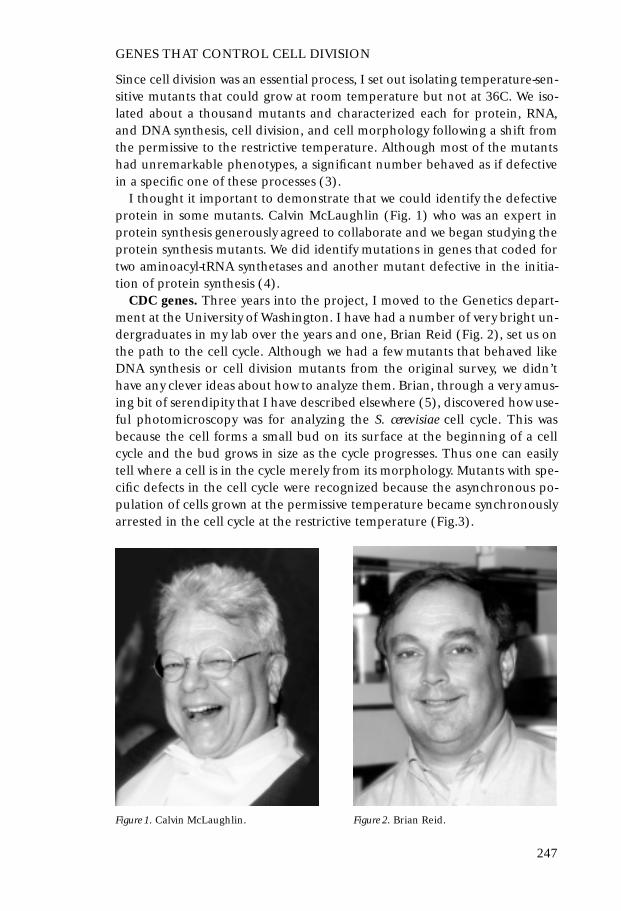
I thought it important to demonstrate that we could identify the defectiveprotein in some mutants. Calvin McLaughlin (Fig. 1) who was an expert inprotein synthesis generously agreed to collaborate and we began studying theprotein synthesis mutants. We did identify mutations in genes that coded fortwo aminoacyl-tRNA synthetases and another mutant defective in the initia-tion of protein synthesis (4).
CDC genes. Three years into the project, I moved to the Genetics depart-ment at the University of Washington. I have had a number of very bright un-dergraduates in my lab over the years and one, Brian Reid (Fig. 2), set us onthe path to the cell cycle. Although we had a few mutants that behaved likeDNA synthesis or cell division mutants from the original survey, we didn’t have any clever ideas about how to analyze them. Brian, through a very amus-ing bit of serendipity that I have described elsewhere (5), discovered how use-ful photomicroscopy was for analyzing the S. cerevisiae cell cycle. This wasbecause the cell forms a small bud on its surface at the beginning of a cell cycle and the bud grows in size as the cycle progresses. Thus one can easilytell where a cell is in the cycle merely from its morphology. Mutants with spe-cific defects in the cell cycle were recognized because the asynchronous po-pulation of cells grown at the permissive temperature became synchronouslyarrested in the cell cycle at the restrictive temperature (Fig.3).
247
Figure 1. Calvin McLaughlin. Figure 2. Brian Reid.

When we realized this, we were able to find hundreds of cell cycle mutantsin a few months. C.F. Robinow agreed to visit the lab and teach us how tostain the nuclei of cells, and Joe Culotti (Fig. 4), a new graduate student, wasable to show that the mutants that arrested with a uniform cell morphologyalso arrested with a uniform nuclear morphology. At this point we were ex-cited that we had some very interesting mutants (Fig. 5) and we were confi-dent that they would reveal new insights into the cell cycle (6).
Pathways. We ordered the mu-tants in a number of ways. First, byhow far they traversed the cycle be-fore arresting. We analyzed the fol-lowing cell cycle events: budding,DNA synthesis, nuclear division cy-tokinesis and cell division. Theevent that stopped first after a shiftfrom the permissive to the restricti-ve temperature was considered theprimary defect. After the primarydefect, other cell cycle events wouldoccur or not depending upon theparticular mutant. Eventually, thecell would arrest development andgenerate a terminal phenotype, de-pending on which events occurredand which did not. Mutants werefound with primary defects in eachof the cell cycle events that we mo-nitored. The phenotypes of the mu-tants suggested a relatively simplepathway of dependent events lead-ing to cell division. The first event,
248
Figure 3. Time-lapse photomicroscopy of a cdc mutant cells growing at the permissive temperature(A) and several hours after a shift to the restrictive temperature (B).
Figure 4. Joe Culotti.

controlled by the CDC28 gene, was required for initiating two pathways, oneof which led to budding, nuclear migration, cytokinesis and cell division (Fig.6). The second led to DNA replication, nuclear division and joined the firstprior to cytokinesis and cell division. We tested our conclusions by construc-ting all possible double mutants and, indeed, the phenotypes of double mu-tants was precisely that expected for the pathway (7).
It is interesting, in hindsight, that the pathway of dependent events was notthe least surprising to me. Although the results could have been very diffe-rent, the pathway we found was sensible with late events requiring the com-pletion of earlier events in the cycle. This is just the way that bacteriophagemorphogenesis was organized.
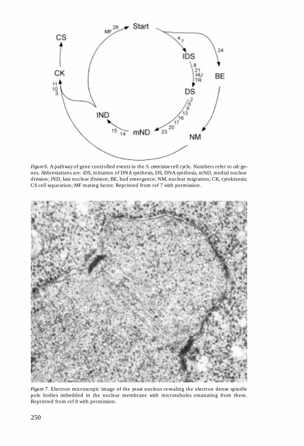
I thought we might learn more about the mutants by looking at them athigher resolution in the electron microscope. When I approached BreckByers about this, he immediately responded that there was only one structureworth looking at and that was the spindle and its poles. He and LorettaGoetsch examined the spindle and were able to resolve additional events ofspindle pole duplication, separation of the two poles, and elongation of thespindle (Fig. 7). This analysis resolved three mutants, all required for the ini-tiation of DNA synthesis into successive steps involving duplication of thespindle pole body (CDC28), separation of the poles (CDC4) and the initiationof DNA synthesis (CDC7) (8).
249
Figure 5. Normal cells and cdc mutant cells several hours after incubation at the restrictive tem-perature. (A) wild type, (B) cdc8 (C) cdc24 (D) cdc10.
250
Figure 6. A pathway of gene controlled events in the S. cerevisiae cell cycle. Numbers refer to cdc ge-nes. Abbreviations are: iDS, initiation of DNA synthesis, DS, DNA synthesis, mND, medial nucleardivision; lND, late nuclear division; BE, bud emergence; NM, nuclear migration; CK, cytokinesis;CS cell separation; MF mating factor. Reprinted from ref 7 with permission.
Figure 7. Electron microscopic image of the yeast nucleus revealing the electron dense spindlepole bodies imbedded in the nuclear membrane with microtubules emanating from them.Reprinted from ref 8 with permission.

The methods that we had used to order mutants into pathways utilized thephenotypes of single and double mutants. Lynna Hereford and Joe Culottithought of a method we called “reciprocal shifts” that utilized two conditionalagents that arrested the cell cycle independently. Jarvik and Botstein (9) in-dependently introduced the same rationale to study bacteriophage morpho-genesis. Lynna applied this method to mating factor (see below) and the cdcmutants showing that mating factor arrested cells at the CDC28 step prior tothe CDC4 and CDC7 steps (10). These results were in agreement with thespindle pole phenotypes determined by Byers & Goetsch.
Mating and cell division. Wolfgang Duntze and Tom Maney had discoveredthe presence of a mating pheromone made by Matα cells and Duntze hadfound that it inhibited DNA synthesis of Matα cells (11). We collaboratedwith them to analyze the cell cycle response to mating pheromone and foundthat it arrested cells at the CDC28 step (12). Over the next few years, we in-vestigated the relationship between the mating pathway and the division path-way. I found that when asynchronous cultures of Mata and Matα cells weremixed, they arrested one another in G1, and at the time of cell fusion, bothcells were at the G1 stage (13; Fig. 8). John Pringle and Linda Wilkensonfound evidence for a Mata pheromone that arrested Matα cells at the CDC28step (14). These experiments demonstrated that cell division was integratedwith cell mating because both haploid cell types produced pheromones thatarrested the other cell at the beginning of the cell cycle.
Breck Byers studied mating cells by electron microscopy and found thatnuclear fusion in the zygote occurred by fusion of the two unduplicatedspindle pole bodies resulting in a single nucleus with a single large spindlepole body (8). This assures that the newly formed diploid nucleus begins thecell cycle in G1 at the unduplicated spindle pole body stage. Brian Reid chal-lenged cells to mate at other stages of the cell cycle by first arresting themwith different cell cycle mutations. He found that only cells arrested at theCDC28 controlled step were capable of mating (15).
251
Figure 8. Stained cells revealing the earliest stage of cell fusion during mating with two unbudded,G1 cells undergoing cell fusion (A) followed by nuclear fusion (B). Reprinted from ref 13 withpermission.

Growth and division. John Pringle(Fig. 9) became interested in theregulation of growth in the cell cy-cle. Williamson and Scopes hadfound that stationary cultures ofyeast cells were unbudded (16).John Pringle and Gerry Johnstonfollowed up on this and showedthat growth is integrated with thedivision cycle prior to budding(17), once again at the CDC28 step.They found that cells were able tocomplete the cell cycle after abruptnitrogen starvation in the absenceof any net growth, but did not startnew cell cycles (Fig. 10). However,if division was interrupted with acell cycle mutation, the growth ofthe cell continued unabated for se-veral hours.
Yeast monitored other essentialnutrients prior to budding as well.Michael Unger used a series of mu-
tations in the sulfur assimilation pathway to ask whether an intermediate insulfur assimilation acted as a signal to arrest cells. The idea was that interrup-tion of the pathway after the signal would fail to produce arrest as unbudded
252
Figure 9. John Pringle.
Figure 10. Growing (A) and nitrogen starved (B) yeast cells illustrating that starved cells comple-te cycles without growth and generate very tiny cells from new buds; all cells end up arrested inG1 as unbudded cells. Reprinted from ref 17 with permission.
cells. He found that limitation for sulfur assimilation at any step, includingthe charging of methionyl-tRNA resulted in arrest at the CDC28 step, sugges-ting that the cell was monitoring protein synthesis or the accumulation ofprotein (18).
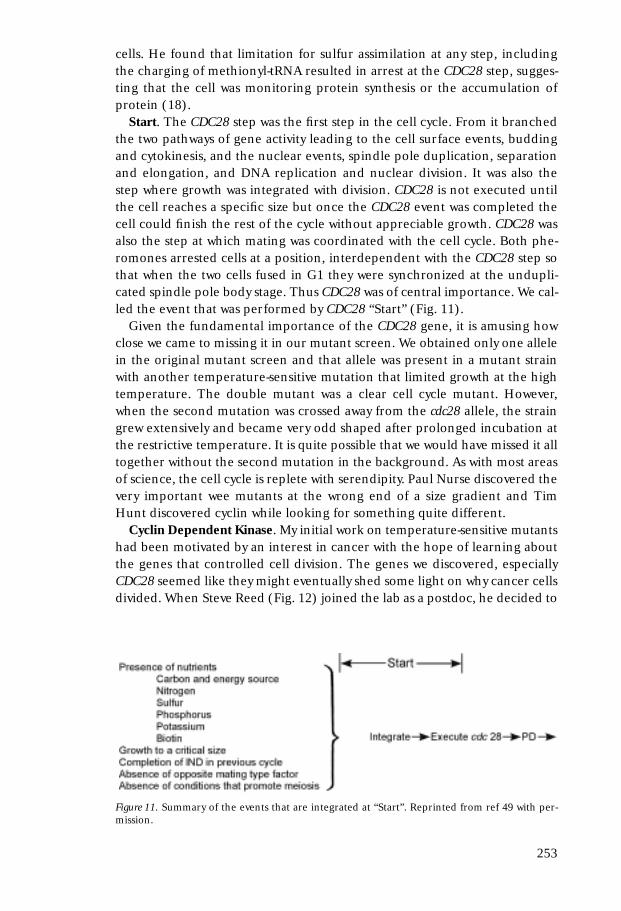
Start. The CDC28 step was the first step in the cell cycle. From it branchedthe two pathways of gene activity leading to the cell surface events, buddingand cytokinesis, and the nuclear events, spindle pole duplication, separationand elongation, and DNA replication and nuclear division. It was also thestep where growth was integrated with division. CDC28 is not executed untilthe cell reaches a specific size but once the CDC28 event was completed thecell could finish the rest of the cycle without appreciable growth. CDC28 wasalso the step at which mating was coordinated with the cell cycle. Both phe-romones arrested cells at a position, interdependent with the CDC28 step sothat when the two cells fused in G1 they were synchronized at the undupli-cated spindle pole body stage. Thus CDC28 was of central importance. We cal-led the event that was performed by CDC28 “Start” (Fig. 11).
Given the fundamental importance of the CDC28 gene, it is amusing howclose we came to missing it in our mutant screen. We obtained only one allelein the original mutant screen and that allele was present in a mutant strainwith another temperature-sensitive mutation that limited growth at the hightemperature. The double mutant was a clear cell cycle mutant. However,when the second mutation was crossed away from the cdc28 allele, the straingrew extensively and became very odd shaped after prolonged incubation atthe restrictive temperature. It is quite possible that we would have missed it alltogether without the second mutation in the background. As with most areasof science, the cell cycle is replete with serendipity. Paul Nurse discovered thevery important wee mutants at the wrong end of a size gradient and TimHunt discovered cyclin while looking for something quite different.
Cyclin Dependent Kinase. My initial work on temperature-sensitive mutantshad been motivated by an interest in cancer with the hope of learning aboutthe genes that controlled cell division. The genes we discovered, especiallyCDC28 seemed like they might eventually shed some light on why cancer cellsdivided. When Steve Reed (Fig. 12) joined the lab as a postdoc, he decided to
253
Figure 11. Summary of the events that are integrated at “Start”. Reprinted from ref 49 with per-mission.

focus on the CDC28 gene, settingup selections that uncovered newalleles as well as new genes that ar-rested at the same step. In 1980 heand Kim Nasmyth used recently de-veloped techniques to clone theCDC28 gene (19) and after Steveset up his own laboratory, he foundthat it had protein kinase activity(20). The ultimate unification ofthe cell cycle field with the discove-ries relating CDC28 of S. cerevisiae,cdc2 of S. pombe, and MPF of Xeno-pus embryos to cyclin dependentkinases centered largely in the labo-ratory of Paul Nurse with importantcontributions by many others andhas been elegantly described re-cently by Kim Nasmyth (21).
Expectations fulfilled. I find it quite interesting philosophicallythat two different groups ap-proached the cell cycle with diffe-rent expectations and each found
exactly what they were looking for. At first, the results looked very different,perhaps because we were imposing our expectations. I approached the studyof the cell cycle with the paradigm of bacteriophage morphogenesis in mind.Indeed what we found looked a lot like phage morphogenesis – a series of gene controlled, dependent events leading to progressive stages in division.Paul Nurse, on the other hand, was most interested in rate limiting steps. Hefound a rate-limiting step at mitosis controlled by the cdc2 gene of S. pombe(22). From this point of view, the most important gene of fission yeast, cdc2,seemed very different than what we viewed as the most important gene inbudding yeast, CDC28, because the former acted in G2 while the latter actedin G1. Once it became clear that the two genes could substitute for one anot-her (23), the apparent differences were resolved. In fact, both proteins actedin each organism at G1 and G2. Under rapid growth conditions the G1 acti-vation of CDC28 in S. cerevisiae is dependent on cell growth while in S. pombe itis the G2 activation of cdc2 that is dependent on growth.
GENOME FIDELITY
Cancer cells not only divide when they shouldn’t but they also reproduce withfar poorer fidelity than normal cells. Chromosome aberrations and chromo-some losses are very common in cancer cells. This loss of fidelity seemedcentral to cancer because it would permit the cancer cells to evolve more
254
Figure 12. Steve Reed.

quickly. We thought we might be able to learn more about how chromosomefidelity is maintained in the normal cell cycle by studying the fidelity of chro-mosome transmission in yeast cells.
Essential machinery. We found that normal yeast cells lose a chromosomeor undergo mitotic recombination at rates of about one event in 105 cells. Wewondered whether limitation of any of the essential cell cycle functions wouldalter that fidelity. An undergraduate, David Smith and I studied chromosomeloss, recombination and mutation in temperature-sensitive cell cycle mutantswhen they were grown at their maximum permissive temperature. We foundthat most mutants had greatly elevated rates of chromosome loss, recombi-nation or mutation (Fig. 13), implying that defects in these functions couldbe important in the fidelity of cell division (24).
Other essential components were also important for fidelity. DougKoshland examined the role of centromere structure and dicentrics on chro-mosome fidelity (25); Meeks-Wagner defined the importance of correct ratiosof histones (26); and Megan Brown studied a new gene involved in centro-mere function, MIF2 (27).
255
Figure 13. Rates of mitotic recombination and chromosome loss in cdc mutants grown at theirmaximum permissive temperature. Reproduced from ref 24 with permission.
These results suggested that mutations in essential components of the cellcycle machinery might be responsible for the genetic instability of tumor cellsin addition to the known role of DNA repair defects in certain forms of can-cer susceptibility (e.g. DNA excision repair in skin cancer and mismatch re-pair in colon cancer).
DNA damage checkpoint. Fortu-nately, my interest in genomic insta-bility coincided with Ted Weinert’s(Fig. 14) interest in studying the regulation of cell division. Hethought it likely that all of our cellcycle mutants were identifying ge-nes that contributed to the machi-nery of cell division and was in-terested in studying something thatwas more clearly an example of cellcycle regulation. I had remembe-red noticing that yeast cells becamearrested synchronously in the cellcycle by radiation and mutagensand he began looking at radiationsensitive mutants to see if any werealtered for their cell cycle response.He quickly found that some radia-tion sensitive mutants failed to ar-rest the cell cycle in response to ra-diation (28). He demonstrated that
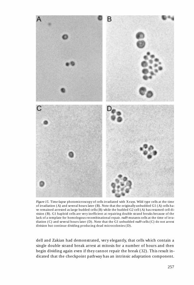
deletion of the RAD9 gene eliminated the regulation of the cell cycle by radi-ation, demonstrating a regulatory role for this gene (Fig. 15) and discovereda number of additional genes involved in the DNA damage checkpoint (29).
Ted’s discovery led to an appreciation of the role of checkpoints in the fi-delity of chromosome transmission. The rad9 mutant exhibited a 20 fold in-crease in the rate of chromosome loss in the absence of any extrinsic DNA da-mage. An explanation for the increase in chromosome loss in checkpointdefective cells was provided by Ted’s discovery that the same mutations thatrendered cells insensitive to arrest of mitosis by radiation also rendered cellsinsensitive to arrest by defects in DNA replication (29; Fig. 16). This sug-gested that intrinsic errors in DNA replication occurred stochastically in rarecells of the population and that the checkpoint function was needed in thosecells to assure correct repair of the damage. The same checkpoint genes were later found by Mandy Paulovitch to control the rate of replication overdamaged DNA (30) and by Siede, Friedberg and Friedberg to control the rate at which cells enter S phase when they experience damage in G1 (31).
The role of the DNA damage checkpoint in assuring genomic fidelity wasfurther elucidated by Daivd Toczyski when he was a postdoc in the lab. San-
256
Figure 14. Ted Weinert.
dell and Zakian had demonstrated, very elegantly, that cells which contain asingle double strand break arrest at mitosis for a number of hours and thenbegin dividing again even if they cannot repair the break (32). This result in-dicated that the checkpoint pathway has an intrinsic adaptation component.
257
Figure 15. Time-lapse photomicroscopy of cells irradiated with X-rays. Wild type cells at the timeof irradiation (A) and several hours later (B). Note that the originally unbudded G1 (A) cells ha-ve remained arrested as large budded cells (B) while the budded G2 cell (A) has resumed cell di-vision (B). G1 haploid cells are very inefficient at repairing double strand breaks because of thelack of a template for homologous recombinational repair. rad9 mutants cells at the time of irra-diation (C) and several hours later (D). Note that the G1 unbudded rad9 cells (C) do not arrestdivision but continue dividing producing dead microcolonies (D).

David discovered mutants defective in adaptation of the DNA damage check-point and showed that wild type cells actually transmit more damage to off-spring than adaptation defective cells (33). The p53 tumor suppressor gene(34) acts as a cell cycle checkpoint gene and its function is also regulated byadaptation. Several other cell cycle checkpoints have been discovered. Onecontrols mitosis if chromosomes are not attached to the mitotic spindle (35).A recent review of the DNA damage checkpoint has appeared (36).
Paradox resolved. After the discovery of cyclin dependant kinases, there re-mained an important paradox in our understanding of the cell cycle. By 1988it was clear that the CDK played a controlling role in the cell cycle of frogs aswell as yeast. Gerhart, Wu and Kirschner had demonstrated that the matura-tion promoting activity (CDK) of Xenopus eggs displayed a periodic activa-tion at the onset of mitosis (37). Murray, Solomon and and Kirschner had
258
Figure 16. Cells defective in telomere replication fail to arrest cell division at the restrictive tem-perature. cdc13 RAD9 cells incubated at the restrictive temperature for several hours have ar-rested cell division and retain viability (A) but cdc13 rad9 cells failed to arrest and die (B).Reproduced from ref 29 with permission.

shown that accumulation of the cyclin mRNA was the limiting component foractivating mitosis (38). Thus it seemed that the Xenopus cell cycle was drivenby periodic activation of the CDK through cyclin synthesis. Moreover, thisCDK “clock” was autonomous from the events of the cell cycle. Inhibition ofDNA replication does not block mitosis during the earliest divisions of theegg.
How could we reconcile this picture with that derived from the two yeasts?The yeast cell cycle did not behave as though it were being run by a clock un-dergoing periodic activation. Instead, it had the appearance of a series of de-pendent events where each event provided the substrate or activated the sub-sequent event. Growth was the activator of the CDK each cycle and there wasno need for a clock. To phrase the problem more precisely, if periodic activa-tion of the CDK was driving cell cycle events, why did the cell cycle stop inyeast when the machinery for DNA replication or mitosis was defective? Whywouldn’t the clock keep ticking and activate mitosis in the absence of DNA re-plication or cytokinesis in the absence of mitosis? The discovery of mutantsthat no longer arrested division in response to radiation provided the answer– a series of regulatory signaling pathways, checkpoints, that keep the cell in-formed of each event’s progress. If an event stops, checkpoint signaling ar-rests the clock. The frog embryo is different because the checkpoint only be-comes active after about the twelfth embryonic division (39).
A unified view. The genetic control of cell division provided two importantlessons that have been repeated over and over in molecular, cellular and de-velopmental biology. The first is the conservation of proteins and their func-tions throughout evolution. This was not a surprising conclusion because allliving organisms share a common ancestor. However, we did not know howconserved the machinery would be. We did not know that a newly sequencedgene from nearly any organism would be seen to fit into a family of proteinslike kinases, myosins, or anthranylate synthetases. Indeed, the molecular con-servation of many of the proteins involved in the cell cycle and in cell cyclecheckpoints is not very evident across the prokaryotic / eukaryotic boundarybut within eukaryotes the conservation is striking. A second, and of course re-lated issue is how easily biology uses the same parts to make different things.Here again, the cell cycle is a dramatic example. Of the three organisms moststudied, Xenopus, fission yeast and budding yeast there appeared to be littlein common. The frog embryonic cell cycle was controlled by a clock, the fis-sion yeast by a rate controlling step in G2 and the budding yeast by a genethat initiated spindle pole duplication in G1. Yet all three activities turned outto encode the same cyclin dependent protein kinase, and the differences were only superficial.
CANCER THERAPEUTICS
The genetic instability of cancer cells are due to genetic defects that affect thecell cycle machinery, DNA repair, or cell cycle checkpoints. Examples of thelatter two are well known. Since we know a lot about the biochemistry of each
259

of these processes it has been possible to define the genetic changes that leadto loss of fidelity in some cancers and it should be possible in many cancers.These defects should create a vulnerability for the cancer cell relative to thenormal cell that could provide a powerful therapeutic advantage if the ap-propriate vulnerability were targeted for therapeutic intervention.
This brings me to a third application of the yeast model to cancer research.A new adventure was in the making when I had the good fortune to be invitedto give a seminar at Massachusetts General Hospital at about the time thatStephen Friend was exploring his interest in drug discovery. Stephen hadspent some time investigating the drug discovery programs at some of the ma-jor pharmaceutical companies. Mark Groudine, Jim Roberts, and Bob Eisen-man had gotten me interested in the potential for drug discovery in the areaof cell cycle checkpoints. Stephen got interested in this idea as well and wasrecruited to the FHCRC to head up a program in molecular pharmacology.Stephen and I set up a drug discovery program that was originally supportedby Merck and the NCI to use yeast genetics to discover and validate cancertargets.
Our goal was to identify drugs and drug targets that would present a thera-peutic advantage. That is, where the cancer cells would be more sensitive tothe drug or to inhibition of the target than the normal cells. The idea was toconstruct yeast cells that contained mutations characteristic of specific tumors(altered in mismatch repair, cyclin, activated telomerase, etc.). The mutantand normal cells were then screened for drugs that killed the mutant yeastmore effectively than a wild type yeast (40). The results have been encourag-ing. A test of 23 commonly used chemotherapeutic agents revealed that somehad a high “therapeutic advantage” (41). Cis-platin preferentially kills cellswith defects in post-replication repair genes, rad6 and rad18 (Fig. 17); thenucleotide analogue, cytarabine is preferential for an sgs1 mutant, a homolo-gue of the Bloom’s syndrome and Werner’s Syndrome helicases; camptothe-cin, a topoisomerase I inhibitor, and idarubicin and mitoxantone, two to-poisomerase II inhibitors, are specific for cells with defects in double strandbreak repair, rad50, rad51 and rad52. Other agents were selective for a broader range of DNA damage repair mutants and some agents were non-specific.
This approach identifies drugs with therapeutic advantage. We were also in-terested in identifying protein targets which if inhibited by drugs would pro-vide therapeutic advantage. These would be “validated” targets. Yeast lends it-self to this goal as well. There are methods for identifying mutations in anunknown gene that are synthetic-lethal with mutations in a second known ge-ne (42). So, we could start with a mutation characteristic of certain types ofcancer and identify mutations in other genes that would be synthetic-lethalwith the first. These genes would be potential drug targets. Targets were iden-tified that would kill mutants defective in the DNA damage checkpoint.These targets were enzymes of deoxynucleotide biosynthesis and componentsof the DNA replication apparatus (Eric Foss and Patrick Paddison, un-published).
260

NATURAL GENETIC VARIATION
I am currently interested in the individual differences in susceptibility to can-cer. How does the genetic variation that exists in the human population ma-nifest in differences in disease susceptibility? Will we be able to assess diseasepredisposition for most people at an early age so they can choose life styles orfrequent screening that will lead to better health? Will there be an individua-lized medicine?
The current preoccupation in molecular genetics is in the use of inbred or-ganisms as models of human disease. When a gene is assigned a function in amouse we reason that it is likely to play a similar role in the human. Yet themodel systems that we study are genetically homogeneous except for the sing-le gene perturbation that we study, whereas the human population is geneti-cally outbred such that all individuals are very different (except for identicaltwins). It is clear that this genetic individuality has a very important role in thetransformation of genotype into phenotype because the same mutation in dif-ferent inbred strains of mice or yeast often have different phenotypes. Are wemissing something by studying inbred model organisms?
I seem to have extremely good luck in finding colleagues who point theway. Just as I was considering these issues, I had the good fortune of meetingStan Leibler who taught me about the robustness of biological circuits (43). Ihave come to think that this concept of robustness is critical to understandingthe phenotypic consequences of natural genetic variation. Because of buffer-ing, some genetic variation in protein activity or amount will have no pheno-typic consequence.
There are several seminal contributions to our understanding of robust-ness. Redundant pathways, feed back control, and certain metabolic pathwayshave this robust behavior. One is the insight of Kczar and Burns to the ro-bustness of metabolic pathways (44). They demonstrated that the flux of me-
261
Figure 17. Sensitivities of different DNA repair and checkpoint mutants to common chemothera-peutic agents. (A) Cisplatin, (B) Cytarabine, (C) Camptothecin. The horizontal axis is a log sca-le. Reproduced from 41 with permission.

tabolic pathways (in which the enzymes followed Michaelis-Menton kineticsand none of the enzymes are saturated for their substrates) would be relati-vely insensitive to changes in the amount or activity of individual enzymes inthe pathway. Stan Leibler and his colleagues have come to the same conclu-sion for the more complicated bacterial chemotaxis behavioral responsebecause of its intrinsic feed-back regulation (45). An investigation of the ge-netic circuitry that controls Drosophila segmentation by vonDassow, Meir,Munroe and Odell has come to the surprising conclusion that robustness maybe a property of almost any circuit with a significant complexity (46).
Since this buffering capacity is a product of the circuit and its componentsthere will be natural genetic variants that compromise the buffering capacityitself. The consequence will be that many polymorphisms in a population willpresent no phenotypic consequence in the most prevalent genotypes.However, in rare genotypes that happen to compromise the buffering capaci-ty for the particular polymorphism in question, a phenotype may result. Ifthis reasoning is correct, then many disease susceptibilities will not be assign-able to single polymorphisms but only to combinations.
How can we identify alleles that interact to produce phenotypes in combi-nation that neither produce alone? Here again, yeast is an ideal organism inwhich to investigate this issue at its most fundamental level. As describedabove, there are methods in yeast for identifying synthetic-lethal interactionsor synthetic-phenotypes (42).
For my purpose, which is to understand the genetic basis of phenotypic va-riation in an outbred population, it is important to understand the numberof synthetic relationships that each gene is subject to. Obviously, the more in-teractions that can create synthetic phenotypes, the greater the genetic com-plexity potentially underlying a phenotype. Recent work in yeast from Tong,et al. (47) and unpublished work by John Hartman in my own laboratory sug-gest that genes interact with 20 or more other genes in the genome to createsynthetic phenotypes. Or, to put it another way, variations in the activity of anysingle protein are buffered by interactions with as many as 20 other proteinsin the genome.
It is clear that the prospects for genetic complexity underlying disease phe-notypes in outbred populations is very great. So great, in fact, that if even asmall percentage of the potential genetic variation were present in the popu-lation, then nearly all phenotypes would be the result of highly complex ge-netic interactions and most individuals with the same phenotype would havedifferent combinations of alleles generating that phenotype. Attempts toidentify natural polymorphisms that account for the differences in cancer susceptibility between two inbred mouse strains often does uncover greatcomplexity (48). What is not clear, is how much functional variation is actu-ally present in the human population. This depends on the history of the po-pulation, its size, its age, etc. For the human population, we know that about1 nucleotide in 600 is polymorphic but we do not have any idea how many ofthese polymorphisms effect function or expression of gene products.
In order to learn more about the principles underlying the genetic com-
262

plexity of human phenotypes we can hope to learn again from yeast. Im-portant questions are the number and identity of genes that can interact syn-ergistically to generate a phenotype, the patterns in which they interact, andthe fraction of natural polymorphisms that have functional consequences. Aninvestigation of these questions in yeast might help identify the set of candi-date genes that could be associated with a particular human disease.
We still have a lot to learn from yeast and other model organisms about thenature of human disease.
REFERENCES
1. Williamson, D.H. 1965. The timing of deoxyribonucleic acid synthesis in the cell cycleof Saccharomyces cerevisiae. J Cell Biol 25(3); 517–28.
2. Robinow, C.F., Marak, J. 1966. A fiber apparatus in the nucleus of the yeast cell. J CellBiol 29(1): 129–51.
3. Hartwell, L.H. 1967. Macromolecule synthesis in temperature-sensitive mutants ofyeast. J. Bact. 93: 1662–1670.
4. Hartwell, L.H. and McLaughlin, C.S. 1968. Mutants of yeast with temperature-sensitiveisoleucyl-tRNA systhetases. Proc. Nat. Acad. Sci 59: 422–428.
5. Hartwell, L.H. 1993. Getting started in the cell cycle. In the early days of yeast genetics,307–314. M.N. Hall and P. Linder. Cold Spring Harbor Laboratory Press, Cold SpringHarbor, NY.
6. Hartwell, L.H., Culotti, J. and Reid, B. 1970. Genetic control of the cell-division cyclein yeast, I. Detection of mutants. Proc. Nat. Acad. Sci 66:352–359.
7. Hartwell, L.H., Culotti, J., Pringle, J.R. and Reid, B.J. 1974. Genetic control of the celldivision cycle in yeast. Science 183: 46–51.
8. Byers, B. and Goetsch, L. 1975. Behaviors of spindles and spindle plaques in the cellcycle and conjugation in Saccharomyces cerevisiae. J. Bacteriol. 124: 511–523.
9. Jarvik, J. and Botstein, D. 1973. A genetic method for determining the order of eventsin a biological pathway. Proc. Natl. Acad. Sci. 70: 2046–2050.
10. Hereford, L.M. and Hartwell, L.H. 1974. Sequential gene function in the initiation ofSaccharomyces cerevisiae DNA synthesis. J. Mol. Biol. 84: 445–461.
11. Duntze, W., MacKay, V., and Manney, T. 1970. Saccharomyces cerevisiae: A diffusible sexfactor. Science 168: 1472–1743.
12. Bucking-Throm, E., Duntze, W., Hartwell, L.H. and Manney, T.R. 1973. Reversible ar-rest of haploid yeast cells at the initiation of DNA synthesis by a diffusible sex factor.Exp. Cell Res. 76:99–110.
13. Hartwell, L.H. 1973. Synthronization of haploid yeast cell cycles, a prelude to con-jugation. Exp. Cell Res. 76:111–117.
14. Wilkinson, L.E. and Pringle, J.R. 1974. Transient GI arrest of S. cerevisiae cells of matingtype α by a factor produced by cells of mating type α Exp. Cell Res. 89: 175–187.
15. Reid, Brian J. and Hartwell, L.H. 1977. Regulation of mating in the cell cycle ofSaccharomyces cerevisiae. J. Cell Biol. 75: 355–365.
16. Okasis.17. Johnston, G.C., Pringle, J.R. and Hartwell, L.H. 1977. Coordination of growth with cell
division in the yeast Saccharomyces cerevisiae. Exp. Cell Res. 105: 79–98.18. Unger, M.W. and Hartwell, L.H. 1976. Control of cell division in Saccharomyces cerevisiae
by methionyl-tRNA. Proc. Natl. Acad. Sci. 73: 1664–1668.19. Nasmyth, K.A. and Reed, S.I. 1980. Isolation of genes by complementation in yeast:
Molecular cloning of a cell-cycle gene. Proc. Nat. Acad. Sci. 77: 2119–2123.
263

20. Reed, S.I., Hadwiger, J.a., and Lorinez, A.T. 1985. Protein kinase activity associatedwith the product of the yeast cell division cycle gene CDC28. Proc. Natl. Acad. Sci82(12): 4055–9.
21. Nasmyth, K. 2001. A Prize for Proliferation. Cell 107:689–701.22. Nurse, P. and Thuriaux, P. 1980. Regulatory genes controlling mitosis in the fission
yeast Schizosaccharomyces pombe. Genetics 96: 627–637.23. Beach, D., Durkacz, B., and Nurse, P. 1982. Functionally homologous cell cycle control
genes in budding and fission yeast. Nature 300: 706–709.24. Hartwell, L.H. and Smith, D. 1985. Altered fidelity of mitotic chromosome transmis-
sion in cell cycle mutants of S. cerevisiae. Genetics 110: 381–395.25. Koshland, D., Kent., J.C. and Hartwell, L. 1985. Genetic analysis of mitotic transmis-
sion of minichromosomes. Cell 40: 393–403.26. Meeks–Wagner, D. and Hartwell, L.H. 1986. Normal stoichiometry of histone dimer
sets is necessary for the high fidelity of mitotic chromosome transmission. Cell 44:43–52.
27. Brown, M.T., Goetsch, L. and Hartwell, L.H. 1993. MIF2 is required for mitotic spind-le integrity during anaphase spindle elongation in Saccharomyces cerevisiae. J. Cell Biol.123: 387–403.
28. Weinert, T.A., and Hartwell, L.H. 1988. The RAD9 gene controls the cell cycle respon-se to DNA damage in Saccharomyces cerevisiae. Science 241: 317–322.
29. Weinert, T.A., Kiser, G.L. and Hartwell, L.H. 1994. Mitotic checkpoint genes in bud-ding yeast and the dependence of mitosis on DNA replication and repair. Genes Dev. 8:652–665.
30. Paulovich, A.G. and Hartwell, L.H. 1995. A checkpoint regulates the rate of progres-sion through S phase in S. cerevisiae in response to DNA damage. Cell 82: 841–7.
31. Siede, W., Friedberg, A.S., and Friedberg, E.C. 1993. RAD9-dependent G1 arrest defi-nes a second checkpoint for damaged DNA in the cell cycle of Saccharomyces cerevisiae.Proc. Natl. Acad. Sci. Sep 1 90(17): 7985–9.
32. Sandell, L.L. and Zakian, V.a. 1993. Loss of a yeast telomere: arrest, recovery, and chro-mosome loss. Cell 75: 729–739.
33. Toczyski, D.P., Galgoczy, D.J., and Hartwell, L.H. 1997. CDC5 and CKII control adap-tation to the yeast DNA damage checkpoint. Cell 90:1097–1106.
34. Kastan, M.B., Zhan, Q., El-Deiry, W.S., Carrier, F., Jacks, T., Walsh, W.V., Plunkett, B.S.,Vogelstein, B., and Fornace, A.J. Jr. 1992. A mammalian cell cycle checkpoint pathwayutilizingi p53 and GADD45 is defective in ataxia-telangiectasia. Cell 71(4): 587–97.
35. Rudner, A.D. and Murray, A.W. 1996. The spindle assembly checkpoint. Curr. Opin. CellBiol. 8(6): 773–80.
36. Zhou, B.S. and Elledge, S.J. 2000. The DNA damage response: putting checkpoints inperspective. Nature 408:433–9.
37. Gerhart, J., Wu, M., Kirschner, M. 1984. Cell cycle dynamics of an M-phase-specific cy-toplasmic factor in Xenopus laevis oocytes and eggs. J. Cell Biol. 98(4): 1247–55.
38. Murray, A.W., Solomon, M.J., and Kirschner, M.S. 1989. The role of cyclin synthesisand degradation in the control of maturation promoting factor activity. Nature 339:280–286.
39. Newport, J. and Dasso, M. 1989. On the coupling between DNA replication and mito-sis. J. Cell. Sci. Suppl. 12:149–60.
40. Hartwell, L.H., Szankasi, P., Roberts, C.J., Murray, A.W., and Friend, S.H. 1997. Inte-grating Genetic Approaches into the discovery of anti-cancer drugs. Science 278(5340):1064–1068.
41. Simon, J.A., Szankasi, P., Nguyen, D.K., Ludlow, C., Dunstan, H.M., Roberts, C.J.,Jensen, E.L., Hartwell, L.H., Friend, S.H. 2000. Differential toxicities of anticanceragents among DNA repair and checkpoint mutants of Saccharomyces cerevisiae. CancerRes. 60(2): 328–333.
264

42. Bender, A., Pringle, J.R. 1991. Use of a screen for synthetic lethal and multicopy sup-pressee mutants to identify two new genes involved in morphogenesis in Saccharomycescerevisiae. Mol Cell Biol. 11(3): 1295–1305.
43. Alon, U., Surette, M.G., Barkai, N., and Leibler, S. 1999. Robustness in bacterial che-motaxis. Nature 397: 168–71.
44. Kacser, H. and Burns, J. 1981. The molecular basis of dominance. Genetics 97: 639–666.45. Alon, U., Surett, M.G., Barkai, N. and Leibler, S. 1999. Robustness in bacterial chemo-
taxis. Nature 397:168–71.46. vonDassow, G., Meir, E., Munroe, E.M., Odell, G.M. 2000. The segment polarity
network is a robust developmental module. Nature 406(6792): 188–192.47. Tong, A.H., Evangelista, M., Parsons, A.B., Xu, H., Bader, G.D., Page, N., Robinson, M.,
Raghibizadeh, S., Hogue, C.W., Bussey, H., Andrews, B., Tyers, M., Boone, C. 2001.Systematic genetic analysis with ordered arrays of yeast deletion mutants. Science294(5550): 2364–8.
48. Tripodis, N., Hart, Augustinus A.M., Fijneman, Remond J.A., Demant, P. 2001.Complexity of lung cancer modifiers: Mapping of thirty genes and twenty-five interac-tions in half of the mouse genome. JNCI 93(10): 1484–1491.
49. Hartwell, L.H. 1974. Saccharomyces cerevisiae Cell Cycle. Bact. Rev. 38(2): 164–198.
265