Embed Size (px)
Citation preview

Y-90 SIR-Spheres Treatment of Unresectable Primary or Metastatic Liver TumorsJ. Machac1, S. Heiba1, M. Jiang1, Z. Zhang1,
R. R. P. Warner2, J. Weintraub3
Division of Nuclear Medicine1, Dept. of Medicine2, Dept. of Radiology3
Mount Sinai School of Medicine, NY.

Localised Liver Cancer
Surgery Laser & Sclerotherapy
RF Ablation Cryotherapy

Advanced Liver Cancer

Hepatic Vasculature

Blood Supply of Liver Cancer

Blood Supply to Liver Cancer

Concept of SIRTS(selective) I(internal) R(radiation) T(therapy)
To selectively target a very high radiation dose to all tumors within the liver, regardless of their cell of origin,
number, size or location
while at the same time
Maintaining a low radiation dose to the normal liver tissue
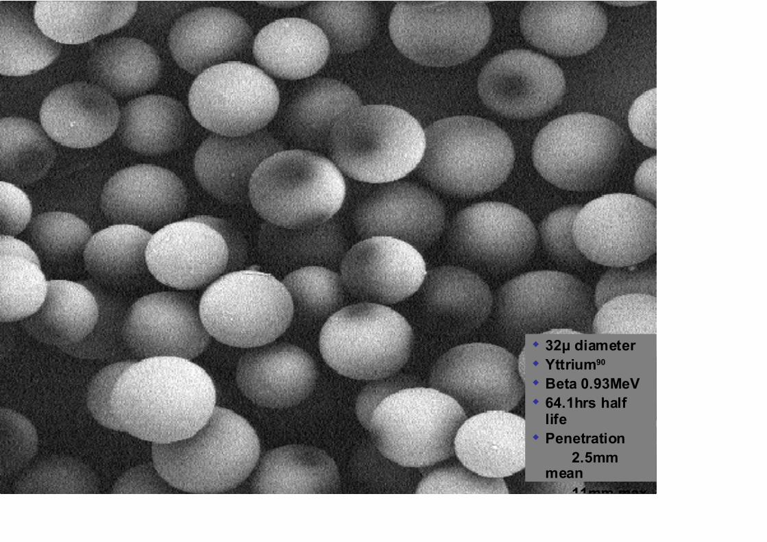
32 diameterYttrium90
Beta 0.93MeV64.1hrs half lifePenetration
2.5mm mean
11mm max

SIR-Spheres in Liver Cancer

Entrapment of SIR-Spheres in Vascular Bed

Dosimetry
HYPERVASCULAR RIM
HYPOVASCULAR CORE
1. ARTERIAL BLOOD FLOW2. SPHERE DENSITY3. SPHERE SPECIFIC ACTIVITY4. SPHERE NUMBER

Types of SIRT• Y-90 Theraspheres
– Nordion– Glass beads (heavier)– High specific activity– Fewer number of beads– Approved for HCC only (with IRB approval)
• Y-90 SIR Spheres– SIRTEX– Plastic– Lower specific activity– Large number of beads– Approved for Colorectal Carcinoma– Used off-label for many other liver tumors

SIR-Spheres

Administration technique

Y-90 microsphere selective radiation therapy
• A microcatheter is selectively placed in the hepatic artery or its branches through the femoral artery and celiac trunk by IR.
• Microspheres lodged in the microvasculature in the tumor, deliver localized radiation.

Transfemoral Injection

OBJECTIVESTo evaluate the efficacy and safety of Y-90 SIR-Spheres (SirTex) treatment in pts with unresectable liver tumors.

• 109 Pts. (130 treatments) METHODS
Disease No. %
Carcinoid 46 42.2
NETs 10 9.2
HCC 32 29.4
Colon ca 10 9.2
Other ca 11 10.1

Patient Population
Patients No. % Median Age
Male 69 63.3 60 (38-85)
Female 40 36.7 67 (45-75)
Total 109 100 63 (38-85)

Tumours in both lobes of Liver

Super Selective Injection ofSIR-Spheres

• To assess liver arterial perfusion, lung & intra-abdominal shunting• Dose: 5 mCi 99mTc-MAA • Inject into the hepatic artery via a percutaneous catheter• Anterior and posterior planar images from the head to the thighs• SPECT/CT images of the abdomen

MAA Hepatic perfusion Imaging is routinely performed prior to Y-90 therapy
Whole Body Planar A/P SPECT/CT images of abdomen

Assess Lung Shunting• ROIs were drawn around the liver
and each lung in the anterior & posterior views

Assess Lung Shunting
• Geometric mean counts for the liver & the lungs were then obtained.
% Shunt = Lung Counts x 100%(Liver + Lung) Counts

Diagnostic Studies: Arterial Tc-99m MAA

Diagnostic Studies: Arterial Tc-99m MAA

SIRT Dose CalculationSIR-Spheres Dose (GBq)
= BSA (m2) – 0.2 + volume tumour volume tumour + liver
% Lung Shunting % Dose Reduction
0-10 % 0 %10-15 % 20 %15-20 % 40 %
40% Coldwell Reduction (before 7/2006)25% Coldwell Reduction (after 7/2006)Volume reduction

Post-Therapy Y-90 Bremmsstrahlung Scan
Anterior Posterior Anterior Posterior

Post-Therapy Y-90 Bremmsstrahlung Scan

Example of good MAA / Y-90 match
MAA
Y-90

Follow Up
• Radiological (CT, MR, PET/CT or Octreoscan)
• Tumor markers
• F/U of at least 3 months was available
• Toxicity - symptoms

Pts. (Tx)Carcinoid
+ NETHCC + Others
Total 109 (130) 58 (74) 51 (56)
NA 38 (48) 16 (24) 22 (24)
Follow up 71(82) 42 (50) 29 (32)
Follow Up Patient Population

Dose No. % Median (GBq)
Min. (GBq)
Max. (GBq)
Std. Dose. 18 21.95 1.55 1.20 2.48
Dose Reduction (25-40%)
64 78.05 1.20 0.30 2.05
Prescribed Dose*
* Std. Dose (GBq) = BSA(m2) - 0.2 + Tumor Vol (Tumor + perfused liv) Vol

Targeted Lobes Tx No. (%)Entire Liver 20 (24.4%)Right Lobe 47 (57.3%) Left Lobe 15 (18.3%)
SIR-Spheres Infusion

RESULTS
Lung Shunting
LungShunting
(%)
Median Min. Max.
4.6 1.1 18.0

% of Volume No. (%) Range (%)
< 25% 24 (29.3%) 8.7-24.0
25-50% 50 (61%) 25-48.5
> 50% 8 (9.7%) 51.0-72.0
RESULTS: Hepatic Tumor Volume%

Absorbed Dose (Gy) Median Min. Max.
Lung 9.88 1.8 30
Liver 21.9 5.2 70
Tumor 60.3 6.4 204
T/N 2.9 0.76 11.4
RESULTS: SIRT Dosimetry
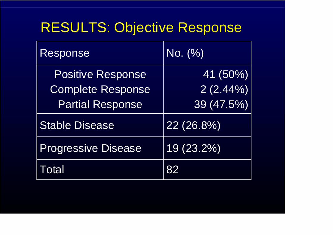
RESULTS: Objective Response
Response No. (%)
Positive ResponseComplete Response
Partial Response
41 (50%)2 (2.44%)
39 (47.5%)
Stable Disease 22 (26.8%)
Progressive Disease 19 (23.2%)
Total 82

12 month post therapy CT showing interval regression in tumor volume.

Follow-up CT showing interval progression of disease

0.0
10.0
20.0
30.0
40.0
50.0
60.0
1 2 3
Carcinoid+NETHCCOthers
Treatment Response
54.8%
30.6%
15.5%
44%
33.3%
22%
52.6%
11.1%
38.9%
CR+PR SD PD

% of Rx Dose Tx No. (%)
100 63 (76.8%)
47-91% (Flow stasis) 19 (23.2%)
RESULTS : Delivered Dose
• There was no statistically significant difference in response between the groups treated with complete dose vs. partial dose due to flow stasis.

Side Effects
• Occasional symptoms Nausea Anorexia
• No radiation pneumonitis and treatment-induced liver failure • 3 pts developed gastric ulceration (3%)
2 pts were managed conservatively 1 pt had surgical treatment

The Images of Gastric Biopsy
Ulceration and radiation fibroblasts
Microspheres in Stomach

Potential complications of Y-90 therapy caused by extra-hepatic distribution
• Radiation pneumonitis from hepatopulmonary shunting.
• Gastrointestinal ulcers and pancreatitis from gastrointestinal or pancreatic deposition of Y-90 microspheres.

• Y-90 SIR-Spheres treatment is a useful modality with remarkable objective response rate in the treatment of pts with unresectable primary or metastatic liver tumors.
• Pts with carcinoid or NET and other cancers showed a higher PR rate compared to pts with HCC .
• Partially delivered dose due to flow stasis could still produce a response.
• The procedure had acceptable toxicity.
CONCLUSIONS

Characterization of extra-hepatic distribution on MAA hepatic perfusion
imaging for Yttrium-90 therapy
M Jiang, J Machac, S Heiba, Z Zhang, K Knesaurek, A Stangl, E Kim, SF Nowakowski. J Weintraub
Mount Sinai Medical Center, NY

Objectives of the study
• We retrospectively reviewed all the MAA hepatic perfusion studies between 2007 to 2008 in Mount Sinai hospital.
• To characterize the extra-hepatic MAA distribution and correlate with diagnosis, infusion site, and angiograms.
• To characterize the main cause of the abnormal findings on MAA hepatic perfusion imaging.

Primary diagnosis Number
Carcinoid 83 (42%) HCC 66 (33%)Colon Ca 25 (13%) Cholangio Ca 7 ( 4%) Other 17 (8%)
Total Total 198198
Patient population

MAA injection site
Studies
198
RHA
101
LHA
43
PHA/
CHA
54

RESULTS I: Hepatopulmonary Shunting
Moderate (10-20%) 17 20-40% dose reduction Severe (>20%) 6 Rejected for therapy
Severity No. Consequences

HCCCarcinoidColon Cancer
Moderate and severe lung shunting is overrepresented by the HCC group
HCC
Colangiocarci
10/17 (59%) 5/6
(83%)
Moderate lung shunting (10-20%)
Severe lung shunting (>20%)
5/17(29%)
1/6(17%)
HCCCarcinoidColon caCholangio
Total studies
66/198 (33%)
83/198 (42%)
2/17(12%)

Hepatic arterial-portal shunting
2
Severe lung shunting
(89%)
Significant hepatic arterial to portal shunting
Esophageal varices

Mechanism of gastrointestinal and pancreatic deposition
Celiac trunk Splenic Artery
PHA
GDAR Gastric Artery
CHA
L Gastric Artery
1. Reflux (retrograde flow)
2. Anterograde flow through GDA, R/L gastric artery.

RESULTS II: Abdominal extra-hepatic MAA deposition
GI/pancreas 12 9 (GDA/gastric A) 3
Number of studies
Cause identified & corrected Rejected
Umbilical vein 5 1 4
Spleen 2 1 (reflux to splenic A) 1

Abdominal distribution is injection site dependent
Umbilical vein 3 2 0 5
GI, Pancreas 5 7 0 12
LHA P/C HA RHA TotalLHA P/C HA RHA Total

Distribution in the stomach and pancreas Distribution in the stomach and pancreas due to gastroduodenal artery (GDA) due to gastroduodenal artery (GDA)
12 34
5
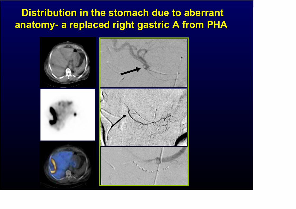
Distribution in the stomach due to aberrant Distribution in the stomach due to aberrant anatomy- a replaced right gastric A from PHA anatomy- a replaced right gastric A from PHA

MAA Deposition in the spleen and small intestine due to hepatic artery stenosis and
extensive collateral flow

Conclusion I For hepatopulmonary shunting:
1. Hepatopulmonary shunting appears to be associated with HCC more than other tumors.
2. Patients with severe lung shunting predominantly have a diagnosis of HCC.

Aberrant Arteries
• 50% of patients have aberrant arteries supplying the liver
• 15% of patients have aberrant arteries from liver and supplying the gut

For abdominal extra-hepatic MAA deposition
1. MAA deposition in the stomach, intestine, pancreas and umblical vein predominantly occurs when injection is made into the LHA or P/CHA.
2. GDA and small gastric arteries are the main causes of distribution in the GI and pancreas, which are potentially correctable.
Conclusion II

Conclusion III
• MAA hepatic perfusion imaging is useful in detection of extra-hepatic deposition.

Carcinoid Cancer FoundationTM
Acknowledgement

Results of treating Colorectal Metastases with SIRT

Phase 2 Trial of SIR-Spheres
SIR-Spheres Alone 16 PatientsResponse
• CT 73% decrease in tumor size (mean 48%)
• CEA 100% decrease in CEA (mean 78%)• Gray BN, Anderson JE, Burton MA et al: Regression of liver metastases following treatment with
Yttrium 90 microspheres. Aust. N.Z. J. Surg. 1992; 62:105-110.

Phase 2 Trial of SIR-Spheres
SIR-Spheres plus HAC 71 PatientsResponse
– CT- 86% decreased tumor size (75% PR + CR)– CEA – 95% decreased (89% PR + CR)
Gray BN, Van Hazel G, Buck M, et al Treatment of colorectal liver metastases with SIR-Spheres plus chemotherapy. GI Cancer 3 (4):249-257, 2000

1st Randomised trial in advanced colorectal liver metastases
(74 patients)HAC versus HAC + SIRT
Response (CR + PR)(CT Response)
HAC HAC + SIRTCR + PR 6 (18%) 16 (44%)
Difference between groups p=0.02
Gray BN, Van Hazel G, Anderson J et al: Randomised trial of SIR-Spheres plus chemotherapy versus chemotherapy alone for treating patients with liver metastases from primary large bowel cancer. Annals Oncology 2001, 12:1711-1720

1st Randomised trial in colorectal liver metastases
HAC versus HAC + SIRTTime to Disease Progression
(CT Response)
HAC HAC + SIRTMean 10.1 months 19.2 months
Median 9.8 months 16.0 monthsDifference between groups (Logrank test) p=0.001
Gray BN, Van Hazel G, Anderson J et al: Randomised trial of SIR-Spheres plus chemotherapy versus chemotherapy alone for treating patients with liver metastases from primary large bowel cancer. Annals Oncology 2001, 12:1711-1720

1st Randomised trial in colorectal liver metastases
HAC versus HAC + SIRT
Survival1 year 2 years 3 years 5 years
HAC 68% 29% 6% 0%
HAC + SIRT 72% 39% 17% 4%
Gray BN, Van Hazel G, Anderson J et al: Randomised trial of SIR-Spheres plus chemotherapy versus chemotherapy alone for treating patients with liver metastases from primary large bowel cancer. Annals Oncology 2001, 12:1711-1720

2nd Randomised trial in colorectal liver metastases
Systemic FU/LV versus SIRT + FU/LV
Time to Disease Progression Data:
Comparison between groups p < 0.0005ASCO 2002:599
Median
Chemotherapy 4.7 months
SIRT + Chemotherapy 15.6 months

2nd Randomised trial in colorectal liver metastases
Systemic FU/LV versus SIRT + FU/LV
Survival
ASCO 2002:599
Median
Chemotherapy 12.8 months
SIRT + Chemotherapy 27.1 months

Patient with colorectal liver metastases.Response 15 months after treatment with
SIR-Spheres + 5FU/LV

Patient with colorectal liver metastases.Response 10 months after treatment with
SIR-Spheres + 5FU/LV

Down-staging Advanced Liver Cancer to Resectability

C/R mets: R lobe only treated CT Scan before/after
SIRT

C/R mets: R lobe only treated PET Scan before/after SIRT

SIRT complementsSurgery & Chemotherapy
SIRT increases surgical options
SIRT increases chemotherapy

Other Cancers
SIRT is targeted radiotherapy and therefore will have effect in all
cancers


R hepatic artery CT scan – Pancreas Cancer Liver Mets

Primary HCC

SIR-Spheres in HCC
71 Patients with advanced HCC
• 89% a-Fetoprotein Response (22% CR 67% PR)
• 27% Tumour Volume Response
W Lau, S Ho, T Leung, M Chan, R Ho, P Johnson, A Li:Int. J. Rad. Oncol. Biol. Phys. 1999, 40:583
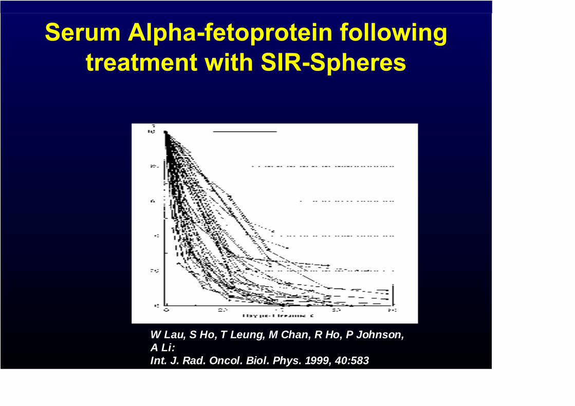
Serum Alpha-fetoprotein following treatment with SIR-Spheres
W Lau, S Ho, T Leung, M Chan, R Ho, P Johnson, A Li:Int. J. Rad. Oncol. Biol. Phys. 1999, 40:583

SIR-Spheres in HCC
71 Patients with advanced HCC
• 5% of patients were down-staged to become resectable.
• Only 15-30 Gy mean dose can be delivered.
• Dose limited by flow stasis
W Lau, S Ho, T Leung, M Chan, R Ho, P Johnson, A Li:Int. J. Rad. Oncol. Biol. Phys. 1999, 40:583

Theraspheres in HCC
• Y-90 Theraspheres have higher specific activity – smaller number of particles.
• Higher doses can be delivered (50-500 Gy).
• Clinical trials show a 50% response rate for HCC compared with our 33% with SIR Spheres.
• Longer period of tumor regression

Theraspheres in HCC• Patients with Stage T1 or T2 eligible for liver
transplant.• 10% recurrence of tumor after transplant (90%
cure).• Of 35 patients with stage T3 HCC, who received
Theraspheres, 19/35 (56%) were downstaged to T2 and became eligible for liver transplant.
• Of those, 11 actually underwent liver transplant.
(Kulik and Salem et al. J Surg Oncol 2006)

Dangers of SIRT
•GI ulceration•Radiation pneumonitis•Pancreatitis•Radiation hepatitis

Lung Tolerance
• 3 of 5 patients with >30Gy total lung dose developed radiation pneumonitis
• No patient receiving <30Gy developed pneumonitis
• Animal studies of 30-33Gy showed little histologic damage

Lung ShuntingActivity
administered (GBq)Lung shunting
(%)Lung radiation dose
(Gray)
1 10% 5
1.5 10% 7.5
2 10% 10
2.5 10% 12.5
3 10% 15
1 15% 7.5
1.5 15% 11.25
2 15% 15
2.5 15% 18.75
3 15% 22.5
1 20% 10
1.5 20% 15
2 20% 20
2.5 20% 25
3 20% 30

Liver Tolerance and Dosimetry
• External beam 40Gy has 50% probability of significant complications
• SIRT: 86% of cells get < mean liver dose and 33% get less than 30% of the dose
• Dose escalation in 10 patients showed that up to 138Gy did not cause clinical radiation hepatitis (Gray et al J Surg Oncol 1989)
• Biopsies in 4 patients receiving up to 75Gy has minimal histologic effect in healthy liver (Gray et al )
• 70Gy is tolerable in cirrhosis (Lau et al)

SIRT Dosimetry
• SIR-Spheres go where the arterial blood goes• SIRT is point source radiation - not homogeneous
radiation• The liver size has a small effect on effect on liver
radiation• Blood flow has a large effect on normal liver radiation
dose• Blood flow has a large effect on tumour radiation dose• The rim of tumours is always hyper-vascular• The centre of tumours is hypo-vascular• Small tumours are more vascular than large tumours• Large tumour mass gets more blood flow than small
tumour mass

Radiation Safety
• Exposure– Bremsstrahlung is typically 15 uSv per Gbq at 15cm from
the patient’s right side (initially)• Precautions
– Non pregnant nursing staff /visitors– 1 foot distance from patient’s side for 3-5 days– Efficiency in patient observations– Nursing from left hand side of patient– Shielding unnecessary