Embed Size (px)
Citation preview
12/10/17
1
X ray physics
Lectures DTU
Mikael Jensen oct.2017 .
Two types of x-ray imaging,- and two lectures
• Planar x-ray • CT

12/10/17
2
Why use x-rays ?
Non invasive, high resolution Quick, widespread
Konrad Røntgen, 1896
X-rays give rapid, high resolution anatomical information
(many photons, good S/N) Rapid introduction, simple technology

12/10/17
3
Electromagnetic spectrum
X-ray
gamma-rays
1 nm
1 fm ν·λ = c E = h · ν
Planar x-ray imaging
• Direct projection ( image) : 2D shadow! ”Black” is low attenuation: Air, lung… ”White” is high attenuation : Bone…. • Geometry factors: FilmFocusDistance,
FilmObjectDistance, FocusObjectDistance
Geometrical enlargement
12/10/17
4
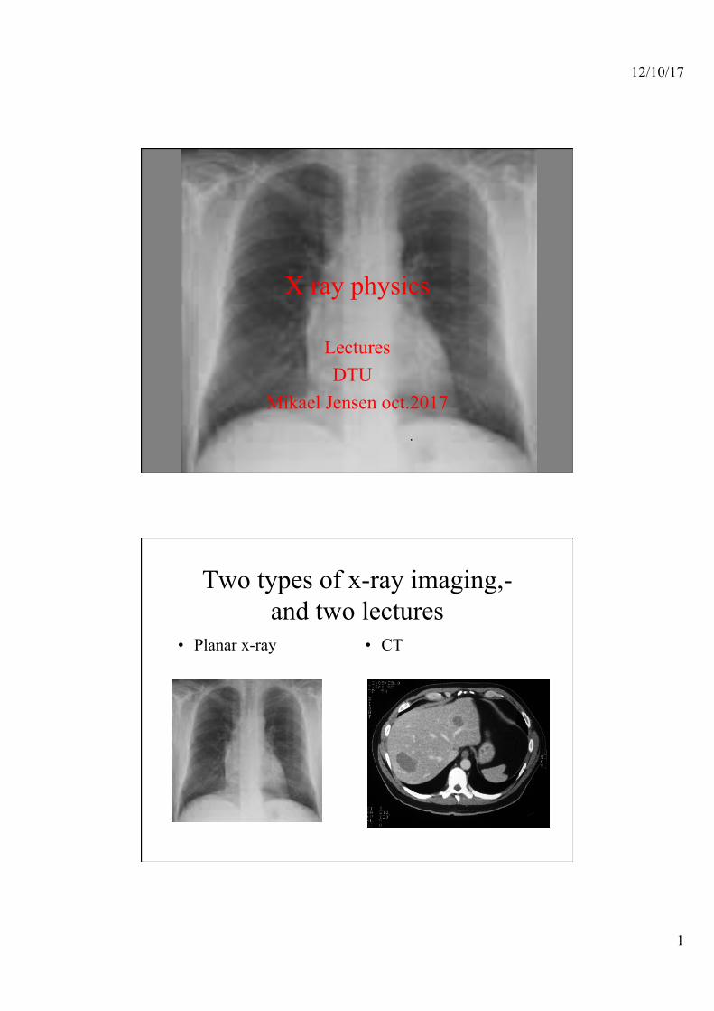
Skull X ray image is shadow image
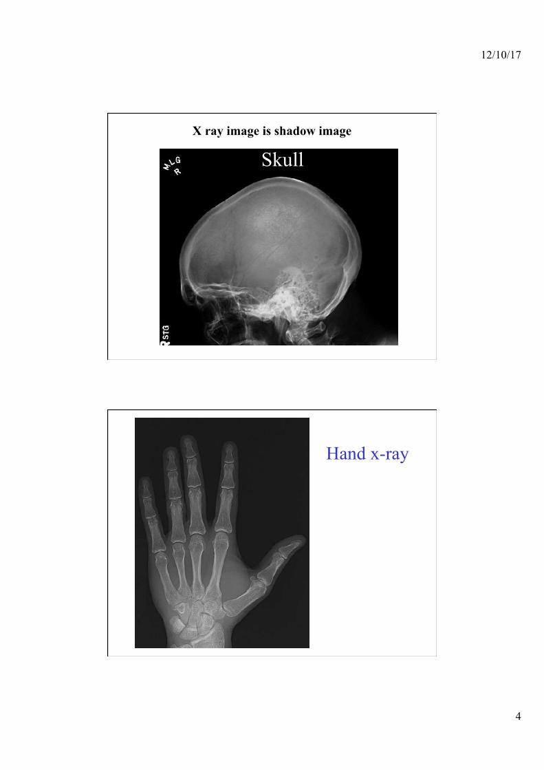
Hand x-ray

12/10/17
5
Chest
And possibility of tomography
Much can be gained from hgh S/N
12/10/17
6
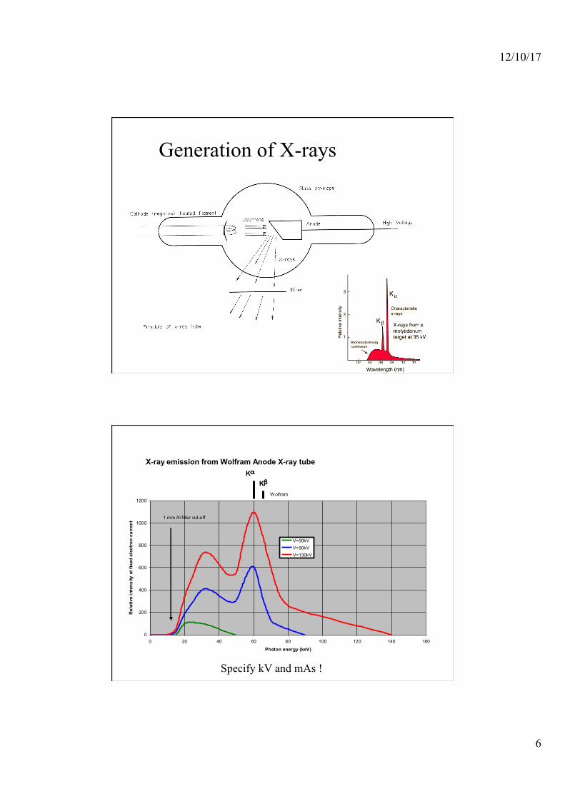
Generation of X-rays
X-ray emission from Wolfram Anode X-ray tube
0
200
400
600
800
1000
1200
0 20 40 60 80 100 120 140 160
Photon energy (keV)
Rel
ativ
e in
tens
ity a
t fix
ed e
lect
ron
curr
ent
V=50kVV=90kVV=130kV
W olfram
Kα
Kβ
1 mm Al f ilter cut-off
Specify kV and mAs !

12/10/17
7
https://www.oem-xray-components.siemens.com/x-ray-spectra-simulation
Spectrum simulation:
Parameters: Anode, kV, mAs, filter
Radiation interaction
• Ionization • Direct kinetic energy transfer • Atomic and molecular exitation • Radiative processes • Nuclear reactions

12/10/17
8
Ionising radiation
• Releases energy through Ionisation
• But also Recombination • And through Secondary
radiation • Allways ending as heat • And perhaps chemical
change
A biological relevant measure for energy transfer
• LET = Linear Energy Transfer. Measured in keV/µm
• Dose =
Energy deposited per unit mass Measured in Gray (Gy)= J/Kg
x
LET=-dE/dx
D=dE/dM M
12/10/17
9
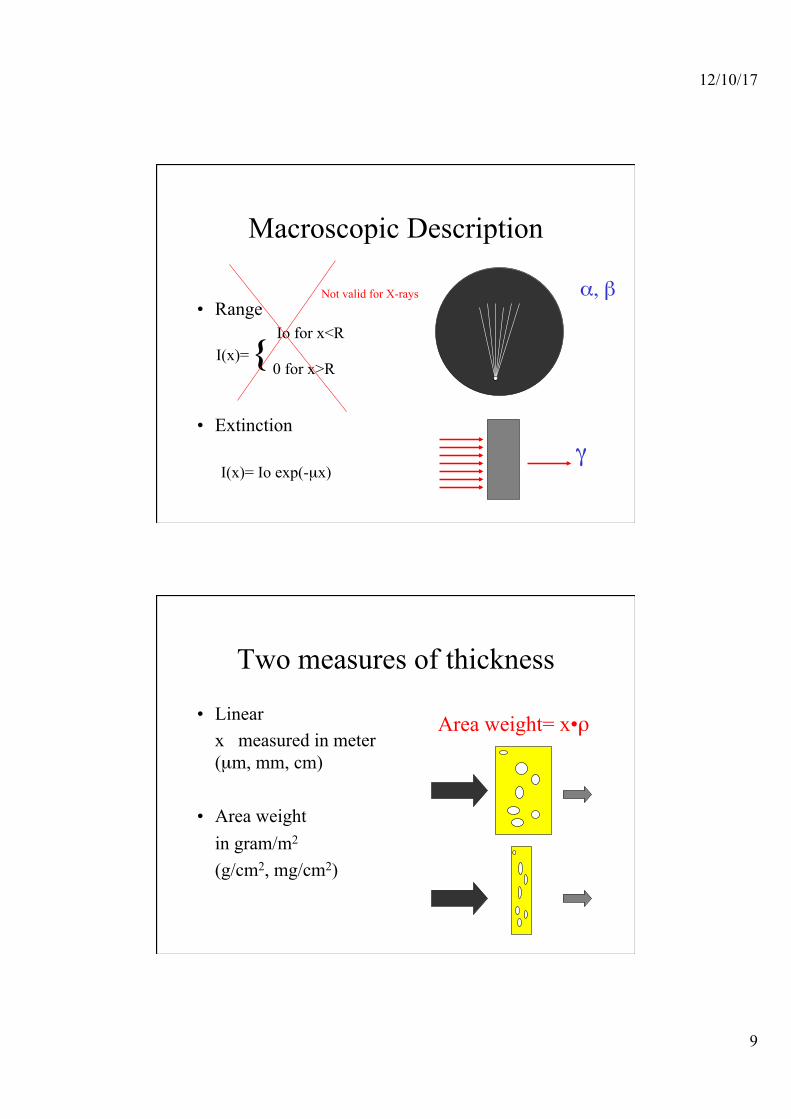
Macroscopic Description
• Range
• Extinction
I(x)=
I(x)= Io exp(-µx)
{ Io for x<R
0 for x>R
α, β
γ
Not valid for X-rays
Two measures of thickness
• Linear x measured in meter (µm, mm, cm)
• Area weight
in gram/m2
(g/cm2, mg/cm2)
Area weight= x•ρ
12/10/17
10
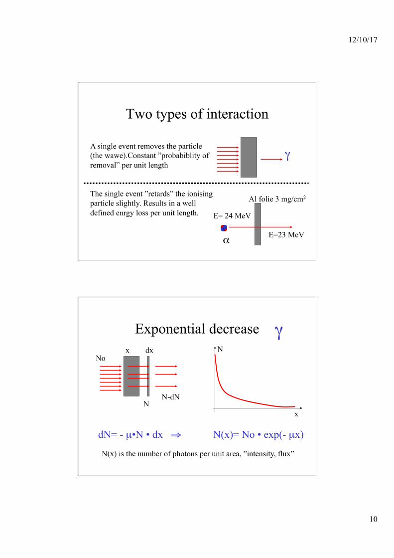
Two types of interaction
A single event removes the particle (the wawe).Constant ”probabiblity of removal” per unit length
The single event ”retards” the ionising particle slightly. Results in a well defined enrgy loss per unit length.
γ
E= 24 MeV
E=23 MeV α
Al folie 3 mg/cm2
Exponential decrease γ
x dx No
N N-dN
dN= - µ•N • dx ⇒ N(x)= No • exp(- µx)
N(x) is the number of photons per unit area, ”intensity, flux”
x
N

12/10/17
11
Microscopic interaction Probability measured in barns
(10-24 cm2)
and milliBarn (mB, 10-27 cm2)
10-10 m
10-15 m
?
Number of Reactions P
P=σNtarget Nbeam
Ntarget number Nbeam number per area
Ntarget number per area Nbeam number
12/10/17
12
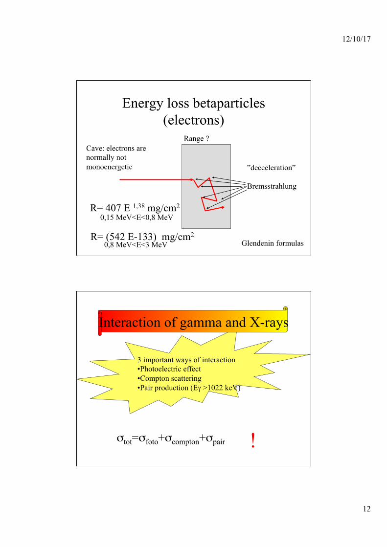
Energy loss betaparticles (electrons)
Range ?
R= 407 E 1,38 mg/cm2
Cave: electrons are normally not monoenergetic
0,15 MeV<E<0,8 MeV
R= (542 E-133) mg/cm2 0,8 MeV<E<3 MeV Glendenin formulas
”decceleration” Bremsstrahlung
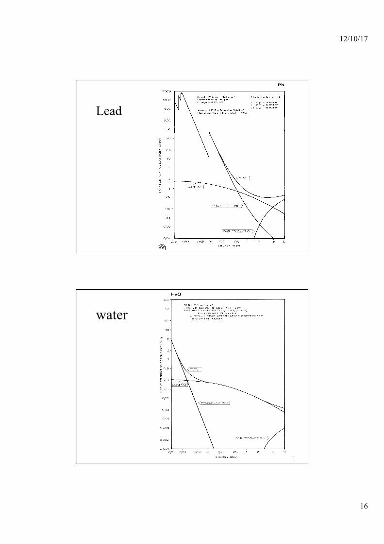
Interaction of gamma and X-rays
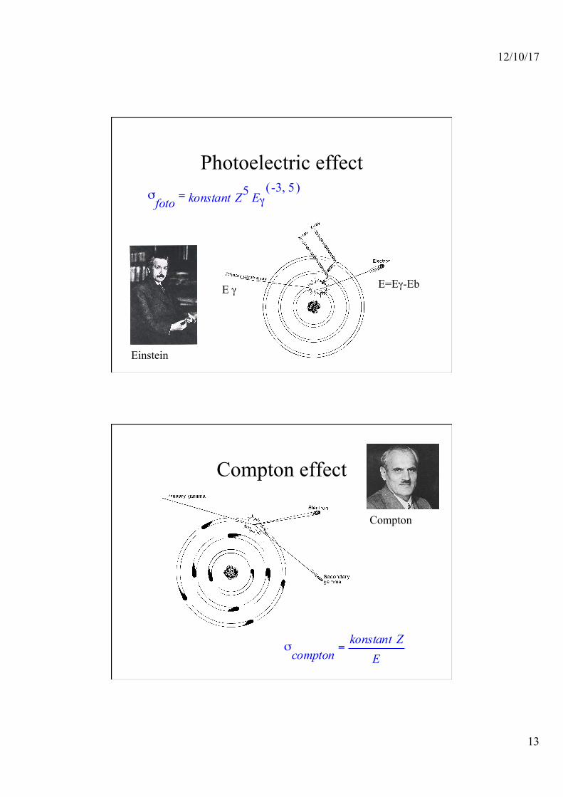
3 important ways of interaction • Photoelectric effect • Compton scattering • Pair production (Eγ >1022 keV)
σtot=σfoto+σcompton+σpair !
12/10/17
13
Photoelectric effect = σ
foto konstant Z5 Eγ( ),-3 5
E=Eγ-Eb E γ
Einstein
Compton effect
Compton
= σcompton
konstant ZE

12/10/17
14
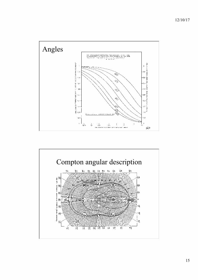
Compton scattering
Klein-Nishina formula for Compton scattering
θ Is the angle of deflection for the photon α Is the energy of the primary gamma ray relative to 511keV (mec2)
12/10/17
15
Angles
Compton angular description
12/10/17
16
Lead
water
12/10/17
17
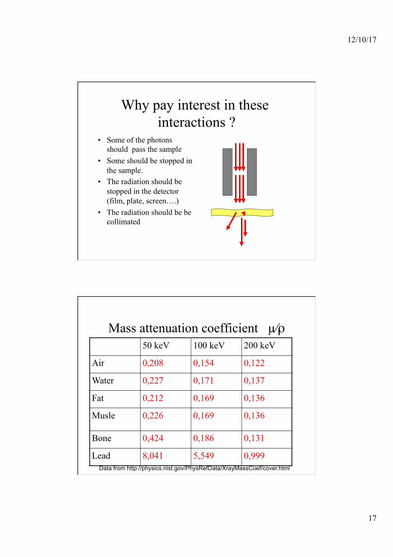
Why pay interest in these interactions ?
• Some of the photons should pass the sample
• Some should be stopped in the sample.
• The radiation should be stopped in the detector (film, plate, screen….)
• The radiation should be be collimated
Mass attenuation coefficient µ⁄ρ 50 keV 100 keV 200 keV
Air 0,208 0,154 0,122
Water 0,227 0,171 0,137
Fat 0,212 0,169 0,136
Musle 0,226 0,169 0,136
Bone 0,424 0,186 0,131
Lead 8,041 5,549 0,999 Data from http://physics.nist.gov/PhysRefData/XrayMassCoef/cover.html
12/10/17
18
Soft tissue contrast
Mainly due to the Compton effect - Because Z is tpically low - And - E xray is ”high”
The image is really ”electron density”
The film / detector
Should stop the x-rays ! ( Silver is good ) but old- fashioned

12/10/17
19
Late 1990’s
• A new approach to imaging appeared
• DR or DDR or Direct Capture imaging
• Too early to tell which system will prevail

12/10/17
20
IMAGE CAPTURE
CR – PSP – photostimulable phosphor plate – REPLACES FILM IN THE CASSETTE
DR – NO CASSETTE – PHOTONS – CAPTURED DIRECTLY – ONTO A TRANSISTOR – SENT DIRECTLY TO A MONITOR
CR vs DR CR • imaging plate
• processed in a Digital Reader
• Signal sent to computer
• Viewed on a monitor
DR • transistor receiver (like
bucky)
• directly into digital signal
• seen immediately on monitor –
12/10/17
21
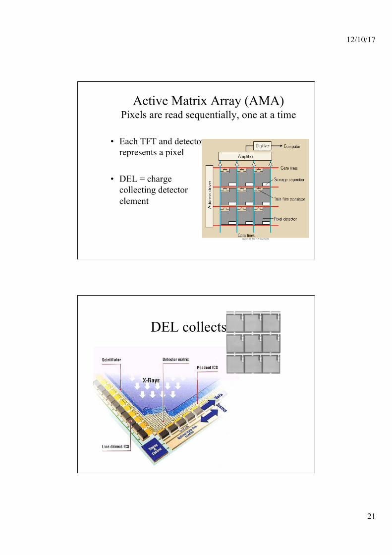
Active Matrix Array (AMA) Pixels are read sequentially, one at a time
• Each TFT and detector represents a pixel
• DEL = charge collecting detector element
DEL collects e-

12/10/17
22
DDR only using amorphous selenium (a-Se)
• The exit x-ray photon interact with the a-Si (detector element/DEL). Photon energy is trapped on detector (signal)
• The TFT stores the signal until readout, one pixel at a time
Active matrix array of silicon photodiodes
12/10/17
23
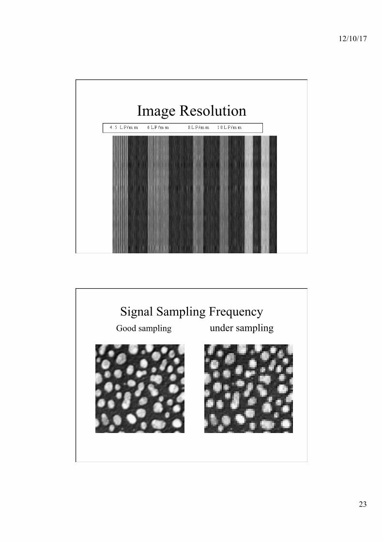
Image Resolution
Signal Sampling Frequency Good sampling under sampling

12/10/17
24
DR
• Initial expense high • very low dose to pt – • image quality of 100s using a 400s
technique • Therfore ¼ the dose needed to make the
image
Flat Panel TFT Detectors • Have to be very careful with terminology • One vendor claims: “Detector has sharpness of 100
speed screen” • May be true: TFT detectors can have very sharp edges
due to DEL alignment • But ! • Spatial resolution is not as good as 100 speed screen. • TFT detector = 3.4 lp/mm • 100 speed screen = 8 – 10 lp/mm
12/10/17
25
TFT Array Detectors
• Detector is refreshed after exposure • If no exposures are produced. . . detector
refreshed every 30 – 45 sec • Built in AEC, An ion chamber between
grid and detector
Patient Dose
• Important factors that affect patient dose • DQE: when using CsI systems • Both systems “fill factor”
– The percentage of the pixel face that contains the x-ray detector.
– Fill factor is approximately 80%

12/10/17
26
DDR has all the advantages of CR imaging techniques
• Post processing & PACS
Questions ?
DR , original and processed

12/10/17
27
Damage
due to ionisation
proportional to energy loss
Biological damage from ionisation
1. Ionisation
2. Free radicals
3. DNA change
4. Lack of repair
H- OH-
4 steps:
12/10/17
28
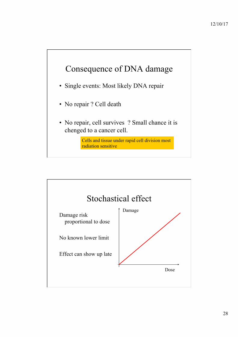
Consequence of DNA damage
• Single events: Most likely DNA repair
• No repair ? Cell death
• No repair, cell survives ? Small chance it is chenged to a cancer cell.
Cells and tissue under rapid cell division most radiation sensitive
Stochastical effect
Damage risk proportional to dose
No known lower limit Effect can show up late
Dose
Damage
12/10/17
29
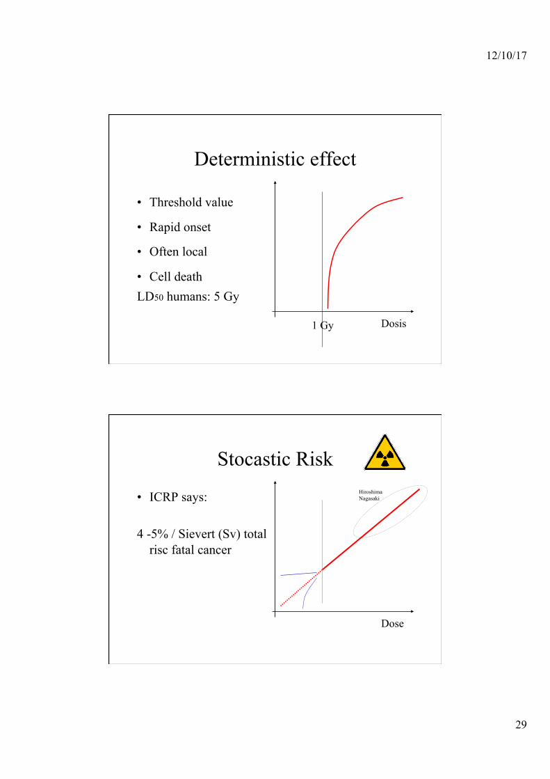
Deterministic effect
• Threshold value
• Rapid onset
• Often local
• Cell death LD50 humans: 5 Gy
Dosis 1 Gy
Stocastic Risk
• ICRP says: 4 -5% / Sievert (Sv) total
risc fatal cancer
Dose
Hiroshima Nagasaki

12/10/17
30
A biological relevant measure for energy transfer
• LET = Linear Energy Transfer. Measured in keV/µm
• Dose =
Energy deposited per unit mass Measured in Gray (Gy)= J/Kg
x
LET=-dE/dx
D=dE/dM M
Unit of dose
• Gray ( J/kg)
• With important ”biological” weight factors linked to Sievert (Sv) (still J/kg)

12/10/17
31
X ray doses
• Single exposure, small area, short path • -limb, teeth, chest • few micro Sievert
• Multiple exposures whole body, low energy to enhance contrast (CT….)
• several milli Sievert
X –ray doses
Note that this is given as effective dose - In Sievert ! - -Why ?

12/10/17
32
Radiation sources (a) • X-ray (diagnostic)
Dose rate is high ! Dose depends on: • Distance • Area of collimation • HV • mAs • Filtering
Surface dose is always higher than depth dose.
Doses 10-100 µGy per image
Justified? • Some procedures are not justified
“Acceptability”
Outcome
Dose