Embed Size (px)
Citation preview

Trans la t iona l Onco logy Volume 2 Number 1 March 2009 pp. 31–38 31
www.transonc.com
X Box–BindingProtein 1 RegulatesAngiogenesis in HumanPancreatic Adenocarcinomas1,2
Lorenzo Romero-Ramirez*, Hongbin Cao*,Maria Paz Regalado*, Neeraja Kambham†,Dietmar Siemann‡, Jeff J. Kim*, Quynh T. Le*and Albert C. Koong*
*Department of Radiation Oncology, Stanford University,Stanford, CA 94305-5152, USA; †Department of Pathology,Stanford University, Stanford, CA 94305-5152, USA;‡Department of Radiation Oncology, University of Florida,Gainesville, FL, USA
AbstractPURPOSE: Tumors encounter endoplasmic reticulum stress during tumor growth and activate an adaptive pathwayknown as the unfolded protein response (UPR). Because this pathway is induced by the tumor microenvironment,it is a promising target for cancer therapy. We have previously demonstrated that X-box binding protein 1 (XBP-1), akey regulator of the UPR, was required for survival under hypoxia and critical for tumor growth in tumor xenografts.In this study, we investigated the role of XBP-1 in regulating tumor angiogenesis. METHODS: We used an intra-dermal angiogenesis model to quantify the effect of XBP-1 on angiogenesis. We also used a human tumor xeno-graft model to assay for tumor growth delay. We determined vascular endothelial growth factor (VEGF) expressionby quantitative polymerase chain reaction and ELISA. Finally, we stained human pancreatic adenocarcinoma speci-mens for XBP-1 expression and correlated the expression pattern of XBP-1 with CD31 (endothelial cell marker)expression. RESULTS: We demonstrated that XBP-1 is essential for angiogenesis during early tumor growth. Inhibit-ing XBP-1 expression by short-hairpin RNA sequence specific for XBP-1 reduced blood vessel formation in tumorsfrom mouse embryonic fibroblast cells and human fibrosarcoma tumor cells (HT1080). Expressing a dominant-negative form of IRE1α also reduced blood vessel formation in tumors. Moreover, expression of spliced XBP-1(XBP-1s) restored angiogenesis in IRE1α dominant-negative expressing cells. We further demonstrated that XBP-1–mediated angiogenesis does not depend on VEGF. CONCLUSIONS: We propose that the IRE1α–XBP-1 branch ofthe UPR modulates a complex proangiogenic, VEGF-independent response that depends on signals received fromthe tumor microenvironment.
Translational Oncology (2009) 2, 31–38
Address all correspondence to: Albert C. Koong, 269 Campus Dr West, CCSR-1245C,Stanford, CA 94305-5152. E-mail: [email protected] work was supported by PO1 CA67166 (Q.T.L. and A.C.K.).2This article refers to supplementary materials, which are designated by Figures W1 toW3 and are available online at www.neoplasia.com.Received 11 November 2008; Revised 31 December 2008; Accepted 6 January 2009
Copyright © 2009 Neoplasia Press, Inc. All rights reserved 1944-7124/09/$25.00DOI 10.1593/tlo.08211
IntroductionTumors experience hypoxia and endoplasmic reticulum (ER) stressduring growth when the energy demands exceed the capacity of thevasculature to supply nutrients. Under these pathophysiological condi-tions, activation of the unfolded protein response (UPR) triggers anadaptive pathway that allows cells to survive in this microenvironmentcharacterized by hypoxia, low glucose, and low pH. Inhibiting theUPR under these conditions is a promising therapeutic strategy [1,2].We have previously demonstrated that the IRE1-XBP1 branch of
the UPR mediated survival under hypoxia and was essential for tumorgrowth. Transformed mouse embryonic fibroblasts (MEFs) [3] orhuman fibrosarcoma tumor cells (HT1080) [4] that are deficient inX-box binding protein 1 (XBP-1) are impaired in their ability to grow
as tumor xenografts in SCID mice. Similarly, PKR-like ER kinase(PERK), another branch of the UPR responsible for attenuation ofprotein translation during hypoxia and ER stress, also plays an impor-tant role in regulating tumor growth [5,6]. Both PERK and XBP-1–deficient cells showed increased apoptosis and decreased clonogenic

32 XBP-1 Regulates Angiogenesis Romero-Ramirez et al. Translational Oncology Vol. 2, No. 1, 2009
survival during ER stress/hypoxia. These findings strongly suggest thatthe UPR represents an important signaling pathway that is critical fortumor growth.
UPR target genes are expressed in a variety of human tumors [7]and have important implications in cancer therapy [8,9]. XBP-1 hasbeen reported to be overexpressed in breast cancer [10], hepatocel-lular carcinoma [11], and colorectal cancer [12]. In this study, weinvestigated the role of XBP-1 in regulating tumor angiogenesis.
Materials and Methods
Cell Culture and Hypoxia TreatmentsMEF and human HT1080 fibrosarcoma cells were maintained in
Dulbecco’s modified Eagle’s medium supplemented with fetal bovineserum (10%) at 37°C in a 5% CO2 incubator. For the hypoxia ex-periments, cells were treated at 70% to 80% confluency and main-tained in an anaerobic chamber (Sheldon Corp., Cornelius, OR) withPO2 levels <0.02%.
Constructs, Reporter Assays, and Production of StablyExpressing Cells
Human XBP-1–specific sequence (5′-GCTCTTTCCC TCATG-TATACT-3′) was used for short-hairpin RNA (shRNA) and cloned inpSIREN-RetroQ vector (Clontech, Mountain View, CA). We usedthe following sequences as shRNA controls: scrambled (SC; 5′-CA-CATGTTCCGATCTCGGC-3′), nontarget sequence obtained fromSigma-Aldrich, St. Louis, MO (NT; 5′-CAACAAGATGAAGAGC-ACCAA-3′), green fluorescent protein sequence (GFP; 5′-TACAA-CAGCCACAACGTCTAT-3′), and a sequence with four mismatchesof the human XBP-1–specific sequence (MM; 5′-GCTgTaTgCCTg-ATGTATACT-3′).
Additional details are available in Supplementary Material. A flag-tagged dominant-negative form of IRE1α and the XBP-1 spliced form(XBP-1s) was cloned into pBabe-Puromycin and pWZL-hygromycinretroviral vectors, respectively. Infected cells were selected with hygro-mycin (375 μg/ml) or puromycin (1 μg/ml) for 10 days. The expres-sion of XBP-1 was confirmed by Western blot analysis, quantitativepolymerase chain reaction (qPCR), and the UPRE-Luciferase reporterassay techniques as described below. HT1080 cells (1.5 × 10⁵) werecultured in 12-well plates. The next day, cells were cotransfected witha pGL3-5×UPRE-luc (containing five repetitions of the XBP-1 DNAbinding site), and a plasmid containing the β-galactosidase enzymewas used for transfection efficiency. All data were normalized by β-galactosidase activity and expressed as a ratio of luciferase/β-galactosidaseactivity. All results were normalized to the control whose value wasarbitrarily set to 1. Lipofectamine and Plus reagent were used accord-ing to the manufacturer’s protocol (Invitrogen, Carlsbad, CA). Lucif-erase assay kit and β-galactosidase assay kit were used according tothe manufacturer’s protocol (Promega, San Luis Obispo, CA).
Western Blot AnalysisCell extracts were prepared in 9 M urea–75 mM Tris-HCl (pH 7.5)
and 0.15 M β-mercaptoethanol (Sigma, St. Louis, MO), sonicatedbriefly, boiled at 95°C for 5 minutes, and loaded onto SDS-PAGE gels.After electrophoresis, the proteins were transferred onto a nitrocellu-lose membrane (Bio-Rad, Hercules, CA) and blocked in 5% nonfatmilk with 0.1% Tween-20 in Tris-buffered saline for 30 minutes atroom temperature. An affinity-purified rabbit polyclonal antibody,
generated against a peptide fragment specific for human XBP-1s, wasused at 1:1000 dilution or a rabbit polyclonal antibody from Biolegend(San Diego, CA) at 1:500 dilution. A monoclonal β-actin antibody(1:3000; Sigma) was used for loading control. After incubation withthe primary antibody, the membranes were washed and hybridizedwith peroxidase-conjugated IgG antibodies (Jackson Immunoresearch,West Grove, PA) as secondary antibodies. The blots were devel-oped with enhanced chemoluminiscence (ECL) substrate (Amersham,Piscataway, NJ).
Quantitative PCRTotal RNA was extracted from cultured cells using TRIzol reagent.
The cDNA was synthesized from 2 μg of total RNA using SuperscriptIII Reverse Transcriptase kit (Invitrogen) according to the manufac-turer’s protocol. The cDNAwas subjected to qPCR on an ABI PRISM7900HT (Applied Biosystems, Foster City, CA) using SYBR GreenPCR Kit. PCR amplifications were performed with specific primersin a total volume of 10 μl containing 1 μl of sense and antisenseprimer mixture (5 μM of each primer), 5 μl of 2xSYBR Green QPCRMaster Mix (Applied Biosystems), 1 μl of diluted cDNA and nuclease-free PCR-grade water. The mixture was used as a template for theamplification after initial denaturation at 95°C and 40 cycles (95°Cfor 30 seconds, 60°C for 1 minute, and 72°C for 30 seconds). Prim-ers sequences used were as follows: for human XBP-1, 5′-AGCCA-AGGGGAATGAAGTGA-3′ (sense) and 5′-GGGGAAGGGCATTTGAAGAA-3′ (antisense); for mouse XBP-1, 5′-TCCGCAGCAC-TCAGACTATG -3′ (sense) and 5′-ACAGGGTCCAACTTGTC-CAG-3′ and for human VEGF 5′-ATCTTCAAGCCATCCTGTGT-GC-3′ (sense) and 5′-GCTCACCGCCTCGGCTTGT-3′ (antisense).SYBR Green fluorescence was measured, and quantification of eachPCR product was expressed relative to beta-actin or 18S rRNA.
Angiogenesis Assay In VivoEach SCID mouse was implanted intradermally on the ventral
surface with four tumors symmetrically equidistant as described byDanielsen and Rofstad [13]. Cells (1 × 10⁵ for MEF cells and 2 ×10⁵ for HT1080 cells) were injected in a volume of 10 μl togetherwith one drop of 0.4% trypan blue to visualize the sites of injection.After 3 and 6 days for MEF cells or after 7 days for HT1080 cells,the mice were killed, the skin carefully separated, and the number ofvessels reaching the edge of the tumor was scored by independentobservers blinded to the treatment group with the use of a dissectingmicroscope at 15× magnification. Results from different controls forshRNA are included in Supplementary Material. Uninfected parentalHT1080 cells were used as an additional control.
VEGF Secretion AssayCells (4 × 10⁵) were seeded in 6-cm dishes and incubated over-
night. Fresh media were added, and cells were exposed to 24 hoursof normoxia or hypoxia (O2 < 0.02%). VEGF was measured byELISA according to the manufacturer’s protocol (R&D Systems,Indianapolis, IN).
Immunohistochemical StainingThe immunohistochemical stains were performed using an affinity-
purified rabbit polyclonal antibody, generated against a peptide frag-ment specific for human XBP-1s antibody directed against humanXBP-1. Serial sections of 4 μm were obtained from the selected par-affin blocks, deparaffinized in xylene, and hydrated in a graded series
Translational Oncology Vol. 2, No. 1, 2009 XBP-1 Regulates Angiogenesis Romero-Ramirez et al. 33
of alcohol. Heat-induced antigen retrieval was carried out by micro-wave pretreatment in citric acid buffer (10 mM, pH 6.0) for 10 min-utes. The XBP-1s antibody was used at a dilution of 1:1000, and thetissue was incubated at room temperature for 30 minutes. The en-dogenous peroxidase was blocked, and the DAKO Envision system(DAKO Corporation, Carpinteria, CA) was used for detection. A tis-sue microarray of formalin-fixed paraffin-embedded normal and car-cinoma tissues was used for initial titration of the XBP-1 antibody.On the basis of these results, colon carcinoma was selected as a positivecontrol, and normal colon mucosa was used as a negative control. TheXBP-1 staining was scored semiquantitatively in the invasive carci-noma, stroma within the carcinoma, and nonneoplastic pancreas asfollows: 0, no staining; 1+, weak and focal (<10% of lesional cells posi-tive); 2+, patchy weak or strong staining (10-30% of lesional cells posi-tive); 3+, strong and diffuse (>30% of lesional cells positive).
Results
XBP-1 Is Essential for Tumor Angiogenesis in MEF Cellsand in Human Fibrosarcoma HT1080 CellsWe have previously demonstrated that constitutive XBP-1 deficiency
impairs tumor growth in human and mouse cell lines [3,4]. In thisstudy, we used an intradermal angiogenesis assay [13] to determinethe role of XBP-1 on tumor angiogenesis. We expressed a shRNA se-quence to inhibit XBP-1 expression (Figure 1A) in mouse embryonic
Figure 1. XBP-1 is essential for tumor angiogenesis in MEF cells. (A) Rwas verified by qPCR for total XBP-1 messenger. (B) Tumor growtshRNA–expressing cells (shXBP-1 MEF). Each SCID mouse was106 shControl MEF cells in one flank and 2 × 10⁶ shXBP-1 MEF cetumors. (C) Angiogenesis assay in vivo for XBP-1 control MEF cells (sSCID mice were intradermally implanted with 1 × 10⁵ MEF cells. T6 days after implantation with each observer blinded to the treatmeStatistical significance was determined using a 2-tailed t test.
fibroblasts (shXBP-1 MEF) and found that their growth as tumorxenografts were impaired compared with the control cells (Figure 1B).The impaired growth of the tumor xenografts expressing shXBP-1is consistent with the decreased expression of XBP-1 observed inthese cells. After implanting these cells intradermally, shXBP-1MEFcells showed significantly less blood vessel formation than the controlcells (shControl MEF; Figure 1C). In these studies, we implanted 1 ×10⁵ cells and had two independent observers score the number of tu-mor capillaries for each tumor using a dissection microscope. Each ob-server was blinded to the cell type for each tumor. We also performedthe same experiments in a human fibrosarcoma cell line (HT1080).As shown in Figure 2A (right panel ), the shXBP-1HT1080 cells hadsignificantly less XBP-1 protein expression under hypoxic conditionsthan the control cells. Similarly, using an XBP-1 responsive reporterconstruct (5×-UPRE-luciferase), cells inhibited in XBP-1 expressionalso showed less XBP-1–dependent reporter transactivation after treat-ment with tunicamycin, an inhibitor of glycosylation (Figure 2A, leftpanel ). Overall, the number of capillaries was significantly reducedin shXBP-1HT1080 cells compared with the control shSC-HT1080cells (Figure 2, B and C , and Figure W2). There was no differencein cell proliferation between these cell lines when assayed for growthin cell culture (data not shown). As additional controls to rule outthe possibility of “off-target” effects of XBP-1 shRNA, we comparedthe blood vessel formation of shXBP-1HT1080 cells with severalHT1080 control cells expressing a scrambled shRNA sequence, a 4-bp
eduction of XBP-1 expression by a short-hairpin specific sequenceh data from XBP-1 control MEF cells (shControl MEF) and XBP-1implanted subcutaneously with two tumors consisting of 2 ×lls in the contralateral flank. Error bars, SD of the mean from fourhControl MEF) and XBP-1 shRNA–expressing cells (shXBP-1 MEF).he number of vessels growing into the tumor was scored 3 andnt condition. Error bars, SE of the mean from at least six tumors.

Figure 2. XBP-1 is essential for tumor angiogenesis in HT1080 cells. Reduction of XBP-1 expression by a short-hairpin specific sequencewas verified by (A) UPRE reporter assay (left panel) and Western blot analysis for spliced XBP-1 (XBP-1s–specific antibody, right panel).Cells were exposed to 8 hours of tunicamycin (5 μg/ml for 8 hours) for the reporter assays and 24 hours of hypoxia (O2 < 0.02%) for theWestern blot analysis to induce XBP-1 splicing. Expression of β-actin is included as a loading control. (B) Angiogenesis assay in vivo forXBP-1 scramble control HT1080 cells (shSc-HT1080) and XBP-1 shRNA–expressing cells (shXBP-1HT1080). SCID mice were intrader-mally implanted with 2 × 10⁵ HT1080 cells. The number of vessels growing into the tumor was scored 7 days after implantation. Eachobserver quantitated the number of blood vessels independently and was blinded to the treatment condition. Error bars, SE of the meanfrom at least 10 tumors. Statistical significance was determined using a 2-tailed t test. (C) Representative photomicrographs of the tumorsfrom angiogenesis assays. As shown in the bottom panel, there are significantly fewer capillaries growing into the HT1080 tumors withreduced XBP-1 expression (sh-XBP-1HT1080) compared with the scrambled shRNA control–expressing cells (top panel, shSCHT1080).
34 XBP-1 Regulates Angiogenesis Romero-Ramirez et al. Translational Oncology Vol. 2, No. 1, 2009
mismatch of the original knockdown sequence, a non target shRNA se-quence, a GFP shRNA sequence, and uninfected HT1080 parental cells(Figure 2B and Figure W2). Functionally, only the shXBP-1HT1080cells were able to block transactivation of an XBP-1–dependent re-porter construct (UPRE-luciferase; Figure W1). We have previouslydemonstrated that HT1080 cells expressing XBP-1 shRNA are inhib-ited in tumor growth compared with control HT1080 cells [14]. Col-lectively, these studies demonstrate that specific inhibition of XBP-1results in decreased tumor growth and decreased angiogenesis.
IRE1α Is Essential for Tumor Angiogenesis in HT1080 CellsXBP-1 protein has two isoforms that are expressed from the same
mRNA: an unspliced (XBP-1u) and spliced form (XBP-1s). Under hyp-oxia andER stress conditions, XBP-1mRNA is spliced by the ERproteinIRE1α. To determine the role of IRE1α on angiogenesis, we over-expressed a truncated version of this protein that contained only the car-boxy terminus. Expression of this dominant-negative form of IRE1α
inhibited the induction of XBP-1 splicing under hypoxia (Figure 3A).Using the same intradermal angiogenesis assays, cells expressing thisdominant-negative form of IRE1α (HT1080-IΔC), also demonstratedfewer capillaries than control cells (HT1080-pBabe; Figure 3B). BecauseIRE1α is required for XBP-1 splicing, these results suggest a key rolefor both IRE1α and spliced XBP-1 in tumor angiogenesis.
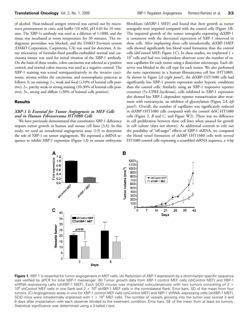
XBP-1s Restores Tumor Angiogenesis in IRE1αDominant-Negative Expressing Cells
We generated additional HT1080 cell lines expressing dominant-negative IRE1α alone (ΔC), spliced XBP-1 alone (S), or both simul-taneously (ΔCS). Spliced XBP-1 expression was confirmed by Westernblot analysis with an antibody that only recognizes the spliced form(Figure 4A, top panel, lanes 3, 4, 7, and 8). Dominant-negative IRE1αexpression (Flag-tagged) was confirmed with anti-Flag antibody (Fig-ure 4A, lower panel, lanes 2, 3, 6, 7 ). As expected in control cells,tunicamycin induced XBP-1 splicing (compare top panel, lane 1 vs 5),

Figure 3. A dominant-negative form of IRE1α inhibits angiogenesis in HT1080 cells. (A) Western blot for spliced XBP-1 (XBP-1s), Flag-tagged IRE1α dominant-negative protein (IΔC), and β-actin (as loading control). Cells were treated with 24 hours of hypoxia (O2 < 0.02%)to induce XBP-1 splicing. There was no XBP-1 splicing in the IRE1α dominant-negative expressing cells. (B) Angiogenesis assay in vivofor empty vector control cells (HT1080-pBabe) and flag-tagged IRE1α dominant-negative expressing cells (HT1080-IΔC). Error bars, SE ofthe mean from 10 tumors. Statistical significance was determined using a 2-tailed t test.
Translational Oncology Vol. 2, No. 1, 2009 XBP-1 Regulates Angiogenesis Romero-Ramirez et al. 35
and XBP-1 splicing was blocked when dominant-negative IRE1α wasexpressed (compare top panel, lane 2 vs 6). Figure 4B shows that an XBP-1–responsive reporter construct (5×-UPRE-luciferase) was blocked bydominant-negative IRE1α expression (middle columns) and that thisinhibition can be overcome with overexpression of spliced XBP-1 ( farright columns). And finally, HT1080 cells expressing dominant-negativeIRE1α (HT1080dvector-ΔC) demonstrated reduced blood vessel for-mation compared with the control cells (HT1080dvector-φ). However,cells coexpressing dominant-negative IRE1α and spliced XBP-1(HT1080dvector-ΔCS) restored angiogenesis to similar levels as thecontrol cells (HT1080dvector-φ; Figure 4C ). Next, we determinedwhether XBP-1 expression influenced VEGF expression in these cells.Using qPCR, we did not observe any difference in either basal VEGFexpression or hypoxic induction of VEGF expression between HT1080control cells (shSC-HT1080) and HT1080 cells expressing an XBP-1shRNA construct (shXBP-1HT1080; Figure 4D). Similar findings werenoted for SU86.86 cells, a human pancreatic cancer cell line. VEGF pro-tein secretion was also not significantly different between these cell linesunder normoxic and hypoxic conditions (FigureW3). These studies sug-gest that although XBP-1s plays an important role in angiogenesis, theregulation of this process occurs independently of VEGF.
XBP-1 Expression in Human Pancreatic Tumors Correlateswith CD31 ExpressionBecause of the inherent limitations of the animal models of angio-
genesis used, we further extended our studies of XBP-1s–mediatedangiogenesis in a defined set of human pancreatic tumors. We stained32 consecutive human pancreatic adenocarcinoma resection specimens(mucinous tumors excluded) for spliced XBP-1 and CD31 (endothe-lial cell marker) expression. In general, the XBP-1s staining was specificfor the tumor cells, whereas the CD31 staining tended to localize tothe tumor stromal cells. Neither did we find a correlation betweenpancreatic tumor size and XBP-1s expression nor did we observe a cor-relation between CD31 staining and pancreatic tumor size. Table 1shows the pathologic characteristics of the pancreatic tumor samplesused in this study. A board-certified pathologist (N.K.) scored the in-tensity of XBP-1s staining in these sections as follows: 0 (no expres-sion), 1 (weak expression), 2 (moderate expression), and 3 (strongexpression). On consecutive sections, these specimens were also as-sessed for CD31 staining. Shown in Figure 5A are representative stain-
ing of two adjacent pancreatic tumor sections showing high XBP-1sexpression from the tumor cells with high CD31 staining from the ad-jacent stroma. CD31-positive blood vessels were counted at 300× mag-nification from three different areas within each tumor, and a meanCD31 score was determined for each tumor. We found a significant cor-relation between XBP-1 staining and mean CD31 score within thesetumors. Specifically, the group of patients with mod-strong XBP-1s ex-pression had higher CD31 scores than those patients with neg-weakXBP-1s expression (Figure 5B). These results indicate that XBP-1 ex-pression in human pancreatic tumors is clinically relevant to angiogen-esis and suggests a therapeutic opportunity in treating this disease.
DiscussionWe have previously reported that human pancreatic tumors are
extremely hypoxic as determined by intraoperative measurementsusing a polar graphic microelectrode technique [15]. What remainsunclear is whether tumor angiogenesis occurs as a response to tumorhypoxia or whether tumor hypoxia is a consequence of insufficientangiogenesis. Most likely, within human tumors, both processes occurand a dynamic interplay exists between the tumor microenvironmentand angiogenesis. The ultimate consequence of this interaction istumor growth.
In prior studies, we found that XBP-1 was a critical mediator oftumor growth, and the data from the current study support the hy-pothesis that at least part of the mechanism for impaired growth ofXBP-1 deficient tumors was through decreased angiogenesis. Ourprevious study demonstrated that VEGF secretion was not signifi-cantly different between XBP-1 wild-type and knockout MEFs [3],suggesting that VEGF does not play a direct role in XBP-1–regulatedangiogenesis. In the current study, we also did not observe any sig-nificant differences in VEGF expression in two different tumor celllines (HT1080, human fibrosarcoma, and SU.86.86, human pancre-atic cancer) expressing shRNA constructs to block XBP-1 (Figure 4Dand Figure W3). However, angiogenesis is a complex sequence ofevents, and up-regulation of other proangiogeneic factors as well asdown-regulation of antiangiogeneic factors also plays an importantrole. With regards to other UPR signaling pathways, PERK has alsobeen implicated in tumor growth and angiogenesis [16]. Interest-ingly, VEGF secretion also was not regulated in a PERK-dependentmanner [5].
Figure 4. XBP-1 spliced form (XBP-1s) rescues tumor angiogenesis in IRE1α dominant-negative expressing cells. (A) Expression of XBP-1sand IRE1α dominant-negative in HT1080 cells was confirmed by Western blot analysis. Lanes 1 and 5 are HT1080 vector control cells.Lanes 2 and 6 are HT1080 cells transfected with a Flag-tagged IRE1α dominant-negative construct. Lanes 3 and 7 are HT1080 cells trans-fected with both Flag-tagged IRE1α dominant-negative construct and an XBP-1 overexpression construct. Lanes 4 and 8 are HT1080 cellstransfected with XBP-1 overexpression construct. Tunicamycin (5 μg/ml for 8 hours) was used to induce XBP-1 splicing. β-Actin was usedas a loading control. Western blot analysis was carried out using XBP-1s–specific antibody (Biolegend). (B) XBP-1 reporter (UPRE-luciferase)assay in HT1080 cells expressing vector alone (vector-φ), IRE1α dominant-negative (vector-ΔC), or IRE1α dominant-negative and splicedXBP-1 (ΔCS). All cells were treated with tunicamycin (5 μg/ml for 8 hours). Expression of IRE1α dominant-negative blocked transactivationof the UPRE-luciferase reporter during tunicamycin treatment. Inhibition of the UPRE-luciferase reporter could be reversed by overexpres-sion of XBP-1s. (C) Angiogenesis assay for HT1080 cells expressing vector alone (vector-φ), IRE1α dominant-negative (φ-ΔC), or IRE1αdominant-negative and spliced XBP-1 (ΔCS). Error bars, SE of the mean from at least eight tumors. Statistical significance was determinedusing a 2-tailed t test. (D) VEGF expression by qPCR in HT1080 control cells (shSC-HT1080) or HT1080 cells inhibited in XBP-1 expressionby shRNA (shXBP-1HT1080). There was no difference in VEGF expression at baseline or during hypoxia between these two cell lines.
36 XBP-1 Regulates Angiogenesis Romero-Ramirez et al. Translational Oncology Vol. 2, No. 1, 2009
The cytoplasmic portion of IRE1α contains both a kinase and anendonuclease domain. Whereas the kinase domain is required forthe endonuclease activity of the protein, it can also activate the JNKpathway through its interaction with TRAF2 [17]. The IRE1 endo-nuclease is responsible for splicing XBP-1 and has also been shownto degrade a set of target mRNA in Drosophila, including some mRNAthat encode for proteins involved in angiogenesis [18]. Drogat et al.showed that in cell culture, tumor cells expressing a kinase inactivemutant for IRE1α (IRE1αK599A) had decreased VEGFmRNA induc-tion during relatively mild hypoxia (3% O2) or hypoglycemia [19]. Fur-thermore, these investigators used an orthotopic glioma tumor model
Table 1. Pancreatic Adenocarcinoma Tumor Characteristics (n = 32).
Patients’ age (years), mean (range)
69 (43-85) Tumor grade, n (%)Well differentiated 7 (22) Moderately differentiated 19 (59) Poorly differentiated 6 (19)Lymph node status, n (%)Positive
16 (50) Negative 16 (50)Resection margin, n (%)Positive
10 (31) Negative 22 (69)Tumor size (cm), median (range)
3.80 (1.10-6.00)
Translational Oncology Vol. 2, No. 1, 2009 XBP-1 Regulates Angiogenesis Romero-Ramirez et al. 37
to show that IRE1αK599A expressing tumors grew slower and showeddecreased blood vessel formation compared with control tumors.Our data are consistent with these investigators and also demon-
strate that IRE1α plays an important role in angiogenesis. In bothtumor model systems, overexpression of a dominant-negative formof IRE1α resulted in reduced blood vessel formation. Moreover, over-expression of spliced XBP-1 was sufficient to overcome the effect ofIRE1α dominant-negative expression on angiogenesis. However, ourfindings differ from Drogat et al. in that we did not observe any sig-nificant differences in VEGF expression in two different cell lines in-hibited in XBP-1 expression (Figure 4D and Figure W3). These resultssuggest that VEGF expression may be altered depending on whetherXBP-1 or IRE1α is blocked. Another possibility is that these dif-ferences may be accounted for by the different cell types used inthese experiments. Because the XBP-1 shRNA experiments blockedexpression of both the spliced and unspliced form of XBP-1, we can-not exclude the possibility that the unspliced form of XBP-1 may alsocontribute to angiogenesis. Although unspliced XBP-1 can function asa negative regulator of spliced XBP-1 [20,21], it may also differentiallyactivate UPR target genes if its stability is altered [22].Our results demonstrate that XBP-1 expression is important for
angiogenesis in a variety of tumor xenografts derived from multipledifferent cell types including MEFs and HT1080 cells. These datasuggest that our results may be generally applied to multiple tumortypes. Moreover, we report a strong correlation between XBP-1 acti-vation and increased vessel density from human pancreatic adeno-carcinoma specimens, which strongly suggests that this pathway isrelevant in human cancer. And finally, although multiple factorscan affect tumor growth (proliferation, apoptosis, adaptation), angio-genesis is an important component of the tumor microenvironmentthat influence overall tumor growth kinetics. We propose that IRE1/XBP-1 functions as part of a complex network of UPR signaling thatdepends on the tumor microenvironment for the appropriate angio-
genic stimuli. These data have important implications in developingantiangiogenic tumor treatment strategies in pancreatic cancers.
AcknowledgmentsThe authors thank the members of Koong and Le Laboratory for theircomments and suggestions.
References[1] Feldman DE, Chauhan V, and Koong AC (2005). The unfolded protein response: a
novel component of the hypoxic stress response in tumors.MolCancer Res3, 597–605.[2] Ma Y and Hendershot LM (2004). The role of the unfolded protein response
in tumour development: friend or foe? Nat Rev Cancer 4, 966–977.[3] Romero-Ramirez L, Cao H, Nelson D, Hammond E, Lee AH, Yoshida H, Mori
K, Glimcher LH, Denko NC, Giaccia AJ, et al. (2004). XBP1 is essential for sur-vival under hypoxic conditions and is required for tumor growth. Cancer Res 64,5943–5947.
[4] Chen Y, Feldman DE, Deng C, Brown JA, De Giacomo AF, Gaw AF, Shi G, LeQT, Brown JM, and Koong AC (2005). Identification of mitogen-activated proteinkinase signaling pathways that confer resistance to endoplasmic reticulum stress inSaccharomyces cerevisiae. Mol Cancer Res 3, 669–677.
[5] Bi M, Naczki C, Koritzinsky M, Fels D, Blais J, Hu N, Harding H, Novoa I,Varia M, Raleigh J, et al. (2005). ER stress–regulated translation increases toler-ance to extreme hypoxia and promotes tumor growth. EMBO J 24, 3470–3481.
[6] Koumenis C, Naczki C, Koritzinsky M, Rastani S, Diehl A, Sonenberg N,Koromilas A, and Wouters BG (2002). Regulation of protein synthesis by hypoxiavia activation of the endoplasmic reticulum kinase PERK and phosphorylation ofthe translation initiation factor eIF2alpha. Mol Cell Biol 22, 7405–7416.
[7] Li J and Lee AS (2006). Stress induction of GRP78/BiP and its role in cancer.Curr Mol Med 6, 45–54.
[8] Fels DR and Koumenis C (2006). The PERK/eIF2alpha/ATF4 module of theUPR in hypoxia resistance and tumor growth. Cancer Biol Ther 5, 723–728.
[9] Koong AC, Chauhan V, and Romero-Ramirez L (2006). Targeting XBP-1 as anovel anti-cancer strategy. Cancer Biol Ther 5, 756–759.
[10] Fujimoto T, Onda M, Nagai H, Nagahata T, Ogawa K, and Emi M (2003).Upregulation and overexpression of human X-box binding protein 1 (hXBP-1)gene in primary breast cancers. Breast Cancer 10, 301–306.
[11] Shuda M, Kondoh N, Imazeki N, Tanaka K, Okada T, Mori K, Hada A, AraiM, Wakatsuki T, Matsubara O, et al. (2003). Activation of the ATF6, XBP1 and
Figure 5. VEGF mRNA expression is not regulated by XBP-1. (A) The top panel shows strong CD31 staining of endothelial cells in a humanpancreatic adenocarcinoma. The bottom panel is an adjacent pancreatic tumor section showing strong XBP-1s expression. (B) Thirty-twoconsecutive pancreatic tumor resection specimens were stained for CD31 and XBP-1s expression. We found a strong correlation betweenXBP-1s staining and CD31 staining (P = .016) suggesting that XBP-1s is a clinically relevant to angiogenesis in pancreatic cancer.
38 XBP-1 Regulates Angiogenesis Romero-Ramirez et al. Translational Oncology Vol. 2, No. 1, 2009
grp78 genes in human hepatocellular carcinoma: a possible involvement of theER stress pathway in hepatocarcinogenesis. J Hepatol 38, 605–614.
[12] Fujimoto T, Yoshimatsu K, Watanabe K, Yokomizo H, Otani T, Matsumoto A,Osawa G, Onda M, and Ogawa K (2007). Overexpression of human X-boxbinding protein 1 (XBP-1) in colorectal adenomas and adenocarcinomas. Anti-cancer Res 27, 127–131.
[13] Danielsen Tand Rofstad EK (1998). VEGF, bFGF and EGF in the angiogenesisof human melanoma xenografts. Int J Cancer 76, 836–841.
[14] Chen Y, Shi G, Xia W, Kong C, Zhao S, Gaw AF, Chen EY, Yang GP, Giaccia AJ,Le QT, et al. (2004). Identification of hypoxia-regulated proteins in head andneck cancer by proteomic and tissue array profiling. Cancer Res 64, 7302–7310.
[15] Koong AC, Mehta VK, Le QT, Fisher GA, Terris DJ, Brown JM, Bastidas AJ,and Vierra M (2000). Pancreatic tumors show high levels of hypoxia. Int J RadiatOncol Biol Phys 48, 919–922.
[16] Blais JD, Addison CL, Edge R, Falls T, Zhao H, Wary K, Koumenis C, HardingHP, Ron D, Holcik M, et al. (2006). Perk-dependent translational regulationpromotes tumor cell adaptation and angiogenesis in response to hypoxic stress.Mol Cell Biol 26, 9517–9532.
[17] Urano F, Wang X, Bertolotti A, Zhang Y, Chung P, Harding HP, and Ron D(2000). Coupling of stress in the ER to activation of JNK protein kinases bytransmembrane protein kinase IRE1. Science 287, 664–666.
[18] Hollien J and Weissman JS (2006). Decay of endoplasmic reticulum–localizedmRNAs during the unfolded protein response. Science 313, 104–107.
[19] Drogat B, Auguste P, Nguyen DT, Bouchecareilh M, Pineau R, Nalbantoglu J,Kaufman RJ, Chevet E, Bikfalvi A, andMoennerM (2007). IRE1 signaling is essen-tial for ischemia-induced vascular endothelial growth factor-A expression and contri-butes to angiogenesis and tumor growth in vivo. Cancer Res 67 (14), 6700–6707.
[20] Lee AH, Iwakoshi NN, Anderson KC, and Glimcher LH (2003). Proteasomeinhibitors disrupt the unfolded protein response in myeloma cells. Proc Natl AcadSci USA 100, 9946–9951.
[21] Yoshida H, Oku M, Suzuki M, and Mori K (2006). pXBP1(U) encoded in XBP1pre-mRNA negatively regulates unfolded protein response activator pXBP1(S) inmammalian ER stress response. J Cell Biol 172, 565–575.
[22] Tirosh B, Iwakoshi NN, Glimcher LH, and Ploegh HL (2006). Rapid turnoverof unspliced Xbp-1 as a factor that modulates the unfolded protein response.J Biol Chem 281, 5852–5860.

Figure W1. UPRE reporter assays with HT1080 cells expressing different shRNA controls and a specific sequence for XBP-1. UPRE-Luciferase construct and a plasmid containing β-galactosidase (as a transfection control) were transiently transfected in HT1080 cells.UPRE reporter was induced with 4 μg/ml of tunicamycin (Tm) for 8 hours. We used HT1080 cells stably expressing a shRNA sequencespecific for XBP-1 (shXBP-1HT1080) and four control sequences as follows: a sequence with four mismatches from the original shRNA-specific sequence for XBP-1 (shMM-HT1080); a nontarget sequence (shNT-HT1080); a sequence from GFP (shGFP-HT1080); and pBabeempty vector–expressing cells (HT1080-pBabe). Error bars, SE of the mean from a triplicate.
Figure W2. Angiogenesis assays in HT1080 cells stably expressing different shRNA controls and a specific sequence for XBP-1. We usedHT1080 cells stably expressing a shRNA specific for XBP-1 (shXBP-1HT1080) and compared blood vessel formation in control cells ex-pressing four different shRNA sequences: a scramble sequence (shSC-HT1080); a sequence with four mismatches from the originalshRNA specific for XBP-1 (shMM-HT1080); a nontarget sequence (shNT-HT1080); and a sequence from GFP (shGFP-HT1080). UninfectedHT1080 parental cells were used as additional controls. Error bars, SE of the mean from 10 tumors.

Figure W3. VEGF secretion assay in media conditioned during24 hours of normoxia or hypoxia (O2 < 0.02%). There was no differ-ence in VEGF secretion between Su.86.86 shRNA scramble controlcells (shSc.Su.86.86) and cells expressing a specific short-hairpinsequence for XBP-1 (shXBP-1Su.86.86). Error bars, SD.