Embed Size (px)
Citation preview
www.soran.edu.iq
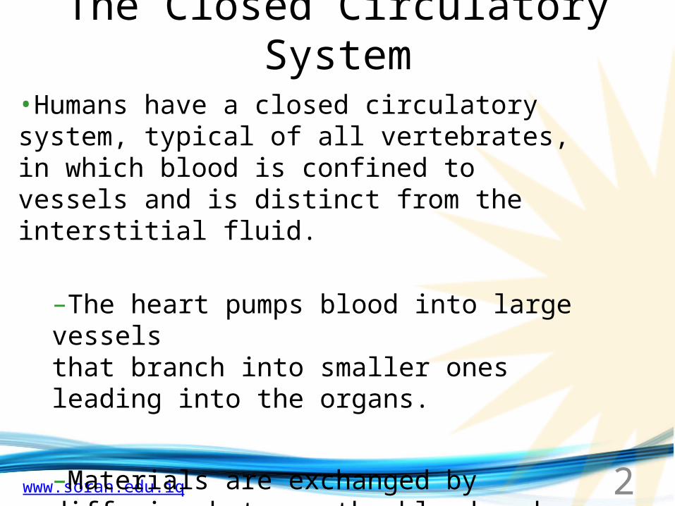
The Closed Circulatory System
•Humans have a closed circulatory system, typical of all vertebrates, in which blood is confined to vessels and is distinct from the interstitial fluid.
–The heart pumps blood into large vessels that branch into smaller ones leading into the organs.
–Materials are exchanged by diffusion between the blood and the interstitial fluid bathing the cells.
2
www.soran.edu.iq
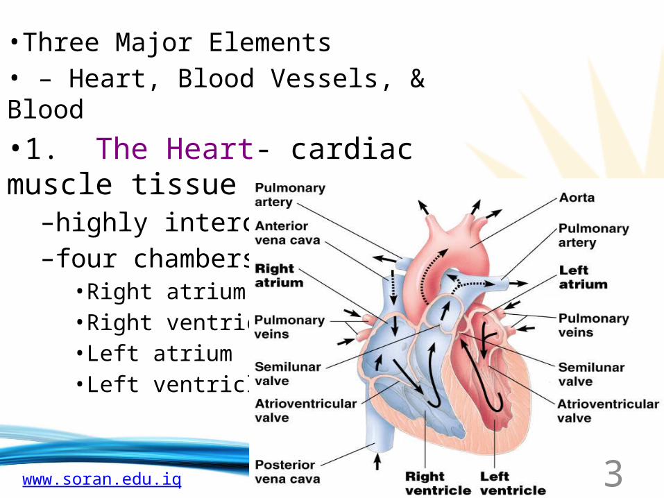
•Three Major Elements• – Heart, Blood Vessels, & Blood•1. The Heart- cardiac muscle tissue
–highly interconnected cells–four chambers
•Right atrium•Right ventricle•Left atrium•Left ventricle
3
www.soran.edu.iq
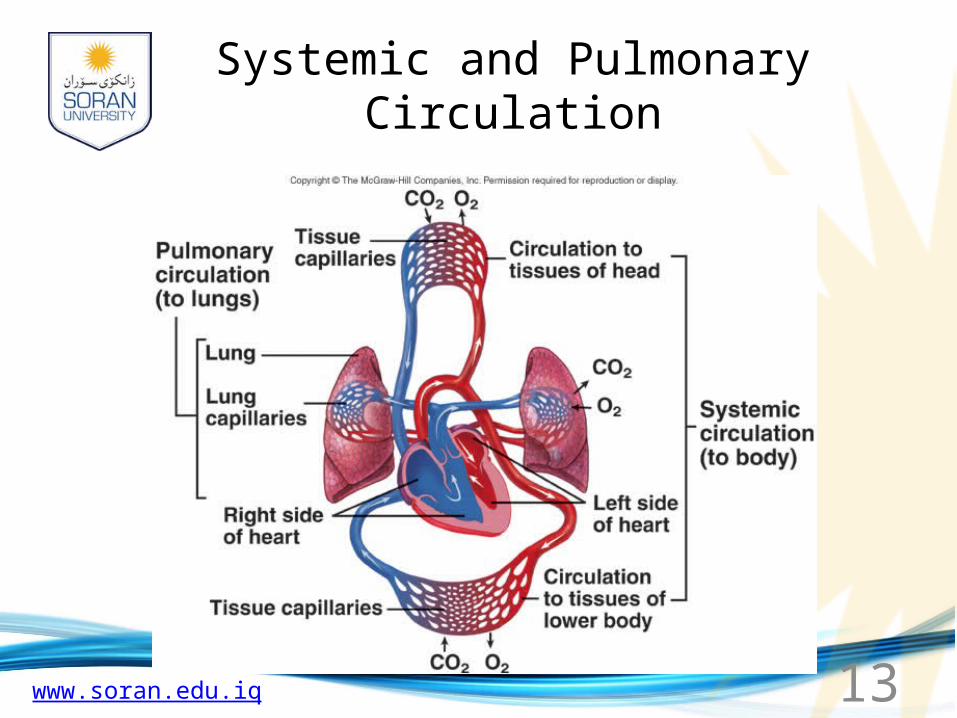
Circuits
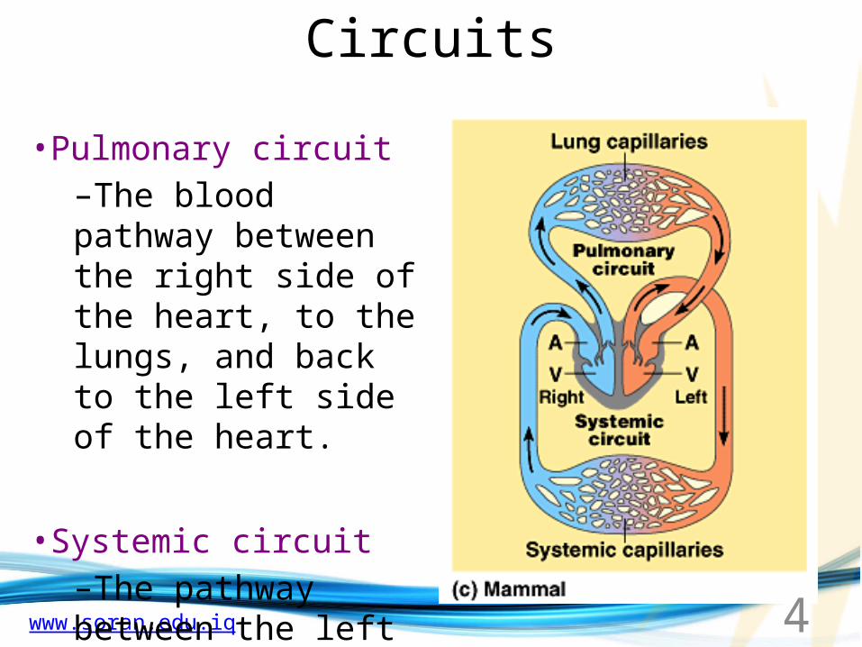
•Pulmonary circuit–The blood pathway between the right side of the heart, to the lungs, and back to the left side of the heart.
•Systemic circuit–The pathway between the left and right sides of the heart.
4
www.soran.edu.iq
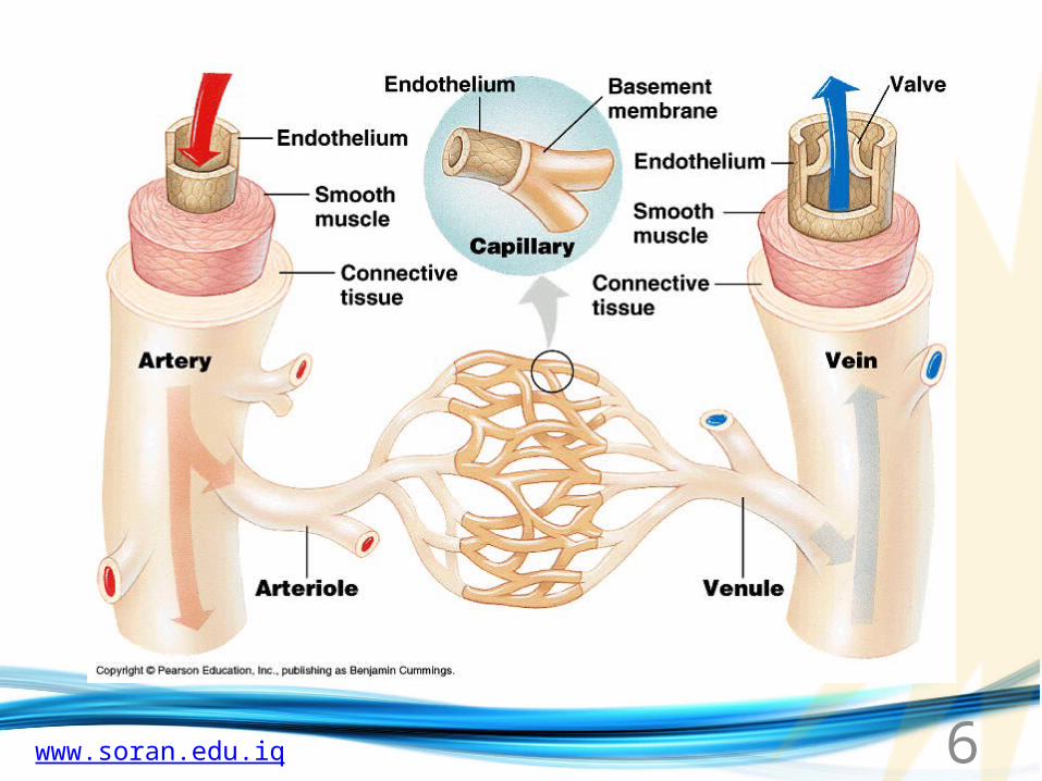
Blood Vessels -
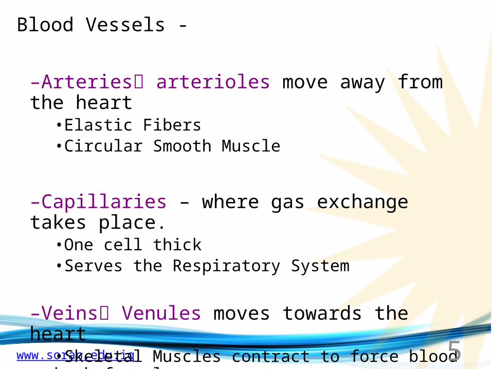
–Arteries arterioles move away from the heart•Elastic Fibers•Circular Smooth Muscle
–Capillaries – where gas exchange takes place.•One cell thick•Serves the Respiratory System
–Veins Venules moves towards the heart•Skeletal Muscles contract to force blood back from legs•When they break - varicose veins form
5
www.soran.edu.iq
The BloodA. Plasma
Liquid portion of the blood. Contains clotting factors, hormones, antibodies, dissolved gases, nutrients and waste
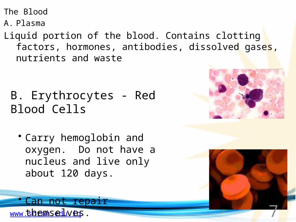
B. Erythrocytes - Red Blood Cells
• Carry hemoglobin and oxygen. Do not have a nucleus and live only about 120 days.
• Can not repair themselves.
7
www.soran.edu.iq
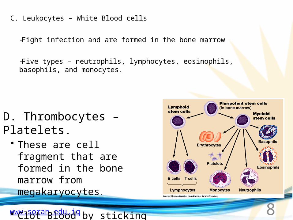
C. Leukocytes – White Blood cells
–Fight infection and are formed in the bone marrow
–Five types – neutrophils, lymphocytes, eosinophils, basophils, and monocytes.
D. Thrombocytes – Platelets.• These are cell fragment that
are formed in the bone marrow from megakaryocytes.
• Clot Blood by sticking together – via protein fibers called fibrin.
8
www.soran.edu.iq 9
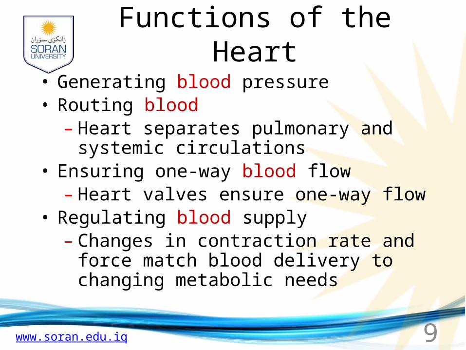
Functions of the Heart• Generating blood pressure• Routing blood
– Heart separates pulmonary and systemic circulations
• Ensuring one-way blood flow– Heart valves ensure one-way flow
• Regulating blood supply– Changes in contraction rate and force match
blood delivery to changing metabolic needs
www.soran.edu.iq
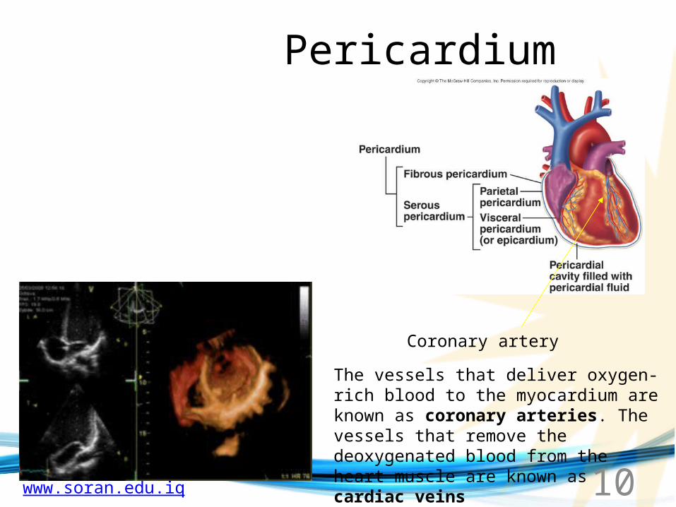
Pericardium
Coronary artery
The vessels that deliver oxygen-rich blood to the myocardium are known as coronary arteries. The vessels that remove the deoxygenated blood from the heart muscle are known as cardiac veins
10
www.soran.edu.iq 11
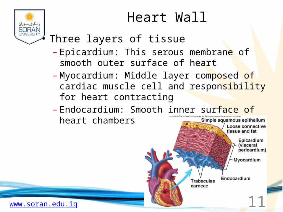
Heart Wall• Three layers of tissue
– Epicardium: This serous membrane of smooth outer surface of heart
– Myocardium: Middle layer composed of cardiac muscle cell and responsibility for heart contracting
– Endocardium: Smooth inner surface of heart chambers
www.soran.edu.iq
Heart Valves
•Atrioventricular–Tricuspid; prevent back flow of blood into the right atrium.
–Bicuspid or mitral; lie between the atria and the ventricles of the heart and control the flow of blood.
•Semilunar–Aortic; When the pressure in the left ventricle rises above the pressure in the aorta, the aortic valve opens, allowing blood to exit the left ventricle into the aorta.
–Pulmonary; At the end of ventricular systole, when the pressure in the right ventricle falls rapidly, the pressure in the pulmonary artery will close the pulmonary valve
•Prevent blood from flowing back
12

www.soran.edu.iq
Cardiac Muscle
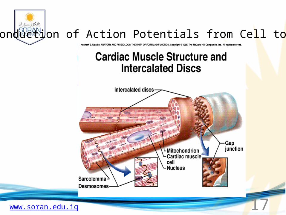
• Elongated, branching cells containing 1-2 centrally located nuclei• Contains actin and myosin myofilaments • Intercalated disks: Specialized cell-cell contacts• Desmosomes hold cells together and gap junctions allow action potentials• Electrically, cardiac muscle behaves as single unit
14
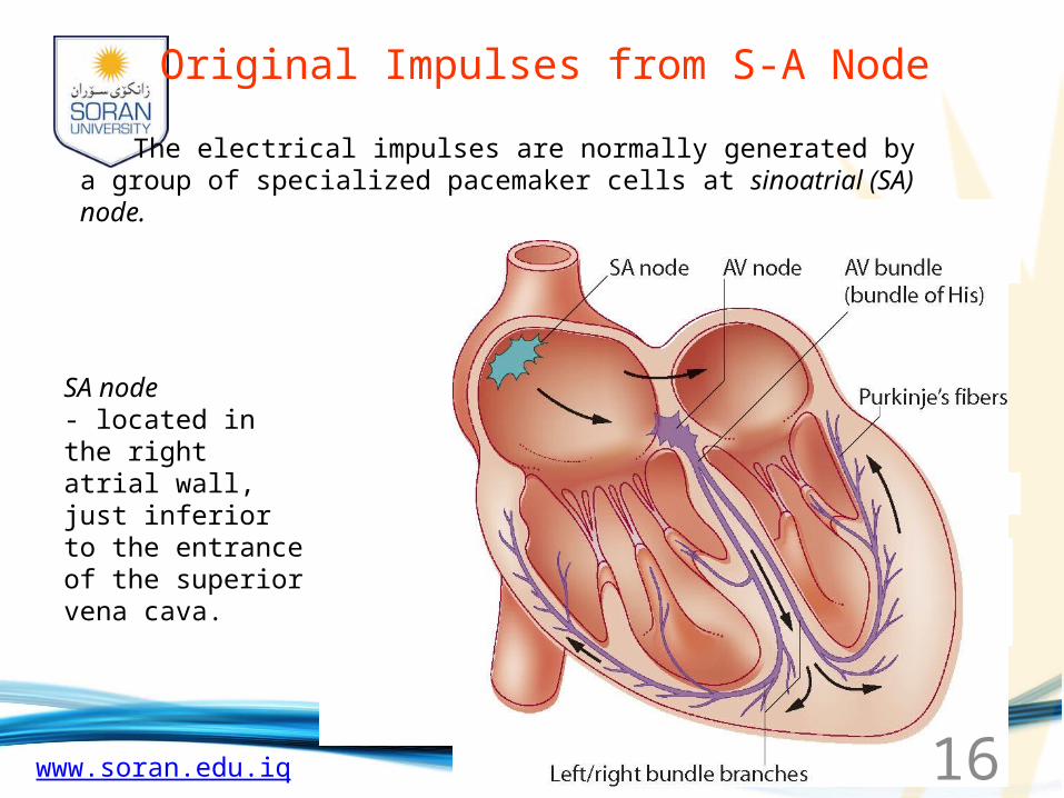
www.soran.edu.iq 16
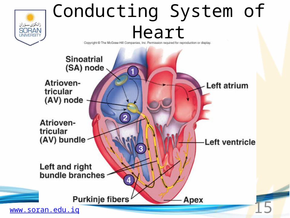
SA node- located in the right atrial wall, just inferior to the entrance of the superior vena cava.
Original Impulses from S-A Node
The electrical impulses are normally generated by a group of specialized pacemaker cells at sinoatrial (SA) node.
www.soran.edu.iq 17
Conduction of Action Potentials from Cell to Cell
through gap junctions in intercalated discs (electrical synapses)
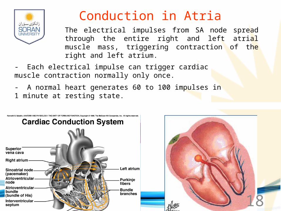
www.soran.edu.iq 18
Conduction in AtriaThe electrical impulses from SA node spread through the entire right and left atrial muscle mass, triggering contraction of the right and left atrium.
- Each electrical impulse can trigger cardiac muscle contraction normally only once.
- A normal heart generates 60 to 100 impulses in 1 minute at resting state.
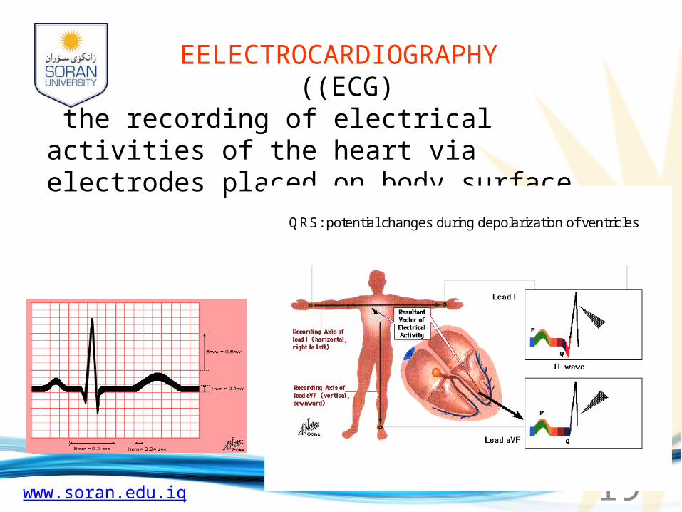
www.soran.edu.iq 19
QRS: potential changes during depolarization of ventricles
EELECTROCARDIOGRAPHY ((ECG)
the recording of electrical activities of the heart via electrodes placed on body surface.
www.soran.edu.iq 21
Regulation of the Heart
• Intrinsic regulation: Results from normal functional characteristics, not on neural or hormonal regulation– Starling’s law of the heart
• Extrinsic regulation: Involves neural and hormonal control– Parasympathetic stimulation
• Supplied by vagus nerve, decreases heart rate, acetylcholine secreted– Sympathetic stimulation
• Supplied by cardiac nerves, increases heart rate and force of contraction, epinephrine and norepinephrine released
www.soran.edu.iq 23
Effects of Aging on the Heart• Gradual changes in heart function, minor under
resting condition, more significant during exercise• Hypertrophy of left ventricle• Maximum heart rate decreases• Increased tendency for valves to function
abnormally and arrhythmias to occur• Increased oxygen consumption required to pump
same amount of blood