Embed Size (px)
Citation preview

REVIEW Open Access
Biological rationale for the use of DNAmethyltransferase inhibitors as new strategy formodulation of tumor response to chemotherapyand radiationGiovanni L Gravina1,2*, Claudio Festuccia2, Francesco Marampon1,2,3, Vladimir M Popov3, Richard G Pestell3,Bianca M Zani2, Vincenzo Tombolini1,2
Abstract
Epigenetic modifications play a key role in the patho-physiology of many tumors and the current use of agentstargeting epigenetic changes has become a topic of intense interest in cancer research. DNA methyltransferase(DNMT) inhibitors represent a promising class of epigenetic modulators. Research performed yielded promisinganti-tumorigenic activity for these agents in vitro and in vivo against a variety of hematologic and solid tumors.These epigenetic modulators cause cell cycle and growth arrest, differentiation and apoptosis. Rationale for com-bining these agents with cytotoxic therapy or radiation is straightforward since the use of DNMT inhibitor offersgreatly improved access for cytotoxic agents or radiation for targeting DNA-protein complex. The positive resultsobtained with these combined approaches in preclinical cancer models demonstrate the potential impact DNMTinhibitors may have in treatments of different cancer types. Therefore, as the emerging interest in use of DNMTinhibitors as a potential chemo- or radiation sensitizers is constantly increasing, further clinical investigations areinevitable in order to finalize and confirm the consistency of current observations.The present article will provide a brief review of the biological significance and rationale for the clinical potential ofDNMT inhibitors in combination with other chemotherapeutics or ionizing radiation. The molecular basis andmechanisms of action for these combined treatments will be discussed herein.
A significant number of tumors are classified as poorlyor non-responsive to therapeutic drugs or radiotherapy.Increasing the chemotherapeutic dosage or radiationdose not only fails in improving the therapeuticresponse, but it also contributes to the development ofside effects and resistance to therapy. An ideal strategywould consist of the identification of anticancer agentsable to act synergistically with standard treatments suchas radiotherapy and chemotherapy, which would resultin triggering the cell death preferentially in tumor cells.Many patients with neoplastic diseases exhibit hyper-methylation of cytosine residues in gene promoterswhich induce silencing of key tumor suppressor genes.
Since methylation of CpG islands occurs infrequently innormal cells, the modulation of this post-translationalmodification may provide a selective tumor-specifictherapeutic target.The packaging of DNA is critical for many DNA
metabolic processes including transcription, replicationand DNA repair. DNA is normally tightly wrappedaround histone octamers to form nucleosomes. Theseprimary elements have been traditionally thought asstable DNA packaging units. However, it is now evidentthat they are dynamic structures that can be altered bydifferent molecular processes [1-3]. These include (i)incorporation of histone variants, which are thought tohave specialized functions [4], (ii) replacement, reposi-tioning or, in certain cases, the removal of nucleosomesby chromatin remodeling complexes, and finally (iii)post-translational modifications.
* Correspondence: [email protected] of Experimental Medicine, Division of Radiation Oncology,S. Salvatore Hospital, L’Aquila, University of L’Aquila, Medical School, L’Aquila67100, ItalyFull list of author information is available at the end of the article
Gravina et al. Molecular Cancer 2010, 9:305http://www.molecular-cancer.com/content/9/1/305
© 2010 Gravina et al; licensee BioMed Central Ltd. This is an Open Access article distributed under the terms of the Creative CommonsAttribution License (http://creativecommons.org/licenses/by/2.0), which permits unrestricted use, distribution, and reproduction inany medium, provided the original work is properly cited.

Post-translational modifications include (i) lysine acet-ylation and deacetylation, (ii) methylation, (iii) serinephosphorylation and ubiquination and (iv) lysine sumoy-lation. These modifications play a major role in model-ing higher-order chromatin conformation and modifyingthe DNA accessibility to transcription factors [5,6].Therefore, such changes are not strictly “genetic,” eventhough the specific chromatin patterns are usuallyinherited by daughter cells during replication.In cancer, epigenetic silencing through methylation
occurs just as frequently as mutations or deletions andleads to aberrant silencing of genes with tumor-suppres-sor functions [2,3].Among the post-translational processes, DNA methy-
lation is one of the most extensively characterized epige-netic modifications and its biological role is to maintainDNA transcriptionally quiescent, resulting in gene silen-cing (Figure 1) [7-9]. This process is dependent uponthe action of DNA methyltransferases (DNMTs),enzymes that catalyze the addition of methyl groups tothe 5’ carbon of the cytosine residues (Figure 1) [7]. Sev-eral isoforms of DNMTs are present in normal cells aswell as in tumor cells [9-11]. Existing evidence indicatesthat DNMT1 appears to be responsible for maintenanceof established patterns of methylated DNA, whileDNMT-3a and -3b seem to mediate de novo DNAmethylation patterns [9-11]. Interestingly DNMT1 aloneis not sufficient for maintenance of abnormal genehypermethylation but the cooperation with DNMT3bmust occur for this function [12-14]. In the last yearsmany different DNMT inhibitors have been developed(Table 1) and multiple molecular mechanisms by which
DNMT inhibitors induce anti-cancer effects have beenidentified. These mechanisms are partially mediated bythe hypomethylation of DNA with cytotoxic effectsdocumented at higher concentrations [8,15]. The neteffect is the modulation of specific genes involved in cel-lular processes such as apoptosis, cytostasis, differentia-tion and tumor angiogenesis [8,15]. Therefore, it is notsurprising that DNMT inhibitors are emerging as pro-mising class of drugs in cancer treatment, especially incombination with other agents or with other treatmentslike radiotherapy. Even though some DNMT inhibitorshave entered into clinical trials, we currently have lim-ited understanding of their precise mechanisms ofaction, especially when combined with other availabletreatments.The present article will provide a brief review of the
biological significance and scientific rationale for theclinical potential of DNMT inhibitors in combinationwith chemotherapy or radiotherapy.
Combined therapy: Published Experience,Ongoing Studies and Future directionsThe goal of combining different treatments in the man-agement of cancer is to increase and prolong theresponse rate as well as to decrease the toxicity asso-ciated with each treatment. Two different strategies canbe utilized to achieve these objectives. Treatments maybe combined based on the absence of overlapping orsynergistic toxicities leading to empiric combinations.A more sound approach is based upon the combinationof treatments with known convergent molecularmechanisms.It is well known that epigenetic abnormalities in can-
cer affect a plethora of genes involved in key cellularpathways including cell cycle control, apoptosis, immunerecognition, angiogenesis tumor invasion and metastasis.Consistent with the functional diversification of epige-netic alterations, epigenetic drugs are characterized bypleiotropic effects which affect key aspects of tumorbiology leading to an overall impairment of the neoplas-tic potential of tumor cells. These are the reasons thatconstitute the rationale for the proposed usage ofDNMT inhibitors as anticancer agents, alone or in com-bination with other treatments.
DNMT inhibitors and chemotherapyDespite the promising anticancer activity in haematolo-gical malignancies [16], early clinical trials showed thatDNMT inhibitors have low anticancer activity and sig-nificant toxicity as single agent in solid tumors. Recentstudies, however, suggest that low concentrations ofDNMT inhibitors such as 5-Aza and decitabine may actsynergistically when combined with chemotherapyand contribute to overcoming intrinsic or acquired
Figure 1 Epigenetic modulation of gene expression by post-translational DNA methylation. Transcriptionally inactivechromatin is characterized by the presence of methylated cytosineswithin CpG dinucleotides (CH3), which is sustained by DNAmethyltransferases (DNMTs).
Gravina et al. Molecular Cancer 2010, 9:305http://www.molecular-cancer.com/content/9/1/305
Page 2 of 16
chemoresistance [17-19]. These properties are consid-ered clinically significant as the resistance of tumor cellsto cytotoxic agents remains the major obstacle in che-motherapeutic-based treatments. The mechanismsunderlying chemoresistance remain in some measureelusive even though multifactorial mechanisms, includ-ing epigenetic modifications may drive this mechanism[20-25]. Therefore, any effort to overcome multi-drugresistance represents the primary goal in cancerresearch. Based on the chemical mechanisms, DNMTinhibitors act through different mechanisms. Among thedifferent mechanisms postulated, alterations in differen-tiation, changes in apoptosis, and induction of a benefi-cial immune response are considered of mainimportance [19]. Finally, the induction of DNA damagedue to the formation of irreversible covalent enzyme-DNA adducts has also been taken into consideration.
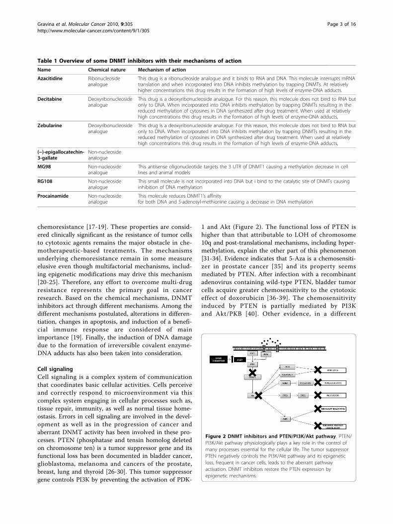
Cell signalingCell signaling is a complex system of communicationthat coordinates basic cellular activities. Cells perceiveand correctly respond to microenvironment via thiscomplex system engaging in cellular processes such as,tissue repair, immunity, as well as normal tissue home-ostasis. Errors in cell signaling are involved in the devel-opment as well as in the progression of cancer andaberrant DNMT activity has been involved in these pro-cesses. PTEN (phosphatase and tensin homolog deletedon chromosome ten) is a tumor suppressor gene and itsfunctional loss has been documented in bladder cancer,glioblastoma, melanoma and cancers of the prostate,breast, lung and thyroid [26-30]. This tumor suppressorgene controls PI3K by preventing the activation of PDK-
1 and Akt (Figure 2). The functional loss of PTEN ishigher than that attributable to LOH of chromosome10q and post-translational mechanisms, including hyper-methylation, explain the other part of this phenomenon[31-34]. Evidence indicates that 5-Aza is a chemosensiti-zer in prostate cancer [35] and its property seemsmediated by PTEN. After infection with a recombinantadenovirus containing wild-type PTEN, bladder tumorcells acquire greater chemosensitivity to the cytotoxiceffect of doxorubicin [36-39]. The chemosensitivityinduced by PTEN is partially mediated by PI3Kand Akt/PKB [40]. Other evidence, in a different
Table 1 Overview of some DNMT inhibitors with their mechanisms of action
Name Chemical nature Mechanism of action
Azacitidine Ribonucleosideanalogue
This drug is a ribonucleoside analogue and it binds to RNA and DNA. This molecule interrupts mRNAtranslation and when incorporated into DNA inhibits methylation by trapping DNMTs. At relativelyhigher concentrations this drug results in the formation of high levels of enzyme-DNA adducts.
Decitabine Deoxyribonucleosideanalogue
This drug is a deoxyribonucleoside analogue. For this reason, this molecule does not bind to RNA butonly to DNA. When incorporated into DNA inhibits methylation by trapping DNMTs resulting in thereduced methylation of cytosines in DNA synthesized after drug treatment. When used at relativelyhigh concentrations this drug results in the formation of high levels of enzyme-DNA adducts,
Zebularine Deoxyribonucleosideanalogue
This drug is a deoxyribonucleoside analogue. For this reason, this molecule does not bind to RNA butonly to DNA. When incorporated into DNA inhibits methylation by trapping DNMTs resulting in thereduced methylation of cytosines in DNA synthesized after drug treatment. When used at relativelyhigh concentrations this drug results in the formation of high levels of enzyme-DNA adducts,
(−)-epigallocatechin-3-gallate
Non-nucleosideanalogue
MG98 Non-nucleosideanalogue
This antisense oligonucleotide targets the 3 UTR of DNMT1 causing a methylation decrease in celllines and animal models
RG108 Non-nucleosideanalogue
This small molecule is not incorporated into DNA but i bind to the catalytic site of DNMTs causinginhibition of DNA methylation
Procainamide Non-nucleosideanalogue
This molecule reduces DNMT1’s affinityfor both DNA and S-adenosyl-methionine causing a decrease in DNA methylation
Figure 2 DNMT inhibitors and PTEN/PI3K/Akt pathway. PTEN/PI3K/Akt pathway physiologically plays a key role in the control ofmany processes essential for the cellular life. The tumor suppressorPTEN negatively controls the PI3K/Akt pathway and its epigeneticloss, frequent in cancer cells, leads to the aberrant pathwayactivation. DNMT inhibitors restore the PTEN expression byepigenetic mechanisms.
Gravina et al. Molecular Cancer 2010, 9:305http://www.molecular-cancer.com/content/9/1/305
Page 3 of 16

experimental model, suggests that the transfection ofTC-32 Ewing sarcoma cells with Akt/PKB inhibits dox-orubicin-induced apoptosis suggesting that PTENincreases doxorubicin cytotoxicity through the PI3K sig-naling pathway. Additional indication of chemosensitiz-ing properties of PTEN derived from data obtained inendometrial cancer cells [41]. In this system, PTEN sig-nificantly enhanced chemosensitivity to doxorubicin.This effect was associated with the levels of phospho-Akt/PKB and phospho-Bad (Ser-136), which werereduced in the PTEN expressing clones. Results fromother studies performed on brain tumors clearly showthat decreasing activity of the PI3K/Akt pathway intumor cells with mutant PTEN may contribute to theincreased sensitivity to chemotherapy [42]. Down-regu-lating the Akt pathway by inducing PTEN also increasesthe sensitivity of glioblastoma cells to temozolomide.
DNA base damageSome kind of acquired or intrinsic chemoresistance maybe epigenetic in nature and operate at DNA mismatchrepair level [43-48]. Interestingly, suppression of a DNAmismatch repair mechanism seems [49] to act in concertwith other independent DNA mismatch repair machi-neries [50,51], resulting in drug resistance and geneticinstability [52]. The suppression of DNA mismatch repairmechanism can occur at epigenetic level and in theabsence of heritable inactivating mutations. Demethylatingagents have been shown to reverse drug resistance to alky-lating agents in some preclinical models of ovarian andcolorectal cancer [53]. This process may happen throughup-regulation of MLH1, and is mediated by 5-Aza treat-ment, which sensitizes tumor cells to cisplatin. However,5-Aza treatment does not sensitize MLH1 mutant cells tocisplatin, indicating that MLH1 gene reactivation isrequired for the sensitization [53,54]. If MLH1 reactivationis required for the sensitization to cisplatin, several con-cerns still exist about the value of epigenetic modulationof DNA repair genes in inducing chemosensitization. It iswell known that MGMT, another DNA repair mediator, isfrequently epigenetically silenced [55]. Consistent with itsrole in protecting the genome from G to A transitions,induced by alkylating agents, MGMT inactivation throughits promoter hypermethylation has been associated with Gto A mutations in k-ras and p53 genes in colorectal cancer[56,57]. Even though DNMT inhibitors, such as 5-Aza anddecitabine, have proven effective in re-expressing MGMTin cancer cells, the clinical advantage of the restoredMGMT expression is doubtful [58-60] since tumors withthe unmethylated promoter of MGMT gene appear signif-icantly less susceptible to the cytotoxic effects of alkylatingdrugs [56,57,60].Although the biological effects of DNMT inhibitors on
methylation and demethylation status of DNA mismatch
repair genes have been extensively studied, DNAdamage-related sequelae of these agents is still not fullyunderstood. DNA Double Strand Breaks (DSBs) are themost cytotoxic DNA lesions. One-ended DSBs can beformed via collapse of a replication fork at the site of ablocking DNA lesion [61-64]. Given that DNMT inhibi-tors, as well as some chemotherapeutics, create irreversi-ble covalent DNA-enzyme adducts, the convergence ofthese phenomena may be one possible mechanism bywhich these two agents synergize and induce cytotoxicity[65,66]. Several reports indicate that 5-Aza, decitabineand zebularine induce DSB responses followed byinduced apoptosis. These responses may be mediated viaATR or ATM, which are two key mediators promotingDNA DSB response signalling [67-69]. Given that theDSB responses induced by 5-Aza and doxorubicin engagein distinct signaling pathways, the combination of thesetwo agents may cooperate and synergistically induce celldeath [70]. The induction of DNA damage response byChk2 and p53 phosphorylation could be anothermechanism by which DNMT inhibitors induce DNAdamage-related sequelae and cooperate with chemothera-peutics [71-75]. In this regard, some studies revealed thatdecitabine may be cytotoxic against both p53 wild-typeand p53 mutant containing tumor cells, suggesting thatp53 function is not always required in order to mediatethe apoptotic process of DNMT inhibitors. Finally, it hasbeen demonstrated that these agents can induce DNAdamage in a dose-dependent manner, while the degreeand the kind of DNA damage induced parallels theamount of incorporated DNMT inhibitor [76]. This maybe an important cooperative mechanism since when thecytotoxic effect of DNMT inhibitors takes place in closeproximity of a single- or double-strand break induced bychemotherapeutics the damage may be lethal.
ApoptosisDefects in apoptotic pathways promote chemoresistance.DNMT inhibitors are known to potentiate apoptoticprocesses through different pathways. Evidence suggeststhat the induction of TRAIL by decitabine is critical forsensitizing breast cancer cells to Adriamycin. The silen-cing of TRAIL decreases caspase activation and abro-gates chemosensitization mediated by decitabine. Severalmechanisms by which DNMT inhibitors induce TRAILhave been postulated. One of the possible mechanismsis the activation of TRAIL gene expression [77,78].Additional evidence suggests that the induction ofTRIAL by decitabine is mediated by the increase in thehalf-life of TRAIL protein [78] or by the induction ofTRAIL via Akt. It is known that the PI3K inhibitorwortmannin can induce TRAIL [79], and that the over-expression of the active form of Akt can abolish TRAILinduction mediated by wortmannin. In agreement with
Gravina et al. Molecular Cancer 2010, 9:305http://www.molecular-cancer.com/content/9/1/305
Page 4 of 16
this evidence, Akt, working as a negative modulator ofTRAIL, is modulated by 5-Aza resulting in a decrease ofphosphorylated Akt and enhanced TRAIL expression.If TRAIL plays a key role in the apoptotic process
mediated by DNMT inhibitors, other investigators sug-gest that the methylation in the promoter region of cas-pase 8 and caspase 9 is another well known mechanismby which tumors acquire chemoresistance. Mechanisticstudies indicate that decitabine induces caspase-8 andcaspase-9 and sensitizes tumor cells to TRAIL, etopo-side, cisplatin [80,81] and paclitaxel [82].Overexpression of the Activator protein 2a (AP-2a) is
another mechanism through which DNMT inhibitorsmay induce apoptosis and influence chemosensitivity.AP-2a is a sequence-specific DNA-binding transcriptionfactor that is required for regulation of many genesinvolved in many biological functions [83-85]. Growthinhibitory activity of AP-2a is mediated through directinteraction with p53 [86] and the overexpression of thistranscription factor induces cell cycle arrest and apopto-sis [87,88]. Epigenetic targeting of AP-2a inhibits tumorproliferation and increases tumor cell death [89]. Thisacquires a meaningful clinical significance consideringthat 75% of invasive breast tumors have epigeneticallysilenced AP-2a. Therefore, the use of DNMT inhibitorsmay provide the unique opportunity for modifying thechemosensitivity of breast cancer containing hyper-methylated and silenced AP-2a [90-95].
Oxidative stressAgents inducing oxidative stress determine cellulardamage by reactive oxygen intermediates (ROI). Smallamounts of ROI may act as signalling molecules but ifROI production exceeds the endogenous intracellularantioxidative capacities [96,97] an oxidative stress occurs[98] resulting in cell death. Increased ROI levels contri-bute to the development of chemoresistance and grow-ing evidence supports a role of epigenetic processes inROS-induced generation of oxidative stress [99-105].The proto-oncogene AP-1 plays a central role in thecontrol of cellular response to oxidative stress [106-109].It modulates the expression of target genes involved inprotective and/or reparative cellular responses to thedamaging effects of oxidative stress [110-113]. Experi-mental data suggest that H2O2 stress-resistant tumorcells have increased AP-1 DNA-binding activity and areresistant to the damaging effects of chemotherapeuticagents [114]. The inhibition of the AP-1 complexreverses the multimodality resistance phenotype(MMRP) in response to oxidative stress through theinhibition of Fos activity [114]. Other studies haveexpanded these observations showing that DNMT1, adownstream target of Fos, is upregulated in chemoresis-tant tumor cells [114]. These results indicate that the
epigenome may play a critical role to oxidative stressand highlights a potential role of DNMT1 activity abro-gation as a potential molecular target in chemoresistanttumor cells. This evidence has been further confirmedshowing that the selective silencing of DNMT1 andDNMT3b greatly reduces the chemoresistance of tumorcells overexpressing DNMTs isoenzymes [115].
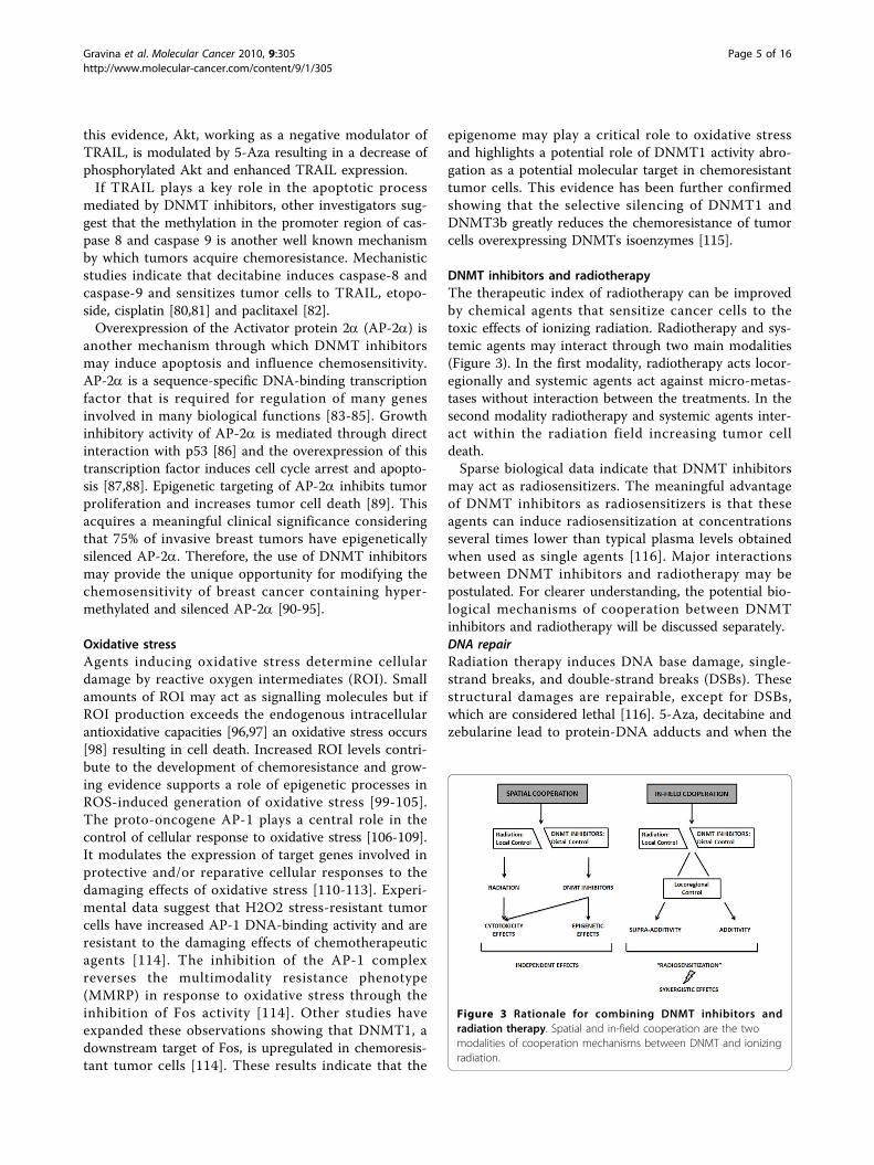
DNMT inhibitors and radiotherapyThe therapeutic index of radiotherapy can be improvedby chemical agents that sensitize cancer cells to thetoxic effects of ionizing radiation. Radiotherapy and sys-temic agents may interact through two main modalities(Figure 3). In the first modality, radiotherapy acts locor-egionally and systemic agents act against micro-metas-tases without interaction between the treatments. In thesecond modality radiotherapy and systemic agents inter-act within the radiation field increasing tumor celldeath.Sparse biological data indicate that DNMT inhibitors
may act as radiosensitizers. The meaningful advantageof DNMT inhibitors as radiosensitizers is that theseagents can induce radiosensitization at concentrationsseveral times lower than typical plasma levels obtainedwhen used as single agents [116]. Major interactionsbetween DNMT inhibitors and radiotherapy may bepostulated. For clearer understanding, the potential bio-logical mechanisms of cooperation between DNMTinhibitors and radiotherapy will be discussed separately.DNA repairRadiation therapy induces DNA base damage, single-strand breaks, and double-strand breaks (DSBs). Thesestructural damages are repairable, except for DSBs,which are considered lethal [116]. 5-Aza, decitabine andzebularine lead to protein-DNA adducts and when the
Figure 3 Rationale for combining DNMT inhibitors andradiation therapy. Spatial and in-field cooperation are the twomodalities of cooperation mechanisms between DNMT and ionizingradiation.
Gravina et al. Molecular Cancer 2010, 9:305http://www.molecular-cancer.com/content/9/1/305
Page 5 of 16

cytotoxic effect takes place in close proximity to a radia-tion-induced single-strand break, the damage may besignificantly more difficult to repair (Figure 4). TheDNA cytosine-C5 methyltransferase (MTase) acts on acytosine residue through its recognition sequence bycovalently binding to C6, and then transferring themethyl group from S-adenosylmethionine to C5. Thecovalent protein-DNA linkage is then reversed and theenzyme dissociates from the DNA. In this context, 5-Aza, decitabine and zebularine substitution at the targetcytosine interferes with the reaction cycle, which resultsin long-lived or irreversible MTase-DNA adducts[117-121]. The cytotoxic mechanism of DNMT inhibi-tors has been documented in in vivo models. Resultsfrom these studies suggest that (i) the formation of pro-tein-DNA adducts mediate decitabine cytotoxicity [122],(ii) the cytotoxicity levels correlate positively with
MTase levels [123], and (iii) decitabine induce p53 DNAdamage response by MTase-DNA adducts [124-127].Cell cycleThe radiosensitivity of tumor cells is dependent on thephase of the cell cycle. Cells in the S-phase are the mostradioresistant, whereas cells in the G2-M phase are themost radiosensitive. Evidence indicates that DNMT inhi-bitors synchronize tumor cells preferentially in the G1or G2/M phase of the cell cycle increasing the efficacyof radiotherapy (Figure 5). In this way, if administeredconcurrently to radiotherapy, the inhibitors may coop-erate to produce additive or synergistic antitumoreffects. Qui and co-workers demonstrated that low con-centrations of decitabine synchronize gastric tumor cellsin the G2/M phase of the cell cycle and induce radio-sensitization [116]. The radiosensitizing effect of thisinhibitor seems to be partly mediated by p53, RASSF1,and DAPK [116]. RASSF1 and DAPK modulate multipleapoptotic and cell-cycle checkpoint pathways [128,129]and the loss of RASSF1 and DAPK expression is docu-mented in a wide range of human tumors as a result ofsilencing, primarily from promoter hypermethylation[130]. Therefore, the epigenetic modulation of RASSF1and DAPK shines some light on the synergism betweenDNMT inhibitors and radiotherapy in terms of apopto-tic signaling modulation.AngiogenesisInduction of anti-angiogenic activity in radiotherapy as aresult of combined treatments with DNMT inhibitors isbacked by a clear rationale. Common in many cancershypoxia has been indicated as a marker of aggressiveclinical behaviour, poor prognosis and unsatisfyingradiation response. Therefore, any compound able toincrease perfusion and oxygenation reduces the radiore-sistant hypoxic areas counteracting the onset of hypoxicand radioresistant clones.Experimental data suggest that many key modulators of
angiogenesis are under epigenetic control. The promoterof von Hippel-Lindau (VHL) tumor suppressor gene ishypermethylated and its absence leads to a failure indegradation of hypoxia inducible factor (HIF)-1 whoseaccumulation favors tumor angiogenesis [131]. This aber-rant pathway has been successfully inactivated by decita-bine, which in addition to restoring VHL expression,down-regulates the vascular endothelial growth factor(VEGF), the glucose transporter (GLUT)-1 [132] and thethrombospondin-1 [133]. Radiotherapy has been shownto kill proliferating endothelial cells. Therefore, any drugthat acts on endothelial cells induces a significantincrease in the cytotoxic effect of radiotherapy decreasingthe levels of angiogenesis. DNMT inhibitors act directlyon activated endothelial cells and inhibit angiogenesis invitro and in vivo [132]. Decitabine and its analogue zebu-larine exhibit significant angiostatic activity. This is
Figure 4 Cooperative cytotoxic mechanism between DNMTinhibitors and radiation. Ionizing radiation induces DNA basedamage, single-strand breaks, and double-strand breaks (DSBs). Allof these errors can be rapidly repaired except for DSBs, which if notrepaired are considered lethal. The cytotoxic effect of DNMTinhibitors in close proximity to a radiation-induced single-strandbreak can act synergistically to make the defect significantly moredifficult to repair, consequently resulting in the induction of cellulardeath.
Figure 5 Cell cycle, DNMT inhibitors and radiosensitivity. Theradiosensitivity of cells is dependent on the phase of the cell cycle.Cells in the S phase are the most radio resistant, and cells in theG2-M phase of the cell cycle are the most radiosensitive. DNMTinhibitors synchronize with the cell cycle of tumor cells increasingthe efficacy of subsequent radiotherapy.
Gravina et al. Molecular Cancer 2010, 9:305http://www.molecular-cancer.com/content/9/1/305
Page 6 of 16
accompanied by a significant effect on the expressionlevels of angiogenesis inhibiting genes (TSP1, JUNB, andIGFBP3). TSP1 is known to block endothelial cell migra-tion and to induce apoptosis. JUNB negatively regulatescell growth by activating p16INK4A and decreasingcyclin D1 expression [132], while IGFBP3, a key regulatorof cell growth and apoptosis, inhibits VEGF-mediatedHUVEC proliferation [133] and angiogenesis [133]. Re-expression of these growth-inhibiting genes by DNMTinhibitors, in activated endothelial cells, may contributeto a decrease in angiogenesis and improvement in intrin-sic radiosensitivity.ApoptosisApoptosis is a well known mechanism through whichantitumor agents induce radiosensitization. DNMT inhi-bitors sensitize tumor cells to apoptosis either by restor-ing the defective expression of apoptotic effectorproteins, or by re-establishing the expression of signaltransducing/mediators of the apoptotic signals. 5-Azarestores the expression of DAPK1 in bladder carcinomaand B-cell lines [134] and the re-expression of DAPK1in Burkitt’s lymphoma cell lines restores the susceptibil-ity to IFN-a triggered apoptosis [135]. Similarly, DNMTinhibitors sensitize NSCLC cells to TRAIL-inducedapoptosis by inducing DAPK1 expression [136]. BesidesDAPK1, 5-Aza and DNMT1 antisense oligonucleotidesare able to restore the sensitivity of cancer cells to IFN-triggered apoptosis despite the re-expression of the pro-apoptotic gene RASSF1A and XAF1 which are fre-quently silenced by epigenetic mechanisms [137,138].Caspases are not spared from epigenetic inactivationduring tumor transformation. Hypermethylation of cas-pase-8 promoter leads to its either reduced expressionor complete absence in neoplastic cells resulting in theirresistance to death receptor and drug-induced apoptosis.However, 5-Aza has proven to be effective in re-estab-lishing caspase-8 expression in cancer cells, restoringtheir sensitivity to TRAIL-, anti-FAS-, and drug-trig-gered apoptosis [35,139-141].Cell signalingDNMT inhibitors are modulators of gene expressionand may increase the expression levels of many keygenes, specifically the ones involved in the radiosensitiz-ing processes. NF-�B is capable of activating a numberof genes involved in stress response, inflammation, andapoptosis. Loss or inhibition of NF-�B activation leadsto radio-sensitization [142-144]. The elevated basal NF-�B activity in certain cancers has been linked withtumor resistance to chemotherapy and radiation [145].Together with the assumption that NF-�B is capable ofregulating more than 150 effector genes, this transcrip-tion factor plays a key role in tumor radioadaptive resis-tance under fractional ionizing radiation. 5-Aza canrapidly induce inhibition of NF-kB [146]. This effect
may be achieved via down-regulation of pro-survivaland anti-apoptotic (IL-6, IL-6Ra, Bcl-XL) mediators orabrogating drug-induced NF-kB stress responses[147,148]. PARP-1 represents the essential transcrip-tional co-regulator implicated in radiation-induced NF-�B, AP-1, Oct1, and HIF-1a activation [149]. Experi-mental results demonstrate that the ATM gene is a tar-get for silencing through aberrant methylation of itsproximal promoter region [150,151]. This epigeneticevent can result in a decreased expression of ATM,resulting in a radioresistant phenotype consistent withreduced ATM function. DNMT inhibitors, positivelyaffecting the levels of ATM, increase radiosensitivity inhuman colorectal tumor cell lines. In this regard, themoderate radiosensitivity displayed by HCT-116 cellscan be increased by 5-azacitidine treatment, correlatingwith ATM levels [151].
Epigenetic control of oncogenes: implication for standardtreatmentsGlobal genomic hypomethylation has been documentedin most solid tumors [152-154]. Evidence suggests thatthis post-translational mechanism supports tumor devel-opment [155]. In solid human tumors, a correlationbetween global genomic hypomethylation and advancedtumor stage has been established [154].Methylation has been primarily considered as a
mechanism for tumor suppressor genes silencing andgenome profiling approaches have identified severalputative tumor suppressor genes silenced by promoterhypermethylation. So far, unmasked expression of puta-tive oncogenes has been sporadically reported [156].Although c-myc was among the very earliest onco-
genes identified and the subject of intense study, it hasnonetheless proven to be an enduring enigma. Resultsto date suggest that Myc-Max influences cell growthand proliferation through direct activation of genesinvolved in DNA synthesis, RNA metabolism and cell-cycle progression [157]. Early studies showed that c-mycis under epigenetic control and its functional silencingsensitizes cancer cells to chemotherapy and radiotherapy[158-160]. These sensitizing effects of c-Myc were pri-marily achieved by inhibiting MLH1 and MSH2 mis-match repair proteins [161]. Evidence suggests thatdecitabine is unable to modify the expression of c-mycin gastric cancer [161]. Other evidence, however, suggestthat several proto-oncogenes, whose promoters areunder epigenetic control, may be down-regulated ratherthan up-regulated after treatment with epi-drugs [162].Microarray data revealed that the treatment of myelomamultiple cells by decitabine and TSA resulted in down-regulation of several proto-oncogenes such as membersof myc family [162]. Of note, the down-regulation ofthese genes was more a response to TSA and
Gravina et al. Molecular Cancer 2010, 9:305http://www.molecular-cancer.com/content/9/1/305
Page 7 of 16

decitabine/TSA than to decitabine alone. The biologicalrationale for this surprising phenomenon is not wellknown although this effect may be explained either by adirect inhibitory action of decitabine and TSA or by anindirect down-regulation by decitabine and TSA affectedgenes [162].Therefore, these conflicting data may have important
therapeutic implications since demethylation-based ther-apy can cause unintended effects. It may be possiblethat in certain tissues and under selective biological con-ditions epi-drugs may result in either up- or down-regu-lation of proto-oncogenes [163]. These concerns mayexplain either some of the side effects or the unsuccess-ful results documented upon demethylation-based ther-apy in solid tumors.
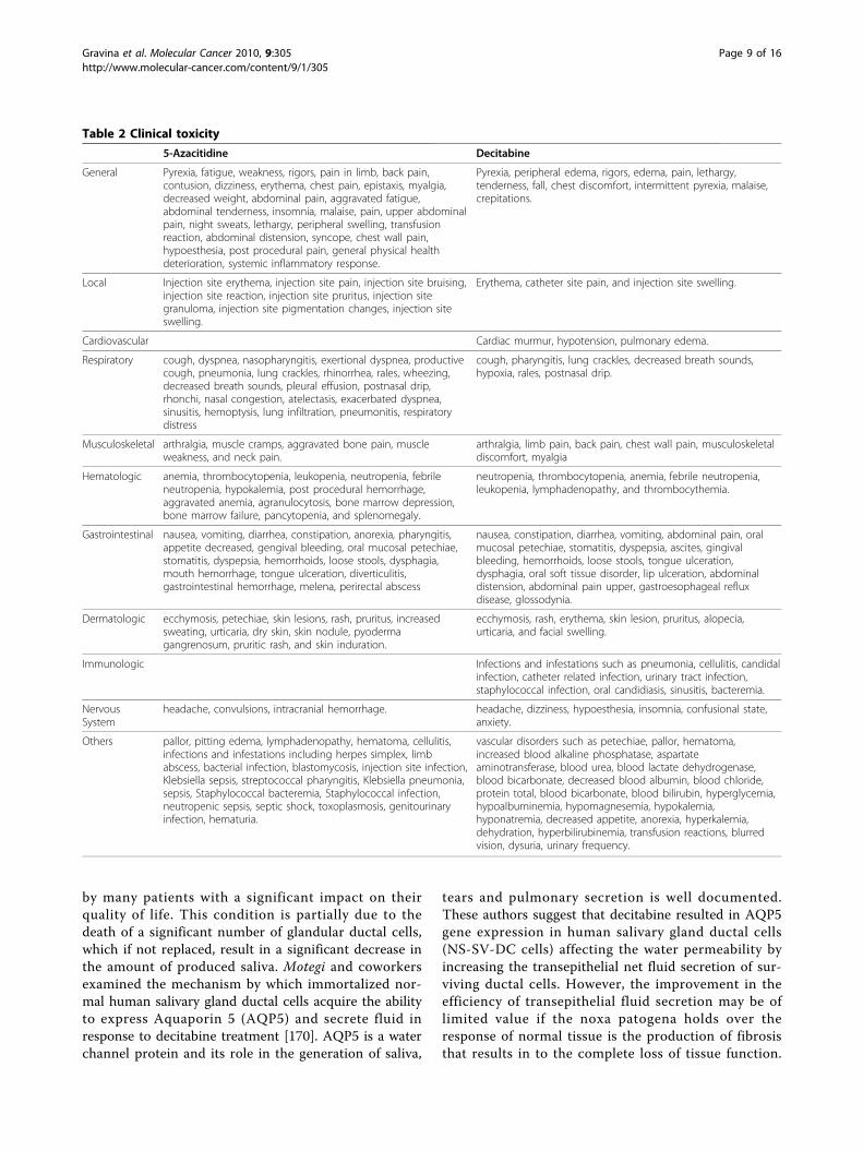
Response of normal tissues to DNMT inhibitorsThe use of DNMT inhibitors raises questions regardingtheir potential to epigenetically affect non-cancerouscells. Therefore, an important issue is a need for amore complete understanding of the potential benefitsand limitation of DNA methylation as a human cancerdrug target. Conflicting evidence exist in literatureregarding the effect of DNMT inhibitors on normalcells. Even though well known toxicity profile has beendocumented for DNMT inhibitors, especially fornucleoside analogues such as 5-Aza and decitabine inthe clinical setting (Table 2), many doubts exist abouttheir long-term safety as well as about their mutagenicand carcinogenic potential [164]. Some evidence indi-cate that intraperitoneal injection of 5-Aza at dosesranging from 2.0 to 2.2 mg/kg for 50-52 weeks in mur-ine models increased the incidence of malignanttumors of hematopoietic and lymphoreticular systemas well as of lung, mammary glands and skin [165].The mutagenic potential of 5-Aza [165] and decitabine[166] was tested in vitro and in vivo systems. Both ana-logues increased mutation frequency in L5178Y mouselymphoma cells, and mutations were produced in anEscherichia coli lac-I transgene in colonic DNA of dec-itabine-treated mice [166]. Decitabine, moreover,resulted in chromosomal rearrangements in larvae offruit flies. The effect of decitabine and 5-Aza on post-natal development and reproductive capacity was eval-uated in murine models. Administration of theseinhibitors in male mice resulted in decreased fertilityand loss of offspring during subsequent embryonic andpostnatal development. Decreased weight of the testesand epididymides with reduced sperm counts werealso detected. Body weights of males and femalesexposed in utero to decitabine were significantlyreduced at all postnatal time points. No consistenteffect on fertility was seen when female mice exposedin utero.
However, if the aforementioned data indicate thatboth 5-Aza and decitabine have a tangible toxicity onnormal tissues, recent biological data seem to suggestthat normal cells may interact differently with DNMTinhibitors than malignant cells. In this regard, some datasuggest that normal cells, dividing at a slower rate thanmalignant cells, incorporate less drug than cancer cellsinto their DNA resulting in decreased effect on DNAmethylation. Zebularine, a novel DNA methyltransferaseinhibitor, has properties of acting differentially on can-cerous and normal cells. Continuous treatment with thisdrug substantially reduces the growth rate of humancancer cells with less effect on normal human fibro-blasts. Growth inhibition in cancer cells was found to beassociated with the induction of p21 which was unmodi-fied in human fibroblasts. This suggests that the growth-suppressive potential of zebularine in tumor cells is see-mingly p21-dependent and its differential effects waspartially due to preferential incorporation of zebularineinto DNA of tumor cells as documented by the uridine/cytidine kinase activity levels that were generally higherin cancerous than normal cells. Therefore, the preferen-tial effects of zebularine in cancer cells in terms ofincorporation into DNA, growth inhibition, demethyla-tion, and depletion of DNMTs are probably due to dif-ferential metabolism compared to normal cells.If different metabolism between normal and malignant
cells may partially explain the preferential effect ofDNMT inhibitors on tumor cells [167], the differentialgenes expression that these inhibitors induce in differentcellular sub-populations is equally relevant. Karpf andcoworker conducted a genomic analysis of gene expres-sion modifications upon decitabine treatment both innormal and cancerous cell lines. In their analysis theauthors concluded that decitabine (i) elicited changes ina limited number of genes, (ii) regulated gene expressionsimilarly between normal and cancer cells, and (iii)changes in the expression of specific genes required thepresence of transcriptional activators competent for acti-vation of the target promoter [168,169]. However, if thedifferences in gene expression patterns between normaland malignant cells upon DNMT inhibitors seem to beless marked than previously seen, the selective activationof specific genes in tumor cells is clearly documented inliterature. In this regard, decitabine leads to the selectiveactivation of specific genes only in tumor cells openingto the intriguing hypothesis that DNMT inhibitors mayincrease the therapeutic index of specific antitumor stra-tegies consenting of targeting the gene products differ-entially expressed in tumor cells [169].Xerostomia is a common complication of radiotherapy
on head and neck cancer due to irreparable damagecaused to the salivary glands if they are included in theradiation fields. This side effect is perceived negatively
Gravina et al. Molecular Cancer 2010, 9:305http://www.molecular-cancer.com/content/9/1/305
Page 8 of 16
by many patients with a significant impact on theirquality of life. This condition is partially due to thedeath of a significant number of glandular ductal cells,which if not replaced, result in a significant decrease inthe amount of produced saliva. Motegi and coworkersexamined the mechanism by which immortalized nor-mal human salivary gland ductal cells acquire the abilityto express Aquaporin 5 (AQP5) and secrete fluid inresponse to decitabine treatment [170]. AQP5 is a waterchannel protein and its role in the generation of saliva,
tears and pulmonary secretion is well documented.These authors suggest that decitabine resulted in AQP5gene expression in human salivary gland ductal cells(NS-SV-DC cells) affecting the water permeability byincreasing the transepithelial net fluid secretion of sur-viving ductal cells. However, the improvement in theefficiency of transepithelial fluid secretion may be oflimited value if the noxa patogena holds over theresponse of normal tissue is the production of fibrosisthat results in to the complete loss of tissue function.
Table 2 Clinical toxicity
5-Azacitidine Decitabine
General Pyrexia, fatigue, weakness, rigors, pain in limb, back pain,contusion, dizziness, erythema, chest pain, epistaxis, myalgia,decreased weight, abdominal pain, aggravated fatigue,abdominal tenderness, insomnia, malaise, pain, upper abdominalpain, night sweats, lethargy, peripheral swelling, transfusionreaction, abdominal distension, syncope, chest wall pain,hypoesthesia, post procedural pain, general physical healthdeterioration, systemic inflammatory response.
Pyrexia, peripheral edema, rigors, edema, pain, lethargy,tenderness, fall, chest discomfort, intermittent pyrexia, malaise,crepitations.
Local Injection site erythema, injection site pain, injection site bruising,injection site reaction, injection site pruritus, injection sitegranuloma, injection site pigmentation changes, injection siteswelling.
Erythema, catheter site pain, and injection site swelling.
Cardiovascular Cardiac murmur, hypotension, pulmonary edema.
Respiratory cough, dyspnea, nasopharyngitis, exertional dyspnea, productivecough, pneumonia, lung crackles, rhinorrhea, rales, wheezing,decreased breath sounds, pleural effusion, postnasal drip,rhonchi, nasal congestion, atelectasis, exacerbated dyspnea,sinusitis, hemoptysis, lung infiltration, pneumonitis, respiratorydistress
cough, pharyngitis, lung crackles, decreased breath sounds,hypoxia, rales, postnasal drip.
Musculoskeletal arthralgia, muscle cramps, aggravated bone pain, muscleweakness, and neck pain.
arthralgia, limb pain, back pain, chest wall pain, musculoskeletaldiscomfort, myalgia
Hematologic anemia, thrombocytopenia, leukopenia, neutropenia, febrileneutropenia, hypokalemia, post procedural hemorrhage,aggravated anemia, agranulocytosis, bone marrow depression,bone marrow failure, pancytopenia, and splenomegaly.
neutropenia, thrombocytopenia, anemia, febrile neutropenia,leukopenia, lymphadenopathy, and thrombocythemia.
Gastrointestinal nausea, vomiting, diarrhea, constipation, anorexia, pharyngitis,appetite decreased, gengival bleeding, oral mucosal petechiae,stomatitis, dyspepsia, hemorrhoids, loose stools, dysphagia,mouth hemorrhage, tongue ulceration, diverticulitis,gastrointestinal hemorrhage, melena, perirectal abscess
nausea, constipation, diarrhea, vomiting, abdominal pain, oralmucosal petechiae, stomatitis, dyspepsia, ascites, gingivalbleeding, hemorrhoids, loose stools, tongue ulceration,dysphagia, oral soft tissue disorder, lip ulceration, abdominaldistension, abdominal pain upper, gastroesophageal refluxdisease, glossodynia.
Dermatologic ecchymosis, petechiae, skin lesions, rash, pruritus, increasedsweating, urticaria, dry skin, skin nodule, pyodermagangrenosum, pruritic rash, and skin induration.
ecchymosis, rash, erythema, skin lesion, pruritus, alopecia,urticaria, and facial swelling.
Immunologic Infections and infestations such as pneumonia, cellulitis, candidalinfection, catheter related infection, urinary tract infection,staphylococcal infection, oral candidiasis, sinusitis, bacteremia.
NervousSystem
headache, convulsions, intracranial hemorrhage. headache, dizziness, hypoesthesia, insomnia, confusional state,anxiety.
Others pallor, pitting edema, lymphadenopathy, hematoma, cellulitis,infections and infestations including herpes simplex, limbabscess, bacterial infection, blastomycosis, injection site infection,Klebsiella sepsis, streptococcal pharyngitis, Klebsiella pneumonia,sepsis, Staphylococcal bacteremia, Staphylococcal infection,neutropenic sepsis, septic shock, toxoplasmosis, genitourinaryinfection, hematuria.
vascular disorders such as petechiae, pallor, hematoma,increased blood alkaline phosphatase, aspartateaminotransferase, blood urea, blood lactate dehydrogenase,blood bicarbonate, decreased blood albumin, blood chloride,protein total, blood bicarbonate, blood bilirubin, hyperglycemia,hypoalbuminemia, hypomagnesemia, hypokalemia,hyponatremia, decreased appetite, anorexia, hyperkalemia,dehydration, hyperbilirubinemia, transfusion reactions, blurredvision, dysuria, urinary frequency.
Gravina et al. Molecular Cancer 2010, 9:305http://www.molecular-cancer.com/content/9/1/305
Page 9 of 16

Therefore, drugs capable of reducing the fibrinogenicpotential of specific treatments are of value in clinicalsetting.Some authors documented that DNA methylation
exert epigenetic control over fribrinogenesis and woundhealing. These events need a process of cell transdiffer-entiation of resident cells to stellate cells, a particularsubtype of myofibroblast, which seems to be a commonprocess in the majority of soft tissues. Myofibroblastsare highly profibrinogenic and produce proinflammatorymediators (IL-6, MCP-1, PFGF, TGFb-1) resulting inthe secretion of a large quantities of collagen I and III[171]. MeCP2 is a methyl-CpG-binding protein that hasthe potential to exert regulation over the expression ofmultiple genes via its interaction with methylated DNA.MeCP2 is a repressor of IkBa which is required fortransdifferentiation and profibrinogenic activity of stel-late cells. Another important mediator of transdifferen-tiation and fribrinogenesis is PPARg whosetranscriptional silencing activity is required for conver-sion of hepatic stellate cells to myofibroblast [172].Forced expression of PPARg in hepatic myofibroblastresults in reversing the transdifferentiation and profibri-nogenic potential of stellate cells with down-regulationof type I collagen, loss of proliferation, and reacquisitionof their adipogenic characteristics. Experimental datasuggest that MeCP2 is recruited to the IkBa promoterand decitabine treatment modulating epigenetically theexpression these mediator exerts control over key fibri-nogenic and inflammatory transcriptional regulatorsreducing greatly the fibrogenic potential of stellate cells[173].Another significant property of DNMT inhibitors is
their capability to act as antioxidants under specific bio-logical setting. This property is very important, espe-cially in conditions where an excess of free radicalsresults in tissue damage. EGCG is a major element ofgreen tea with a documented activity as DNMT inhibi-tor. Conflicting data are available in literature on theeffect of EGCG on normal and tumoral cells. In animmortalized normal breast epithelial cell line(MCF10A), EGCG induced growth arrest prior to thecell cycle restriction point, with elevated p21, hypopho-sphorylation of Rb, and decreased cyclin D1, suggestingthat higher concentrations of EGCG may be toxic tonormal mammary epithelial cells [174]. However, EGCGmay have both antioxidant and prooxidative activitiesinvolved in redox cycling and quinone formation [175]and may induce oxidative stress in vivo [176,177].Cysteine conjugates, indicative of reactive species devel-opment, have been detected after 200 and 400 mg/kg i.p. EGCG [178]. EGCG was also reported to be capableof inducing liver, kidney and gastrointestinal toxicitywhich seemed to be correlated with bioavailability of
EGCG [176,179]. Different results were documented byYamamoto and his group [180] since new mechanismsby which EGCG may act differentially in tumor andnormal cells were identified. In humans, EGCG israpidly absorbed through the oral mucosa and secretedback into the oral cavity by saliva, suggesting that sali-vary glandular cells may tolerate high concentrations ofEGCG [181]. Accordingly, this evidence suggests thatEGCG may differentially affect oxidative status and mayact as either a ROS inducer or a ROS suppressordepending upon the cell type [182]. EGCG concentra-tions higher than plasma Cmax do not produce ROS incells derived from the normal epidermis and oral cavitybut rather protect these cells by decreasing ROS pro-duction. Mechanisms responsible for the differentialeffects could rely on distinctive signal pathways activatedby EGCG in a tissue-specific manner. High concentra-tion of EGCG failed to produce ROS in normal epider-mal keratinocytes, and immortalized normal salivarygland cells. In contrast, EGCG elevated ROS levels upontreatment in a dose-dependent manner in oral carci-noma cells. The ROS levels were significantly higher inthe tumor cell lines that possessed low catalase activity.Therefore, EGCG may potentially simultaneouslyenhance tumor cell death rate and protect normal cellsfrom chemo/radiation-induced oxidative stress intumors such as skin and head neck [182].If uncertainty exists regarding the effect of DNMT
inhibitors on normal tissues, scanty direct evidenceexists regarding the biological effects these inhibitorshave in combination with chemotherapy or ionizingradiation. Clinical data indicate that, when decitabine isadministered in association with cisplatin in subjectswith advanced squamous cell carcinoma of the cervix, asignificant hematological toxicity is documented [183].Recent study reported interesting evidence of biologi-
cal interaction of a DNMT inhibitor with chemotherapy.In this report 5-Aza was combined with cisplatin inorder to measure the improvement in the therapeuticindex of these two drugs. Interestingly, 5-Aza preventedthe nephrotoxicity related to cisplatin, which was furtherrelated with the lowering in the levels of BUN and crea-tinine in the murine model. The mechanism by which5-Aza decreased the nephrotoxicity involved the reduc-tion of the levels of mediators such as metallothioneins,which are able to induce oxidative stress or indirectlyactivate gene responsible for preventing oxidative stress.This represents the first evidence that an epigenetictreatment with a DNMT inhibitor reduces the toxicityrelated to chemotherapy [184]. A single study dealingwith the combination of decitabine with ionizing radia-tion reports in a differentiated effect in terms of growthinhibition normal and cancerous cells [185]. When deci-tabine was combined with radiation no significant
Gravina et al. Molecular Cancer 2010, 9:305http://www.molecular-cancer.com/content/9/1/305
Page 10 of 16
synergism in terms of growth inhibition was found onhuman fibroblasts. On contrary, irradiation aloneresulted in a significant decrease in the proliferation ofnormal fibroblasts, suggesting that decitabine signifi-cantly modified cellular response to irradiation.
Off-target effects of DNMT inhibitors and potentialcontribution to radiosensitizationThe mechanisms by which DNMT inhibitors exert theireffects on cells may be divided into those related toDNMT inhibitors and those not related to demethyla-tion of DNA. The latter mechanisms may be defined asoff-target effects and although observed at higher con-centrations may substantially contribute to the antitu-mor properties of nucleoside analogues alone or incombination with chemotherapy or ionizing radiation.Numerous chemicals as well as radiation can lead tocovalent protein-DNA adducts. 5-Aza, one of the mostimportant nucleoside analogue, leads to protein-DNAadducts. 5-Aza is a cytidine analogue in which carbon-5(C5) of the pyrimidine ring is replaced with nitrogen.Normally, a DNA cytosine-C5 methyltransferase(MTase) acts on cytosine residue in its recognitionsequence by covalent binding to C6, and then transfer-ring the methyl group from S-adenosylmethionine toC5; the covalent protein-DNA adduct is then reversedand the enzyme dissociates from the DNA [186]. 5-Azasubstitution at target cytosine interferes with the reac-tion cycle and results in long-lived or irreversibleMTase-DNA adduct. A major consequence of 5-Azatreatment is loss of cytosine MTase activity [186]. Inmammalian cells, 5-Aza results in defective tRNA andrRNAs and inhibits protein synthesis [187]. Additionally,decitabine results in the induction of p53 DNA damageresponse, proposed to be dependent on formation ofMTase adducts [188]. Covalent protein-DNA adducts ortightly bound proteins represent a major challenge tothe DNA replication machinery. DNA replication forkscan be blocked in vivo by replication termination com-plexes [189]. As previously described, scanty evidencesindicate that nucleoside analogues may be efficientradiosensitizers. The cytotoxic mechanisms of nucleo-side analogues may potentiate the effects of ionizingradiation for several theoretical reasons. First, as DNAsynthesis inhibitors, nucleoside analogues have a poten-tial to inhibit the repair of genomic damage induced byionizing radiation. Second, because they are preferen-tially cytotoxic to proliferative cells, these analogues maydecrease the number of tumor clonogens and thus slowdown cell repopulation during fractionated radiotherapy.Tumor shrinkage induced by these compounds mayimprove tumor oxygenation and counter the detrimentaleffect of tumor hypoxia on radiation response. Third, anucleoside analogue with DNA chain terminator
property may, following their incorporation into theDNA repair patch, trigger an apoptotic response similarto that observed during the replication phase. SinceDNA damage is induced in all phase of the cell cycle byradiation, this mechanism offers the prospect of extend-ing the cytotoxicity of these analogues to non-S-phasecells. Due to the very low number of studies facing thistopic the aforementioned mechanisms are all putativeand specific experimental studies in vitro and in vivosettings should be performed in order to confirm thesehypotheses.
Combination of DNMT inhibitors with HDAC inhibitorsDue to their relative antitumor selectivity DNMT inhibi-tors have been used in combination with HDAC inhibi-tors. Recently, there have been several excellent reviewsof the histone deacetylase inhibitors (HDACIs) fieldused as a therapeutic option alone or in association witha variety of novel and conventional anticancer agents[190-192]. Optimal re-expression of methylated genessuch as tumor suppressor or other cancer relevant genesfollowing the serial application of a DNA methyltrans-ferase inhibitor followed by an HDAC inhibitor createdsignificant interest in combination epigenetic therapy.Among these p16INK4A and p14INK4b, Apaf-1 andcaspase-8 are efficiently re-expressed when DNMT inhi-bitors and HDACIs are combined [190-192]. DNMTand a group of methyl-cytosine binding proteins, e.g.,MeCP2, can also recruit and direct HDACs to the chro-matin associated with silenced genes. Combined treat-ment with a DNMT inhibitor and HDACI has beenshown to be superior in de-repressing silenced tumorsuppressor genes, as well as in inducing increasedgrowth inhibition, differentiation and apoptosis of can-cerous cells. The best results in de-repressing silencedgenes are observed when DNMT inhibitors are usedfirst at relatively low doses followed by exposure to theHDACIs.
Conclusions and Future DirectionsNo doubt exists that combining traditional cancer ther-apy with epigenetic modulators and reversing thechanges of DNA methylation pattern holds a hugepotential for successful treatment of haematological andsolid malignancies. There are a number of importantsteps that need to be accomplished on the path towardsefficient epigenetic therapy. Firstly, it is important togain more insight into the diverse molecular mechan-isms of the epi-drugs available today. A body of experi-mental evidences suggests that DNMT inhibitors mayserve as efficient chemo- and radiosensitizers in solidtumors. However, the observation reported need moresupport in order to indicate intimate molecular mechan-isms of chemo- and radiosensitization. Additionally, the
Gravina et al. Molecular Cancer 2010, 9:305http://www.molecular-cancer.com/content/9/1/305
Page 11 of 16
understanding of different mechanisms as well as thelong term safety of DNMT inhibitors on normal cellsalone or in combination with standard treatmentsremains very limited and requires further researchefforts. Although clinical and preclinical data indicate asubstantial toxicity of these inhibitors, other evidenceseems to suggest that these drugs may have potential inreducing toxicity under specific conditions. Therefore,the future goal is to indentify compounds able toenhance the therapeutic index and protect the nonmalignant tissues from side effects. Finally, other precli-nical data suggest that the sensitizing effects of DNMTinhibitors seem to depend on the epigenetic modulationof a wide array of genes. A non negligible part of thiseffect may be related to the off-target mechanisms.Future research will focus on establishing clinically rele-vant combinations of DNMT inhibitors and conven-tional cancer therapies. In particular, a longstandinginterest exists in the development of molecules that canmodify cellular responses to radiation and chemother-apy. The future perspectives lie in identifying more simi-lar compounds and elucidating their mechanisms ofaction in order to develop more effective cancer thera-pies and treatments.
AcknowledgementsThe authors wish to express the gratitude to Dr. Mario Di Staso (Division ofRadiotherapy, San Salvatore Hospital, L’Aquila), Dr. Pierluigi Bonfili (Division ofRadiotherapy, San Salvatore Hospital, L’Aquila), and Dr. Mario Tombolini(Department of Otorhinolaryngology, Audiology and Phoniatrics ‘G. Ferreri’,University ‘La Sapienza’, Rome, Italy) for critical review and helpful discussionwith the manuscript.
Author details1Department of Experimental Medicine, Division of Radiation Oncology,S. Salvatore Hospital, L’Aquila, University of L’Aquila, Medical School, L’Aquila67100, Italy. 2Department of Experimental Medicine, Laboratory ofRadiobiology, University of L’Aquila, Medical School, L’Aquila 67100, Italy.3Department of Cancer Biology and Medical Oncology, Kimmel CancerCenter, Thomas Jefferson University, Philadelphia, PA 19107, USA.
Authors’ contributionsGLG, CF, FM, RGP and VT drafted and wrote the manuscript. VP and BMZrevised the manuscript critically for important and intellectual content. GLGand FM supervised the project. All authors read and approved the finalmanuscript.
Competing interestsThe authors declare that they have no competing interests.
Received: 18 February 2010 Accepted: 25 November 2010Published: 25 November 2010
References1. Horn PJ, Peterson CL: Heterochromatin assembly: A new twist on an old
model. Chromosome Res 2006, 14:83-94.2. Luger K: Structure and dynamic behavior of nucleosomes. Curr Opin
Genet Dev 2003, 13:127-135.3. Saha A, Wittmeyer J, Cairns BR: Chromatin remodelling: The industrial
revolution of DNA around histones. Nat Rev Mol Cell Biol 2006, 7:437-47.
4. Marks PA, Rifkind RA, Richon VM, Breslow R: Inhibitors of histonedeacetylase are potentially effective anticancer agents. Clin Cancer Res2001, 7:759-60.
5. Iizuka M, Smith MM: Functional consequences of histone modifications.Curr Opin Genet Dev 2003, 13:154-60.
6. Spotswood HT, Turner BM: An increasingly complex code. J Clin Invest2002, 110:577-82.
7. Bird A: DNA methylation patterns and epigenetic memory. Genes Dev2002, 16:6-21.
8. Jones PA, Baylin SB: The fundamental role of epigenetic events in cancer.Nat Rev Genet 2002, 3:415-428.
9. Bestor TH: The DNA methyltransferases of mammals. Hum Mol Genet2000, 9:2395-2402.
10. Okano M: DNA methyltransferases Dnmt3a and Dnmt3b are essential forde novo methylation and mammalian development. Cell 1999,99:247-257.
11. Okano M: Cloning and characterization of a family of novel mammalianDNA (cytosine-5) methyltransferases. Nat Genet 1998, 19:219-220.
12. Rhee I: DNMT1 and DNMT3b cooperate to silence genes in humancancer cells. Nature 2002, 416:552-556.
13. Rhee I: CpG methylation is maintained in human cancer cells lackingDNMT1. Nature 2000, 404:1003-1007.
14. Gravina GL, Festuccia C, Millimaggi D, Dolo V, Tombolini V, De Vito M:Chronic Azacitidine Treatment Results in Differentiating Effects,Sensitizes Against Bicalutamide in Androgen-Independent ProstateCancer Cells. The Prostate 2008, 68:793-801.
15. Herman JG, Baylin SB: Gene silencing in cancer in association withpromoterhypermethylation. N Engl J Med 2003, 349:2042-2054.
16. Silverman LR, Demakos EP, Peterson BL: Randomized controlled trial ofazacitidine in patients with the myelodysplastic syndrome: a study ofthe cancer and leukemia group B. J Clin Oncol 2002, 20:2429-40.
17. Cameron EE, Bachman KE, Myohanen S, Herman JG, Baylin SB: Synergy ofdemethylation and histone deacetylase inhibition in the re-expression ofgenes silenced in cancer. Nat Genet 1999, 21:103-7.
18. kristensen LS, Nielsen HM, Hansen LL: Epigenetic and cancer treatment.European Juornal of Pharmacology 2009, 625:131-142.
19. Oki Y, Aoki E, Issa JP: Decitabine–Bedside to bench. Crit Rev Oncol 2007,61:140-152.
20. Larsen AK, Escargueil AE, Skladanowski A: Resistance mechanismsassociated with altered intracellular distribution of anticancer agents.Pharmacol Ther 2000, 85:217-29.
21. Larsen AK, Skladanowski A: Cellular resistance to topoisomerase-targeteddrugs: from drug uptake to cell death. Biochim Biophys Acta 1998,1400:257-74.
22. Kern MA, Helmbach H, Artuc M, Karmann D, Jurgovsky K, Schadendorf D:Human melanoma cell lines selected in vitro displaying various levels ofdrug resistance against Cisplatin, fotemustine, vindesine or Etoposide:modulation of proto-oncogene expression. Anticancer Res 1997,17:4359-70.
23. Spitz DR, Kinter MT, Roberts RJ: Contribution of increased glutathionecontent to mechanisms of oxidative stress resistance in hydrogenperoxide resistant hamster fibroblasts. J Cell Physiol 1995, 165:600-9.
24. Spitz DR, Li GC: Heat-induced cytotoxicity in H2O2-resistant Chinesehamster fibroblasts. J Cell Physiol 1990, 142:255-60.
25. Spitz DR, Phillips JW, Adams DT, Sherman CM, Deen DF, Li GC: Cellularresistance to oxidative stress is accompanied by resistance to Cisplatin:the significance of increased catalase activity and total glutathione inhydrogen peroxide-resistant fibroblasts. J Cell Physiol 1993, 156:72-9.
26. Steck PA, Pershouse MA, Jasser SA, Yung WK, Lin H, Ligon AH, Langford LA,Hattier T, Davis T, Frye C, Hu R, Swedlund B, Teng DH, Tavtigian SV:Identification of a candidate tumour suppressor gene, MMAC1, atchromosome 10q23.3 that is mutated in multiple advanced cancers. NatGenet 1997, 15:356-362.
27. Li J, Yen C, Liaw D, Podsypanina K, Bose S, Wang SI, Puc J, Miliaresis C,Rodgers L, McCombie R, Bigner SH, Giovanella BC, Ittmann M, Tycko B,Hibshoosh H, Wigler MH, Parsons R: PTEN, a putative protein tyrosinephosphatase gene mutated in human brain, breast, and prostate cancer.Science 1997, 275:1943-1947.
28. Wang SI, Puc J, Li J, Bruce JN, Cairns P, Sidransky D, Parsons R: PTEN, aputative protein tyrosine phosphatase gene mutated in human brain,breast, and prostate cancer. Cancer Res 1997, 57:4183-4186.
Gravina et al. Molecular Cancer 2010, 9:305http://www.molecular-cancer.com/content/9/1/305
Page 12 of 16

29. Guldberg P, thor Straten P, Birck A, Ahrenkiel V, Kirkin AF, Zeuthen J:Disruption of the MMAC1/PTEN gene by deletion or mutation is afrequent event in malignant melanoma. Cancer Res 1997, 57:3660-3663.
30. Cairns P, Okami K, Halachmi S, Halachmi N, Esteller M, Herman JG, Jen J,Isaacs WB, Bova GS, Sidransky D: Frequent inactivation of PTEN/MMAC1 inprimary prostate cancer. Cancer Res 1997, 57:4997-5000.
31. Gravina G, Biordi L, Martella F, Flati V, Ricevuto E, Ficorella C, Tombolini V,Festuccia C: Epigenetic modulation of PTEN expression duringantiandrogenic therapies in human prostate cancer. International Journalof Oncology 2009, 35:1133-9.
32. Soria JC, Lee HY, Lee JI, Wang L, Issa JP, Kemp BL, Liu DD, Kurie JM, Mao L,Khuri FR: Lack of PTEN Expression in Non-Small Cell Lung Cancer CouldBe Related to Promoter Methylation. Clin Cancer Res 2002, 8:1178-1184.
33. Whang YE, Wu X, Suzuki H: Inactivation of the tumor suppressorPTENyMMAC1 in dvanced human prostate cancer through loss ofexpression. Proc Natl Acad Sci 1998, 95:5246-5250.
34. Davies MA, Koul D, Dhesi H, Berman R, McDonnell TJ: Regulation of AKT/PKB activity, cellular growth, and apoptosis in prostate carcinomacellsby MMAC/PTEN. Cancer Res 1999, 59:2551-2556.
35. Festuccia C, Gravina GL, D’Alessandro AM, Muzi P, Millimaggi D: Azacitidineimproves antitumor effects of docetaxel and cisplatin in aggressiveprostate cancer models. Endocr Relat Cancer 2009, 16:401-413.
36. Tanaka Motoyoshi, Koul Dimpy, Davies AMichael, Liebert Monica,Steck APeter, Grossman HBarton: MMAC1/PTEN inhibits cell growth andinduces chemosensitivity to doxorubicin in human bladder cancer cells.Oncogene 2000, 19:5406-5412.
37. Fujiwara T, Grimm EA, Mukhopadnyay T, Zhang WW: Induction ofchemosensitivity in human lung cancer cells in vivo by adenovirus-mediated transfer of the wild type p53 gene. Cancer Res 1994,54:2287-2291.
38. Ogawa N, Fujiwara T, Kagawa S: Novel combination therapy for humancolon cancer with adenovirus-mediated wild-type p53 gene transfer andDNA-damaging chemotherapeutic agent. Int J Cancer 1997, 73:367-70.
39. Gurnani M, Lipari P, Dell J, Shi B, Nielsen LL: Adenovirus-mediated p53gene therapy has greater efficacy when combined with chemotherapyagainst human head-neck, ovarian, prostate, and breast cancer. CancerChemother Pharmacol 1999, 44:143-151.
40. Thoretsky JA, Takar M, Eskenazi AE, Frantz CN: Phosphoinositide 3-hydroxide kinase blockade enhances apoptosis in the Ewing’s sarcomafamily tumors. Cancer Res 1999, 59:5745-50.
41. Wan X, Li J, Lu X: PTEN augments doxorubicin-induced apoptosis inPTEN-null Ishikawa cells. Int J Gynecol Cancer 2007, 17:808-812.
42. Mayo LD, Dixon JE, Durden DL, Tonks NK, Donner DB: PTEN protectsp53from Mdm2 and sensitizes cancer cells to chemotherapy. J Biol Chem2002, 277:5484-9.
43. Green SK, Frankel A, Kerbel RS: Adhesion-dependent multicellular drugresistance. Anti-Cancer Drug Design 1999, 14:153-68.
44. St Croix B, Rak JW, Kapitain S, Sheehan C, Graham CH, Kerbel RS: Reversalby hyaluronidase of adhesion-dependent multicellular drug resistance inmammary carcinoma cells. J Natl Cancer Inst 1996, 88:1285-96.
45. St Croix B, Florenes VA, Rak JW, Flanagan M, Bhattacharya N, Slingerland JM,Kerbel RS: Impact of the cyclin dependent kinase inhibitor p27Kip1 onadhesion-dependent resistance of tumor cells to anticancer agents. NatMed 1996, 2:1204-10.
46. St Croix B, Sheehan C, Rak JW, Florenes VA, Slingerland JM, Kerbel RS: E-cadherin-dependent growth suppression is mediated by the cyclindependent kinase inhibitor p27KIP1. J Cell Biol 1998, 142:557-71.
47. Green SK, Francia G, Isidoro C, Kerbel RS: Anti-adhesive antibodiestargeting E-cadherin sensitize multicellular tumor spheroids tochemotherapy in vitro. Mol Cancer Ther 2004, 3:149-59.
48. Teicher BA, Herman TS, Holden SA, Wang YY, Pfeffer MR, Crawford JW,Frei E: Tumor resistance to alkylating agents conferred by mechanismsoperative only in vivo. Science 1990, 247:1457-61.
49. Youn CK, Cho HJ, Kim SH, Kim HB, Kim MH, Chang IY, Lee JS, Chung MH,Hahm KS, You HJ: Bcl-2 expression suppresses mismatch repair activitythrough inhibition of E2F transcriptional activity. Nat Cell Biol 2005,7:137-47.
50. Kobayashi H, Man S, Kapitain SJ, Teicher BA, Kerbel RS: Acquiredmulticellular-mediated resistance to alkylating agents in cancer. Proc NatlAcad Sci USA 1993, 90:3294-8.
51. Jain RK: Delivery of novel therapeutic agents in tumors: physiologicalbarriers and strategies. J Natl Cancer Inst 1989, 81:570-6.
52. Bindra RS, Schaffer PJ, Meng A, Woo J, Måseide K, Roth ME, Lizardi P,Hedley DW, Bristow RG, Glazer PM: Down-regulation of Rad51 anddecreased homologous recombination in hypoxic cancer cells. Mol CellBiol 2004, 24:8504-18.
53. Plumb JA, Strathdee G, Sludden J, Kaye SB, Brown R: Reversal of drugresistance in human tumor xenografts by 2V-deoxy-5-azacytidineinduced demethylation of the hMLH1 gene promoter. CancerRes 2000, 60:6039-44.
54. Chan AT, Tao Q, Robertson KD, Flinn IW, Mann RB, Klencke B, Kwan WH,Leung TW, Johnson PJ, Ambinder RF: Azacitidine induces demethylationof the Epstein-Barr virus genome in tumors. J Clin Oncol 2004, 22:1373-81.
55. Esteller M, Hamilton SR, Burger PC, Baylin SB, Herman JG: Inactivation ofthe DNArepair geneO6-methylguanine-DNA methyltransferase bypromoter hypermethylation is a common event in primary humanneoplasia. Cancer Res 1999, 59:793-797.
56. Esteller M, Herman JG: Generating mutations but providingchemosensitivity: the role of O6-methylguanine DNA methyltransferasein human cancer. Oncogene 2004, 23:1-8.
57. Hermann A, Gowher H, Jeltsch A: Biochemistry and biology ofmammalian DNA methyltransferases. Cell Mol Life Sci 2004, 61:2571-2587.
58. Bae SI, Lee HS, Kim SH, Kim WH: Inactivation of O6-methylguanine-DNAmethyltransferase by promoter CpG island hypermethylation in gastriccancers. Br J Cancer 2002, 86:1888-1892.
59. Danam RP, Howell SR, Brent TP, Harris LC: Epigenetic regulation ofO6-methylguanine-DNA methyltransferase gene expression by histoneacetylation and methyl-CpG binding proteins. Mol Cancer Ther 2005,4:1-69.
60. Wang W, Huper G, Guo Y, Murphy SK, Olson JA Jr, Marks JR: Analysis ofmethylation sensitive transcriptome identifies GADD45a as a frequentlymethylated gene in breast cancer. Oncogene 2005, 24:705-2714.
61. Michel B, Flores MJ, Viguera E, Grompone G, Seigneur M, Bidnenko V:Rescue of arrested replication forks by homologous recombination. ProcNatl Acad Sci USA 2001, 98:8181-8.
62. McGlynn P, Lloyd RG: Recombinational repair and restart of damagedreplication forks. Nat Rev Mol Cell Biol 2002, 3:859-70.
63. Szostak JW, Orr-Weaver TL, Rothstein RJ, Stahl FW: The doublestrand-breakrepair model for recombination. Cell 1983, 33:25-35.
64. Fishman-Lobell J, Rudin N, Haber JE: Two alternative pathways of double-strand break repair that are kinetically separable and independentlymodulated. Mol Cell Biol 1992, 12:1292-303.
65. Burma S, Chen BP, Murphy M, Kurimasa A, Chen DJ: ATM phosphorylateshistone H2AX in response to DNA double-strand breaks. J Biol Chem2001, 276:42462-7.
66. Rothkamm K, Kruger I, Thompson LH, Lobrich M: Pathways of DNAdouble-strand break repair during the mammalian cell cycle. Mol Cell Biol2003, 23:5706-15.
67. Durocher D, Jackson SP: DNA-PK, ATM and ATR as sensors of DNAdamage: variations on a theme? Curr Opin Cell Biol 2001, 13:225-31.
68. Roos WP, Kaina B: DNA damage-induced cell death by apoptosis. TrendsMol Med 2006, 12:440-50.
69. Kurz EU, Douglas P, Lees-Miller SP: Doxorubicin activates ATM dependentphosphorylation of multiple downstream targets in part through thegeneration of reactive oxygen species. J Biol Chem 2004, 279:53272-81.
70. Kiziltepe T, Hideshima T, Catley L, Raje N, Yasui H: 5-Azacytidine, a DNAmethyltransferase inhibitor, induces ATR-mediated DNA double-strandbreak responses, apoptosis, and synergistic cytotoxicity withdoxorubicine and bortezomib against multiple myeloma cells. MolCancer Ther 2007, 6:1718-1727.
71. Taylor WR, Stark GR: Regulation of the G2/M transition by p53. Oncogene2001, 20:1803-15.
72. Wichmann A, Jaklevic B, Su TT: Ionizing radiation induces caspasedependent but Chk2- and p53-independent cell death in Drosophilamelanogaster. Proc Natl Acad Sci USA 2006, 103:9952.
73. Aliouat-Denis CM, Dendouga N, Van den Wyngaert I, Goehlmann H,Steller U, van de Weyer I, Van Slycken N, Andries L, Kass S, Luyten W,Janicot M, Vialard JE: p53-independent regulation of p21Waf1/Cip1expression and senescence by Chk2. Mol Cancer Res 2005, 3:627-34.
74. Pepper CJ, Hambly RM, Fegan CD, Delavault P, Thurston DE: The novelsequence-specific DNA cross-linking agent SJG-136 (NSC 694501) has
Gravina et al. Molecular Cancer 2010, 9:305http://www.molecular-cancer.com/content/9/1/305
Page 13 of 16

potent and selective in vitro cytotoxicity in human B-cell chroniclymphocytic leukemia cells with evidence of a p53-independentmechanism of cell kill. Cancer Res 2004, 64:6750-5.
75. Tse AN, Schwartz GK: Potentiation of cytotoxicity of topoisomerase inpoison by concurrent and sequential treatment with the checkpointinhibitor UCN-01 involves disparate mechanisms resulting in either p53-independent clonogenic suppression or p53-dependent mitoticcatastrophe. Cancer Res 2004, 64:6635-44.
76. Chai Guolin, Li Lian, Zhou Wen, Wu Lipeng, Zhao Ying, Wang Donglai,Lu Shaoli, Yu Yu, Wang Haiying, McNutt AMichael, Hu Ye-Guang,Chen Yingqi, Yang Yang, Wu Xin, Otterson AGregory, Zhu Wei-Guo: HDACInhibitors Act with 5-aza-2’-Deoxycytidine to Inhibit Cell Proliferation bySuppressing Removal of Incorporated Abases in Lung Cancer Cells. PLoSOne 2008, 3:,3412-20.
77. Nebbioso A, Clarke N, Voltz E, Germain E, Ambrosino C, Bontempo P,Alvarez R, Schiavone EM, Ferrara F, Bresciani F, Weisz A, de Lera AR,Gronemeyer H, Altucci L: Tumor-selective action of HDAC inhibitorsinvolves TRAIL induction in acute myeloid leukemia cells. Nat Med 2005,11:77-84.
78. Xu J, Zhou JY, Tainsky MA, Wu GS: Evidence that Tumor Necrosis Factor-Related Apoptosis-Inducing Ligand Induction by 5-Aza-2¶-DeoxycytidineSensitizes Human Breast Cancer Cells to Adriamycin. Cancer Res 2007,67:1203-1211.
79. Wang Q, Wang X, Hernandez A, Hellmich MR, Gatalica Z, Evers BM:Regulation of TRAIL expression by the phosphatidylinositol 3-kinase/Akt/GSK-3 pathway in human colon cancer cells. J Biol Chem 2002,277:36602-10.
80. Fulda S, Kufer MU, Meyer E, van Valen F, Dockhorn-Dworniczak B,Debatin KM: Sensitization for death receptor- or drug-induced apoptosisby re-expression of caspase-8 through demethylation or gene transfer.Oncogene 2001, 20:5865-77.
81. Fulda S, Debatin KM: 5-Aza-2¶-deoxycytidine and IFN-gamma cooperateto sensitize for TRAIL-inducedapoptosis by upregulating caspase-8.Oncogene 2006, 25:5125-33.
82. Gomyo Y, Sasaki J, Branch C, Roth JA, Mukhopadhyay T: 5-aza-2¶-deoxycytidine upregulates caspase-9 expression cooperating with p53-induced apoptosis in human lung cancer cells. Oncogene 2004,23:6779-87.
83. Nottoli T, Hagopian-Donaldson S, Zhang J, Perkins A, Williams T: AP-2-nullcells disrupt morphogenesis of the eye, face, and limbs in chimericmice. Proc Natl Acad Sci USA 1998, 95:13714-9.
84. Feng W, Williams T: Cloning and characterization of the mouse AP-2egene: a novel family member expressed in the developing olfactorybulb. Mol Cell Neurosci 2003, 24:460-75.
85. Higler-Eversheim K, Moser M, Schorle H, Buettner R: Regulatory roles ofAP-2 transcription factors in vertebrate development, apoptosis and cellcycle control. Gene 2000, 260:1-12.
86. McPherson LA, Loktev AV, Weigel RJ: Tumor suppressor activity of AP2amediated through a direct interaction with p53. J Biol Chem 2002,277:45028-33.
87. Zeng YX, Somasundaram K, El-Deiry WS: AP2 inhibits cancer cell growthand activates p21WAF1/CIP1 expression. Nat Genet 1997, 15:28-32.
88. Wajapeyee N, Somasundaram K: Cell cycle arrest and apoptosis inductionby activator protein 2a (AP-2a) and the role of p53 and p21WAF1/CIP1in AP-2amediated growth inhibition. J Biol Chem 2003, 278:52093-101.
89. Zhang J, Brewer S, Huang J, Williams T: Overexpression of transcriptionfactor AP-2a suppresses mammary gland growth and morphogenesis.Dev Biol 2003, 256:127-45.
90. Douglas DB, Akiyama Y, Carraway H: Hypermethylation of a smallCpGuanine rich region correlates with loss of activator protein-2aexpression during progression of breast cancer. Cancer Res 2004,64:1611-20.
91. Anttila MA, Kellokoski JK, Moisio KI, Mitchell PJ, Saarikoski S, Syrjänen K,Kosma VM: Expression of transcription factor AP-2a predicts survival inepithelial ovarian cancer. Br J Cancer 2000, 82:1974-83.
92. Ropponen KM, Kellokoski JK, Pirinen RT, Moisio KI, Eskelinen MJ, Alhava EM,Kosma VM: Expression of transcription factor AP-2 in colorectaladenomas and adenocarcinomas; comparison of immunohistochemistryand in situ hybridisation. J Clin Pathol 2001, 54:533-8.
93. Heimberger AB, McGary EC, Suki D, Ruiz M, Wang H, Fuller GN, Bar-Eli M:Loss of the AP-2a transcription factor is associated with the gradeofhuman gliomas. Clin Cancer Res 2005, 11:267-72.
94. Ruiz M, Pettaway C, Song R, Stoeltzing O, Ellis L, Bar-Eli M: Activator protein2a inhibits tumorigenicity and represses vascular endothelial growthfactortranscription in prostate cancer cells. Cancer Res 2004, 64:631-8.
95. Gershenwald JE, Sumner W, Calderone T, Wang Z, Huang S, Bar-Eli M:Dominant-negative transcription factor AP-2 augments SB-2 melanomatumor growthin vivo. Oncogene 2001, 20:3363-75.
96. Dicato M, Duhem C, Pauly M, Ries F: Multidrug resistance: molecular andclinical aspects. Cytokines Cell Mol Ther 1997, 3:91-9.
97. Hunt CR, Sim JE, Sullivan SJ, Featherstone T, Golden W, Von Kapp-Herr C,Hock RA, Gomez RA, Parsian AJ, Spitz DR: Genomic instability and catalasegene amplification induced by chronic exposure to oxidative stress.Cancer Res 1998, 58:3986-92.
98. Bergelson S, Pinkus R, Daniel V: Intracellular glutathione levels regulateFos/Jun induction and activation of glutathione S-transferase geneexpression. Cancer Res 1994, 54:36-40.
99. Dewey WC: Arrhenius relationships from the molecule and cell to theclinic. Int J Hyperthermia 1994, 10:457-83.
100. Diamond DA, Parsian A, Hunt CR, Lofgren S, Spitz DR, Goswami PC, Gius D:Redox factor-1 (Ref-1) mediates the activation of AP-1 in HeLa and NIH3T3 cells in response to heat shock. J Biol Chem 1999, 274:16959-64.
101. Kerppola T, Curran T: Transcription. Zen and the art of Fos and Jun.Nature 1995, 373:199-200.
102. Martins NM, Santos NA, Curti C, Bianchi ML, Santos AC: Cisplatin inducesmitochondrial oxidative stress with resultant energetic metabolismimpairment, membrane rigidification and apoptosis in rat liver. J ApplToxicol 2007, 23:234-244.
103. Simons AL, Ahmad IM, Mattson DM, Dornfeld KJ: Spitz DR. 2-Deoxy-Dglucose combined with cisplatin enhances cytotoxicity via metabolicoxidative stress in human head and neck cancer cells. Cancer Res 2007,67:3364-70.
104. Siitonen T, Alaruikka P, Mantymaa P, Savolainen ER, Kavanagh TJ, Krejsa CM,Franklin CC, Kinnula V, Koistinen P: Protection of acute myeloblasticleukemia cells against apoptotic cell death by high glutathione andgammaglutamylcysteine synthetase levels during etoposide-inducedoxidative stress. Ann Oncol 1999, 10:1361-7.
105. Mairesse N, Bernaert D, Del Bino G, Horman S, Mosselmans R, Robaye B,Galand P: Expression of HSP27 results in increased sensitivity to tumornecrosis factor, etoposide, and H2O2 in an oxidative stress-resistant cellline. J Cell Physiol 1998, 177:606-17.
106. Furuta Y, Hunter N, Barkley T, Hall E, Milas L: Increase in radioresponse ofmurine tumors by treatment with indomethacin. Cancer Res 1988,48:3008-13.
107. Iliakis G, Seaner R, Okayasu R: Effects of hyperthermia on the repair ofradiation-induced DNA single- and double-strand breaks in DNA double-strand break repair-deficient and repair-proficient cell lines. Int JHyperthermia 1990, 6:813-33.
108. Jurivich DA, Pachetti C, Qiu L, Welk JF: Salicylate triggers heat shock factordifferently than heat. J Biol Chem 1995, 270:24489-95.
109. Jurivich DA, Sistonen L, Kroes RA, Morimoto RI: Effect of sodium salicylateon the human heat shock response. Science 1992, 255:1243-5.
110. Bhushan A, Abramson R, Chiu JF, Tritton TR: Expression of c-fos in humanand murine multidrug-resistant cells. Mol Pharmacol 1992, 42:69-74.
111. Dignam JD: Preparation of extracts from higher eukaryotes. MethodsEnzymol 1990, 182:194-203.
112. Maity A, Kao GD, Muschel RJ, McKenna WG: Potential molecular targetsfor manipulating the radiation response. Int J Radiat Oncol Biol Phys 1997,37:639-53.
113. Yao KS, Godwin AK, Johnson SW, Ozols RF, O’Dwyer PJ, Hamilton TC:Evidence for altered regulation of gamma-glutamylcysteine synthetasegene expression among cisplatin-sensitive and cisplatin-resistant humanovarian cancer cell lines. Cancer Res 1995, 55:4367-74.
114. Bradbury CM, Locke JE, Wei SJ, Shah S, Rene LM, Clemens RA, Roti Roti J,Horikoshi N, Gius D: Increased activator protein 1 activity as well asresistance to heat-induced radiosensitization, hydrogen peroxide, andcisplatin are inhibited by indomethacin in oxidative stress-resistant cells.Cancer Res 2001, 61:3486-92.
115. Mishra MV, Bisht K S, Sun L, Muldoon-Jacobs K, Awwad R, Kaushal A,Nguyen P, Huang L, Pennington JD, Markovina S, Bradbury CM, Gius D:
Gravina et al. Molecular Cancer 2010, 9:305http://www.molecular-cancer.com/content/9/1/305
Page 14 of 16
DNMT1 as a Molecular Target in a Multimodality-Resistant Phenotype inTumor Cells. Mol Cancer Res 2008, 6:243-249.
116. Qiu H, Yashiro M, Shinto O, Matsuzaki T, Hirakawa K: DNAmethyltransferase inhibitor 5-aza-CdR enhances the radiosensitivity ofgastric cancer cells. Cancer Sci 2009, 1:181-188.
117. Santi DV, Norment A, Garrett CE: Covalent bond formation between aDNA-cytosine methyltransferase and DNA containing 5-azacytosine. ProcNatl Acad Sci USA 1984, 81:6993-7.
118. Friedman S: The irreversible binding of azacytosine-containing DNAfragments to bacterial DNA(cytosine-5)methyltransferases. J Biol Chem1985, 260:5698-705.
119. Gabbara S, Bhagwat AS: The mechanism of inhibition of DNA (cytosine-5-)-methyltransferases by 5-azacytosine is likely to involve methyl transferto the inhibitor. Biochem J 1995, 307:87-92.
120. Friedman S, Som S: Induction of EcoRII methyltransferase: evidence forautogenous control. J Bacteriol 1993, 175:6293-8.
121. Liu K, Wang YF, Cantemir C, Muller MT: Endogenous assays of DNAmethyltransferases: evidence for differential activities of DNMT1,DNMT2, and DNMT3 in mammalian cells in vivo. Mol Cell Biol 2003,23:2709-19.
122. Juttermann R, Li E, Jaenisch R: Toxicity of 5-aza-2deoxycytidine tomammalian cells is mediated primarily by covalent trapping of DNAmethyltransferase rather than DNA demethylation. Proc Natl Acad Sci USA1994, 91:11797-801.
123. Ferguson AT, Vertino PM, Spitzner JR, Baylin SB, Muller MT, Davidson NE:Role of estrogen receptor gene demethylation and DNAmethyltransferase DNA adduct formation in 5-aza-2¶-deoxycytidine-induced cytotoxicity in human breast cancer cells. J Biol Chem 1997,272:32260-6.
124. Karpf AR, Moore BC, Ririe TO, Jones DA: Activation of the p53 DNAdamage response pathway after inhibition of DNA methyltransferase by5-aza-2-deoxycytidine. Mol Pharmacol 2001, 59:751-7.
125. Christman JK: 5-azacytidine and 5-aza-2-deoxycytidine as inhibitors ofDNA methylation: mechanistic studies and their implications for cancertherapy. Oncogene 2002, 21:5483-95.
126. Barbe J, Gibert I, Guerrero R: 5-azacytidine: survival and induction of theSOS response in Escherichia coli K-12. Mutat Res 1986, 166:9-16.
127. Lal D, Som S, Friedman S: Survival and mutagenic effects of 5-azacytidinein Escherichia coli. Mutat Res 1988, 193:229-36.
128. Donninger H, Vos MD, Clark GJ: The RASSF1A tumor suppressor. J Cell Sci2007, 120:3163-72.
129. Dammann R, Li C, Yoon JH, Chin PL, Bates S, Pfeifer GP: Epigeneticinactivation of a RAS association domain family protein from the lungtumour suppressor locus 3p21.3. Nat Genet 2000, 25:315-19.
130. Schildhaus HU, Krockel I, Lippert H, Malfertheiner P, Roessner A, Schneider-Stock R: Promoter hypermethylation of p16INK4a, E-cadherin, O6-MGMT,DAPK and FHIT in adenocarcinomas of the esophagus, esophagogastricjunction and proximal stomach. Int J Oncol 2005, 26:1493-500.
131. Herman JG, Latif F, Weng Y, Lerman MI, Zbar B, Liu S, Samid D, Gnarra JR,Linehan WM: Silencing of the VHL tumor-suppressor gene by DNAmethylation in renal carcinoma. Proc Natl Acad Sci USA 1994,91:9700-9704.
132. Alleman WG, Tabios RL, Chandramouli GV, Aprelikova ON, Torres-Cabala C,Mendoza A, Rogers C, Sopko NA, Linehan WM, Vasselli JR: The in vitro andin vivo effects of reexpressing methylated von Hippel-Lindau tumorsuppressor gene in clear cell renal carcinoma with 5-aza-20-deoxycytidine. Clin Cancer Res 2004, 10:7011-7021.
133. Li Q, Ahuja N, Burger PC, Issa JP: Methylation and silencing of theThrombospondin-1 promoter in human cancer. Oncogene 1999,18:3284-3289.
134. Kissil JL, Feinstein E, Cohen O, Jones PA, Tsai YC, Knowles MA,Eydmann ME, Kimchi A: DAP-kinase loss of expression in variouscarcinoma and B-cell lymphoma cell lines: possible implications for roleas tumor suppressor gene. Oncogene 1997, 15:403-407.
135. Katzenellenbogen RA, Baylin SB, Herman JG: Hypermethylation of theDAP-kinase CpG island is a common alteration in B-cell malignancies.Blood 1999, 93:4347-4353.
136. Tang X, Sun SY, Wistuba II, Hong WK, Mao L: Hypermethylation of thedeathassociated protein kinase promoter attenuates the sensitivity toTRAIL-induced apoptosis in human non-small cell lung cancer cells. MolCancer Res 2004, 2:685-691.
137. Reu FJ, Bae SI, Cherkassky L, Leaman DW, Lindner D, Beaulieu N,MacLeod AR, Borden EC: Overcoming resistance to interferon-inducedapoptosis of renal carcinoma and melanoma cells by DNAdemethylation. J Clin Oncol 2006, 24:3771-3779.
138. Reu FJ, Leaman DW, Maitra RR, Bae SI, Cherkassky L, Fox MW, Rempinski DR,Beaulieu N, MacLeod AR, Borden EC: Expression of RASSF1A, anepigenetically silenced tumor suppressor, overcomes resistance toapoptosis induction by interferons. Cancer Res 2006, 66:2785-2793.
139. Hopkins-Donaldson S, Bodmer JL, Bourloud KB, Brognara CB, Tschopp J,Gross N: Loss of caspase-8 expression in highly malignant humanneuroblastoma cells correlates with resistance to tumor necrosis factor-related apoptosis-inducing ligand-induced apoptosis. Cancer Res 2000,60:4315-4319.
140. Fulda S, Kufer MU, Meyer E, van Valen F, Dockhorn-Dworniczak B,Debatin KM: Sensitization for death receptor- or drug-induced apoptosisby re-expression of caspase-8 through demethylation or gene transfer.Oncogene 2001, 20:5865-5877.
141. Eramo A, Pallini R, Lotti F, Sette G, Patti M, Bartucci M, Ricci-Vitiani L,Signore M, Stassi G, Larocca LM, Crino L, Peschle C, De Maria R: Inhibitionof DNA methylation sensitizes glioblastoma for tumor necrosis factor-related apoptosis-inducing ligand mediated destruction. Cancer Res 2005,65:11469-11477.
142. Sen R, Baltimore D: Inducibility of kappa immunoglobulin enhancer-binding protein NF-kappa B by a posttranslational mechanism. Cell 1986,47:921-928.
143. Criswell T, Leskov K, Miyamoto S, Luo G, Boothman DA: Transcriptionfactors activated in mammalian cells after clinically relevant doses ofionizing radiation. Oncogene 2003, 22:5813-5827.
144. Jung M, Dritschilo A: NF-kappa B signaling pathway as a target forhuman tumor radiosensitization. Semin Radiat Oncol 2001, 11:346-351.
145. Orlowski RZ, Baldwin AS Jr: NF-kappaB as a therapeutic target in cancer.Trends Mol Med 2002, 8:385-389.
146. Khong T, Sharkey J, Spencer A: The effect of azacitidine on interleukin-6signaling and nuclear factor-kB activation and its in vitro and in vivoactivity against multiple myeloma. Haematologica 2008, 93:860-869.
147. Sharkey J, Khong T, Spencer A: PKC412 demonstrates JNK-dependentactivity against human multiple myeloma cells. Blood 2006, 109:1712-9.
148. Mitsiades N, Mitsiades CS, Richardson PG, Poulaki V, Tai YT, Chauhan D,Fanourakis G, Gu X, Bailey C, Joseph M, Libermann TA, Schlossman R,Munshi NC, Hideshima T, Anderson KC: The proteasome inhibitor PS-341potentiates sensitivity of multiple myeloma cells to conventionalchemotherapeutic agents: therapeutic applications. Blood 2003,101:2377-80.
149. Martin-Oliva D, Aguilar-Quesada R, O’valle F, Munoz-Gamez JA, Martinez-Romero R, Garcia Del Moral R, Ruiz de Almodóvar JM, Villuendas R, Piris MA,Oliver FJ: Inhibition of poly(ADP-ribose) polymerase modulates tumor-related gene expression, including hypoxia-inducible factor-1 activation,during skin carcinogenesis. Cancer Res 2006, 66:5744-5756.
150. Vo QN, Kim WJ, Cvitanovic L: The ATM gene is target for epigeneticsilencing in locally advanced breast cancer. Oncogene 2004, 23:9432-9437.
151. Kim WJ, Vo QN, Shrivastav M, Lataxes TA, Brown KD: Aberrant methylationof the ATM promoter correlates with increased radiosensitivity in ahuman colorectal tumor cell line. Oncogene 2002, 21:3864-3871.
152. Das P: Singal R DNA methylation and cancer. J Clin Oncol 2004,22:4632-4642.
153. Dunn BK: Hypomethylation: one side of a larger picture. Ann N Y Acad Sci2003, 983:28-42.
154. Ehrlich M: DNA methylation in cancer: too much, but also too little.Oncogene 2002, 21:5400-5413.
155. Gaudet F, Hodgson JG, Eden A, Jackson-Grusby L, Dausman J: Induction oftumors in mice by genomic hypomethylation. Science 2003, 30:0:489-492.
156. Nishigaki M, Aoyagi K, Danjoh I, Fukaya M, Yanagihara K: Discovery ofaberrant expression of R-RAS by cancer-linked DNAhypomethylation ingastric cancer using microarrays. Cancer Res 2005, 65:2115-2124.
157. Lutz W, Leon J, Eilers M: Contributions of Myc to tumorigenesis.Biochimica et Biophysica Acta (BBA) 2002, 1602(1):61-71.
158. Wang J, Whang H, Li Z, Wu Q: c-Myc is required for maintenance ofglioma cancer stem cells. PLoS ONE 2008, 3:e3769.
159. Jupin P, Heber AO, Littelwood T, Evan G: c-Myc-induced sensitization toapoptosis is mediated through cytocrome c release. Genes Dev 1999,13:1367-1381.
Gravina et al. Molecular Cancer 2010, 9:305http://www.molecular-cancer.com/content/9/1/305
Page 15 of 16

160. Bucci B, D’Agnano I, Amendola D, Citti A: Myc down-regulation sensitizesmelanoma cells to radiotherapy by inhibiting MLH1 and MSH2mismatch repair proteins. Clin Cancer Res 2005, 11(7):2756-2767.
161. Fang JY, Yang L, Zhu HY, Chen YX, Lu J, Lu R: 5-Aza-2’-deoxycitidineindices demethylation and up-regulates transcription of p16INK4A genein human gastric cancer cell lines. Chin Med J (Engl) 2009, 117(1):99-103.
162. Heller G, Schmidt WM, Ziegler B, Holzer S, Mullauer L: Genome-widetranscriptional response to 5-Aza-2’-deoxycitdine and tricostatin A inmultiple myeloma cells. Cancer Res 2008, 68(1):44-54.
163. Eden A, Gaudet F, Waghmare A, Jaenisch R: Chromosomal instability andtumors promoted by DNA hypomethylation. Science 2003, 300:455.
164. Yang AS, Estecio MR, Garcia-Manero G, Kantarjian HM, Issa JP: Comment on“Chromosomal instability and tumors promoted by DNAhypomethylation” and “Induction of tumors in mice by genomichypomethylation”. Science 2003, 302:1153.
165. [Http://www.vidaza.com/patient/default.aspx].166. Dagon® (decitabine for injection). [http://www.supergen.com/].167. Cheng JC, Yoo CB, Weisenberger DJ, Chuang J, Wozniak C, Liang G,
Marquez VE, Greer S, Orntoft TF, Thykjaer T, Jones PA: Preferential responseof cancer cells to zebularine. Cancer Cell 2004, 6:151-158.
168. Karpf AR, Peterson PW, Rawlins JT, Dalley BK: Inhibition of DNAmethyltransferase stimulates the expression of signal transduction andactivator transcription 1,2 and 3 genes in colon tumor cells. Proc NatlAcad Sci 1999, 96:14007-1412.
169. Karpf AR, Lasek AW, Ririe TO, Hanks AN: Limited gene activation in tumorand normal epithelial cells treated with the DNA methyltransferaseinhibitor 5-Aza-2’-deoxycytidine. Molecular Parmacology 2004, 65:18-27.
170. Motegi K, Azuma M, Tamatani T, Ashida Y, Sato M: Expression ofaquaporin-5 and fluid secretion from immortalized human salivary glandcells by treatment with 5-Aza2’-deoxycytidine: a possibility forimprovement of xerostomia in patients with Sjogren syndrome. LabInvestigation 2005, 85:342-353.
171. Benyon RC, Arthur MJ: Extracellular matrix degradation and the role ofhepatic stellate cells. Semin Liver Dis 2001, 21:373-384.
172. Hazra S, Xiong S, Wang J: Peroxisome proliferator-activated receptorgamma induces a phenotypic switch from activated to quiescenthepatic stellate cells. J Biol Chem 2004, 27(9):11392-11401.
173. Mann J, Oakley F, Akiboye F, Elsharkawy A, Thorne AW, Mann DA:Regulation of myofibroblast transdifferentiation by DNA methylationand MePC2: implication for wound healing and fribrinogenesis. CellDeath and Differentiation 2007, 14:275-285.
174. Liberto M, Cobrinik D: Growth factor-dependent induction of p21CIP1 bythe green tea polyphenol, epigallocatechin gallate. Cancer Lett 2000,154:151-161.
175. Sang S, Hou Z, Lambert JD, Yang CS: Redox Properties of Tea Polyphenolsand Related Biological Activities. Antioxidants & Redox Signaling 2005,7:1704-1714.
176. Lambert JD, Sang S, Yang CS: Possible Controversy over DietaryPolyphenols: Benefits vs Risks. Chem Res Toxicol 2007, 20:583-585.
177. Sang S, Lambert JD, Hong J, Tian S, Lee MJ, Stark RE, Ho CT, Yang CS:Synthesis and Structure Identification of Thiol Conjugates of(-)-Epigallocatechin Gallate and Their Urinary Levels in Mice. Chem ResToxicol 2005, 18:1762-176.
178. Sang S, Hou Z, Lambert JD, Yang CS: Redox Properties of Tea Polyphenolsand Related Biological Activities. Antioxidants & Redox Signaling 2005,7:1704-1714.
179. Isbrucker RA, Edwards JA, Wolz E, Davidovich A, Bausch J: Safety studies onepigallocatechin gallate (EGCG) preparations. Part 2: dermal, acute andshort-term toxicity studies. Food Chem Toxicol 2006, 44:636-650.
180. Yamamoto T, Hsu S, Lewis J, Wataha J: Green Tea Polyphenol CausesDifferential Oxidative Environments in Tumor versus Normal EpithelialCells. Journal of Pharmacology and Experimental therapeutics 2003,307:230-237.
181. Yang CS, Lee MJ, Chen L: Human salivary tea catechin levels andcatechin esterase activities: implication in human cancer preventionstudies. Cancer Epidemiol Biomarkers Prev 1999, 8:83-89.
182. Yamamoto T, Hsu S, Lewis J, Wataha J: Green Tea Polyphenol CausesDifferential Oxidative Environments in Tumor versus Normal EpithelialCells. Journal of Pharmacology and Experimental therapeutics 2003,307:230-237.
183. Pohlmann P, DiLeone LP, Cancella AI, Caldas AP: Phase II trial of cisplatinplus decitabine, a new DNA hypomethylating agent, in patients withadvanced squamous cell carcinoma of the cervix. Am J Clin Oncol 2002,25:496-501.
184. Tikoo K, Yunus Ali I, Gupta J, Gupta C: 5-Azaytidine prevents Cisplatininduced nephrotoxicity and potentiates anticancer activity of Cisplatinby involving inhibition of metallothionenin, pAKT and DNMT1expression in chemical induced cancer rats. Toxicology Letters 2009,191:158-166.
185. Qui H, Hashimiro M, Shinto O, Matsuzaki T, Hirakawa K: DNAmethyltransferase inhibitor 5-Aza-CdR enhances the radiosensitivity ofgastric cancer cells. Cancer Sci 2009, 100:181-188.
186. Santi DV, Garret CE, Barr Pj: On the mechanism of inhibition of DNAcytosine methyltransferase by cytosine anlogs. Cell 1983, 33:9-10.
187. Christman JK: 5-azacytidine and 5-Aza-2’deoxycytidine as inhibitors ofDNA methylation: mechanistic studies and their implications for cancertherapy. Oncogene 2002, 21:5483-5495.
188. Karpf AR, Moore BC, Ririe TO, Jones DA: Activation of the p53 DNAdamage response pathway after inhibition of DNA methyltransferase by5-Aza-2’-deoxycytidine. Mol Pharmacol 2001, 59:751-757.
189. Neylon C, Kralicek AV, Hill TM, Dixon NE: Replication termination inEscherichia coli: structure and antihelicase activity of the Tus-Tercomplex. Microbiol Mol Biol Rev 2005, 69:501-526.
190. Siaglotti L, Fratta E, Coral S, Cortini E, Covre A, Nicolay HY, Anzalone L,Pezzani L, Di Giacomo AM, Fonsatti E, Coalizzi F, Altomonte M, Calbrò L,Maio M: Epigenetic Drugs as Pleiotropic Agents in Cancer Treatment:Biomolecular Aspects and Clinical Applications. J Cell Physiol 2007,212:330-344.
191. Mund C, Brueckner B, Lyko F: Reactivation of Epigenetically SilencedGenes by DNA Methyltransferase Inhibitors: Basic Concepts and ClinicalApplications. Epigenetics 2006, 1:1, 7-13.
192. Griffiths EA, Gore SV: DNA Methyltransferase and Histone DeacetylaseInhibitors in the Treatment of Myelodysplastic Syndromes. SeminHematol 2008, 45(1):23-30.
doi:10.1186/1476-4598-9-305Cite this article as: Gravina et al.: Biological rationale for the use of DNAmethyltransferase inhibitors as new strategy for modulation of tumorresponse to chemotherapy and radiation. Molecular Cancer 2010 9:305.
Submit your next manuscript to BioMed Centraland take full advantage of:
• Convenient online submission
• Thorough peer review
• No space constraints or color figure charges
• Immediate publication on acceptance
• Inclusion in PubMed, CAS, Scopus and Google Scholar
• Research which is freely available for redistribution
Submit your manuscript at www.biomedcentral.com/submit
Gravina et al. Molecular Cancer 2010, 9:305http://www.molecular-cancer.com/content/9/1/305
Page 16 of 16