Embed Size (px)
Citation preview

Wounds of Many Colors:Case Review of Atypical Wounds
Lindsey C. Graff, MSN, AGNP-C, CWOCN-AP
Aspirus Wound & Hyperbaric Center

Educational Activity
� Case review of atypical wounds.

Purpose
� Review defining characteristics of selected atypical wounds.
� Identify relevant diagnostics, treatments, and appropriate referrals for multi-disciplinary care.

Disclosures
� Principle Investigator for Aspirus Wound & Hyperbaric Center site patient enrollment in The RESPOND Registry.
� This product is not discussed during this presentation.
� Off-label medication use will be identified during the presentation.

Introduction
“when you hear hoof beats, don’t think zebras”…
…or maybe we should.

Red/Orange
� 71 y.o. female� Medical Hx: HTN,
Hyperlipidemia, controlled NIDDM, venous insufficiency w/out compression use. No history of non healing wounds.
� Metformin� 3 month history of left
tibial ulcer� 0.8 x 0.7 x 0.2 cm (PD)

Red/Orange
�Initial appointment: vascular assessment, pain 7/10.
�Treatment: Debridement. Non adherent, nano-crystalline, 7 day dressing and light bilayer compression system.
�Referrals: Vascular duplex. Positive left GSV reflux.

Red/Orange
�4 weeks: 0.4 x 0.5 cm, pain 4/10. but increased redness, induration, and pain�Debridement and Cx: Augmentin.
�6 weeks: 0.8 x 0.8 x 0.1 cm. Biopsy.�“solar keritosis with scar, irritated, with no
noted full thickness or atypia nor is there invasive malignancy”
�8 weeks: Dermatology referral.�Re-biopsied ulcer and two suspicious lesions.

Red/Orange
� Biopsy results:
� Well-differentiated, invasive Squamous Cell Carcinoma.
� Plastics and Oncology referrals.
� Mohs surgery at 18 since first appointment.

Red/Orange: SCC
� 2nd most common� Immunosuppression� Actinic Keratosis=>
round, indurated, elevated bases that are dull red with telangiectasia
� Primary treatment: surgical excision, Mohs surgery

References
�James, W.D., Berger, T.G, Elston, D.M., (2006). Andrews’ Diseases of the Skin: Clinical Dermatology, 10th Ed. Canada: Saunders Elsevier.
�James, W.D., Berger, T.G, Elston, D.M., (2006). Andrews’ Diseases of the Skin: Clinical Dermatology, 10th Ed. Canada: Saunders Elsevier.

Yellow
� 84 y.o. female
� Medical Hx: NIDDM, RLE DVT, CKD 3B, CAD, CHF, gout, inconsistent compression, Hx vein surgeries.
� 2 month history right lateral calf ulcer, Keflex for MSSA
� 0.6 x 0.8 x 0.2 cm

Yellow
�Initial assessment: 2+ pitting edema of the ankles=>4+ at tibial tuberosity. Arterial assessment adequate.
�Treatment: silver hydrogel, gauze. Daily. Ace bandage compression.

Yellow
� 2 weeks: 0.7 x 0.9 x 0.2 cm. Debridement reveals calcium deposit. Palpable extensive plaque.
� Tib/fib x-ray
� Nephrology case review.
� Sodium chloride dressing.

Yellow

Yellow

Yellow
� 4 weeks: 0.5 x 0.7 x 0.2 cm.
� Thin margin of pink, dermal tissue. Cleaner. Increased drainage.
� Dressing: hydrophilic polyurethane foam with surfactant.

Yellow
� 6 weeks: 0.9 x 1.2 x 0.2 cm. Cellulitis.
� New distal tissue necrosis. 0.4 x 0.4 x 0.2 cm.
� Admission
� Inpatient Dermatology Consult
� Biopsy

Yellow: Calcinosis Cutis
�Biopsy results: No vascular calcifications=>calciphylaxis, consistent with calcinosis cutis.
�Pertinent history: previous hyperparathyroid levels, which in the setting of chronic venous inflammation, could have triggered calcium deposition in the soft tissue.

Yellow: Calcinosis Cutis
�6 weeks-5 months: Dermatology managed.
�Pseudomonas: topical gentamycin
�Bisphosphonates (alendronate), diltiazem, and minocycline.
�Orthopedic referral: too extensive for excision
�Topical sodium thiosulfate 25% / zinc oxide 20% topical therapy�Disclaimer: off-label use

Yellow: Calcinosis Cutis� 5 months: Venous compression pumps. Dc constant
compression. � Distal: 4.0 x 4.2 x 1.0 cm. Proximal: 1.7 x 3.0 x 0.8 cm.� Ankle: 1.5 x 0.5 x 0.4 cm.

Yellow: Calcinosis Cutis
� 5.5 months
� Pain 10/10
� Exposed calcium/necrotic tissue
� Wet/dry, antimicrobial dressings, BID.
� Sodium thiosulfate infusions 3x weekly w/ concurrent topical soaks (disclaimer: off-label use)

Yellow: Calcinosis Cutis
6 months
Pain 3/10
New granular tissue
Venous pumps at 40 mmHg, BID
Proximal: 1.0 x 1.8 x 0.5 cm. Distal: 10.0 x 9.8 x 1.0 cm. Ankle: 2.2 x 1.4 x 0.2 cm.
Yellow: Calcinosis Cutis
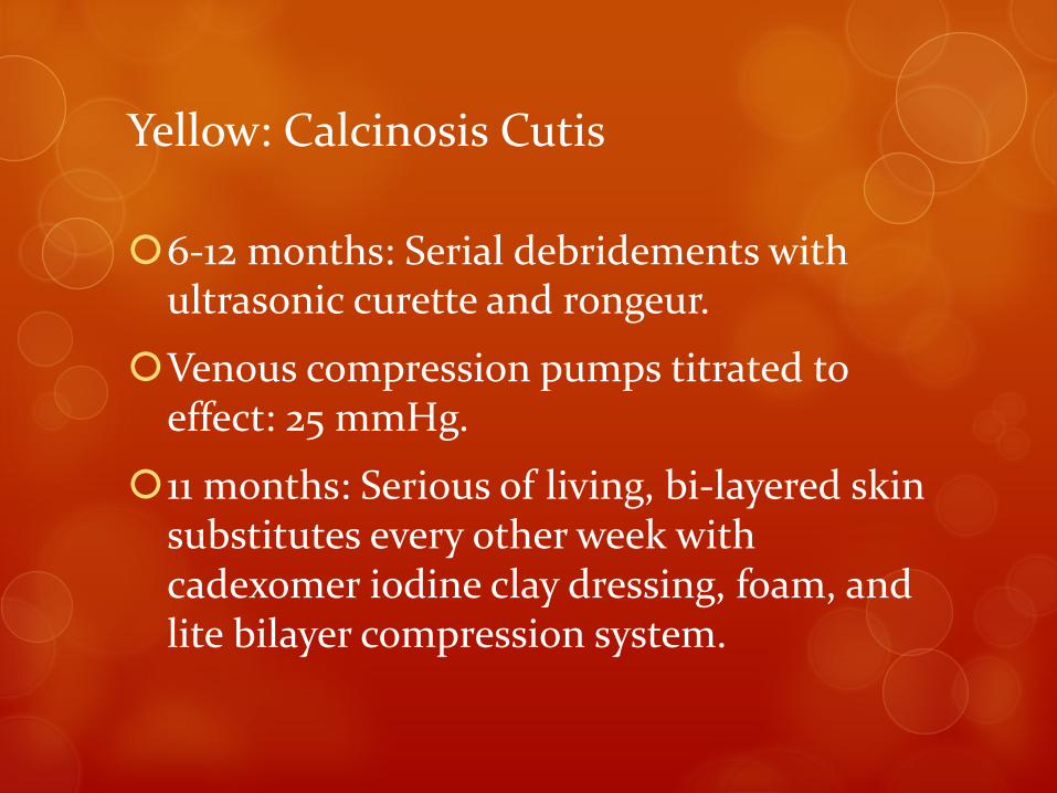
�6-12 months: Serial debridements with ultrasonic curette and rongeur.
�Venous compression pumps titrated to effect: 25 mmHg.
�11 months: Serious of living, bi-layered skin substitutes every other week with cadexomer iodine clay dressing, foam, and lite bilayer compression system.

Yellow: Calcinosis Cutis7 months

Yellow: Calcinosis Cutis8 & 9 months

Yellow: Calcinosis Cutis10 & 11 months

Yellow: Calcinosis Cutis12 & 15 months

Yellow: Calcinosis Cutis�Dystrophic calcinosis-subtype of CC that
effects elastic or collagen fibers�Serum calcium and phosphate levels are
normal�Linked to autoimmune connective tissue DO�Tx: excision for comfort, bisphosphonates,
calcium-channel blockers, warfarin, colchicine, probenecid, low calcium/phosphate diet with aluminum hydroxide.

References
�Fernandez, K.H. Calcinosis cutis: Management. UpToDate. Retrieved September 24, 2017.
�James, W.D., Berger, T.G, Elston, D.M., (2006). Andrews’ Diseases of the Skin: Clinical Dermatology, 10th Ed. Canada: Saunders Elsevier.

Green
� 56 y.o. male�Medical Hx: gout�No relevant
medications�6 month history of
left lateral ankle ulcer
� 5.4 x 6.6 x 0.5 cm� Tx: Biopsy, Cx, Chest x-ray

Green: Blastomycosis
� 1 month
� 3.1 x 5.9 x 0.3 cm
� No pulmonary symptoms
� No pain
� Care transferred to Infections Disease

Green: Blastomycosis
� Blastomyces dermatitides
� Dimorphic fungus
� KOH vs Cx
� Endemic to Ohio and Mississippi River basins and Great Lakes
� Highest annual incidence: central WI

Green: Blastomycosis
� Inoculation
� Testing
� Presentation
� Pulmonary vs Cutaneous
� Primary vs Secondary

References
� Blastomycosis Statistics. CDC Center for Disease Control. Retrieved on September 25, 2017. https://www.cdc.gov/fungal/diseases/blastomycosis/statistics.html.
� James, W.D., Berger, T.G, Elston, D.M., (2006). Andrews’ Diseases of the Skin: Clinical Dermatology, 10th Ed. Canada: Saunders Elsevier.
� Sources of Blastomycosis: CDC Center for Disease Control. Retrieved on September 25, 2017. https://www.cdc.gov/fungal/diseases/blastomycosis/causes.html.

Blue
� 60 y.o. male
� Medical Hx: non occlusive heart disease, hyperlipidemia
� 48 hours prior, 5000# pipes rolled up his legs
� No fractures or vascular injury/compartment syndrome

Blue
� Initial assessment: lower extremity edema
� Full thickness abrasions to the left thigh, lateral knee, left tibia, left medial knee, right tibia and right wrist
� Contusion of left thigh
� Tx: antibiotic ointment and non- adherant dressing, BID.

Blue: Crush
� 1 week: returned to work with no restrictions� worsening edema and pain, focal tenderness of
left knee� Dx: Crush injury with worsening edema=>hypoxia� Tx: HBOT next day x 10, elevation, off work,
continue topical wound care, knee US (small hematoma)
� Referral: Orthopedics re: Left knee pain=ACL/PCL instability, bilateral labral cuff tears and rotator injuries.

Blue: Crush
�3 months:
�Able to tolerate 8 hours of ambulation with 40 mmHg stocking
�Venous duplex shows no residual venous injury
�Case turned over to Orthopedics.

Blue: Crush
� 52 y.o. male
� Medical Hx: nicotine use
� No relevant medications
� 11 days prior, blunt force injury to right tibia
� No compartment syndrome, wound debrided.
� Patient left open to air with eschar, 11.5 x 0.9.

Blue: Crush
� Tx: Admitted, Plastics consult and OR debridement. NPWT applied.
� Declined STSG, weaned off nicotine.
� Plastics followed until closured by secondary intention.

Blue: Crush
�Hyperbaric Oxygen Therapy (HBOT) indicated for acute crush or de-gloving injuries
�Prevents reperfusion injury�Secondary edema => secondary ischemia
injury�Optimal w/in 5-6 hours or ASAP�Arterial vs Venous injuries

References
�Kindwall, E.P. & Niezgoda, J.A., (2006). Hyperbaric Medicine Procedures: The Kindwall HBO Handbook, 9th. Aurora St. Luke’s Medical Center: Milwaukee, WI.

Violet
�75 y.o. male�Medical Hx: gout, HTN, NIDDM,
hyperlipidemia�Medications: cephalexin, allopurinol,
metformin, amlodipine, atorvastatin, fosinopril, aspirin
�8 day history of increased BLE swelling and rash, rash converting to wounds; - DVT

Violet
� Initial assessment: ascending palpable purpura converting to hemorrhagic wounds, 4+ pitting edema.
� Tx: Absorbant foam dressings and Ace bandage compression

Violet: Vasculitis�Tx: biopsy and admission, 15 days in hospital
complicated by GI bleed and worsening renal function.
�Dermatology Consult: agreed with Vasculitis syndrome with renal and GI involvement.
�Nephrology, GI, Rheumatology consults.�ANA, C3/C4 normal. ANCA and cryoglobulins
negative. Renal biopsy showed IgA nephropathy.
�New incidental diagnosis of Diastolic CHF

Violet: Vasculitis
� Tx: IV methylprednisolone=> 80 mg prednisone daily.
� Continued absorbent foam/Ace bandage compression per home health RN.
� 3 weeks: single right tibia ulcer, 0.2 x 0.2 x 0.1 cm, 4+ pitting edema

Violet: Vasculitis
�Long term maintenance: orthotic compression wraps, diuretics, ACEI for renal protection, avoid nephro-toxic medications, and HTN control.
�Prednisone tapered off.
�Follows with Nephrology.

Violet: Vasculitis � IgA Vasculitis, Henoch-Schoenlein Purpura, sub-type
of small-vessel vasculitis characterized by arthralgias(74-84%), abdominal pain (61-76%), and renal disease (44-47%).
�Symptoms can proceed signs by 2 weeks�Abnormal immune response, etiology unclear�Biopsy reveals deposition of immunoglobulin A�Other tests exclude other forms of vasculitis and
differential diagnoses�General vasculitis is characterized with petechial rash,
palpable purpura converting to hemorrhagic wounds with disproportionate pain.

References
�Bryant, R.A. & Nix, D.P. (2012). Acute & Chronic Wounds: Current Management Concepts, 4th Ed. St. Louis, MO: Elsevier Mosby.
�James, W.D., Berger, T.G, Elston, D.M., (2006). Andrews’ Diseases of the Skin: Clinical Dermatology, 10th Ed. Canada: Saunders Elsevier.

Conclusion
� Thank you