Embed Size (px)
Citation preview

Received: 31st May-2012 Revised: 06th August -2012 Accepted: 10th August -2012
Review article
WOUND REPAIRMENT: IMMUNE MACHINERY OF AQUATIC INVERTEBRATE
Sanjib Saha
Bioscience and Environmental Studies Section, Pranta Palli High School, J-Block, Baghajatin Palli, Kolkata - 32, West Bengal, India
E- mail: [email protected] ABSTRACT: Aquatic invertebrates are small animals, such as poriferans, cnidarians, nematodes, annelids, insects, crustaceans, mollusks, and echinoderms that live in water. Aquatic invertebrates found in pools, lakes, springs and river. Some of them exist in highly saline waters, lagoons, hot springs. In the highly competitive such challenging environment, invertebrates experiences physical wounding due to inter and intraspecific struggle and struggle with environmental adversities. Animals physiologically respond to tissue repairment following external injury and repairment of wound and rapid sealing of the exoskeleton are required to prevent the loss of hemolymph and opportunistic invasion of pathogens. Tissue repairment involves coagulation of plasma proteins which are degranulated from adherent hemocytes and degranulated flattened cells get adhered over the damage area and form a membranous structure which traps other type of surrounding cells. Plasma gelation involves the activation of enzyme prophenoloxidase which in turn activates phenoloxidase which results in sequential activation of a cascade of biochemical reaction. Phenoloxidase cascade resembles the mammalian complement pathway and leads to killing of invading pathogens. Invertebrate wound repairing immune response mainly based on innate immune effector cells or hemocytes and hemolymph. Such immunocytes show diverse functional activities during wound repairment including aggregation, adhesion, coagulation, phagocytosis, degranulation, generation of cytotoxic agents and hemolymph consists of different proteins and enzymes in inactive forms to get active to protect the invertebrates invading pathogens and toxins of unknown chemical nature within hostile environment. Key words: Aquatic invertebrate, wound repairment, immune response. AQUATIC INVERTEBRATE There are over million described species of animals. of this number about 95% contain invertebrates (without backbone). Invertebrates do not hold single positive features in common, aside from general animal characteristics also shared with vertebrates. The range of size, in structural diversity and in adaptations to different modes of existence is enormous. Aquatic invertebrates are small animals, such as poriferans, cnidarians, nematodes, annelids, insects, crustaceans, mollusks, and echinoderms that live in water [8]. Aquatic invertebrates found in pools, lakes, springs and river. Some of them exist in highly saline waters, lagoons, hot springs. In lentic water (standing water) invertebrates occur at the bottoms of deep lakes, open water while in lotic water (flowing water) aquatic invertebrates under stones, woody debris, buried in sand and sediment. Aquatic invertebrates are a major source of food for birds, mammals, amphibians, reptiles, fish and other invertebrates in both aquatic and terrestrial habitats. E.g. female ducks take invertebrates during the breeding season and water fowl shift their winter to spring by invertebrates. Changes in a food source of such importance as aquatic invertebrates can have repercussions in many parts of the food web. The life cycles of aquatic invertebrates are intricately connected to land as well as water. Most invertebrates are found living in the stream bottom among the rocks and gravel. Many types of insects, such as mosquitoes, dragonflies, and mayflies, begin their life cycle in water, but they are perhaps more familiar and more frequently encountered as adults that fly. The stream bottom may appear to be just a bunch of rocks, but in a healthy stream there may be thousands of individual animals and 100 different species living in a single square meter of stream bottom. Aquatic invertebrates are also used to assess the health of water bodies like lakes, wetlands because different species have different tolerances to a variety of pollutants. Aquatic invertebrates are useful for water quality measurement or acts as bio-indicator–
International Journal of Plant, Animal and Environmental Sciences Page: 18 Available online at www.ijpaes.com

Sanjib Saha Copyrights@2012 IJPAES ISSN 2231-4490 • Aquatic invertebrates are relatively easy to collect and identify without a lot of specialized equipment. • Like fish, most aquatic invertebrates living in streams get oxygen directly from the water itself, not from the air.
When animals that require a good supply of dissolved oxygen are not found, it may indicate types of pollution that take oxygen out of the water.
• Aquatic invertebrates generally cannot move around very much. Unlike fish, these tiny bugs cannot swim away from pollution.
• Based on years of study, scientists know which species of invertebrates are sensitive to pollution. Some selected species of aquatic invertebrate taxa are described below –
Porifera [sponges], Cnidaria [hydras & jellyfish], Turbellaria [flatworms & planarians], Gastrotricha, Bryozoa (Ectoprocta), Entoprocta, Nematomorpha [horsehair worms], Nemertea, Tardigrada [water bears], Nematoda (free-living) [roundworms], Rotifera, Annelida, Polychaeta [bristleworms & sandworms], Branchiobdellida, Oligochaeta [aquatic earthworms], Hirudinea [leeches], Gastropoda [snails & limpets], Bivalvia [mussels & clams], Cladocera [water fleas], Copepoda, Ostracoda [seed shrimp], Amphipoda [scuds, sandfleas], Isopoda [isopods], Decapoda [crayfish, shrimp, crabs], Bathynellacea, Anostraca [fairy shrimp], Arguloida [fish lice], Conchostraca [Clam Shrimp], Mysida [opossum shrimp], Notostraca [tadpole shrimp], Thermosbaenacea, Acari [water mites], Insecta, Plecoptera [stoneflies], Ephemeroptera [mayflies], Odonata [damselflies & dragonflies], Collembola [springtails], Megaloptera [dobsonflies, fishflies, hellgrammites], Neuroptera [spongilla flies], Lepidoptera [aquatic moths], Orthoptera, Hymenoptera [wasps], Heteroptera, Coleoptera [beetles], Trichoptera [caddisflies], Diptera [mosquitoes & flies]. AQUATIC INVERTEBRATE IMMUNO MACHINERY The defense or immune system is commonly divided into two major categories named innate or nonspecific and acquired or specific. The innate immune system classified into humoral and cellular defense responses. This innate immunity is more ancient from of defense mechanisms than acquired immunity. The innate or nonspecific immune response depends mainly on recognition and killing of invading xenobiotics. Invertebrate innate immunity recognizes invading pathogens by pattern recognition receptors including Toll receptors, peptidoglycan recognition proteins and lectins which bind to microbial components [3]. However innate immunity is crutial for the first line of defense which include blood cells. Most of the aquatic invertebrates possess white blood cells or immunocytes that probably evolved from free living protozoa like ancestor [3]. The immunocytes are free within blood vessels or occupy fluid filled hemocoel. Invertebrate host defense mechanisms do not show an advance degree of specificity of innate immune system. Innate immunity consists of physico-chemical barrier to potential microorganisms are the cuticle and mucous layer. Rigid and wax covered cuticle serving as a mechanical barrier and they can also rapidly produce during infection. The materials in the cuticle are chitin which is the most abundant skeletal material in invertebrates and it is chemically similar to cellulose [16]. It is a polysaccharide that is synthesized by the animal, and it can be degraded by extracellular enzymes chitinases released by bacteria and fungi. Among the 3 forms of chitin, crustacean cuticle is α–chitin which is most stable due to large number of hydrogen bonds present and the cuticle consists of 4-layers: the external layer epicuticle, exocuticle, endocuticle-I, last layer endocuticle-II [5]. When the cuticle is damaged due to injury or infection, the wound is rapidly clotted and this prevents loss of hemolymph. Once a clot is formed, the area is melanized and wound appears dark black. Melanin in addition to sealing the wound and epidermis will form a new cuticle beneath the melanized layer. Apart from these, other important innate defense is hemolymph and hemocytes (immunocytes) which are activated upon discrete immunological challenges.
Hemocytes are the circulating immunoeffector blood cells of the aquatic invertebrates, which perform diverse immunological activities including aggregation, coagulation, adhesion, phagocytosis, generation of cytotoxic molecules, antioxidant enzymes and wound repairment [4, 12, 13, 14, 15]. Basic scheme of classification of hemocytes according to function is 5 types – progenitor, phagocytic, hematopoietic, nutritive and pigmented [3]. • Progenitor cell = acts similar as hematopoietic or stem cell that production of other types of blood cells. • Phagocytic cell = helps in phagocytosis of invading pathogens or xenobiotics, wound repairing and encapsulation.
Phagocytosis is essential for host defence against infection and clearance of apoptotic cells generated during development.
International Journal of Plant, Animal and Environmental Sciences Page: 19 Available online at www.ijpaes.com

Sanjib Saha Copyrights@2012 IJPAES ISSN 2231-4490 • Hematopoietic cell = these granular cells are involved coagulation, wound repairing, adhesion etc. • Nutritive cell = this cell help in nutrition. But proper function still now unknown. • Pigmented cell = cell present in many species but in a few species they contain respiratory pigment. They have also
role in defence. In general function, cytochemistry and ultrastructure of hemocytes suggest there are three major type blood
cells present in the invertebrate: hyalinocyte, semigranulocyte and granulocyte [9, 17, 18]. Hyalinocytes are small, spherical and no or few granules and capable of phagocytosis. Semigranulocytes are generally oval which contain small granules and capable of encapsulation, phagocytosis and cytotoxic response. Granulocytes are round in shape and contain huge no of large eosinophilic granules that responsible for repairment of wound and cytotoxicity. Insect produce several types of hemocytes that are prohemocyte, granulocyte, plasmocytes, spherulocyte and oenocytoid [3]. Generally granulocyte and plasmocyte are adhering cells to foreign surface. Spherulocyte transports cuticular components while oenocytoids contain cytoplasmic phenoloxidase precursors for melanization. Prohemocyte acts as stem cell that differentiates into different hemocyte types.
In Mollusca contain different types of hemocytes i.e. asterocyte, prohemocyte, hyalinocyte, granulocyte, lymphoblast, agranulocyte, hyalinocyte [4]. Invertebrates rapidly seal wounds caused by injury or pathogens invasion and prevent loss of excess hemolymph through the process of muscular contraction, coagulation of fluid, melanization and formation of new tissue [19]. Nodulation involve to multiple hemocytes (plasmocytes) binding to aggregations of bacteria where as encapsulation refers to binding of hemocytes (plasmocytes) to relatively large particles. Crystal cells play an important role in clotting or coagulation during repairment. Epidermal tissue and hemolymph play a vital role in melanization. Mollusca hemocytes involve in elicitation of immunological response under the challenge of foreign invaders and exhibit diverse physiological functions like phagocytosis, aggregation, generation of cytotoxic molecules etc. Insects also continue to produce hemocytes during larval or nymph stages via division of stem cells in mesodermally derived hematopoietic organs. Echinoderm cellular responses are mediated by coelomic celomocytes that exert immunological functions including phagocytosis, clotting, encapsulation, antibacterial activity. Invertebrates’ body fluids contain a range of humoral factors. These factors are agglutinins, lysozyme, lysins, non-lysozyme bactericidins, hemolin etc [2]. The humoral immune response of insect is based on the products of characterized immune genes induced by microbial infection and encodes antimicrobial peptides, which are synthesized predominantly in fat body and released into hemolymph and Over 150 antimicrobial peptides have been isolated and characterized in insects [2]. In general antimicrobial peptides have four groups - cecropins, cysteine-rich peptides, proline-rich peptides, and glycine-rich peptides. Integrins surface proteins present in sponges to humans that participate in adhesion, migration, tissue organization and recognize and bind protein motifs in specific cell-surface or extracellular matrix or soluble collagen, laminins, fibronectins. Invertebrate integrins are primary molecules for the recognition of foreign agents and the initiation of immune response. Lectin plays an important role in immune recognition and adhesion in invertebrate that helps to distinguish self from nonself surface. They are characterized by a wide range of binding activities. Adhesion to foreign surface occurs through pattern recognition proteins (receptors) that recognize and bind conserved domains or patterns located on the pathogen surface, which are called pathogen-associated molecular patterns. Humoral immune responses include activation of enzymatic cascades that regulate coagulation and melanization of hemolymph, and production of reactive oxygen and nitrogen species (ROS-RNS). Absences of specific immune cells in Cnidaria have effective epithelial cells to defend against microbial recognition through pattern recognition receptor (toll and nod like receptors). Cnidarian immune system has capability to distinguish between beneficial and pathogenic microorganisms and such system plays a vital role in controlling mutualistic symbionts or pathogens. Cnidarian innate immunity produces huge amounts of antimicrobacterial peptides along with further potential bactericidal molecule against environmental bacterial contamination. Toll-like receptor pathway, complement C3, several membrane attack complex, perforin domain (MAC/PF) proteins and interleukin are present in Cnidarians. Innate and humoral immunity of annelid plays a vital role in defence mechanism against microorganisms or pathogens. Innate immune responses include pattern recognition receptors, phagocytosis, encapsulation, cytotoxicity, clotting through blood cells or celomocytes. Various types of free-floating cells are found in the coelomic fluid of representatives of several annelid groups.
International Journal of Plant, Animal and Environmental Sciences Page: 20 Available online at www.ijpaes.com

Sanjib Saha Copyrights@2012 IJPAES ISSN 2231-4490 Humoral immunity based on antimicrobial peptides, cytokines and hemolytic components. Insect immune response depends on both humoral and cellular responses that are mediated via certain recognizing receptors (toll receptor) and activation of several signaling pathways. Fat body and hemocytes are the main source of the synthesis and secretion of antimicrobial peptides and regulator agents of cellular responses, while cell mediated immunity is performed by hemocytes. Hemocytes have numerous functional activities - hemolymph clotting after cuticle rupture, wound repairing, self and nonself recognition, release of cytotoxic agents. Hemocytes also synthesis and store antibacterial peptides, serine protease, prophenoloxidase substances which may be release after lectins, hemolysins, infections and environmental contaminants. The defense mechanisms of crustaceans rely on the innate immune system that is activated when pathogen-associated molecular patterns are recognized by soluble or by cell surface host proteins (lectins, antimicrobial, clotting, and pattern recognition proteins). Innate immunity activates cellular or humoral effector mechanisms to destroy invading pathogens. Discrimination of self and nonself surface adhesion, Phagocytosis, degranulation of chemical agents by hemocytes is regarded as classical immunological response in invertebrates. Hemocyte deficiency during infection in invertebrate refers a serious threat to the health status and a rapid supply of fresh hemocytes is essential in order to destroy invasive micro-organisms instantly and combat infection of invading microbes. Phagocytosis through hemocyte is a classical immune response of invertebrates. Phagocytes capable of generating cytotoxic agent like reactive oxygen intermediates (superoxide anion, hydrogen peroxide, phenoloxidase) and reactive nitrogen intermediates (nitric oxide) which are toxic to pathogens. Three major enzymes act jointly for the destruction of the reactive oxygen intermediates in hemocyte i.e. SOD, GST, catalase. A variety of cytokines are reported in the hemocytes of invertebrates. Cell types isolated from the hemolymph of invertebrates are positive for cytokines like interleukin (IL 1α, IL 1β, IL 2, IL 6 and TNFα). Presence of IL 2 in the hemocyte expressed phagocytic activity is of evolutionary significance. Presence of IL 8 in mollusks expressed chemotaxis, changes in cell shape, bacterial clearance activity. Humoral factors reported in the hemolymph of invertebrate including agglutinins, lysozymes, bactericidins etc. Nodulation in invertebrate is the process of hemocytic aggregates that encircle a large number of microorganisms and melanized or non-melanized nodules are formed. Nodule formation appears to be related with eicosanoids in many insect species or prophenoloxidase and dopa decarboxylase in medfly hemocytes. Generally two types of immune responses are available in echinoderms. They comprise cellular and humoral. Echinoderms can recognize nonself surface of pathogens and readily neutralize it. Cell mediated immunity is carried out by circulatory free blood cells or celomocytes that perform diverse activities (chemotaxis, phagocytosis, encapsulation, clotting, cytotoxicity, agglutination, wound repairing etc.). Basic types of blood cells are discoidal, polygonal, small phagocyte, amebocytes, spherule, lymphocyte, crystal and hemocyte. On the other hand, humoral immunity is mediated by several compounds like lectins, hemolysins, agglutinins, perforins, cytokines and complement.
Figure 1: Hematopoietic activation mechanism in arthropods.
International Journal of Plant, Animal and Environmental Sciences Page: 21 Available online at www.ijpaes.com

Sanjib Saha Copyrights@2012 IJPAES ISSN 2231-4490 Hematopoiesis is vital process for invertebrate animals. In vertebrate hematopoietic cellular immune response consists of the lymphoid and myeloid lineages. The lymphoid lineage is the main component of acquired immune response. In invertebrate there is no lymphoid lineage and cellular immune response consists of circulating immunocytes or hemocytes [18]. Hemocytes play a key role in immune surveillance and are active against pathogen and environmental xenobiotics [20]. When an invading particulate (living or nonliving) is recognized as nonself or foreign part, circulating hemocytes should release particulate through the process of phagocytosis or encapsulation and generation of cytotoxic molecules. Therefore a continuous supply of mature new hemocyte needs to be produced and released from hematopoietic tissue (stem cell) against infection or damaged part of the animal for sealing and healing processes. Invertebrate cytokine present within blood that is involve in hematopoiesis. Such cytokine has structural similarities with vertebrate protein kinase. Identification of interleukin (IL) and tumor necrosis factor in invertebrates indicated the possible presence of other form of cytokines of phagocytic and T-cell origin in these animals. Activation of immunocytes of mollusks by IL 1 and TNF is suggestive to the possible existence of intricate cell communicative and proliferative response in invertebrate. Prohemocytes are believed to be the stem cells (hematopoietic cells) in arthropods from which fresh and mature hemocytes involved proliferation, commitment and differentiation. The hematopoietic tissue comprises small lobules that surrounded by thin collegenous tissue sheath and close contact with blood sinuses. The number of hemocytes in the circulating system is regulated by hematopoiesis in hematopoietic tissue or by storage at other sites [19]. Insects can produce hemocytes during larval stages derived from stem cells (Hpt). Fruit fly and vertebrates share several common important hematopoietic factors. During embryogenesis, the only hemocytes present are known as macrophages or plasmocytes. In larvae, plasmocytes remain the most abundant hemocyte but lamelocytes and crystal cells are also present. Several signaling pathways associated with hematopoiesis in fruit fly larvae. Insects produce immunocytes in nymph phase through cell division from hematopoietic organs and release hemocytes into circulation. Prohemocytes of hematopoietic organs transfer into plasmocytes which differentiate into granulocytes, spherulocytes and oenocytoids. It is reported overactivation of Ras/Raf pathway and epidermal growth factor lead to uncontrolled overproliferation of hemocytes. In crustacean different types of hematopoietic tissue cells are morphologically detected and locate on collegenous sheath of heart or stomach. Generally there are five types of hematopoietic tissue cell, from which granular and semi granular cells are differentiated and release to the circulation in Crustacea. In invertebrates several signaling pathways associated with hematopoiesis activation. For activation different hematopoietic transcription factors and mechanisms require which include GATA, RUNX, NF-kB, JAK/STAT, Ras/Ras etc. overactivation of Ras or Raf pathway leads to uncontrolled proliferation of hemocytes. Arthropoda genome encodes five GATA and three RUNX transcription factors and GATA expressed before all differentiation markers in embryonic prohemocytes while RUNX is involved in hematopoiesis along with function of RUNX. In absence of both GATA and RUNX transcription factors plasmocyte differentiate normally. It is thought that platelet derived growth factor and epidermal growth factor act as cytokine like activity while active Ras signal upstream induce the over proliferation process where as downstream of Ras/Raf/MAPK pathway are involved in hemocyte specification. During hematopoiesis the GATA factor Serpent is required for the specification of all hemocytes and RUNX factor Lozenge are required for the specification of plasmocytes and crystal cells respectively. The Toll-NF-kB and JAK/STAT pathways, both play a role in hemocyte proliferation and likely in lamellocytes specification.
Toll receptor is membrane glycoproteins consisting of an ectodomain, leucine rich repeats and a globular cytoplasmic domain. Such receptor detects microbes on the basis of conserved Pathogen Associated Molecular Pattern. Toll receptors share molecular structure and common ancestors with arthropod Toll molecules [1]. Toll pathway activated mainly by fungi and Gram-positive bacteria and the immune deficiency pathway. Activation of the Toll receptor by its ligend leads to the formation of a multimeric receptor-adaptor complex which comprises three death-domain proteins: MyD88, Tube, and the kinase Pelle. The assembly of these proteins into a complex induces phosphorylation of Ik-B-like inhibitor Cactus Phosphorylated cactus is degraded by proteasomes and dissociates from Rel transcription factor dorsal-related immunity-factor which is then free to translocate into the nucleus and activate numerous antimicrobial genes of Drosophila.
International Journal of Plant, Animal and Environmental Sciences Page: 22 Available online at www.ijpaes.com
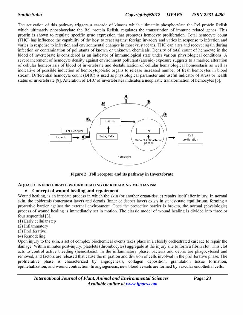
Sanjib Saha Copyrights@2012 IJPAES ISSN 2231-4490 The activation of this pathway triggers a cascade of kinases which ultimately phosphorylate the Rel protein Relish which ultimately phosphorylate the Rel protein Relish, regulates the transcription of immune related genes. This protein is shown to regulate specific gene expression that promotes hemocyte proliferation. Total hemocyte count (THC) has influence the capability of the host to react against foreign invaders and varies in response to infection and varies in response to infection and environmental changes in most crustaceans. THC can alter and recover again during infection or contamination of pollutants of known or unknown chemicals. Density of total count of hemocyte in the blood of invertebrate is considered as an indicator of immunological state under various physiological conditions. A severe increment of hemocyte density against environment pollutant (arsenic) exposure suggests to a marked alteration of cellular homeostasis of blood of invertebrate and destabilization of cellular hematological homeostasis as well as indicative of possible induction of hemocytopoietic organs to release increased number of fresh hemocytes in blood stream. Differential hemocyte count (DHC) is used as physiological parameter and useful indicator of stress or health status of invertebrate [8]. Alteration of DHC of invertebrates indicates a neoplastic transformation of hemocytes [5].
Figure 2: Toll receptor and its pathway in Invertebrate.
AQUATIC INVERTEBRATE WOUND HEALING OR REPAIRING MECHANISM • Concept of wound healing and repairment
Wound healing, is an intricate process in which the skin (or another organ-tissue) repairs itself after injury. In normal skin, the epidermis (outermost layer) and dermis (inner or deeper layer) exists in steady-state equilibrium, forming a protective barrier against the external environment. Once the protective barrier is broken, the normal (physiologic) process of wound healing is immediately set in motion. The classic model of wound healing is divided into three or four sequential [3]. (1) Early cellular step (2) Inflammatory (3) Proliferative (4) Remodeling Upon injury to the skin, a set of complex biochemical events takes place in a closely orchestrated cascade to repair the damage. Within minutes post-injury, platelets (thrombocytes) aggregate at the injury site to form a fibrin clot. This clot acts to control active bleeding (hemostasis). In the inflammatory phase, bacteria and debris are phagocytosed and removed, and factors are released that cause the migration and division of cells involved in the proliferative phase. The proliferative phase is characterized by angiogenesis, collagen deposition, granulation tissue formation, epithelialization, and wound contraction. In angiogenesis, new blood vessels are formed by vascular endothelial cells.
International Journal of Plant, Animal and Environmental Sciences Page: 23 Available online at www.ijpaes.com

Sanjib Saha Copyrights@2012 IJPAES ISSN 2231-4490 In fibroplasia and granulation tissue formation, fibroblasts grow and form a new, provisional extracellular matrix by excreting collagen and fibronectin. Concurrently, re-epithelialization of the epidermis occurs, in which epithelial cells proliferate and 'crawl' atop the wound bed, providing cover for the new tissue. In contraction, the wound is made smaller by the action of myofibroblasts, which establish a grip on the wound edges and contract themselves using a mechanism similar to that in smooth muscle cells. When the cells' roles are close to complete, unneeded cells undergo apoptosis. In the maturation and remodeling phase, collagen is remodeled and realigned along tension lines and cells that are no longer needed are removed by apoptosis. However, this process is not only complex but fragile, and susceptible to interruption or failure leading to the formation of non-healing chronic wounds. Factors which may contribute to this include diabetes, venous or arterial disease, old age, and infection. Wound healing divided into Early phase, Inflammatory phase, Proliferative phase and remodeling phase. 1. Early cellular phase = wound healing is classically divided into hemostasis, inflammation, proliferation, and
remodeling. Although a useful construct, this model employs considerable overlapping among individual phases. Recently, a complementary model has been described, such that the many elements of wound healing are more-clearly delineated. The importance of this new model becomes more apparent through its utility in the fields of regenerative medicine and tissue engineering. In this construct, the process of wound healing is divided into major two phases: early phase and cellular phase: The early phase, which begins immediately following skin injury, involves cascading molecular and cellular events leading to hemostasis and formation of an early, makeshift extracellular matrix—providing structural support for cellular attachment and subsequent cellular proliferation. The cellular phase follows the early phase, and involves several types of cells working together to mount an inflammatory response, synthesize granulation tissue, and restore the epithelial layer. Subdivisions of the cellular phase are: Macrophages and inflammatory components (within 1–2 days), Epithelial-mesenchymal interaction: re-epithelialization (phenotype change within hours, migration begins on day 1 or 2), Fibroblasts and myofibroblasts: progressive alignment, collagen production, and matrix contraction (between day 4 day 14), Endothelial cells and angiogenesis (begins on day 4), Dermal matrix: elements of fabrication (begins on day 4, lasting 2 weeks) and alteration/remodeling (begins after week 2, lasting weeks to months—depending on wound size.).
2. Inflammatory phase = Just before the inflammatory phase is initiated, the clotting cascade takes place in order to obtain hemostasis, or stop blood loss by way of a fibrin clot. Thereafter, various soluble factors (including chemokines and cytokines) are released to attract cells that phagocytise debris, bacteria, and damaged tissue, in addition to releasing signaling molecules that initiate the proliferative phase of wound healing.
a) Clotting or coagulation cascade: When tissue is first wounded, blood comes in contact with collagen, triggering blood platelets to begin secreting inflammatory factors. Platelets also express glycoproteins on their cell membranes that allow them to stick to one another and to aggregate, forming a mass. Fibrin and fibronectin cross-link together and form a plug that traps proteins and particles and prevents further blood loss. This fibrin-fibronectin plug is also the main structural support for the wound until collagen is deposited. Migratory cells use this plug as a matrix to crawl across, and platelets adhere to it and secrete factors. The clot is eventually lysed and replaced with granulation tissue and then later with collagen. Platelets, the cells present in the highest numbers shortly after a wound occurs, release a number of things into the blood, including ECM proteins and cytokines, including growth factors. Growth factors stimulate cells to speed their rate of division. Platelets also release other proinflammatory factors like serotonin, bradykinin, prostaglandins, prostacyclins, thromboxane, and histamine which serve a number of purposes, including to increase cell proliferation and migration to the area and to cause blood vessels to become dilated and porous.
b) Vasoconstriction and vasodilation: Immediately after a blood vessel is breached, ruptured cell membranes release inflammatory factors like thromboxanes and prostaglandins that cause the vessel to spasm to prevent blood loss and to collect inflammatory cells and factors in the area. This vasoconstriction lasts five to ten minutes and is followed by vasodilation, a widening of blood vessels, which peaks at about 20 minutes post-wounding. Vasodilation is the result of factors released by platelets and other cells. The main factor involved in causing vasodilation is histamine. Histamine also causes blood vessels to become porous, allowing the tissue to become edematous because proteins from the bloodstream leak into the extravascular space, which increases its osmolar load and draws water into the area. Increased porosity of blood vessels also facilitates the entry of inflammatory cells like leukocytes into the wound site from the bloodstream.
International Journal of Plant, Animal and Environmental Sciences Page: 24 Available online at www.ijpaes.com

Sanjib Saha Copyrights@2012 IJPAES ISSN 2231-4490 c) Polymorphonuclear neutrophils: Within an hour of wounding, polymorphonuclear neutrophils arrive at the wound
site and become the predominant cells in the wound for the first two days after the injury occurs, with especially high numbers on the second day. They are attracted to the site by fibronectin, growth factors, and substances such as kinins. Neutrophils phagocytise debris and bacteria and also kill bacteria by releasing free radicals in what is called a 'respiratory burst'. They also cleanse the wound by secreting proteases that break down damaged tissue. Neutrophils usually undergo apoptosis once they have completed their tasks and are engulfed and degraded by macrophages. Other leukocytes to enter the area include helper T cells, which secrete cytokines to cause more T cells to divide and to increase inflammation and enhance vasodilation and vessel permeability. Cells also increase the activity of macrophages.
d) Macrophages: Macrophages are essential for wound healing. They replace polymorphonuclear neutrophils as the predominant cells in the wound by two days after injury. Attracted to the wound site by growth factors released by platelets and other cells, monocytes from the bloodstream enter the area through blood vessel walls. Numbers of monocytes in the wound peak one to one and a half days after the injury occurs. Once they are in the wound site, monocytes mature into macrophages. The spleen contains half the body's monocytes in reserve ready to be deployed to injured tissue. The macrophage's main role is to phagocytize bacteria and damaged tissue and they also debride damaged tissue by releasing proteases. Macrophages also secrete a number of factors such as growth factors and other cytokines, especially during the third and fourth post-wounding days. These factors attract cells involved in the proliferation stage of healing to the area although they may restrain the contraction phase. Macrophages are stimulated by the low oxygen content of their surroundings to produce factors that induce and speed angiogenesis and they also stimulate cells that reepithelialize the wound, create granulation tissue, and lay down a new extracellular matrix. By secreting these factors, macrophages contribute to pushing the wound healing process into the next phase.
e) Decline of inflammatory phase: As inflammation dies down, fewer inflammatory factors are secreted, existing ones are broken down, and numbers of neutrophils and macrophages are reduced at the wound site. These changes indicate that the inflammatory phase is ending and the proliferative phase is underway. In vitro evidence, obtained using the dermal equivalent model, suggests that the presence of macrophages actually delays wound contraction and thus the disappearance of macrophages from the wound may be essential for subsequent phases to occur. Because inflammation plays roles in fighting infection, clearing debris and inducing the proliferation phase, it is a necessary part of healing. However, inflammation can lead to tissue damage if it lasts too long. Thus the reduction of inflammation is frequently a goal in therapeutic settings. Inflammation lasts as long as there is debris in the wound. Thus the presence of dirt or other objects can extend the inflammatory phase for too long, leading to a chronic wound.
3. Proliferative phase = About two or three days after the wound occurs, fibroblasts begin to enter the wound site, marking the onset of the proliferative phase even before the inflammatory phase has ended. As in the other phases of wound healing, steps in the proliferative phase do not occur in a series but rather partially overlap in time.
a) Angiogenesis: Also called neovascularization, the process of angiogenesis occurs concurrently with fibroblast proliferation when endothelial cells migrate to the area of the wound. Because the activity of fibroblasts and epithelial cells requires oxygen and nutrients, angiogenesis is imperative for other stages in wound healing, like epidermal and fibroblast migration. The tissue in which angiogenesis has occurred typically looks red (is erythematous) due to the presence of capillaries. Stem cells of endothelial cells, originating from parts of uninjured blood vessels, develop pseudopodia and push through the ECM into the wound site to establish new blood vessels. Endothelial cells are attracted to the wound area by fibronectin found on the fibrin scab and chemotactically by angiogenic factors released by other cells e.g. from macrophages and platelets when in a low-oxygen environment. Endothelial growth and proliferation is also directly stimulated by hypoxia, and presence of lactic acid in the wound. To migrate, endothelial cells need collagenases and plasminogen activator to degrade the clot and part of the ECM. Zinc-dependent metalloproteinases digest basement membrane and ECM to allow cell migration, proliferation and angiogenesis. When macrophages and other growth factor-producing cells are no longer in a hypoxic, lactic acid-filled environment, they stop producing angiogenic factors. Thus, when tissue is adequately perfused, migration and proliferation of endothelial cells is reduced. Eventually blood vessels that are no longer needed die by apoptosis.
International Journal of Plant, Animal and Environmental Sciences Page: 25 Available online at www.ijpaes.com

Sanjib Saha Copyrights@2012 IJPAES ISSN 2231-4490
b) Fibroplasia and granulation tissue formation: Simultaneously with angiogenesis, fibroblasts begin accumulating in the wound site. Fibroblasts begin entering the wound site two to five days after wounding as the inflammatory phase is ending, and their numbers peak at one to two weeks post-wounding. By the end of the first week, fibroblasts are the main cells in the wound Fibroplasia ends two to four weeks after wounding. In the first two or three days after injury, fibroblasts mainly migrate and proliferate, while later, they are the main cells that lay down the collagen matrix in the wound site. Origins of these fibroblasts are thought to be from the adjacent uninjured cutaneous tissue (although new evidence suggests that some are derived from blood-borne, circulating adult stem cells/precursors). Initially fibroblasts utilize the fibrin cross-linking fibers (well-formed by the end of the inflammatory phase) to migrate across the wound, subsequently adhering to fibronectin. Fibroblasts then deposit ground substance into the wound bed, and later collagen, which they can adhere to for migration. Granulation tissue functions as rudimentary tissue, and begins to appear in the wound already during the inflammatory phase, two to five days post wounding, and continues growing until the wound bed is covered. Granulation tissue consists of new blood vessels, fibroblasts, inflammatory cells, endothelial cells, myofibroblasts, and the components of a new, provisional extracellular matrix. The provisional ECM is different in composition from the ECM in normal tissue and its components originate from fibroblasts. Such components include fibronectin, collagen, glycosaminoglycans, elastin, glycoproteins and proteoglycans. Its main components are fibronectin and hyaluronan, which create a very hydrated matrix and facilitate cell migration. Later this provisional matrix is replaced with an ECM that more closely resembles that found in non-injured tissue. Growth factors (PDGF, TGF-β) and fibronectin encourage proliferation, migration to the wound bed, and production of ECM molecules by fibroblasts. Fibroblasts also secrete growth factors that attract epithelial cells to the wound site. Hypoxia also contributes to fibroblast proliferation and excretion of growth factors, though too little oxygen will inhibit their growth and deposition of ECM components, and can lead to excessive, fibrotic scarring.
c) Collagen deposition: One of fibroblasts' most important duties is the production of collagen. Collagen deposition is important because it increases the strength of the wound; before it is laid down, the only thing holding the wound closed is the fibrin-fibronectin clot, which does not provide much resistance to traumatic injury. Also, cells involved in inflammation, angiogenesis, and connective tissue construction attach to, grow and differentiate on the collagen matrix laid down by fibroblasts. Type III collagen and fibronectin are generally beginning to be produced in appreciable amounts at somewhere between approximately 10 hours and 3 days depending mainly on wound size. Their deposition peaks at one to three weeks. They are the predominating tensile substances until the later phase of maturation, in which they are replaced by the stronger type I collagen. Even as fibroblasts are producing new collagen, collagenases and other factors degrade it. Shortly after wounding, synthesis exceeds degradation so collagen levels in the wound rise, but later production and degradation become equal so there is no net collagen gain. This homeostasis signals the onset of the later maturation phase. Granulation gradually ceases and fibroblasts decrease in number in the wound once their work is done. At the end of the granulation phase, fibroblasts begin to commit apoptosis, converting granulation tissue from an environment rich in cells to one that consists mainly of collagen.
d) Epithelialization: The formation of granulation tissue in an open wound allows the reepithelialization phase to take place, as epithelial cells migrate across the new tissue to form a barrier between the wound and the environment. Basal keratinocytes from the wound edges and dermal appendages such as hair follicles, sweat glands and sebacious (oil) glands are the main cells responsible for the epithelialization phase of wound healing. They advance in a sheet across the wound site and proliferate at its edges, ceasing movement when they meet in the middle. Keratinocytes migrate without first proliferating. Migration can begin as early as a few hours after wounding. However, epithelial cells require viable tissue to migrate across, so if the wound is deep it must first be filled with granulation tissue. Thus the time of onset of migration is variable and may occur about one day after wounding. Cells on the wound margins proliferate on the second and third day post-wounding in order to provide more cells for migration. If the basement membrane is not breached, epithelial cells are replaced within three days by division and upward migration of cells in the stratum basale in the same fashion that occurs in uninjured skin. However, if the basement membrane is ruined at the wound site, reepithelization must occur from the wound margins and from skin appendages such as hair follicles and sweat and oil glands that enter the dermis that are lined with viable keratinocytes. If the wound is very deep, skin appendages may also be ruined and migration can only occur from wound edges.
International Journal of Plant, Animal and Environmental Sciences Page: 26 Available online at www.ijpaes.com

Sanjib Saha Copyrights@2012 IJPAES ISSN 2231-4490
Migration of keratinocytes over the wound site is stimulated by lack of contact inhibition and by chemicals such as nitric oxide. Before they begin to migrate, cells must dissolve their desmosomes and hemidesmosomes, which normally anchor the cells by intermediate filaments in their cytoskeleton to other cells and to the ECM. Transmembrane receptor proteins called integrins, which are made of glycoproteins and normally anchor the cell to the basement membrane by its cytoskeleton, are released from the cell's intermediate filaments and relocate to actin filaments to serve as attachments to the ECM for pseudopodia during migration. Thus keratinocytes detach from the basement membrane and are able to enter the wound bed. Before they begin migrating, keratinocytes change shape, becoming longer and flatter and extending cellular processes like lamellipodia and wide processes that look like ruffles. Actin filaments and pseudopodia form. During migration, integrins on the pseudopod attach to the ECM, and the actin filaments in the projection pull the cell along. The interaction with molecules in the ECM through integrins further promotes the formation of actin filaments, lamellipodia, and filopodia. Epithelial cells climb over one another in order to migrate. This growing sheet of epithelial cells is often called the epithelial tongue. The first cells to attach to the basement membrane form the stratum basale. These basal cells continue to migrate across the wound bed, and epithelial cells above them slide along as well. The more quickly this migration occurs, the less of a scar there will be. Fibrin, collagen, and fibronectin in the ECM may further signal cells to divide and migrate. Like fibroblasts, migrating keratinocytes use the fibronectin cross-linked with fibrin that was deposited in inflammation as an attachment site to crawl across. As keratinocytes migrate, they move over granulation tissue but underneath the scab (if one was formed), separating it from the underlying tissue. Epithelial cells have the ability to phagocytize debris such as dead tissue and bacterial matter that would otherwise obstruct their path. Because they must dissolve any scab that forms, keratinocyte migration is best enhanced by a moist environment, since a dry one leads to formation of a bigger, tougher scab. To make their way along the tissue, keratinocytes must dissolve the clot, debris, and parts of the ECM in order to get through. They secrete plasminogen activator, which activates plasminogen, turning it into plasmin to dissolve the scab. Cells can only migrate over living tissue, so they must excrete collagenases and proteases like matrix metalloproteinases to dissolve damaged parts of the ECM in their way, particularly at the front of the migrating sheet. Keratinocytes also dissolve the basement membrane, using instead the new ECM laid down by fibroblasts to crawl across. As keratinocytes continue migrating, new epithelial cells must be formed at the wound edges to replace them and to provide more cells for the advancing sheet. Proliferation behind migrating keratinocytes normally begins a few days after wounding and occurs at a rate that is 17 times higher in this stage of epithelialization than in normal tissues. Until the entire wound area is resurfaced, the only epithelial cells to proliferate are at the wound edges. Growth factors, stimulated by integrins and polymorphonuclear neutrophils, cause cells to proliferate at the wound edges. Keratinocytes themselves also produce and secrete factors, including growth factors and basement membrane proteins, which aid both in epithelialization and in other phases of healing. Growth factors are also important for the innate immune defense of skin wounds by stimulation of the production of antimicrobial peptides and neutrophil chemotactic cytokines in keratinocytes. Keratinocytes continue migrating across the wound bed until cells from either side meet in the middle, at which point contact inhibition causes them to stop migrating. When they have finished migrating, the keratinocytes secrete the proteins that form the new basement membrane. Cells reverse the morphological changes they underwent in order to begin migrating; they reestablish desmosomes and hemidesmosomes and become anchored once again to the basement membrane. Basal cells begin to divide and differentiate in the same manner as they do in normal skin to reestablish the strata found in reepithelialized skin.
e) Contraction: Contraction is a key phase of wound healing. If contraction continues for too long, it can lead to disfigurement and loss of function. Thus there is a great interest in understanding the biology of wound contraction, which can be modelled in vitro using the collagen gel contraction assay or the dermal equivalent model. Contraction commences approximately a week after wounding, when fibroblasts have differentiated into myofibroblasts. In full thickness wounds, contraction peaks at 5 to 15 days post wounding. Contraction can last for several weeks and continues even after the wound is completely reepithelialized. A large wound can become 40 to 80% smaller after contraction. Wounds can contract at a speed of up to 0.75 mm per day, depending on how loose the tissue in the wounded area is.
International Journal of Plant, Animal and Environmental Sciences Page: 27 Available online at www.ijpaes.com

Sanjib Saha Copyrights@2012 IJPAES ISSN 2231-4490
f) Contraction usually does not occur symmetrically; rather most wounds have an 'axis of contraction' which allows for greater organization and alignment of cells with collagen. At first, contraction occurs without myofibroblast involvement. Later, fibroblasts, stimulated by growth factors, differentiate into myofibroblasts. Myofibroblasts, which are similar to smooth muscle cells, are responsible for contraction. Myofibroblasts contain the same kind of actin as that found in smooth muscle cells. Myofibroblasts are attracted by fibronectin and growth factors and they move along fibronectin linked to fibrin in the provisional ECM in order to reach the wound edges. They form connections to the ECM at the wound edges, and they attach to each other and to the wound edges by desmosomes. Also, at an adhesion called the fibronexus, actin in the myofibroblast is linked across the cell membrane to molecules in the extracellular matrix like fibronectin and collagen. Myofibroblasts have many such adhesions, which allow them to pull the ECM when they contract, reducing the wound size. In this part of contraction, closure occurs more quickly than in the first, myofibroblast-independent part. As the actin in myofibroblasts contracts, the wound edges are pulled together. Fibroblasts lay down collagen to reinforce the wound as myofibroblasts contract. The contraction stage in proliferation ends as myofibroblasts stop contracting and commit apoptosis. The breakdown of the provisional matrix leads to a decrease in hyaluronic acid and an increase in chondroitin sulfate, which gradually triggers fibroblasts to stop migrating and proliferating. These events signal the onset of the maturation stage of wound healing.
4. Maturation and remodeling = When the levels of collagen production and degradation equalize, the maturation phase of tissue repair is said to have begun. During maturation, type III collagen, which is prevalent during proliferation, is gradually degraded and the stronger type I collagen is laid down in its place. Originally disorganized collagen fibers are rearranged, cross-linked, and aligned along tension lines. The onset of the maturation phase may vary extensively, depending on the size of the wound and whether it was initially closed or left open, ranging from approximately 3 days to 3 weeks. The maturation phase can last for a year or longer, similarly depending on wound type. As the phase progresses, the tensile strength of the wound increases, with the strength approaching 50% that of normal tissue by three months after injury and ultimately becoming as much as 80% as strong as normal tissue. Since activity at the wound site is reduced, the scar loses its red appearance as blood vessels that are no longer needed are removed by apoptosis. The phases of wound healing normally progress in a predictable, timely manner; if they do not, healing may progress inappropriately to either a chronic wound, such as a venous ulcer or pathological scarring such as a scar.
Wound repair in invertebrates is a natural phenomenon that aid in stopping the fatal loss of blood from body, maintenance of tissue architecture and minimization of opportunistic growth of pathogens. The end product of prophenoloxidase cascade is melanin which is able to seal the wounds and minimize bacterial and fungal infections by encapsulation. Arsenic is one of the few elements accumulated by crabs in the contaminated area, which exhibits toxic effect on the tissue responsible for cuticle synthesis and wound repair. Healing of wound in invertebrates involves sequential steps of cellular activity which was studied in depth, using histological and immunological methods. The cellular events are involved in regeneration of damaged tissues and repairment of integument. • Components and responses of wound repairment
Animals physiologically respond to tissue repairment following external injury and repairment of wound with rapid sealing of the exoskeleton are required to prevent the loss of hemolymph and opportunistic invasion of pathogens. Invertebrate tissue repairment involves hemocyte aggregation, phagocytosis, degranulation, coagulation or clotting of plasma proteins, activation of prophenoloxidase system, formation of new cuticle (Ray and Saha, 2011). a) Hemocyte Aggregation = The first and essential internal defense mechanism of hemocytes is the recognition of self
as well as nonself surface through their surface protein for serving hemeostasis and defense against invading (Ray et al., 2011). When hemolymph is removed from invertebrate, the free hemocytes form aggregates during the bleeding or immediately thereafter. When hemocytes aggregate and free cells settle down on a foreign surface, they adhere and usually spread and later migrate away. In some cases when hemocytes remove from circulation rapidly aggregate due to participation of sulfhydryl groups and undergo vast morphological alterations.
International Journal of Plant, Animal and Environmental Sciences Page: 28 Available online at www.ijpaes.com

Sanjib Saha Copyrights@2012 IJPAES ISSN 2231-4490
b) Hemocyte aggregation is an important cellular behaviour involved in recognition of self as well as nonself foreign
surfaces, clot formation at wound site, encapsulation reaction and intercellular communication. Recognition and aggregation to self surface is an important immune response against invading pathogens. Attachment, spreading and migration of hemocytes are the physiological sequences during self and nonself surface adhesion process. During wound repairment process, cellular clot of hemocytes are formed and adhere to the surfaces of the wound. Migration and aggregation of semigranulocytes and granulocytes at the wound site exhibited degranulation, subsequent flattening and phenoloxidase activity which are physiological responses of healing and repairment. After injury the surround cells at wound site become elongated, spread outwards, swollen etc. After that epidermal cells start to migrate and crowd along injured margin and surround cells lie as rows outwards of above cells and a cytoplasmic arrangement happen between them. Now the hemocytes aggregate along the cut margin within few hours. After that a solid plug formed over perforation and spread out on the basement membrane. Cell division and proliferation begin after the processes of activation and migration are finished. Cell-cell aggregation occurs due to presence of different divalent cations and various cell surface sugars. Interference of cell aggregation was extensively studied by exposing the arsenic treated cells to chelating, anticoagulant and carbohydrate agents. Interference of cell aggregation was studied by chelating divalent cations with varying concentrations of EDTA. Similar experiment was carried out by treating the hemocytes with two anticoagulant i.e. sodium citrate and heparin. Both the agent resulted a shift in interference. Cell surface sugars residues of immunoactive cells play important roles in epitope expressing and functioning. Many of these cells express surface receptors for carbohydrates. Study of aggregation interference was carried out in presence of fructose and mannose. Both fructose and mannose resulted interference of aggregation of hemocytes of crustaceans.
c) Nonself recognition = The first and essential internal defence process is the recognition of invading microorganisms which is mediated by the hemocytes and plasma proteins. The invertebrate immune system recognizes large groups of pathogens, represented by fixed common molecular patterns that are known as pathogen associated molecular pattern [10]. Since these molecules are absent on host cells they can serve as discriminators between self and nonself. The receptors that bind these conserved structures are called pattern recognition receptors. The porPO activating system including the cell adhesive and opsonic protein peroxinectin from the storage granules. Finally released peroxinectin can stimulate the phagocytosis by hyaline cells or encapsulation by semigranular cells [7].
d) Phagocytosis = The phagocytosis process, by which cells engulf relatively small size particle from its own size found in their environment, is essential for host defense against infections microorganisms and for the clearance of apoptotic cells generated during development and wound healing process [10]. This process is characterized and initiated by the recognition of target particle to a phagocytic cell followed by binding, ingestion and finally killing and clearance of invading pathogen in the wound site. Phagocytes recognition of foreign particles or damaged self-tissue may be achieved directly by means of membrane recognition molecules or may be indirect, depending on soluble factors that bind to the foreign or damaged surface, thus marking it for the phagocyte. Such soluble factors are termed as opsonins. In crustaceans, the opsonins are peroxidase; peroxinectin and masquerade-like protein are involved in phagocytosis process. Phagocytosis activity is related to an increase in aerobic respiration, and is believed to be the major cellular defense mechanisms, which is associated with the production of reactive oxygen (superoxide anions) and reactive nitrogen intermediates (nitric oxide, hydrogen peroxide). It is evident that suppression of phagocytosis is correlated with hyperproduction of reactive oxygen and reactive nitrogen intermediates under exposure to xenobiotics including heavy metals in crustaceans. Antioxidative enzymes (Super oxide dismutase and catalase) that participated in the production of reactive oxygen compounds used in the destruction of engulfed or encapsulated parasites and indicated as a marker of overall health status in contaminated environment of crustaceans.
e) Degranulation = Degranulation or exocytosis and release of previously cell bound factors are essential compound of a cellular reaction against foreign invaders in invertebrates (Saha et al. 2009). Such process resulting release of lysosomal hydrolases is assumed responsible for associated host and non-host tissue damage and may be one mechanism by which bacterial activity is seen to rise in invertebrate hemolymph after inoculation of formalin killed bacteria.
International Journal of Plant, Animal and Environmental Sciences Page: 29 Available online at www.ijpaes.com

Sanjib Saha Copyrights@2012 IJPAES ISSN 2231-4490
f) Degranulation from the hemocyte is the most convenient way to deliver the compound or agents of the proPO system which is activated by Lipopolysaccharides (LPS, bacterial cell wall component) and the β-1,3-glucan (fungal cell wall component). Exocytosis of isolated hemocytes in vitro could be evoked by the Calcium ion. The proPO system was released from the cellular vesicles in its inactive form, since the secreted material contained protease and prophenoloxidase as inactive proenzymes form that could be activated in presence of LPS or β-1,3-glucans. The anion channel inhibits exocytosis in several systems, prevented degranulation triggered by β-1,3-glucan, LPS, or ionophore. This system is involved in host defence mechanism through coagulation, opsonization, antimicrobial and lytic activities. Recent work on degranulation of human eosinophils has suggested that the eosinophils do not discharge granules to the cell surface (exocytosis) but by lysis and the process in crustacean hemocytes may be the same.
g) Coagulation or clotting = The clotting or coagulation system is an important reaction in invertebrates to prevent blood loss through wounds and at the same time to restrict spreading of microbes (Ray et al. 2011). Therefore crustaceans have evolved efficient clotting mechanisms like vertebrates. Only two different coagulation mechanisms have been characterised in molecular detail in crustaceans. So far, the hemolymph derived clotting cascade in crustaceans and the transglutaminase dependent clotting reaction in crayfish. Hyalinocyte initiate coagulation, while granulocyte and semigranulocyte responsible for coagulation due to presence of three clotting factors and two antimicrobial factors within the dense granules in granular cells. The protein participating in the crustacean clotting system all reside in the hemocytes and upon activation they are released from the cytoplasmic L-granules into the hemolymph through rapid exocytosis. In crustaceans the coagulation cascade has been shown biochemically to involve 4 serine protease zymogens, viz factor C, factor B, factor G and Proclotting enzyme and a gelation protein, coagulogen. The factor C indicates that hemocytes play an important role at the initial stage of coagulation. The crustacean clotting system is a proteolytic cascade and is activated by LPS or β-1,3-glucan or endotoxin and the resulting clotting enzyme catalyses the conversion of a soluble protein (coagulogen) into an insoluble aggregate (coagulin) (Iwanaga et al., 2005). In the presence of LPS, factor C is autocatalytically activated to an active form. Factor B is then activated by active factor C. clotting enzyme that converts coagulogen of non-covalent hemopolymers, through head tail interaction. On the other hand, factor G is autocatalytically activated in presence of β-1,3-glucan, in absence of any other protein. The resulting active factor G activates proclotting enzyme directly, resulting in coagulin gel formation. The active component of the cell lysate is a calcium-dependent transglutaminase. The transglutaminase dependent clotting protein reaction in crayfish is induced when a transglutaminase is released from hemocytes or tissues, become activated by the calcium in plasma and start to crosslink the clotting protein molecules into large aggregates. Transglutaminase is similar to the way a vertebrate clot is crosslinked by factor XIIIa, which is also a transglutaminase. Hyalinocytes are most abundant in crustaceans with factor C and least abundant in crustaceans with factor A. Recently it is found that noncovalent coagulin homopolymers are cross-linked by hemocyte cell surface proteins named proxins, in the presence of hemocyte derived transglutaminase. This indicates that cross-linking is important at the final stage of the hemolymph clotting to facilitate hemostasis and wound healing that has been reported in the mammalian blood clotting system. Crosslinking of the clot might depend on hemocyanin that can be converted to phenol oxidase (PO) by non-catalytic interaction with either activated factor B or clotting enzyme. Coagulation inhibitors (LICI) play an important role in regulating proteolytic cascade reaction of coagulation. In general three is three coagulation inhibitors for preventing clotting within blood stream – Limulus intracellular coagulation inhibitor – 1 (LICI - 1), Limulus intracellular coagulation inhibitor – 2 (LICI - 2), Limulus intracellular coagulation inhibitor – 3 (LICI - 3).
h) Prophenoloxidase activating system = Importance of phenoloxidase in different invertebrate Phyla as a defence enzyme has increased considerably since its localization was reported in hemocytes and hemolymph [6]. The enzyme has been reported in various animal tissues and cell types including circulating blood cells, neurons and melanocytes. Phenoloxidase is a copper containing protein that is activated by limited proteolysis and it catalyzes both the O- hydroxylation of monophenols to diphenols and oxidizes diphenols to quinines, which can polymerize non-enzymatically to insoluble melanin. The enzyme is involved in the defence mechanism of many invertebrates by catalyzing the oxidation involved in sclerotisation and wound healing and participates in the process of encapsulation of foreign bodies and melanization. The enzyme usually exists in the cell as prophenoloxidase – an inactive precursor. It has been demonstrated that bacterial cell wall can activate prophenoloxidase in invertebrates.
International Journal of Plant, Animal and Environmental Sciences Page: 30 Available online at www.ijpaes.com

Sanjib Saha Copyrights@2012 IJPAES ISSN 2231-4490
Table 1. Clotting molecules in Crustacea (Iwanaga and Lee, 2005.)
Protein and peptide Mass (kDa) Function/specificity Localization
Factors Factor C 123 Serine protease L-granule Factor B 64 Serine protease L-granule Factor G 110 Serine protease L-granule
Proclotting enzyme 54 Serine protease L-granule Coagulogen 20 Gelation L-granule
Protease inhibitors Limulus intracellular coagulation
inhibitor – 1(LICI - 1)
48
Serpin / factor C
L-granule Limulus intracellular coagulation
inhibitor – 2 (LICI - 2)
42 Serpin / clotting
enzyme
L-granule Limulus intracellular coagulation
inhibitor – 3 (LICI - 3)
53
Serpin / factor G
L-granule Others
Transglutaminase 86 Crosslinking Cytosol Proxins 80 Transglutaminase
substrate L-granule
Figure 3. proPO activating system in Invertebrate (Pro-ppA=pro form of pro-phenoloxidase activating enzyme; ppA=pro-phenoloxidase activating enzyme; proPO=prophenoloxidase enzyme; PO = phenoloxidase enzyme).
International Journal of Plant, Animal and Environmental Sciences Page: 31 Available online at www.ijpaes.com

Sanjib Saha Copyrights@2012 IJPAES ISSN 2231-4490
Figure 4. Coagulation mechanism of invertebrate.
Figure 5. The clotting reaction in invertebrate.
International Journal of Plant, Animal and Environmental Sciences Page: 32 Available online at www.ijpaes.com

Sanjib Saha Copyrights@2012 IJPAES ISSN 2231-4490 The reaction process is very specific and sensitive, involves the activation of enzyme and initiates a cascade of serine protease activation. Serine protease in turn, proteolytically cleaves prophenoloxidase and thus activates the precursor to an active phenoloxidase. Activation of prophenoloxidase by serine protease is similar to that of alternative pathway of complement activation in mammals. Physicochemical factors like pH and calcium ion concentration activate the prophenoloxidase cascade which in turn, initiate and augment other classical defence reactions namely; phagocytosis and exocytosis. Induction of phenoloxidase is established in crustaceans and was estimated in granular hemocytes by reacting with its substrate dihydroxyphenylalanine (L-DOPA). The prophenoloxidase and superoxide anion activity were studied in hemocyte of Indian River prawn under various elicitor factors like temperature, glucan, zymosan and vitamins. It is reported melanization as a common response against invading pathogens in invertebrates, involving activation of prophenoloxidase activating system. The activity of phenoloxidase in the hemocytes of prawn by treating them with experimental temperature, beta glucan, zymogan etc. Activation of prophenoloxidase in invertebrate is regarded as an innate immune response. Prophenoloxidase is an inactive proenzymatic defence molecule reported in Crustacea. The effects of environmental changes on immune response in crustacean animals. They studied the effects of temperature and ammonia on phenoloxidase activity. Activation of prophenoloxidase in invertebrate is an innate immune response under the state of physiological stress and it acts as an inactive proenzymatic defence molecule reported in Crustacea. This enzyme is responsible for destruction of toxic microorganisms and melanization as effective defence response. Activation of prophenoloxidase pathway bears immunological resemblance with vertebrate complement system. The effects of hypoxia and pH on phenoloxidase activity in Atlantic blue crab were examined and the study showed a decreasing trend of phenoloxidase activity at low oxygen and pH. The cytotoxicity in molluscs throughout the year and concluded cytotoxicity as an useful parameter for evaluation of immune activity. Disruption of hemocyte density and inhibition of intrahemocyte cytotoxic response in gastropod exposed to toxic exposure of field smoke.
Figure: 6. Prophenoloxidase activating system of invertebrates.
• Steps involved in wound repairment The process of wound healing in invertebrates is a natural phenomenon that aid in stopping the fatal loss of hemolymph from body, maintenance of tissue architecture and minimization of opportunistic invasion of pathogens. Wounds of invertebrates are caused by tissue injury (accidental and natural), parasitic invasion, food procurement, competition etc. Wounds are rapidly sealed by extrusion of fat body, muscular contraction, hemocyte aggregation, coagulation and deposition of melanin through activation of prophenoloxidase activating system.
International Journal of Plant, Animal and Environmental Sciences Page: 33 Available online at www.ijpaes.com

Sanjib Saha Copyrights@2012 IJPAES ISSN 2231-4490 Prophenoloxidase activating system is similar to the activation of alternative complement pathway of mammals. Invertebrates lack adaptive immune or non-lymphoid system and depend mainly upon on innate immune defence. Chitinous exoskeleton or cuticle serves as the first line of defence in crab. This external physical barrier protects the animal from damage and infectious external agents. To prevent the loss of hemolymph and closure of wound, following physiological events or steps are observed in invertebrates. First, hemocytes aggregation occurs for primary sealing of the wound. The aggregation of hemocytes in bivalves is responsible and most aggregated cells later disperse, re-entering the circulatory system as wound repair progress. Second, degranulation (exocytosis) and activation of phenoloxidase cascade and melanin formation at wound site. Degranulation of granular hemocytes involves the secretion of the proenzymes prophenoloxidase (proPO) and protease. The end product of the proPO activating cascade of the melanization process. Melanization of the wound area form a dense black membrane beneath which the new epidermis forms. This epidermal layer forms new cuticle and lies beneath the melanin membrane at wound place and replace the scab.
Figure: 7. Proposed general scheme of wound repair in invertebrates
• Wound healing and repairing in different aquatic invertebrates The capability to repair tissue injury is a fundamental feature of all multicellular organisms including cnidarians that response against molecular pattern agents (lipopolysaccharide and endogenously derived signal molecules like heat shock protein, uric acid, HMGB1) released from damaged cells. Different biomolecular factors helps as regeneration process during wound healing. Such factors are TGF, Bmp and matrix metalloproteinase. Recent studies demonstrated that the Wnt and TGF/Bmp signaling pathway components are transcriptionally up regulated early during regeneration and Dickkopf-like proein expression that antagonizes Wnt/β-catenin signaling is stimulated by the injury signal itself.
International Journal of Plant, Animal and Environmental Sciences Page: 34 Available online at www.ijpaes.com

Sanjib Saha Copyrights@2012 IJPAES ISSN 2231-4490 Wound healing in earthworms is continuing process from open to healed wound with different stages and activities involves the inflammatory response with various cell types, including immunoactive macrophage-like celomocytes. Also the influence on cell membranes, cell division, energy production or use, synthesis of DNA or RNA and enzymatic pathways should be sufficient to interfere with the complex processes required to heal damaged tissue. Tissue repair in annelids shows a high degree of similarity to wound healing in vertebrates in biochemical and structural-functional points of view. The wound healing process in annelids can be divided into three stages – inflammation, granulation tissue and scar tissue remodeling. Granulation stage is characterized by re-epithelization, angiogenesis and fibroplasias. During these steps occurs the formation of new epithelium, followed by the blood vessels and then by connective tissues. The cellular events that occur during wound healing, namely the pattern of cell division, ECM remodeling, and muscle dedifferentiation are similar to those that take place during regeneration of complex structures in the animals that posses a high regeneration capacity.
In molluscans wound are closed by muscular contraction and or by hemocytes (amebocytes) clump formation (aggregation). The aggregates of hemocytes are reversible and most aggregated cells later disperse, re-enter the circulatory system as wound repaie progresses. When hemolymph is removed from wound, the free hemocytes aggregates immediately. Whether or not blood clotting substances, comparable to vertebrate fibrinogen which forms a web of fibrils, are present in Molluscs is doubtful. It has been suggested that in the course of wound healing amebocytes transform into fibroblasts of the connective ans muscular tissue in the wound area. Various cellular and humoral activities of the wound repair process and the effects of PDGF-AB andTGF-b1 on tissue repair mechanisms in the mollusk are studied by histological and immunocytochemical procedures. In the first hours, an infiltration phase is activated 24 h after the incision; hemocytes stratify at wound margins and actively phagocitize cell debris and damaged tissue in the surrounding area. Moreover, the cells are immunoreactive to anti-IL–1a, IL–8 and TNF–a antibodies. After 24–72 h, granulation tissue rich in small blood lacunae is formed and the provisional matrix is synthesized and deposited on the base of the new tissue. In histological sections 72 h after injury, the incision is filled with granulation tissue, and at the wound base, a layer of fibrous connective tissue is formed. Hemocytes present in the newly formed blood lacunae and fibroblasts are involved in the synthesis and deposit of extracellular matrix components, i.e. fibronectin, reticular and collagen fibres. 96 hr after wound production, the repair process continues and the granulation tissue is more developed. At 192 h, re-epithelialization begins, and this is more evident in the histological sections after 14 days. Hemocytes are immunoreactive to the cytokines at all the times examined. Exogenous administration of PDGF–AB and TGF–b1 stimulates the tissue healing process through a general acceleration of the activities involved. A larger closing area of clumped hemocytes and a smaller damaged tissue area are observed 24 h after treatment of the wound. At 72 hr, the granulation tissue is more developed and more extracellular matrix components are deposited than in the control incision. A larger number of cells express cytokine-like molecules, and re-epithelialization of the wound is accelerated, as 14 days after growth factor treatments almost all the wound area is covered by a layer of cubic epithelial cells, and the alcianophilic cell coat is restored.
Arthropods have open circulatory systems and wound healing through efficient clotting system present in blood. Transglutaminase enzyme cross links the clot and prophenoloxidase is activated through a proteolytic cascade. The well characterised clotting cascade in arthropods is strongly activated by bacterial elicitors. After wounding arthropods must establish a matrix that quickly stops the loss of blood or hemolymph but also aids in trapping invading pathogens into hemocoel. During clot formation phenoloxidase involves to hardening of the clot and also to the killing of pathogens through production of reactive intermediates, leading ultimately to the visible deposition of melanin. In arthropods generally two types of hemocytes are involved – granulocytes and plasmocytes. Four steps are reported during healing and repairing process: i. Degranulation or disintegration of hemocytes leads to establishment of extracellular aggregates that include
hemocytes, cell debris and extracellular matrix. These aggregate seal the wound. ii. Activation of the prophenoloxidase cascade leads to crosslinking of the clot (hard clot). iii. Plasmocytes spread over the clot and seal it off from the hemocoel (scab formation). iv. Regeneration of the epidermis occurs, growing across the wound site and replacing the scab.
International Journal of Plant, Animal and Environmental Sciences Page: 35 Available online at www.ijpaes.com

Sanjib Saha Copyrights@2012 IJPAES ISSN 2231-4490 Wound repair in crustaceans involve not only the immune responses but also the neuroendocrine system that regulating the synthesis of steroid molting hormones (ecdysteroids). Wound promotes a swift rise of ecdysteroids in hemolymph followed by a premolt peak and molting. As a result new cuticle is secreted by epidermis. Ecdysteroids are synthesized and secreted by Y-organs under the negative control of molt-inhibiting-hormone (MIH). It is found that hemocytes are principal targets of ecdysteroids and wounding results in elevation of phenoloxidase activity. So nature of hemocyte responsiveness to ecdysteroids is activation of immune responses. Schematic diagram of the following is shown that several triggering factors in the wound repair cascade are known that helps in synthesis of repairing molecules from target cells through several pathways. The mechanisms of wound healing and repairing in Crustacea are as following (Ray and Saha, 2011): Hemocytes migration and aggregation: Hemocyte aggregation occurs at the site of accidental wounding for the purpose of bio-sealing. Situation leads to arrestation of loss of blood and subsequent recovery. The aggregation of hemocytes is reversible and after that most aggregated cells later disperse, re-entering into the circulatory system. Lyses and degranulation of hemocytes: At the site of injury activated granular hemocytes get lysed and discharge their contents through degranulation. These cells get flattened after degranulation, stuck along the margins of the wound area and form a lamina. This result release of healing contents and activation of proclotting enzymes into active clotting enzymes and other factors and initiate coagulation. Coagulation and clotting: Two different coagulation mechanisms have been characterized in crustaceans., the hemolymph derived clotting cascade and the transglutaminase which is stored in the granules of hemocytes and depending clotting reaction. Transglutaminase become polymer after activation and clot formation. Activation of phenoloxidase and melanin formation: The prophenoloxidase (proPO) activating system comprises of activation of enzyme cascade leading to activation of proPO and other compounds with related activities. The intrahemocytic proPO undergo zymosan mediated cleavage to form phenoloxidase. PO is a copper containing protein which is activated by limited proteolysis and it catalyzes both the O-hydroxylation of monophenols to diphenols and oxidizes diphenols to quinines, which can polymerize non-enzymatically to insoluble melanin. Melanin deposits around the damaged tissue and adhere to the surface of pathogens. Melanin bears the sealing ability of wound. Hardening the clot and killing pathogens: Cross linking of melanin as well as transglutaminase harden clot and seal permanently the wound. Phenoloxidase has antimicrobial properties that inactivation and elimination of pathogens by the process of opsonization, encapsulation and phagocytosis. Regeneration of the new cuticle: Formation of the epidermal layer beneath the melanin layer of wound site and replace the scab.
Figure 8. Cascade events of wound healing and repair in Crustacea.
International Journal of Plant, Animal and Environmental Sciences Page: 36 Available online at www.ijpaes.com

Sanjib Saha Copyrights@2012 IJPAES ISSN 2231-4490 Regeneration and wound repairing are most potential activities in larva and adult echinoderms. Regeneration involves two alternative mechanisms – epimorphosis and morphallaxis. In epimorphosis, new tissues arise from stem or dedifferentiated cells that are recruited to develop a typical blastema. In morphallaxis extensive phenomena of rearrangement from differentiated tissues take place and no blastema is formed – only limited and localized proliferation activity involves cells derived from existing tissues by reversal of differentiation and / or migration. Regeneration or wound repair is extensively employed to reconstruct external parts and internal organs often subjected to predation or amputation, self-induced or traumatic, rapidly followed by complete regrowth of the lost parts.
• Relationship between hematopoiesis and wound repairment Hematopoiesis is a complex mechanism by which different types of blood cells are differentiated from specific stem cell through the life to compentiate the cell density and specific type of cells (totipotency and plueuropotency) during wound healing [21]. So hemocyte play a vital role in immune response including wound repairing where new blood cells are formed by hematopoietic tissue within the wall of hematopoietic tubules. The hematopoietic lumen of each lobule is lined by endothelial cells and hemocytes enter the peripheral circulation. Wounds of animals are caused by tissue injury that may accidental or natural. Such cells release differentiation mature hemocytes according to necessity. The classic wound healing involving stem cells restricted to organ-specific lineages had never been seriously challenged. Since then, the notion of adult stem cells having cellular plasticity or the ability to differentiate into non-lineage cells has emerged as an alternative explanation. To be more specific, hematopoietic progenitor cells may have the ability de-differentiate back into hematopoietic stem cells and or transdifferentiate into non-lineage cells, such as fibroblasts. Multipotent adult stem cells have the capacity to be self-renewing and give rise to different cell types. Stem cells give rise to progenitor cells, which are cells that are not self-renewing, but can generate several types of cells. The extent of stem cell involvement in cutaneous (skin) wound healing is complex. It is thought that the epidermis and dermis are reconstituted by mitotically active stem cells that reside at the apex of rete ridges (basal stem cells) the bulge of hair follicles (hair follicular stem cell) and the papillary dermis (dermal stem cells). Moreover, bone marrow may also contain stem cells that play a major role in cutaneous wound healing. In rare circumstances, such as extensive cutaneous injury, self-renewal subpopulations in the bone marrow are induced to participate in the healing process, whereby they give rise to collagen-secreting cells that seem to play a role during wound repair. These two self-renewal subpopulations are - (1) Bone marrow-derived mesenchymal stem cells (MSC), (2) Hematopoietic stem cells (HSC). Bone marrow also harbors a progenitor subpopulation (endothelial progenitor cells) that in the same type of setting, are mobilized to aid in the reconstruction of blood vessels. Moreover, it thought that, extensive injury to skin also promotes the early trafficking of a unique subclass of leukocytes (circulating fibrocytes) to the injured region, where they perform various functions related to wound healing. Repair refers to the physiologic adaptation of an organ after injury in an effort to re-establish continuity without regards to exact replacement of lost damaged tissue. True tissue regeneration refers to the replacement of lost or damaged tissue with an exact copy, such that both morphology and functionality are completely restored. Through the process of hematopoiesis, hematopoietic tissues (HPT) generate and supply hemocytes during the immunological reaction and developmental process. The hemocyte proliferation occurs mainly in circulating hemolymph, and contains different stages of maturation of the cell. Wound repair in crustaceans is a natural phenomenon that aid in stopping the fatal loss of blood from body, maintenance of tissue architecture and minimization of opportunistic growth of pathogens. The end product of prophenoloxidase cascade is melanin which is able to seal the wounds and minimize bacterial and fungal infections by encapsulation. Healing of wound in invertebrates involves sequential steps of cellular activity. The cellular events are involved in regeneration of damaged tissues and repairment of integument. In crustaceans cellular defence response during healing injury follows following mechanism: maintenance of exoskeleton integrity, foreign agent recognition, repair of damage.
CONCLUSION Many aquatic invertebrates are economically important species due to its palatability, nutritive value, large size, high unit price and great demand in the local and international markets. These species are mostly harvested indiscriminately from its natural habitat and selected areas of the estuarine for human consumption. In the competitive environment animals struggle for food, space, mate and are often subjected to accidental tissue injury.
International Journal of Plant, Animal and Environmental Sciences Page: 37 Available online at www.ijpaes.com

Sanjib Saha Copyrights@2012 IJPAES ISSN 2231-4490 Due to continuous struggle against environmental adversities and potential enemies, animals get wounded in their natural habitat. Hemocyte aggregation and degranulation are reported to be major mechanisms of clot formation at the site of injury arresting the loss of blood. Animals physiologically respond against external injury and rapid sealing of wound over the exoskeleton is required to prevent the loss of hemolymph and minimize opportunistic invasion of pathogens. The wound or tissue damage is repaired by muscular contraction by coagulation reaction of body fluid, aggregation of hemocytes, melanin deposition, cuticle formation. Tissue repairment involves coagulation of plasma associated with flattened cells which get attached on the damage area to form a membrane like structure entrapping the surrounding cells. Plasma gelation involves the enzyme prophenoloxidase which depend upon the conversion of phenoloxidase and activation the cascade of serine proteases and production of reactive intermediates leading to killing of invading pathogens. Transglutaminase enzyme cross links the clot and prophenoloxidase is activated through a proteolytic cascade. During clot formation, phenoloxidase might contribute to healing the injured surface by deposition of melanin. Generally several steps are involved during wound healing and repair. Firstly, migration and aggregation of hemocytes at the site of injury. Secondly, lyse and degranulation of hemocytes at wound region and form a lamina. Thirdly, clotting factors cascade activation for clot formation through transglutaminase enzyme cross-linking of dimeric protein. Fourthly, activation of prophenoloxidase to phenoloxidase and subsequently insoluble melanin formation and sealing the wound. Fifthly, melanin and transglutaminase hardening the clot and permanently repair the wound through antimicrobial and phagocytic activities. Sixthly, degranulated hemocytes spread over the clot and seal it off from the hemocoel (scab formation). Seventhly, beneath the harden melanin layer epidermal layer is formed and replacing the scab. The wound healing process of integument as a biomarker for determining sublethal chemical toxicity of heavy metal in earthworm. Histopathological data obtained from the artificial damage in invertebrates until the wound was repaired. Xenobiotics affects the immunological response of hemocytes in respect to their tissue repair. Hemocytes (blood cells) play an important role in the immune response including wound repairing in aquatic invertebrates and thus the regulation of hemocyte homeostasis (hematopoiesis) is essential for the host survival against pathogens. Such injured tissue cells release signal to prevent blood loss as well as to differentiate mature hemocytes according to necessity form hematopoietic tissue cells or stem cells. The adult stem cells having cellular plasticity or the ability to differentiate and proliferate totipotent and pluripotent cells. The basic mechanism of wound healing in invertebrate is divided into four sequential, yet overlapping phases – neoplastic proliferation of hemocyte through hematopoiesis and infiltration, degranulation, clot formation, melanin deposition, new cuticle formation. So hematopoiesis is part of wound healing and repairing process.
REFERENCES
[1] Coscia MR, Giacomelli S and Oreste U. 2011. Toll like receptors: an overview from invertebrates to
vertebrates. ISJ, 8: 210 – 226. [2] Iwanaga S and Lee B L. 2005. Recent Advances in the Innate Immunity of Invertebrate Animals.
Journal of Biochemistry and Molecular Biology, 38(2): 128-150. [3] Roitt I Brostoff J and Male D. 1996. Immunology, 6th ed. (Dianne Zack, USA). [4] Ray S Ray M and Bhunia NS. 2009. Toxicity of smoke in an amphibian mollusc from India. In
Environmental and Regional Air Pollution. Chapter 14 (Editor Gallo, D. and Mancini, R.) Nova Science Publishers, Inc. NY, pp. 1 – 16.
[5] Ray S and Saha S. 2011. Arsenic toxicity of estuarine mudcrab. LAP LAMBERT Academic Publishing GmbH & Co. KG, Germany.
[6] Ray S Ray M and Saha S. 2011a. Aggregation and chemical induced interference of aggregation of hemocytes of Indian mud crab exposed to arsenic; in Animal Science and Issues (eds) Jose Rosa Gomes (Nova Publisher, New York, USA) pp. 45 – 56.
[7] Ray S Saha S and Ray M. 2011b. Immunotoxicological reactivity of hemocyte of juvenile mudcrab of Sundarbans biosphere reserve of India. In: David L. Eder (Ed.), Aquatic animals: Biology, Habitats and Threat. Nova Publisher, New York, USA, Chapter 2: pp. 53 – 76.
International Journal of Plant, Animal and Environmental Sciences Page: 38 Available online at www.ijpaes.com

Sanjib Saha Copyrights@2012 IJPAES ISSN 2231-4490 [8] Saha S Ray S, 2006. Enumeration and hemocyte profile of the estuarine mud crab, Scylla serrata.
Environ. Ecol. 24S (3A) 818-819. [9] Saha S Ray M Ray S. 2007. Analyses of total count of hemocytes of estuarine crab, Scylla serrata
under acute arsenic exposure. Icfai J. Life Sci. 1 75-78. [10] Saha S Ray M Ray S. 2008a. Kinetics of nonself surface adhesion and phagocytic response of
hemocyte of Scylla serrata exposed to sodium arsenite. Toxicol. Inter. 15 15-19. [11] Saha S Ray M Ray S. 2008b. Nitric oxide generation by immunocytes of mud crab exposed to sodium
arsenite; in Zoological Research in Human Welfare (ZSI, Kolkata), paper 44, pp. 425-428. [12] Saha S Ray M Ray S. 2009a. Recognition of antilymphocyte and antihemocyte sera by crab (Scylla
serrata) hemocytes exposed to arsenic. Res. Environ. Life Sci. 2 1-6. [13] Saha S Ray M, Ray S. 2009b. Activity of Phosphatase in the Hemocytes of Estuarine edible mud crab,
Scylla serrata Exposed to Arsenic. J. Environ. Biol. 30 655-658 (2009b). [14] Saha S Ray M Ray S. 2010a. Shift in cytoarchitecture of immunocytes of mudcrab exposed to arsenic.
Inter. J. Appl. Biol. Pharm. Technol. 1 234-246. [15] Saha S Ray M Ray S. 2010b. Screening of phagocytic response and intrahemocytotoxicity of Scylla
serrata under the challenge of charcoal particle exposed to arsenic. Asian J. Exper. Biol. Sci. 1 47-54. [16] Saha S. 2010c. Studies on the toxicity of arsenic in Scylla serrata, Ph.D. thesis, University of Calcutta,
Kolkata, India. [17] Saha S Ray M and Ray S. 2011a. Effect of sublethal concentration of arsenic on hemocyte density in
edible crab, Scylla serrata; in Animal Science and Issues (eds) Jose Rosa Gomes (Nova Publisher, New York, USA) pp. 27 – 36.
[18] Saha S. 2011b. Innate immune source and functional machinery in decapods of Crustacea. Indian Journal of Fundamental and Applied Life Sciences, 1(3): 310 – 324.
[19] Saha S Ray M and Ray S. 2011c. Healing of experimental wound of estuarine crab exposed to arsenic. Proceeding in the ‘International Conference on Biodiversity and Aquatic Toxicology’, Vijayawada, AP, India, 12 – 14 February p. 131.
[20] Saha S. 2012. Environmental Education. APH Publishing Corporation, New Delhi, India. [21] Saha S. 2012. Hematopoiesis and wound repairment of Crustacea. LAP LAMBERT Academic
Publishing GmbH & Co. KG, Germany.
International Journal of Plant, Animal and Environmental Sciences Page: 39 Available online at www.ijpaes.com