Embed Size (px)
Citation preview

Wound Management Dr. Nancy Mettee, WIDECASTDirector of Sea Turtle Medicine

Sharp trauma
• Clean/sharp margins
• Typically from propellor, skeg, or propulsion system but also some bite wounds
• Stab = deeper than long
• Incision= longer than deep

Blunt trauma• Ragged wound margins
• Crushing/laceration (tear in dermis, often with bridging)
• Compression/cavitation
• Contusion = bruise, closed injury
• Abrasion, scrape caused by friction or stretching

Constriction injury• Slow crushing
• Circumferential loss of blood supply
• Tourniquet effect
• May result in traumatic amputation
• Typically affect mid shaft extremity
• Treatment varies on severity

Proliferation phase
Aging of injuries
Acute/inflammatory phase
Maturation phase
3 STAGES OF HEALING
by convention, “acute” is
within 24 hours of injury

Inflammatory phase• Days
• Active bleeding
• Oozing serum
• Sharp edges
• No necrosis (dead tissue)
• No exudate (pus)
• No malodor

Proliferation phase
• Weeks
• Well vascularized (red or pink) scar tissue
• Wound margins rounded
• Necrosis evident
• Infection (pus and malodor) evident, yellowish scab like material adherent to wound
• Tissue reorganization to epitheleize wound (cover over defect)

Maturation phase• Months
• Tissue remodeling to form dense scar
• Organized, smooth granulation tissue
• Low vascularity (pale or pigmented in final stages)
• No bleeding
• No exudate
• No infection/odor

Individual factors affecting healing• All sea turtle wounds are contaminated, culture and sensitivity is ideal
• Common culprits: E. Coli, Pseudomonas (often very resistant), Vibrio, Citrobacter, Aeromonas, and Salmonella
• Anorexia and hypoproteinemia (TS <1.5 mg/dl) will delay healing
• Necrotic tissue in the wound is food for infectious organisms!
• Dry dock results in impaired circulation
• Fungal infections are common in cold stunned and immune compromised individuals
• Appropriate diet, vitamin supplementation, stress

Wound management technique will depend on resources available

Environmental factors affecting healing• Holding temperatures of 75-85 degrees F (23-29 degrees C) will stimulate the
immune system and improve metabolism
• Fresh water can damage exposed tissue
• Water quality : “pre load” bacteria present in incoming water, may be reduced or eliminated with filtration or by manufacture of sea water
• Water quality: “load” bacteria originating from patient exudate, decaying food material, or excrement, may be reduced by addition of chlorine to water (0.5-1 ppm chlorine), hygiene, and high flow rate of water
• Water quality: “after load” bacteria originating from other tanks/pipes as backwash, may be prevented with one way valves (require maintenance)
• Ultraviolet light?

Simplest Remove necrotic tissue (debride) weeklyClean source of high flow waterExtended antibioticsRelatively slow healing, 6-12 month rehab time
Open wound management

-Short term only due to dry dock issues-Daily bandages changes-Honey pack is a very effective and economical antimicrobial-Wet to dry bandages -SSD ointment, other topical antimicrobials-Can do compression for hemostasis, grenades for drainage-Increased healing time (4-6 months)
Bandaging out of the water
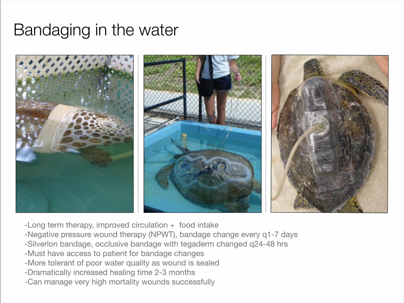
-Long term therapy, improved circulation + food intake-Negative pressure wound therapy (NPWT), bandage change every q1-7 days-Silverlon bandage, occlusive bandage with tegaderm changed q24-48 hrs-Must have access to patient for bandage changes-More tolerant of poor water quality as wound is sealed-Dramatically increased healing time 2-3 months-Can manage very high mortality wounds successfully
Bandaging in the water

Lung leaks can result in collapse without interventionNegative pressure needed to prevent lung scarringVAC therapy is ideal
Open pneumocoelom

Drainage needed to prevent coelomitis/sepsis (ventral placement needed if no suction available)Barrier may be needed to maintain viscera in place if defect is present (implantable mesh, prosthetic)
Open coelom

Head trauma
• If neurocranium fractured very high mortality
• Head trauma + neurologic signs (tremors, spastic movement, hyperesthesia) is grounds for euthanasia
• Encephalitis common but may take weeks to develop
• MRI or CT scan ideal
• DO NOT VAC!

• Only if fracture results in marked instability
• Bone to bone healing unlikely (will form fibrous union)
• Drilling holes in carapace is painful,a source for infection, and screws will loosen due to micromovement of shell and localized osteomyelitis
• Fiberglass or other occlusive NOT advised due to risk of infection with out improved outcome
Fracture repair

Carapace and skull are dermal bone
• Growth occurs at suture lines--not end plates as with osteochondral bone
• Growth can be manipulated by distraction (pulling apart) and compression (pulling together) to reduce defects
• Implants or semipermanent fixtures will actually restrict growth and result in deformities (particularly an issue with juveniles and sub adults)
• Individuals should not be released with an implant in place due to risk of complications