Embed Size (px)
Citation preview
Work Related Musculoskeletal Disorders
Dr. Majid GolabadiOccupational Medicine Specialist
Isfahan University of Medical Sciences
Job Risk Factors
• Repetition
• Force
• Awkward posture
• Contact stress
• Vibration

Upper Extremity Disorders
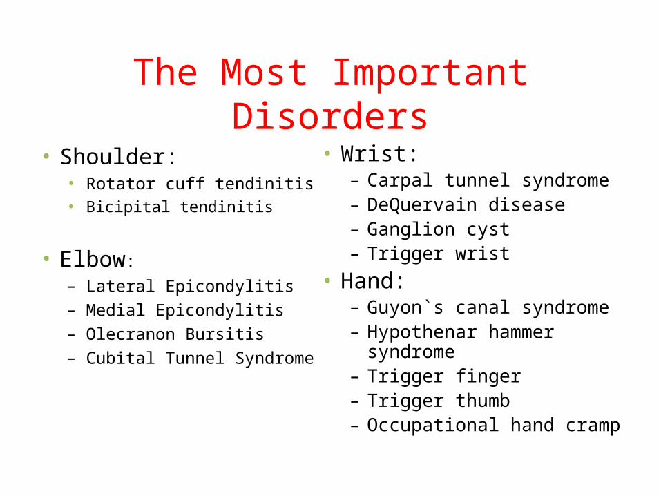
The Most Important Disorders
• Shoulder:• Rotator cuff tendinitis• Bicipital tendinitis
• Elbow:– Lateral Epicondylitis– Medial Epicondylitis– Olecranon Bursitis– Cubital Tunnel Syndrome
• Wrist:– Carpal tunnel syndrome– DeQuervain disease– Ganglion cyst– Trigger wrist
• Hand:– Guyon`s canal syndrome– Hypothenar hammer syndrome– Trigger finger– Trigger thumb– Occupational hand cramp

Lateral Epicondylitis(Tennis Elbow)

Lateral Epicondylitis (Tennis Elbow)
Inflammation, at the muscular origin of the extensor carpi radialis brevis (ECRB).
the most common overuse injury of the elbow
up to 10 times more frequently than medial epicondylitis
most often occurs between the third and fifth decades of life.


Ergonomic Stressors
• Frequent lifting
• Repetitive wrist dorsiflexion with force
• Sustained power gripping. • Repetitive forearm supination
• Sudden elbow extension• Tool use, shaking hand,
twisting movement

Clinical Presentationslateral elbow pain of gradual onset. pain generally increases with
activityPicking up a cup of coffee or a gallon of
milkHeavy liftingGripping
Pain may be present at night.
Symptoms are typically unilateral.
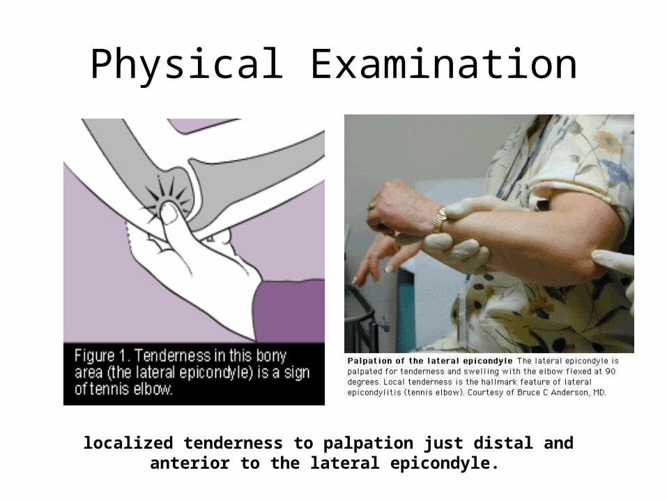
Physical Examination
localized tenderness to palpation just distal and anterior to the lateral epicondyle.
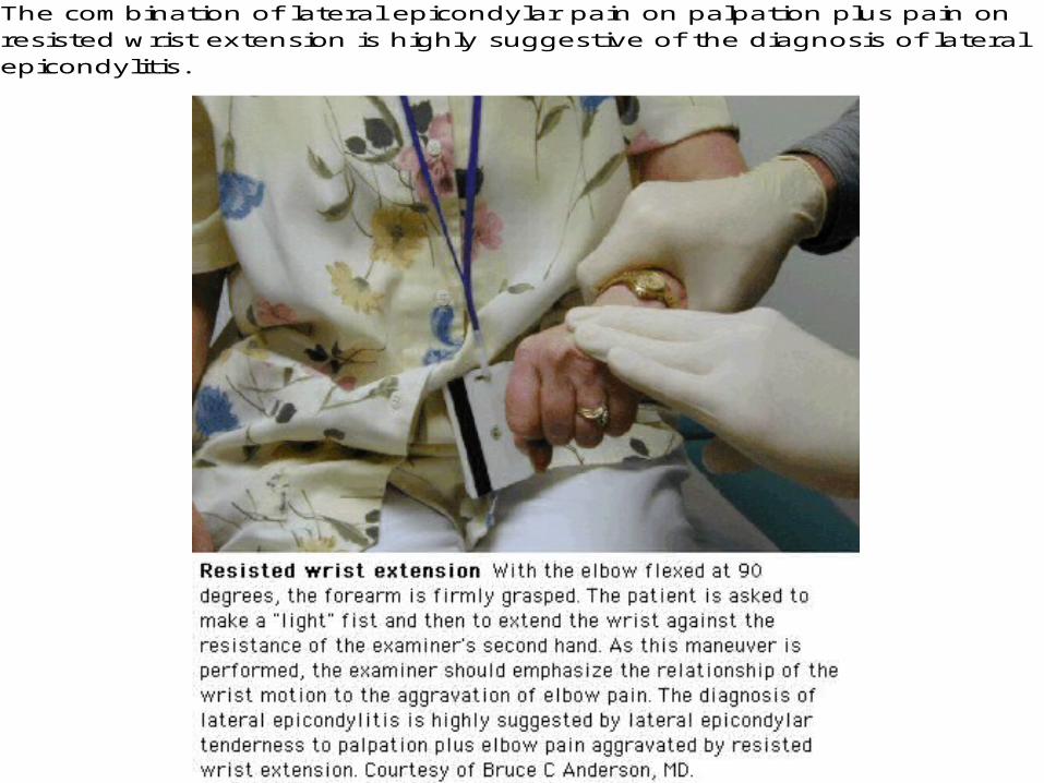
The combination of lateral epicondylar pain on palpation plus pain on resisted wrist extension is highly suggestive of the diagnosis of lateral epicondylitis.

Presumptive Diagnosis Requires:
• Local tenderness directly over the lateral epicondyle
• Pain aggravated by resisted wrist extension and radial deviation
• Pain aggravated by strong gripping
• Normal elbow range of motion
Paraclinical Testing
• No specific test is required

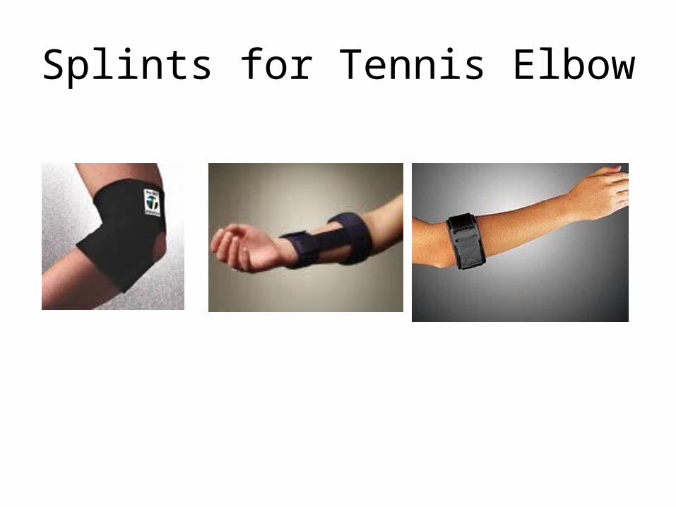
Splints for Tennis Elbow

Carpal Tunnel Syndrome
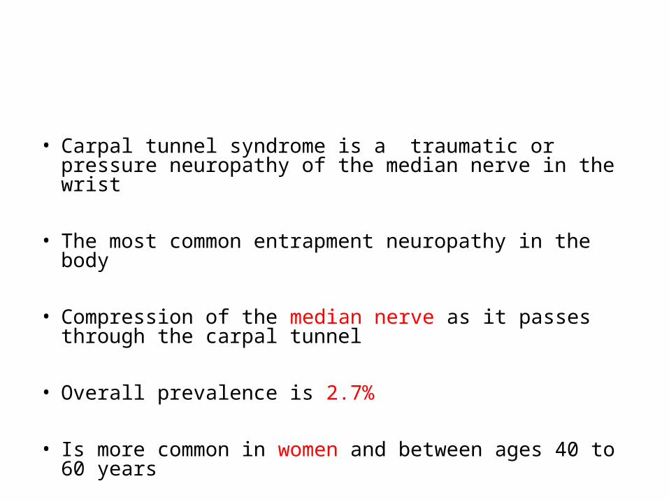
• Carpal tunnel syndrome is a traumatic or pressure neuropathy of the median nerve in the wrist
• The most common entrapment neuropathy in the body
• Compression of the median nerve as it passes through the carpal tunnel
• Overall prevalence is 2.7%
• Is more common in women and between ages 40 to 60 years


Etiology
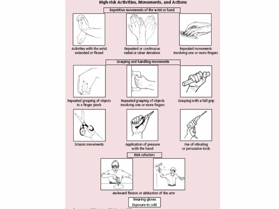
Work Related Risk Factors
Occupations that require Repetitive Flexion and extension of the fingers and wrist
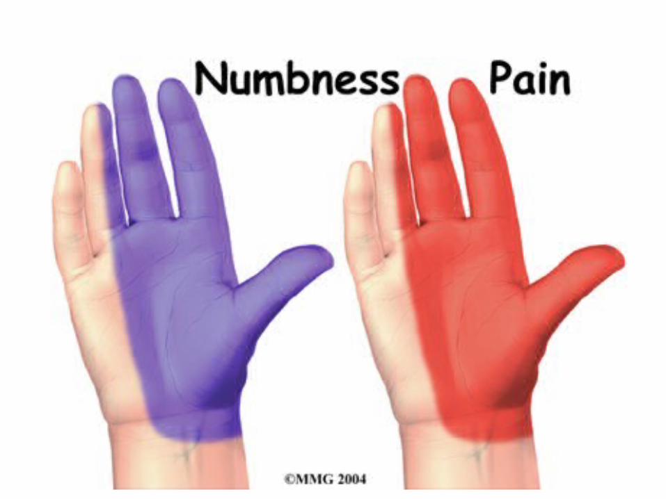

Symptoms
• Paresthesias in the median nerve distribution, gradually and spontaneously
• With progression: pain, numbness, tingling and burning
• In more progressed cases: Reduced force, Skin sensory deficit and Thenar Atrophy
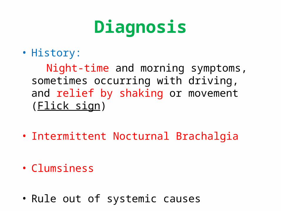
Diagnosis• History:
Night-time and morning symptoms, sometimes occurring with driving, and relief by shaking or movement (Flick sign)
• Intermittent Nocturnal Brachalgia
• Clumsiness
• Rule out of systemic causes
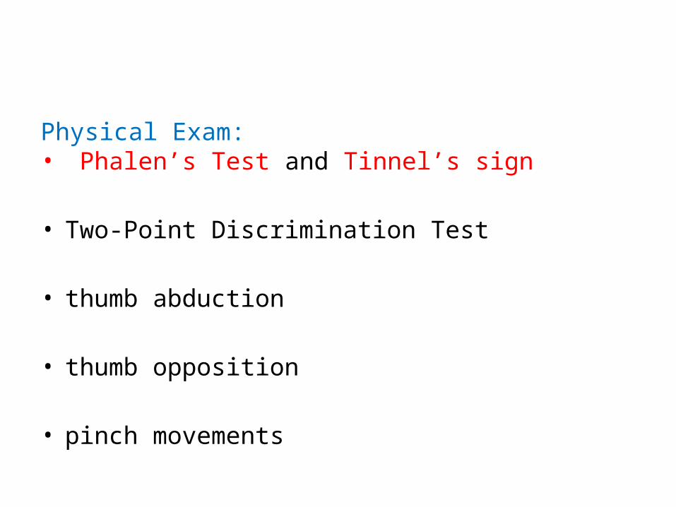
Physical Exam: • Phalen’s Test and Tinnel’s sign
• Two-Point Discrimination Test
• thumb abduction
• thumb opposition
• pinch movements
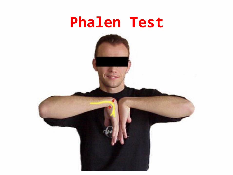
Phalen Test

Tinnel sign

• Electrodiagnostic studies: EMG/NCV
confirm diagnosis
• Thenar weakness should warrant full EMG studies
Treatment1- Treatment of associated conditions
2- Splinting the wrist in a neutral position at night and during the day . For 2 to 4 weeks
Job task modification is often critical in this phase
3- Corticosteroid injection into the carpal tunnel
4- Surgery. After 3 month of conservative treatment

Surgery indications
• Progressive symptoms
• Persistent symptoms
• Thenar Atrophy
• EMG abnormalities

De Quervain’s Disease

De Quervain’s Disease
• Inflammation of the tendon sheath of the extensor pollicis brevis and abductor pollicis longus
• Combination of Tendonitis and Tenosynovitis.
• In individuals between 30 and 50 years of age and is ten times more prevalent among women than men
• May be caused by OVER USE of thumb, like repetitive work and forceful gripping

Symptoms• pain at the base of the thumb.
• swelling
Differential diagnosis• Old nonunion of navicular bone• Osteoartritis of first carpometacarpal joint

Finkelstein test

Treatment• Modifying hand activity
• Immubilization of thumb (3-6 weeks)
• NSAIDs
• Local Injection of Lidocain-triamcinolone into tendon sheat (Standard Treatment)
• Surgical decompression
Trigger Finger
• Stenosing tenosinovitis of the flexor tendon of the finger
• Painful snap or jerking movements in PIP
• Collapse the joint suddenly like a trigger
• Usually associated with using tools that have handles with hard or sharp edges.

• Trauma, • Rheumatoid arthritis, • CTS
Differential diagnosis• De Qurvein• Dupuytren ContracturesTrauma, liver diseases, Alcohol Abuse
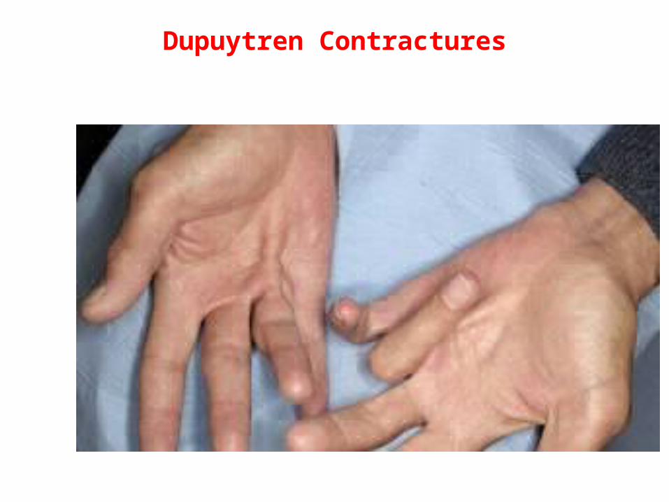
Dupuytren Contractures
Treatment
• Local Injection of Lidocain-triamcinolone into tendon sheet (Standard Treatment)
• Surgical decompression

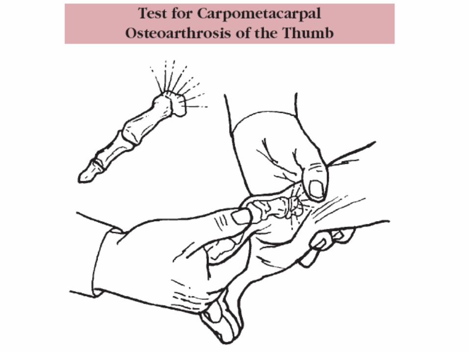
Osteoarthritis of the first carpometacarpal joint
• In 25% of women older than 55 years
• Unknown cause
• Pain at the base of thumb when grasping
• Squaring of the base of thumb
• Diagnosis with radiographs

Tratment
• Avoid repetitive painful activities
• Immobilization
• NSAIDs
• Arthroplasty or arthrodesis

Scaphoid Fractures

• Occur in younger people
• Pain at the base of the thumb or wrist pain
• Tenderness of the tuberosity of scaphoid
• PA, Lateral and Scaphoid view Ragiographs
• MRI or Bone Scan

Treatment
• Nondisplaced12 weaks immobilization
• DisplasedOpen reduction and Internal Fixation

Mallet Finger
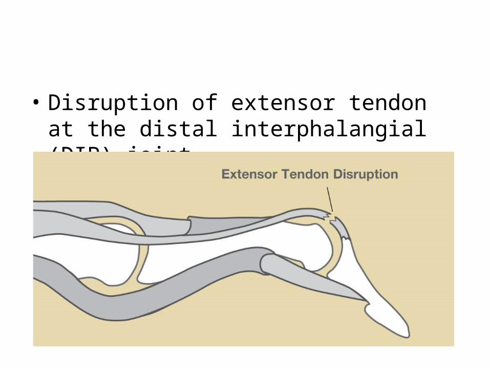
• Disruption of extensor tendon at the distal interphalangial (DIP) joint


Some Useful Tests

Apley Scratch Test
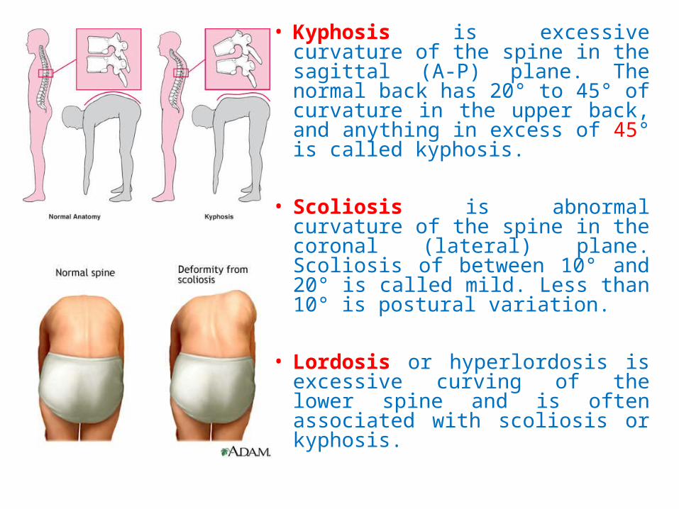
• Kyphosis is excessive curvature of the spine in the sagittal (A-P) plane. The normal back has 20° to 45° of curvature in the upper back, and anything in excess of 45° is called kyphosis.
• Scoliosis is abnormal curvature of the spine in the coronal (lateral) plane. Scoliosis of between 10° and 20° is called mild. Less than 10° is postural variation.
• Lordosis or hyperlordosis is excessive curving of the lower spine and is often associated with scoliosis or kyphosis.

Straight Leg RaiseSLR
Examiner raises straight leg (30 to 60 degrees) eliciting radicular pain on same
side (Lasegue Sign). Then lowers leg until pain goes away, the foot is then
dorsiflexed causing return of pain
Sensitivity 91%Specificity 26 %
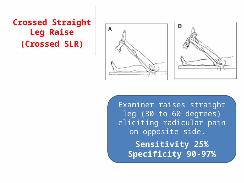
Crossed Straight Leg Raise
(Crossed SLR)
Examiner raises straight leg (30 to 60 degrees) eliciting radicular pain on
opposite side.
Sensitivity 25%Specificity 90-97%
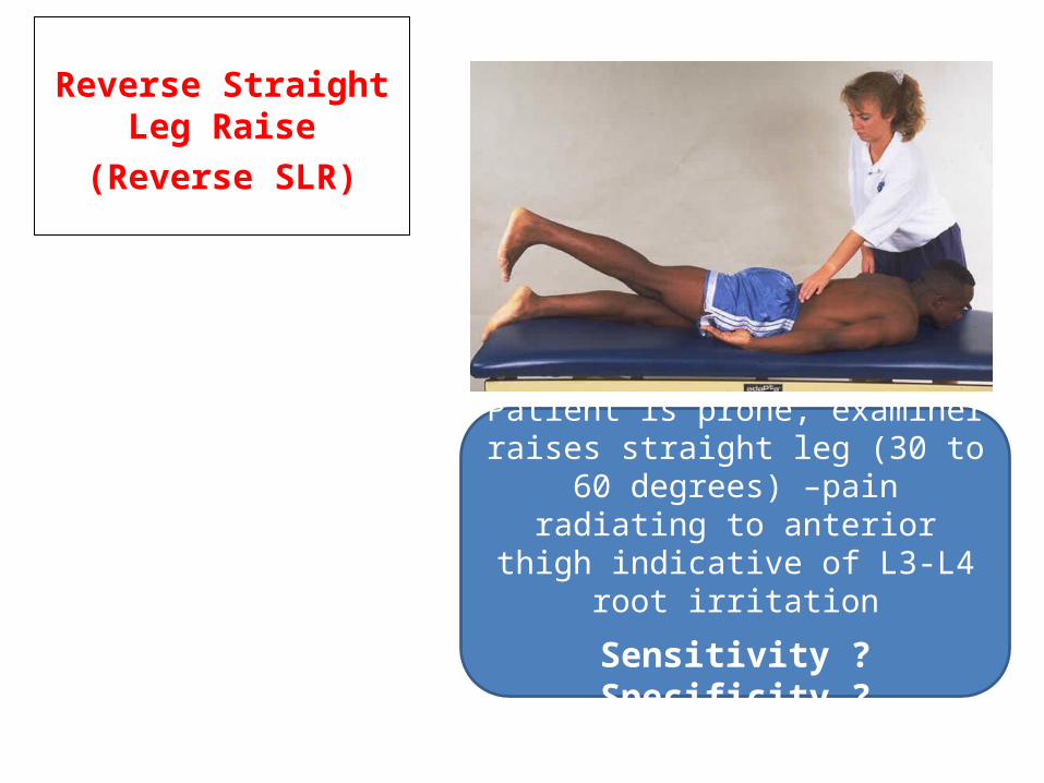
Reverse Straight Leg Raise
(Reverse SLR)
Patient is prone, examiner raises straight leg (30 to 60 degrees) –pain radiating to anterior thigh indicative
of L3-L4 root irritation
Sensitivity ?Specificity ?
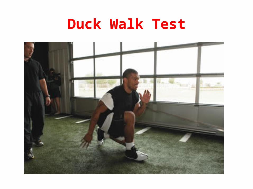
Duck Walk Test
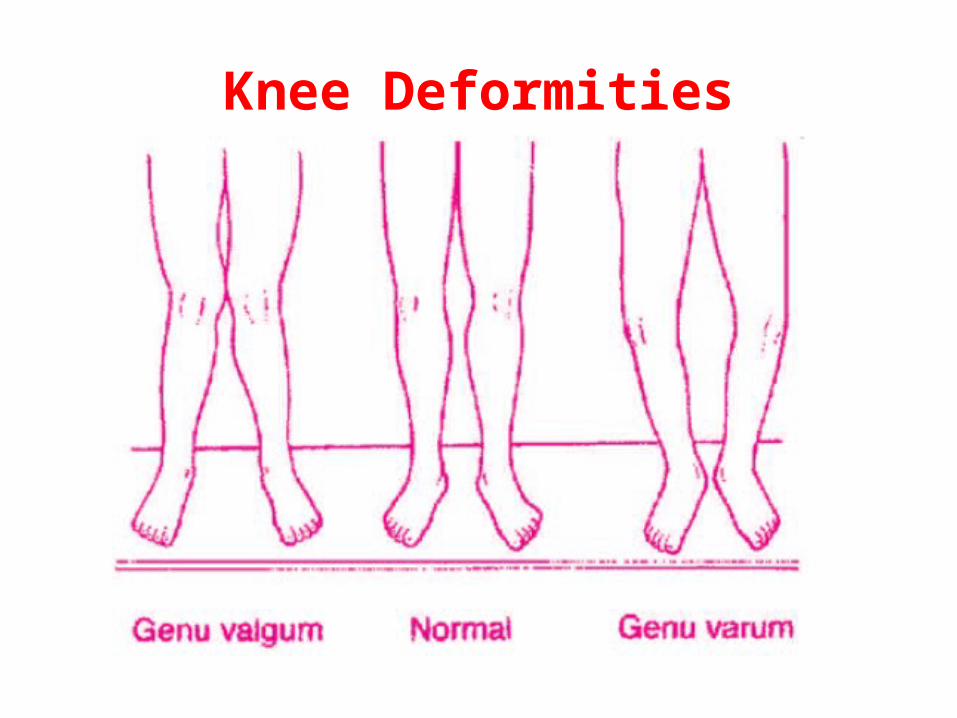
Knee Deformities