Embed Size (px)
Citation preview
* CorrespondE-mail addr
1760-2734/$ -http://dx.doi.
Journal of Veterinary Cardiology (2012) 14, 511e518
www.elsevier.com/locate/jvc
Within-day and between-day variabilityof transthoracic anatomic M-modeechocardiography in the awake bottlenosedolphin (Tursiops truncatus)
Valerie Chetboul, DVM, PhD a,b,*, Jonathan Lichtenberger, DVM a,Mathieu Mellin, DVM c, Birgitta Mercera c, Anne-Cecile Hoffmann, DVM a,Gwendoline Chaix, DVM a, Emilie Trehiou-Sechi, DVM a,Charlotte Misbach, DVM a, Amandine Petit, DVM a, Herve P. Lefebvre,DVM, PhD d, Nicolas Gaide, MSc a, Renaud Tissier, DVM, PhD b,e,Fabienne Delfour, PhD c
aUniversite Paris-Est, Ecole Nationale Veterinaire d‘Alfort, Unite de Cardiologie d‘Alfort (UCA), CentreHospitalier Universitaire Veterinaire d‘Alfort (CHUVA), 7 avenue du General de Gaulle,94704 Maisons-Alfort cedex, Franceb INSERM, U955, Equipe 03, 51 avenue du Marechal de Lattre de Tassigny, 94010 Creteil cedex, FrancecDelphinarium du Parc Asterix, Plailly, FrancedUnite de Recherche Clinique Sante des Populations, Ecole Nationale Veterinaire de Toulouse,23 chemin des Capelles, 31076 Toulouse cedex 03, FranceeUniversite Paris-Est, Ecole Nationale Veterinaire d‘Alfort, Unite de Pharmacie-Toxicologie, 7 avenue duGeneral de Gaulle, 94704 Maisons-Alfort cedex, France
Received 25 March 2012; received in revised form 30 May 2012; accepted 4 July 2012
KEYWORDSAquatic mammal;Heart;Repeatability;Reproducibility
ing author.ess: vchetboul@vet-alf
see front matter ª 201org/10.1016/j.jvc.2012
Abstract The use of transthoracic echocardiography in dolphins has been limitedso far owing to technical and anatomical specificities. Anatomic M-mode (AMM) isa postprocessing echocardiographic technique generating M-mode studies fromtwo-dimensional (2D) cineloops independently of the ultrasound beam orientation.The aim of the present study was to determine the within-day (repeatability) andbetween-day (reproducibility) variability of AMM echocardiography in awakehealthy bottlenose dolphins (BN, Tursiops truncatus). Four adult BN trained to lie
ort.fr (V. Chetboul).
2 Elsevier B.V. All rights reserved..07.002

512 V. Chetboul et al.
Abbreviations
2D two-dimensionalAMM anatomic M-modeAomin minimal aortic diamAomax maximal aortic diamBN bottlenose dolphinCV coefficient of variatLV left ventricle/left vLVDD left ventricular end-LVDS left ventricular end-M-mode motion modeSD standard deviationTEE transesophageal echTTE transthoracic echoc
in left recumbency at the water surface were involved in the protocol. A total of 96echocardiographic examinations were performed on 4 different days by a trainedobserver examining each BN 6 times per day. Video clips of 2D left parasternallong-axis views showing the left ventricle (LV) ventrally and the aortic root dorsallywere recorded at each examination and analyzed for AMM measurements ina random order. A general linear model was used to determine the within-dayand between-day coefficients of variation (CV). All examinations were interpret-able allowing calculation of 10 AMM variables (i.e., end-diastolic and end-systolicventral and dorsal LV myocardial wall thicknesses as well as LV and aortic diame-ters, mean aortic diameter, and LV shortening fraction). Most within- andbetween-day CV values (18/20) were <15%, the lowest being observed for theend-diastolic LV diameter (1.6%). In conclusion, AMM provides a simple non-invasive evaluation of heart morphology and function in the awake BN with goodrepeatability and reproducibility of the measurements. Further studies arerequired to determine the corresponding reference intervals.ª 2012 Elsevier B.V. All rights reserved.
etereter
ionentriculardiastolic diametersystolic diameter
ocardiographyardiography
Introduction
Aquatic mammals may be affected by miscella-neous heart diseases including congenital cardiacdefects, cardiomyopathies, parasitic heartdiseases, degenerative valvular diseases, valvularendocarditis, and myocarditis.1e4 Transthoracicechocardiography (TTE) is a well-established tech-nique for the antemortem diagnosis of heartdiseases both in humans and in small animals,allowing qualitative description of cardiac abnor-malities and quantitative assessment of heartanatomy and function using combined two-dimensional (2D) and M-mode (motion mode).However, the use of TTE to evaluate the dolphinheart has been limited so far because of anatomicbarriers (thick integument and blubber layers, largesternum, and circumferential lung) and technicalchallenges (difficulty for non-sedated animals to
stay in a stable appropriate position for severalminutes and relatively high sedation-relatedrisks).4,5 Dolphin heart diseases are thereforecurrently diagnosed by necropsy and histopatho-logical examinations only, and cardiology knowl-edge of this species remains very limited.
In one study specifically dedicated to echocar-diography in the bottlenose dolphin (BN, Tursiopstruncatus), TTE yielded poor-quality images ofonly small portions of the heart, and the authorsconcluded that transesophageal echocardiography(TEE) was more effective than TTE for non-invasiveexamination of the dolphin heart.4 Nevertheless,the authors reported that the TEE window was farmore limited in dolphins than in primates, and thatbreath-holding following forced exhalation wasnecessary to obtain good quality TEE images (whiledecreasing lung interference and increasing thecontact between heart and esophagus). Thisparticular breathing behavior required 4 months ofspecific training of the BN involved in the study,which represents a technical limitation for thepractical use of TEE in this species.
Anatomic M-mode (AMM) is a post-processingechocardiographic technique capable of gener-ating M-mode studies from stored 2D cineloopsindependently of the ultrasound beam ori-entation.6e9 This technique overcomes the mainlimitation of conventional M-mode echocardiog-raphy, i.e., fixed origin of the M-mode line at thesector apex, which may result in an incorrectalignment with the cardiac structures studied, andmay thus lead to unreliable measurements of thelatter. Using AMM, the observer has the opportu-nity to position the M-mode line in any spatialorientation defined from 2D views, thus improvingmeasurements variability and increasing theability to measure cardiac structures from various
Echocardiography in dolphins 513
spatial directions.6e11 We hypothesize that TTEusing AMM could be suitable to assess the unse-dated BN heart. To the best of our knowledge, LVmorphology and function have never been quan-titatively evaluated in this species.
The aims of this prospective study were there-fore 1) to assess the feasibility of transthoracicAMM in healthy awake BN (optimal animal position,views, and probes), and then 2) to determine theintra-observer within-day (repeatability) andbetween-day (reproducibility) variability of thecorresponding AMM measurements.
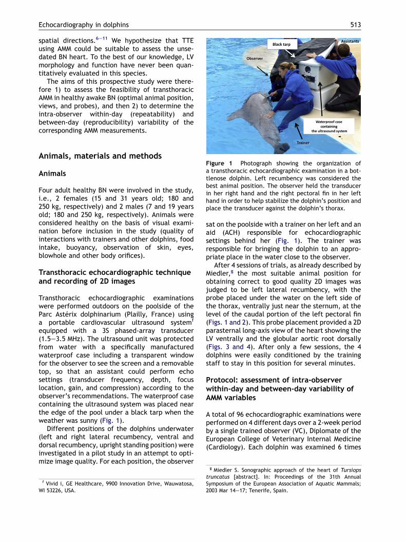
Figure 1 Photograph showing the organization ofa transthoracic echocardiographic examination in a bot-tlenose dolphin. Left recumbency was considered thebest animal position. The observer held the transducerin her right hand and the right pectoral fin in her lefthand in order to help stabilize the dolphin’s position andplace the transducer against the dolphin’s thorax.
Animals, materials and methods
Animals
Four adult healthy BN were involved in the study,i.e., 2 females (15 and 31 years old; 180 and250 kg, respectively) and 2 males (7 and 19 yearsold; 180 and 250 kg, respectively). Animals wereconsidered healthy on the basis of visual exami-nation before inclusion in the study (quality ofinteractions with trainers and other dolphins, foodintake, buoyancy, observation of skin, eyes,blowhole and other body orifices).
Transthoracic echocardiographic techniqueand recording of 2D images
Transthoracic echocardiographic examinationswere performed outdoors on the poolside of theParc Asterix dolphinarium (Plailly, France) usinga portable cardiovascular ultrasound systemf
equipped with a 3S phased-array transducer(1.5e3.5 MHz). The ultrasound unit was protectedfrom water with a specifically manufacturedwaterproof case including a transparent windowfor the observer to see the screen and a removabletop, so that an assistant could perform echosettings (transducer frequency, depth, focuslocation, gain, and compression) according to theobserver’s recommendations. The waterproof casecontaining the ultrasound system was placed nearthe edge of the pool under a black tarp when theweather was sunny (Fig. 1).
Different positions of the dolphins underwater(left and right lateral recumbency, ventral anddorsal recumbency, upright standing position) wereinvestigated in a pilot study in an attempt to opti-mize image quality. For each position, the observer
f Vivid i, GE Healthcare, 9900 Innovation Drive, Wauwatosa,WI 53226, USA.
sat on the poolside with a trainer on her left and anaid (ACH) responsible for echocardiographicsettings behind her (Fig. 1). The trainer wasresponsible for bringing the dolphin to an appro-priate place in the water close to the observer.
After 4 sessions of trials, as already described byMiedler,g the most suitable animal position forobtaining correct to good quality 2D images wasjudged to be left lateral recumbency, with theprobe placed under the water on the left side ofthe thorax, ventrally just near the sternum, at thelevel of the caudal portion of the left pectoral fin(Figs. 1 and 2). This probe placement provided a 2Dparasternal long-axis view of the heart showing theLV ventrally and the globular aortic root dorsally(Figs. 3 and 4). After only a few sessions, the 4dolphins were easily conditioned by the trainingstaff to stay in this position for several minutes.
Protocol: assessment of intra-observerwithin-day and between-day variability ofAMM variables
A total of 96 echocardiographic examinations wereperformed on 4 different days over a 2-week periodby a single trained observer (VC), Diplomate of theEuropean College of Veterinary Internal Medicine(Cardiology). Each dolphin was examined 6 times
g Miedler S. Sonographic approach of the heart of Tursiopstruncatus [abstract]. In: Proceedings of the 31th AnnualSymposium of the European Association of Aquatic Mammals;2003 Mar 14e17; Tenerife, Spain.
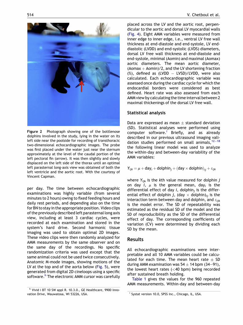
Figure 2 Photograph showing one of the bottlenosedolphins involved in the study, lying in the water on itsleft side near the poolside for recording of transthoracictwo-dimensional echocardiographic images. The probewas first placed under the water just near the sternumapproximately at the level of the caudal portion of theleft pectoral fin (arrow). It was then slightly and slowlydisplaced on the left side of the thorax until an optimalleft parasternal long-axis view was obtained of both theleft ventricle and the aortic root. With the courtesy ofVincent Capman.
514 V. Chetboul et al.
per day. The time between echocardiographicexaminations was highly variable (from severalminutes to 2 hours) owing to fixed feeding hours anddaily rest periods, and depending also on the timefor BN to stay in theappropriateposition. Video clipsof the previously described left parasternal long axisview, including at least 3 cardiac cycles, wererecorded at each examination and stored in thesystem’s hard drive. Second harmonic tissueimaging was used to obtain optimal 2D images.These video clips were then randomly analyzed forAMM measurements by the same observer and onthe same day of the recordings. No specificrandomization criteria was used except that thesame animal could not be used twice consecutively.Anatomic M-mode images, showing motions of theLV at the top and of the aorta below (Fig. 5), weregenerated from digital 2D cineloops using a specificsoftware.h The electronic AMM cursor was carefully
h Vivid i BT 10 SW appl R. 10.3.0., GE Healthcare, 9900 Inno-vation Drive, Wauwatosa, WI 53226, USA.
placed across the LV and the aortic root, perpen-dicular to the aortic and dorsal LV myocardial walls(Fig. 4). Eight AMM variables were measured frominner edge to inner edge, i.e., ventral LV free wallthickness at end-diastole and end-systole, LV end-diastolic (LVDD) and end-systolic (LVDS) diameters,dorsal LV free wall thickness at end-diastole andend-systole, minimal (Aomin) and maximal (Aomax)aortic diameters. The mean aortic diameter,(Aomax þ Aomin)/2, and the LV shortening fraction(%), defined as (LVDD � LVSD)/LVDD, were alsocalculated. Each echocardiographic variable wasassessed once during the cardiac cycle for which theendocardial borders were considered as bestdefined. Heart rate was also assessed from eachAMMviewby calculating the time interval between2maximal thickenings of the dorsal LV free wall.
Statistical analysis
Data are expressed as mean � standard deviation(SD). Statistical analyses were performed usingcomputer software.i Briefly, and as alreadydescribed in our previous ultrasound imaging vali-dation studies performed on small animals,12e18
the following linear model was used to analyzethe within-day and between-day variability of theAMM variables:
Yijk ¼ mþ dayi þ dolphinj þ ðday� dolphinÞij þ 3ijk
where Yijk is the kth value measured for dolphin jon day i, m is the general mean, dayi is thedifferential effect of day i, dolphinj is the differ-ential effect of dolphin j, (day � dolphin)ij is theinteraction term between day and dolphin, and 3ijk
is the model error. The SD of repeatability wasestimated as the residual SD of the model and theSD of reproducibility as the SD of the differentialeffect of day. The corresponding coefficients ofvariation (CV) were determined by dividing eachSD by the mean.
Results
All echocardiographic examinations were inter-pretable and all 10 AMM variables could be calcu-lated for each time. The mean heart rate � SDduring AMM examination was 54 � 14 bpm (34e91),the lowest heart rates (<40 bpm) being recordedafter sustained breath holding.
Table 1 gives the values for the 960 repeatedAMM measurements. Within-day and between-day
i Systat version 10.0, SPSS Inc., Chicago, IL, USA.
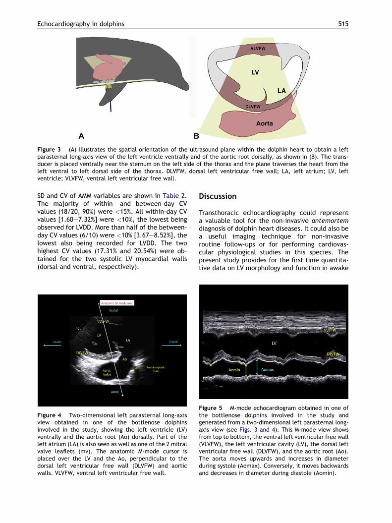
Figure 3 (A) illustrates the spatial orientation of the ultrasound plane within the dolphin heart to obtain a leftparasternal long-axis view of the left ventricle ventrally and of the aortic root dorsally, as shown in (B). The trans-ducer is placed ventrally near the sternum on the left side of the thorax and the plane traverses the heart from theleft ventral to left dorsal side of the thorax. DLVFW, dorsal left ventricular free wall; LA, left atrium; LV, leftventricle; VLVFW, ventral left ventricular free wall.
Echocardiography in dolphins 515
SD and CV of AMM variables are shown in Table 2.The majority of within- and between-day CVvalues (18/20, 90%) were <15%. All within-day CVvalues [1.60e7.32%] were <10%, the lowest beingobserved for LVDD. More than half of the between-day CV values (6/10) were <10% [3.67e8.52%], thelowest also being recorded for LVDD. The twohighest CV values (17.31% and 20.54%) were ob-tained for the two systolic LV myocardial walls(dorsal and ventral, respectively).
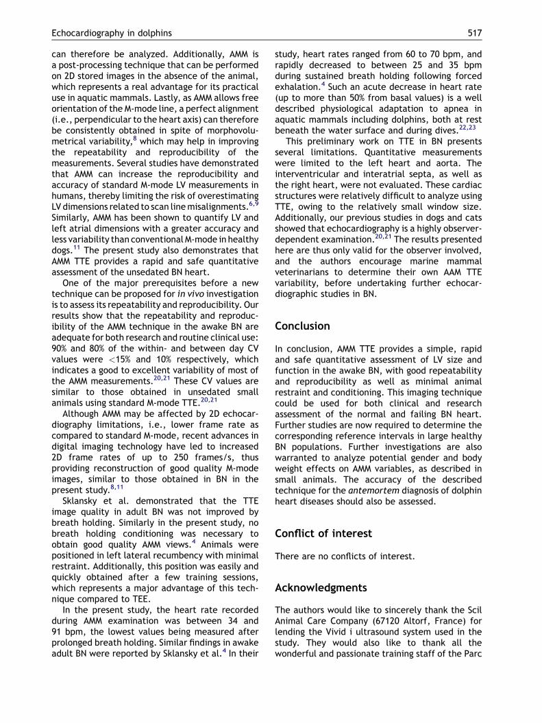
Figure 4 Two-dimensional left parasternal long-axisview obtained in one of the bottlenose dolphinsinvolved in the study, showing the left ventricle (LV)ventrally and the aortic root (Ao) dorsally. Part of theleft atrium (LA) is also seen as well as one of the 2 mitralvalve leaflets (mv). The anatomic M-mode cursor isplaced over the LV and the Ao, perpendicular to thedorsal left ventricular free wall (DLVFW) and aorticwalls. VLVFW, ventral left ventricular free wall.
Discussion
Transthoracic echocardiography could representa valuable tool for the non-invasive antemortemdiagnosis of dolphin heart diseases. It could also bea useful imaging technique for non-invasiveroutine follow-ups or for performing cardiovas-cular physiological studies in this species. Thepresent study provides for the first time quantita-tive data on LV morphology and function in awake
Figure 5 M-mode echocardiogram obtained in one ofthe bottlenose dolphins involved in the study andgenerated from a two-dimensional left parasternal long-axis view (see Figs. 3 and 4). This M-mode view showsfrom top to bottom, the ventral left ventricular free wall(VLVFW), the left ventricular cavity (LV), the dorsal leftventricular free wall (DLVFW), and the aortic root (Ao).The aorta moves upwards and increases in diameterduring systole (Aomax). Conversely, it moves backwardsand decreases in diameter during diastole (Aomin).

Table 1 Mean � SD, minimum and maximum values of repeated measurements of echocardiographic variablesobtained by a trained observer in 4 bottlenose dolphins (Tursiops truncatus) from 96 transthoracic examinations usinganatomic M-mode.
Echocardiographic parameter Mean � SD Minimumemaximum
Ventral left ventricular free wall thickness in diastole (mm) 12.7 � 0.44 11.5e13.9Ventral left ventricular free wall thickness in systole (mm) 21.2 � 1.24 18.5e23.8Left ventricular end-diastolic diameter (mm) 66.2 � 3.80 61.3e75.6Left ventricular end-systolic diameter (mm) 40.3 � 2.00 35.0e43.9Dorsal left ventricular free wall thickness in diastole (mm) 12.7 � 0.50 11.3e14.4Dorsal left ventricular free wall thickness in systole (mm) 21.0 � 1.13 18.6e23.0Shortening fraction (%) 39.0 � 4.07 32.0e52.0Minimal aortic diameter (mm) 40.0 � 1.28 36.3e42.9Maximal aortic diameter (mm) 46.8 � 1.72 43.0e50.2Mean aortic diameter (mm) 43.4 � 1.27 40.2e46.4
516 V. Chetboul et al.
BN using a safe, effective ultrasound technique,i.e., AMM TTE. This non-invasive imaging approachrepresents an initial step in the development offunctional cardiology in cetaceans.
Few data are available regarding echocardio-graphic evaluation of the dolphin heart. Fetalechocardiography was recently described in BN bySklansky et al.19 The authors reported thata detailed assessment of fetal BN cardiovascularstatus, including 2D imaging and color flowmappingof the heart and great arteries as well as pulsedDoppler evaluation of the umbilical artery and vein,could be obtained between 8 and 9 months ofgestation.19 Another study, performed by the sameauthors on 4 adult BN trained to hold their breathfollowing forced exhalation, showed that the TEEtechnique yielded high-quality images of the entireheart (atrioventricular and arterial valves, intera-trial and interventricular septa, left and right atrialcavities, left and right ventricles, ascending aortaand main pulmonary artery).4 The latter report alsodemonstrated mild tricuspid regurgitation in all BN,and mild aortic regurgitation in one BN using color
Table 2 Within-day and between-day variability, expressed(CV), of anatomic M-mode variables obtained by a trained ob96 transthoracic echocardiographic examinations.
Echocardiographic parameter
Ventral left ventricular free wall thickness in diastole (mm)Ventral left ventricular free wall thickness in systole (mm)Left ventricular end-diastolic diameter (mm)Left ventricular end-systolic diameter (mm)Dorsal left ventricular free wall thickness in diastole (mm)Dorsal left ventricular free wall thickness in systole (mm)Shortening fraction (%)Minimal aortic diameter (mm)Maximal aortic diameter (mm)Mean aortic diameter (mm)
flow Doppler mode.4 Nevertheless, despite breathholding, reliable quantitative TEE measurements ofventricular size and function could not be obtainedbecause of inconsistent animal positioning, andonly maximal valve diameters could be measured.4
Advances in conventional ultrasound imagingtechniques, including AMM echocardiography, haveafforded newopportunities for non-invasive cardiacanalysis in humans and various animal species.6e11
The standard M-mode provides monodimensionalechocardiograms and is commonly used for linearcardiac measurements, including ventriculardiameters and myocardial wall thicknesses. Thisconventional echocardiographic technique is char-acterized by a high temporal resolution, and istherefore suitable for studying mobile structures.8
However the major limitation of the standard M-mode is that the analysis line can only rotate ona fixed point, i.e., the sector apex.8 The AMMtechnique overcomes this fixity drawback, as itprovides M-mode images by orienting the analysisline in any direction according to the observer’sdesire.8 Any cardiac structure in every angle shot
as standard deviations (SD) and coefficients of variationserver on 4 bottlenose dolphins (Tursiops truncatus) from
Within-day Between-day
SD CV (%) SD CV (%)
0.40 3.16 0.85 6.691.00 4.72 4.35 20.541.06 1.60 2.43 3.671.92 4.76 3.44 8.520.46 3.65 1.03 8.130.94 4.47 3.64 17.312.86 7.32 3.84 9.830.96 2.39 4.41 11.041.11 2.36 2.93 6.250.81 1.87 4.91 11.31
Echocardiography in dolphins 517
can therefore be analyzed. Additionally, AMM isa post-processing technique that can be performedon 2D stored images in the absence of the animal,which represents a real advantage for its practicaluse in aquatic mammals. Lastly, as AMM allows freeorientation of the M-mode line, a perfect alignment(i.e., perpendicular to the heart axis) can thereforebe consistently obtained in spite of morphovolu-metrical variability,8 which may help in improvingthe repeatability and reproducibility of themeasurements. Several studies have demonstratedthat AMM can increase the reproducibility andaccuracy of standard M-mode LV measurements inhumans, thereby limiting the risk of overestimatingLV dimensions related to scan linemisalignments.6,9
Similarly, AMM has been shown to quantify LV andleft atrial dimensions with a greater accuracy andless variability than conventional M-mode in healthydogs.11 The present study also demonstrates thatAMM TTE provides a rapid and safe quantitativeassessment of the unsedated BN heart.
One of the major prerequisites before a newtechnique can be proposed for in vivo investigationis to assess its repeatability and reproducibility. Ourresults show that the repeatability and reproduc-ibility of the AMM technique in the awake BN areadequate for both research and routine clinical use:90% and 80% of the within- and between day CVvalues were <15% and 10% respectively, whichindicates a good to excellent variability of most ofthe AMM measurements.20,21 These CV values aresimilar to those obtained in unsedated smallanimals using standard M-mode TTE.20,21
Although AMM may be affected by 2D echocar-diography limitations, i.e., lower frame rate ascompared to standard M-mode, recent advances indigital imaging technology have led to increased2D frame rates of up to 250 frames/s, thusproviding reconstruction of good quality M-modeimages, similar to those obtained in BN in thepresent study.8,11
Sklansky et al. demonstrated that the TTEimage quality in adult BN was not improved bybreath holding. Similarly in the present study, nobreath holding conditioning was necessary toobtain good quality AMM views.4 Animals werepositioned in left lateral recumbency with minimalrestraint. Additionally, this position was easily andquickly obtained after a few training sessions,which represents a major advantage of this tech-nique compared to TEE.
In the present study, the heart rate recordedduring AMM examination was between 34 and91 bpm, the lowest values being measured afterprolonged breath holding. Similar findings in awakeadult BN were reported by Sklansky et al.4 In their
study, heart rates ranged from 60 to 70 bpm, andrapidly decreased to between 25 and 35 bpmduring sustained breath holding following forcedexhalation.4 Such an acute decrease in heart rate(up to more than 50% from basal values) is a welldescribed physiological adaptation to apnea inaquatic mammals including dolphins, both at restbeneath the water surface and during dives.22,23
This preliminary work on TTE in BN presentsseveral limitations. Quantitative measurementswere limited to the left heart and aorta. Theinterventricular and interatrial septa, as well asthe right heart, were not evaluated. These cardiacstructures were relatively difficult to analyze usingTTE, owing to the relatively small window size.Additionally, our previous studies in dogs and catsshowed that echocardiography is a highly observer-dependent examination.20,21 The results presentedhere are thus only valid for the observer involved,and the authors encourage marine mammalveterinarians to determine their own AAM TTEvariability, before undertaking further echocar-diographic studies in BN.
Conclusion
In conclusion, AMM TTE provides a simple, rapidand safe quantitative assessment of LV size andfunction in the awake BN, with good repeatabilityand reproducibility as well as minimal animalrestraint and conditioning. This imaging techniquecould be used for both clinical and researchassessment of the normal and failing BN heart.Further studies are now required to determine thecorresponding reference intervals in large healthyBN populations. Further investigations are alsowarranted to analyze potential gender and bodyweight effects on AMM variables, as described insmall animals. The accuracy of the describedtechnique for the antemortem diagnosis of dolphinheart diseases should also be assessed.
Conflict of interest
There are no conflicts of interest.
Acknowledgments
The authors would like to sincerely thank the ScilAnimal Care Company (67120 Altorf, France) forlending the Vivid i ultrasound system used in thestudy. They would also like to thank all thewonderful and passionate training staff of the Parc

518 V. Chetboul et al.
Asterix dolphinarium. This study was presented atthe 40th Annual Symposium of the EuropeanAssociation of Aquatic Mammals (EAAM, March 10th2012) and received the “Outstanding OralCommunication by a student Award” (JL). Theauthors would like to thank the EAAM committee.
References
1. Gulland FMD, Lowenstine LJ, Spraker TR. Non infectiousdiseases. In: Dierauf LA, Gulland FMD, editors. CRChandbook of marine mammal medicine. 2nd ed. BocaRaton: CRC Press; 2001. p. 535e536.
2. Gonzalez-Barrientos R, Morales JA, Hernandez-Mora G,Barquero-Calvo E, Guzman-Verri C, Chaves-Olarte E,Moreno E. Pathology of striped dolphins (Stenella coeru-leoalba) infected with Brucella ceti. J Comp Pathol 2010;142:347e352.
3. Lipscomb TP, Kennedy S, Moffett D, Ford BK. Morbilliviraldisease in an Atlantic bottlenose dolphin (Tursiops trunca-tus) from the Gulf of Mexico. J Wildl Dis 1994;30:572e576.
4. Sklansky M, Levine G, Havlis D, West N, Renner M,Rimmerman C, Stone R. Echocardiographic evaluation ofthe bottlenose dolphin (Tursiops truncatus). J Zoo WildlMed 2006;37:454e463.
5. Brook F, Van Bonn W, Jensen E. Ultrasonography. In:Dierauf LA, Gulland FMD, editors. CRC handbook of marinemammal medicine. 2nd ed. Boca Raton: CRC Press; 2001. p.596e597.
6. Mele D, Pedini I, Alboni P, Levine RA. Anatomic M-mode:a new technique for quantitative assessment of leftventricular size and function. Am J Cardiol 1998;81(12A):82Ge85G.
7. Strotmann JM, Kvitting JP, Wilkenshoff UM, Wranne B,Hatle L, Sutherland GR. Anatomic M-mode echocardiog-raphy: a new approach to assess regional myocardial func-tion e a comparative in vivo and in vitro study of bothfundamental and second harmonic imaging modes. J Am SocEchocardiogr 1999;1(2):300e307.
8. Carerj S, Micari A, Trono A, Giordano G, Cerrito M, Zito C,Luzza F, Coglitore S, Arrigo F, Oreto G. Anatomical M-mode:an oldenew technique. Echocardiography 2003;20:357e361.
9. Donal E, Coisne D, Pham B, Ragot S, Herpin D, Thomas JD.Anatomic M-mode, a pertinent tool for the daily practice oftransthoracic echocardiography. J Am Soc Echocardiogr2004;17:962e967.
10. Grenacher PA, Schwarzwald CC. Assessment of leftventricular size and function in horses using anatomicalM-mode echocardiography. J Vet Cardiol 2010;12:111e121.
11. Oyama MA, Sisson DD. Assessment of cardiac chamber sizeusing anatomic M-mode. Vet Radiol Ultrasound 2005;46:331e336.
12. Chetboul V, Athanassiadis N, Carlos C, Nicolle A,Zilberstein L, Pouchelon JL, Lefebvre HP, Concordet D.Assessment of repeatability, reproducibility, and effect ofanesthesia on determination of radial and longitudinal leftventricular velocities via tissue Doppler imaging in dogs. AmJ Vet Res 2004;65:909e915.
13. Chetboul V, Carlos Sampedrano C, Gouni V, Concordet D,Lamour T, Ginesta J, Nicolle AP, Pouchelon JL, Lefebvre HP.Quantitative assessment of regional right ventricularmyocardial velocities in awake dogs by Doppler tissueimaging: repeatability, reproducibility, effect of bodyweight and breed, and comparison with left ventricularmyocardial velocities. J Vet Intern Med 2005;19:837e844.
14. Chetboul V, Carlos Sampedrano C, Gouni V, Nicolle AP,Pouchelon J-L. Ultrasonographic assessment of regionalradial and longitudinal systolic function in healthy awakedogs. J Vet Intern Med 2006;20:885e893.
15. Gouni V, Serres FJ, Pouchelon JL, Tissier R, Lefebvre HP,Nicolle AP, Carlos Sampredrano C, Chetboul V. Quantificationofmitral valve regurgitation in dogswith degenerativemitralvalve disease by use of the proximal isovelocity surface areamethod. J Am Vet Med Assoc 2007;231:399e406.
16. Chetboul V, Serres F, Gouni V, Tissier R, Pouchelon JL.Radial strain and strain rate by two-dimensional speckletracking echocardiography and the tissue velocity basedtechnique in the dog. J Vet Cardiol 2007;9:69e81.
17. Serres F, Chetboul V, Tissier R, Poujol L, Gouni V, CarlosSampedrano C, Pouchelon JL. Comparison of 3 ultrasoundmethods for quantifying left ventricular systolic function:correlationwith disease severity and prognostic value in dogswithmitral valve disease. J Vet InternMed 2008;22:566e577.
18. Chetboul V, Serres F, Gouni V, Tissier R, Pouchelon JL.Noninvasive assessment of systolic left ventricular torsionby 2-dimensional speckle tracking imaging in the awakedog: repeatability, reproducibility, and comparison withtissue Doppler imaging variables. J Vet Intern Med 2008;22:342e350.
19. Sklansky M, Renner M, Clough P, Levine G, Campbell M,Stone R, Schmitt T, Chang RK, Shannon-Rodriguez J. Fetalechocardiographic evaluation of the bottlenose dolphin(Tursiops truncatus). J Zoo Wildl Med 2010;41:35e43.
20. Chetboul V, Concordet D, Pouchelon JL, Athanassiadis N,Muller C, Benigni L, Munari AC, Lefebvre HP. Effects ofinter- and intra-observer variability on echocardiographicmeasurements in awake cats. J Vet Med A Physiol PatholClin Med 2003;50:326e331.
21. Chetboul V, Athanassiadis N, Concordet D, Nicolle A,Tessier D, Castagnet M, Pouchelon JL, Lefebvre HP.Observer-dependent variability of quantitative clinicalendpoints: the example of canine echocardiography. J VetPharmacol Ther 2004;27:49e56.
22. Elsner R, Kenney DW, Burgess K. Diving bradycardia in thetrained dolphin. Nature 1966;212:407e408.
23. Noren SR, Cuccurullo V, Williams TM. The development ofdiving bradycardia in bottlenose dolphins (Tursiopstruncatus). J Comp Physiol B 2004;174:139e147.
Available online at www.sciencedirect.com