Embed Size (px)
Citation preview

Page 1/28
“Prevalence, Predictors and Outcomes of Surgicaland Gastrointestinal Bleeding Events in Patientswith COVID-19 Infection on Anticoagulation”Ahmed Alkhamis ( [email protected] )
Kuwait University https://orcid.org/0000-0002-4732-8471Yousef Alshamali
Ministry of HealthKhaled Alyaqout
Ministry of Health: Gobierno de Chile Ministerio de SaludEisa Lari
Ministry of HealthMoh A. Alkhamis
Kuwait UniversitySaad Althuwaini
Ministry of HealthAli Lari
Ministry of HealthMaryam Al�li
Ministry of HealthAli Alkhayat
Ministry of HealthMohammad H Jamal
Kuwait UniversitySalman Alsabah
Kuwait University
Research article
Keywords: COVID-19 Infection, Anticoagulation, Gastrointestinal Bleeding Events, Patients, InfectiousDiseases
Posted Date: March 31st, 2021
DOI: https://doi.org/10.21203/rs.3.rs-366340/v1

Page 2/28
License: This work is licensed under a Creative Commons Attribution 4.0 International License. Read Full License

Page 3/28
AbstractBackground
This study aims to examine risk factors and complications associated with bleeding events in patientswith coronavirus 19 (COVID-19) who are on anticoagulation.
Method
We conducted retrospective review of a prospectively maintained database of all patients who wereadmitted with COVID-19 and developed bleeding events.
Results
Of 122 bleeds, there were 67 (28%) non-gastrointestinal (GI) and 55 (28%) GI bleeds. Overall mortalitywas 59% (n= 72), 34 (28%) and 38 (31%) following non-GI and GI bleeds respectively. The prevalence oftherapeutic invasive interventions was 7.5% and 16.3% in non-GI and GI bleeds respectively and all weresuccessful in resolving the bleeding event. We found that having a GI bleeds was associated with higherrisk of mortality compared to non-GI bleeds (p= 0.04) and having occult bleeds to be associated with 15times increased risk of mortality. Furthermore, patients who were on therapeutic dose of anticoagulationwere more likely to die compared to patients who were on none (odds ratio (OR) 0.1, 95%CI 0.01-0.86), onprophylactic (OR 0.07,95%CI 0.02-0.28) or intermediate (OR 0.36,95%CI 0.09-1.34) anticoagulation doses.
Conclusions
Routine prescription of supra-prophylactic dose anticoagulation should be revisited as it appears to beassociated with increased of mortality and so more individualized approach to prescription should be thenorm. Regardless of the cause of bleeding event it appears that the majority of bleeding events resolvewith noninvasive interventions, correction and optimization sepsis therapies. However, when invasiveinterventions were necessary, they were associated with high success rate despite the delay.
BackgroundThe rapid emergence of the novel coronavirus 19 (COVID-19) has brought the world to a standstill. Thetransmissibility and associated morbidity and mortality of this virus have overwhelmed many worldwidehealthcare systems, resulting in an urgent need to understand this virus and its associated effects better.
It appears that the principal cause of death is acute respiratory failure complicated by a concomitantcoagulation disorder that can induce disseminated intravascular coagulation (DIC) (1). In light of this,anticoagulation therapy has been introduced recently as an adjuvant treatment, showing promisingresults in term of reducing mortality rate in several small retrospective studies (2). As a result, manyorganizations including the international society of thrombosis and hemostasis (ISTH) arerecommending speci�c anticoagulation regimens for COVID-19 patients (3). Recommendations included
Page 4/28
the use of low molecular weight heparin (LMWH) at various doses or unfractionated heparin (UFH)infusions in COVID-19 patients with elevated D-dimer levels but no known thrombotic complications (4–5). However, others have argued against empiric escalation of anticoagulation due to fears of a potentialstill unquanti�ed increased risk of bleeding (6). Furthermore, COVID-19 induced thrombocytopenia andDIC has been hypothesis to contribute to further increased risk of bleeding as a direct or a sepsis inducedeffect (7).
Because of these changes in practice, we predicted an increase in inpatients’ surgical and gastrointestinalconsults to manage patients with bleeding and hence it was imperative to appropriately identify high riskpatients for bleeding so that we can mitigate potential bleeding episodes and the associated morbiditiesand mortality. To date there is a lack in studies evaluating risk factors associated with increased risk ofgastrointestinal (GI) and non GI bleeding events, as well as factors associated with the resolution ofbleeding episodes after interventions and the risk of mortality following bleeding events in patients withCOVID-19 on anticoagulation. Our study aims to be the �rst to investigate these.
MethodsThe study was approved by Kuwait Ministry of Health Ethical Review Board.The study was approved byKuwait Ministry of Health Ethical Review Board. All patients admitted to Jaber Al-Ahmad Al-Sabahhospital in Kuwait, with a diagnosis of COVID-19, based on the World Health Organization (WHO) interimguidance (8) and have been con�rmed by laboratory testing using polymerase chain reaction (PCR)testing, between March 2020 and June 2020 were included. Patients who had equivocal PCR tests wereexcluded from the study.
Data collectionData regarding patients’ demographics, baseline characteristics, inpatient therapies, complications, attime of consultations symptoms, laboratory values and interventions were collected retrospectively fromthe hospital electronic medical record system. These data were entered by the admitting residentprospectively.
De�nitionsWith regard to the anticoagulation dose, this variable was divided to either none, prophylactic,intermediate or therapeutic dose. Intermediate dose was de�ned as a dose which was higher the than thecriteria for prophylactic dose and lower than the one for therapeutic. The intermediate dose was adjustedaccording to the patient’s comorbidities, laboratory values such as D-dimers, and the risk of bleeding asdeemed by the treating physician. Patients were counted as being on systemic steroids if they receivedoral or intravenous steroids for over 24 hours. Systemic steroids included prednisolone,methylprednisolone, dexamethasone, and hydrocortisone. Acute respiratory distress syndrome (ARDS)variable was classi�ed as normal, mild, moderate or severe. Severe ARDS was de�ned as havingPaO2/FiO2 ratio of 100 or less. Moderate ARDS was de�ned as PaO2/FiO2 ratio of 100–200, mild ARDS

Page 5/28
as PaO2/FiO2 ratio of 200–300, and normal as PaO2/FiO2 over 300. World health organization bleedingscale was used to classify bleeding severity. Grade 0 was de�ned as no bleeding, grade 1 was de�ned asminor bleeding such as petechial or mucosal bleeding, vaginal spotting and nasopharyngeal bleedinglasting less than 30 minutes. Grade 2 was de�ned as mild blood loss (clinically signi�cant) such ashaving hematemesis, melena, gross hematuria and persistent nasopharyngeal bleeding. Grade 3 wasde�ned as gross blood loss severe enough to require blood transfusion. Grade 4 was de�ned asdebilitating blood loss associated with hemodynamic instability and or associated with fatality. Occultbleeding was de�ned as failure to identify the source of bleeding after dropping over 2 grams ofhemoglobin. Patient was deemed to have sepsis if they were found to have systemic in�ammatoryresponse syndrome in response to an infectious process. We have also calculated the quick sequentialorgan failure assessment score (qSOFA) (value 0 to 3) and charlson comorbidity index (CCI) (value 0 to9) scores for all patients. All patients diagnosed with COVID-19 stayed in the hospital until they hadresolution of symptoms; de�ned as being afebrile for more than 72 hours and having oxygen saturationsequal to or above 94%, Discharge occurred after two consecutive negative PCR tests for COVID-19, morethan 24 hours apart. Patients’ mortality was tracked up to 30 days after bleeding event consultation.
Outcome measuredData were analyzed in accordance with three major outcomes. First was mortality within 30 days ofbleeding episode. Second was resolution of the bleeding event after consultation. Third was bleedingevent outcome which was de�ned as either developing a GI or non-GI bleeding event. Gastrointestinalbleeding included bleeding from the upper or lower GI systems. Non-GI bleeding included all bleedingevents other than GI bleeds such as retroperitoneal bleeding, intraperitoneal bleeding, abdominal wallhematoma, genitourinary bleed, nasopharyngeal (NPA) bleed, and central nervous system bleeds.Interventions were de�ned as invasive, noninvasive or hemostatic. Noninvasive intervention was de�nedas withholding anticoagulation/antiplatelet therapies, reducing the dose of anticoagulation, transfusionof blood products, nasal packing, bladder continuous irrigation, and or instigating medications such asproton pump inhibitors. Invasive interventions included upper endoscopy (gastroscopy, laryngoscopy andbronchoscopy), lower GI endoscopy and traditional angiography. Hemostatic interventions included asurgical operation to control the bleeding, angioembolization, use of gold probe, epinephrine injection,argon positron coagulation (APC) and hemoclips to control an active GI bleed.
Statistical analysisQualitative variables were expressed as numbers and percentages while quantitative variables wereexpressed as means and standard deviations and/or medians and interquartile ranges (IQR). Weperformed univariate and multivariate analyses using R statistical software package (9). We imputed themissing data using the random forest algorithm implemented in MissForest R package (10). We used theunivariate analyses, which included the chi-square test, two-sample t-tests and Mann-Whitney U test, toassess the degree of statistical signi�cance between the risk factors and the three selected outcomes,described above. We set a p-value equal or less than 0.1 as a threshold for selecting the risk factors forthe subsequent multivariate analyses. Our multivariate analyses include three independent logistic
Page 6/28
regression models for each of the selected outcomes. We used a backward elimination approach andselected the �nal models based on the largest pseudo R2 as well as the smallest Akaike informationcriterion. Confounding by the demographic characteristics of the patients was assessed using the 10%threshold change in the regression coe�cient approach. Finally, we evaluated how well the �nal models�t the data using the Hosmer – Lemeshow goodness-of-�t statistic.
Results1) Overall descriptive data
Our surgical and GI divisions were consulted to manage bleeding episodes in 122 COVID-19 patients.There were 67 non-GI bleeds (55%) and 55 GI bleeds (45%). Overall mortality was 59% (n= 72), 34 (28%)and 38 (31%) deaths followed non-GI and GI bleeds respectively. World health organization grade 2 and 3were the most common bleeding grades in the series. For non-GI bleeds the distribution of WHO bleedinggrades were; WHO 0 (n=0), WHO 1 (n= 12), WHO 2 (n= 36), WHO 3 (n= 18), WHO 4 (n=1). For GI bleeds;WHO 0 (n=6), WHO 1 (n=7). WHO 2 (n=20), WHO 3 (n=22), WHO 4 (n=0).
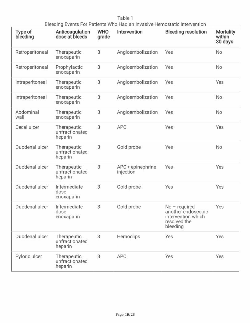
Of all 67 non-GI bleeds 5 patients (7.5%) required invasive interventions to control the bleeding with 100%success rate to resolve the bleeding event (Table 1). Thirty-four patients died within 30 days; 7 in patientswho had retroperitoneal and abdominal bleeds, 2 in patients who had hematuria, 15 in patients who hadNPA bleeds, 5 in patients who had brain bleeds and 5 were unclear.
At initial assessment, of all 55 GI bleeds, the source of bleeding was deemed to be upper GI in 12 patients,lower GI in 4 patients and 39 were unknown. Eighteen patients had esophagogastroduodenoscopy (EGD)(32.7%), and 5 had colonoscopy (9.1%). Of the 18 EGD, 13 (72.2%) were performed at time ofconsultation and 5 (27.3%) after failure of conservative treatment. Of the 5 colonoscopies, 4 (90%) wereperformed at time of consultation and 1 (10%) after failure of conservative treatment. The yield rate(having a positive �nding) for EGD was 88.8% (only two patients had normal EGDs) and 40% forcolonoscopies (three patients had normal colonoscopes and or were diagnosed with piles). Overall, theprevalence of invasive interventions for GI bleeds was 16.3% (8 upper and one lower GI bleeds) with 100%success rate (Table 1).
DOSE OF ANTICOAGULATION
Overall, 79 patients (67.7%) were on therapeutic anticoagulation, 15 patients (12.3%) were onintermediate dose, 22 patients (18%) were on prophylactic dose and 6 patients (5%) were on none. In thenon-GI bleeding group, 45 (67%) patients were on therapeutic anticoagulation, 9 (13.4%) were onintermediate dose, 11 (16.4%) patients were on prophylactic dose and 2 (3%) were on none. In the GIbleeding group, 34 (62%) were on therapeutic dose, 6 (11%) were on intermediate dose, 11 (20%) were onprophylactic dose and 4 (7.2%) were on none.
NON-GI BLEEDING

Page 7/28
a) Retroperitoneal, intraperitoneal and abdominal wall hematoma
We identi�ed 9 cases of retroperitoneal bleeding (7.3%). Three patients were on prophylacticanticoagulation, �ve were on therapeutic dose and one was on none. Only one patient was on antiplatelet(aspirin) as well as anticoagulation. Two patients (both were on prophylactic dose) requiredangioembolization to control the bleeding and both were resolved. The rest resolved with no intervention.Of the 7 patients who did not require interventions 3 died within 30 days of consultation from sepsis.
We identi�ed 2 intraperitoneal bleeding. Both patients were on therapeutic dose anticoagulation and bothrequired angioembolization to successfully control the bleeding. However, one of the two patients diedlater within 30 days from sepsis.
Five patients with abdominal wall hematomas were encountered. One patient was on intermediate doseand 4 on therapeutic dose. All bleeding resolved with noninvasive interventions except one which requitedangioembolization to control the bleeding. Of all patients with abdominal wall bleeding, one died within30 days after the consultation from COVID-19 multiorgan failure.
b) Hematuria
Twelve patients had hematuria. Four patients were on prophylactic dose, �ve on full dose and three onintermediate dose anticoagulation. All bleeding resolved without the need for invasive intervention. Twopatients died within 30 days.
c) Brain
Seven patients had cerebral bleeding events. Two patients were not on anticoagulation; one patientdeveloped subarachnoid hemorrhage which had stabilized without intervention, however, patient laterdied from sepsis and the other patient survived the bleeding event. Five patients were on therapeuticanticoagulation; 4 patients expired; one as a direct consequence from intracerebral bleeding (deemedinoperable) other following combined spontaneous subdural and epidural hematomas (deemedinoperable), and 2 had hemorrhagic infarcts (died from other comorbidities within 30 days).
d) Nasopharyngeal bleeding
We identi�ed 25 patients who had NPA bleeding. Diagnoses ranged from epistaxis, oral bleeding andtracheal site bleeding. Interventions, which were all noninvasive, included holding the anticoagulation tillbleeding resolved, nasal packing and administering vitamin K. All bleeding resolved with thesetechniques. Fifteen patients died within 30 days from sepsis. Of all NPA consultations, four patients wereon prophylactic dose, 3 on intermediate dose, and 18 on therapeutic dose anticoagulation. Of patientswho died, all were on therapeutic dose anticoagulation except two, one was on intermediate dose, and theother was on none.
GI BLEEDING

Page 8/28
Out of 55 patients who had signs of GI bleed at initial presentation 47 patients were managed with fulldose proton pump inhibitors (PPI) and active observation, of whom 6 failed this approach and requiredendoscopic intervention, �ve EGD and 1 colonoscopy.
Of the 19 patients who had endoscopic procedures 18 EGDs and 5 colonoscopies were performed. Ninepatients (16.3%) required an endoscopic hemostatic intervention to control the bleeding, all weresuccessful. Eight patients required upper GI hemostatic intervention and 1 lower GI hemostaticintervention. The upper GI interventions included gold probe, epinephrine injection, APC and hemoclipsapplications for bleeding ulcers. Bleeding resolved for all cases. Etiology for bleeding events wereduodenal & gastric ulcers in 10 patients (55.5%), esophagitis and Roux-en-Y anastomosis ulcer in 1patient, and gastritis in 5 patients (27.7%). Three patients died within 30 days of consultation. The onlylower GI endoscopic intervention was for large colonic ulcer in the cecum which was treated with APC.Bleeding resolved but patient later died from sepsis. Other colonoscopies done without interventions werefor; small rectal ulcer with piles for 1 patient, ischemic colitis for 1 patient, piles for 1 patient, and onecolonoscopy was essentially normal.
Overall, of 55 patients with signs of GI bleed, 38 (69.1%) expired within 30 days of consultation. Twopatients had unclear source of bleeding and died while still bleeding, both were on Plavix and therapeuticanticoagulation at the time of consultation. The rest died after resolution of the bleeding episode fromCOVID-19 complications.
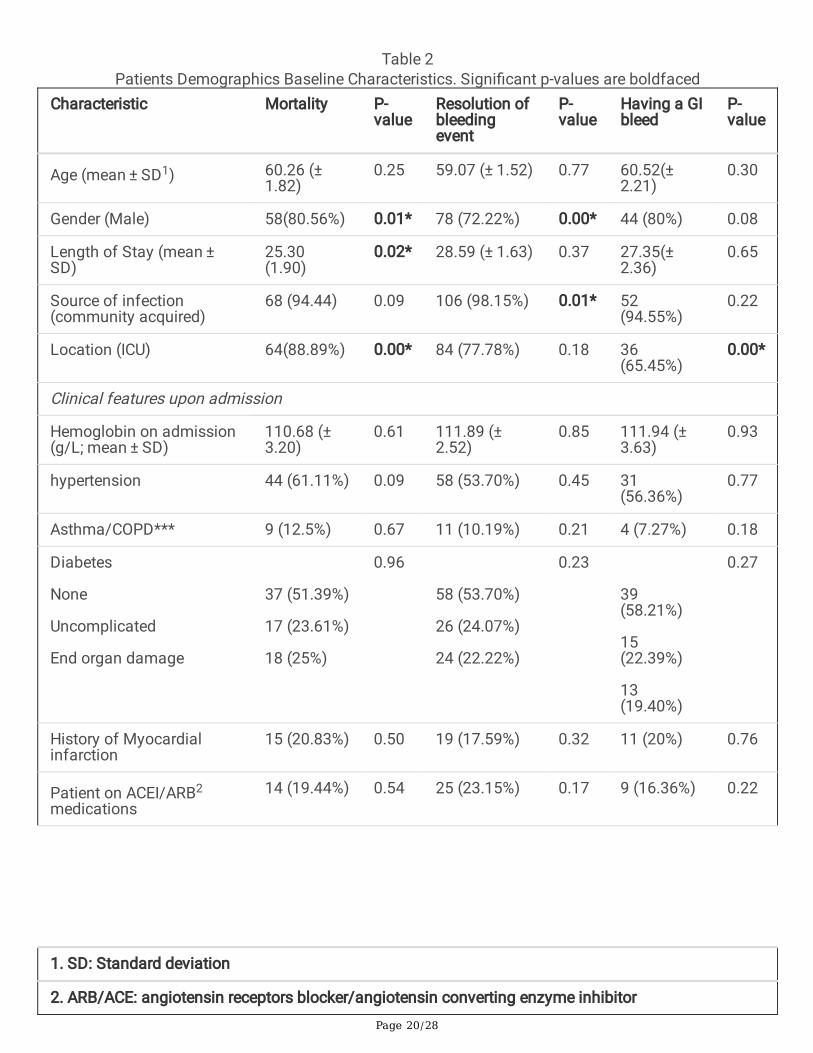
2) Demographics and baseline characteristics by primary outcomes, Univariate analysis (1) – Table 2
Mortality outcome
Male patients (80.56%) were signi�cantly more likely to die than female patients. The mean age forpatients who died was 60 years old. They were also signi�cantly more likely to be admitted to intensivecare unit (ICU) and had shorter hospital stay compared to patients who survived. Medical comorbiditiesat the time of admission did not have signi�cant implication on the risk of mortality. These includedhaving a history of hypertension requiring medications, asthma or chronic obstructive pulmonary disease(COPD), having complicated diabetes, remote history of myocardial infarction, or being on antiplatelets(single or dual) at the time of admission. Moreover, admission baseline hemoglobin level, before theonset of bleeding episode, did not affect the risk of mortality.
Resolution of bleeding event & having GI bleed outcomes
Gender appears to have a signi�cant association with bleeding resolution outcome, but it did not appearto in�uence the risk of having a GI bleeding compared to a non-GI bleed. ICU patients were more likely tohave a GI bleed than non-GI bleed.
3) Inpatient therapies and complications developed during patient’s admission by primary outcomes,Univariate analysis (2) – Table 2

Page 9/28
Mortality, resolution of bleeding event, & having GI bleed outcomes
The use of PPI, vasopressors, and inotropes appears to be signi�cantly associated with the risk of death.We also found GI bleeds to be signi�cantly associated with the need to use PPI and inotropes but notvasopressors. Furthermore, the dose but not the type of anticoagulation at the time of admission wassigni�cantly associated with risk of death following the bleeding episode. The dose of anticoagulationhowever did not in�uence bleeding event resolution or the type of bleeding.
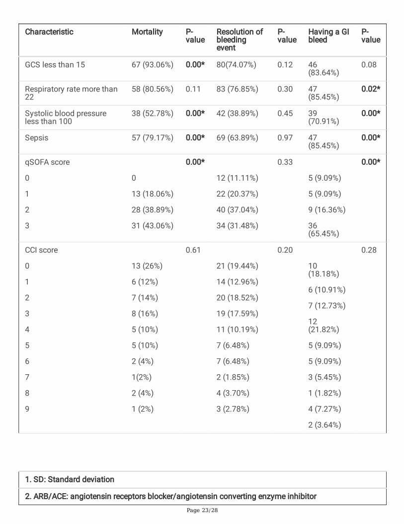
Being on systemic steroids appears to be signi�cantly associated with increased risk of death but not thebleeding resolution or the type of bleeding. Furthermore, being on invasive ventilation was associatedwith increased risk of mortality and having a GI bleed rather than a non-GI bleed. Moreover, severe ARDSwas associated with higher risk of mortality, but it did not affect the bleeding resolution or the type ofbleeding event. With regard to mortality outcome, having a cardiac injury, liver injury, acute kidney injury,Glasgow coma scale (GCS) less than 15, systolic blood pressure less than 100 and sepsis and highqSOFA score were associated with higher risk of death. However, the CCI score and the WHO bleedinggrade did not affect the risk of death. Furthermore, having cardiac injury, liver injury, being on renalreplacement therapy, having a respiratory rate over 22, systolic blood pressure less that 100, and sepsisappears to signi�cantly in�uence the type of bleeding event. Also, the qSOFA and WHO bleeding gradebut not CCI score signi�cantly affected the type of bleeding event. With regard to resolution of bleedingevent outcome, there was a signi�cant association with the WHO bleeding grade. None of thecomplications the patients developed during admission affected bleeding resolution chance.
4) At consultation symptoms, laboratory values and interventions by primary outcomes; Univariateanalysis (3) – Table 2
Mortality
Having an occult source of bleeding rather than a speci�c symptoms and signs indicative of a source ofbleeding was signi�cantly associated with the risk of death. Furthermore, hemoglobin level, white bloodcell (WBC) count, platelet, international normalized ratio (INR), D-dimer level, e-glomerular �ltration rate(eGFR) level, urea, creatinine, and C reactive protein (CRP) were signi�cantly associated with the risk ofdeath. There was no relationship between the type of intervention (invasive, noninvasive or hemostatic)and the risk of death within 30 days.
Resolution of bleeding event
We did not identify a signi�cant relationship between any coagulation pro�le derangements and theability to control bleeding episode. However, CRP and Procalcitonin (PCT) were signi�cantly associatedwith bleeding resolution.
Having GI bleed outcome

Page 10/28
Having an occult bleed appeared to be signi�cantly associated with having a GI bleed. Also, having highurea but not deranged coagulation pro�le was signi�cantly associated with having a GI bleed.Furthermore, there was a signi�cant relationship between the type of intervention (invasive, noninvasiveor hemostatic) and the type of bleeding event.
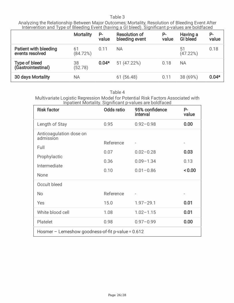
5) Relationships between major outcomes – Univariate analysis (4) – Table 3
We found having a GI bleeding event was signi�cantly associated with the risk of death (P = 0.04). Theprevalence of death following bleeding event was higher following a GI bleed compared to a non-GI bleed,52.7% vs. 48.3% respectively.
6) Risk factors predictors of primary outcomes
Mortality - Multivariate logistic regression (1) – Table 4
Patient who had longer hospital stay appeared to be less likely to die, odds ratio (OR) 0.95 (95% CI, 0.92-0.98, p= 0.003). We also found that patients who were on therapeutic dose of anticoagulation were morelikely to die compared to patients who were on none, on prophylactic or intermediate anticoagulationdoses. This risk appears to be signi�cant when therapeutic dose was compared to prophylactic dose, OR0.07 (95%CI 0.02-0.028, p=0.03) and no anticoagulation, OR 0.1 (95%CI0.97-0.99, p < 0.00) but notsigni�cant when compared to intermediate dose, OR 0.36 (95%CI 1.02-1.15, p=0.13). Furthermore, havingan occult bleeding appeared to be a signi�cant predictor of risk of death, OR 15 (95% CI 1.97-29.1, p=0.013). Also, WBC and platelet levels appeared to independently affect risk of death.
Resolution of bleeding event - multivariate logistic regression (2) – Table 5
Patients who were on PPI were more likely to have resolution of bleeding event compared to patients whowere not. Out of all GI symptoms and signs melena appeared to be signi�cantly associated with lowerodds of bleeding resolution, OR 0.03 (95%CI 0.01-0.18, p < 0.00). C-reactive protein appeared as well to besigni�cantly associated with lower odd of bleeding resolution, OR 0.98 (95% CI 0.97-0.99, p < 0.00).
Type of bleeding event - multivariate logistic regression (3)- Table 6
The risk of GI bleeding increased when patient was on inotropes (OR 7.33, 95%CI 1.03-55.28, p= 0.005),had cardiac injury (OR 6.73, 95%CI 0.92-49.43, p= 0.06), had liver injury (OR 74.08, 95%CI 4.18-132.08, p=0.03), had qSOFA score of 3 (OR 23.43, 95%CI 4.94-374.73, p= 0.02), had hematemesis (OR 19.79, 95%CI2.23-175.74 p= 0.00), and had occult bleed (OR 32.24, 95%CI 3.34-311.08, p= 0.00). The mortality variablehad poor correlation with the type of bleeding event on multivariate analysis model and so it wasremoved from the model.
Discussion
Page 11/28
To our knowledge, this is the largest and �rst study to evaluate the risk factors associated with GI andsurgical bleeding events in patients with COVID-19.
In our population WHO grade, which is representative of the volume of blood lost and thus indirectlyblood transfusion requirements, did not affect the risk of mortality. The majority of the bleeding eventsencountered in our population were WHO 2 or 3 (78.6%). This moderate degree of bleeding likely did notlead to hemodynamic instability and thus did not generate enough force to tip the patient toward shockwhen they were not or worsened an existing shock. Furthermore, we found newly developed, in hospital,medical comorbidities rather than pre-existing ones before admission to carry more weight on increasingthe risk of mortality following a bleeding event. Charlson score, contains both pre-existing and newlydeveloped medical comorbidities, and qSOFA score contains only acute ones. We found CCI score did notto affect the risk of mortality, but qSOFA score, acute cardiac injury, acute liver injury, acute kidney injury,in hospital GCS less than 15, in hospital systolic blood pressure less than 100, and sepsis, weresigni�cantly associated with increased risk of mortality.
Due to the hypothesized hemostatic derangement observed with COVID-19 which causes amicrothrombosis induced multiorgan failure and death (11) clinicians have been routinely prescribingintermediate and full therapeutic doses rather than prophylactic dose anticoagulation to prevent thispresumed phenomenon. Helms et al recently reported at least 40% thrombotic complications in patientswith COVID-19 (12), and Tang et. (13) al. has suggested mortality bene�ts with the use of anticoagulationin COVID-19 patients. However, all these studies suffer from small samples size and limited explorationof the known potential negative implications of higher doses of anticoagulation use. In our population wenoticed that the majority of patients who had head and neck bleeds (brain and NPA) ended up dying.Patients with brain bleeds who died in our population were deemed inoperable and all of them were ontherapeutic anticoagulation. Of all deaths in NPA bleeds all were on therapeutic and or intermediate doseanticoagulation except one. Dogra et al. (14) reported 4.4% of 755 patients diagnosed with COVID-19were found to have ICH on concurrent neuroimaging, of whom the majority of these patients were ontherapeutic anticoagulants. These are all indications that being on “supra-prophylactic” doseanticoagulation can put patient at increased risk of fatal head and neck bleeds. In our population, withregards to brain bleeds, it appears to be associated with high risk of mortality because by the time ithappened intervention seems too late in typical patient with limited physiological reserve in context ofCOVID-19 sepsis. With regards to NPA bleeds, despite bleeding resolution in all events, signi�cantproportion of patients eventually expired. This might be because the progressive “lingering” COVID-19associated platelet dysfunction and DIC rather than the acute bleeding event itself are the majorcontributors to the eventual death of patient with COVID-19 and so NPA bleeds should be considered red�ags for aggressive persistent COVID-19 coagulopathy, multiorgan failure and eventual death. Thus,efforts should be focused on correction and optimization of COVID-19 sepsis therapies andanticoagulation should be administered with caution in head neck bleeds subpopulation.
We are the �rst to identify a signi�cant association between the dose of anticoagulation in the setting ofbleeding event and risk of death. Speci�cally, we found patients who were on prophylactic dose or no

Page 12/28
anticoagulation appeared to be on lower risk of death compared to patient on therapeutic anticoagulationby 7%, and 10% respectively. When therapeutic dose was compared to intermediate dose, there was nosigni�cant difference in the risk of death. These �ndings put into question the routine unopposed practiceof prescribing “supra-prophylactic dose” anticoagulation to newly admitted COVID-19 patients andprobably these doses should be prescribed in selective cases only.
Moreover, we found that the dose of anticoagulation did not in�uence the risk of bleeding resolution northe type of bleeding. This might suggest that regardless of the dose of anticoagulation patient is on,noninvasive interventions such as withholding anticoagulation following bleed event, rather than aninvasive or hemostatic intervention are the major determinants of bleeding resolution, and so shouldalways be considered as �rst and primary line of intervention. This approach will save valuable resourcesand spare health care professionals unnecessary exposure.
Samkari et. al. (5) retrospective study of 400 admitted COVID-19 patients who were primarily receivingprophylactic dose of anticoagulation reported thrombocytopenia at initial presentation to be signi�cantpredictor of bleeding. In our study, we found, “chemical thrombocytopenia”, being on antiplatelets therapy(single, dual or even the novel ones), did not affect the risk of mortality, resolution of bleeding events northe type of bleeding. This might indicate that sepsis induced platelet dysfunction and eventually shockrather than thrombocytopenia itself signi�cantly interact with bleeding events variables.
In general, in acute GI bleeding events, endoscopy remains the �rst line intervention within 24 hours ofpatient stabilization. However, with the era of COVID-19, the risk bene�ts equation got more complex byconcerns for provider safety and a need to preserve personal protective equipment. Moreover, there arelimited data on the diagnostic and therapeutic bene�ts of endoscopy in this cohort, leaving endoscopistwith inadequate information and algorithms to guide their decision of when the risk of endoscopyoutweigh the bene�ts. In our population the prevalence of instigating diagnostic or therapeuticendoscopic is relatively low. However, this does not appear to be unique to our center. Salerno et. al. (15)looked at the impact of COVID-19 on urgent endoscopy in Italy. They reported a signi�cant reduction inthe number of urgent upper and lower GI endoscopy by 80% and 55% respectively. This reduction inendoscopy use was replicated in a Belgium study which reported 40% reduction in upper GI bleedingevents requiring endoscopy (16). Salerno et. al also reported that the signi�cant reduction in endoscopyuse was associated with increase in diagnostic yield by over 10% in the upper GI endoscopy group. Thiscorrelate well with our �ndings. Where even though our use of endoscopy to investigate our GI bleedingevents was relatively low, as endoscopy was preserved for patients with clinically signi�cant GI bleed andthose who failed conservative therapy, our diagnostic yield was relatively high for both upper and lower GIendoscopy, 88.8% and 60% respectively.
Martin et. al. (17) conducted a match case control study of 41 patients with COVID-19 who had bleedingevents (31 upper GI and 10 lower GI bleeds) compared to 82 COVID-19 patients who did not have GIbleeds. They found no difference in presenting symptoms and signs, no difference in severity of COVID-19 manifestations, and no difference in anticoagulation use. They reported most common cause of upper

Page 13/28
GI bleed was duodenal ulcer (80%), ours was 55.5% duodenal & gastric ulcers. For lower GI bleeding eventthey reported rectal tube insertions to be the most common cause (60%), our hospital does not use rectaltubes routinely. In their study, hemostatic interventions where successful in all cases who requiredintervention (n = 7), with no immediate postprocedural complications happened, and no interventionalradiology or surgical procedures were required. We had similar experience with 100% successfulinterventions for both non-GI and GI bleeds for all hemostatic interventions attempts. On multivariateanalysis they found having a previous history of upper GI bleed to be the only predictor of upper GIbleeding. We found being on inotropes, having liver injury, high qSOFA score, having hematemesis, andoccult bleeding to be signi�cant predictor of having a GI bleed. Furthermore, they found trends towardhigher risk of upper and lower bleeding events with being on anticoagulation, but this was not staticallysigni�cant. We as well did not �nd a signi�cant relationship between the dose of anticoagulation and therisk of having a GI bleed. Similar to Martin et. al. the majority of our study bleeding events ultimately hadcessation of bleeding without the need for hemostatic intervention. Furthermore, our GI bleedingpopulation had high mortality rate (69%) despite bleeding resolution in the majority of cases. Based on allthat, in COVID-19 era it appears to be safe to delay instigating endoscopic an intervention for GI bleeds,contrary to most guidelines which recommends early intervention for bleeding events. This will helpalleviate concerns toward patient’s respiratory status or illness severity, provider safety, PPE conservation,and the preservation of ventilators and avoiding procedural related intubations.
Finally, we found having a GI bleed was signi�cantly associated with increased risk of mortality (p = 0.04), despite bleeding event resolution in the majority of cases. We also found having an occult bleedwas associated with 15 times increased risk of death. This suggests that slow non profound bleeding asa consequence of COVID-19 coagulopathy might be a more signi�cant contributor to increased risk ofmortality rather an acute bleeding episode which is normally associated with hard signs of bleeding suchas hematemesis and melena. Based on that, efforts probably should be focused on correction ofcoagulopathy and sepsis rather than none targeted invasive GI interventions in patient with COVID-19,since most of these patients do not have identi�able source of bleeding amendable to invasivehemostatic intervention.
Limitations
One major limitation of the present study was that it was derived from a single institutional cross-sectional study with inherent selection and information bias, hence generalizability of the �ndings tolarger populations might not be representative. Further, our study had a limited sample size which led tothe in�ation of the ORs 95% CI, rendering them notably less precise. However, our inferences were basedon all available data on rare outcomes that were collected within a short time during the currentpandemic. Therefore, future studies should be focused on collecting more data to additionally validateour results.
Conclusions

Page 14/28
As COVID-19 pandemic evolves, surgeons and gastroenterologists are being confronted with uniquechallenges, particularly understanding the bleeding sequala of this novel virus. With the increasing use ofsupra-prophylactic doses of anticoagulation in this subpopulation the incidence of bleeding events, bothsurgical and GI, will be on the increase. We are the �rst to identify a signi�cant association between thedose of anticoagulation and risk of mortality. The previously unchallenged recommendation to prescribetherapeutic and or intermediate doses of anticoagulation to all newly admitted patients with COVID-19should be revisited and more individualized approach to prescription should be the norm.
Gastrointestinal bleeds appear to be associated with increased risk of mortality compared to non-GIbleeds, however, regardless of the source of bleeds the majority of bleeding events in COVID-19 patientsappear to resolve with noninvasive interventions and when hemostatic interventions were necessary ithad high success rate, despite the delay. This means that conservative management at the time ofconsultation seems to be a reasonable initial approach managing these complex cases, as most caseswill resolve without the need for intervention. This alleviates concerns regarding provider safety, and theneed to preserve personal protective equipment without jeopardizing patient safety and outcomes.
With regards to GI bleeds, endoscopic intervention should be limited to patients with hard signs of GIbleeds, such as hematemesis or melena. In patients with occult bleed efforts might be better gearedtoward optimizing therapies that manage COVID-19 sepsis induced coagulopathy, DIC and shock ratherthan none target low yield endoscopic interventions.
AbbreviationsCOVID-19: Coronavirus disease 19
GI: gastrointestinal
DIC: disseminated intravascular coagulation
ISTH: the international society of thrombosis and hemostasis
LMWH: low molecular weight heparin
UFH: unfractionated heparin
WHO: World Health Organization
PCR: polymerase chain reaction
ARDS: Acute respiratory distress syndrome
qSOFA: quick sequential organ failure assessment score
CCI: charlson comorbidity index
Page 15/28
NPA: nasopharyngeal
APC: argon positron coagulation
IQR: interquartile ranges
EGD: esophagogastroduodenoscopy
PPI: proton pump inhibitors
ICU: intensive care unit
COPD: chronic obstructive pulmonary disease
GCS: Glasgow coma scale
WBC: white blood cells
INR: international normalized ratio
eGFR: e-glomerular �ltration rate
CRP: C reactive protein
PCT: Procalcitonin
OR: odds ratio
Declarations
Ethics approval & consent to participate:The Ministry of Health of Kuwait which is the government branch at the state of Kuwait which manageJabir AL-Ahmad Hospital (our study center) approved the study.
Since the this is a retrospective review of prospectively maintained database, patient consent was notrequired to get ethical approval. To ensure patient con�dentiality patient identi�cation data wereanonymized.
Consent for publication section:The ethics committee approved publication of this manuscript
Availability of data and material:

Page 16/28
The dataset used and/or analysed during the current study available from the corresponding author onreasonable request
Finding:No funding was obtained for this study
Authors’ contribution:All authors read and approved the manuscript
AA contributed to study concept, design, data collection, analysis, draft preparation and �nal submission
YA contributed to study concept, design, data collection, analysis, draft preparation and �nal submission
KA contributed to study concept, design, data collection, analysis, draft preparation and �nal submission
EL contributed to study concept, design, data collection, analysis, draft preparation and �nal submission
MA contributed to study concept, design, data collection, analysis, draft preparation and �nal submission
AL contributed to study concept, design, data collection, analysis, draft preparation and �nal submission
MA contributed to study concept, design, data collection, analysis, draft preparation and �nal submission
MJ contributed to study concept, design, data collection, analysis, draft preparation and �nal submission
SA contributed to study concept, design, data collection, analysis, draft preparation and �nal submission
Acknowledgments:Not applicable
Data availability:All data used to create the analysis can be provided upon request
References1. Chen T, Wu D, Chen H, Yan W, Yang D, Chen G, Ma K, Xu D, Yu H, Wang H, Wang T, Guo W, Chen J,
Ding C, Zhang X, Huang J, Han M, Li S, Luo X, Zhao J, Ning Q. Clinical characteristics of 113deceased patients with coronavirus disease 2019: retrospective study. BMJ. 2020 Mar

Page 17/28
26;368:m1091. doi: 10.1136/bmj.m1091. Erratum in: BMJ. 2020 Mar 31;368:m1295. PMID:32217556; PMCID: PMC7190011.
2. Tang N, Bai H, Chen X, Gong J, Li D, Sun Z. Anticoagulant treatment is associated with decreasedmortality in severe coronavirus disease 2019 patients with coagulopathy. J Thromb Haemost. 2020May;18(5):1094-1099. doi: 10.1111/jth.14817. Epub 2020 Apr 27. PMID: 32220112.
3. Thachil J, Tang N, Gando S, Falanga A, Cattaneo M, Levi M, Clark C, Iba T. ISTH interim guidance onrecognition and management of coagulopathy in COVID-19. J Thromb Haemost. 2020May;18(5):1023-1026. doi: 10.1111/jth.14810. Epub 2020 Apr 27. PMID: 32338827.
4. Obi AT, Barnes GD, Wake�eld TW, Brown S, Eliason JL, Arndt E, Henke PK. Practical diagnosis andtreatment of suspected venous thromboembolism during COVID-19 pandemic. J Vasc Surg VenousLymphat Disord. 2020 Jul;8(4):526-534. doi: 10.1016/j.jvsv.2020.04.009. Epub 2020 Apr 17. PMID:32305585; PMCID: PMC7162794.
5. Al-Samkari H, Karp Leaf RS, Dzik WH, Carlson JCT, Fogerty AE, Waheed A, Goodarzi K, Bendapudi PK,Bornikova L, Gupta S, Leaf DE, Kuter DJ, Rosovsky RP. COVID-19 and coagulation: bleeding andthrombotic manifestations of SARS-CoV-2 infection. Blood. 2020 Jul 23;136(4):489-500. doi:10.1182/blood.2020006520. PMID: 32492712; PMCID: PMC7378457.
�. Cattaneo M, Bertinato EM, Birocchi S, Brizio C, Malavolta D, Manzoni M, Muscarella G, Orlandi M.Pulmonary Embolism or Pulmonary Thrombosis in COVID-19? Is the Recommendation to Use High-Dose Heparin for Thromboprophylaxis Justi�ed? Thromb Haemost. 2020 Aug;120(8):1230-1232.doi: 10.1055/s-0040-1712097. Epub 2020 Apr 29. PMID: 32349132; PMCID: PMC7516356.
7. Lucatelli P, De Rubeis G, Citone M, Lucarelli NM, Pasqualini V, Sturiale M, Giuliani S, Rosati M,Ceccherini C, Corona M, Mosconi C, Utili A, Argirò R. Heparin-Related Major Bleeding in Covid-19-Positive Patient: Perspective from the Outbreak. Cardiovasc Intervent Radiol. 2020 Aug;43(8):1216-1217. doi: 10.1007/s00270-020-02532-3. Epub 2020 May 28. PMID: 32468143; PMCID:PMC7255445.
�. World Health Organization. Clinical management of severe acute respiratory infection when novelcoronavirus (nCoV) infection is suspected. WHO; 2020.
9. R Core Team. R: A language and environment for statistical computing. R Foundation for StatisticalComputing, Vienna, Austria.URL: http://www.R-project.org/. . 2013; ISBN 3-900051-07-0.
10. Stekhoven, D. J. & Bühlmann, P. Missforest-Non-parametric missing value imputation for mixed-typedata. Bioinformatics 28, 112-118, doi:10.1093/bioinformatics/btr597 (2012).
11. Bikdeli B, Madhavan MV, Jimenez D, Chuich T, Dreyfus I, Driggin E, Nigoghossian C, Ageno W, MadjidM, Guo Y, Tang LV, Hu Y, Giri J, Cushman M, Quéré I, Dimakakos EP, Gibson CM, Lippi G, Favaloro EJ,Fareed J, Caprini JA, Tafur AJ, Burton JR, Francese DP, Wang EY, Falanga A, McLintock C, Hunt BJ,Spyropoulos AC, Barnes GD, Eikelboom JW, Weinberg I, Schulman S, Carrier M, Piazza G, BeckmanJA, Steg PG, Stone GW, Rosenkranz S, Goldhaber SZ, Parikh SA, Monreal M, Krumholz HM,Konstantinides SV, Weitz JI, Lip GYH; Global COVID-19 Thrombosis Collaborative Group, Endorsed bythe ISTH, NATF, ESVM, and the IUA, Supported by the ESC Working Group on Pulmonary Circulation

Page 18/28
and Right Ventricular Function. COVID-19 and Thrombotic or Thromboembolic Disease: Implicationsfor Prevention, Antithrombotic Therapy, and Follow-Up: JACC State-of-the-Art Review. J Am CollCardiol. 2020 Jun 16;75(23):2950-2973. doi: 10.1016/j.jacc.2020.04.031. Epub 2020 Apr 17. PMID:32311448; PMCID: PMC7164881.
12. Helms J, Tacquard C, Severac F, Leonard-Lorant I, Ohana M, Delabranche X, Merdji H, Clere-Jehl R,Schenck M, Fagot Gandet F, Fa�-Kremer S, Castelain V, Schneider F, Grunebaum L, Anglés-Cano E,Sattler L, Mertes PM, Meziani F; CRICS TRIGGERSEP Group (Clinical Research in Intensive Care andSepsis Trial Group for Global Evaluation and Research in Sepsis). High risk of thrombosis in patientswith severe SARS-CoV-2 infection: a multicenter prospective cohort study. Intensive Care Med. 2020Jun;46(6):1089-1098. doi: 10.1007/s00134-020-06062-x. Epub 2020 May 4. PMID: 32367170;PMCID: PMC7197634.
13. Tang N, Bai H, Chen X, Gong J, Li D, Sun Z. Anticoagulant treatment is associated with decreasedmortality in severe coronavirus disease 2019 patients with coagulopathy. J Thromb Haemost. 2020May;18(5):1094-1099. doi: 10.1111/jth.14817. Epub 2020 Apr 27. PMID: 32220112.
14. Dogra S, Jain R, Cao M, Bilaloglu S, Zagzag D, Hochman S, Lewis A, Melmed K, Hochman K, HorwitzL, Galetta S, Berger J. Hemorrhagic stroke and anticoagulation in COVID-19. J Stroke CerebrovascDis. 2020 Aug;29(8):104984. doi: 10.1016/j.jstrokecerebrovasdis.2020.104984. Epub 2020 May 23.PMID: 32689588; PMCID: PMC7245254.
15. Salerno R, Conti CB, De Silvestri A, Campbell Davies SE, Mezzina N, Ardizzone S. The impact ofcovid-19 pandemic on urgent endoscopy in Italy: a nation-wide multicenter study. Scand JGastroenterol. 2020 Jul;55(7):870-876. doi: 10.1080/00365521.2020.1782466. Epub 2020 Jul 2.PMID: 32615891.
1�. Schmiderer A, Schwaighofer H, Niederreiter L, Profanter C, Steinle H, Ziachehabi A, Tilg H. Decline inacute upper gastrointestinal bleeding during COVID-19 pandemic after initiation of lockdown inAustria. Endoscopy. 2020 Nov;52(11):1036-1038. doi: 10.1055/a-1178-4656. Epub 2020 May 14.PMID: 32408356; PMCID: PMC7653543.
17. Martin TA, Wan DW, Hajifathalian K, Tewani S, Shah SL, Mehta A, Kaplan A, Ghosh G, Choi AJ, KriskoTI, Fortune BE, Crawford CV, Sharaiha RZ. Gastrointestinal Bleeding in Patients With CoronavirusDisease 2019: A Matched Case-Control Study. Am J Gastroenterol. 2020 Oct;115(10):1609-1616. doi:10.14309/ajg.0000000000000805. PMID: 32796176; PMCID: PMC7446989.
Tables
Page 19/28
Table 1Bleeding Events For Patients Who Had an Invasive Hemostatic Intervention
Type ofbleeding
Anticoagulationdose at bleeds
WHOgrade
Intervention Bleeding resolution Mortalitywithin30 days
Retroperitoneal Therapeuticenoxaparin
3 Angioembolization Yes No
Retroperitoneal Prophylacticenoxaparin
3 Angioembolization Yes No
Intraperitoneal Therapeuticenoxaparin
3 Angioembolization Yes Yes
Intraperitoneal Therapeuticenoxaparin
3 Angioembolization Yes No
Abdominalwall
Therapeuticenoxaparin
3 Angioembolization Yes No
Cecal ulcer Therapeuticunfractionatedheparin
3 APC Yes Yes
Duodenal ulcer Therapeuticunfractionatedheparin
3 Gold probe Yes No
Duodenal ulcer Therapeuticunfractionatedheparin
3 APC + epinephrineinjection
Yes Yes
Duodenal ulcer Intermediatedoseenoxaparin
3 Gold probe Yes Yes
Duodenal ulcer Intermediatedoseenoxaparin
3 Gold probe No – requiredanother endoscopicintervention whichresolved thebleeding
Yes
Duodenal ulcer Therapeuticunfractionatedheparin
3 Hemoclips Yes Yes
Pyloric ulcer Therapeuticunfractionatedheparin
3 APC Yes Yes
Page 20/28
Table 2Patients Demographics Baseline Characteristics. Signi�cant p-values are boldfaced
Characteristic Mortality P-value
Resolution ofbleedingevent
P-value
Having a GIbleed
P-value
Age (mean ± SD1) 60.26 (± 1.82)
0.25 59.07 (± 1.52) 0.77 60.52(± 2.21)
0.30
Gender (Male) 58(80.56%) 0.01* 78 (72.22%) 0.00* 44 (80%) 0.08
Length of Stay (mean ± SD)
25.30(1.90)
0.02* 28.59 (± 1.63) 0.37 27.35(± 2.36)
0.65
Source of infection(community acquired)
68 (94.44) 0.09 106 (98.15%) 0.01* 52(94.55%)
0.22
Location (ICU) 64(88.89%) 0.00* 84 (77.78%) 0.18 36(65.45%)
0.00*
Clinical features upon admission
Hemoglobin on admission(g/L; mean ± SD)
110.68 (± 3.20)
0.61 111.89 (± 2.52)
0.85 111.94 (± 3.63)
0.93
hypertension 44 (61.11%) 0.09 58 (53.70%) 0.45 31(56.36%)
0.77
Asthma/COPD*** 9 (12.5%) 0.67 11 (10.19%) 0.21 4 (7.27%) 0.18
Diabetes
None
Uncomplicated
End organ damage
37 (51.39%)
17 (23.61%)
18 (25%)
0.96
58 (53.70%)
26 (24.07%)
24 (22.22%)
0.23
39(58.21%)
15(22.39%)
13(19.40%)
0.27
History of Myocardialinfarction
15 (20.83%) 0.50 19 (17.59%) 0.32 11 (20%) 0.76
Patient on ACEI/ARB2
medications14 (19.44%) 0.54 25 (23.15%) 0.17 9 (16.36%) 0.22
1. SD: Standard deviation
2. ARB/ACE: angiotensin receptors blocker/angiotensin converting enzyme inhibitor

Page 21/28
Characteristic Mortality P-value
Resolution ofbleedingevent
P-value
Having a GIbleed
P-value
Patient on antiplateletstherapy before admission
yes
no
18 (25%)
54 (75%)
0.36
23(21.30%)
85(78.70%)
0.53
13(23.64%)
42(76.36%)
0.71
Inpatient therapies and Complications developed during patients’ admissions
Use of proton pumpinhibitors
None
Prophylactic
Therapeutic
3 (4.17%)
53 (73.61%)
16 (22.22%)
0.04*
7 (6.48%)
87(80.56%)
14(12.96%)
0.06
6 (10.91%)
33 (60%)
16(29.09%)
0.00*
Vasopressor 45 (62.50%) 0.04* 57 (52.78%) 0.18 27(49.09%)
0.24
Inotropes 23 (31.94%) 0.00* 22 (20.37%) 0.92 22 (40%) 0.00*
Patient on antiplatelettherapy during admission
Yes
no
20 (27.78%)
52 (72.22%)
0.21
24(22.22%)
84(77.78%)
0.26
13(23.64%)
42(76.36%)
0.97
Anticoagulation dose onadmission
None
Prophylactic
Intermediate
Full
2 (2.78%)
5 (6.94%)
8 (11.1%)
57 (79.17%)
0.00*
6 (5.56%)
21(19.44%)
13(12.04%)
68(62.96%)
0.50
4 (7.27%)
11 (20%)
6 (10.91%)
34(61.82%)
0.65
1. SD: Standard deviation
2. ARB/ACE: angiotensin receptors blocker/angiotensin converting enzyme inhibitor

Page 22/28
Characteristic Mortality P-value
Resolution ofbleedingevent
P-value
Having a GIbleed
P-value
Type of anticoagulationused
Enoxoparin
Fondoparinux
Unfractionated heparin
34 (48.57%)
4 (5.71%)
32 (45.71%)
0.09
59(57.84%)
4(3.92%)
39(38.24%)
0.16
28(54.90%)
4 (7.84%)
19(37.25%)
0.06
Inhaled steroid 11 (15.28%) 0.39 16 (14.81) 0.12 11 (20%) 0.04*
Systemic steroid (dose inmg)
748.33 (± 98.60)
0.10 665.54 (± 67.96)
0.85 676.87(± 110.99)
0.82
systemic steroid
yes
no
56 (77.78%)
16 (22.22%)
0.00*
73 (67.59%)
35 (32.41%)
0.77
39(70.91%)
16(20.09%)
0.54
Oxygen therapy
Nasal
Mask
Invasive
1 (1.39%)
0
71 (98.61%)
0.00*
9(8.74%)
5(4.85%)
89(86.41%)
0.36
8 (14.55%)
0
47(85.45%)
0.00*
ARDS
Normal to mild
Moderate to severe
6 (8.33%)
66 (91.67%)
0.00*
24(23.30%)
79(76.70%)
0.00*
14(25.45%)
41(74.55%)
0.21
Cardiac injury 22 (30.56%) 0.00* 24(22.22%) 0.49 23(41.82%)
0.00*
Liver injury 11 (15.28%) 0.04* 11(10.19%) 0.64 12(21.82%)
0.00*
Acute kidney injury 56 (77.78%) 0.01* 75(69.44%) 0.88 40 (72.73) 0.50
Renal replacement therapy 28 (38.89%) 0.08 35(32.41%) 0.80 25(45.45%)
0.00*
1. SD: Standard deviation
2. ARB/ACE: angiotensin receptors blocker/angiotensin converting enzyme inhibitor
Page 23/28
Characteristic Mortality P-value
Resolution ofbleedingevent
P-value
Having a GIbleed
P-value
GCS less than 15 67 (93.06%) 0.00* 80(74.07%) 0.12 46(83.64%)
0.08
Respiratory rate more than22
58 (80.56%) 0.11 83 (76.85%) 0.30 47(85.45%)
0.02*
Systolic blood pressureless than 100
38 (52.78%) 0.00* 42 (38.89%) 0.45 39(70.91%)
0.00*
Sepsis 57 (79.17%) 0.00* 69 (63.89%) 0.97 47(85.45%)
0.00*
qSOFA score
0
1
2
3
0
13 (18.06%)
28 (38.89%)
31 (43.06%)
0.00*
12 (11.11%)
22 (20.37%)
40 (37.04%)
34 (31.48%)
0.33
5 (9.09%)
5 (9.09%)
9 (16.36%)
36(65.45%)
0.00*
CCI score
0
1
2
3
4
5
6
7
8
9
13 (26%)
6 (12%)
7 (14%)
8 (16%)
5 (10%)
5 (10%)
2 (4%)
1(2%)
2 (4%)
1 (2%)
0.61
21 (19.44%)
14 (12.96%)
20 (18.52%)
19 (17.59%)
11 (10.19%)
7 (6.48%)
7 (6.48%)
2 (1.85%)
4 (3.70%)
3 (2.78%)
0.20
10(18.18%)
6 (10.91%)
7 (12.73%)
12(21.82%)
5 (9.09%)
5 (9.09%)
3 (5.45%)
1 (1.82%)
4 (7.27%)
2 (3.64%)
0.28
1. SD: Standard deviation
2. ARB/ACE: angiotensin receptors blocker/angiotensin converting enzyme inhibitor

Page 24/28
Characteristic Mortality P-value
Resolution ofbleedingevent
P-value
Having a GIbleed
P-value
WHO grade
0
1
2
3
4
0
8 (16%)
26 (52%)
16 (32%)
0
0.05
6 (5.56%)
13 (12.04%)
51 (47.22%)
38 (35.19%)
0
0.00*
6 (10.91%)
3 (5.45%)
20(36.36%)
26(47.27%)
0
0.00*
At consultation Symptoms, laboratory values, and interventions
Hematemesis 10 (13.89%) 0.76 14(12.96%) 0.89 14(74.55%)
0.00*
Melena 26 (36.11%) 0.09 28 (25.93%) 000* 29(52.73%)
0.00*
Occult bleed 11 (15.28%) 0.04* 13 (12.04%) 0.17 11 (20%) 0.00*
Hemoglobin on consultday (g/L ;mean ± SD)
77.58 (± 2.26)
0.02* 81.81 (± 2.14) 0.39 75.30 (± 3.25)
0.00*
White blood cell (109/Lmean ± SD)
17.38 (± 1.20)
0.00* 15.63 (± 0.87) 0.92 16.49 (± 1.39)
0.31
Neutrophil (109/L ;mean ± SD)
15.88 (± 1.40)
0.08 14.25 (± 1.19) 0.81 13.18 (± 1.13)
0.32
Lymphocytes (109/L;mean ± SD)
1.85 (± 0.67)
0.87 1.44 (± 1.44) 0.01* 1.18 (± 0.11)
0.17
Hematocrit (L/L; mean ± SD)
0.26 (± 0.01)
0.22 0.87 (± 0.59) 0.70 0.26 (± 0.01)
0.36
Platelet (109/L; mean ± SD)
199.41(± 14.48)
0.00* 237.01 (± 13.05)
0.20 219.87 (± 17.03)
0.41
Prothrombin time(seconds; mean ± SD)
16.81 (± 0.58)
0.05 16.03 (± 0.39) 0.16 16.17 (± 0.38)
0.91
Activated partialthromboplastin time(seconds ; mean ± SD)
48.15 (± 2.63)
0.05 44.77 (± 2.03) 0.44 45.98 (± 3.39)
0.72
1. SD: Standard deviation
2. ARB/ACE: angiotensin receptors blocker/angiotensin converting enzyme inhibitor

Page 25/28
Characteristic Mortality P-value
Resolution ofbleedingevent
P-value
Having a GIbleed
P-value
International normalizedratio
1.23 (± 0.03)
0.01* 1.17 (± 0.01) 0.00* 1.19 (± 0.02)
0.66
D dimer (ng/mL ; mean ± SD)
3421.37 (± 234.11)
0.03* 3030.11 (± 203.31)
0.34 3062.92 (± 311.61)
0.88
Fibrinogen (g/L ; mean ± SD)
5.24 (± 0.18)
0.12 5.38 (± 0.12) 0.51 5.36 (± 0.17)
0.74
Estimated glomerular�ltration rate eGFR(mL/mins/1.73m2 ; mean ± SD)
42.31 (± 3.81)
0.00* 50.48 (± 3.54) 0.31 45.36 (± 4.95)
0.27
Urea (mmol/L) 26.19 (± 1.74)
0.00* 22.37 (± 1.42) 0.33 26.17 (± 2.27)
0.02*
Creatinine (µmol/L ; mean ± SD)
255.44 (± 24.98)
0.02* 214.38 (± 18.14)
0.21 246.34 (± 27.41)
0.21
CRP (mg/L ; mean ± SD) 173.69 (± 12.01)
0.00* 144.71 (± 9.76)
0.01* 145.25 (± 15.22)
0.44
Procalcitonin (ng/mL ;mean ± SD)
11.91 (± 3.16)
0.11 7.20 (± 1.11) 0.00* 5.44 (± 0.73)
0.06
Noninvasive intervention 60 (83.33%) 0.69 92 (85.19%) 0.52 41(74.55%)
0.00*
Invasive intervention 12 (16.67%) 0.92 17 (15.74%) 0.59 14(25.45%)
0.01*
Hemostatic intervention 6 (8.33%) 0.31 13 (12.04%) 0.17 8 (14.55%) 0.20
1. SD: Standard deviation
2. ARB/ACE: angiotensin receptors blocker/angiotensin converting enzyme inhibitor
Page 26/28
Table 3Analyzing the Relationship Between Major Outcomes; Mortality, Resolution of Bleeding Event After
Intervention and Type of Bleeding Event (having a GI bleed). Signi�cant p-values are boldfaced
Mortality P-value
Resolution ofbleeding event
P-value
Having aGI bleed
P-value
Patient with bleedingevents resolved
61(84.72%)
0.11 NA 51(47.22%)
0.18
Type of bleed(Gastrointestinal)
38(52.78)
0.04* 51 (47.22%) 0.18 NA
30 days Mortality NA 61 (56.48) 0.11 38 (69%) 0.04*
Table 4
Multivariate Logistic Regression Model for Potential Risk Factors Associated withInpatient Mortality. Signi�cant p-values are boldfaced
Risk factor Odds ratio 95% con�denceinterval
P-value
Length of Stay 0.95 0.92–0.98 0.00
Anticoagulation dose onadmission
Full
Prophylactic
Intermediate
None
Reference
0.07
0.36
0.10
-
0.02–0.28
0.09–1.34
0.01–0.86
-
0.03
0.13
< 0.00
Occult bleed
No
Yes
Reference
15.0
-
1.97–29.1
-
0.01
White blood cell 1.08 1.02–1.15 0.01
Platelet 0.98 0.97–0.99 0.00
Hosmer – Lemeshow goodness-of-�t p-value = 0.612

Page 27/28
Table 5Multivariate Logistic Regression Model for Potential Risk Factors
Associated With Resolved Bleeding Events. Signi�cant p-values areboldfaced
Risk factor Odds ratio 95% con�denceinterval
P-value
Use of proton pumpinhibitors
None
Prophylactic
Therapeutic
Reference
13.04
1.24
-
3.2–136.94
0.13–14.84
-
0.03
0.86
Melena
No
Yes
Reference
0.03
-
0.01–0.18
-
< 0.00
Melena
GI bleed
No GI bleed
-
0.03
-
0.01–0.26
-
0.00
CRP 0.98 0.97–0.99 0.00
Hosmer – Lemeshow goodness-of-�t p-value = 0.130

Page 28/28
Table 6Multivariate Logistic Regression Model for Potential RiskFactors Associated with the Type of Bleeding (having a
GI bleed). Signi�cant p-values are boldfacedRisk factor Odds
ratio95%con�denceinterval
P-value
Inotropes
No
Yes
Reference
7.33
-
1.03–55.28
-
0.05
Cardiac injury
No
Yes
Reference
6.73
-
0.92–49.43
-
0.06
Liver injury
No
Yes
Reference
74.08
-
4.18–132.08
-
0.00
Q-sofa score
0
1
2
3
Reference
0.19
0.08
23.43
-
0.02–1.89
0.06–2.18
4.94–374.73
-
0.15
0.06
0.02
Hematemesis
No
Yes
Reference
19.79
-
2.23–175.74
-
0.00
Occult bleed
No
Yes
Reference
32.24
-
3.34–311.08
-
0.00
Hosmer – Lemeshow goodness-of-�t p-value = 0.99