Embed Size (px)
Citation preview

Wireless Physiological Monitoring and Ocular Tracking: 3D Calibrationin a Fully-Immersive Virtual Health Care Environment
Lelin Zhang1, Yu Mike Chi1, Eve Edelstein1,2, Jurgen Schulze1, Klaus Gramann1, Alvaro Velasquez3,Gert Cauwenberghs1, and Eduardo Macagno1,2
Abstract— Wireless physiological/neurological monitoring invirtual reality (VR) offers a unique opportunity for unob-trusively quantifying human responses to precisely controlledand readily modulated VR representations of health careenvironments. Here we present such a wireless, light-weighthead-mounted system for measuring electrooculogram (EOG)and electroencephalogram (EEG) activity in human subjectsinteracting with and navigating in the Calit2 StarCAVE, afive-sided immersive 3-D visualization VR environment. Thesystem can be easily expanded to include other measurements,such as cardiac activity and galvanic skin responses. Wedemonstrate the capacity of the system to track focus of gazein 3-D and report a novel calibration procedure for estimatingeye movements from responses to the presentation of a setof dynamic visual cues in the StarCAVE. We discuss cyberand clinical applications that include a 3-D cursor for visualnavigation in VR interactive environments, and the monitor-ing of neurological and ocular dysfunction in vision/attentiondisorders.
I. INTRODUCTION
The complexity of modern health care introduces manyconfounding elements that may be associated with medicalerror and health care acquired harm [1], [2]. Although today’sclinicians have a multitude of electronic devices designedto promote safe medication practices, little has been doneto determine which visual stimuli distract clinicians duringperformance, or to design effective visual cues to reduceerror. Indeed, it is the multiplicity of equipment itself thatcan lead to delay or distraction when attempting to providecare [3], [4]. A key obstacle to determining the object ofattention, or inattention, is the lack of tracking devices ableto compute focus of gaze in space and time.
Here we report on the development of a unique human-machine interface that both records and responds to physio-logical and behavioral measures of subjects or patients im-mersed in virtual reality simulations of health care scenarios.The system is embedded in the StarCAVE, a fully immersive3-D visualization virtual reality (VR) environment in theCalifornia Institute of Telecommunications and InformationTechnology (CalIT2) [5]. CAVE-CAD, computer aided de-sign software developed by our interdisciplinary team foruse within the StarCAVE, maps user responses in 3-D spaceplus time. A real-time ‘bio-cursor’ uses electrooculography(EOG) synchronized with VR head tracking to reveal atten-tion to specific elements in the virtual environment. The bio-cursor is programmed to detect focus of gaze, and is furthercapable of detecting muscle and neural responses fromelectromyogenic (EMG) and electroenphalographic (EEG)
1 University of California San Diego, La Jolla, CA 920932 Academy of Neuroscience for Architecture, San Diego, CA 921013 HMC Architects, San Diego, CA 92101
Fig. 1. CalIT2 StarCAVE immersive visualization virtual reality environ-ment [6] for controlled human experiments in interactive health care andarchitecture [5].
biopotentials. The system provides real-time feedback ofeach eye’s location, providing a means to indicate the objectof focus within models projected in the StarCAVE. The bio-cursor’s ocular coordinate signals can be harnessed to enablehands-free control of the VR display, react to simulations,and to drive the interactive CAVE-CAD modeling softwarethat allows clinicians and architects to assay the functionof health care environments during the design process. Ulti-mately, we envision that the wireless bio-tracker will be usedto assay visual attention during real clinical procedures, inreal health care environments.
II. IMMERSIVE 3-D VISUALIZATION AND VIRTUALHEALTH CARE
A. StarCAVEThe StarCAVE at CalIT2, a five-sided virtual reality room
with stereo projections on 360-degree screens surroundingthe viewer [6], provides a central resource to this projectand serves as an immersive visualization virtual environmentfor controlled experiments in interactive health care [5].The StarCAVE offers 3-D stereo, 20/40 vision in a fullyhorizontally enclosed space with a diameter of 3 m andheight of 3.5 m. A combined resolution of over 68 millionpixels–34 million per eye, distributed over 15 rear-projectedwalls and two floor screens. Each of the five sides of theroom has three stacked screen tiles, with the bottom and topscreens titled inward by 15 degrees to increase the immersiveexperience, while reducing stereo ghosting. Each screen tileis served by a polarized pair of projectors, powered by ahigh-end, quad-core PC running on Linux, with dual nVIDIAgraphics processing units (GPUs) to generate highly complexstereo images, and with dual network cards to achieve gigabitEthernet/10GigE networking.
The StarCAVE environment is fully immersive, and inter-acts with the subject through a 3-D joystick as well as a head

tracking sensor system. The head tracking system installedon a hat worn by the subject registers the subject’s locationand orientation in space and projects 3-D visual fieldsaccordingly. Both the joystick and the head tracking systemuse four infra-red cameras that detect infra-red reflective ballsto map position and orientation. The actual 3-D position forthe viewer’s head as well as the joystick are calculated andlogged over time so that the viewer position and interactionsare dynamically tracked in the virtual setting.
B. CAVE-CADA major advantage of the StarCAVE VR environment
for fully immersive virtual health care is the capability todynamically alter the environment while logging subjectresponses in the design of controlled experiments. Our teamhas developed novel interactive computer-aided design soft-ware (CAVE-CAD) that enables experimenters to change thevisual configuration of scenarios while they are immersedin the StarCAVE. This approach eliminates the traditionalstep of creating a 3-D model at a desktop computer, beforebringing it into a virtual environment, thus allowing for muchshorter turnaround times when changes to the model areto be made and immediately visualized in VR. The useris immersed in the CAD “drawing” in full-scale, and hasthe ability to directly interact in 3D with the geometry andimmediately respond to changing geometries, materials andlighting. An example of a user navigating in virtual spacein the StarCAVE emulating an architectural environment isdepicted in Fig. 1.
The immersive and interactive capabilities of the Star-CAVE VR environment are further augmented with simul-taneous physiological and neurological monitoring of thesubject responses to enable a new class of controlled ex-periments in virtual health care.
III. PHYSIOLOGICAL AND NEUROLOGICAL MONITORING
We have developed and tested a customized non-contactbiopotential sensing and logging device that can detect andcollect EEG, EMG, EOG, and ECG (electrocardiogram)signals from the body and transmit the digitized waveformsover a Bluetooth wireless link [9]. The unobtrusive sensoroperates without conductive contact to the skin, and canbe mounted over hair or over clothing without conductivegel or other skin preparation. Other versions of the sensormake use of dry-contact sensors [7] as well as conductivefabric to integrate sensing into apparel worn by the user.These advances contribute to the mobility and simplicity ofthe subject experience during continuous brain and ocularactivity monitoring in the StarCAVE VR environment. TheEEG/EOG system directly interfaces with the StarCAVEcomputing platform through a Bluetooth communicationlink.
A. Wireless Integrated Biopotential SensorsThe recording system consists of a chain of active elec-
trodes connected along a single common wire. While thesystem is designed to operate with non-contact electrodesfor EEG and ECG use [8], [9] as shown in Fig. 2, it alsooperates with dry-contact [7] or standard gel-based wet-contact electrodes, and for other signal modalities such asEOG and EMG. The sensors can be either in direct contact
a ba b
Fig. 2. Non-contact EEG/ECG biopotential recording [9]. (a) Integratedbiopotential acquisition, filtering and decimation unit operating at 600µWpower. (b) EEG alpha wave and eye blink activity, recorded from theoccipital lobe over haired skull.
Fig. 3. Bio-cursor head-mask with six EOG electrodes.
with the skin or embedded within fabric and clothing. Asmall base unit powers the entire system and contains awireless transmitter to send data to a computer or otherexternal device. Near the base unit, a single adhesive ordry contact sensor placed anywhere convenient is used toestablish the ground reference for the system.
B. Wireless EOG 3-D Eye Tracking Bio-CursorAlthough EOG signals have found widespread use in
biomedicine for monitoring of ocular and vestibular eyemovement disorders as well as identifying of REM activityduring sleep staging, only recently EOG has revealed a con-sistent measure of eye gaze direction for tracking directionof gaze in human-computer interfaces [10]. Here we reportthe first use of EOG for tracking focus of gaze in 3-D, asa ‘bio-cursor’ user interface embedded in the StarCAVE VRenvironment.
We have adapted the wireless integrated biopotential arrayfor EOG use in the prototype 3-D eye tracking bio-cursor.The system uses DC-coupled gel-based wet-contact activeelectrodes rather than intrinsically AC-coupled capacitivenon-contact sensors [8], [9] to capture low-frequency com-ponents in the EOG signal that are required for continuouseye tracking.
The EOG head-mask is depicted in Figure 3. Six elec-trodes are positioned on the facial skin symmetrically aroundboth eyes, to record both horizontal and vertical differentialcomponents in the EOG signal. These EOG signals relay thedipole moments of both eyeballs and are sufficient to registerazimuth, elevation, and vergence of gaze through calibrationas outlined in Section IV.
Software embedded in the CAVE-CAD environment si-multaneously logs stimulus, head position and EOG/EEGanalog data converted at a rate of 400 samples/s to 2byte-digits, sent via Bluetooth to a Linux system. Thesystem is designed to record and log the EOG/EEG sig-nals synchronously with the user’s position and interactions

in the virtual world, cueing analysis based on physiolog-ical/neurological events of interest and virtual stimuli ofinterest.
A calibration procedure to quantify and optimize thecapacity of the EOG system to identify focus of gaze inthe 3-D VR field of view is presented next.
IV. CALIBRATION OF 3-D OCULAR MOVEMENTS
The EOG bio-cursor serves to dynamically track in 3-Dthe visual focus of the subject interacting with the VR healthcare environment. It is therefore critical to the performanceof the system to calibrate the mapping from EOG signals toa reliable and reproducible estimate of 3-D ocular focus inthe VR field of view. Since currently existing eye trackingsystems are limited to 2-D for use with standard flat displays,we developed a novel calibration method to perform themapping.
The calibration procedure correlates eye position withEOG within the 3-D field of view, linked to the viewer’s headlocation, in the StarCAVE. A dynamic calibration stimulusis presented in the form of a yellow ball moving throughvirtual 3-D space. The position of the ball is modulatedby three independent periodic wave functions, which eachindependently scan the space uniformly in the azimuth, ele-vation, and vergence dimensions. This modulation of the ballposition in 3-D virtual space relative to the head coordinatesof the subject guarantees a uniform spread in coverage of theangular deflections of the eye ball tracking stimulus acrossthe field of view.
A. EOG Model of Ocular Angular Deflection
Differentials between horizontally positioned EOG elec-trodes measure horizontal ocular deflection (azimuth), anddifferentials between vertically positioned EOG electrodesmeasure vertical ocular deflection (elevation) [10]. Further-more, a simple geometric model accounting for the EOG sig-nals in response to 3-D focus of gaze in stereo vision showsthat vergence in stereo vision can be obtained by differencingthe azimuth estimates of both eyes. The geometry of themodel is illustrated in Figure 4 (a), and yields approximateexpressions for the spherical coordinates (r, θ, φ) of thevisual target (focus of gaze) in terms of the azimuthal andelevation angular deflections (θl, φl) and (θr, φr) of the leftand right eyeballs, in the limit where the distance r (or‘vergence’ between the target and the center of the eyes issignificantly larger than the distance d between the eyes:
θ ≈ θl + θr2
φ ≈ φl + φr2
(1)
r ≈ d cos θ
cosφ (θl − θr)These expressions show that while the azimuth θ and eleva-tion φ of the focus of gaze directly correspond to the averageazimuth and elevation of the ocular angular deflections, thevergence is inversely proportional to the difference betweenthe two ocular azimuth angles θl − θr. The challenge inaccurately estimating vergence is to resolve small azimuthdifferences in already small differential EOG signals.
For small angular deflections θ and φ, the EOG electrodevoltages Vi (i = 1, . . . 6) are approximately linear in θ andφ, and furthermore cos(θ) ≈ 1 and cos(φ) ≈ 1 in (1) so thatan approximate linear relationship can be assumed betweenthe vergence coordinates and EOG electrode voltages: θ
φdr
=W
V1...V6
(2)
where W is a matrix of parameters that depend on thegeometry of EOG sensor placements relative to the ocularframe of reference, and where constant DC offsets have beensubtracted out. Note the inverse dependence on vergencer in the variable d/r for linearity and the scaling by theinter-ocular spacing d for a dimensionless representation.The linear relationship (2) is the basis of the calibrationprocedure.
B. Calibration StimulusRather than computing W from the geometry, we calibrate
the parameters in W from measurements by regressingthe model (2) under a known calibration visual stimulus.For effective calibration under noisy EOG measurementconditions, it is important to choose a calibration visualstimulus that most uniformly excites the dynamic range ofthe variables under regression. We chose a triple-harmonicstimulus
θ = A1 cos(ω1t)
φ = A2 cos(ω2t) (3)d
r= A3 cos(ω3t)
with angular frequencies ω1, ω2 and ω3 randomly in the [0.8Hz, 1.2 Hz] interval, and suitably small amplitudes A1, A2
and A3. The length of the calibration interval is chosen muchlarger than 1/mini 6=j |ωi − ωj |, and is 200 seconds in thispilot study.
V. RESULTS
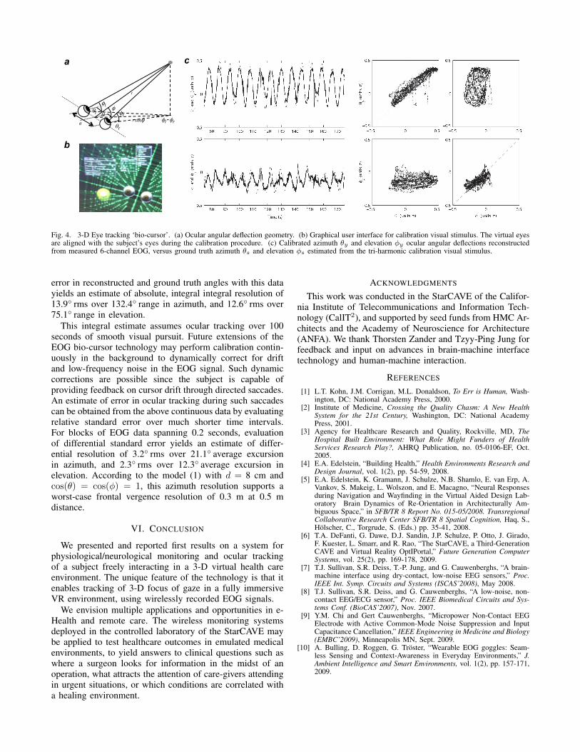
The above calibration procedure was performed off-line ontime-stamped EOG data recorded simultaneously with thesequence of the dynamic calibration stimulus presented tothe subject in StarCAVE. A screenshot of the graphical userinterface for EOG data collection, integrated in the CAVE-CAD software environment, is shown in 4 (b). The grid(green dots) super-imposed on the calibration stimulus (yel-low ball) serves as a reference for head fixation throughoutthe data collection. Multiple sessions, each with 200 secondsof continuous 6-channel EOG data, and with various tri-harmonic calibration stimuli (3), were collected on a malesubject.
Results of the calibration on a 100 second fragment of themeasured EOG data are given in Figure 4 (c), showing thecalibrated azimuth θy and elevation φy reconstructed fromthe EOG data, relative to the ground truth azimuth θs andelevation φs estimated from the calibration visual stimulus.No effort was made to eliminate signal artifacts in the data,which are clearly visible, such as eye blinks and involuntarysaccades during the recording, as well as sources of impulsenoise in the EOG measurement. Evaluation of the standard
a
b
c
r cos!"
r
!!l
!r #l
#r
# d
!l - !r
Fig. 4. 3-D Eye tracking ‘bio-cursor’. (a) Ocular angular deflection geometry. (b) Graphical user interface for calibration visual stimulus. The virtual eyesare aligned with the subject’s eyes during the calibration procedure. (c) Calibrated azimuth θy and elevation φy ocular angular deflections reconstructedfrom measured 6-channel EOG, versus ground truth azimuth θs and elevation φs estimated from the tri-harmonic calibration visual stimulus.
error in reconstructed and ground truth angles with this datayields an estimate of absolute, integral integral resolution of13.9◦ rms over 132.4◦ range in azimuth, and 12.6◦ rms over75.1◦ range in elevation.
This integral estimate assumes ocular tracking over 100seconds of smooth visual pursuit. Future extensions of theEOG bio-cursor technology may perform calibration contin-uously in the background to dynamically correct for driftand low-frequency noise in the EOG signal. Such dynamiccorrections are possible since the subject is capable ofproviding feedback on cursor drift through directed saccades.An estimate of error in ocular tracking during such saccadescan be obtained from the above continuous data by evaluatingrelative standard error over much shorter time intervals.For blocks of EOG data spanning 0.2 seconds, evaluationof differential standard error yields an estimate of differ-ential resolution of 3.2◦ rms over 21.1◦ average excursionin azimuth, and 2.3◦ rms over 12.3◦ average excursion inelevation. According to the model (1) with d = 8 cm andcos(θ) = cos(φ) = 1, this azimuth resolution supports aworst-case frontal vergence resolution of 0.3 m at 0.5 mdistance.
VI. CONCLUSION
We presented and reported first results on a system forphysiological/neurological monitoring and ocular trackingof a subject freely interacting in a 3-D virtual health careenvironment. The unique feature of the technology is that itenables tracking of 3-D focus of gaze in a fully immersiveVR environment, using wirelessly recorded EOG signals.
We envision multiple applications and opportunities in e-Health and remote care. The wireless monitoring systemsdeployed in the controlled laboratory of the StarCAVE maybe applied to test healthcare outcomes in emulated medicalenvironments, to yield answers to clinical questions such aswhere a surgeon looks for information in the midst of anoperation, what attracts the attention of care-givers attendingin urgent situations, or which conditions are correlated witha healing environment.
ACKNOWLEDGMENTS
This work was conducted in the StarCAVE of the Califor-nia Institute of Telecommunications and Information Tech-nology (CalIT2), and supported by seed funds from HMC Ar-chitects and the Academy of Neuroscience for Architecture(ANFA). We thank Thorsten Zander and Tzyy-Ping Jung forfeedback and input on advances in brain-machine interfacetechnology and human-machine interaction.
REFERENCES
[1] L.T. Kohn, J.M. Corrigan, M.L. Donaldson, To Err is Human, Wash-ington, DC: National Academy Press, 2000.
[2] Institute of Medicine, Crossing the Quality Chasm: A New HealthSystem for the 21st Century, Washington, DC: National AcademyPress, 2001.
[3] Agency for Healthcare Research and Quality, Rockville, MD, TheHospital Built Environment: What Role Might Funders of HealthServices Research Play?, AHRQ Publication, no. 05-0106-EF, Oct.2005.
[4] E.A. Edelstein, “Building Health,” Health Environments Research andDesign Journal, vol. 1(2), pp. 54-59, 2008.
[5] E.A. Edelstein, K. Gramann, J. Schulze, N.B. Shamlo, E. van Erp, A.Vankov, S. Makeig, L. Wolszon, and E. Macagno, “Neural Responsesduring Navigation and Wayfinding in the Virtual Aided Design Lab-oratory Brain Dynamics of Re-Orientation in Architecturally Am-biguous Space,” in SFB/TR 8 Report No. 015-05/2008. TransregionalCollaborative Research Center SFB/TR 8 Spatial Cognition, Haq, S.,Holscher, C., Torgrude, S. (Eds.) pp. 35-41, 2008.
[6] T.A. DeFanti, G. Dawe, D.J. Sandin, J.P. Schulze, P. Otto, J. Girado,F. Kuester, L. Smarr, and R. Rao, “The StarCAVE, a Third-GenerationCAVE and Virtual Reality OptIPortal,” Future Generation ComputerSystems, vol. 25(2), pp. 169-178, 2009.
[7] T.J. Sullivan, S.R. Deiss, T.-P. Jung, and G. Cauwenberghs, “A brain-machine interface using dry-contact, low-noise EEG sensors,” Proc.IEEE Int. Symp. Circuits and Systems (ISCAS’2008), May 2008.
[8] T.J. Sullivan, S.R. Deiss, and G. Cauwenberghs, “A low-noise, non-contact EEG/ECG sensor,” Proc. IEEE Biomedical Circuits and Sys-tems Conf. (BioCAS’2007), Nov. 2007.
[9] Y.M. Chi and Gert Cauwenberghs, “Micropower Non-Contact EEGElectrode with Active Common-Mode Noise Suppression and InputCapacitance Cancellation,” IEEE Engineering in Medicine and Biology(EMBC’2009), Minneapolis MN, Sept. 2009.
[10] A. Bulling, D. Roggen, G. Troster, “Wearable EOG goggles: Seam-less Sensing and Context-Awareness in Everyday Environments,” J.Ambient Intelligence and Smart Environments, vol. 1(2), pp. 157-171,2009.