Embed Size (px)
Citation preview
Wilms Tumor Gene on X Chromosome (WTX) InhibitsDegradation of NRF2 Protein through Competitive Binding toKEAP1 Protein*□S
Received for publication, October 24, 2011, and in revised form, December 16, 2011 Published, JBC Papers in Press, January 3, 2012, DOI 10.1074/jbc.M111.316471
Nathan D. Camp‡, Richard G. James‡, David W. Dawson§, Feng Yan¶, James M. Davison¶, Scott A. Houck¶,Xiaobo Tang�, Ning Zheng�1, Michael B. Major¶2, and Randall T. Moon‡1,3
From the ‡Howard Hughes Medical Institute, Department of Pharmacology, Institute for Stem Cell and Regenerative Medicine,University of Washington School of Medicine, Seattle, Washington 98195, the §Department of Pathology and Laboratory Medicine,Jonsson Comprehensive Cancer Center, David Geffen School of Medicine, UCLA, Los Angeles, California 90095-1732, the¶Department of Cell and Developmental Biology, Lineberger Comprehensive Cancer Center, University of North Carolina School ofMedicine, Chapel Hill, North Carolina 27599, and the �Howard Hughes Medical Institute, Department of Pharmacology, Universityof Washington School of Medicine, Seattle, Washington 98195
Background:KEAP1 is a ubiquitin ligase adaptor that promotes the ubiquitination anddegradation ofNRF2, a transcriptionfactor that drives the antioxidant response.Results:Wilms tumor gene on the X chromosome (WTX) stabilizes NRF2 by competing with NRF2 for binding to KEAP1.Conclusion:WTX regulates the antioxidant response.Significance: This study reveals a novel regulatory mechanism governing the antioxidant response.
WTX is a tumor suppressor protein that is lost or mutated inup to 30% of cases ofWilms tumor. Among its known functions,WTX interacts with the �-transducin repeat containing familyof ubiquitin ligase adaptors and promotes the ubiquitinationanddegradationof the transcription factor�-catenin, a key con-trol point in the WNT/�-catenin signaling pathway. Here, wereport that WTX interacts with a second ubiquitin ligase adap-tor, KEAP1, which functions to regulate the ubiquitination ofthe transcription factor NRF2, a key control point in the antiox-idant response. Surprisingly, we find that unlike its ability topromote the ubiquitination of �-catenin, WTX inhibits theubiquitination ofNRF2.WTXandNRF2 compete for binding toKEAP1, and thus loss ofWTX leads to rapid ubiquitination anddegradation ofNRF2 and a reduced response to cytotoxic insult.These results expand our understanding of themolecularmech-anisms of WTX and reveal a novel regulatory mechanism gov-erning the antioxidant response.
FAM123B/WTX/AMER1 (hereafter referred to as WTX) islocated on the X chromosome and encodes a tumor suppressorprotein that is lost ormutated in up to 30%of the cases ofWilmstumor, the most common pediatric kidney cancer (1–3).Recently, germ line mutations inWTX were also discovered infamilies suffering from osteopathia striata congenital with cra-
nial sclerosis (OSCS),4 a debilitating skeletal dysplasia that isoften accompanied by developmental abnormalities and fatal-ity in males (4, 5).We previously reported that WTX regulates the stability of
�-catenin (6). The regulation of the stability of �-catenin is akey control point of theWNT/�-catenin signaling pathway andthe broader protein networks with which it interacts (7). In theabsence ofWNT ligand, �-catenin is phosphorylated by a mul-tiprotein complex often called the “destruction complex” and issubsequently recognized by the SCFBTRC (Skp, Cullin, F-box)ubiquitin ligase complex where it is ubiquitinated and targetedfor proteasomal degradation (8–10). In the presence of WNTligand, phosphorylation of �-catenin is attenuated, resulting inthe nuclear accumulation of �-catenin and the regulation oftranscription. Through proteomic and functional dissection ofthe WNT/�-catenin signaling pathway, we discovered thatWTX associates with �-catenin as well as proteins in thedestruction complex, including adenomatous polyposis coli(APC), AXIN1, BTRC (commonly referred to as �-TrCP), andFBXW11 (commonly referred to as�-TrCP2) (6). Although theprecise mechanism(s) is unknown, WTX promotes the ubiq-uitination and degradation of �-catenin.
In addition to regulating the stability of �-catenin, WTX hasalso been shown to play a role in regulating WNT signal trans-duction at the membrane (11). Furthermore, WTX controlscell-cell adhesion through interactions with APC at the plasmamembrane (12) and modulates the activity of theWT1 (Wilmstumor 1) transcription factor in the nucleus (13). Thus, it ispossible that the loss of WTX contributes to disease throughdistinct mechanisms in specific subcellular compartments.
* This work was supported, in whole or in part, by National Institutes of HealthGrant T32 GM07270 from USPHS and NRSA (to N. D. C.).Author’s Choice—Final version full access.
□S This article contains supplemental Figs. S1–S3.1 Investigator of the Howard Hughes Medical Institute.2 Supported by a National Institutes of Health Directors New Innovator Award
and a Sidney Kimmel Scholar Award. To whom correspondence may beaddressed: 450 West Dr., Rm. 31-351, Chapel Hill, NC 27599. Tel.: 919-259-2695; E-mail: [email protected].
3 To whom correspondence may be addressed: 815 Mercer St., Rm. S524,Seattle, WA 98109-358056. Tel.: 206-543-1722; E-mail: [email protected].
4 The abbreviations used are: OSCS, osteopathia striata congenital withcranial sclerosis; APC, adenomatous polyposis coli; ARE, antioxi-dant-response element; BTRC, �-transducin repeat containing; tBHQ,tert-butylhydroquinone.
THE JOURNAL OF BIOLOGICAL CHEMISTRY VOL. 287, NO. 9, pp. 6539 –6550, February 24, 2012Author’s Choice © 2012 by The American Society for Biochemistry and Molecular Biology, Inc. Published in the U.S.A.
FEBRUARY 24, 2012 • VOLUME 287 • NUMBER 9 JOURNAL OF BIOLOGICAL CHEMISTRY 6539
at University of N
orth Carolina at C
hapel Hill, on A
ugust 23, 2012w
ww
.jbc.orgD
ownloaded from
http://www.jbc.org/content/suppl/2012/01/03/M111.316471.DC1.html Supplemental Material can be found at:
Our previous analysis of the WTX protein interaction net-work identified an association between WTX and KEAP1(Kelch-like ECH-associated protein 1) (6). KEAP1 is a substraterecognitionmodule for theCUL3-basedE3ubiquitin ligase thatconstitutively ubiquitinates the transcription factor NRF2 (NF-E2-related factor-2; NFE2L2) (14–16). In the presence of cyto-toxic stress such as xenobiotics and antioxidants, KEAP1 isinhibited, and NRF2 is no longer targeted for ubiquitinationand degradation. NRF2 then accumulates in the nucleus whereit regulates transcription of genes involved in the “phase II”antioxidant response (17–19).Although many studies have reported that small molecules
and stressors regulate NRF2 stability and function, relativelylittle is known about regulation of KEAP1 and NRF2 throughprotein-protein interactions. Here, we report that WTXdirectly binds to KEAP1. Using gain-of-function and loss-of-function approaches, we found that WTX stabilizes NRF2 andpositively regulates its transcriptional activity.We identified anETGEmotifwithin theKEAP1 interacting domain ofWTX thatis nearly identical to that of NRF2. We show that this motif isnot only required for the interaction ofWTXwithKEAP1 but isalso required for the ability of WTX to regulate NRF2 stabilityand activity. Our observations support a model whereby WTXcompetes withNRF2 for binding to KEAP1, thereby promotingthe NRF2-mediated antioxidant response.
EXPERIMENTAL PROCEDURES
Tissue Culture, Transfections, and Small Interfering RNAs—All cell lines were grown in DMEM supplemented with 10%fetal bovine serum in a 37 °C humidified incubator with 5%CO2. Selection and passage of stable cell lines were performedwith 1.5 �g/ml puromycin until cell death was no long appar-ent. Expression constructs were transiently transfected inHEK293T cells with Lipofectamine 2000 as directed by themanufacturer (Invitrogen). Transient transfection of siRNAwas performed with Lipofectamine RNAiMAX, as directed bythe manufacturer (Invitrogen). Sequences of the siRNA sensestrands are as follows: WTX-A (CCU GGA GAU GAC UGCCUU U dTdT), WTX-B (UAU GCC AGG GAG GCC CAC AdTdT), NRF2 (GUAAGAAGCCAGAUGUUAA dTdT), andKEAP1 (GGG CGU GGC UGU CCU CAA U dTdT). ControlsiRNA was acquired from Ambion.Plasmids and Expression Vectors—NRF2, WTX, KEAP1,
CUL3, and GFP cDNAs were created with standard PCR-basedcloning strategies. The reporter gene fusion construct forhuman NQO1-ARE (hNQO1-ARE-luciferase) was a kind giftfrom Jeffrey Johnson.WTX Antibody Production—Amino acids 212–438 of WTX
were expressed as a GST fusion protein in Escherichia coli.Purified GST-tagged WTX fragment was used as an immuno-gen in rabbits, following established protocols by Cocalico Bio-logicals, Inc. Prior to use in Western blot analysis or immuno-staining, the antibody was affinity-purified over protein Gbeads.Affinity Pulldowns and Western Blotting—For streptavidin
affinity purification, cells were lysed in radioimmunoprecipita-tion buffer (RIPA: 25 mM Tris-HCl, pH 8.0, 150 mM NaCl, 10%glycerol, 1% Triton X-100, 0.25% deoxycholic acid, 2 mM
EDTA) containing protease inhibitor mixture (Roche AppliedScience) and phosphatase inhibitormixture (Calbiochem). Celllysates were cleared by centrifugation and incubated withstreptavidin resin (Amersham Biosciences) before washingwith lysis buffer and eluting in NuPAGE loading buffer (Invit-rogen). Detection of proteins by Western blot was performedusing the following antibodies: anti-NRF2 (H-300) polyclonal(sc-13032, Santa Cruz Biotechnology), anti-KEAP1 polyclonal(10503–2-AP, Proteintech), anti-HMOX1 monoclonal(ab13248; Abcam), anti-FLAG M2 monoclonal (Sigma),anti-HA polyclonal (1867423; Roche Applied Science), anti-CTNNB1 polyclonal (9562; Cell Signaling Technology), anti-GFP polyclonal (ab290, Abcam), and anti-TUBB1 monoclonal(T7816; Sigma).In Vitro Binding Experiments—HumanGST-VSV-WTXwas
purified fromE. coli andmixedwith purifiedCUL3orKEAP1 inbuffered 150mMNaCl. Following incubation for 30min at 4 °C,complexes were washed with five bed volumes of 350 mM buff-ered NaCl before elution and Western blot.ARE-Luciferase Quantification—For DNA, HEK293T cells
were transfected with expression constructs, ARE-luciferase(firefly), and a control plasmid containing Renilla luciferasedriven by a constitutive cytomegalovirus (CMV) promoter fornormalization. Approximately 6 h post-transfection, cells weretreated with 100 �M tBHQ and incubated for an additional12–16 h. Activation was measured as the ratio of firefly toRenilla luciferase activity. For siRNA, HEK293T cells stablyexpressing the ARE-luciferase and Renilla control reporterswere transfected with siRNA. Approximately 48 h later, 100�M
tBHQ was added, and cells were incubated for an additional16 h. Activation was then measured as described above.NQO1-GFP Quantification—H1299 cells containing a YFP
fragment retrovirally inserted into intron 1 of the NQO1 gene(170407PL1A2-NQO1) were a gift fromUri Alon and the KahnProtein Dynamics group. Ten thousand cells were seeded intoeach well of a 96-well clear bottom white plate. Following an18-h incubation period, cells were transfected with siRNA.After an additional 24 h, 100 �M tBHQ was added, and cellswere incubated for an additional 24 h. Cells were lysed in RIPAbuffer, and fluorescence at �ex � 485 nm was determined.Cell Viability—HEK293T cells were transfected with siRNA
as described above in 6-well tissue-culture plates. Twenty hourspost-transfection, 30,000 cells were seeded into each well of a96-well plate. Approximately 8 h later, cells were treated witheither DMSO (vehicle) or etoposide. The 3-(4,5-dimethylthi-azol-2-yl)-2,5-diphenyltetrazolium bromide cell proliferationassay was performed 36 h post-treatment as described by themanufacturer (ATCC, catalog no. 30-1010K-A).RNA Isolation, Reverse Transcription, and Semi-quantitative
Real Time-PCR—Total RNA fromcellswas harvested inTRIzol(Invitrogen) reagent according to the manufacturer’s instruc-tions. RNA was quantified by UV spectrophotometry, andcDNAwas created using the RevertAid First Strand cDNA syn-thesis kit (Fermentas). PCR was performed in duplicate withthe LightCycler FastStart DNASYBRGreen kit (RocheAppliedScience) using the LightCycler 480 instrument (Roche AppliedScience). The PCR conditions are as follows: 35 cycles of ampli-fication with 1 s denaturation at 95 °C and 5 s annealing at
WTX/FAM123B Regulates NRF2 Protein Stability
6540 JOURNAL OF BIOLOGICAL CHEMISTRY VOLUME 287 • NUMBER 9 • FEBRUARY 24, 2012
at University of N
orth Carolina at C
hapel Hill, on A
ugust 23, 2012w
ww
.jbc.orgD
ownloaded from
58 °C. A template free negative control was included in eachexperiment. Quantitative light cycler PCR primers used are asfollows:WTX (GAC CCAAAAGGATGAAGC T and reverseCCC CTC CAA AGA AAC TAG GC) and �-actin (AGA GCAAGA GAG GCA TCC TC and reverse CTC AAA CAT GATCTG GGT CA).Cellular Extract Degradation—HEK293T cells were trans-
fected with eitherWTX-A or control siRNA. Forty eight hourspost-transfection, cells were resuspended in swelling buffer (20mMHEPES, pH 7.7, 5mMMgCl2, 5mMKCl, 1mMDTT, 0.2mM
ATP, and protease inhibitor) at a ratio of 4:3. Resuspended pel-lets were subjected to two rounds of freeze/thaw and passedthrough a chilled 27-gauge needle two times. Lysates were thencentrifuged at 2,300 � g for 5 min, and supernatant was trans-ferred to a new tube and centrifuged for 30 min at 16,000 � g.The middle layer containing the cytosolic fraction was isolatedandused in subsequentNRF2degradation experiments. In vitrotranscription and translation of 35S-labeled NRF2 was per-formed using the coupled transcription-translation T7 system(Promega). Reactions consisted of 0.25 �l of 35S-labeled NRF2,15�l of cell extract, and 1�l of NRGmixture containing a 1:1:1ratio of energy mix (150 mM creatine phosphate, 20 mMATP, 2mM EGTA, and 20mMMgCl2), cycloheximide (0.1 �g/ml), and0.1 �g/ml recombinant ubiquitin. One �l of each reaction wastaken at 0, 30, 120, and 240 min and snap-frozen in loadingbuffer. Samples were then run on a 3–12% gradient gel (Invit-rogen), dried onto filter paper, and exposed to a phosphorscreen overnight. Bands were quantified, and percent of NRF2remaining was determined as the ratio of each time point com-pared with the zero time point.InVitroUbiquitination—In vitro transcription/translation of
CUL3, NRF2, KEAP1, WTX, and GFP was performed with theTNT� Quick coupled Transcription/Translation System (Pro-mega). Five �l of each product was incubated in various com-binations with recombinant ubiquitin (1.25 �g), UBE1 (12.5�g), and UBCH5B (20 �g) in the presence of 2 mM ATP, 5 mM
MgCl2. The mixtures were incubated at room temperature for45 min. NRF2 was immunoprecipitated, and complexes wereanalyzed by Western blot for ubiquitin.Immobilized Metal Affinity Chromatography and Mass
Spectrometry—Affinity purification of GLUE-WTX andFLAG-KEAP1 was performed as described previously (6). Theprecipitated proteinswere trypsinized directly off beads follow-ing reduction with 5 mM DTT and alkylation with 15 mM chlo-roacetamide. Following “stage-tip” desalting (20), peptideswere resuspended in binding buffer (250 mM acetic acid and30% acetonitrile) and incubated for 30 min with immobilizedmetal affinity chromatography gel (I1408, Sigma). The phos-phorylated peptides were washed three times with bindingbuffer, eluted with 200 mM ammonium phosphate buffer, andprocessed for mass spectrometry. Raw mass spectrometry datawere searched with SEQUEST (ThermoFisher), and phosphor-ylation was queried via specification of a differential modifica-tion of 79.6 atomic mass units on serine, threonine, or tyrosine.Proteins were scored using the Institute for Systems Biologytrans-proteomic pipeline (21), and phosphosite accuracy wasevaluated using Ascore (22).
AlphaScreen—Recombinant GST-NRF2 and HIS6-hKELCHwere purified using standard techniques. Seventy ng ofAlphaScreen GSH-Donor and AlphaLISA nickel-acceptorbeads (PerkinElmer Life Sciences) were mixed with 100 nMHIS6-hKELCH, 20 nM GST-NRF2, and respective peptides inbinding buffer containing 20 mM Tris, pH 8, 200 mM NaCl, 1mM DTT, and 0.05% Tween 20. Reactions were incubated atroom temperature for 30 min, and AlphaScreen Signal wasdetermined with an Envision plate-reader (PerkinElmer LifeSciences). Peptide sequences are as follows: NRF2 16-mer,AFFAQLQLDEETGEFL; WTX 16-mer, ASSLEEPHSPET-GEKV; and �-catenin 26-mer, KAAVSHWQQQSYLDSGIH-SGATTTAP.Statistical Analysis—Student’s t test was used to assess the
statistical significance of the differences between the differentgroups; a p value of �0.05 was considered significant.
RESULTS
WTX Directly Interacts with KEAP1—The WTX geneencodes an 1135-amino acid protein (WTX-WT) characterizedby multiple protein-protein interaction domains and an N-ter-minal phosphatidylinositol 4,5-bisphosphate binding domainthat mediates its localization to the plasma membrane (12).Through in-frame alternative splicing, a shorter isoform(WTX-S) of 858 amino acids is produced that lacks residues50–326 and does not localize to the plasma membrane (13).Here, we employ an 804-amino acid variant ofWTX, previouslyreported by us and by Grohmann et al. (12) that shares perfectidentity with WTX-WT through residue 785. This isoformcontains theN-terminal phosphatidylinositol 4,5-bisphosphatebinding domain, as well as the binding domains for KEAP1,�-catenin, APC, AXIN1, BTRC, and FBXW11.
We previously observed that KEAP1 co-purifies with WTXprotein complexes (6) as determined by mass spectrometry-based proteomics. To validate this interaction, we engineeredHEK293T cells to express a fusion protein containing anN-ter-minal streptavidin-binding protein, calmodulin-binding pro-tein, the hemagglutinin epitope (HA), and WTX (GLUE-WTX). Affinity purification of GLUE-WTX and Westernblotting confirmed that endogenous KEAP1 forms a complexwith GLUE-WTX (Fig. 1A, compare lane 10 with control lane7). This interactionwas independent of the activity of either theKEAP1/NRF2 or the WNT/�-catenin signaling pathways asneither the KEAP1 antagonist tBHQ (tert-butylhydroquinone)nor WNT3A conditioned media affected the interaction ofWTX with KEAP1 (Fig. 1A, compare lanes 10, 11, and 12).To determine whether WTX directly interacts with KEAP1,
we tested whether recombinant GST-WTX was able to pulldown recombinant KEAP1 in vitro. Using this method, wedetermined that WTX directly interacts with KEAP1 but notwith the KEAP1-associated CUL3 protein (Fig. 1B, comparelanes 4 and 5). Together with our previous work (6), these datademonstrate thatWTXdirectly binds the substrate recognitionmodules of two different E3 ubiquitin ligases, namely BTRCand KEAP1.KEAP1 has three well defined domains, an N-terminal BTB
domain that binds CUL3 (14), an intervening region, and theC-terminal KELCH repeats that form a �-propeller fold that
WTX/FAM123B Regulates NRF2 Protein Stability
FEBRUARY 24, 2012 • VOLUME 287 • NUMBER 9 JOURNAL OF BIOLOGICAL CHEMISTRY 6541
at University of N
orth Carolina at C
hapel Hill, on A
ugust 23, 2012w
ww
.jbc.orgD
ownloaded from
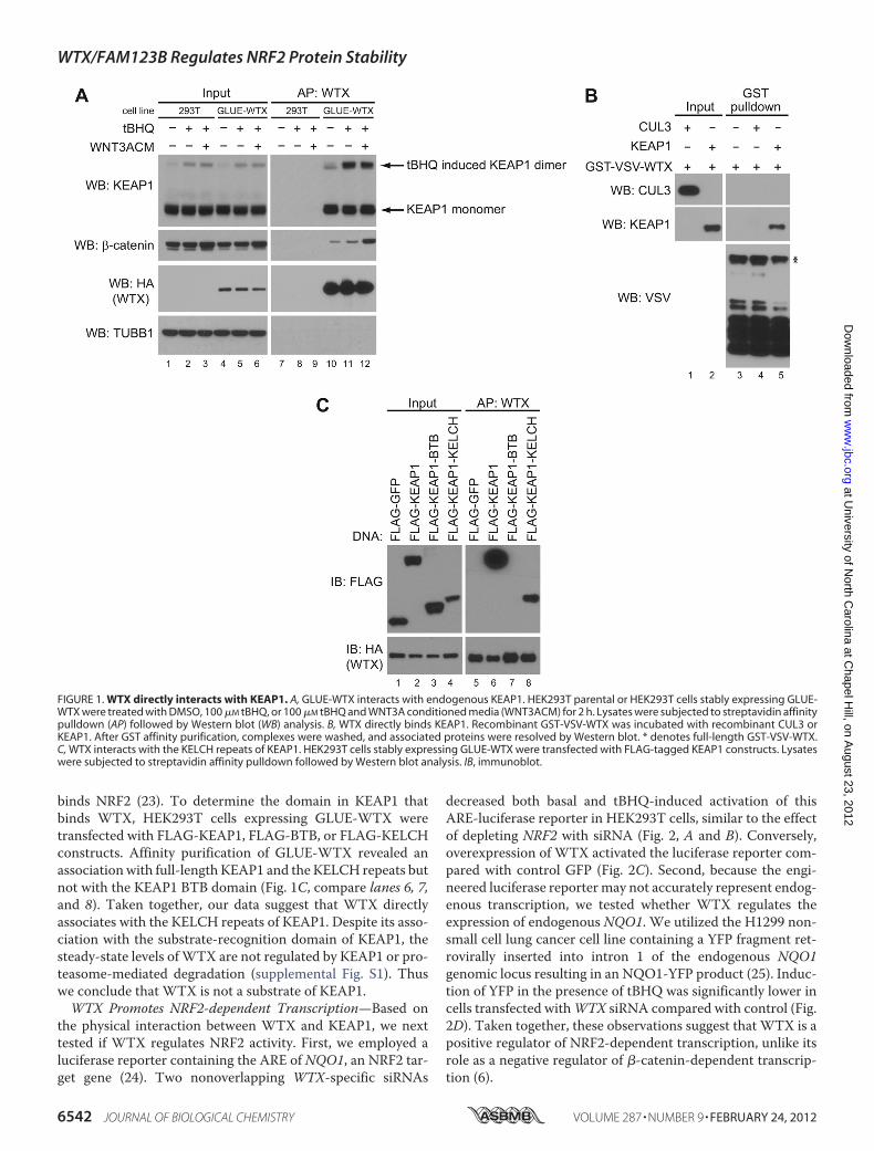
binds NRF2 (23). To determine the domain in KEAP1 thatbinds WTX, HEK293T cells expressing GLUE-WTX weretransfected with FLAG-KEAP1, FLAG-BTB, or FLAG-KELCHconstructs. Affinity purification of GLUE-WTX revealed anassociationwith full-length KEAP1 and the KELCH repeats butnot with the KEAP1 BTB domain (Fig. 1C, compare lanes 6, 7,and 8). Taken together, our data suggest that WTX directlyassociates with the KELCH repeats of KEAP1. Despite its asso-ciation with the substrate-recognition domain of KEAP1, thesteady-state levels ofWTX are not regulated by KEAP1 or pro-teasome-mediated degradation (supplemental Fig. S1). Thuswe conclude that WTX is not a substrate of KEAP1.WTX Promotes NRF2-dependent Transcription—Based on
the physical interaction between WTX and KEAP1, we nexttested if WTX regulates NRF2 activity. First, we employed aluciferase reporter containing the ARE of NQO1, an NRF2 tar-get gene (24). Two nonoverlapping WTX-specific siRNAs
decreased both basal and tBHQ-induced activation of thisARE-luciferase reporter in HEK293T cells, similar to the effectof depleting NRF2 with siRNA (Fig. 2, A and B). Conversely,overexpression of WTX activated the luciferase reporter com-pared with control GFP (Fig. 2C). Second, because the engi-neered luciferase reporter may not accurately represent endog-enous transcription, we tested whether WTX regulates theexpression of endogenous NQO1. We utilized the H1299 non-small cell lung cancer cell line containing a YFP fragment ret-rovirally inserted into intron 1 of the endogenous NQO1genomic locus resulting in an NQO1-YFP product (25). Induc-tion of YFP in the presence of tBHQ was significantly lower incells transfected withWTX siRNA compared with control (Fig.2D). Taken together, these observations suggest that WTX is apositive regulator of NRF2-dependent transcription, unlike itsrole as a negative regulator of �-catenin-dependent transcrip-tion (6).
FIGURE 1. WTX directly interacts with KEAP1. A, GLUE-WTX interacts with endogenous KEAP1. HEK293T parental or HEK293T cells stably expressing GLUE-WTX were treated with DMSO, 100 �M tBHQ, or 100 �M tBHQ and WNT3A conditioned media (WNT3ACM) for 2 h. Lysates were subjected to streptavidin affinitypulldown (AP) followed by Western blot (WB) analysis. B, WTX directly binds KEAP1. Recombinant GST-VSV-WTX was incubated with recombinant CUL3 orKEAP1. After GST affinity purification, complexes were washed, and associated proteins were resolved by Western blot. * denotes full-length GST-VSV-WTX.C, WTX interacts with the KELCH repeats of KEAP1. HEK293T cells stably expressing GLUE-WTX were transfected with FLAG-tagged KEAP1 constructs. Lysateswere subjected to streptavidin affinity pulldown followed by Western blot analysis. IB, immunoblot.
WTX/FAM123B Regulates NRF2 Protein Stability
6542 JOURNAL OF BIOLOGICAL CHEMISTRY VOLUME 287 • NUMBER 9 • FEBRUARY 24, 2012
at University of N
orth Carolina at C
hapel Hill, on A
ugust 23, 2012w
ww
.jbc.orgD
ownloaded from
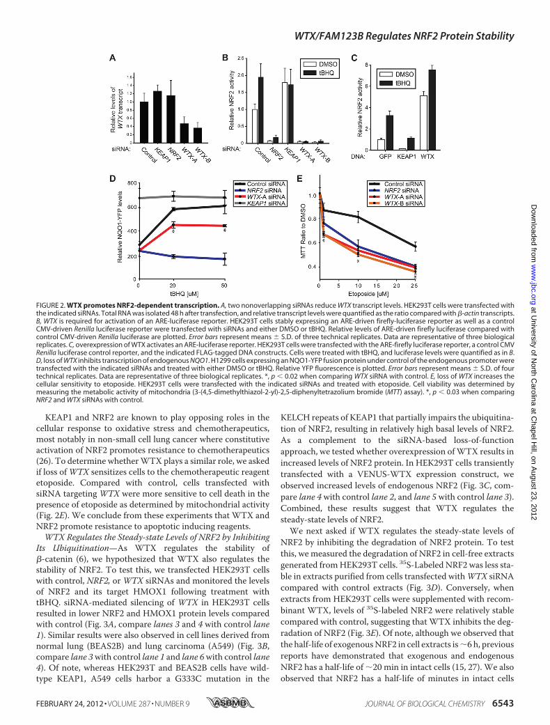
KEAP1 and NRF2 are known to play opposing roles in thecellular response to oxidative stress and chemotherapeutics,most notably in non-small cell lung cancer where constitutiveactivation of NRF2 promotes resistance to chemotherapeutics(26). To determine whetherWTX plays a similar role, we askedif loss ofWTX sensitizes cells to the chemotherapeutic reagentetoposide. Compared with control, cells transfected withsiRNA targeting WTX were more sensitive to cell death in thepresence of etoposide as determined by mitochondrial activity(Fig. 2E). We conclude from these experiments that WTX andNRF2 promote resistance to apoptotic inducing reagents.WTX Regulates the Steady-state Levels of NRF2 by Inhibiting
Its Ubiquitination—As WTX regulates the stability of�-catenin (6), we hypothesized that WTX also regulates thestability of NRF2. To test this, we transfected HEK293T cellswith control, NRF2, orWTX siRNAs and monitored the levelsof NRF2 and its target HMOX1 following treatment withtBHQ. siRNA-mediated silencing of WTX in HEK293T cellsresulted in lower NRF2 and HMOX1 protein levels comparedwith control (Fig. 3A, compare lanes 3 and 4 with control lane1). Similar results were also observed in cell lines derived fromnormal lung (BEAS2B) and lung carcinoma (A549) (Fig. 3B,compare lane 3with control lane 1 and lane 6with control lane4). Of note, whereas HEK293T and BEAS2B cells have wild-type KEAP1, A549 cells harbor a G333C mutation in the
KELCH repeats of KEAP1 that partially impairs the ubiquitina-tion of NRF2, resulting in relatively high basal levels of NRF2.As a complement to the siRNA-based loss-of-functionapproach, we tested whether overexpression ofWTX results inincreased levels of NRF2 protein. In HEK293T cells transientlytransfected with a VENUS-WTX expression construct, weobserved increased levels of endogenous NRF2 (Fig. 3C, com-pare lane 4with control lane 2, and lane 5with control lane 3).Combined, these results suggest that WTX regulates thesteady-state levels of NRF2.We next asked if WTX regulates the steady-state levels of
NRF2 by inhibiting the degradation of NRF2 protein. To testthis, we measured the degradation of NRF2 in cell-free extractsgenerated fromHEK293T cells. 35S-Labeled NRF2 was less sta-ble in extracts purified from cells transfected withWTX siRNAcompared with control extracts (Fig. 3D). Conversely, whenextracts from HEK293T cells were supplemented with recom-binant WTX, levels of 35S-labeled NRF2 were relatively stablecompared with control, suggesting that WTX inhibits the deg-radation of NRF2 (Fig. 3E). Of note, although we observed thatthe half-life of exogenousNRF2 in cell extracts is�6 h, previousreports have demonstrated that exogenous and endogenousNRF2 has a half-life of �20 min in intact cells (15, 27). We alsoobserved that NRF2 has a half-life of minutes in intact cells
FIGURE 2. WTX promotes NRF2-dependent transcription. A, two nonoverlapping siRNAs reduce WTX transcript levels. HEK293T cells were transfected withthe indicated siRNAs. Total RNA was isolated 48 h after transfection, and relative transcript levels were quantified as the ratio compared with �-actin transcripts.B, WTX is required for activation of an ARE-luciferase reporter. HEK293T cells stably expressing an ARE-driven firefly-luciferase reporter as well as a controlCMV-driven Renilla luciferase reporter were transfected with siRNAs and either DMSO or tBHQ. Relative levels of ARE-driven firefly luciferase compared withcontrol CMV-driven Renilla luciferase are plotted. Error bars represent means � S.D. of three technical replicates. Data are representative of three biologicalreplicates. C, overexpression of WTX activates an ARE-luciferase reporter. HEK293T cells were transfected with the ARE-firefly luciferase reporter, a control CMVRenilla luciferase control reporter, and the indicated FLAG-tagged DNA constructs. Cells were treated with tBHQ, and luciferase levels were quantified as in B.D, loss of WTX inhibits transcription of endogenous NQO1. H1299 cells expressing an NQO1-YFP fusion protein under control of the endogenous promoter weretransfected with the indicated siRNAs and treated with either DMSO or tBHQ. Relative YFP fluorescence is plotted. Error bars represent means � S.D. of fourtechnical replicates. Data are representative of three biological replicates. *, p � 0.02 when comparing WTX siRNA with control. E, loss of WTX increases thecellular sensitivity to etoposide. HEK293T cells were transfected with the indicated siRNAs and treated with etoposide. Cell viability was determined bymeasuring the metabolic activity of mitochondria (3-(4,5-dimethylthiazol-2-yl)-2,5-diphenyltetrazolium bromide (MTT) assay). *, p � 0.03 when comparingNRF2 and WTX siRNAs with control.
WTX/FAM123B Regulates NRF2 Protein Stability
FEBRUARY 24, 2012 • VOLUME 287 • NUMBER 9 JOURNAL OF BIOLOGICAL CHEMISTRY 6543
at University of N
orth Carolina at C
hapel Hill, on A
ugust 23, 2012w
ww
.jbc.orgD
ownloaded from
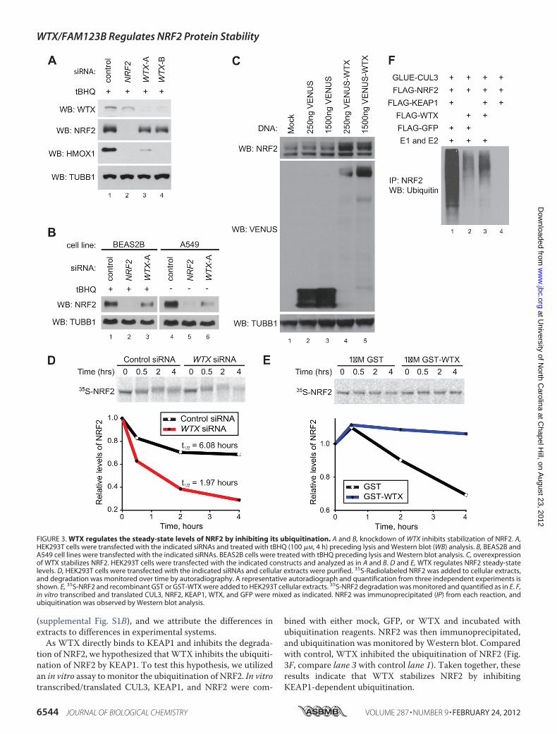
(supplemental Fig. S1B), and we attribute the differences inextracts to differences in experimental systems.As WTX directly binds to KEAP1 and inhibits the degrada-
tion of NRF2, we hypothesized thatWTX inhibits the ubiquiti-nation of NRF2 by KEAP1. To test this hypothesis, we utilizedan in vitro assay tomonitor the ubiquitination of NRF2. In vitrotranscribed/translated CUL3, KEAP1, and NRF2 were com-
bined with either mock, GFP, or WTX and incubated withubiquitination reagents. NRF2 was then immunoprecipitated,and ubiquitination was monitored byWestern blot. Comparedwith control, WTX inhibited the ubiquitination of NRF2 (Fig.3F, compare lane 3 with control lane 1). Taken together, theseresults indicate that WTX stabilizes NRF2 by inhibitingKEAP1-dependent ubiquitination.
FIGURE 3. WTX regulates the steady-state levels of NRF2 by inhibiting its ubiquitination. A and B, knockdown of WTX inhibits stabilization of NRF2. A,HEK293T cells were transfected with the indicated siRNAs and treated with tBHQ (100 �M, 4 h) preceding lysis and Western blot (WB) analysis. B, BEAS2B andA549 cell lines were transfected with the indicated siRNAs. BEAS2B cells were treated with tBHQ preceding lysis and Western blot analysis. C, overexpressionof WTX stabilizes NRF2. HEK293T cells were transfected with the indicated constructs and analyzed as in A and B. D and E, WTX regulates NRF2 steady-statelevels. D, HEK293T cells were transfected with the indicated siRNAs and cellular extracts were purified. 35S-Radiolabeled NRF2 was added to cellular extracts,and degradation was monitored over time by autoradiography. A representative autoradiograph and quantification from three independent experiments isshown. E, 35S-NRF2 and recombinant GST or GST-WTX were added to HEK293T cellular extracts. 35S-NRF2 degradation was monitored and quantified as in E. F,in vitro transcribed and translated CUL3, NRF2, KEAP1, WTX, and GFP were mixed as indicated. NRF2 was immunoprecipitated (IP) from each reaction, andubiquitination was observed by Western blot analysis.
WTX/FAM123B Regulates NRF2 Protein Stability
6544 JOURNAL OF BIOLOGICAL CHEMISTRY VOLUME 287 • NUMBER 9 • FEBRUARY 24, 2012
at University of N
orth Carolina at C
hapel Hill, on A
ugust 23, 2012w
ww
.jbc.orgD
ownloaded from
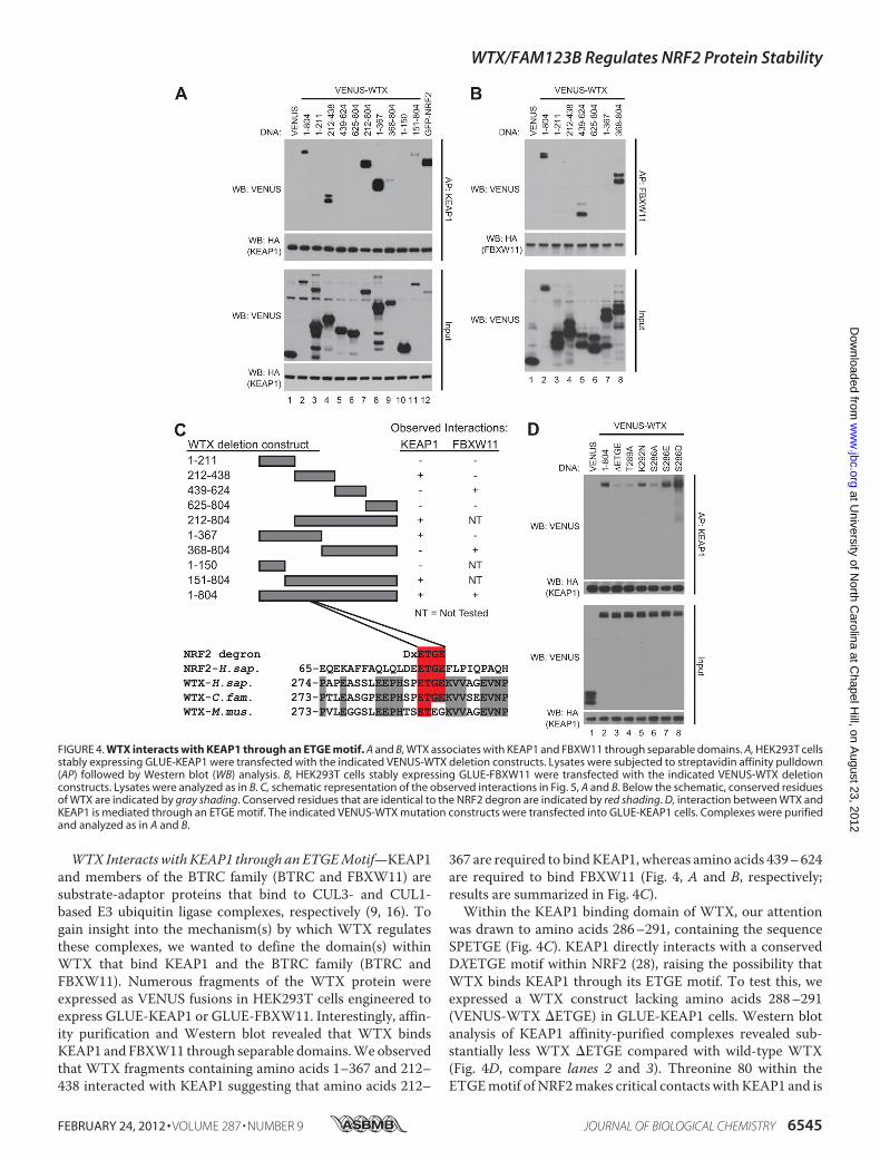
WTX Interacts with KEAP1 through an ETGEMotif—KEAP1and members of the BTRC family (BTRC and FBXW11) aresubstrate-adaptor proteins that bind to CUL3- and CUL1-based E3 ubiquitin ligase complexes, respectively (9, 16). Togain insight into the mechanism(s) by which WTX regulatesthese complexes, we wanted to define the domain(s) withinWTX that bind KEAP1 and the BTRC family (BTRC andFBXW11). Numerous fragments of the WTX protein wereexpressed as VENUS fusions in HEK293T cells engineered toexpress GLUE-KEAP1 or GLUE-FBXW11. Interestingly, affin-ity purification and Western blot revealed that WTX bindsKEAP1 and FBXW11 through separable domains.Weobservedthat WTX fragments containing amino acids 1–367 and 212–438 interacted with KEAP1 suggesting that amino acids 212–
367 are required to bindKEAP1, whereas amino acids 439–624are required to bind FBXW11 (Fig. 4, A and B, respectively;results are summarized in Fig. 4C).Within the KEAP1 binding domain of WTX, our attention
was drawn to amino acids 286–291, containing the sequenceSPETGE (Fig. 4C). KEAP1 directly interacts with a conservedDXETGE motif within NRF2 (28), raising the possibility thatWTX binds KEAP1 through its ETGE motif. To test this, weexpressed a WTX construct lacking amino acids 288–291(VENUS-WTX �ETGE) in GLUE-KEAP1 cells. Western blotanalysis of KEAP1 affinity-purified complexes revealed sub-stantially less WTX �ETGE compared with wild-type WTX(Fig. 4D, compare lanes 2 and 3). Threonine 80 within theETGEmotif of NRF2makes critical contacts with KEAP1 and is
FIGURE 4. WTX interacts with KEAP1 through an ETGE motif. A and B, WTX associates with KEAP1 and FBXW11 through separable domains. A, HEK293T cellsstably expressing GLUE-KEAP1 were transfected with the indicated VENUS-WTX deletion constructs. Lysates were subjected to streptavidin affinity pulldown(AP) followed by Western blot (WB) analysis. B, HEK293T cells stably expressing GLUE-FBXW11 were transfected with the indicated VENUS-WTX deletionconstructs. Lysates were analyzed as in B. C, schematic representation of the observed interactions in Fig. 5, A and B. Below the schematic, conserved residuesof WTX are indicated by gray shading. Conserved residues that are identical to the NRF2 degron are indicated by red shading. D, interaction between WTX andKEAP1 is mediated through an ETGE motif. The indicated VENUS-WTX mutation constructs were transfected into GLUE-KEAP1 cells. Complexes were purifiedand analyzed as in A and B.
WTX/FAM123B Regulates NRF2 Protein Stability
FEBRUARY 24, 2012 • VOLUME 287 • NUMBER 9 JOURNAL OF BIOLOGICAL CHEMISTRY 6545
at University of N
orth Carolina at C
hapel Hill, on A
ugust 23, 2012w
ww
.jbc.orgD
ownloaded from
required for interacting with KEAP1 (29). Similarly, weobserved thatmutation of threonine 289 to alanine in the ETGEmotif of WTX disrupts the interaction with KEAP1 (Fig. 4D,compare lanes 2 and 4). Interestingly, lysine 292, adjacent to theETGE motif, is mutated to asparagine in several reportedWilms tumor cases and a single instance of acute myeloid leu-kemia (1, 3, 30). Comparedwithwild-typeWTX, KEAP1 pulleddown less WTX K292N, although more than WTX �ETGE(Fig. 4D, compare lanes 2, 3, and 5). Combined, these resultssuggest that WTX binds to KEAP1 through a similar motif asNRF2.One notable difference between the KEAP1 interaction
domains of NRF2 andWTX is the two amino acids upstream oftheir respective ETGEmotifs. The humanNRF2motif containsa conserved aspartic acid that interacts with two water mole-cules that in turn hydrogen bond with two arginine residues inKEAP1 (29). In place of the aspartic acid, WTX contains a ser-ine (residue 286). Interestingly, mutation of serine 286 to ala-nine inhibited the ability of KEAP1 to pull downWTX (Fig. 4D,compare lanes 2 and 6). Based on the crystal structure of theKELCH repeats and anNRF2 peptide (23, 29), we reasoned thatmutation of serine 286 to an acidic residue would enhance theinteraction between WTX and KEAP1. Although we onlyobserved a modest increase in the interaction between KEAP1andWTX S286E, WTX S286D was significantly enriched rela-tive to wild-type WTX in KEAP1 pulldowns (Fig. 4D, comparelanes 2, 7, and 8). We conclude from these experiments that inaddition to the ETGE motif, serine 286 is also important in theKEAP1 interacting domain of WTX.Serine 286 Is Phosphorylated inVivo—Given thatmutation of
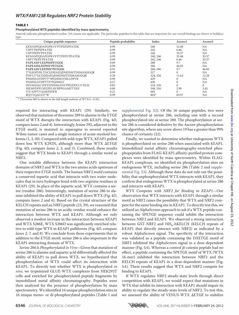
serine 286 to alanine and aspartic acid differentially affected theability of KEAP1 to pull down WTX, we hypothesized thatphosphorylation of WTX could affect its interaction withKEAP1. To directly test whether WTX is phosphorylated invivo, we trypsinized GLUE-WTX complexes from HEK293Tcells and enriched for phosphorylated peptide fragments byimmobilized metal affinity chromatography. Peptides werethen analyzed for the presence of phosphorylation by massspectrometry.We identified 16 unique phosphorylation sites in16 unique mono- or di-phosphorylated peptides (Table 1 and
supplemental Fig. S2). Of the 16 unique peptides, two werephosphorylated at serine 286, including one with a secondphosphorylated site at serine 280. The phosphorylation at ser-ine 286 is considered definitive by the Ascore phosphorylationsite algorithm, where any score above 19 has a greater than 99%chance of certainty (22).Finally, we wanted to determine whether endogenous WTX
is phosphorylated on serine 286 when associated with KEAP1.Immobilized metal affinity chromatography-enriched phos-phopeptides from FLAG-KEAP1 affinity-purified protein com-plexes were identified by mass spectrometry. Within FLAG-KEAP1 complexes, we identified six phosphorylation sites onendogenous WTX, including serine 286 (Table 2 and supple-mental Fig. S3). Although these data do not rule out the possi-bility that unphosphorylatedWTX interacts with KEAP1, theyconfirm that endogenousWTX is phosphorylated at serine 286and interacts with KEAP1.WTX Competes with NRF2 for Binding to KEAP1—Our
observation that WTX interacts with KEAP1 through a similarmotif as NRF2 raises the possibility that WTX and NRF2 com-pete for the samebinding site inKEAP1.Todirectly test this, weutilized an AlphaScreen approach to ask if aWTX peptide con-taining the SPETGE sequence could inhibit the interactionbetween NRF2 and KEAP1. We observed a strong interactionbetween GST-NRF2 and HIS6-hKELCH (KELCH repeats ofKEAP1 that directly interact with NRF2) as indicated by arobust AlphaScreen signal. The specificity of the interactionwas validated as a peptide containing the DXETGE motif ofNRF2 inhibited the AlphaScreen signal in a dose-dependentmanner (Fig. 5A). Whereas a control �-catenin peptide had noeffect, a peptide containing the SPETGE motif of WTX (WTX16-mer) inhibited the interaction between NRF2 and theKELCH repeats of KEAP1 in a dose-dependent manner (Fig.5A). These results suggest that WTX and NRF2 compete forbinding to KEAP1.If WTX regulates NRF2 steady-state levels through direct
competition with KEAP1, we would expect that mutations inWTX that inhibit its interaction with KEAP1 should impair itsability to regulate the steady-state levels of NRF2. To test this,we assessed the ability of VENUS-WTX �ETGE to stabilize
TABLE 1Phosphorylated WTX peptides identified by mass spectrometryAsterisk indicates phosphorylated residue. NAmeans not applicable. The particular peptides in this table that are important for our overall findings are shown in boldfacetype.
Unique peptide sequence Peptide probability Index Ascore1 Ascore2
KENANPQDAPGPKVS*PTPEPSPPATEK 0.99 240 32.48 NAVSPT*PEPSPPATEK 0.99 242 6.46 NAVSPTPEPS*PPATEK 0.99 246 33.57 NAKENANPQDAPGPKVS*PTPEPS*PPATEK 0.99 240, 246 32.48 33.57VSPT*PEPS*PPATEK 0.99 242, 246 6.46 33.57PAPEASS*LEEPHSPETGEK 0.89 280 9.7 NAPAPEASSLEEPHS*PETGEK 0.99 286 46.85 NAPAPEASS*LEEPHS*PETGEK 0.91 280, 286 9.7 46.85T*SLKSFDSLTGCGDIIAEQDMDSMTDSMASGGQR 0.99 317 0 NAFDS*LT*GCGDIIAEQDMDSMTDSMASGGQR 0.29 324, 326 11.63 12.28PNMNLGYHPTT*SPGHHGYMLLDPVR 0.99 429 0 NAPNMNLGYHPTTS*PGHHGY 0.57 430 0 NADSYSGDALYEFYEPDDSLENS*PPGDDCLY*DLH 0.85 518, 526 0 0SSEMFDPFLNFEPFLSS*RPPGAMET*EEE 0.80 548, 556 2.99 5.45ITS*AFPTTASSEPDWR 0.21 683 0 NARQVTQACGT*W 0.99 803a 69.97 NA
a Threonine 803 is absent in the full-length isoform of WTX(1–1135).
WTX/FAM123B Regulates NRF2 Protein Stability
6546 JOURNAL OF BIOLOGICAL CHEMISTRY VOLUME 287 • NUMBER 9 • FEBRUARY 24, 2012
at University of N
orth Carolina at C
hapel Hill, on A
ugust 23, 2012w
ww
.jbc.orgD
ownloaded from
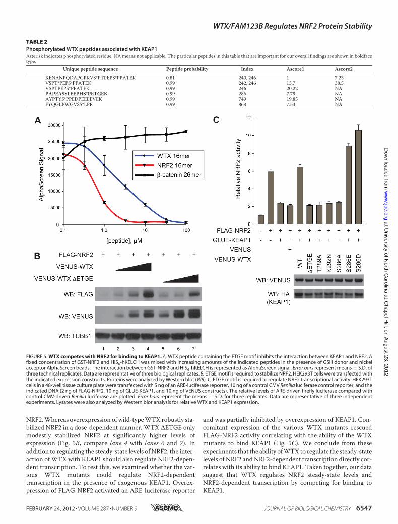
NRF2.Whereas overexpression ofwild-typeWTXrobustly sta-bilized NRF2 in a dose-dependent manner, WTX �ETGE onlymodestly stabilized NRF2 at significantly higher levels ofexpression (Fig. 5B, compare lane 4 with lanes 6 and 7). Inaddition to regulating the steady-state levels of NRF2, the inter-action ofWTXwith KEAP1 should also regulate NRF2-depen-dent transcription. To test this, we examined whether the var-ious WTX mutants could regulate NRF2-dependenttranscription in the presence of exogenous KEAP1. Overex-pression of FLAG-NRF2 activated an ARE-luciferase reporter
and was partially inhibited by overexpression of KEAP1. Con-comitant expression of the various WTX mutants rescuedFLAG-NRF2 activity correlating with the ability of the WTXmutants to bind KEAP1 (Fig. 5C). We conclude from theseexperiments that the ability ofWTX to regulate the steady-statelevels of NRF2 andNRF2-dependent transcription directly cor-relates with its ability to bind KEAP1. Taken together, our datasuggest that WTX regulates NRF2 steady-state levels andNRF2-dependent transcription by competing for binding toKEAP1.
TABLE 2Phosphorylated WTX peptides associated with KEAP1Asterisk indicates phosphorylated residue. NAmeans not applicable. The particular peptides in this table that are important for our overall findings are shown in boldfacetype.
Unique peptide sequence Peptide probability Index Ascore1 Ascore2
KENANPQDAPGPKVS*PTPEPS*PPATEK 0.81 240, 246 1 7.23VSPT*PEPS*PPATEK 0.99 242, 246 13.7 38.5VSPTPEPS*PPATEK 0.99 246 20.22 NAPAPEASSLEEPHS*PETGEK 0.99 286 7.79 NAAYPTYS*PPEDPEEEEVEK 0.99 749 19.85 NAFYQGLPWGVSS*LPR 0.99 868 7.53 NA
FIGURE 5. WTX competes with NRF2 for binding to KEAP1. A, WTX peptide containing the ETGE motif inhibits the interaction between KEAP1 and NRF2. Afixed concentration of GST-NRF2 and HIS6-hKELCH was mixed with increasing amounts of the indicated peptides in the presence of GSH donor and nickelacceptor AlphaScreen beads. The interaction between GST-NRF2 and HIS6-hKELCH is represented as AlphaScreen signal. Error bars represent means � S.D. ofthree technical replicates. Data are representative of three biological replicates. B, ETGE motif is required to stabilize NRF2. HEK293T cells were transfected withthe indicated expression constructs. Proteins were analyzed by Western blot (WB). C, ETGE motif is required to regulate NRF2 transcriptional activity. HEK293Tcells in a 48-well tissue culture plate were transfected with 5 ng of an ARE-luciferase reporter, 10 ng of a control CMV Renilla luciferase control reporter, and theindicated DNA (2 ng of FLAG-NRF2, 10 ng of GLUE-KEAP1, and 10 ng of VENUS constructs). The relative levels of ARE-driven firefly luciferase compared withcontrol CMV-driven Renilla luciferase are plotted. Error bars represent the means � S.D. for three replicates. Data are representative of three independentexperiments. Lysates were also analyzed by Western blot analysis for relative WTX and KEAP1 expression.
WTX/FAM123B Regulates NRF2 Protein Stability
FEBRUARY 24, 2012 • VOLUME 287 • NUMBER 9 JOURNAL OF BIOLOGICAL CHEMISTRY 6547
at University of N
orth Carolina at C
hapel Hill, on A
ugust 23, 2012w
ww
.jbc.orgD
ownloaded from
DISCUSSION
The WTX gene encodes a tumor suppressor protein; it islocated on the X chromosome and is somatically lost ormutated in 7–30% of cases of Wilms tumor (1–3). Germ linemutations in WTX give rise to OSCS, a debilitating and fataldisease that largely affects the skeletal system (4, 5). As thediscovery of WTX and its mutations in human disease is rela-tively new, the molecular function(s) and developmental orhomeostatic consequences of its loss is only beginning to beunraveled. Compelling data from numerous research groupshave described functions for WTX in controlling WNT/�-catenin signaling, cell-cell adhesion, apoptosis, and transcrip-tion (1, 6, 11–13). Recently, characterization ofWtx deletion inmice revealed that Wtx regulates mesenchymal progenitor cellfate specification in part through �-catenin (31).
Here, we expand on the knowledge of the molecular mecha-nisms of action of WTX with the observation that WTX regu-lates the steady-state levels ofNRF2 andNRF2-dependent tran-scription by competing with NRF2 for binding to KEAP1.KEAP1 forms a homodimer and interacts with CUL3 throughits BTBdomain (32, 33) and interactswith theN-terminalNeh2domain of a single NRF2 molecule through its KELCH repeats.The Neh2 domain of NRF2 contains two highly conservedregions, one bearing anLXXQDXDLG (DLG)motif (34, 35) andthe other bearing a DXETGE motif (28). These motifs interactwith a separate KEAP1 molecule in the KEAP1 homodimer,although the DXETGE motif binds with �100-fold higheraffinity (36). WTX contains a similar SPETGE motif that candirectly inhibit the interaction between NRF2 and the KELCHrepeats of KEAP1 in vitro. As WTX does not contain a DLGmotif similar to NRF2, it is possible that NRF2 can still interactwith a KEAP1 dimer through this low affinity interactionmotif.We hypothesize that WTX enhances NRF2 steady-state levelsby disrupting the conformation of the E3 ubiquitin ligase com-plex, resulting in lower ubiquitination of NRF2. In support ofour findings, expression of the NRF2 target genes NQO1 andHMOX1 was significantly enhanced in HEK293T cells overex-pressing a fragment ofWTX containing the KEAP1 interactiondomain (37).In addition to the ETGEmotif, we determined that serine 286
is also required for the ability of WTX to interact with KEAP1and regulate NRF2. Interestingly, mutation of serine 286 toeither glutamic or aspartic acid enhanced the functional effectsof WTX. Although these mutations may only present confor-mational changes inWTX that make it more suitable for bind-ing to KEAP1, both glutamic and aspartic acid resemble phos-phorylated serine. Thus, phosphorylation ofWTX at serine 286may increase its affinity forKEAP1. Furthermore,we found thatWTX is phosphorylated in vivo, and KEAP1 interacts withendogenous WTX that is phosphorylated at serine 286, raisingthe intriguing possibility that the interaction between WTXand KEAP1 is regulated by a yet to be identified kinase(s).Under normal conditions, NRF2 is constitutively ubiquiti-
nated through its association with KEAP1 (14–16). In the pres-ence of oxidative stress, cysteine residues in KEAP1 are modi-fied resulting in a conformational change that disrupts theubiquitination of NRF2, and NRF2 accumulates in the nucleus
where it regulates gene transcription (16, 38). In addition to ourfindings, several recent studies have also revealed that NRF2signaling is regulated through protein-protein interactions.The cyclin-dependent kinase inhibitor p21, a p53-regulatedgene with pro-survival properties, was recently shown to bindthe DLGmotif of NRF2 and inhibit its interaction with KEAP1,resulting in elevated NRF2 levels under both basal and chemi-cally induced conditions (39). Another recent study identifiedp62 as a novel interactor of KEAP1 (40). During autophagy, p62directs ubiquitinated proteins to degradation by the lysosome.By binding to KEAP1 through an ETGE-like motif similar tothat of NRF2 and WTX, p62 targets KEAP1 for autophagicdegradation, thus contributing to the stabilization of NRF2.Combined with WTX, these examples highlight the complexregulation of NRF2 degradation and its importance in bothhomeostasis and response to cytotoxic stress.Whereas WTX inhibits the ubiquitination of NRF2, we pre-
viously reported that WTX promotes the ubiquitination of�-catenin by the SCFBTRC ubiquitin ligase complex. To ourknowledge, WTX is the first described protein to interact withtwo E3 ubiquitin ligase adaptors and have opposite regulatoryeffects on their respective substrates. How is this possible?WTX interacts with the KELCH repeats that form the �-pro-peller fold of KEAP1 and likely inhibits the formation of a func-tional CUL3-KEAP1-NRF2 complex. Through a separabledomain, WTX interacts with both BTRC and �-catenin, sug-gesting that it binds to an intact SCFBTRC-substrate complex(6). BTRC also employs a �-propeller fold for substrate capture(41), but it is unlikely that WTX interacts with the �-propellerfold of BTRC in a similar fashion as KEAP1 as this would inhibitthe formation of a functional CUL1-SKP1-BTRC-�-catenincomplex and result in elevated �-catenin levels. Interestingly, arecent study demonstrated that NRF2 is phosphorylated byGSK3� in the central Neh6 domain (DSGIS, residues 334–338), creating an SCFBTRC destruction motif similar to that of�-catenin (42). This phosphorylated form of NRF2 is recog-nized by BTRC. Additionally, KEAP1 and BTRC have beenidentified in the same complex bymass spectrometry (43). Thisraises the possibility that multiple E3 ubiquitin ligase com-plexes consisting of a KEAP1 homodimer, a BTRChomodimer,or a KEAP1/BTRC heterodimer regulate the ubiquitination ofNRF2. AsWTX interacts with BTRC and KEAP1 through sep-arable domains, we hypothesize that WTX coordinates theadaptors in the E3 ubiquitin ligase complexes, resulting in vari-able ubiquitination of substrates.Although most commonly lost through gene-encompassing
deletions, a small percentage of WTX mutations identified inWilms tumor yield single amino acid substitutions and trun-cated proteins. Conversely, the majority of WTX mutationsidentified in OSCS yield truncated proteins. Aligning the loca-tion of these mutations with the WTX protein interactiondomains suggests relationships of these diseases with specificbinding interfaces. The KEAP1 interaction domain lies N-ter-minal to the domains that bind �-catenin/BTRC/APC andWT1 and remains intact in 9 out of the 20 reported mutationproducts. Of note, we determined that the K292N substitutioninhibited the association of WTX and KEAP1. The �-catenin/BTRC/APC interacting domains encompass residues 280–
WTX/FAM123B Regulates NRF2 Protein Stability
6548 JOURNAL OF BIOLOGICAL CHEMISTRY VOLUME 287 • NUMBER 9 • FEBRUARY 24, 2012
at University of N
orth Carolina at C
hapel Hill, on A
ugust 23, 2012w
ww
.jbc.orgD
ownloaded from
839. Of the 20 reported mutations, 11 are predicted to alterthese binding activities. As the C terminus ofWTXbindsWT1,this interaction is lost in all truncation products derived frommutations in WTX. These correlations suggest that whereasWT1 is likely central to WTX-associated diseases, the WTX-KEAP1 and WTX-�-catenin/BTRC/APC functional relation-ships may contribute to a subset of Wilms tumor and OSCS,perhaps accounting for variability in disease onset orprogression.The observation that WTX regulates the NRF2-mediated
antioxidant response supports further study of its role in dis-ease beyond Wilms tumor and OSCS. One of the best-studieddiseases with altered KEAP1/NRF2 activity is non-small celllung cancer, where mutations in KEAP1 lead to constitutiveNRF2-mediated transcription. Consequently, cultured lungcancer cell lines with constitutive NRF2 activity are resistant tocell death induced by etoposide (26). Similarly, silencing NRF2restores resistance to chemotherapeutics in KEAP1 mutantcells (44).We find that silencingWTX sensitizesHEK293T cellsto death induced by etoposide, similar to loss of NRF2. Inter-estingly, although WTX expression is diminished in adultmouse brain and kidney compared with embryonic tissues,expression levels remain high in the lung, suggesting it maycontribute to lung cell homeostasis in the adult (1). Coupledwith the established roles ofWTX inWilms tumor andKEAP1/NRF2 in lung cancer, our data support future investigationsinto a functional role for WTX in lung cancer and other dis-eases associated with aberrant NRF2 activity.
Acknowledgments—We thank Priscila Siesser for unpublished data,Jeffrey Johnson and Seth Goldenberg for reagents, and other membersof the Moon laboratory andMajor laboratory for helpful discussions.
REFERENCES1. Rivera, M. N., Kim, W. J., Wells, J., Driscoll, D. R., Brannigan, B. W., Han,
M., Kim, J. C., Feinberg, A. P., Gerald,W. L., Vargas, S. O., Chin, L., Iafrate,A. J., Bell, D. W., and Haber, D. A. (2007) An X chromosome gene, WTX,is commonly inactivated in Wilms tumor. Science 315, 642–645
2. Perotti, D., Gamba, B., Sardella, M., Spreafico, F., Terenziani, M., Collini,P., Pession, A., Nantron, M., Fossati-Bellani, F., and Radice, P. (2008)Functional inactivation of theWTX gene is not a frequent event inWilms’tumors. Oncogene 27, 4625–4632
3. Ruteshouser, E. C., Robinson, S. M., and Huff, V. (2008) Wilms tumorgenetics. Mutations inWT1,WTX, and CTNNB1 account for only aboutone-third of tumors. Genes Chromosomes Cancer 47, 461–470
4. Perdu, B., de Freitas, F., Frints, S. G., Schouten,M., Schrander-Stumpel, C.,Barbosa, M., Pinto-Basto, J., Reis-Lima, M., de Vernejoul, M. C., Becker,K., Freckmann, M. L., Keymolen, K., Haan, E., Savarirayan, R., Koenig, R.,Zabel, B., Vanhoenacker, F. M., and Van Hul, W. (2009) Osteopathia stri-atawith cranial sclerosis owing toWTX gene defect. J. BoneMiner. Res. 25,82–90
5. Jenkins, Z. A., van Kogelenberg, M., Morgan, T., Jeffs, A., Fukuzawa, R.,Pearl, E., Thaller, C., Hing, A. V., Porteous, M. E., Garcia-Miñaur, S.,Bohring, A., Lacombe, D., Stewart, F., Fiskerstrand, T., Bindoff, L., Ber-land, S., Adès, L. C., Tchan, M., David, A., Wilson, L. C., Hennekam, R. C.,Donnai, D., Mansour, S., Cormier-Daire, V., and Robertson, S. P. (2009)Germ line mutations in WTX cause a sclerosing skeletal dysplasia but donot predispose to tumorigenesis. Nat. Genet. 41, 95–100
6. Major, M. B., Camp, N. D., Berndt, J. D., Yi, X., Goldenberg, S. J., Hubbert,C., Biechele, T. L., Gingras, A.C., Zheng,N.,Maccoss,M. J., Angers, S., andMoon, R. T. (2007) Wilms tumor suppressor WTX negatively regulates
WNT/�-catenin signaling. Science 316, 1043–10467. van Amerongen, R., Mikels, A., and Nusse, R. (2008) Alternative wnt sig-
naling is initiated by distinct receptors. Sci. Signal. 1, re98. Yost, C., Torres,M.,Miller, J. R., Huang, E., Kimelman,D., andMoon, R. T.
(1996) The axis-inducing activity, stability, and subcellular distribution of�-catenin is regulated in Xenopus embryos by glycogen synthase kinase 3.Genes Dev. 10, 1443–1454
9. Winston, J. T., Strack, P., Beer-Romero, P., Chu, C. Y., Elledge, S. J., andHarper, J.W. (1999) The SCF�-TRCP-ubiquitin ligase complex associatesspecifically with phosphorylated destructionmotifs in I�B� and�-cateninand stimulates I�B� ubiquitination in vitro. Genes Dev. 13, 270–283
10. Liu, C., Kato, Y., Zhang, Z., Do, V. M., Yankner, B. A., and He, X. (1999)�-Trcp couples�-catenin phosphorylation-degradation and regulatesXe-nopus axis formation. Proc. Natl. Acad. Sci. U.S.A. 96, 6273–6278
11. Tanneberger, K., Pfister, A. S., Brauburger, K., Schneikert, J., Hadjihannas,M. V., Kriz, V., Schulte, G., Bryja, V., and Behrens, J. (2011) Amer1/WTXcouples Wnt-induced formation of PtdIns(4,5)P2 to LRP6 phosphoryla-tion. EMBO J. 30, 1433–1443
12. Grohmann, A., Tanneberger, K., Alzner, A., Schneikert, J., and Behrens, J.(2007) AMER1 regulates the distribution of the tumor suppressor APCbetween microtubules and the plasma membrane. J. Cell Sci. 120,3738–3747
13. Rivera, M. N., Kim, W. J., Wells, J., Stone, A., Burger, A., Coffman, E. J.,Zhang, J., andHaber, D. A. (2009) The tumor suppressorWTX shuttles tothe nucleus andmodulatesWT1 activity. Proc. Natl. Acad. Sci. U.S.A. 106,8338–8343
14. Cullinan, S. B., Gordan, J. D., Jin, J., Harper, J. W., and Diehl, J. A. (2004)TheKeap1-BTBprotein is an adaptor that bridgesNrf2 to aCul3-based E3ligase. Oxidative stress sensing by a Cul3-Keap1 ligase.Mol. Cell. Biol. 24,8477–8486
15. Kobayashi, A., Kang, M. I., Okawa, H., Ohtsuji, M., Zenke, Y., Chiba, T.,Igarashi, K., and Yamamoto, M. (2004) Oxidative stress sensor Keap1functions as an adaptor for Cul3-based E3 ligase to regulate proteasomaldegradation of Nrf2.Mol. Cell. Biol. 24, 7130–7139
16. Zhang, D. D., Lo, S. C., Cross, J. V., Templeton, D. J., and Hannink, M.(2004) Keap1 is a redox-regulated substrate adaptor protein for a Cul3-dependent ubiquitin ligase complex.Mol. Cell. Biol. 24, 10941–10953
17. Kensler, T. W., Wakabayashi, N., and Biswal, S. (2007) Cell survival re-sponses to environmental stresses via the Keap1-Nrf2-ARE pathway.Annu. Rev. Pharmacol. Toxicol. 47, 89–116
18. Hayes, J. D., and McMahon, M. (2009) NRF2 and KEAP1 mutations. Per-manent activation of an adaptive response in cancer. Trends Biochem. Sci.34, 176–188
19. Venugopal, R., and Jaiswal, A. K. (1996)Nrf1 andNrf2 positively and c-Fosand Fra1 negatively regulate the human antioxidant response element-mediated expression of NAD(P)H:quinone oxidoreductase1 gene. Proc.Natl. Acad. Sci. U.S.A. 93, 14960–14965
20. Rappsilber, J., Mann, M., and Ishihama, Y. (2007) Protocol for micro-purification, enrichment, pre-fractionation, and storage of peptides forproteomics using StageTips. Nat. Protoc. 2, 1896–1906
21. Nesvizhskii, A. I., Keller, A., Kolker, E., and Aebersold, R. (2003) A statis-tical model for identifying proteins by tandem mass spectrometry. Anal.Chem. 75, 4646–4658
22. Beausoleil, S. A., Villén, J., Gerber, S. A., Rush, J., and Gygi, S. P. (2006) Aprobability-based approach for high throughput protein phosphorylationanalysis and site localization. Nat. Biotechnol. 24, 1285–1292
23. Padmanabhan, B., Tong, K. I., Ohta, T., Nakamura, Y., Scharlock, M.,Ohtsuji, M., Kang, M. I., Kobayashi, A., Yokoyama, S., and Yamamoto, M.(2006) Structural basis for defects of Keap1 activity provoked by its pointmutations in lung cancer.Mol. Cell 21, 689–700
24. Lee, J. M., Hanson, J. M., Chu,W. A., and Johnson, J. A. (2001) Phosphati-dylinositol 3-kinase, not extracellular signal-regulated kinase, regulatesactivation of the antioxidant-responsive element in IMR-32 human neu-roblastoma cells. J. Biol. Chem. 276, 20011–20016
25. Cohen, A. A., Geva-Zatorsky, N., Eden, E., Frenkel-Morgenstern, M., Is-saeva, I., Sigal, A.,Milo, R., Cohen-Saidon,C., Liron, Y., Kam,Z., Cohen, L.,Danon, T., Perzov, N., and Alon, U. (2008) Dynamic proteomics of indi-vidual cancer cells in response to a drug. Science 322, 1511–1516
WTX/FAM123B Regulates NRF2 Protein Stability
FEBRUARY 24, 2012 • VOLUME 287 • NUMBER 9 JOURNAL OF BIOLOGICAL CHEMISTRY 6549
at University of N
orth Carolina at C
hapel Hill, on A
ugust 23, 2012w
ww
.jbc.orgD
ownloaded from
26. Singh, A., Misra, V., Thimmulappa, R. K., Lee, H., Ames, S., Hoque,M. O.,Herman, J. G., Baylin, S. B., Sidransky, D., Gabrielson, E., Brock,M. V., andBiswal, S. (2006) Dysfunctional KEAP1-NRF2 interaction in non-smallcell lung cancer. PLoS Med. 3, e420
27. Nguyen, T., Sherratt, P. J., Huang, H. C., Yang, C. S., and Pickett, C. B.(2003) Increased protein stability as a mechanism that enhances Nrf2-mediated transcriptional activation of the antioxidant response element.Degradation of Nrf2 by the 26 S proteasome. J. Biol. Chem. 278,4536–4541
28. Kobayashi, M., Itoh, K., Suzuki, T., Osanai, H., Nishikawa, K., Katoh, Y.,Takagi, Y., and Yamamoto, M. (2002) Identification of the interactiveinterface and phylogenic conservation of the Nrf2-Keap1 system. GenesCells 7, 807–820
29. Lo, S. C., Li, X., Henzl, M. T., Beamer, L. J., and Hannink, M. (2006)Structure of the Keap1:Nrf2 interface provides mechanistic insight intoNrf2 signaling. EMBO J. 25, 3605–3617
30. Owen, C., Virappane, P., Alikian,M., Stasevich, I., Summers, K., Lillington,D., Bonnet, D., Burnett, A., Mills, K., Lister, T. A., and Fitzgibbon, J. (2008)WTX is rarely mutated in acute myeloid leukemia. Haematologica 93,947–948
31. Moisan, A., Rivera, M. N., Lotinun, S., Akhavanfard, S., Coffman, E. J.,Cook, E. B., Stoykova, S., Mukherjee, S., Schoonmaker, J. A., Burger, A.,Kim, W. J., Kronenberg, H. M., Baron, R., Haber, D. A., and Bardeesy, N.(2011) The WTX tumor suppressor regulates mesenchymal progenitorcell fate specification. Dev. Cell 20, 583–596
32. Ogura, T., Tong, K. I., Mio, K., Maruyama, Y., Kurokawa, H., Sato, C., andYamamoto, M. (2010) Keap1 is a forked-stem dimer structure with twolarge spheres enclosing the intervening double glycine repeat and C-ter-minal domains. Proc. Natl. Acad. Sci. U.S.A. 107, 2842–2847
33. Zipper, L. M., and Mulcahy, R. T. (2002) The Keap1 BTB/POZ dimeriza-tion function is required to sequester Nrf2 in cytoplasm. J. Biol. Chem.277, 36544–36552
34. McMahon, M., Thomas, N., Itoh, K., Yamamoto, M., and Hayes, J. D.(2004) Redox-regulated turnover of Nrf2 is determined by at least twoseparate protein domains, the redox-sensitive Neh2 degron and the re-dox-insensitive Neh6 degron. J. Biol. Chem. 279, 31556–31567
35. Katoh, Y., Iida, K., Kang, M. I., Kobayashi, A., Mizukami, M., Tong, K. I.,McMahon,M., Hayes, J. D., Itoh, K., and Yamamoto,M. (2005) Evolution-
ary conserved N-terminal domain of Nrf2 is essential for the Keap1-me-diated degradation of the protein by proteasome. Arch. Biochem. Biophys.433, 342–350
36. Tong, K. I., Katoh, Y., Kusunoki, H., Itoh, K., Tanaka, T., and Yamamoto,M. (2006) Keap1 recruitsNeh2 through binding to ETGE andDLGmotifs.Characterization of the two-site molecular recognition model. Mol. Cell.Biol. 26, 2887–2900
37. Kim, M. K., Min, D. J., Rabin, M., and Licht, J. D. (2011) Functional char-acterization ofWilms tumor-suppressorWTXand tumor-associatedmu-tants. Oncogene 30, 832–842
38. Yamamoto, T., Suzuki, T., Kobayashi, A., Wakabayashi, J., Maher, J., Mo-tohashi, H., and Yamamoto, M. (2008) Physiological significance of reac-tive cysteine residues of Keap1 in determining Nrf2 activity. Mol. Cell.Biol. 28, 2758–2770
39. Chen, W., Sun, Z., Wang, X. J., Jiang, T., Huang, Z., Fang, D., and Zhang,D. D. (2009) Direct interaction between Nrf2 and p21(Cip1/WAF1) up-regulates the Nrf2-mediated antioxidant response.Mol. Cell 34, 663–673
40. Komatsu, M., Kurokawa, H., Waguri, S., Taguchi, K., Kobayashi, A.,Ichimura, Y., Sou, Y. S., Ueno, I., Sakamoto, A., Tong, K. I., Kim, M.,Nishito, Y., Iemura, S., Natsume, T., Ueno, T., Kominami, E., Motohashi,H., Tanaka, K., and Yamamoto, M. (2010) The selective autophagy sub-strate p62 activates the stress-responsive transcription factor Nrf2through inactivation of Keap1. Nat. Cell Biol. 12, 213–223
41. Wu, G., Xu, G., Schulman, B. A., Jeffrey, P. D., Harper, J.W., and Pavletich,N. P. (2003) Structure of a �-TrCP1-Skp1-�-catenin complex. Destruc-tion motif binding and lysine specificity of the SCF(�-TrCP1) ubiquitinligase.Mol. Cell 11, 1445–1456
42. Rada, P., Rojo, A. I., Chowdhry, S., McMahon, M., Hayes, J. D., andCuadrado, A. (2011) SCF/�-TrCP promotes glycogen synthase kinase3-dependent degradation of the Nrf2 transcription factor in a Keap1-independent manner.Mol. Cell. Biol. 31, 1121–1133
43. Sowa,M. E., Bennett, E. J., Gygi, S. P., andHarper, J.W. (2009)Defining thehuman deubiquitinating enzyme interaction landscape. Cell 138,389–403
44. Homma, S., Ishii, Y., Morishima, Y., Yamadori, T., Matsuno, Y., Haragu-chi, N., Kikuchi, N., Satoh, H., Sakamoto, T., Hizawa, N., Itoh, K., andYamamoto, M. (2009) Nrf2 enhances cell proliferation and resistance toanticancer drugs in human lung cancer. Clin. Cancer Res. 15, 3423–3432
WTX/FAM123B Regulates NRF2 Protein Stability
6550 JOURNAL OF BIOLOGICAL CHEMISTRY VOLUME 287 • NUMBER 9 • FEBRUARY 24, 2012
at University of N
orth Carolina at C
hapel Hill, on A
ugust 23, 2012w
ww
.jbc.orgD
ownloaded from