Embed Size (px)
Citation preview

1Scientific RepoRts | 6:38821 | DOI: 10.1038/srep38821
www.nature.com/scientificreports
Widespread episodic thiamine deficiency in Northern Hemisphere wildlifeLennart Balk1, Per-Åke Hägerroth1, Hanna Gustavsson1, Lisa Sigg1, Gun Åkerman1, Yolanda Ruiz Muñoz2, Dale C. Honeyfield3, Ulla Tjärnlund1, Kenneth Oliveira4, Karin Ström1, Stephen D. McCormick5, Simon Karlsson6,7, Marika Ström1,8, Mathijs van Manen1,9, Anna-Lena Berg10, Halldór P. Halldórsson11, Jennie Strömquist7, Tracy K. Collier12, Hans Börjeson13, Torsten Mörner14 & Tomas Hansson1
Many wildlife populations are declining at rates higher than can be explained by known threats to biodiversity. Recently, thiamine (vitamin B1) deficiency has emerged as a possible contributing cause. Here, thiamine status was systematically investigated in three animal classes: bivalves, ray-finned fishes, and birds. Thiamine diphosphate is required as a cofactor in at least five life-sustaining enzymes that are required for basic cellular metabolism. Analysis of different phosphorylated forms of thiamine, as well as of activities and amount of holoenzyme and apoenzyme forms of thiamine-dependent enzymes, revealed episodically occurring thiamine deficiency in all three animal classes. These biochemical effects were also linked to secondary effects on growth, condition, liver size, blood chemistry and composition, histopathology, swimming behaviour and endurance, parasite infestation, and reproduction. It is unlikely that the thiamine deficiency is caused by impaired phosphorylation within the cells. Rather, the results point towards insufficient amounts of thiamine in the food. By investigating a large geographic area, by extending the focus from lethal to sublethal thiamine deficiency, and by linking biochemical alterations to secondary effects, we demonstrate that the problem of thiamine deficiency is considerably more widespread and severe than previously reported.
Thiamine (vitamin B1), an aminopyrimidine ring linked by a methylene bridge to a thiazolium ring, is a water-soluble vitamin needed in all living cells, and as such it is a possible target for noxious influence by envi-ronmental disturbances. Inside animal cells, non-phosphorylated thiamine (T) is phosphorylated to thiamine diphosphate (TDP), which functions as a cofactor in at least five life-sustaining enzymes required for basic cellu-lar metabolism, whereas thiamine monophosphate (TMP) is a degradation product, which is recycled or excreted. During the last few decades, severe thiamine deficiency has been observed in wildlife, such as fish1, reptiles2, and birds3. A number of research projects, in both North America and Europe, have investigated the problem and
1Department of Environmental Science and Analytical Chemistry (ACES), Stockholm University, SE-10691 Stockholm, Sweden. 2Department of Biochemistry, Genetics and Immunology, University of Vigo, Lagoas-Marcosende, ES-36310 Vigo, Spain. 3Leetown Science Center, Northern Appalachian Research Laboratory, U.S. Geological Survey (USGS), Wellsboro, Pennsylvania 16901, USA. 4Department of Biology, University of Massachusetts Dartmouth, Dartmouth, Massachusetts 02747, USA. 5Leetown Science Center, Conte Anadromous Fish Research Laboratory, U.S. Geological Survey (USGS), Turners Falls, Massachusetts 01376, USA. 6River Ecology and Management, Karlstad University, SE-65188 Karlstad, Sweden. 7Department of Aquatic Resources, Institute of Freshwater Research, Swedish University of Agricultural Sciences (SLU), SE-17893 Drottningholm, Sweden. 8Department of Medicine Solna and Center for Molecular Medicine, Karolinska Institutet, SE-17176 Stockholm, Sweden. 9Institute for Risk Assessment Sciences (IRAS), Utrecht University, NL-3508 TD Utrecht, the Netherlands. 10Medical Products Agency, Box 26, SE-75103 Uppsala, Sweden. 11The University of Iceland’s Research Centre in Suðurnes, IS-245 Sandgerði, Iceland. 12NOAA Fisheries, Northwest Fisheries Science Center, Seattle, Washington 98112, USA. 13Department of Aquatic Resources, Fisheries Research Station, Swedish University of Agricultural Sciences (SLU), Brobacken, SE-81494 Älvkarleby, Sweden. 14Department of Disease Control and Epidemiology, National Veterinary Institute (SVA), SE-75189 Uppsala, Sweden. Correspondence and requests for materials should be addressed to L.B. (email: [email protected]) or T.H. (email: [email protected])
Received: 19 September 2016
Accepted: 14 November 2016
Published: 13 December 2016
OPEN
www.nature.com/scientificreports/
2Scientific RepoRts | 6:38821 | DOI: 10.1038/srep38821
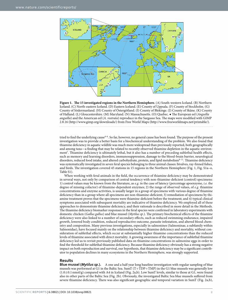
tried to find the underlying cause4–6. So far, however, no general cause has been found. The purpose of the present investigation was to provide a better basis for a biochemical understanding of the problem. We also found that thiamine deficiency in aquatic wildlife was much more widespread than previously reported, both geographically and among taxa—a finding that may be related to recently observed thiamine depletion in the aquatic environ-ment7. Thiamine deficiency is ultimately lethal, but it also has a number of preceding sublethal health effects, such as memory and learning disorders, immunosuppression, damage to the blood-brain barrier, neurological disorders, reduced food intake, and altered carbohydrate, protein, and lipid metabolism8–11. Thiamine deficiency was systematically investigated in seven feral species belonging to three animal classes: bivalves, ray-finned fishes, and birds. The investigation covered 45 stations in 15 regions in the Northern Hemisphere (Fig. 1, Fig. S1a–o, Table S1).
When working with feral animals in the field, the occurrence of thiamine deficiency may be demonstrated in several ways, not only by comparison of central tendency with non-thiamine-deficient (control) specimens: 1) control values may be known from the literature, as e.g. in the case of latency (percentage apoenzyme, i.e. the degree of missing cofactor) of thiamine-dependent enzymes; 2) the range of observed values, of e.g. thiamine concentrations and enzyme activities, is usually larger in a group of specimens with various degree of thiamine deficiency than in a group where all specimens are non-thiamine-deficient; 3) remediation of specimens by thi-amine treatment proves that the specimens were thiamine-deficient before the treatment; and 4) typical clinical symptoms associated with subsequent mortality are indicative of thiamine deficiency. We employed all of these approaches to demonstrate thiamine deficiency, and their rationale is described in more detail in the Methods. The thiamine deficiency biomarker responses in the feral species were confirmed in laboratory experiments with domestic chicken (Gallus gallus) and blue mussel (Mytilus sp.). The primary biochemical effects of the thiamine deficiency were also linked to a number of secondary effects, such as reduced swimming endurance, impaired growth, lowered body condition, reduced reproductive outcome, parasite infestation, and altered blood chem-istry and composition. Many previous investigations, especially in salmonines (Salmoninae, a subfamily within Salmonidae), have focused mainly on the relationship between thiamine deficiency and mortality, without con-sideration of sublethal effects, which occur at substantially higher thiamine concentrations than the reduced levels of thiamine associated with direct mortality. A growing awareness of the importance of sublethal thiamine deficiency led us to revisit previously published data on thiamine concentrations in salmonine eggs in order to find the threshold for sublethal thiamine deficiency. Because thiamine deficiency obviously has a strong negative impact on both reproduction and survival, our hypothesis, that thiamine deficiency may be a significant contrib-utor to population declines in many ecosystems in the Northern Hemisphere, was strongly supported.
ResultsBlue mussel (Mytilus sp.). A one and a half year long baseline investigation with regular sampling of blue mussels was performed at G1 in the Baltic Sea. SumT (T+ TDP+ TMP) in the G1 blue mussels was generally low (1.0± 0.1 nmol/g) compared with A4 in Iceland (Fig. 2a,b). Low SumT levels, similar to those at G1, were found also in other parts of the Baltic Sea (Fig. 2b). Obviously, the investigated Baltic Sea blue mussels suffered from severe thiamine deficiency. There was also significant geographic and temporal variation in SumT (Fig. 2a,b),
Figure 1. The 15 investigated regions in the Northern Hemisphere. (A) South-western Iceland. (B) Northern Iceland. (C) North-eastern Iceland. (D) Eastern Iceland. (E) County of Uppsala. (F) County of Stockholm. (G) County of Södermanland. (H) County of Östergötland. (I) County of Blekinge. (J) County of Skåne. (K) County of Halland. (L) Gloucestershire. (M) Maryland. (N) Massachusetts. (O) Quebec. • The European eel (Anguilla anguilla) and the American eel (A. rostrata) reproduce in the Sargasso Sea. The maps were modified with GIMP 2.8.16 (http://www.gimp.org/downloads/) from Free World Maps (http://www.freeworldmaps.net/printable/).

www.nature.com/scientificreports/
3Scientific RepoRts | 6:38821 | DOI: 10.1038/srep38821
indicating that the thiamine deficiency occurs episodically. The data in Fig. 2b do not, however, warrant conclu-sions about trends over the years. The even SumT level in the groups with lowest SumT 2011–2013 (Fig. 2b) may indicate a threshold for survival of ca 1.0 nmol/g. It is important that SumT concentrations as high as 3.4 nmol/g do occur (Fig. 2a,b), and may be indicative of a more normal thiamine status. The proportions of T, TDP, and TMP were very similar in specimens from Iceland and the Baltic Sea differing in SumT levels (Fig. 2c, Fig. S2a). This finding suggests that the cause of the thiamine deficiency is not malfunction of the thiamine phosphoryla-tion within the cells. As expected, but not previously shown in blue mussels, digestive gland transketolase (TK)
Figure 2. Blue mussel (Mytilus sp.). (a) Soft body SumT at G1 in the County of Södermanland (red or orange) and at A4 in Iceland (blue). (b) Soft body SumT in Iceland (blue) and the Baltic Sea area (red). ANOVA-type regression model. (c) Concentration and proportion soft body T, TDP, and TMP in Icelandic (blue) specimens with high SumT and Baltic Sea (red) specimens with low SumT. (d) Digestive gland TK latency and soft body (except digestive gland) SumT in the Baltic Sea. (e) Digestive gland TK activity and latency at G1 during four seasons. (f) Soft body SumT at G1 (red) and chlorophyll a in the water at G2 (green) in the County of Södermanland. Dashed line: five weeks delayed chlorophyll a curve. (g,h) Dry body weight in specimens of different lengths at G1 2011 (red/black, this investigation) and Vrångskär in the County of Södermanland 1973 (green, adapted from Kautsky16). (g) The shaded area indicates the reproductive period (March 1–June 10) defined by Kautsky16. (h) Week 17–19. Dashed lines: 95% CI. • Bars: arithmetic means. Whiskers: 95% CI. C: control. Group comparisons are indicated by brackets.

www.nature.com/scientificreports/
4Scientific RepoRts | 6:38821 | DOI: 10.1038/srep38821
activity was positively related to soft body SumT in a subsample from the Baltic Sea (Fig. S2c), whereas digestive gland TK latency (proportion of apoenzyme) was negatively related to soft body SumT in the same material (Fig. 2d). A negative relationship was also demonstrated between digestive gland TK latency and digestive gland SumT in a number of aquaculture blue mussels experimentally subjected to hypoxia and hyperthermia (Fig. S2d). These relationships demonstrate that TK activity and latency are relevant biomarkers of thiamine deficiency also in this invertebrate species. Negative relationships between digestive gland TK activity and latency were observa-ble at A4 (Fig. S2e) and in three of four seasons at G1 (Fig. 2e).
In the aquatic food web, thiamine is produced mainly by micro and macro algae, and to a more limited extent by fungi and bacteria12–15. Hence, SumT concentrations in the blue mussels are expected to be positively corre-lated with chlorophyll a in the water, but we found a negative correlation between blue mussel SumT and water chlorophyll a (Fig. 2f). This finding indicates a disturbed production and/or flux of thiamine in the aquatic food web today. If the chlorophyll a curve was delayed five weeks, however, there was a tendency towards a positive correlation (Fig. 2f, dashed line). An explanation of this observation could be that thiamine in the blue mussels comes mainly from fungi and bacteria feeding on settling dead phytoplankton some time after the blooms.
In Fig. 2g,h and Fig. S2i, dry body weight data on the blue mussels from G1 2011–2012 are compared with corresponding data from a nearby (ca 12 km) station in region G in 1973 presented by Kautsky16. The most notable observation was the almost complete absence of a peak in the dry body weight during the reproductive period in 2011 (Fig. 2g, shaded area). This difference was also reflected in Fig. 2h, illustrating the respective dry body weights in week 17–19, which was the time of the peak in the dry body weight in 1973. Lower dry body weight during the reproductive period was also observed 2011–2012 compared with corresponding data from south-western Finland in 1992 (Fig. S2j) presented by Öst & Kilpi17. These findings suggest impaired reproduc-tion in the blue mussels today, possibly as a result of thiamine deficiency. During the rest of the year (outside the reproductive period) the dry body weight was instead higher 2011–2012 than in 1973 (Fig. S2i). Explanations of this observation could be a trade-off between somatic growth and gonadal growth, or a generally higher avail-ability of food for the blue mussels today due to eutrophication. Important, though, is that the blue mussels do not have a generally lower dry body weight today than 40 years ago. This finding speaks against the hypothesis that the recent population declines in several diving duck species (including the common eider) feeding on blue mussels in the Baltic Sea18 would be caused by generally lower flesh content in the blue mussels.
Common eider (Somateria mollissima). For assessment of thiamine status in common eider females, we used domestic chicken as a surrogate control (Fig. 3a–f, Fig. S3a–e). A thiamine “complete” group and a thia-mine “deficient” group were produced in the laboratory by giving domestic chickens different concentrations of thiamine in their fodder during 11–12 days. The mean dose (nmol T per g dry fodder) was 17.5 for the complete group and 1.2 for the deficient group. Figure 3a and Fig. S3a illustrates that the deficient group had substantially lower SumT and TMP concentrations in the liver than the complete group, and that these concentrations were very similar to those found in the common eider females from Iceland (D3) and Sweden (I10–14). Fig. 3b–f and Fig. S3b show the results of six other thiamine deficiency biomarkers in the same material: brain SumT, liver α -ketoglutarate dehydrogenase (KGDH) activity and latency, brain KGDH activity and latency, and proportion liver TDP. The general pattern was that thiamine status was substantially lower in the deficient group than in the complete group, and that the common eider females were more similar to the deficient group, except for brain SumT and brain KGDH latency in the Icelandic material. Moreover, when there was a difference between Icelandic and Swedish common eider females, the thiamine deficiency was more severe in Sweden. Thiamine deficiency is often associated with a biphasic relationship between proportion TDP and T concentration in var-ious tissues (Fig. S3c–e). The mechanism behind this relationship is described in detail in the Supplementary Results. Accordingly, thiamine deficiency is indicated in the common eider females by the negative relationship between the proportion TDP and the T concentration in both the liver and brain (Fig. S3f,g). There was also a negative relationship between activity and latency for liver TK (Fig. S3h), liver KGDH (Fig. S3i), and brain KGDH (Fig. S3j).
Yolk T concentration was analysed in Icelandic common eider eggs sampled on several occasions between 2005 and 2013 (Fig. 3g). There was significant geographic and temporal variation in yolk T (Fig. 3g), indicating that the thiamine deficiency occurs episodically. The data in Fig. 3g do not, however, warrant conclusions about temporal trends. A majority of the eggs from D1 in 2008 and 2009 had a yolk T concentration under 5 nmol/g (Fig. 3g), which may be a threshold for the production of viable offspring. This threshold is based on the mean yolk T concentration 4.7 nmol/g, which was coincident with an almost complete absence of common eider pulli in the County of Södermanland (region G) during the breeding season 20053. More recently, extremely low num-bers of common eider pulli were observed also in the County of Blekinge (region I) coincidently with abnormal behaviour of both females and pulli (see Supplementary Results).
Figure 3h illustrates a 100 year survey of more than 20,000 common eider nests on 28 islands in an area (F9) within the Stockholm Archipelago. The dip between 1939 and 1961 was probably due to excessive hunting and environmental disturbances during the economic depression and the Second World War. Between 1964 and 2001, the population was relatively stable at around 1,100 nests per year. From 2001, there was a dramatic decline through 2016, when the total number of nests was only two, the lowest number ever. This development was antic-ipated in our work 2005–20093. In recent years, large common eider population declines have been observed also in Iceland19. We suspect thiamine deficiency to be the cause of these recent population declines.
Anguillid eels (Anguilla spp.). Elvers were sampled at the mouth of River Severn (L1) in south-western England, UK, i.e. in the middle of the geographic range of the European eel (Anguilla anguilla). The proportions of T, TDP, and TMP in the muscle (Fig. 4a) were similar to those commonly reported in non-thiamine-deficient fish20, although the T proportion was somewhat lower. This finding suggests normal function of the thiamine

www.nature.com/scientificreports/
5Scientific RepoRts | 6:38821 | DOI: 10.1038/srep38821
phosphorylation within the cells. Thiamine deficiency was indicated, however, by the negative relationship between the proportion muscle TDP and the muscle T concentration (Fig. S4a). Thiamine deficiency was also indicated by the positive response to bathing in a T solution. Such treatment should have no effect on non-thiamine-deficient individuals, but muscle TDP, muscle TMP, and liver TK activity all increased (Fig. 4b–d), whereas liver TK latency decreased (Fig. 4e). Further indications of thiamine deficiency were illustrated by the negative relationships between liver TK activity and latency (Fig. S4b) and between liver TK latency and muscle TDP (Fig. S4c). The latter relationship indicated that the thiamine deficiency was of systemic nature. There was also a positive relationship between liver TK activity and muscle TDP (P = 0.028, R2 = 0.12, n = 40, not shown).
Figure 3. Common eider (Somateria mollissima) and domestic chicken (Gallus gallus). (a–f) Domestic chicken thiamine “complete” group (green, 17.5 nmol T per g dry fodder) and thiamine “deficient” group (orange, 1.2 nmol T per g dry fodder), common eider females at D3 in eastern Iceland (blue) and at I10–14 in the County of Blekinge (red). Student’s t-test. (a) Liver SumT. (b) Brain SumT. (c) Liver KGDH activity. (d) Liver KGDH latency. (e) Brain KGDH activity. (f) Brain KGDH latency. (g) Box plot of common eider egg yolk T in Iceland. Data from 2005 were adapted from Balk et al.3. Wilcoxon-Mann-Whitney test. (h) A 100 year survey of more than 20,000 common eider nests on 28 islands in the archipelago at F9 in the County of Stockholm. • Bars: arithmetic means. Whiskers: 95% CI (except in the box plot). C: control. Group comparisons are indicated by brackets.

www.nature.com/scientificreports/
6Scientific RepoRts | 6:38821 | DOI: 10.1038/srep38821
For silver eels there was a multitude of variables indicating severe thiamine deficiency both in the European eel and the American eel (A. rostrata). Liver TK latency was negatively related to liver SumT (Fig. 4f). Liver TK activity was positively related to liver SumT (Fig. S4d) and negatively related to liver TK latency (P = 0.042, R2 = 0.18, n = 24, not shown). There were also negative relationships between proportion TDP and T concen-tration in the white muscle (Fig. S4e), brain (Fig. S4f), and liver (Fig. S4g). The systemic nature of the thiamine deficiency was further indicated by the positive relationships between brain SumT and liver SumT (Fig. S4h), between somatic growth (SG) and liver SumT (Fig. 4g), between SG and liver/brain SumT ratio (Fig. S4i), and
Figure 4. European eel (Anguilla anguilla) and American eel (A. rostrata). (a–e) Untreated (blue) and T-treated (green) elvers from L1 in the UK. The T-treated specimens were bathed twice in a 100 mg/L T solution for 48 h each time. (a) Concentration and proportion muscle T, TDP, and TMP. (b–e) Student’s t-test. (b) Muscle TDP. (c) Muscle TMP. (d) Liver TK activity. (e) Liver TK latency. (f–h) Female European silver eels in Sweden. (f) Liver TK latency and SumT. (g) SG and liver SumT. (h) Brain TK activity and BCI. (i) White muscle SumT in female European silver eels in Sweden (red) and two groups of American silver eels in St. Lawrence River, Canada (pink/black), adapted from Fitzsimons et al.21. The range is indicated to the right of each coloured bar. (j) Critical swim speed and white muscle SumT in experimental American yellow eels from M1 in Maryland, USA. (k) Number of the swim bladder parasite Anguillicola crassus and proportion liver TDP in female European silver eels in Sweden. • Bars: arithmetic means. Whiskers: 95% CI. OLS: ordinary least squares regression. GM: geometric mean regression.

www.nature.com/scientificreports/
7Scientific RepoRts | 6:38821 | DOI: 10.1038/srep38821
between brain TK activity and body condition index (BCI) (Fig. 4h). Mean white muscle SumT concentrations in the European and American silver eels were 1.6–3.6 nmol/g (Fig. 4i), although single specimens of the latter species had considerably higher white muscle SumT21. For example, the maximum white muscle SumT concen-tration observed in American silver eels 2005 was 15.9 nmol/g (Fig. 4i). It is thus likely that a white muscle SumT concentration of 1.6–3.6 nmol/g is far from sufficient for migration to the Sargasso Sea and production of viable (non-thiamine-deficient) eggs, as also suggested by Fitzsimons et al.21. The mass balance of SumT in the spawning female silver eel was modelled for two scenarios, which are available as Supplementary Information. The effect of thiamine deficiency on swimming endurance was analysed in American yellow eels (Fig. 4j). Swimming endur-ance was reduced at the white muscle SumT concentrations observed in many feral European and American eels today (Fig. 4i,j). Thiamine deficiency has been associated with immunosuppression in several investigationse.g. 22, as well as with increased parasite infestation23. Hence, it is interesting to note the negative relationship between the number of the swim bladder parasite Anguillicola crassus and proportion liver TDP in silver eels (Fig. 4k), further analysed in the Supplementary Results. There was also a positive relationship between brain TK latency and the number of this parasite in the same specimens (P = 0.039, R2 = 0.20, n = 28, not shown).
Salmonines (Salmoninae). Here, we focus primarily on sublethal thiamine deficiency in both parents and their offspring of a number of salmonines. This approach differed from previous investigations within this scien-tific field, which focused mainly on mortality in the offspring. Our current focus provides a new perspective to the problem, and we have revisited existing literature to evaluate the prevalence of sublethal thiamine deficiency among salmonines in the Northern Hemisphere.
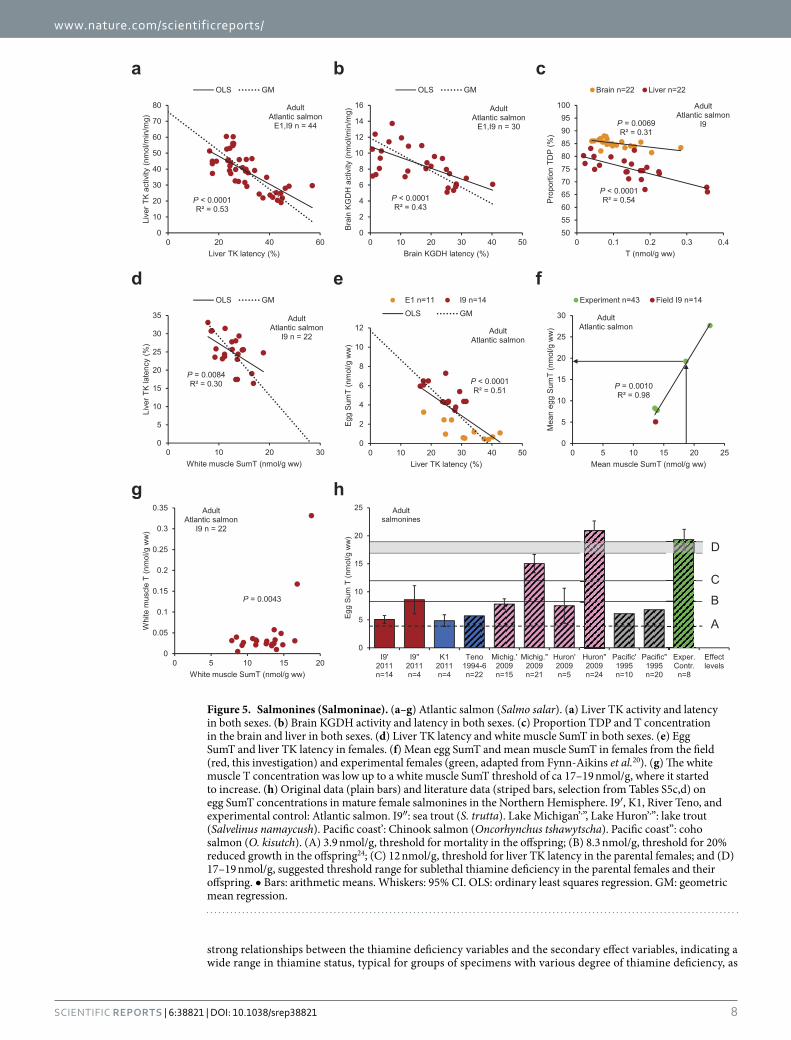
In parental Atlantic salmon (Salmo salar) of both sexes from E1 and I9 in the Baltic Sea area, thiamine defi-ciency was indicated by the negative relationship between liver TK activity and latency (Fig. 5a), and even the liver TK latencies of 16–57% alone were evidence of severe sublethal thiamine deficiency. Liver TK activity and latency were also related to uncoordinated swimming (wiggling) in the parents (Fig. S5a,b). Brain KGDH activity and latency were negatively related and latencies of 0–40% were observed (Fig. 5b). The proportion TDP was neg-atively related to the T concentration, both in the liver and the brain (Fig. 5c). The thiamine status in the females was mostly lower at E1 than at I9, and lowest in the E1 females with mortality (lethal thiamine deficiency) in the offspring (Fig. S5c–h). The systemic nature of the thiamine deficiency was indicated by the negative relationships between liver TK latency and white muscle SumT (Fig. 5d) and egg SumT and liver TK latency (Fig. 5e), as well as the positive relationships between egg SumT and liver TK activity (Fig. S5j), white muscle SumT and total weight (Fig. S5k), liver SumT and heart SumT (Fig. S5l), liver SumT and brain SumT (Fig. S5m), and heart SumT and liver/brain SumT ratio (P = 0.0076, R2 = 0.31, n = 22, not shown).
Extrapolation to zero latency in Fig. 5d gave a white muscle SumT concentration of 28.2± 6.8 nmol/g, and extrapolation to zero latency in Fig. 5e gave an egg SumT concentration of 11.7± 2.3 nmol/g. These white mus-cle and egg SumT concentrations were not essentially different from those obtained by direct measurement in Atlantic salmon females, differing in thiamine status, by Fynn-Aikins et al.20. Their data demonstrated a strong positive relationship between mean egg and muscle SumT over a large range of SumT concentrations, and the means of our data from I9 fitted into this relationship (Fig. 5f), in which e.g. a muscle SumT concentration of 18.6 nmol/g corresponded to an egg SumT concentration of 19.3 nmol/g (arrows). An indication of how high the white muscle SumT concentration at least should be in a non-thiamine-deficient individual was provided by the threshold in Fig. 5g, where the white muscle T concentration was low up to a white muscle SumT concentration of ca 17–19 nmol/g, where it started to increase. The significance of this threshold was analysed with Fisher’s exact test (P = 0.0043), where the P-value represents the probability to obtain the two higher values at the end of the data array by pure chance. Egg SumT concentrations in a selection of mature female salmonines in the Northern Hemisphere are presented in Fig. 5h. (A more complete compilation of literature data on egg SumT concentra-tions of > 4.0 nmol/g is given in the Table S5c,d). The indicated levels are as follows: (A) 3.9 nmol/g, threshold for mortality in the offspring (see Supplementary Results); (B) 8.3 nmol/g, threshold for 20% reduced growth24; (C) 12 nmol/g, threshold for liver TK latency (Fig. 5e); and (D) 17–19 nmol/g, suggested threshold range for sublethal thiamine deficiency (see Supplementary Results).
Herring (Clupea harengus). The herrings sampled here turned out to be essentially non-thiamine-deficient. Because they consisted of mature specimens that had migrated to their spawning grounds at the correct time of the year and were strong enough to catch the jig of spinning rods, they may constitute a non-random sample of the Baltic Sea herring population. The importance of these herrings was to show that non-thiamine-deficient individuals can still be found in the field, and that they exhibit biomarker responses that are not typical of thia-mine deficiency (Fig. S6a–f, Table S6).
DiscussionThis investigation demonstrated severe thiamine deficiency in six out of seven studied aquatic species present in the Northern Hemisphere: blue mussel, common eider, European eel, American eel, Atlantic salmon, and sea trout. The analysed variables included activity and latency of a cytosolic (TK) and a mitochondrial (KGDH) thiamine-dependent enzyme in the digestive gland (blue mussels) and the liver and brain (vertebrates), as well as concentrations of T, TDP, and TMP in the digestive gland, liver, brain, muscle, heart, and eggs. Further analyses are presented in the Supplementary Results. The enzyme and thiamine variables indicated low thiamine status in a majority of the investigated specimens. We also analysed a number of secondary effect variables, such as—in the blue mussel: growth, condition, and reproduction—in the common eider: condition, liver size, histopathological alterations, and reproduction—in the European and American eel: swimming endurance, growth, condition, blood chemistry and composition, parasite infestation, and reproduction—and in the Atlantic salmon: swim-ming behaviour, growth, condition, blood chemistry and composition, and reproduction. There were several
www.nature.com/scientificreports/
8Scientific RepoRts | 6:38821 | DOI: 10.1038/srep38821
strong relationships between the thiamine deficiency variables and the secondary effect variables, indicating a wide range in thiamine status, typical for groups of specimens with various degree of thiamine deficiency, as
Figure 5. Salmonines (Salmoninae). (a–g) Atlantic salmon (Salmo salar). (a) Liver TK activity and latency in both sexes. (b) Brain KGDH activity and latency in both sexes. (c) Proportion TDP and T concentration in the brain and liver in both sexes. (d) Liver TK latency and white muscle SumT in both sexes. (e) Egg SumT and liver TK latency in females. (f) Mean egg SumT and mean muscle SumT in females from the field (red, this investigation) and experimental females (green, adapted from Fynn-Aikins et al.20). (g) The white muscle T concentration was low up to a white muscle SumT threshold of ca 17–19 nmol/g, where it started to increase. (h) Original data (plain bars) and literature data (striped bars, selection from Tables S5c,d) on egg SumT concentrations in mature female salmonines in the Northern Hemisphere. I9′, K1, River Teno, and experimental control: Atlantic salmon. I9″: sea trout (S. trutta). Lake Michigan’,”, Lake Huron’,”: lake trout (Salvelinus namaycush). Pacific coast’: Chinook salmon (Oncorhynchus tshawytscha). Pacific coast”: coho salmon (O. kisutch). (A) 3.9 nmol/g, threshold for mortality in the offspring; (B) 8.3 nmol/g, threshold for 20% reduced growth in the offspring24; (C) 12 nmol/g, threshold for liver TK latency in the parental females; and (D) 17–19 nmol/g, suggested threshold range for sublethal thiamine deficiency in the parental females and their offspring. • Bars: arithmetic means. Whiskers: 95% CI. OLS: ordinary least squares regression. GM: geometric mean regression.
www.nature.com/scientificreports/
9Scientific RepoRts | 6:38821 | DOI: 10.1038/srep38821
described in the Methods. These relationships also indicated that many health effects, including abnormal behav-iour, observed in biota today may well be due to thiamine deficiency. In many cases, we estimated the expected thiamine status of non-thiamine-deficient individuals, e.g. by extrapolation to zero latency, and generally, the hypothesized non-thiamine-deficient individuals had much higher thiamine status than the thiamine-deficient individuals. The episodic nature of the thiamine deficiency, in both time and space, was demonstrated by large differences in thiamine status between sampling groups. From a biochemical point of view, it is unlikely that the thiamine deficiency is caused by impaired phosphorylation within the cells, because the proportions of the phosphorylated forms were normal and the proportion TDP initially increased with increasing thiamine defi-ciency. Rather, the results point towards insufficient amount of thiamine in the food, as indicated by the relation-ship between the common eider and its main prey, blue mussels (see Supplementary Results). Such knowledge constitutes foundational information needed for the search for the causative agent(s). By investigating a large geographic area, by extending the focus from lethal to sublethal thiamine deficiency, and by linking biochemical alterations to secondary effects, we were able to demonstrate that the problem of thiamine deficiency is much more widespread and severe than previously reported.
The biogeochemical environment is highly impacted by human activities, both directly through the release of pollutants and nutrients, and indirectly through altered biogeochemical cycles of substances. The degree of human impact today, e.g. the number of man-made substances being released into the environment, is enormous compared with previous centuries. Consequently, the work to find a causative agent or agents for the thiamine deficiency will be a multi-step procedure. Before attempting to test possible candidates among a myriad of specific substances in exposure experiments, it is absolutely necessary to know which species, trophic levels, and geo-graphic areas are affected by the thiamine deficiency, as well as its temporal variation. Such information should facilitate finding of the toxic mechanism, which in turn will be critical for finding the causative agent(s).
Decades of environmental research have proved that in order to avoid toxic effects in biota, including humans, man must stop releasing persistent pollutants into the environment. It is well known that lipophilic persistent pollutants biomagnify in the food web in a way such that top predators are most affected. Impaired health as a result of deficiency of an essential vitamin, however, surely differs from this paradigm, a presumption supported by the episodic nature of thiamine deficiency and its lack of correlation with exposure to classic lipophilic per-sistent pollutants. Although severe thiamine deficiency in salmonines has been observed since the early 1970s in both northern Europe and North America1, most of the research has focused mainly on mortality25,26 and not on sublethal effects. The present investigation sets out to demonstrate that this is inadequate, and it is tempting to draw a parallel to classic environmental pollutants, which also were investigated predominantly with respect to their lethal effects in the beginning (ca 1950–1970). It was not until the mid-1960s that concern started to arise about their sublethal effects, and doses orders of magnitude lower than those causing direct mortality were found to be important. Also for the classic environmental pollutants, the extension of the focus from lethal to sublethal effects revealed that the problem was much more widespread and severe than previously realized.
In order to understand the ecological implications of thiamine deficiency, it is important to remember that it always starts with molecular alterations on the subcellular level. Inactivation of thiamine-dependent enzymes is followed by an increase in toxic metabolites, such as lactate, glyoxals, and phytanic acid, as well as a decrease in life sustaining molecules, such as ATP, NADH, and NADPH. Such alterations compro-mise the integrity of tissues and organs, and their malfunction results in systemic disorders affecting cen-tral functions of life, such as growth, reproduction, immune defence, behaviour, nerve function, sensory functions, learning, and memory. Moreover, the metabolic disorders caused by thiamine deficiency are often only partially reversible, i.e. physiological conditions may not be fully restored, even though thi-amine supply is fully restored. This has been demonstrated for both TK27–30 and KGDH31–33, as well as for other thiamine-dependent enzymes and metabolites27,32,33. One phenomenon that contributes to irreversi-ble damage is focal cell necrosis, where dead cells are not replaced34–37. These relatively recent observations that a short-lasting (days–weeks) episode of thiamine deficiency may cause long-lasting (many years or for the rest of an organism’s life) sublethal effects38,39 add another dimension to the problem. In fact, many feral animals may have gone through one or more episodes of thiamine deficiency in recent years and thus suf-fer from a long-lasting or permanent reduction in thiamine-dependent enzyme activities. This phenom-enon may have dramatic consequences for the majority of ecological research performed during the last decades, because it is not certain that animals display normal characteristics when subject to past and/or present thiamine deficiency. All kinds of characteristics may be affected, e.g. feeding, migration, behaviour, habitat preference, reproduction, and sex ratio. This is important to be aware of, especially since thiamine defi-ciency has been observed even in remote areas. For example, thiamine deficiency must be taken into account in the attempts to track animal movements, such as the silver eels’ migration to the Sargasso Sea, by use of telemetry equipment40–42. It is also possible that thiamine treatment of silver eels is necessary before viable offspring can be produced by hormonal treatment in the laboratory and in future aquaculture43–46. Another important phenomenon is that thiamine-deficiency-induced immunosuppression in feral animals involves increased spreading of pathogens between species. The avian influenza is a striking example, because feral water birds form the natural reservoir of these viruses47–49. During the last decade, several investigations of feral birds have pointed at the increased disease risk by immunosuppression, which is also reflected in altered disease dynamics3,50–54.
It is well known that populations lose much of their genetic variation when passing through a bottleneck. Recently, it has been demonstrated in seabirds that such loss of genetic variation also directly reduces the fitness of single individuals55. Despite the fact that biodiversity loss56,57 may well be the most important global threat to sustainability58, relatively few scientists have attempted to estimate the relative importance of the different fac-tors contributing to this serious problem. Factors that have been proposed include habitat loss, global warming, and lack of food, although surprisingly often without explicit scientific references. Although the first two factors

www.nature.com/scientificreports/
1 0Scientific RepoRts | 6:38821 | DOI: 10.1038/srep38821
constitute serious problems, they are insufficient to explain the already ongoing global biodiversity loss57,59. For example, it is now 30 years since agricultural intensification ceased to explain the population declines of many farmland bird species60,61. It should also be remembered that global warming still mainly is a risk scenario of the future. The increase in global average surface temperature from 1951 to 2010 was within the range 0.6–0.7 °C62, which is notably less than feared future temperature increases. It may thus be time to revise several opinions about the dramatic current population declines63,64, sometimes referred to as the sixth mass extinction65. From 1970 to 2010/2012, population sizes of both terrestrial and marine vertebrate species dropped by half66,67, and from 1950 to 2010, the global seabird population declined overall by ca 70%68. We suggest that thiamine deficiency may be a significant contributor to this fatal process, and that the cause must be searched for at the chemical and biochemical levels.
MethodsThiamine concentrations were measured with High Performance Liquid Chromatography (HPLC) with fluores-cence detection. Thiamine-dependent enzyme activities were measured spectrophotometrically in subcellular fractions. The detailed methods are described in the Supplementary Methods.
How thiamine deficiency is demonstrated. Several facts support the concept that the latency of thiamine-dependent enzymes should be zero or very low in non-thiamine-deficient individuals. Literature data on TK and KGDH in organs, such as liver, kidney, and brain, in animals, such as mouse, rat, domestic chicken, European eel, and rainbow trout (Oncorhynchus mykiss), show that the mean latency of these enzymes in groups of control specimens is mostly 0–6%3,28,69–77, whereas higher latencies are often associated with formation of toxic metabolitese.g. 78 and disease with more or less permanent damagee.g. 38,39. For example, the decrease in TK activity during an episode of severe thiamine deficiency is only partly reversible28,69,79. Moreover, urinary excretion of thiamine strongly declines when latency starts to increase80. Obviously, the body strives to keep the latency low. From an evolutionary point of view, the ability to synthesize thiamine is believed to have been lost in animals during evolution, because it always has been abundant in the food81. Hence, throughout the biochemical litera-ture, thiamine is customarily assumed to be present in sufficient amounts in non-thiamine-deficient individuals. Apparently, there is no such thing as “natural latency” and thus latencies higher than 6% are indicative of thiamine deficiency. Thiamine deficiency may, however, sometimes be present even in animals with low latencies, because the apoenzyme is not always maintained in thiamine-deficient tissues73–75,82.
Another important property of thiamine is that there is an upper limit for how much thiamine each organ in the body can hold, and any excess thiamine is excreted within a relatively short time20,80,81,83–85. Because thiamine normally should be present in sufficient amounts in the diet, there is no need for storing extra thiamine. To the best of our knowledge, there are no data in the biochemical literature indicating storage of excess thiamine in somatic tissues81. Together, these properties of thiamine suggest that the natural thiamine concentration in a non-thiamine-deficient tissue should not be substantially lower than the maximum observed thiamine concen-tration in this tissue. Therefore, thiamine concentrations substantially lower than the maximum concentration are indicative of thiamine deficiency.
Literature data show that thiamine concentrations and activities of thiamine-dependent enzymes may decrease to as little as 5–10% of control levels in single thiamine-deficient individuals77,79,86. Hence, the range of measured thi-amine deficiency biomarker values are significantly larger in a group of specimens with different degrees of thiamine deficiency than in a corresponding control group, where all specimens are non-thiamine-deficiente.g. 71,74,77,78,84,86,87. As a consequence, bivariate relationships between thiamine-deficiency biomarkers are often discernible only in groups of specimens with different degrees of thiamine deficiency, where the range of measured biomarker values is large, and not in non-thiamine-deficient groups of specimens, where this range is much smaller. This phenomenon has been observed in numerous investigationse.g. 70,78–80,87–91. The observed relationships between thiamine deficiency biomarkers and secondary effects are also in agreement with the scientific literature on thi-amine deficiency.
Relationships between latency and another biomarker allow extrapolation to zero latency, as an indication of what a control value of the other biomarker might be. The observed covariance between independently measured biomarkers also corroborate that our analyses were correctly performed. Finally, another way to demonstrate thiamine deficiency is remediation. Administration of thiamine to a non-thiamine-deficient individual should have no effect at all, whereas a therapeutic effect of thiamine proves that the specimen was thiamine-deficient prior to treatment.
Animal care. All use of animals in this investigation was performed in accordance with the permits required by the respective national laws and local regulations. The sampling of common eiders (including eggs and pulli) in Sweden was approved by the Swedish Environmental Protection Agency, SEPA (Dnr. NV-02953–11) and the Stockholm Northern Research Ethics Committee (Dnr. N58/11). Permission to temporarily visit bird protec-tion areas in the County of Blekinge 2010–2014 was granted by the County Administrative Board of Blekinge (Dnr. 521–441–10). The sampling of common eiders and their eggs in Iceland was approved by the Icelandic Ministry for the Environment and Natural Resources (UMH13060039, UMH05040093/12–1). The domestic chicken experiments were approved by the Stockholm Northern Research Ethics Committee (Dnr. N351/10, Dnr. N209/13). The purchase and direct import of elvers from the UK to Stockholm University was approved by the Swedish Board of Agriculture (Dnr. 6.2. 18–4928/13). The American eel swimming endurance experiment was approved by U.S. Geological Survey, Leetown Science Center Institutional Animal Care and Use Committee. The American eel maturation experiment was approved by the University of Massachusetts Dartmouth’s Institutional Animal Care and Use Committee (Protocol No. 09–03).

www.nature.com/scientificreports/
1 1Scientific RepoRts | 6:38821 | DOI: 10.1038/srep38821
References1. Fitzsimons, J. D. The effect of B-vitamins on a swim-up syndrome in Lake Ontario lake trout. Suppl. J. Great Lakes Res. 21, 286–289
(1995).2. Sepúlveda, M. S. et al. Organochlorine pesticides and thiamine in eggs of largemouth bass and American alligators and their
relationship with early life-stage mortality. J. Wildlife Dis. 40, 782–786 (2004).3. Balk, L. et al. Wild birds of declining European species are dying from a thiamine deficiency syndrome. Proc. Natl. Acad. Sci. USA
106, 12001–12006 (2009).4. McDonald, G., Fitzsimons, J. D. & Honeyfield, D. C. (eds). Early life stage mortality syndrome in fishes of the Great Lakes and Baltic
Sea. American Fisheries Society, Symposium 21 (1998).5. Bengtsson, B.-E., Hill, C., Nellbring, S. & Kessler, E. (eds). Reproductive disturbances in Baltic Sea fish: An international perspective.
Ambio 28(1) (1999).6. Blazer, V. S. & Brown, L. L. (eds). Early mortality syndrome in Great Lakes salmonines. Journal of Aquatic Animal Health 17(1)
(2005).7. Sañudo-Wilhelmy, S. A. et al. Multiple B-vitamin depletion in large areas of the coastal ocean. Proc. Natl. Acad. Sci. USA 109,
14041–14045 (2012).8. Singleton, C. K. & Martin, P. R. Molecular mechanisms of thiamine utilization. Curr. Mol. Med. 1, 197–207 (2001).9. Combs, G. F. The Vitamins 265–280 (Elsevier, 2008).
10. Manzetti, S., Zhang, J. & van der Spoel, D. Thiamin function, metabolism, uptake, and transport. Biochemistry 53, 821− 835 (2014).11. Mkrtchyan, G. et al. Molecular mechanisms of the non-coenzyme action of thiamin in brain: Biochemical, structural and pathway
analysis. Sci. Rep. 5, 12583, 10.1038/srep12583 (2015).12. Provasoli, L. & Carlucci, A. F. In Algal Physiology and Biochemistry (ed. Stewart, W. D. P.) 741–787 (University of California Press, 1974).13. Nishijima, T., Shiozaki, R. & Hata, Y. Production of vitamin B12, thiamine, and biotin by freshwater phytoplankton. B. Jpn. Soc. Sci.
Fish. 45, 199–204 (1979).14. Donderski, W. & Strzelczyk, E. Studies on B vitamins production by heterotrophic benthic bacteria of lakes of different trophy. II.
Mesotrophic Lake Jasne. Acta Microbiol. Pol. 36, 357–363 (1987).15. Nishijima, T., Sun, X. & Hata, Y. B group vitamin requirements of marine yeasts isolated from coastal sea-waters. B. Mar. Sci. Fish.
Kochi Univ. 11, 61–67. (1989).16. Kautsky, N. Quantitative studies on gonad cycle, fecundity, reproductive output and recruitment in a Baltic Mytilus edulis
population. Mar. Biol. 68, 143–160 (1982).17. Öst, M. & Kilpi, M. Blue mussels Mytilus edulis in the Baltic: Good news for foraging eiders Somateria mollissima. Wildlife Biol. 4,
81–89 (1998).18. Skov, H. et al. Waterbird populations and pressures in the Baltic Sea. TemaNord 2011:550 (2011).19. Jónsson, J. E. et al. Relationships between long-term demography and weather in a sub-arctic population of common eider. PLoS
ONE 8, e67093, 10.1371/journal.pone.0067093 (2013).20. Fynn-Aikins, K., Bowser, P. R., Honeyfield, D. C., Fitzsimons, J. D. & Ketola, H. G. Effect of dietary amprolium on tissue thiamin and
Cayuga syndrome in Atlantic salmon. Trans. Am. Fish. Soc. 127, 747–757 (1998).21. Fitzsimons, J. D. et al. Impacts of diet on thiamine status of Lake Ontario American eels. Trans. Am. Fish. Soc. 142, 1358–1369
(2013).22. Shoji, S. et al. Thiamine disulfide as a potent inhibitor of human immunodeficiency virus (type-1) production. Biochem. Bioph. Res.
Co. 205, 967–975 (1994).23. Prasad, R., Rao, Y. V. B. G., Mehta, K. & Subrahmanyam, D. Effect of thiamine deficiency on the filarial infection of albino rats with
Litomosoides carinii. Int. J. Parasitol. 10, 93–96 (1980).24. Fitzsimons, J. D. et al. Influence of thiamine deficiency on lake trout larval growth, foraging, and predator avoidance. J. Aquat. Anim.
Health 21, 302–314 (2009).25. Honeyfield, D. C., Fitzsimons, J. D., Brown, S. B., Marcquenski, S. V. & McDonald, G. Introduction and overview of early life stage
mortality. Am. Fish. Soc. Symp. 21, 1–7 (1998).26. Norrgren, L., Amcoff, P., Börjeson, H. & Larsson, P.-O. Reproductive disturbances in Baltic fish: A review. Am. Fish. Soc. Symp. 21,
8–17 (1998).27. McCandless, D. W., Schenker, S. & Cook, M. Encephalopathy of thiamine deficiency: Studies of intracerebral mechanisms. J. Clin.
Invest. 47, 2268–2280 (1968).28. Gibson, G. E., Ksiezak-Reding, H., Sheu, K.-F. R., Mykytyn, V. & Blass, J. P. Correlation of enzymatic, metabolic, and behavioral
deficits in thiamin deficiency and its reversal. Neurochem. Res. 9, 803–814 (1984).29. Butterworth, R. F., Giguere, J.-F. & Besnard, A.-M. Activities of thiamine-dependent enzymes in two experimental models of
thiamine-deficiency encephalopathy: 1. The pyruvate dehydrogenase complex. Neurochem. Res. 10, 1417–1428 (1985).30. Giguère, J.-F. & Butterworth, R. F. Activities of thiamine-dependent enzymes in two experimental models of thiamine deficiency
encephalopathy: 3. Transketolase. Neurochem. Res. 12, 305–310 (1987).31. Parker, W. D. et al. Brain mitochondrial metabolism in experimental thiamine deficiency. Neurology 34, 1477–1481 (1984).32. Héroux, M. & Butterworth, R. F. Reversible alterations of cerebral γ -aminobutyric acid in pyrithiamine-treated rats: Implications for
the pathogenesis of Wernicke’s encephalopathy. J. Neurochem. 51, 1221–1226 (1988).33. Butterworth, R. F. & Héroux, M. Effect of pyrithiamine treatment and subsequent thiamine rehabilitation on regional cerebral
amino acids and thiamine-dependent enzymes. J. Neurochem. 52, 1079–1084 (1989).34. Lê, O., Héroux, M. & Butterworth, R. F. Pyrithiamine-induced thiamine deficiency results in decreased Ca2+-dependent release of
glutamate from rat hippocampal slices. Metab. Brain Dis. 6, 125–132 (1991).35. Leong, D. K., Le, O., Oliva, L. & Butterworth, R. F. Increased densities of binding sites for the “peripheral-type” benzodiazepine
receptor ligand [3H]PK11195 in vulnerable regions of the rat brain in thiamine deficiency encephalopathy. J. Cereb. Blood Flow Metab. 14, 100–105 (1994).
36. Hamada, S. et al. Thiamine deficiency induces massive cell death in the olfactory bulbs of mice. J. Neuropathol. Exp. Neurol. 72, 1193–1202 (2013).
37. Zhao, Y. et al. Downregulation of transketolase activity is related to inhibition of hippocampal progenitor cell proliferation induced by thiamine deficiency. BioMed Res. Int. 2014, 572915, 10.1155/2014/572915 (2014).
38. Fattal-Valevski, A. et al. Outbreak of life-threatening thiamine deficiency in infants in Israel caused by a defective soy-based formula. Pediatrics 115, e233–e238, 10.1542/peds.2004–1255 (2005).
39. Mimouni-Bloch, A. et al. Thiamine deficiency in infancy: Long-term follow-up. Pediatr. Neurol. 51, 311–316 (2014).40. Aarestrup, K. et al. Oceanic spawning migration of the European eel (Anguilla anguilla). Science 325, 1660 (2014).41. Westerberg, H., Sjöberg, N., Lagenfelt, I., Aarestrup, K. & Righton, D. Behaviour of stocked and naturally recruited European eels
during migration. Mar. Ecol. Prog. Ser. 496, 145–157 (2014).42. Béguer-Pon, M., Castonguay, M., Shan, S., Benchetrit, J. & Dodson, J. J. Direct observations of American eels migrating across the
continental shelf to the Sargasso Sea. Nat. Commun. 6, 8705, 10.1038/ncomms9705 (2015).43. Sorensen, P. W. & Winn, H. E. The induction of maturation and ovulation in American eels, Anguilla rostrata (LeSueur), and the
relevance of chemical and visual cues to male spawning behavior. J. Fish Biol. 25, 261–268 (1984).

www.nature.com/scientificreports/
1 2Scientific RepoRts | 6:38821 | DOI: 10.1038/srep38821
44. Tanaka, H., Kagawa, H. & Ohta, H. Production of leptocephali of Japanese eel (Anguilla japonica) in captivity. Aquaculture 201, 51–60 (2001).
45. Palstra, A. P., Cohen, E. G. H., Niemantsverdriet, P. R. W., van Ginneken, V. J. T. & van den Thillart, G. E. E. J. M. Artificial maturation and reproduction of European silver eel: Development of oocytes during final maturation. Aquaculture 249, 533–547 (2005).
46. Oliveira, K. & Hable, W. E. Artificial maturation, fertilization, and early development of the American eel (Anguilla rostrata). Can. J. Zool. 88, 1121–1128 (2010).
47. Webster, R. G., Bean, W. J., Gorman, O. T., Chambers, T. M. & Kawaoka, Y. Evolution and ecology of influenza A viruses. Microbiol. Rev. 56, 152–179 (1992).
48. Fouchier, R. A. M. et al. Characterization of a novel influenza A virus hemagglutinin subtype (H16) obtained from black-headed gulls. J. Virol. 79, 2814–2822 (2005).
49. Hall, J. S. et al. Avian influenza virus ecology in Iceland shorebirds: Intercontinental reassortment and movement. Infect. Genet. Evol. 28, 130–136 (2014).
50. Hasselquist, D. Comparative immunoecology in birds: Hypotheses and tests. Suppl. J. Ornithol. 148, S571–S582 (2007).51. Buehler, D. M., Piersma, T., Matson, K. & Tieleman, B. I. Seasonal redistribution of immune function in a migrant shorebird:
Annual‐cycle effects override adjustments to thermal regime. Am. Nat. 172, 783–796 (2008).52. Martin, L. B., Weil, Z. M. & Nelson, R. J. Seasonal changes in vertebrate immune activity: Mediation by physiological trade-offs. Phil.
Trans. R. Soc. B 363, 321–339 (2008).53. Hoque, M. A., Burgess, G. W., Cheam, A. L. & Skerratt, L. F. Epidemiology of avian influenza in wild aquatic birds in a biosecurity
hotspot, North Queensland, Australia. Prev. Vet. Med. 118, 169–181 (2015).54. Stockdale, J. E. et al. The protozoan parasite Trichomonas gallinae causes adult and nestling mortality in a declining population of
European Turtle Doves, Streptopelia turtur. Parasitology 142, 490–498 (2015).55. Velando, A., Barros, Á. & Moran, P. Heterozygosity–fitness correlations in a declining seabird population. Mol. Ecol. 24, 1007–1018
(2015).56. Pimm, S. L., Russell, G. J., Gittleman, J. L. & Brooks, T. M. The future of biodiversity. Science 269, 347–350 (1995).57. Mace, G. M. et al. Quantification of extinction risk: IUCN’s system for classifying threatened species. Conserv. Biol. 22, 1424–1442
(2008).58. Rockström, J. et al. A safe operating space for humanity. Nature 461, 472–475 (2009).59. McCauley, D. J. et al. Marine defaunation: Animal loss in the global ocean. Science 347, 247 & 1255641, 10.1126/science.1255641
(2015).60. Thaxter, C. B., Joys, A. C., Gregory, R. D., Baillie, S. R. & Noble, D. G. Hypotheses to explain patterns of population change among
breeding bird species in England. Biol. Conserv. 143, 2006–2019 (2010).61. Rioux Paquette, S., Pelletier, F., Garant, D. & Bélisle, M. Severe recent decrease of adult body mass in a declining insectivorous bird
population. Proc. R. Soc. B 281, 20140649, 10.1098/rspb.2014.0649 (2014).62. Pachauri, R. K. et al. Climate Change 2014 – Synthesis Report 48–49. https://www.ipcc.ch/pdf/assessment-report/ar5/syr/SYR_AR5_
FINAL_full.pdf (2015).63. Hoffmann, M. et al. The impact of conservation on the status of the world’s vertebrates. Science 330, 1503–1509 (2010).64. Vredenburg, V. T., Knapp, R. A., Tunstall, T. S. & Briggs, C. J. Dynamics of an emerging disease drive large-scale amphibian
population extinctions. Proc. Natl. Acad. Sci. USA 107, 9689–9694 (2010).65. Barnosky, A. D. et al. Has the Earth’s sixth mass extinction already arrived? Nature 471, 51–57 (2011).66. McLellan, R. et al. Living Planet Report 2014 (WWF International, 2014).67. Tanzer, J. et al. Living Blue Planet Report 2015 (WWF International, 2015).68. Paleczny, M., Hammill, E., Karpouzi, V. & Pauly, D. Population trend of the world’s monitored seabirds, 1950–2010. PLoS ONE 10,
e0129342, 10.1371/journal.pone.0129342 (2015).69. Takahashi, K., Nakamura, A. & Nose, Y. Effects of thiamine deficiency and thiamine administration on the thiamine diphosphate-
dependent enzymes in rat liver. J. Vitaminol. 17, 207–214 (1971).70. Tiemann, U. & Lehmitz, R. Frühdiagnose eines Vitamin-B1-Mangels bei der Regenbogenforelle (Salmo gairdneri) und beim Aal
(Anguilla anguilla). Z. Binnenfisch. DDR 22, 312–319 (1975).71. Kreutzmann, H.-L. & Lehmitz, R. Der Einfluß von Vitamin-B1-Mangel, auf physiologische und morphologische Blutparameter, bei
Aalen (Anguilla anguilla). Arch. Tierernähr. 26, 293–303 (1976).72. Nose, Y., Takahashi, K. & Nakamura, A. Thiamine diphosphate-dependent enzyme status due to thiamine deficiency in rat liver.
Suppl. J. Nutr. Sci. Vitaminol. 22, 51–55 (1976).73. Vinogradov, V. V., Mandrik, K. A., Strumilo, S. A. & Matsyuk, S. K. Mechanisms of the activation of liver transketolase of vitamin
B1-deficient rats by administration of thiamine. Biochemistry-Moscow 44, 681–687 (1979).74. Trebukhina, R. V., Ostrovsky, Y. M., Petushok, V. G., Velichko, M. G. & Tumanov, V. N. Effect of thiamine deprivation on thiamine
metabolism in mice. J. Nutr. 111, 505–513 (1981).75. Ruenwongsa, P. & Pattanavibag, S. Decrease in the activities of thiamine pyrophosphate dependent enzymes in rat brain after
prolonged tea consumption. Nutr. Rep. Int. 27, 713–721 (1983).76. Trebukhina, R. V. Activity of transketolase and the TDP-effect in tissues of animals with experimental tumors. Vop. Med. Khim. 29,
85–90 (1983).77. Blair, P. V. et al. Dietary thiamin level influences levels of its diphosphate form and thiamin-dependent enzymic activities of rat liver.
J. Nutr. 129, 641–648 (1999).78. Depeint, F., Shangari, N., Furrer, R., Bruce, W. R. & O’Brien, P. J. Marginal thiamine deficiency increases oxidative markers in the
plasma and selected tissues in F344 rats. Nutr. Res. 27, 698–704 (2007).79. Wildemann, L., Böhm, M., Pabst, W. & Hess, B. Über die Beziehungen zwischen der Thiaminaufnahme und dem Gehalt an Thiamin,
Thiaminpyrophosphat sowie Transketolaseaktivität in Rattenorganen. Enzymol. Biol. Clin. 10, 81–112 (1969).80. Wood, B. et al. A study of partial thiamin restriction in human volunteers. Am. J. Clin. Nutr. 33, 848–861 (1980).81. Kennedy, D. O. B vitamins and the brain: mechanisms, dose and efficacy—A review. Nutrients 8, 68 (2016).82. Jeyasingham, M. D., Pratt, O. E., Thomson, A. D. & Shaw, G. K. Reduced stability of rat brain transketolase after conversion to the
apo form. J. Neurochem. 47, 278–281 (1986).83. Roth-Maier, D. A., Kirchgessner, M. & Rajtek, S. Retention and utilization of thiamin by gravid and non gravid rats with varying
dietary thiamin supply. Int. J. Vitam. Nutr. Res. 60, 343–350 (1990).84. Roth-Maier, D. A., Trübswetter, N., Stangl, G. I. & Kirchgessner, M. Dietary thiamin supply during lactation influences thiamin
status in lactating rats and their offspring and the thiamin level in milk. Z. Ernährungswiss. 36, 169–175 (1997).85. Halver, J. E. In Fish Nutrition (eds. Halver, J. E. & Hardy, R. W.) 61–141 (Academic Press, 2002).86. Jung, E. H., Itokawa, Y. & Nishino, K. Effect of chronic alcohol administration on transketolase in the brain and the liver of rats. Am.
J. Clin. Nutr. 53, 100–105 (1991).87. Butterworth, R. F. Cerebral thiamine-dependent enzyme changes in experimental Wernicke’s encephalopathy. Metab. Brain Dis. 1,
165–175 (1986).88. Salcedo Jr, J., Najjar, V. A., Holt Jr, L. E. & Hutzler, E. W. The relation between urinary excretion and tissue concentrations of
thiamine in rats. J. Nutr. 36, 307–313 (1948).

www.nature.com/scientificreports/
13Scientific RepoRts | 6:38821 | DOI: 10.1038/srep38821
89. Peskin, M. R., Newton, G. & Brin, M. Thiamine deficiency, infantile manipulation and startle response in rats. J. Nutr. 91, 20–24 (1967).
90. Baines, M. & Davies, G. The evaluation of erythrocyte thiamin diphosphate as an indicator of thiamin status in man, and its comparison with erythrocyte transketolase activity measurements. Ann. Clin. Biochem. 25, 698–705 (1988).
91. Gibson, G., Nielsen, P., Mykytyn, V., Carlson, K. & Blass, J. Regionally selective alterations in enzymatic activities and metabolic fluxes during thiamin deficiency. Neurochem. Res. 14, 17–24 (1989).
AcknowledgementsThis work was supported jointly, and with equal amounts, by the two foundations Signhild Engkvists Stiftelse and Baltic Sea 2020. The investigation of blue mussels was also supported by Regionplanekontoret at Stockholms Läns Landsting. The investigation of salmonines in River Mörrumsån was also supported by the Swedish Agency for Marine and Water Management (HaV) and Sveaskog. Stiftelsen Oscar och Lili Lamms Minne funded a PhD student, and Carl Tryggers Stiftelse för Vetenskaplig Forskning funded a postdoctoral fellow. Financial support was also provided by the World Wide Fund for Nature (WWF). The Foundation for Swedish-Icelandic Co-operation funded some of the travel to Iceland. Carl Zeiss AB (Stockholm) supplied optical equipment.
Author ContributionsL.B., P.Å.H., L.S., G.Å., D.C.H., U.T., K.O., S.D.M., S.K., H.P.H., H.B., T.M., and T.H. collected field material and data. L.B., P.Å.H., L.S., G.Å., U.T., S.K., T.M., and T.H. participated in sampling and processing of biological material in the field. L.B., L.S., G.Å., D.C.H., U.T., K.O., K.S., S.D.M., M.v.M., and T.H. performed laboratory experiments. L.B., H.G., Y.R.M., D.C.H., and M.S. quantitated thiamine. L.B., L.S., G.Å., U.T., K.S., and M.v.M. analysed thiamine-dependent enzymes. L.B., P.Å.H., L.S., G.Å., D.C.H., U.T., K.O., K.S., S.D.M., S.K., M.v.M., A.L.B., J.S., T.M., and T.H. performed physiological, histological, and other biological analyses. L.B. and T.H. performed the statistical analysis. L.B., H.G., L.S., Y.R.M., S.K., T.K.C., and T.H. wrote the article with input from all co-authors. All authors reviewed the manuscript. Any use of trade, product or firm names is for descriptive purposes only and does not imply endorsement by the U.S. Government.
Additional InformationSupplementary information accompanies this paper at http://www.nature.com/srepCompeting financial interests: The authors declare no competing financial interests.How to cite this article: Balk, L. et al. Widespread episodic thiamine deficiency in Northern Hemisphere wildlife. Sci. Rep. 6, 38821; doi: 10.1038/srep38821 (2016).Publisher's note: Springer Nature remains neutral with regard to jurisdictional claims in published maps and institutional affiliations.
This work is licensed under a Creative Commons Attribution 4.0 International License. The images or other third party material in this article are included in the article’s Creative Commons license,
unless indicated otherwise in the credit line; if the material is not included under the Creative Commons license, users will need to obtain permission from the license holder to reproduce the material. To view a copy of this license, visit http://creativecommons.org/licenses/by/4.0/ © The Author(s) 2016


![Validation of a HPLC Method for Quantification of Thiamine ... · absorption of thiamine, associated or not with alcohol abuse [6] [7] [8]. In humans, thiamine deficiency (TD) causes](https://img.dokumen.tips/doc/110x75/5e25e129bfdccc50f34a527f/validation-of-a-hplc-method-for-quantification-of-thiamine-absorption-of-thiamine.jpg)