Embed Size (px)
Citation preview

1
UNIVERSITY OF PAPUA NEW GUINEA SCHOOL OF MEDICINE AND HEALTH SCIENCES
DIVISION OF BASIC MEDICAL SCIENCES DISCIPLINE OF BIOCHEMISTRY AND MOLECULAR BIOLOGY
MBBS PROBLEM BASED LEARNING (PBL) SEMINARS – MBBS II
WHY THE NEED FOR OXYGEN?
Significant role of Oxygen (O2) in cellular and whole body metabolism cannot be
overemphasized Daily needs for Oxygen depends on Energy expenditure of individual Energy expended depends on four main factors:
Basal Metabolic Rate (BMR): o Energy expenditure to maintain basic physiologic functions at rest
Thermogenic Effect (Specific dynamic action) of food: o Equivalent to 5 – 10% of total energy expenditure: related to energy for
digestion and stimulation of metabolic processes
Physical Activity: Largest variable affecting energy expenditure of individuals Environmental Temperature:
o At low Temperatures: Shivering causes increased energy expenditure o At high Temperatures: Extra energy is expended in cooling (sweating causes
cooling of body surface) Energy metabolism with special emphasis on Glycolysis: What is Glycolysis?
Glycolysis is: A Major metabolic pathway for Energy production via degradation of Glucose and
other Monosaccharides Unique pathway because it can occur either:
o In the absence of O2 (Anaerobic Glycolysis), and in cells that do not contain mitochondria, or
o In the presence of O2 (Aerobic Glycolysis) in cells that contain mitochondria
What is the significant of Anaerobic Glycolysis?
Anaerobic Glycolysis is of major Biomedical significance, because: o It provides tissues like skeletal muscle with energy (ATP) at low O2 tension, o It allows tissues with significant Glycolytic ability to survive Hypoxic episodes
However, Cardiac muscle, which is adopted for Aerobic oxidation has relatively poor glycolytic ability and poor survival under conditions of Ischaemia
What are some of the consequences of prolonged Anaerobic Glycolysis?
Anaerobic Glycolysis leads to production of 2 molecules of Lactic Acid (Lactate) and a Total of 4 ATP, which ultimately gives a Net of 2 ATP per molecule of Glucose
1

2
o Summary of Overall equation for Anaerobic Glycolysis: (All enzymes are present in Cytosol)
Glucose + 2ADP + 2P ========== 2 Lactate + 2 ATP + 2H+
End product of Anaerobic Glycolysis is Lactic acid Prolonged Anaerobic Glycolysis can affect the blood buffer, causing Lactic Acidosis
o Muscles become Tired and Sore Lungs respond by Hyperventilation, blowing out CO2, which helps to reduce
accumulation of acid in the cells and restore Acid – Base balance Lactic acid is removed from the body under appropriate conditions
What is significant about Aerobic Glycolysis?
Aerobic Glycolysis occurs in the presence of O2 in cells that contain Mitochondria, Aerobic Glycolysis leads to production of 2 molecules of Pyruvic Acid (Pyruvate), a
Total of 10 ATP, which ultimately gives a Net of 8 ATP per molecule of Glucose
Under Aerobic conditions the end product of Glycolysis is Pyruvate o It occurs in cells that contain mitochondria
Pyruvate is then converted to Acetyl-CoA in the mitochondria Acetyl-CoA is then oxidized by enzymes in the TCA cycle Reducing Equivalents (FADH2 and NADH) produced in the TCA cycle are sent to the
Electron Transport Chain (ETC) for production of ATP (via Oxidative Phosphorylation)
Energy (ATP), CO2 and H2O are the end products A Net of 38 ATP are produced per molecule of Glucose oxidized
How significant is O2 for normal function of the Brain?
Adequate amount of energy is required to maintain normal Brain functions Energy is needed for:
o Maintenance of Blood-Brain Barrier, o Impulse transmission and signal Transduction,
Oxygen and Glucose are very important for energy production in cerebral tissue (Brain)
Cerebral tissue appears to utilize O2 more than other tissues; o Example: cerebral tissue utilizes about 20 times more O2 than muscle tissue
when at rest
Aerobic and Anaerobic Glycolysis occurs in the Brain tissue Aerobic Glycolysis occurs mainly in Grey matter Anaerobic Glycolysis occurs mainly in White matter
Energy production is mainly via Aerobic Glycolysis In cerebral tissues, O2 is also used by specific enzymes, such as, Mixed Functional
Oxygenases that require molecular O2 as substrates Continuous replenishment of O2 by the circulation is essential, because O2 stored in
the cerebral tissue is extremely small compared to the rate of utilization
2

3
If cerebral blood flow is completely interrupted (Ischaemia), consciousness is loss within minutes, or the amount of time required for consuming the O2 contained within the brain and its blood content
How does Hypoxia affect energy metabolism?
Major metabolic consequence of Hypoxia is reduction in the rate of Aerobic Glycolysis (Oxidative Phosphorylation) resulting in loss of energy production in cells of most tissues
During hypoxia, as the rate of aerobic glycolysis slows down the amount of ATP in cells reduces, while the amount of AMP increases
Increased level of AMP stimulates production of ATP via Anaerobic Glycolysis leading to production of Lactic acid
o Lactic acid can accumulate in the blood, and in some cases results in low blood
pH and low Bicarbonate level causing Lactic acidosis Lactate accumulates in the blood because the cells of tissues cannot effectively utilize
lactate when Oxygen supply is low (Hypoxia) How does Hypoxia affect energy metabolism in the brain?
Effect of hypoxia on the brain is more severe than on other tissues Brain is one of the most metabolically active tissues in the body Brain depends mainly on Oxidative Phosphorylation (Aerobic respiration) to produce
the amount of energy required for maintenance of its functional and structural integrity A major metabolic change that occurs in brain tissue during hypoxia is a drastic
slowdown in the rate of Oxidative Phosphorylation As a result, there is an increase in Anaerobic Glycolysis, leading to an increase in
cellular levels of Lactate, which consequently can, in some cases result in intracellular acidosis
This compensatory mechanism that occurs in brain tissue during hypoxia is called “Pasteur Effect”
o Pasteur Effect implies inhibition of Anaerobic Glycolysis in the presence of Oxygen
In other words, Anaerobic Glycolysis increases in the absence of Oxygen supply
However, Anaerobic Glycolysis even at its maximum cannot provide sufficient energy to meet the metabolic requirements of the brain
Hypoxia causes: Increase in Glucose utilization by cerebral tissues, together with a decrease in
cerebral glucose concentration The total effect results in an increase of Lactic acid and to a lesser extent
Pyruvic acid in the brain Affect the rate of production of some Neurotransmitters (chemical compounds
that transfer signals from one nerve cell to another nerve cell or to a muscle cell)
Synthesis and metabolism of some of these Neurotransmitters are Oxygen dependent
3

4
UNIVERSITY OF PNG
SCHOOL OF MEDICINE AND HEALTH SCIENCES DIVISION OF BASIC MEDICAL SCIENCES
DISCIPLINE OF BIOCHEMISTRY AND MOLECULAR BIOLOGY PBL SEMINAR
NUTRITION AND ESSENTIAL NUTRIENTS – An Overview What is nutrition?
- Utilization of foods by living organisms - Three areas of Human nutrition:
o Over-nutrition, o Under-nutrition, and o Ideal or optimal nutrition
- Major nutrition problem in developing countries: o Under-nutrition: Synonymous with Malnutrition o Nutritional deficiency diseases common among infants and adults particularly
women What are the Major Indices of Food Quality
- CALORIC VALUE (also called ENERGY VALUE) and - NUTRITIVE VALUE
What is Caloric Value (Energy Value) of foods and how is it related to energy content of food?
- Kilocalorie (i.e. 1000 calories ≡ 1.0 Calorie) is the Classical unit of food energy o Kilocalorie or Calorie is the amount of heat required to raise the temperature of
1000 grams of water by 1ºC.
- Kilo joule (unit of energy in the SI system). o 1.0 Kilocalorie ≡ 4.18 KJ of energy.
- Energy Content of Foodstuff:
o Determined by burning known quantity of the food substance in a Bomb Calorimeter immersed in water
o Take Note:
Energy content of food obtained by this method is the same as heat of combustion of the food substance.
Amount of energy that the body derives from the food is less than the energy content of the food as determined by bomb calorimeter. WHY??
Answer:
- Because the energy yielding nutrients (Carbohydrates, Fats and Proteins) are not usually completely digested, and the digested fractions are not always completely absorbed from the GIT.
- In addition, the nitrogen atoms in protein molecules cannot be oxidized in the body.
4

5
- CALORIC VALUE or ENERGY VALUE of a food:
o Amount of calories (energy) derived from the food or expected to be derived from the food by the BODY.
Is the caloric value (energy value) of a food the same as the Energy content of the food? Answer: No it is not. Why?? Because by definition: Caloric (Energy) Value = ENERGY Content of food – ENERGY Loss during digestion of food
- Take Note:
o ENERGY CONTENT IS ALWAYS HIGHER THAN THE CALORIC (ENERGY) VALUE OF FOODSTUFFS.
How can the energy value of food be calculated? - By convention the energy value of food or diet is calculated from the macronutrient
(Carbohydrate, Fat and Protein) content of the food. - For foods containing alcohol, the amount of alcohol present in the food must be
included in the calculation. - If the amount of protein, carbohydrate and fat are known, then the energy value of the
food or diet can be calculated from this equation:
Energy Value (Kcal) = (P x p) + (F x f) + (C x c)
Where P, F and C represent the amounts (expressed in grams) of Protein, Fat and Carbohydrate, respectively, in the food as determined by chemical analysis or obtained from Food composition Tables. p, f and c, denote the energy conversion factors (i.e. ATWATER energy factors) for protein, fat and carbohydrate respectively.
The respective Atwater energy factors are as follows:
1.0g Protein is equivalent to 4 Kcal of energy 1.0g Fat is equivalent to 9 Kcal of energy 1.0g Carbohydrate is equivalent to 3.75 Kcal of energy 1.0g Alcohol is equivalent to 7 Kcal of energy
Note: Atwater energy factors express the energy value of 1.0 g of the respective macronutrient. Atwater energy factors permit the calculation of Metabolizable Energy of a mixed diet with a considerable degree of accuracy.
5

6
How can the Metabolizable Energy (Energy value) of a food or diet be calculated? Question: Calculate the amount of energy in Kcal derivable on consumption of a diet containing 25.0g dietary protein, 10.0g dietary fat, 120.0g available carbohydrates and 3.0g of ethanol. If the heat of combustion (Energy content) of the diet is 1000.0 Kcal, what percentage of its energy content is available to the body? Answer to the question: The Atwater factors are as follows Protein 4.0Kcal/g; Fat 9.0Kcal/g; Carbohydrate 3.75Kcal/g; Ethanol 7.0Kcal/g. By definition:
Energy value of dietary protein = 25 x 4 = 100 Kcal Energy value of dietary fat = 10 x 9 = 90 Kcal Energy value of dietary carbohydrate = 120 x 3.75 = 450 Kcal Energy value of dietary ethanol = 3 x 7 = 21 Kcal
Total Energy value = 100kcal + 90kcal + 450kcal + 21kcal = 661 Kcal Energy value of the diet = 661 Kcal. Percentage of energy available to the body can be calculated as follows:
Energy value ------------------------- X 100% Heat of combustion
Thus: 661 ------ X 100 = 66.1% 1000 NUTRITIVE VALUE OF FOOD: What is the nutritive value of food?
o Nutritive value of a food refers to the amount of nourishment that is actually derivable from the food.
Is nutritive value of food the same as nutrient composition of food?
o Nutritive value of a food is not the same at its nutrient composition o Nutrient compositions of most major foodstuffs have been determined and the data are
usually available as food composition Tables. o A major significance of the food composition Table is that it facilitates easier
comparison of nutrient contents of different foodstuffs, and it is easier to select a mixture of foodstuffs to meet the nutrient requirements of selected diets.
6

7
TAKE NOTE: Food composition tables can only serve as a guide
o Food composition tables are not standards, o Nutrients values of foods are usually specific for regions/countries, because of crop
varieties and the nutrient composition of the soil on which the crop or foodstuff was grown – in the case of plant based – foods.
o Quality of an animal food source depends on the diet of the livestock What are the major Essential Macronutrients?
Essential Amino Acids (EAA): o Amino acids that cannot be synthesized in the body o They must be obtained from protein in the diet.
Remember the essential amino acids – TV TILL PM and/or (also PVT TIM HALL for infants).
o Cysteine and Tyrosine may be formed from the essential amino acids Methionine and Phenylalanine respectively
o Therefore, if sufficient amount of Cysteine and Tyrosine are present in the diet, they spare the dietary requirement for Methionine and Phenylalanine
Essential Fatty Acids (EFAs):
o Polyunsaturated Fatty Acids that cannot be synthesize in the body o Omega-6 and Omega-3 family of fatty acids
Dietary significant EFAs are LINOLEIC ACID; LINOLENIC ACID and ARACHIDONIC ACID
Arachidonic acid is a semi-essential, or partially essential fatty acid because it can be derived from Linoleic acid or Linolenic acid
What do you understand by the term Protein Quality
o Egg and Milk proteins are usually considered as High-quality proteins because: o They contain all the essential amino acids in the proportions required for good
nutrition o The body efficiently utilizes these proteins o They are used as reference standards against which other proteins are
compared
o Quality of a protein is measured by comparing the proportions of essential amino acids in the protein with the proportions in a standard or reference protein, such as Egg or Milk protein
o The closer the proportions are the higher the protein quality Why is the biological value of plant proteins said to be zero?
o Meat protein is of high protein quality, o Plant proteins are of low protein quality,
o Plant proteins are usually relatively deficient in certain essential amino acids. o For example:
Maize (corn) is deficient in Tryptophan and Lysine; Wheat and other cereals are deficient in Lysine;
7
8
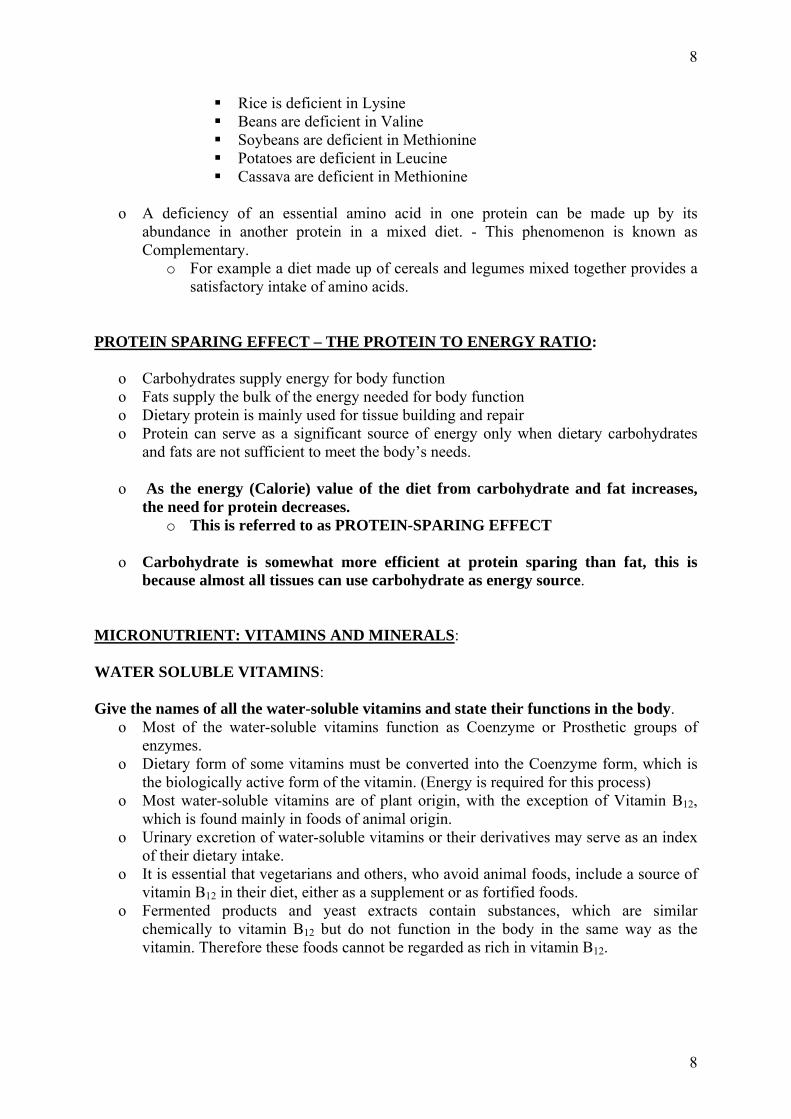
Rice is deficient in Lysine Beans are deficient in Valine Soybeans are deficient in Methionine Potatoes are deficient in Leucine Cassava are deficient in Methionine
o A deficiency of an essential amino acid in one protein can be made up by its
abundance in another protein in a mixed diet. - This phenomenon is known as Complementary.
o For example a diet made up of cereals and legumes mixed together provides a satisfactory intake of amino acids.
PROTEIN SPARING EFFECT – THE PROTEIN TO ENERGY RATIO:
o Carbohydrates supply energy for body function o Fats supply the bulk of the energy needed for body function o Dietary protein is mainly used for tissue building and repair o Protein can serve as a significant source of energy only when dietary carbohydrates
and fats are not sufficient to meet the body’s needs. o As the energy (Calorie) value of the diet from carbohydrate and fat increases,
the need for protein decreases. o This is referred to as PROTEIN-SPARING EFFECT
o Carbohydrate is somewhat more efficient at protein sparing than fat, this is because almost all tissues can use carbohydrate as energy source.
MICRONUTRIENT: VITAMINS AND MINERALS: WATER SOLUBLE VITAMINS: Give the names of all the water-soluble vitamins and state their functions in the body.
o Most of the water-soluble vitamins function as Coenzyme or Prosthetic groups of enzymes.
o Dietary form of some vitamins must be converted into the Coenzyme form, which is the biologically active form of the vitamin. (Energy is required for this process)
o Most water-soluble vitamins are of plant origin, with the exception of Vitamin B12, which is found mainly in foods of animal origin.
o Urinary excretion of water-soluble vitamins or their derivatives may serve as an index of their dietary intake.
o It is essential that vegetarians and others, who avoid animal foods, include a source of vitamin B12 in their diet, either as a supplement or as fortified foods.
o Fermented products and yeast extracts contain substances, which are similar chemically to vitamin B12 but do not function in the body in the same way as the vitamin. Therefore these foods cannot be regarded as rich in vitamin B12.
8

9
WATER SOLUBLE VITAMINS Common Names & Chemical Nature
Biologically Active / Coenzyme forms
Metabolic functions of biologically active forms
Thiamine or Vitamin B1
Thiamine Pyrophosphate (TPP)
Coenzyme in Oxidative Decarboxylase reactions (Pyruvate, Alpha-Oxo-Glutarate, Alpha-Ketobutyrate
Riboflavin or Vitamin B2
Flavin Adenine-Dinucleotide (FAD),
Flavin Adenine-Mononucleotide (FMN)
Coenzyme in some Dehydrogenase reactions, and in some Red-Ox reactions
Niacin: Nicotinic Acid; Nicotinamide
Nicotinamide Adenine-Dinucleotide (NAD)
Nicotinamide Adenine Dinucleotide Phosphate (NADP)
Coenzyme in several Dehydrogenase reactions, and in several Red-Ox reactions
Pyridoxine, Pyridoxal, Pyridoxamine (Vitamin B6)
Pyridoxal-Phosphate (B6-Phosphate)
Coenzyme in several enzymes: Amino Acid Decarboxylase, Transaminases, Delta-amino-Laevulinic Acid Synthetase (ALA-Synthase)
Pantothenic Acid Coenzyme A, Acyl-carrier Protein
(ACP)
Carrier of Acyl groups in Acylation reactions
Cobalamin (Vitamin B12)
Methyl-Cobalamin, 5’-Deoxyadenosyl
Cobalamin
Coenzyme for One-carbon transfer reactions (-CH3)
Folic Acid, Folate, Foliacin (Vitamin M)
Tetra-hydro-folic acid, Tetra-hydro-Folate (FH4, or THF)
Coenzyme for One-carbon transfer reactions
Ascorbic Acid, (Vitamin C)
L-Ascorbic Acid, Dehydro-Ascorbate
Reducing Agent (electron donor), Antioxidant
Biotin Prosthetic group of Carboxylases
Carrier of active CO2 in carboxylation reactions
9

10
FAT SOLUBLE VITAMINS: Give the names all the fat-soluble vitamins and state their functions in the body.
o Prolonged deficiency of vitamin D results in Rickets in children and Osteomalacia in adults.
o Combination of factors may be associated with low vitamin D status. o Such factors include the following:
o Low exposure to sunlight – this may be due to seclusion or strict dress codes limiting vitamin D synthesis in the skin.
o Type of vegetarian diet – vitamin D is found naturally in only a few foods, all of which are of animal origin, (Oily fish such as Mackerel and Sardines, Eggs, Whole Milk and its products).
o Some breakfast cereals and margarines are fortified with vitamin D. o Those who receive little exposure to the sun should rely more on dietary
sources of vitamin D. FAT SOLUBLE VITAMINS: Common Names & Chemical Nature
Biological Active Forms Metabolic functions of Active forms
Retinol (Vitamin A), All trans Retinol
11-cis Retinal, Prosthetic group in visual pigments,
Cofactor role in biosynthesis of Cholesterol,
Role in membrane biogenesis
Role in cell differentiation
Cholecalciferol (Vitamin D3)
Calciferol or Ergocalciferol (Vitamin D2)
1,25-Dihydroxy-Cholecalciferol, 1,25-DihydroxyVitamin D3
Absorption of Calcium in GIT,
Reabsorption & Mobilization of Calcium and Phosphate in Bone
Tocopherols (Vitamin E) Alpha-Tocopherol, Beta-Tocopherol
Antioxidants protecting polyunsaturated fatty acids in membranes,
Phytomenadione (Vitamin K) Vitamin K Cofactor in Post-translational gamma-carboxylation of N-terminal Glutamic acid residue in blood clotting factors
10

11
MINERAL ELEMENTS:
o Two major groups of dietary elements: o Macroelements are usually required in amounts greater than 100 mg per day
Macroelements consist of Calcium, Phosphorus, Potassium, Sodium, Magnesium, Chloride, and Sulfur
o Microelements or Trace elements are required in amounts less than 100 mg per day
Microelements consist of Iron, Copper, Manganese, Zinc, Iodine, Selenium, Cobalt, Molybdenum, Chromium, Fluorine, Silicon, Vanadium, Tin, Arsenic, and Nickel.
NON-NUTRIENTS:
Non-nutrients in food can be separated into two major groups: o Non-Toxic Non-nutrients and o Toxic Non-nutrients.
Non-Toxic Non-nutrients:
Major non-toxic non-nutrient with beneficial effects on the human body are those classified as dietary fiber (or Roughage).
Dietary Fiber – Definition:
o Dietary fibers are non-toxic non-nutrient component of food that cannot be broken down by human digestive enzymes.
o Bacterial enzymes in human intestine can breakdown some of the dietary fibers. o Chemically, dietary fiber can be defined as:
o Non-starch polysaccharide and Lignin. o Non-starch polysaccharide includes cellulose, and non-cellulose
polysaccharides. o Non-cellulose polysaccharides include:
Hemicelluloses (arabinoxylans); Pectin, Plant Gums, Mucilage, and Inulin.
Lignin is a group of polyphenolic compounds of diverse molecular weights.
Lignin is one of the essential components of the cell wall. What are some of the biological effects of Dietary fibers? Chemically there are different types of dietary fiber.
o Dietary fiber has a laxation effect on the functioning of the GIT. o Dietary fiber increases faecal bulk. o Dietary fiber lowers plasma cholesterol level. o Dietary fiber decreases nutrient availability. o Dietary fiber reduces glycaemic response to carbohydrate–containing meals. o Consumption of staple diets that are deficient in dietary fiber has been implicated in
the etiology of a number of human GIT diseases, such as cancer of the colon and rectum, diverticular disease of the colon, hemorrhoids and appendicitis.
11

12
What are some of the factors that affect the bioavailability of nutrients? Some factors affecting bioavailability of nutrients:
o Bioavailability of a nutrient contained in a given food is influenced by many factors, such as:
o Stability to cooking or processing; o Chemical form in which the nutrient is present; o Nature of other constituents of the diet and o Efficiency of an individual’s digestive system. o Negative influence on Bioavailability is exerted by some non-nutrients in
foods, such as: OXALIC ACID; PHYTIC ACID; PROTEINASE INHIBITORS, AVIDIN.
o Oxalic acid forms oxalate precipitate with dietary calcium; o Phytic acid forms insoluble phytates with Ca, Fe, Zn and other divalent metals.
12
13
UNIVERSITY OF PNG SCHOOL OF MEDICINE AND HEALTH SCIENCES
DIVISION OF BASIC MEDICAL SCIENCES DISCIPLINE OF BIOCHEMISTRY AND MOLECULAR BIOLOGY
PBL SEMINAR NUTRIENT REQUIREMENTS, MALNUTRITION- An Overview
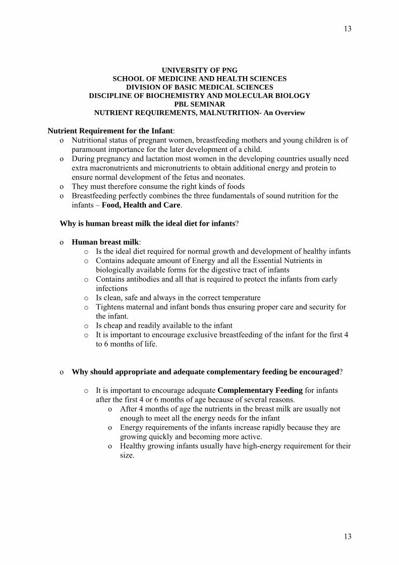
Nutrient Requirement for the Infant:
o Nutritional status of pregnant women, breastfeeding mothers and young children is of paramount importance for the later development of a child.
o During pregnancy and lactation most women in the developing countries usually need extra macronutrients and micronutrients to obtain additional energy and protein to ensure normal development of the fetus and neonates.
o They must therefore consume the right kinds of foods o Breastfeeding perfectly combines the three fundamentals of sound nutrition for the
infants – Food, Health and Care. Why is human breast milk the ideal diet for infants? o Human breast milk:
o Is the ideal diet required for normal growth and development of healthy infants o Contains adequate amount of Energy and all the Essential Nutrients in
biologically available forms for the digestive tract of infants o Contains antibodies and all that is required to protect the infants from early
infections o Is clean, safe and always in the correct temperature o Tightens maternal and infant bonds thus ensuring proper care and security for
the infant. o Is cheap and readily available to the infant o It is important to encourage exclusive breastfeeding of the infant for the first 4
to 6 months of life.
o Why should appropriate and adequate complementary feeding be encouraged?
o It is important to encourage adequate Complementary Feeding for infants after the first 4 or 6 months of age because of several reasons.
o After 4 months of age the nutrients in the breast milk are usually not enough to meet all the energy needs for the infant
o Energy requirements of the infants increase rapidly because they are growing quickly and becoming more active.
o Healthy growing infants usually have high-energy requirement for their size.
13

14
How can the increase in energy intake be achieved?
o To achieve this energy intake: o High-energy foods with good quality proteins eaten, as part of small
and frequent meals, should be given to infants, who do not have large enough stomachs to cope with big meals.
o Intake of adequate amount of high quality protein is necessary o Adequate amount of Micronutrients (Vitamins and Minerals) is
necessary at this time. o Calcium is needed for healthy tooth development and, together with
vitamin D, helps to make bones stronger. Childhood is an important time for tooth and bone development.
o Iron deficiency anemia is associated with frequent infections, poor weight gain and delay in development in children.
o Therefore adequate amount of Iron rich foods must be given to children during the period of rapid growth.
Take Note:
o Adequate amount of the micronutrients are needed at all ages, however, the effects of inadequate intake are particularly serious during periods of rapid growth, pregnancy, lactation and early childhood.
o Trace elements such as, Iron, Zinc, Iodine and Selenium are very important for the physical and cognitive development of children.
If vegetarian diets are excellent for adults why are they not excellent for infants?
o A vegetarian diet that keeps adults in good health is not necessary appropriate for infants and young children, as this is a time of rapid growth and development when a good supply of energy and nutrients is particularly important.
o Diets that are low in energy and fat and high in bulk may pose a nutritional risk for children when stomach capacity is limited.
o The presence of milk and milk products and perhaps eggs in a child’s vegetarian diet is likely to ensure that adequate amounts of calcium, vitamin B12, vitamin D and Riboflavin are supplied.
o Children who are vegetarian must have alternative sources of iron, such as dark green leafy vegetables, pulses (beans), nuts and fortified breakfast cereals.
o Iron from plant sources is less well absorbed than iron from animal sources. o Consuming vitamin C rich foods or drinks with a meal can increase iron absorption
form plant sources, e.g. providing fresh orange juice. o Vegetarian diets are not recommended during the weaning period. o However, for families who are Vegetarians:
o Weaning should follow the same dietary principle as for non-Vegan babies, o At least a pint per day of infant Soya formula should be consumed when breast
milk is no longer given o It is recommended that all Vegan children under five years of age should
receive supplements of Vitamin drops containing Vitamins A, C and D o Foods fortified with vitamin B12 should either be included in the diet or
supplement given.
14

15
MALNUTRITION: What is malnutrition?
o It is a pathological state, general or specific, resulting from a relative or absolute deficiency or excess in the diet of one or more essential nutrients. It may be clinically manifest or detectable only by Biochemical and Physiological tests.
What are the different forms of malnutrition?
o Starvation, Under-nutrition, Specific deficiency, and Over-nutrition. (Some authors do not consider over-nutrition and its resulting obesity under the heading of malnutrition).
Take Note:
o In developing country malnutrition is synonymous with growth failure – malnourished children are shorter and lighter than they should be for their age.
What parameters are used to determine malnutrition in children? Four Indicators or Indices of nutritional status are used to determine if a child is malnourished or well nourished. The four indicators usually used are:
o Weight-for-Length (W/L) or Weight-for-Height (W/H) o Length-for-Age (L/A) or Height-for-Age (H/A) o Weight-for-Age (W/A) o Mid-Upper-Arm-Circumference (MUAC)
There are standard reference tables for each indicator. These tables contain measurements done on healthy and well-nourished children.
o Weight-for-Height or Weight-for-Length: o Indicates wasting and the current nutritional status of the child, because weight
is most sensitive to recent events. o If a child has been sick and has experienced a recent shortage of food, his
weight will decrease but his height will remain the same. In PNG the classification use for Weight-for-Height or Weight-for-Length is as follows: Percentage of Standard W/H (Classification)
o Below 80% (Severe wasting) o 80 – 89% (Moderate wasting) o 90 – 120% (Normal) o Above 120% (Obesity or Over-nutrition)
Example: James is a 4 years old boy. His Weight is 10.0kg and height is 80.0cm. If in the reference table the standard weight for an 80.0cm child is 12.0kg, what is the nutritional status of James? Answer: By checking the Weight-for-Height reference table the standard weight a child of height 80.0cm should be 12.0kg.
15

16
But James weight is 10.0kg. Therefore James will have a percentage of W/H as: Weight of James divided by Standard Weight multiplied by 100 10.0/12 x 100% = 83.3% of standard Weight-for-Height In the Classification above the nutrition status of James is Moderate Wasting. What is Protein-Energy Malnutrition (PEM)?
o Protein-Energy Malnutrition (PEM), or Protein-Calorie Malnutrition (PCM) is characterized by deficit in the diet of:
o Macronutrients (Energy and Protein) and o Some Micronutrients
o PEM represents the various levels of inadequate protein and/or energy intake between
starvation (no food intake) and inadequate nourishment o Although PEM is more commonly found in infants and children in some developing
countries, it can occur in person of any age in any country.
What are the different forms and grades (classification) of PEM?
o Clinically, PEM has three forms, which depends on the balance of Non-protein (Carbohydrate and Fat) and Protein sources of energy.
o Dry (thin, desiccated), o Wet (edematous, swollen), and o A combined form between the two extremes.
o Each of the three forms can be graded as
o Mild, Moderate, or Severe. What are the characteristics of the different forms of PEM?
o Dry form, called Marasmus: o Is due to near starvation with deficiency of Protein, and non-protein
(Carbohydrates and Fats) nutrients. o Marasmic child consumes very little food – often because his mother is unable
to breastfeed – thus the child is very thin from loss of muscle and body fat. o It is the predominant form of PEM in most developing countries. o It is associated with the early abandonment or failure of breastfeeding and o It is usually associated with infections, most notably those causing infantile
gastroenteritis. o The infections usually result from improper hygiene and o Inadequate knowledge of infant rearing that is prevalent in the rapidly growing
slums of developing countries.
16

17
o Wet form is called Kwashiorkor:
o Protein deficiency (lack of an intake of good quality protein) is usually more marked than the Energy deficiency, and
o Kwashiokor child consumes a carbohydrate rich food with very poor quality protein.
o Edema usually occur in such children o Children with kwashiorkor tend to be older than those with Marasmus and tend
to develop the disease after they are weaned from breast milk. o Kwashiorkor is less common and is usually manifested as marasmic
kwashiorkor. o It tends to be confined to developing countries where staple and weaning foods
fed to infants include yam, cassava, sweet potato, and green banana. o These foods are excessively starchy and contain low quality protein. o The combined form of PEM is called Marasmic Kwashiorkor.
o Children with this form have some Edema and more body fat than those with Marasmus.
What are some of the Biochemical basis of Marasmus? In Marasmus:
o Energy intake is insufficient for the body’s requirements, thus it draws on its own stores. Liver glycogen is exhausted within a few hours, and
o Skeletal muscle protein is then used via Gluconeogenesis to maintain adequate plasma glucose.
o Triacylglycerols in fat depots are broken down into free fatty acids, which provide some energy for most tissues, but not for the nervous system.
o When near starvation is prolonged, fatty acids are incompletely oxidized to ketone bodies, which can be used by the brain and other organs for energy.
o The severe energy deficiency of Marasmus adaptation is facilitated by high Cortisol and Growth Hormone levels and depression of Insulin and Thyroid hormone secretion Because amino acids are mobilized from muscle to provide the liver with substrate for protein synthesis, plasma protein levels decrease less in Marasmus than in kwashiorkor.
In Kwashiorkor:
o Relatively increased carbohydrate intake with decreased intake of protein and essential amino acids lead to decreased visceral protein synthesis.
o The resulting Hypoalbuminaemia causes dependent edema, and o Impaired β-lipoprotein synthesis causes fatty liver. o Insulin secretion is initially stimulated but is reduced later in the disease. o Fat mobilization and amino acid release from muscle are reduced, so that less
amino acid substrate is available to the liver. Take Note: List some adaptations that occur in the body during protein deficiency:
o Adaptive enzyme changes occur in the liver, o Amino acid synthetases increase, and o Urea formation diminishes, thus conserving nitrogen and reducing its loss in urine. o Homeostatic mechanisms initially operate to maintain the level of plasma albumin
and other transport proteins.
17
18
o The rates of albumin synthesis eventually decrease, and plasma levels fall, leading to reduced Oncotic pressure and Edema.
o Growth, immune response, repair of tissues, and production of some enzymes and hormones are impaired in severe protein deficiency.
What are some of the symptoms and signs that are typical of (a) Marasmic and (b) Kwashiokor children?
o Some signs and symptoms in Marasmic children include: o Marasmic infants: Hunger, Gross weight loss, Growth retardation, and Wasting of
Subcutaneous Fat and Muscle.
Some signs and symptoms in Kwashiorkor children include: o Generalized edema; “Flaky Paint” Dermatosis, Thinning, De-coloration,
Reddening of the hair; Enlarged fatty liver; and Petulant apathy in addition to retarded growth.
Take Note:
o Alternating episodes of under-nutrition and adequate nutrition may cause the hair to have a dramatic “striped flag” appearance.
o Various types of infections usually occur in all forms of PEM. What are some of the laboratory finding in children with PEM?
o Mild or moderately severe PEM may cause: o Slight depression of plasma albumin, Decrease in urinary excretion of urea,
due to decreased protein intake, and in Hydroxyproline, reflecting impaired growth. Increased urinary 3-methylhistidine reflects muscle breakdown.
o In Kwashiorkor:
o Plasma levels of albumin is low, Transferrin is low, Essential amino acids (especially branched-chain) are low, β-lipoprotein, and Glucose are low, Plasma Cortisol and growth hormone levels are high, Insulin secretion and Insulin-like growth factor are depressed.
o In Marasmus and kwashiorkor,
o The percentage of body water and extracellular water is increased. Electrolytes, especially Potassium and Magnesium, are depleted; Levels of some enzymes and circulating lipids are low, and Blood urea decreases. Anemia, usually due to Iron deficiency and Metabolic Acidosis can occur. Diarrhea is common and is sometimes aggravated by intestinal Disaccharidase deficiency, especially of Lactase.
18

19
University of Papua New Guinea
School of Medicine and Health Sciences Division of Basic Medical Sciences
Discipline of Biochemistry and Molecular Biology PBL SEMINAR
STRESS and CATECHOLAMINES – An Overview What is stress?
Stress can be Physical and Psychological reaction to excessive stimulus Stress can be a Psychological disorder caused by constant mental strain or emotion Stress can be caused by environmental factors like:
o Injury, Trauma, Temperature (very high or very low), Loud noises, etc, Stress is also caused by disease conditions like:
o Renal Failure, Burns, Infections, etc, What are some of the consequences of stress?
Hormonal and Neuronal changes Metabolic changes leading to:
Insulin Resistance, Weight loss, Diabetes What metabolic changes can occur in response to stress?
Some hormonal changes that can occur include o Increase blood levels of some Insulin Counter-Regulatory Hormones:
Cortisol, Glucagon, Catecholamines and Growth hormone
Under constant stress some individual may develop Insulin Resistant because of high blood levels of the Insulin Counter-Regulatory Hormones
o Insulin Resistant may cause elevation of Basal Metabolic Rate, Due to increase Glucose and Free Fatty Acid level in blood
Constant stress may cause increase in Catabolism of Muscle Protein, leading to Negative Nitrogen Balance, which is partly responsible for weight loss in individuals under stress
One explanation for the negative nitrogen is increase production of Mediators, such as Monocyte and Lymphocyte proteins
List some specific mediators produced during stress? Mediators include Monocyte and Lymphocyte proteins (Endogenous Pyrogens, that is,
they cause fever) Interleukin-1, Interleukin-6, Tumor Necrosis Factor (TNF)
What are the actions of these mediators?
Interleukin-1: Activates catabolism of Skeletal Muscle Protein Interleukin-6:
o Stimulates synthesis of Acute Phase Reactants in the liver o Acute Phase Reactants are group of proteins produced during injury or
infection to either serve or active the defense mechanism of the body Examples of Acute Phase Reactants are: Fibrinogen, Complement
Proteins, Some Clotting Factors, Alpha-macroglobulin
19

20
Tumor Necrosis Factor (TNF):
o Tends to suppress synthesis of Triacylglycerides (Fat) in adipose tissue, o Stimulate Lipolysis (breakdown of fat), o Inhibits Lipoprotein Lipase and therefore prevents the uptake of circulating fat
How can environmental factors be related to stress?
Most environmental signals are filtered by Reticular formation in Brain and “Alarm” and other signals are transmitted from CNS to Limbic System (e.g., Hippocampus)
Limbic System then transmits signal to Hypothalamus Hypothalamus generates 2 types of signals:
o Neuronal signal via Neuronal System and o Chemical (Hormonal) signal via Hormonal system
Neuronal Signals:
o Generated via Peripheral Nervous System Neuronal signals act via Cholinergic Neurons located in Adrenal
medulla in Adrenal Gland Cholinergic Neurons cause secretion into general circulation of:
Epinephrine, Enkephalins and Norepinephrine Chemical/Hormonal Signals:
o Generated via Anterior Pituitary by production of Adrenocorticotropic Hormone (ACTH), Beta-Lipotropin (beta-LTH) and Beta-endorphin
ACTH acts on Adrenal Cortex in Adrenal Gland and causes release of Cortisol, which is the major stress adaptational hormone
ACTH also causes release of Aldosterone and Dehydroepiandrosterone (DHEA)
o Beta-endorphin acts on receptors in CNS to produce Analgesia Ultimate effects of ACTH and Beta-endorphin are:
o To limit deleterious effects of stress by providing immediate sources for the energy required to counter the stress
If stress continues for a prolonged period of time Pathological changes can occur in the system
Briefly outline the Humoral Stress Pathway? (SEE DIAGRAM)
Environmental stress event is detected by the CNS Signals are sent to Limbic system (Hyppocampal structure) which, in turn, signals
Hypothalamus to release Corticotrophin Releasing Hormone (CRH) CRH acts on Anterior Pituitary to produce the polypeptide called Pro-Opio-Melano-
Cortin (POMC) POMC is split into ACTH, Beta-LTH, and Beta-endorphin
Beta-endorphin:
o Beta-endorphin acts on CNS to promote Analgesia, possibly by lowering the level of cellular c-AMP in certain cells via a Beta-endorphin receptor coupled to an Inhibitory G-protein transducer and Adenylate cyclase
o Beta-endorphin also binds to receptors on Sommatotrophs and Lactotrophs of the Anterior Pituitary causing secondary release of Growth Hormone and
20

21
Prolactin, which may play some role in stress response by virtue of the Hyperglycemic actions of these hormones in liver cells
ACTH:
o ACTH acts on Adrenal Cortex to release Cortisol, which then circulates in blood bound to Transcortin or Corticosteroid binding globulin (CBG)
o Cortisol (Glucocorticoid) acts on appropriate tissues to produce systemic
effects that constitute stress adaptation, which are useful to the body because the actions of Cortisol tends to limit the deleterious effects of stress
If the stress continues for a long time the effects of this system can become harmful to the body
CATECHOLAMINES
Catecholamines are Biogenic amines derived from L-Tyrosine Catecholamines include the following:
Dopamine, Noradrenaline (also called Norepinephrine) and Adrenaline (also called Epinephrine)
Catecholamines do not cross the blood-brain barrier Catecholamines synthesized within the blood-brain barrier act mainly as
neurotransmitters whereas, those synthesize outside the blood-brain barrier act as hormones
How are the Catecholamines synthesized? (Fig. 1)
Precursor for biosynthesis of catecholamines is mainly L- Tyrosine Rate-limiting step in biosynthesis of Catecholamines is the conversion of L-Tyrosine
to 3,4-Dihydroxyphenylalanine (DOPA) Tyrosine Hydroxylase is a mixed functional Oxygenase that utilizes molecular oxygen
and L-Tyrosine as substrates and Tetrahydrobiopterine as cofactor Tyrosine Hydroxylase is also capable of converting Phenylalanine to Tyrosine, in
conditions where Phenylalanine Hydroxylase is deficient as is the case in Phenylketonurics
DOPA is Decarboxylated to Dopamine in a reaction catalyzed by DOPA -Decarboxylase, an enzyme that utilizes Pyridoxal Phosphate (B6-PO4) as coenzyme
Dopamine is the first member of the Catecholamines
Dopamine is Hydroxylated to Noradrenaline (Norepinephrine) by Dopamine-beta-Hydroxylase, which is a Copper-containing mixed functional Oxygenase that utilizes molecular oxygen as one of its substrates
o Ascorbate is essential for this reaction Noradrenaline is methylated to form Adrenaline in a reaction catalyzed by S-
Adenosyl-Methionine-Phenylethanolamine-N-Methyl Transferase Methyl donor is the high-energy compound S-Adenosyl-Methionine
How does stress affect the synthesis and release of Catecholamines?
Adrenal Medulla releases catecholamines into the blood In humans, catecholamines released from Adrenal Medulla are about 80% Adrenaline
21

22
and 20% Noradrenaline Noradrenaline biosynthesis increases after Acute stress Prolonged Stress accompanied by Chronic Sympathetic nerve activity causes increase
in the activity of both Tyrosine Hydroxylase and Dopamine-beta-Hydroxylase Increase in activity of these enzymes in the Catecholamine biosynthetic pathway is a
means of adapting to Physiologic Stress In Adrenal Medulla, Acetylcholine acting as the neurotransmitter of the sympathetic
ganglion acts on Nicotinic receptors and promotes the release of Catecholamines into the circulation
What is the mode of Action of Adrenalin?
Catecholamines act through two major classes of receptors o Alpha-Adrenergic and Beta-Adrenergic receptors, (Each consists of two
subclasses, i.e. alpha-1, alpha-2, beta-1 and beta-2)
Adrenaline is considered as the "Fright, Flight or Fight” hormone when produced outside the blood-brain barrier
Adrenaline:
Interacts directly with Beta-Adrenergic receptors in Plasma membrane of liver cells to activate Adenylate cyclase, thereby causing:
o Activation of Glycogenolysis and o Inhibition of Glycogenesis and Glycolysis to maximize the release of Glucose
from Hepatic cells (Fig. 2) Adrenalin:
o Interacts with Alpha-Adrenergic receptors to activate Phospholipase C, which then catalyzes hydrolysis of Phosphatidyl-Inositol-4, 5-Bisphosphate (PIP2) to produce 1,2-Diacylglycerol and Inositol-1, 4,5-Ttriphosphate (IP3)
o IP-3 acting as a Second messenger stimulates the release of Ca2+ ions from Endoplasmic Reticulum
o Increase in Ca2+ ions ultimately results in activation of Glycogen Phosphorylase and Inhibition of Glycogen Synthase
Action of Adrenaline results in increased breakdown of Liver Glycogen to produce more blood Glucose for tissues that needs to meet the challenge of the stressful situation that triggered the release of Adrenaline from the Adrenal medulla (Fig. 3)
Adrenalin:
o Interact with Beta-Adrenergic receptors to stimulate degradation of Glycogen in Cardiac and Skeletal muscle tissues;
This does not lead to increase blood glucose, because Cardiac and Skeletal muscle tissues lack Glucose-6-Phosphatase
In addition c-AMP produced in these tissues stimulate Glycolysis
Role of Adrenaline on Glycogen metabolism in Cardiac and Skeletal muscle is to make more Glucose-6-Phosphate available for Glycolysis in these tissues
o ATP generated by Glycolysis can then be used to meet the metabolic demand
imposed on these muscles by the stress that triggered the release of Adrenaline (Fig. 4)
22

23
How are Catecholamines degraded? (Fig. 5 & 6)
Catecholamines that diffuse into the circulation or are released, as neuro-hormones may be taken up into sympathetic nerve terminals by the Na – K pump
Enzymes involved in degradation of Catecholamines are Monoamine Oxidase (MAO), Catechol-O-methyl Transferase (COMT) and Aldehyde Dehydrogenase (ADH)
Depending on the location of the Catecholamine, either COMT or MAO may initiate the reaction
MAO always initiates the degradation of intra-cellular Catecholamines, while COMT initiates the degradation of extra-cellular Catecholamines
Major end product of Dopamine degradation is Homovanillic Acid (HVA) Major end product of Noradrenaline and Adrenaline degradation is Vanilly-Mandelic
Acid (VMA), also called Methoxy-4-hydroxymandelic acid MAO inhibitors have been used to treat Hypertension and Depression, but serious
reaction with foods or drugs that contain Sympathomimetic amines limit their usefulness
23

24
ENVIRONMENTAL STRESS
CNS Analgesia
LIMBIC SYSTEM
HYPOTHALAMUS Produces CRH
ANTERIOR PITUITARY Produces
ACTH Beta-LTH Beta-endophin ADRENAL GLAND
Cortisol
SIMPLIFIED DIAGRAM OF THE OVERVIEW OF HUMORAL STRESS PATHWAY
24

25
UNIVERSITY OF PNG
SCHOOL OF MEDICINE AND HEALTH SCIENCES DISCIPLINE OF BIOCHEMISTRY AND MOLECULAR BIOLOGY
PBL MBBS SEMINAR
CEREBRAL METABOLISM - An Overview • Brain or Cerebral energy metabolism is often considered to reflect predominantly, if not
exclusively, Neuronal energy metabolism or CNS metabolism in general • Other cell types, namely Glial and Vascular Endothelial cells not only consume energy
but also play active role in the flux of energy substrates to Neurons Why does the cerebral tissue need energy? • Neuron is the functional unit of the CNS • Neuron is an excitable cell, because it is capable of generating and conducting electrical
impulse by temporarily reversing its membrane potential • Major functions of neurons are excitation and conduction, which are reflected in the
unceasing electrical activity of Cerebral tissue • Electrical energy is derived from chemical processes • Energy consumption is used for active transport of ions needed to sustain and restore the
membrane potentials discharged during the process of excitation and conduction • Thus, cerebral tissue requires constant supply of energy What substrates are used for energy production in cerebral tissue? • Glucose is the major substrate for energy production in cerebral tissue • Cerebral tissue utilizes glucose directly from arterial blood • Insulin is not required for uptake of glucose by cerebral tissue • Brain can utilize Glycogen store (about 0.1%) to maintain cerebral metabolism for a very
short time, when blood glucose is low • Apart from Glucose, Mannose can be used to sustain normal cerebral metabolism
• Mannose easily crosses the blood–brain barrier, is converted to Fructose-6-phosphate that enters the Glycolytic pathway
• Mannose is not normally present in the blood and cannot therefore be considered as a substrate for cerebral energy metabolism
• Fructose, Galactose, Lactate and Pyruvate have limited permeability across the blood–
brain barrier, therefore cannot directly serve as substrates for cerebral energy metabolism • Lactate and Pyruvate when formed within the blood-brain barrier are useful metabolic
substrates for cerebral metabolism How significant is O2 supply to brain energy metabolism?
• Brain represents about 2 to 3% of total body weight of an average adult, but it utilizes about 20 to 25% of the total O2 consumed by the whole organism
• In children up to 4 years of age, the brain utilizes about 50% of the total O2 consumed by the whole organism
• Cerebral tissue utilizes O2 more than other tissues; o Example: it utilizes about 20 times more O2 than muscle tissue when at rest.
25

26
Oxygen consumption varies throughout the brain: • Grey matter utilizes about twice more O2 than White matter (which contains
fewer cells than the Grey matter) • Cerebral O2 consumption continues unabated day and night, (Sleep reduces
cerebral O2 uptake by only 3%) • Oxygen stored in the brain is extremely small compared to the rate of
utilization, thus the brain requires the continuous replenishment of its oxygen by the circulation
• Consciousness is loss if cerebral blood flow is completely interrupted
TAKE NOTE:
• Reduced cerebral O2 uptake occurs under certain conditions that lead to depressed consciousness
o Examples include: Insulin induced hypoglycemia, Diabetic coma, Cerebral tumors, Uremia, Gross liver damage that culminate in hepatic coma and exposure to depressant drugs used during surgery.
What are some of the uses of O2 consumed by cerebral tissue?
Some uses of O2 include: • Energy metabolism via Oxidative Phosphorylation • Maintenance of energy component in blood-brain barrier • Functioning of specific enzyme systems:
Mixed Functional Oxygenases used in the biosynthesis of Neurotransmitters and other biologically active compounds
Briefly explain how carbohydrate is metabolized in cerebral tissue
• Aerobic and Anaerobic Glycolysis occurs in cerebral tissue • HMP shunt (Pentose Phosphate Pathway) also occurs in cerebral tissue mainly for the
production of NADPH, required for the biosynthesis of Fatty acids and Steroids • Carbohydrates such as Maltose, Fructose, Galactose, Hexose-phosphates and
Intermediate metabolites such as Lactate, Pyruvate and Glyceraldehydes are used only after their conversion to Glucose via Gluconeogenesis
• Thus, these compounds act by raising the Blood Glucose Level
• Cerebral tissue can utilize Mannose directly and rapidly from the blood to restore or maintain normal metabolic functions • Mannose can directly enter the Glycolytic pathway of cerebral tissues, without
raising blood glucose level • Mannose like Glucose can easily cross the blood-brain-barrier and can be
converted to Mannose-6-phosphate by the enzyme Hexokinase
26

27
• Phospho-mannose Isomerase is an active enzyme in cerebral tissue that converts Mannose-6-phosphate to Fructose-6-phosphate, which then enters the Glycolytic pathway.
The reaction is as follows: 1 2
Mannose + ATP ====> Mannose-6-P ======> Fructose-6-Phosphate (NB: 1 = Hexokinase; 2 = Phosphomannose Isomerase)
{Mannose is not normally present in blood in any appreciable amount and is therefore of no Physiological significance}. Briefly comment on the amino acid content in cerebral tissue Cerebral tissue contains:
• Very high concentration of free amino acids compared to that in plasma • Highest amount of free Glutamate, compared to any other mammalian tissue • Some unusual amino acids such as: Gamma-Aminobutyrate (GABA), N-Acetyl-
Aspartate and Cystathione. TAKE NOTE:
• GABA is an inhibitory neurotransmitter that acts by increasing the passage of Chloride ions through the Post-synaptic membrane of Neurons
• Glutamate is involved in several metabolic processes such as: the biosynthesis of GABA, Detoxification of Ammonia and as Neurotransmitter
How is Ammonia formed in cerebral tissue?
Formation of Ammonia in cerebral tissue: • In cerebral tissue ammonia is produced mainly via Adenylate Deaminase reaction
Adenylate Deaminase
AMP ================> IMP + NH4+
• High concentration of Glutamate in blood causes ammonia toxicity • Glutamate Dehydrogenase (GDH) catalyzes the formation of Ammonia from
Glutamate
Glutamate DH Glutamate + NAD + H2O ========= Alpha-oxoglutarate + NH3 + NADH + H+ How is ammonia removed from cerebral tissue?
• Rate of urea formation in the cerebral tissue is too low to account for the removal of ammonia via the urea cycle, WHY???
o Mitochondrial N-Acetyl-Glutamate activated Carbamoyl-Phosphate Synthetase that catalyzes the first reaction in Urea cycle, is low or absent in cerebral tissue
27

28
• Thus, removal of ammonia from Cerebral tissue involves 2 reactions: o First is formation of Glutamate from Alpha-Oxoglutarate and Ammonia, by
Glutamate Dehydrogenase (GDH) Glutamate DH Alpha-oxoglutarate + NH3 + NADH + H+ ====== Glutamate + NAD + H2O
• Second is formation of Glutamine from Glutamate and Ammonia, by Glutamine Synthetase
Glutamine Synthetase
Glutamate + NH3 + ATP ==========> Glutamine + ADP + Pi
• Concentration of Ammonia in Cerebral tissue will be kept low if there is adequate supply of Alpha-Oxoglutarate
• Extensive utilization of Alpha-Oxoglutarate (produced in the TCA cycle) within the cerebral tissue, would deplete Intermediates from the TCA cycle and thus affect energy supply to the Brain, unless a mechanism of replenishing the intermediates is available
• One of such mechanism is known as the Anaplerotic (Filling-up) reaction, which is the formation of TCA cycle intermediates in the cerebral tissue
• Anaplerotic reactions can increase the concentrations of TCA cycle intermediates, allowing an increased rate of oxidation of Acetyl-CoA
List some of the Anaplerotic reactions Anaplerotic reactions include:
• Pyruvate Carboxylase reaction: formation of Pyruvate from Oxaloacetate using ATP and Biotin
• Some Transamination reactions • Glutamate Dehydrogenase reaction to form Alpha-Oxoglutarate • Succinyl-CoA formation from Isoleucine, Valine, Methionine, and Threonine
SOME FACTORS THAT CAN AFFECT CEREBRAL METABOLISM
• Oxygen and Glucose are two major substrates required for normal energy metabolism in cerebral tissue
• Hypoxia and Ischaemia can severely affect energy metabolism in cerebral tissue How does hypoxia affect cerebral metabolism?
• After a brief period of hypoxia: o Drastic slowdown in Oxidative metabolism occurs in Cerebral tissue o Rate of Glycolysis is increased in Cerebral tissue o Lactic acid production is increased, which can consequently leads to
intracellular acidosis
• These changes can be explained on the basis of Pasteur effect o Inhibition of Glycolysis in the presence of oxygen
28

29
o Pasteur effect reflects the increased energy yield obtained via Aerobic metabolism of glucose as compared to Anaerobic metabolism
• Hypoxia causes an increase in Glucose utilization from cerebral blood stream,
followed by a decrease in cerebral glucose concentration o Resulting is an increase in Lactic acid production in cerebral tissue o Gradual increase in Spinal fluid Lactate level occurs during hypoxia.
TAKE NOTE: • The earliest detectable Neuro-chemical change in brain resulting from Hypoxia is not
elevation of cerebral lactate concentration, but a reduction in Acetylcholine Synthesis • Major effect of Hypoxia on the Nervous system is reduction in the rate of conversion
of Pyruvate to Acetyl-CoA with a resultant decrease in both the biosynthesis of Acetylcholine and the activity of the TCA cycle
• In situation of low Acetyl-CoA availability the brain may use the available Acetyl-CoA for energy production so as to maintain membrane potentials in preference to its use in the biosynthesis of any compound
How does Ischaemia affect cerebral metabolism?
• During Ischaemia: o Glucose and O2 supply are deficient o Cerebral glucose concentration and Glycogen store are depleted o Coma can occur leading to cerebral tissue damage
• Hypoglycemia can severely affect cerebral energy metabolism because glucose is
almost exclusively used by the brain as the substrate for energy metabolism TAKE NOTE:
• During starvation cerebral tissue can use Ketone bodies (especially Beta-hydroxybutyrate, and Acetone) as substrate for energy metabolism
• Concentration of Ketone bodies are usually very high in the blood during starvation, thus they are able to cross the blood-brain barrier without much restriction
• Vitamin deficiency can lead to abnormality in cerebral metabolism and function • Effect of vitamin deficiency can be either direct or indirect, because of the role of
vitamins on biochemical processes.
29

30
UNIVERSITY OF PAPUA NEW GUINEA
SCHOOL OF MEDICINE AND HEALTH SCIENCES DIVISION OF BASIC MEDICAL SCIENCES
DISCIPLINE OF BIOCHEMISTRY AND MOLECULAR BIOLOGY PBL SEMINAR MBBS
MECHANISMS OF OEDEMA (EDEMA) – An Overview What is Oedema/Edema?
o Oedema is an accumulation of fluid in the Interstitial Compartments o Oedema occurs when there is more Interstitial Fluid than the Lymphatic system can
return into the circulation How much water (fluid) is contained in the body?
o Water/Fluid is a major body constituent o An average person (weighing about 70 kg) contains about 42 liters of Total Body
Water (TBW) o TBW accounts for about 60% of total body weight
What are the fluid compartments in the body?
Two major fluid compartments: o Intra-Cellular Fluid Compartment (ICF): Volume of Fluid Inside Cells
ICF constitute about 66.6% of TBW o Extra-Cellular Fluid Compartment (ECF): Volume of Fluid Outside Cells
ECF constitute about 33.3% of TBW • ECF is made up of Plasma and Interstitial Fluid
o Plasma is about 25% of ECF o Interstitial Fluid is about 75% of ECF
TBW
ICF ECF ECF Plasma
Interstitial Fluid
30
31
What are some of the consequences of Fluid loss?
o Selective loss of fluid from either ICF or ECF compartments gives rise to distinct signs and symptoms: For example:
Loss of ICF, can cause: • Cellular Dysfunction: resulting in Lethargy, Confusion and Coma
Loss of ECF (e.g., Blood loss) can lead to • Circulatory Collapse, Renal shutdown and Shock
o Loss of TBW produces similar effects as indicated in both cases above
o Signs of (substantial) fluid loss, is usually spread across both ECF and ICF compartments
o State of Hydration (volume of body fluid compartments) of a patient, is usually assessed on Clinical grounds, by looking for appropriate Clinical signs that indicate:
o Dehydration (loss of fluid) or o Over-hydration (accumulation of fluid in body compartments)
What is “Water Steady State” or Water Balance?
o Water steady state is an important concept that simply means that: o Amount of water consumed each day must equal the amount of water
eliminated from the body over the same period of time o If not, then body will have either a net water gain (Over-hydration) or a net
water loss (Dehydration) What are some of the major sources and routes of fluid intake?
o Some major sources of fluid intake include: o Water Drinking; Water contained in our various foodstuffs; Metabolic water
What are some of the major routes in the body for water loss?
o Some major routes of water loss include: o Urinary loss; Fecal loss o Insensible water loss – such as evaporation from the respiratory tract and the
skin surface (not including sweat which is sensible since it has a purpose) o Sweat Losses:
o At room temperature, sweating accounts for about 25% of heat losses o In cold environments, H2O losses in sweat decrease o In warm environments, or with exercise, sweat losses increase
o Pathological losses – Include: vascular bleeding, vomiting, and diarrhea What is “Electrolyte Steady State” or Electrolyte Balance?
o Electrolytes are Na+, K+, Cl- and H2CO3- ions;
o Amount of electrolytes consumed must be equal to amount eliminated within certain period
o Na+, K+, Cl- ions normally enter the body mainly by ingestion o Clinically, Electrolytes can enter the body via parenteral route, e.g., via
administration of Intravenous (i.v.) Solutions o Possible routes for Electrolyte losses: Renal excretion, Stool losses, Sweating,
Pathological routes: e.g., Vomit and Diarrhea
31

32
What is OSMOLALITY or OSMOLARITY?
o Osmolality is the number of solute particles per unit weight of water, irrespective of the size or nature of the particles
o Low molecular weight solutes contribute much more to the Osmolality than high molecular weight solutes
o Osmolality determines the osmotic pressure exerted by a solution across a semi-permeable membrane
o Osmolarity is the number of particles of solute per liter of solution (NB: Osmolarity is now replaced by Osmolality)
o Water moves easily through semi permeable membranes that separate ECF from ICF o Osmolality of ICF is always the same as Osmolality of ECF o ECF and ICF compartments contain Isotonic solutions
How is Osmolality calculated?
o Osmolality of Serum or Plasma is calculated from the concentrations of the major Solutes. One very simple method for calculating Osmolality is:
Serum Osmolality = 2 x molar concentration of serum Sodium ions = 2[Na+] (Note: Unit for Osmolality is either, mmol/kg, or mOsmol/Kg or mOsmol/L; Unit for Plasma or Serum Sodium ion is always in mmol/L)
o This simple formula for calculating Osmolality can be used ONLY if the Serum or Plasma Concentrations of Urea and Glucose are within the reference ranges
o If either or both are abnormally high, the concentration of either or both (in mmol/L) must be included in the calculation of the Osmolality
NB: In human, Normal Osmolality of Serum or Plasma (and other body fluids except urine) is in the range 285 to 295 mmol/kg (285 to 295 mOsmol/L)
Example for calculation of Osmolality: Normal Conditions (i.e., Plasma or Serum concentrations of Urea and Glucose are within normal range) ECF Osmolality can be roughly estimated as:
P Naosm p= ⋅ = −2 270 290[ ] mOsm (Where Posm is plasma Osmolality;. Since intracellular Osmolarity is the same as extra-cellular Osmolality under normal conditions, this also provides an estimate of intracellular Osmolality) Clinical Laboratory Measurement:
o Plasma Osmolarity measured in Clinical laboratory also includes contributions from Glucose and Urea
o Normally the contribution from Glucose and Urea is small
32

33
o Under certain Pathological conditions, the concentrations of these substances can be very high
o Plasma Osmolality measured in clinical laboratory can be calculated as:
P = 2[Na+] + 2[K+] + [Glucose] + [Urea] How is effective Osmole different from ineffective Osmole? Ineffective and Effective Osmoles:
o Urea is an Ineffective Osmole because it crosses cell membranes just as easily as water, therefore it does not contribute to redistribution of water between ECF and ICF
o Glucose, Na+ and Anions associated with Na+ have concentration gradients across the cell membrane and are therefore Effective Osmoles in the sense that they determine the distribution of water between ECF and ICF
How can effective Osmole be calculated? Two ways for calculating effective Osmole:
o Effective Osmole: P (effective) = 2[Na+] + [Glucose]
o Effective Osmole: P (effective) = P (measured) – [Urea] What is Osmolal Gap and how is it calculated?
o Difference between Measured Osmolality (MO) and Calculated Osmolality (CO) is known as Osmolality Gap or Osmolal Gap (OG):
Osmolal Gap (OG) = MO – CO
o Large positive OG helps to identify presence in serum of osmotically active substances, such as, Ethanol, Methanol, Iso-propanol, Ethylene Glycol and Acetone
o Proper interpretation of OG also requires knowledge of Anion Gap (AG), and blood pH
Anion Gap = [Na+] – {[HCO3
-] + [Cl-]} What does “Hyponatraemia” mean?
o Hyponatraemia is a significant fall in serum Na+ ion concentration below the reference range (what reference range is used for Serum Na+ ion in PMGH?)
o “Hypo-Osmolality” is synonymous with Hyponatraemia because Sodium is the only ion present in the ECF in sufficient amount such that a decrease in concentration would significantly affect the Osmolality
List two possibilities of Hyponatraemia?
o Hyponatraemia due to Fluid Retention o More fluid than normal is retained in the body compartments and dilutes the
constituents in ECF causing Hyponatraemia o Hyponatraemia due to Loss of Sodium
o When loss of Sodium ions exceeds loss of fluid, Hyponatraemia may result
33

34
Example: if body fluids (from vomiting or from fistulae) that contain Sodium are replaced simply by water
What are some of the causes of Hyponatraemia with fluid retention?
o Decreased water excretion: Examples: Nephrotic Syndrome, Renal Failure) o Increased Water Intake: Examples: Inappropriate IV Saline, Compulsive water
drinking) TAKE NOTE:
o In general if fluid loss is not apparent from the Clinical history of a patient then the reason for the Hyponatraemia is usually WATER RETENTION
o Hyponatraemia due to water overload without a decrease in total body Sodium is the commonest Biochemical disturbance encountered in clinical practice
o Further consideration of Hyponatraemia of this type, depends on whether the patient has Oedema: Two possible conditions are:
Oedematous Hyponatraemia Non-Oedematous Hyponatraemia
(TAKE NOTE: Arginine Vasopressin (AVP), also called ADH – Anti-diuretic hormone, increases Osmo-receptor, which senses extracellular concentration of Sodium ions, it then increases water re-absorption in Distal Kidney Tubules) OEDEMATOUS HYPONATRAEMIA:
o Patients who have generalized Oedema have an increase in both Total Body Sodium and Water
o Causes of Oedema include: o Heart Failure
o Effective blood volume may be reduced because pumping action of the heart is unable to maintain a satisfactory circulation of Blood and ECF
o Hypo-albuminaemia,
o Effective blood volume may be reduced because Hypo-albuminaemia lowers Plasma Oncotic Pressure, which disrupts normal exchange of solutes and fluid in capillary bed resulting in unsatisfactory circulation of Blood and ECF
o Albumin makes the biggest contribution to the plasma Oncotic pressure o Oedema may occur if blood albumin concentration falls very low
o In response to reduced effective blood volume, Aldosterone is secreted and causes
Sodium retention to allow the ECF volume to expand o Reduction in effective blood volume is one of the Non-Osmotic Stimuli for the secretion
of AVP (Arginine Vasopressin) and consequently water is retained o Hyponatraemia results from the Retention of relatively more water than Sodium in
the ECF
34

35
Sequence of events leading to the development of Hyponatraemia in Patient with Oedema is schematically presented below:
Decreased Blood Volume due to: Heart Failure or Hypo-albuminaemia
Aldosterone secretion AVP secretion Sodium retention
Water retention
Hyponatraemia
35

36
What are some of the causes of Hypo-albuminaemia?
o Decreased biosynthesis of albumin due to: o Liver disease causing inadequate biosynthesis of Albumin o Loss of albumin exceeds biosynthetic capacity of liver as occurs in Nephrotic
syndrome o Malnutrition or Mal-absorption
o Abnormal distribution or dilution:
o Hypo-albuminaemia can be induced by over-hydration or if there is increased capillary permeability as occurs in Septicaemia
o Abnormal excretion or degradation:
o Nephrotic Syndrome, Protein-losing Enteropathies, Burns, Haemorrhage and Catabolic states
NON-OEDEMATOUS HYPONATRAEMIA:
o Patients with Non-Oedematous Hyponatraemia have normal total body sodium and exhibit the features of the so-called Syndrome of Inappropriate Antidiuresis (SIAD)
o Patients are Hyponatraemic, Normotensive, have normal Glomerular Filtration Rate (GFR) and normal serum Urea and Creatinine concentration
o Urine Flow Rate is usually less than 1.5 liter/day SIAD is usually encountered in conditions – such as:
o Infections, e.g. Pneumonia o Malignancy, e.g. Carcinoma of the Bowel or the Lung o Trauma, e.g. Abdominal Surgery o Drug-induced, e.g. Thiazide Diuretics, Chlorpropamide
o Patients suffering from any of the above may have Non-Osmotic AVP stimulation and, if they are exposed to excessive water loads, in the form of oral drinks or intravenous glucose solutions, they will become Hyponatraemic
HYPONATRAEMIA DUE TO SODIUM LOSS:
o Occurs during Pathological Sodium Loss
o May be from GIT or in Urine Vomiting (severe and protracted as occurs in Pyloric Stenosis) Diarrhoea; Fistula
36

37
Table below shows electrolyte composition of GIT
Fluid
Na+ (mmol/L) K+ (mmol/L) Cl- (mmol/L)
Gastric juice 70 10 110 Small intestine fluid 120 10 100 Diarrhoea 50 30 50 Rectal mucus 100 40 100 Bile, Pleural and Peritoneal Fluids
140 5 100
Urinary loss may be due to:
o Aldosterone deficiency due to failure of the Adrenal Glands (Addison’s disease) o Drugs that antagonize Aldosterone action o Initially in all of the above
o Sodium loss is accompanied by Water loss and Serum Sodium ion concentration remains normal
o As Sodium loss proceeds, the reduction in ECF and blood volume stimulates AVP secretion
o Non-osmotic control of AVP secretion overrides osmotic control mechanism o Increased AVP secretion causes water retention and thus the patient becomes
Hyponatraemic o Patient becomes Hyponatraemic because a deficit of Isotonic Sodium-
containing fluid is replaced only by water, either Orally or Intravenously STUDY QUESTIONS FOR MBBS Question 1: A patient presented at the OPD at PMGH complaining of diarrhea after eating food purchase from a trader in the market. Briefly state the changes that can occur in the distribution of fluid in the body. Answer to Question 1: Diarrhea: ECF decreases with no change in Osmolarity. No change will occur in the ICF. Question 2: Briefly indicate the likely changes that may occur in the fluid compartments if a patient is injected with several vials of isotonic saline solution Answer to Question 2: If a patient is given several injections with isotonic saline solution: The Osmolarity of the isotonic saline solution is the same as the Osmolarity of body fluid. The injected saline solution will cause an increase in the volume of the ECF with no change in the volume of ICF. Question 3: What changes will occur in the body fluid compartments of a patient that consumes large amount of salty food without drinking any water?
37
38
Answer to Question 3: Consumption of salty food may result in excessive intake of Sodium Chloride (Na Cl). The Osmolarity of the ECF will increase. Fluid in the ICF will move into the ECF, causing a decrease in the ICF volume and increase in the ECF volume, leading to increase in the Osmolarity of the ICF and ECF. Question 4: What is the effect of Adrenal Insufficiency on the body fluid compartments? Answer to Question 4: Adrenal Insufficiency the proximal tubules are unable to re-absorb sodium ions resulting in loss of Sodium ions. Both the Osmolarity and volume of the ECF will decrease. Thus water will diffuse into the ICF until the Osmolarity becomes equal to that in the ECF. The volume of the ICF increases, while the volume of the ECF decreases. Question 5: It is a very hot day and as usual a farmer has been working on his farm the whole afternoon and sweating a lot. However, on this occasion the farmer has not consumed any fluid the whole day because he forgot to bring his food and water along. What possible changes could occur in his body fluid dynamics and why does it occur. Answer to Question 5: During sweating water loss is greater than salt loss, because the amount of water in sweat is higher than the amount of salt. Thus, there is a net hyper-osmotic volume contraction. The ECF volume decreases, and the Osmolarity increases, therefore water shifts out of ICF leading to increased ICF Osmolarity until it is equal to ECF Osmolarity. The resultant effect is a net loss in Total Body Water, causing ICF and ECF volume loss with increased Osmolarity of the ICF and ECF.
38

39
University of Papua New Guinea
School of Medicine and Health Sciences Division of Basic Medical Sciences
Discipline of Biochemistry & Molecular Biology PBL SEMINAR
INTRODUCTION TO RENAL FUNCTION What are the major components of the Kidneys?
Kidneys are the major excretory system in humans and other Ureotelic Organisms Nephron is the Functional unit of Kidneys (Fig 1a and 1b) Major components of Nephron:
o Glomerulus – where filtration occurs o Proximal tubule – where main reabsorption occurs; o Loop of Henle – where concentration of filtrate occurs; o Distal tubule – where secretion occurs o Collecting duct – where water reabsorption occurs
What are some of the functions of the Kidneys?
o Regulate Extracellular Fluid (ECF) Volume and Electrolyte composition to compensate for wide daily variations in Water and Electrolyte intake
o Regulation of water, o Regulation of Electrolyte, o Regulation of Acid-base balance, which involves maintaining the pH
(acidity/alkalinity) in body fluids o Excretion of metabolic waste products (of Protein and Nucleic acid) – such as Urea,
Creatinine, Creatine, Uric acid, Sulphate and Phosphate State some of the endocrine functions of the Kidneys
Kidneys are under the control of some hormones and the productions of some hormones are under the control of the kidneys (Fig. 2). o Arginine Vasopressin (AVP), {Anti-diuretic Hormone ADH}: Acts on the
kidneys to Regulate Water balance o Aldosterone: Acts on Kidney Tubules to Regulates Sodium balance o Parathyroid Hormone (PTH): Acts via Kidneys:
o To promote Tubular Reabsorption of Calcium; o To promote Phosphate excretion o For biosynthesis of 1,25-Dihydroxy-Cholecalciferol (Vitamin D3) that
regulates Calcium absorption by Gastrointestinal Tract o Renin: an enzyme produced by Juxtaglomerular cells in kidneys
o Renin catalyzes conversion of Angiotensinogen to Angiotensin-1 o Angiotensin Converting Enzyme (ACE) converts Angiotensin-1 to
Angiotensin II o Angiotensin II then stimulates biosynthesis of Aldosterone in the Adrenal
Cortex o Erythropoietin:
A peptide hormone that promotes biosynthesis of Hemoglobin Production of Erythropoietin is partly regulated by kidneys
NB: Endocrine effects of the kidneys remain intact until the End Stage of Renal Failure
39

40
How are the functions of the kidneys assessed?
Functions of the kidneys are assessed by collection of called Renal Function Tests Tests for Glomerular Functions Tests for Tubular Functions
Specimens for Renal Function Tests: Urine and Blood Plasma or Serum Urinalysis is the first line tests for assessing functional state of kidneys
URINE TESTS (URINALYSIS): What tests are carried out during urinalysis?
o Randomly collected urine sample is examined Physically, Chemically and Microscopically
o Physical Examination of Urine sample for:
o Color, Odor, Appearance, Concentration (specific gravity);
o Chemical Examination of urine sample for: o Protein, Glucose, Urine pH (acidity/ alkalinity);
o Examination of urine Microscopically for the presence of: o Cellular elements (red blood cells, white blood cells, and epithelial cells),
Bacteria, Crystals, Casts (structures formed by the deposit of protein, cells, and other substances in the kidneys' tubules).
What is usually done if the Urinalysis tests are positive?
o If Urinalysis indicates possibility of disease or impaired kidney function, additional tests are performed to diagnose the problem.
o These include the following:
o Creatinine Clearance (CC) Test: o CC test evaluates how efficiently the kidneys clear Creatinine from the
blood o Creatinine, a waste product of muscle energy metabolism, is produced at a
constant rate that is proportional to the muscle mass of the individual o Creatinine is filtered and excreted mainly in the urine o CC test is usually performed on urine samples collected over 24-hours o Determination of the concentration of Creatinine in blood is required to
calculate the Rate at which the Kidneys are clearing Creatinine from blood
o Urine Osmolality: o Osmolality is the measurement of urine concentration that depends on the
number of particles dissolved in the urine o Osmolality is a more precise than Specific Gravity for evaluating the
ability of kidneys to Concentrate or Dilute the Urine o For a “healthy” person under normal Physiological conditions the Urine is
usually more concentrated than Plasma Urine Osmolality > Plasma Osmolality
40

41
o Measure Osmolality on early morning urine samples, on multiple timed urine samples, or on 24-hour urine sample
o Inability of kidneys to concentrate the urine in response to restricted fluid intake, or to dilute the urine in response to increased fluid intake during Osmolality testing may indicate decreased kidney function
o Urine Protein (Proteinuria):
o Glomerular filtrate is an ultra-filtrate of plasma o Glomerular basement membrane does not usually allow passage of
albumin and large proteins o Small amount of protein, usually less than 25mg/24h is found in urine o A positive screening test for protein (included in a routine urinalysis) on a
random urine sample is usually followed-up with a test on a 24-hour urine sample that more precisely measures the quantity of protein
o Larger amount of protein, in excess of 250 mg/24h in urine, indicates significant damage to the Glomerular membrane
o Persistent presence of significant amounts of protein in urine, is an important indicator of kidney disease
BLOOD TEST FOR ASSESSING RENAL FUNCTION:
o Plasma Creatinine: o Measures blood levels of Creatinine o Creatinine is filtered from the blood by the kidneys and excreted in urine o Production of Creatinine depends on the muscle mass, which usually fluctuates
very little over 24-hour period o For “Health” individuals with Normal cardiac and kidney functions amount of
Creatinine in blood remains relatively constant and normal o Elevated Plasma or Serum Creatinine Concentration is a sensitive indicator of
impaired kidney function, if the cardiac output and renal blood supply are normal
o Blood Urea Nitrogen (BUN)
o Urea is a by-product of protein metabolism o Urea is formed in the liver, released in the blood then filtered by the
Glomerulus and excreted in the urine
o BUN is the amount of Nitrogen contained in the Urea in blood o High concentration of BUN indicates kidney dysfunction, but because BUN is
also affected by Protein intake and Liver Function, the test is usually done in conjunction with Plasma Creatinine, which is a more specific indicator of kidney function
o An elevated BUN, by itself, is suggestive, but not diagnostic of kidney dysfunction, because BUN can be affected by other factors
41
42
What other parameters in blood can be used for assessing kidney function?
o Electrolytes: Sodium, Potassium, Chloride, Bicarbonate, What is Glomerular Filtration Rate (GFR)?
o Glomerular Filtrate is an Ultra filtrate of Plasma o It has the same biochemical composition as plasma excluding most of the
Plasma Proteins o Normally, Plasma is filtered by Glomeruli at a rate of about 140ml/minute o A normal GFR depends on normal Renal Blood Flow and Pressure o GFR is directly related to body size, thus it is higher in men than in women o GFR is affected by age, with a reduced rate in elderly o If GFR falls due to Restriction of Renal Blood Supply, or Low Cardiac Output, or
Damage of Nephrons by Renal Disease, then waste products of metabolism will accumulate in blood
o As the renal disease progresses, Creatinine and Urea concentrations may increase slowly over many months
o In patients with Chronic Renal disease, a new “Steady State” is reached with a high plasma concentration of Creatinine and Urea
How is Creatinine Clearance calculated?
o Creatinine Clearance is a measure of the GFR (i.e., number of milliliters of filtrate made by the kidneys per minute) of the kidney
o It measures the rate at which the kidneys can clear a compound from the blood o Maximum rate that plasma can be ‘Cleared’ of any substance is equal to the GFR o GFR can be calculated from the Clearance of some plasma constituent, which is freely
filtered at the Glomerulus, and is neither reabsorbed nor secreted by the kidney tubules o In clinical practice Creatinine, which is already present in blood as a normal product
of muscle metabolism, comes close to fulfilling the above requirements o Estimate of GFR can be calculated from Creatinine content of a 24-hour urine
collection, and the Plasma concentration of Creatinine within the period
GFR is calculated as follows: GFR = (U x V)/P Where U = Urine concentration of Creatinine (in mmol/L) P = Plasma or Serum concentration of Creatinine (in mmol/L or as µmol/L) V = Urine Flow Rate (in ml/minute) TAKE NOTE:
o GFR must be corrected for body surface area of patients, this is calculated from the Age and Height of the patient in relation to the “Standard” Average Body Surface Area of normal individual
o ‘Standard’ Average body surface area is usually taken as 1.73m2 o It is a common mistake to consider V as urine volume, which it is not o V is Urine Flow Rate: Volume of Urine collected in 24 hours, expressed per unit time
o Amount of urine collected in 24 hours divided by 24 x 60 to give Urine produced per minute
o An important factor in the estimation of GFR (i.e., the Creatinine Clearance test) is the urine collection
42

43
QUESTION: Your are requested to determine the Creatinine Clearance in a male patient age 45 years admitted with loin pain in the Clinical ward in PMG. The volume of urine collected in 24-hour was 2160ml. The concentration of creatinine in the urine was 7.5mmol/L. The concentration of Creatinine in plasma was 150 µmol/L. Calculate the Creatinine Clearance of the patient and comment on the results. (Assume that the correction factor for body surface area is 0.8). The nursing staff that was helping you to collect the urine came to see you later and told you that the 2160ml of urine was collected in 17hours not 24-hours. How does this affect the result and its interpretation? ANSWER: The creatinine clearance (CC) adjusted for body surface area is calculated using the formula: CC = 1.73(U x V)/ (PA) Where U is the concentration of Creatinine in urine; V is the Urine Flow Rate; P is the concentration creatinine in plasma; 1.73 is the “standard” average body surface area of normal individual; A is the body surface area of the patient obtained from his height and age. As there are 1440 minutes in a day (24 x 60 = 1440), the Urine Flow Rate of this patient is V = 2160/1440 = 1.5 ml/minute. His urinary Creatinine concentration must be in the same units as his Plasma Creatinine concentration. Therefore his urinary Creatinine concentration U = 7.5 mmol/L. His plasma Creatinine concentration P = 150/1000 = 0.15 mmol/L. The correction factor for body surface area 1.73/A is given as 0.8. Thus CC = 0.8(7.5 x 1.5)/0.15 = 60.0 ml/minute This value is low for a young male. (The normal range for male is 80 – 130 ml/minute). When it was discovered that the urine collection was for 17 hours and not 24 hours his Urine Flow Rate was recalculated. V = 2160/1020 = 2.1 ml/minute. Recalculating the CC value gives:
CC = 0.8(7.5 x 2.1)/0.15 = 84.0 ml/minute Comments:
o This is the range expected in a young male o Errors in the timing and collection of urine can significantly influence the calculation
of Creatinine Clearance. o Errors in collection are by far the most common and serious errors encountered when
estimating the Creatinine Clearance. o Low Clearance values for Creatinine and Urea indicate diminished ability of the
kidneys to filter these waste products from the blood and excrete them in the urine. o As Clearance levels decrease, blood levels of Creatinine and Urea Nitrogen increase.
43
44
Factors affecting CC values: CC value can be increased, because of the following:
o Exercise, o Pregnancy (this is due in part to the increased load placed on the kidney by the
growing fetus), o High cardiac output syndromes (note that as blood flow increases to the kidney, GFR
and CC increase) CC value can be reduced, because of the following:
o Impaired kidney function o Conditions causing decreased GFR (e.g., congestive heart failure, cirrhosis with
ascites, shock, and dehydration) – conditions that are associated with decreased blood flow to the kidney will decrease GFR.
RENAL TUBULAR FUNCTION:
o Glomeruli provide an efficient filtration mechanism for removal of waste products and toxic substances
o Tubular reabsorption must be efficient to ensure that important constituents such as Water, Sodium, Glucose, and Amino Acids are not lost in urine
o For example, about 180 litres of fluid pass into the Glomerular filtrate each day, and more than 99% of this is recovered
NB: Compared with GFR as an assessment of Glomerular function, there are no easily performed biochemical tests that measure Tubular function in a quantitative manner
TUBULAR DYSFUNCTION:
o Some disorders of tubular function are inherited; for example, some patients are unable to reduce their urine pH below 6.5, because of a specific failure of Hydrogen ion secretion.
o Of all the tubular functions, the most frequently affected by disease is the ability to concentrate the urine.
o If the tubules and collecting ducts are working efficiently, and if AVP is present, they will be able to reabsorb water.
o This process can be assessed by measuring the Concentration of urine (i.e. the Osmolality of urine) and compare it with plasma.
o The Urine-Plasma Osmolality Ratio is usually between 1.0 and 3.0, because the urine is normally more concentrated than the plasma.
o When this ratio is 1.0 or less, the renal tubules are not reabsorbing water.
44

45
UNIVERSITY OF PAPUA NEW GUINEA
SCHOOL OF MEDICINE AND HEALTH SCIENCES DIVISION OF BASIC MEDICAL SCIENCES
DISCIPLINE OF BIOCHEMISTRY AND MOLECULAR BIOLOGY PBL SEMINAR MBBS
BIOCHEMICAL BASIS OF PAIN – Part 1: PAIN AND EICOSANOIDS – An Overview
What is pain?
Pain is a complex experience consisting of Physiological response to Noxious stimulus, which in some cases is followed by Emotional response
Pain can sometimes serve as a warning mechanism that helps to protect an organism, signalling withdrawal from harmful stimuli causing the pain
Pain can be a symptom of injury, inflammation, heat, or pressure Perception of pain may be enhanced by non-physical factors such as Anxiety, Some pain has no physical cause whatsoever
What do individuals respond differently to pain?
Although the Neuro-anatomic basis for pain reception develops in the fetus, individual pain responses are learned in early childhood and are affected by factors such as:
Social, Cultural, Psychological, Cognitive, and Genetic factors, among others
Combination of factors serve as the basis to explain the apparent difference in pain tolerance among different people or group of individuals
Example: Soldiers are able to withstand or ignore pain while engaged in battle, Certain cultural practices require participants to endure pain that seems
intolerable to others What is Nociception?
Nociception is detection and perception of noxious stimuli, such as pain Pain tends to alert the individual to potential damage (Nociception) Perception of pain is a multi-step process, originating at the site of insult with the
stimulation of specific nerve fibers known as Nociceptors Pain sensation is only one part of the Nociceptive response Other responses include:
Rise in blood pressure, Increase in heart rate, and Reflexive withdrawal from the noxious stimulus
What are Nociceptors?
Nociceptors are Peripheral Sensory Systems that respond to noxious stimuli Nociceptors are connected to the CNS via thinly myelinated or unmyelinated nerve
fibers with free nerve endings
45

46
NOCICEPTORS Receptors Fibre
group Receptor response to
Function Characteristics
Small myelineated
Αδ Noxious stimuli
Sharp, pricking pain
Myelinated fibers with (bare/free) unmyelinated endings
Unmyelinated C Noxious stimuli
Dull, burning pain
Unmyelinated fibers with bare/free nerve endings in Epidermis, Dermis and deeper tissues
Itch receptors C Pruritic stimuli
Itch Unmyelinated fibers that end in or near the Epidermis and are highly sensitive to Histamine
Once activated Nociceptors may become sensitized, i.e., respond to low threshold stimuli and exhibit spontaneous firing
Some Nociceptors react to several kinds of painful stimulation Others are more selective Certain Nociceptors react to Pinprick, but ignore painful heat Link between pain stimulation and pain perception is highly variable Injury may occur without pain, and pain without injury
What mediators are released by Nociceptors?
After nerve fibers or Nociceptors are stimulated, the irritated or damage cells release chemical mediators of pain and inflammation, such as: Bradykinin, Serotonin, Potassium ions, Hydrogen ions, Histamine, Prostaglandins,
etc What are the different types of pain?
Fast pain: o It has rapid onset and offset o It is carried by group III fibers
Slow pain:
o It is poorly localized o It is carried by C fibers
Acute pain can arise from breaking a bone or touching a hot surface
o Two phases are perceived in acute pain: Immediate, Intense feeling of short duration, sometimes described
as Sharp, Pricking sensation, Followed by Dull, Throbbing sensation
Chronic pain, often associated with Pathological conditions such as Cancer or Arthritis,
Chronic pain is sometimes difficult to locate and control
46

47
o If a chronic pain cannot be alleviated, Psychological factors such as depression and anxiety can intensify the condition, complicating an already challenging treatment situation
What are Eicosanoids?
Eicosanoids are a group of compounds derived from metabolism of Eicosa-polyenoic fatty acids (Polyunsaturated fatty acids with 20 Carbon atoms)
What are the clinically relevant Eicosanoids?
Clinically relevant Eicosanoids include: (Fig. 1) Prostaglandins (PGs):
Originally assumed to be produced in Prostate gland Thromboxanes (TXs):
Originally assumed to be produced in Platelets (Thrombocytes) Leukotrienes (LTs):
Originally assumed to be produced in Leukocytes Prostacyclins (PGIs) Lipoxins (LXs)
PGs, TXs and PGIs are collectively known as Prostanoids
What are the precursors for biosynthesis of Eicosanoids?
Principle Eicosanoids are derived from Arachidonic Acid (cis-5, 8, 11, 14 – Eicosatetraenoic acid)
Arachidonic Acid is a member of ω-6-series of fatty acids o Formula for Arachidonic acid: ω - 6; 20:4; 5, 8, 11, 14
Major source of Arachidonic acid is cellular stores Arachidonic acid is predominantly located at the C-2 position of membrane
Phospholipids (Phosphatidyl-Choline) Phospholipase A2 catalyzes the hydrolysis of membrane Phospholipids to produce
Arachidonic acid as one of its products (Fig. 2) Dietary source of Arachidonic acid is Linoleic acid and Linolenic acid Minor Eicosanoids are derived from:
o Dihomo-γ-Linoleic acid and o Eicosapentaenoic Acid (EPA)
Linolenic acid is the precursor for the biosynthesis of Dihomo-γ-Linoleic acid and Eicosapentaenoic acid
Linoleic acid and Linolenic acid are both essential fatty acids Dietary deficiency of Linoleic acid would seriously threaten the ability of the body to
synthesize the Eicosanoids Briefly describe the Cyclic Pathway for Biosynthesis of Eicosanoids
All mammalian cells except Erythrocytes synthesize Eicosanoids Summary of the Cyclic Pathway for biosynthesis of clinically relevant Prostaglandins
(PGs) and Thromboxanes (TXs) from Arachidonic acid is shown in Fig. 3: Cyclic pathway, is initiated through the action of the enzyme Prostaglandin
Endoperoxide Synthase which is made up of two enzymes:
47

48
Cyclooxygenase and Peroxidase Numerous stimuli (e.g. Bradykinin, Epinephrine, Thrombin, etc.) activate
Phospholipase A2 (PLA2), PLA2 hydrolyzes membrane Phospholipids to produce Arachidonic acid, providing
substrate for the cyclic pathway Briefly describe the Linear Pathway for Biosynthesis of Eicosanoids
Summary of Linear Pathway for biosynthesis of clinically relevant Leukotrienes from Arachidonic acid is shown in Fig 4
Linear pathway is initiated by the enzyme Lipoxygenases 5-Lipoxygenase gives rise to Leukotrienes (LTs)
What are some of the functions of Eicosanoids?
Eicosanoids are Autocrine because they function locally at the site of synthesis Eicosanoids produce a wide range of effects, such as:
Inflammatory responses (predominantly those of the joints, skin and eyes), Intensity and duration of pain and fever, Reproductive function (including the induction of labor) Inhibition of Gastric acid secretion, Regulation of Blood pressure through Vasodilatation or Constriction, Inhibition or activation of Platelet Aggregation and Thrombosis
What are some of the specific functions of Eicosanoids?
Prostaglandins (PGE2 and PGE1) induce signs of inflammation, such as: o Redness and Heat (due to Arteriolar Vasodilatation), o Swelling and Edema resulting from increasing capillary permeability
Condition can be treated with Corticosteroids that Inhibit biosynthesis of
Prostaglandins Bradykinin and Histamine can activate biosynthesis of PGE2 in the region of the
Hypothalamus where body temperature is regulated, thus resulting in increasing body temperature causing fever
Interleukin-1 (IL-1α) can act on the Hypothalamus causing an increase in biosynthesis of Prostaglandins, thereby increasing body temperature
Prostaglandins are “Pyrogenic” because they can raise body temperature o Aspirin, which is an Anti-pyretic drug, can inhibit the Pyrogenic effect of
Prostaglandins Prostaglandins (PGE, PGA) and Prostacyclin (PGI2), are Vasodilators
o They lower systemic arterial pressure, thereby increasing local blood flow and decreasing peripheral resistance
What are the sites of Action of Inhibitors of Prostaglandin biosynthesis?
Clinically there are Two types of Therapeutically useful drugs that affect the biosynthesis of Prostaglandins
First type is Non-steroidal anti-inflammatory drug (NSAIDs): o Aspirin (Acetylsalicylic acid), Indomethacin, Phenylbutazone
These drugs block biosynthesis of Prostaglandin by irreversibly inhibiting the enzyme Cyclooxygenase (COX) (Fig: 5)
o Aspirin, inhibition occurs by Acetylation of COX
48

49
Second type is Steroidal Anti-inflammatory Drug Corticosteroid
o Corticosteroid appear to block biosynthesis of Prostaglandin by inhibiting the action of Phospholipase A2,
Thus, interfering with mobilization of Arachidonic acid, which is the substrate for COX (Fig 5)
Factors that control biosynthesis of Prostaglandins are poorly understood, but, in
general, Prostaglandin release seems to be triggered following Hormonal or Neural excitation or after muscular activity
Examples o Histamine stimulates increase in Prostaglandin concentration in Gastric
Perfusates o Prostaglandins are released during labor and after cellular injury
Reference:
1. WWW.archway.ac.uk/activities/Departments/SHHP/ 2. WWW.chem.wsu.edu/Chem102/ 3. WWW.indstate.edu/thcme/ 4. Textbook of Biochemistry, with clinical correlation, edited by T. M. Devlin, 3rd
edition. Page 461 to 470.
49

50
UNIVERSITY OF PAPUA NEW GUINEA
SCHOOL OF MEDICINE AND HEALTH SCIENCES DIVISION OF BASIC MEDICAL SCIENCES
DISCIPLINE OF BIOCHEMISTRY AND MOLECULAR BIOLOGY PBL SEMINAR
BIOCHEMICAL BASIS OF PAIN – Part 2 PAIN RECEPTORS – An Overview
What neurotransmitters are involved in pain perception and transmission of pain signal?
Several Neurotransmitters and Receptors are involved in perception and transmission of pain signals. Examples of neurotransmitters include:
Substance P:
o Substance P is an excitatory neurotransmitter that has a role in pain transmission o Substance P is the predominant Neuropeptide released at Primary Afferent-Second
Order Neuron Synapses upon High-Intensity stimulation of Nociceptive afferents o Substance P is known to produce Slow, Long-Lasting Depolarization of Second Order
Neurons o Causing intensification of Post-synaptic response to Nociceptor Stimulation
and thereby functions as Intensity-Coding Mechanism for Nociceptive transmission
Opioids: Opioid peptides are widely distributed neurotransmitters:
o Met-Enkephalins, Leu-Enkephalins, β-endorphin o Their inhibitory actions on synaptic neurotransmission are mediated by receptors (µ, δ
and κ) that are coupled to G-proteins o Enkephalins constitute the predominant class of Opioids released by Spinal Inhibitory
Inter-neurons They activate receptors on Pre- and Post-synaptic membranes
L-Glutamate:
Amino acid L-Glutamate is a neurotransmitter in many central excitatory pathways L-Aspartate is also an excitatory neurotransmitter
What receptors are involved in transmission of pain signals?
Transmission of signals between nerve cells involves interaction of Neurotransmitters with receptors located on the Postsynaptic membrane of the neighboring neuron
Two major types of receptors: Ionotropic receptors and Metabotropic receptors Excitatory amino acid receptors can be grouped into two:
Ionotropic Receptors:
o Receptor activation is directly coupled to specific membrane ion channel, causing depolarising and excitatory effects
o Receptors allows Na+ ions to move into cells and K+ ions to move out of cells (Na+ Channels and K+ Channels respectively)
o Receptors also allow Ca2+ ions to move into cells (Ca2+ Channels) o Ionotropic receptors are broadly classified based on responses evoked by
selective Agonists:
50

51
N-Methyl-D-Aspartate (NMDA) α-Amino-3-hydroxy-5-Methylisoxazole-4-Propionic Acid (AMPA)
o AMPA is also called Kainate or Non-NMDA NMDA (N-Methyl-D-Aspartate) receptors:
o NMDA receptors have relatively higher Ca2+ permeability than Non-NMDA Ionotropic receptors
o NMDA receptors are blocked by Mg2+ in a Voltage-dependent manner, o NMDA receptors have a requirement for Glycine (or similar ligand) as a co-
agonist, o NMDA receptors potentiate synapses in several neural pathways, including those
involved in chronic pain because of their Slow kinetic properties, High Ca2+-permeability and Blockade by Mg2+ under Physiological synaptic conditions,
o NMDA receptors have modulator sites for Polyamines, Reducing agents, Zn2+ and
Protons α-Amino-3-hydroxy-5-Methylisoxazole-4-Propionic Acid (AMPA) receptor /Kainate receptors/Non-NMDA receptors):
o AMPA/Kainate receptors were originally classified because of their activation by Agonists Quisqualate and Kainate, but not NMDA
o Use of Quisqualate as an Agonist for these receptors has now been abandoned in favour of the more selective Agonist AMPA, and these receptors are thus referred to either as “Non-NMDA Ionotropic receptors” or “AMPA/Kainate receptors”
o Kainate itself can activate AMPA receptors, and AMPA can activate most of the Kainate receptors
What are Metabotropic receptors?
o Receptor activation is coupled to an intracellular Biochemical cascade that results in either the opening or closing of membrane ion channels
o Metabotropic receptors acts via G-protein to produce Second Messenger o Metabolic responses to Excitatory Amino Acid Agonists led to
characterization of Metabotropic Glutamate Receptors (m-GluRs), o There are about eight Metabotropic Glutamate receptors (m-GluR 1- 8)
Briefly comment on the various types of Glutamate receptors.
o Glutamate receptors are classified as: o AMPA, NMDA and m-GluR (Metabotropic Glutamate Receptors) o All 3 sub-types are highly expressed at Nociceptive synapses
o AMPA and NMDA receptors are Agonist-gated Cation Channels, which depolarize synaptic membranes upon activation
o Ion-channels associated with AMPA receptors demonstrate fast activation and inactivation kinetics and mediate rapid excitatory neurotransmission
o The m-GluRs are G-protein-coupled receptors that couple to G-proteins.
51

52
What are chemical Stimuli (Algogens)?
o Chemicals produced endogenously that increase pain (e.g. Prostaglandins) o Prostaglandins are synthesized from membrane phospholipid precursors and
o Prostaglandins do not directly activate Nociceptors, but make them more sensitive to the Mechanical and Thermal forces that impinge upon them (Sensitizing agents)
o Prostaglandin E2 (PGE2) production is increased in Injured or Inflamed tissue, due to the activation of Phospholipase and up regulation of Cyclooxygenase (COX)
o Inhibition of COX by drugs such as Aspirin and Ibuprofen is the main mechanism by which these drugs block pain perception o Aspirin inhibition involves irreversible Acetylation of Serine residue at the
active site of the enzyme o There are different COX isoenzymes:
o COX-1 is critical for many housekeeping functions, including GIT protection
o COX-2 is the relevant isoenzyme for inflammation-evoking pain Current concept of Pain:
o Theories of pain mechanism have evolved from specificity and summation models to the popular gate control theory
o This latter pain theory, proposed by Melzack/Wall/Casey, has become the most important development in the field of pain management
o Pain perception is a complex mechanism involving modulation coming from both peripheral and central nervous system
o Scientific evidence shows that acute persistent pain eventually sensitizes wide dynamic neurons in the dorsal horn of the spinal cord ("wind-up phenomenon"), constituting the basis of developing chronic pain syndromes
o There are several ways of classifying pain: o For example:
Pain can be classified into five different types, i.e., Visceral, Somatic, Referred, Neuropathic and Psychogenic, according to their origins of pain signal generation
o In chronic pain (frequently non-nociceptive), neuropathic and psychogenic
mechanisms prevail, resulting in protracted suffering and disability both physically and mentally.
Reference:
5. WWW.archway.ac.uk/activities/Departments/SHHP/ 6. WWW.chem.wsu.edu/Chem102/ 7. WWW.indstate.edu/thcme/ 8. Textbook of Biochemistry, with clinical correlation, edited by T. M. Devlin, 3rd
edition. Page 461 to 470.
52

53
UNIVERSITY OF PAPUA NEW GUINEA
SCHOOL OF MEDICINE AND HEALTH SCIENCES DIVISION OF BASIC MEDICAL SCIENCES
DISCIPLINE OF BIOCHEMISTRY AND MOLECULAR BIOLOGY MBBS PBL SEMINAR
INTRODUCTION TO HORMONES What are Hormones?
• In multi-cellular organisms cells communicate with one another in order to coordinate their growth and metabolism.
• One of the principle ways by which cells communicate with each other is by means of Extracellular signaling molecules or Hormones.
• Hormones are substances that carry information from Sensor Cells, which sense changes in the environment, to Target Cells that respond to changes
• Hormones also help to coordinate various metabolic processes in the body. How are hormones classified?
• Hormones can be classified according to the various criteria: o By the proximity of their site of synthesis to their site of action, o By their chemical structure, and o By their degree of solubility in aqueous medium.
How can hormones be classified by proximity of site of synthesis to site of action?
• Hormones can be classified into 3 distinct types based on the distance between the Site of Synthesis and Site of Action of the hormone (Fig. 1).
• Autocrine Hormones: these are hormones that affect the same cells that synthesize them.
• Paracrine Hormones: these are hormones that are synthesized very close to their site of action.
• Endocrine Hormones: these are hormones that are synthesized by endocrine glands and transported a considerable distance by the blood to target cells that contain the appropriate receptors
How can hormones be classified according to their Chemical Structure?
• Hormones can be separated into Four broad classes on the basis of their structure: • Peptide or Protein hormones: these are synthesized as larger precursors that
undergo processing and secretion. Examples include the Hypothalamic factor called Thyrotropin releasing hormone (TRH), which is made up of three amino acid residues; Insulin made up of 51 amino acid residues; Pituitary Gonadotrophins, which are large molecules (Glycoproteins with subunits).
• Amino acid derivatives: e.g., Adrenaline, Catecholamines, Thyroid Hormones • Fatty acid derivatives: such as the Eicosanoids (e.g., Prostaglandins). • Steroid hormones: These are all derivatives of Cholesterol.
How can hormones be classified according to solubility in aqueous medium in cells?
• Hormones can be separated into Two Classes on the basis of their solubility in aqueous medium;
53

54
• Location of receptor for each class of hormone is also different. • Hydrophilic Hormones (Lipophobic Hormones):
• Hormones that are soluble in aqueous medium • They cannot cross the cell membrane, therefore they bind to receptor
molecules on the outer surface of target cells, initiating reactions within the cell that ultimately modifies the functions of the cells
• Lipophilic Hormones (Hydrophobic Hormones):
• Hormones that are not soluble in aqueous medium • They can easily cross the cell membrane, therefore they can enter
target cells and bind to intracellular receptors Acronyms of some Hormones Hormones Acronym AdrenoCorticoTrophic Hormone (Corticotrophin) ACTH Arginine Vasopressin (Anti-Diuretic Hormone) AVP (ADH) Corticotrophin Releasing Hormone CRH Follicular Stimulating Hormone FSH Gonadotrophin Releasing Hormone GnRH Growth Hormone GH (HGH) Growth Hormone Releasing Hormone GHRH Luteinizing Hormone LH Parathyroid Hormone PTH Thyroid Stimulating Hormone TSH Thyrotrophin Releasing Hormone TRH Thyroxine Tri-iodothyronine
T4T3
How do hormones exist in blood plasma?
• Hormones are normally present in blood plasma at very low concentrations • In blood, hormone generally binds to specific plasma carrier protein, forming a
complex, which is then transported in the plasma to distant target cells • Plasma carrier proteins exist for all classes of endocrine hormones
What are the functions of carrier proteins for hormones?
• Carrier proteins for: o Peptide Hormones prevent the destruction of the peptide hormones by Protease
enzymes in plasma o Steroid Hormones and Thyroid Hormones allow these very hydrophobic
compounds to be present in the plasma at concentrations several hundred-fold greater than their solubility in water would permit
o Small, Hydrophilic Amino Acids – derived hormones prevent their filtration by the kidneys, thus greatly prolonging their circulating half-life
54

55
TAKE NOTE:
• Because of the very low concentrations of hormones in blood plasma, sensitive protein receptors have evolved in target tissues to detect and interact with hormones
• All tissues that are capable of responding to hormones have two properties in common:
• They posses specific receptors with very high binding affinity for specific hormone
• Each receptor is usually coupled to a process that regulates metabolism of the target cells
What is the mechanism of action of Hydrophilic hormones with receptors in target cells?
• Mechanism of action of Hydrophilic hormones with receptors in target cells is called “Second Messenger”.
• Receptors for Hydrophilic Hormones (Amino Acid – derived Hormones and Peptide Hormones) are located on the Plasma membrane of target cells
• Hormone (First messenger) interacts with the receptor on the cell membrane, • Hormone-receptor complex causes conformational change in membrane proteins that
results in production within the cell of compounds (Second Messenger), such as Cyclic-AMP, or Cyclic-GMP
• Elevation in the cellular level of one or other of these second messengers leads to a rapid alteration in cellular function.
o For example, the action of the hormone called Glucagon on Glycogen metabolism is carried out through the Second Messenger cyclic-AMP.
What are some of the properties of receptors for Hydrophilic hormones?
• Receptors for hydrophilic hormones are large, integral membrane proteins with specificity and high affinity for a given hormone. (Some hormone receptors may be Transmembrane-protein with enzymatic activity).
• Binding between the hormone and receptor is reversible, and the hormonal action declines as the plasma level of the hormone declines
• Hydrophilic hormones can initiate a response without entering target cells • Hydrophilic hormones tend to cause a more rapid response and have a shorter duration
of action than lipophilic hormones • Action of hydrophilic hormone can last from seconds to hours • G – proteins are associated with hormone receptors on the cytosolic side of the cell
membrane. (G – protein is a protein that bound either GTP or GDP) What is the mechanism of action of Lipophilic (Hydrophobic) hormones with receptors in target cells?
• Lipophilic hormone (e.g., Steroid hormones, Thyroid hormones) can move across cell membrane to bind with intracellular receptor, forming the so called Hormone-Receptor Complex
• Hormone-Receptor Complex then bind to Specific Sequence of Nucleotide Bases in the DNA called the Hormone Response Element (HRE)
55

56
• Binding of Hormone-Receptor Complex to HRE results in the production of Messenger-RNA (Transcription of Specific Genes) required for biosynthesis of specific protein (Fig. 2)
• Lipophilic hormones are slower to act and have a longer duration of action than hydrophilic hormones. Their duration of action ranges from hours to days.
What is Negative-Feedback Mechanism for Regulation of Hormone secretion?
• Regulation of the secretion of some hormones from their endocrine glands is controlled through Negative-Feedback Mechanism, (Long-Loop and Short-Loop negative Feedback, Figs. 3a. 3b)
• Hormone released from one endocrine gland often regulates the release of another hormone from a second gland, which in turn controls hormonal production in and release from the first gland
• In addition, plasma concentration of the hormone itself or of a substance produced by the target tissue in response to the hormone regulates the further release of the hormone from the gland
What are some of the factors controlling hormone secretion? • Hormone secretion is under a variety of influences:
o Stimulatory and Inhibitory agents, such as: Hypothalamic Peptides or Neurotransmitters may influence hormone synthesis or release.
o Many hormones, such as Gonadotropin Releasing Hormone (GnRH), are released in a pulsatile fashion
o Some hormones exhibit a Circadian Rhythm, {e.g., AdrenoCorticoTrophic Hormone (ACTH), and Cortisol}; note that Prolactin, Thyroid Stimulating Hormone (TSH), Growth Hormone (GH) and even Parathyroid Hormone (PTH) have peak secretion at different times during the day or night.
o Stress can increase hormone synthesis and release, e.g., are ACTH, GH and Prolactin.
o Hormones synthesized by target cells may feed back to the endocrine glands (Negative Feed Back control).
o Changes in metabolic products as a result of hormone action may likewise exert feedback control.
o Other hormones or drugs may modulate normal endocrine responses. TAKE NOTE:
• Concentration of hormones in blood plasma are usually not constant, Thus: o A single measurement of a hormone in peripheral plasma may suggest,
incorrectly, that there is abnormal endocrine function o Dynamic tests have been developed to give clearer information about
endocrine activity in the patient, especially those with suspected Pituitary or Adrenocortical disorders
56

57
OVERVIEW OF SOME SPECIFIC HORMONES
INSULIN:
Insulin is a Protein Hormone that is secreted by Beta cell in the Islets of Langerhans in the Pancreas
Insulin is the Principal hormone affecting Blood Glucose level, thus an understanding of its mode of action is an important prerequisite to the study of the condition called Diabetes Mellitus (Sweet Urine)
Insulin acts through membrane receptors and its main target tissues are Skeletal Muscle and Adipose tissue
Overall effect of Insulin is to promote cellular uptake and storage of metabolic fuels and these actions can be categorize as follows:
Metabolic functions that are enhanced in the presence of Insulin: • These include – Glucose uptake in muscle and adipose tissue, Glycolysis,
Glycogenesis, Protein synthesis, and Cellular uptake of ions especially K+ ions and PO4
3- ions. Metabolic functions that are decreased in the presence of insulin:
• These include – Gluconeogenesis, Glycogenolysis, Lipolysis, Ketogenesis, and Proteolysis.
Insulin Stimulates Biosynthesis of: Glycogen, Fats and Proteins, Insulin at the same time Inhibits Degradation of: Glycogen, Fat and Proteins. Insulin affects the uptake of Glucose into Muscle cells, Adipose tissue, Connective
tissues and White blood cells Insulin DOES NOT affects uptake of Glucose into the Brain, Liver and Kidneys Insulin regulates metabolism of Glucose in the Liver Insulin Counter Regulatory Hormones, such as, Glucagon, Epinephrine,
Glucocorticoids, and Growth hormone oppose the Actions of Insulin. Homeostatic Regulation of Glucose concentration in blood is the result of Balance
between the Actions of Insulin and the Insulin Counter-Regulatory Hormones (Fig. 4: Stop – Go – Reactions)
GLUCAGON:
Glucagon is a hormone produced in the Alpha cells of the Pancreas Glucagon is an Insulin Counter-Regulatory Hormone, whose action is to increase
Blood Glucose Level from Low to Normal Glucagon acts primarily in the Liver to stimulate the breakdown of Glycogen to
Glucose, which is then released into the blood Glucagon also signals the breakdown of Fats in Adipose Tissue and the conversion of
Fatty Acids to Glucose (Gluconeogenesis) in the Liver Production and release of Glucagon is stimulated by Falling Glucose Level
(Hypoglycemia) and by Increase Absorption of Amino Acids into the blood (as arise after a protein-rich meal)
High Blood Glucose Level Inhibits the production and release of Glucagon
57

58
THYROID HORMONES: What are the Thyroid Hormones?
• Thyroxine (T4) and Tri-Iodothyronine (T3) are together known as the “Thyroid Hormones”
• Thyroid hormones are unique because they contain the Trace element Iodine • T4 contains 4 Iodine atoms • T3 contains 3 Iodine atoms
Where are the Thyroid hormones produced?
• Thyroid hormones are produced by Thyroid Gland • Thyroid gland secretes mostly T4 • T4 is usually called the Pro-hormone because it is later converted to T3 • T4 is converted to T3 by removal of an Iodine atom (De-Iodination) • Peripheral tissues, especially the Liver and Kidney, De-Iodinate T4 to produce
approximately two-thirds of the circulating T3, present in blood plasma • T4 can be metabolised to reverse T3 (rT3), which is biologically inactive.
How do Thyroid hormones exist in blood plasma?
• Both T4 and T3 circulate in plasma bound to two specific binding proteins: Thyroxine Binding Globulin (TBG) and Transthyretin (also called Thyroxine-Binding Pre-Albumin or TBPA) In plasma TBG is quantitatively the most important binding protein for the
Thyroid hormones TBG is synthesized in the Liver Thyroid hormones are also bound to Plasma Albumin
What form of Thyroid hormone is biologically active?
• T3 is the biological active form of the Thyroid hormones • T3 binds to receptors and triggers the end-organ effects of the Thyroid hormones
It is the Unbound, or “Free” T3 concentration that are important for the biological effects of the Thyroid hormone, including the feedback to the Pituitary and Hypothalamus
How are Thyroid hormones secretion regulated?
• Thyroid hormone secretion are regulated by Negative-Feedback mechanism • Feedback regulation of Thyroid hormones occurs via the Hypothalamic-Pituitary-
Thyroid axis (HPT axis) • Thyrotropin Releasing Hormone (TRH) is secreted by Hypothalamus • TRH stimulate Anterior Pituitary to produce Thyroid Stimulating Hormone (TSH) • TSH stimulates the Thyroid glands to produce Thyroid hormones (Fig. 5) • Excess amount of Thyroid hormones will feedback inhibits the Anterior Pituitary and
Hypothalamus (Long-loop feedback) • In addition, excess amount of TSH will feedback inhibits the Hypothalamus to stop
the production of TRH (Short-loop feedback)
58

59
What are some of the actions of Thyroid hormones? • Thyroid hormones act at the cellular level and affect whole body metabolism • Thyroid hormones affect protein, fat and carbohydrate metabolism • Thyroid hormones regulate Gene Expression, Tissue Differentiation, and general
metabolism and development • Thyroid hormones are essential for the Normal Maturation and Metabolism of all the
tissues in the body • Hypothyroid children may have Delayed Skeletal Maturation, Short Stature and
Delayed Puberty
59

60
University of Papua New Guinea School of Medicine and Health Sciences
Division of Basic Medical Sciences Discipline of Biochemistry and Molecular Biology
PBL SEMINAR NUTRITION AND MALIGNANCY – An Overview
Malignancy and Weight loss (Cachexia):
Weight loss (Cachexia) caused by decrease in body fat and wastage of muscles occurs to varying degree in patients with malignancy
Biochemical evidence of multiple vitamin deficiencies can also occur in some patients with malignancy
Unexplained weight loss may be a sign of malignancy, and weight loss is common in advanced cancer
Some clinical features of malignancy include: o Anorexia, Lethargy, Weight loss, Muscle weakness, Anemia and Pyrexia
How does weight loss in malignancy affect muscle mass?
Weight loss in malignancy is largely from Skeletal Muscle and Adipose Tissue, with relative Sparing of Visceral Proteins (i.e., liver, kidney, and heart)
Malignancy results in: o Increased turnover of whole body protein, o Increased rate of protein synthesis in the Liver, o Decreased rate of synthesis in skeletal muscle with corresponding increase in
net degradation of skeletal muscle protein What factors are responsible for weight loss in malignancy?
Biochemical basis for the weight loss may be due to many factors, which are not completely understood
Loss of Taste and the Malaise that accompanies many malignant diseases may contribute to Poor Food Intake leading to malnutrition,
o These do not fully explain the weight loss in malignancy Some cancer patients may have a Negative Nitrogen Balance, Others who are in Positive Nitrogen Balance may show Caloric Deficit In weight loss accompanying malignant diseases, Caloric Expenditure may
remain high, with an Elevated Basal Metabolic Rate despite reduced dietary intake;
o An indication that the phenomenon results from a profound Systemic Derangement of metabolism in the affected individual
What causes the imbalance in caloric intake and energy requirement in malignancy?
In malignancy there is an Imbalance between Dietary Calorie Intake and body Energy Requirements,
Such imbalance may be due to combination of factors, such as: Inadequate food intake Impaired digestion and absorption Competition between the host and tumour for nutrients:
60

61
Growing tumour has a high metabolic rate and may deprive the body of nutrients, especially if it is large
One consequence of this is the Low Blood Cholesterol Level in most cancer patients
Increased energy requirement of the cancer patient: Reaction of the host to the tumour is similar to the metabolic response to stress
and injury, (i.e., increased metabolic rate and altered tissue metabolism) How does stress influence metabolic changes in malignancy?
Cancer can be considered as a Physiological Stress on the organism o Other types of Physiological Stress include: Injury, Surgery, Renal Failure,
Burns, Infections Malignancy may result in:
o Increase rate of Infection, Dysphagia, Persistent vomiting and Diarrhoea, all of which may contribute to the overall picture seen in cancer Cachexia
Different types of stress can cause increase blood levels of Hormones (Insulin counter regulatory hormones), such as:
o Cortisol, Glucagon, Catecholamines, Growth hormones
Some patients in this state may develop Insulin Resistance, resulting in elevated levels of:
o Basal Metabolic Rate, Blood Glucose, Free Fatty Acid Intracellular Muscle Glutamine Pool is reduced, resulting in reduced protein synthesis
and increased protein breakdown Clinically, reversing the protein breakdown is usually very difficult: Management of such patients may include replacing Amino Acids, Glucose, and Fat
by infusing solutions of these nutrients Intravenously o Such solutions for infusion usually lack Glutamine, Tyrosine, and Cysteine
because of stability and solubility constraints o Supplementation of these amino acids, by using more stable Dipeptides, may
help to reverse the catabolic state resulting in Cachexia What factors are responsible for metabolic changes in malignancy?
Both small and large malignant tumors have profound effect on host metabolism o Indicating that malignant cells secrete, or cause the release of, Humoral agents
(Cytokines) that mediate metabolic changes, which may result in weight loss Negative Nitrogen Balance observed in some patients that are injured or infected is
usually mediated by proteins produced by Monocytes and Lymphocytes These proteins (called Cytokines – their primary function is to regulate immune
responses) include: o Interleukin-1, Interleukin-6, Tumor Necrosis Factor-α (TNF-α)
Cytokines are responsible for causing fever and also for much of the wasting seen in chronic infections and malignancy
61
62
What are some of the specific functions of Cytokines? (Fig 1)
Interleukin-1: Activates proteolysis (breakdown of proteins) in skeletal muscle
Interleukin-6: Stimulates synthesis of a number of hepatic proteins called Acute Phase
Reactants; Examples of Acute Phase Reactants include:
Fibrinogen, Complement proteins, Some Clotting Factors, α2-macroglobulin, which are presumed to play a role in defense against injury and infection
TNF-α:
Suppresses Synthesis of Fat in Adipose Tissue, Prevents uptake of circulating fat by inhibiting Lipoprotein Lipase, Stimulates Lipolysis, Inhibits release of Insulin, Promotes Insulin Resistance
TAKE NOTE:
Cancer cell results from permanent genetic change in normal cell Such a change, called Malignant Transformation, may be triggered by:
o External physical agency, such as: o x-Rays, Excess Ultraviolet Irradiation from Sunlight, Carcinogenic (cancer
causing) chemical agents Cancer cells tend to grow aggressively and do not obey normal patterns of tissue
formation, Abnormal growth pattern, which is not necessarily massive but is usually disruptive,
tends to interfere with the normal activities of adjacent cells and tissues Contact inhibition signals are lacking in cancer cells Cancer cells consume less Oxygen than normal cells Cancer cells utilizes about 5 to 10 times more Glucose as normal cells,
What is unique in the metabolism of Glucose in cancer cells?
Cancer cells have a distinctive type of metabolism: Cancer cells have all the enzymes for Glucose metabolism, but cancer cells of
nearly all types cannot link Glycolytic pathway and TCA Cycle The normal allosteric factors regulating the rate of Glycolysis to match the rate of
utilization of Pyruvate by the TCA cycle, (i.e., through the Pasteur Effect), are defective or altered in cancer cells (Pasteur effect states that the rate of Glycolysis is Indirectly Suppressed in the Presence of O2)
Cancer cells convert Glucose into Lactic acid in the presence of O2 Most important systemic effect of this Metabolic Imbalance in Cancer Cells is the
utilization of large amount of Blood Glucose and the release of correspondingly large amounts of Lactic Acid into the Blood
The Lactic Acid is taken up by Liver to produced Glucose via Gluconeogenesis o Glucose formed is then released into the Blood (Cori Cycle: Fig. 2)
62

63
Why are cancer cells referred to as “Metabolic Parasites”?
Cancer cells convert Glucose into Lactic acid in the presence of O2 Lactic acid is released into the blood and picked up by the Liver for conversion to
Glucose via Gluconeogenesis Conversion of Lactic acid to Glucose in the Liver requires 6 ATP Cancer cell produces net of 2 ATP per molecule of Glucose converted into Lactic acid
o Thus, to convert the Lactic acids produce by cancer cell to Glucose the Liver needs to provide an additional 4 ATP
Therefore, Cancer cell can be looked upon as a Metabolic Parasite that depends on the Liver for a substantial part of its energy
Large masses of cancer cells can be a considerable metabolic drain on the host organism, in addition to causing other local and systemic problems
Cancer cells are said to be Metabolic Parasites because:
Cancer cells utilize abnormally large amounts of Glucose, which in the presence of Oxygen, are convert into Lactic acid that is release in the blood
The Lactic acid is then converted back into blood Glucose via Gluconeogenesis in the liver at a large net cost in the ATP stores in the body
63

64
UNIVERSITY OF PNG SCHOOL OF MEDICINE AND HEALTH SCIENCES
DIVISION OF BASIC MEDICAL SCIENCES DISCIPLINE OF BIOCHEMISTRY AND MOLECULAR BIOLOGY
PBL SEMINAR BLOOD-BRAIN BARRIER: An Overview
The Blood-Brain Barrier (BBB) and the Blood-Cerebrospinal fluid barrier isolates brain cells from the normal variations in body fluid composition and regulate the composition of the brain’s extracellular fluid in order to provide a stable environment for nerve cell interactions. Indeed brain ECF is essentially identical to CSF, and substances enter the CSF by filtration, diffusion, facilitated diffusion, and active transport. Several experiments were carried out to show that the CNS is separated from the blood stream by the BBB and Blood-CSF barrier. The cellular basis for these barriers was established with the electron microscope. Furthermore, exchange of materials across the cerebral vessels is so different from that in other capillary beds, and the rate of exchange of many physiologically important substances is so slow that it seems logical to speak specifically of a BBB. The brain endothelial cells differ from the endothelial cells in capillaries of other organs in two ways: The endothelial cells are linked by Tight Junctions that prevent trans-capillary movement
of polar molecules that vary in size from ions to proteins. {What is a tight junction? Tight junctions occur when the outer membranes of neighboring cells are fused at several points, such that nothing can pass at the points of fusion}. A contributing factor to the BBB is the tight junction between the epithelial cells lining the capillaries in the brain.
There are no detectable trans-endothelial pathways, thus resulting in the absence of trans-cellular channels.
Because of these special features the endothelial cells in the brain seems to provide a continuous cellular barrier between the blood and the interstitial fluid. Some unique features of the BBB: Not all areas of the brain contain capillaries that produce a barrier. There are areas
referred to as the circumventricular organs that have no barriers. These areas in the brain are said to be “outside the BBB”. Many of these areas appear to contain hypothalamic hormones. Some of them function as neurohemal organs, i.e. areas in which substances secreted by the neurons enter the circulation; e.g. Oxytocin and Vasopressin enter the general circulation in the posterior pituitary.
The cerebral capillaries are much more permeable at birth than in adulthood, and the BBB develops during the early years of life. In severely jaundiced infants, bilirubin penetrates into the nervous system and, can damage the basal ganglia (causing kernicterus). However, in jaundiced adults, the nervous system is unstained and not directly affected by bilirubin.
Interaction between Astrocytes (a Glial cell) and endothelial cells plays an important role in the formation and functioning of the BBB. Support for this idea is found in brain tumors and non-barrier regions of the brain, where the absence of close Astrocytes-endothelial cell contact is associated with the absence of BBB.
64

65
No substance is completely excluded from the brain. The important consideration is the rate of transfer of the substance across the BBB. Certain compounds cross the BBB slowly, whereas closely related compounds enter rapidly.
SUBSTANCES THAT CAN CROSS THE BBB: In general the rapidity with which substances penetrate the brain tissue is inversely related
to their molecular size and directly related to their lipid solubility; water-soluble polar compounds generally cross slowly.
The rate of entry of compounds that diffuse into the brain depends on their lipid solubility. For example, the permeability of very lipid soluble compounds, such as, ethanol, and nicotine is very high, thus their uptake by the brain is limited only by blood flow. On the other hand polar molecules, such as glycine and catecholamines are quite slow to enter the brain, thus isolating the brain from neurotransmitters in plasma.
H2O, CO2, O2, N2O and some volatile anesthetics enter the brain by diffusion. Thus, the rate at which the concentration of CO2, O2, and N2O in the brain equilibrate with plasma concentration is limited mainly by the rate of cerebral blood flow.
The permeability of the BBB for CO2 is higher than for H+, therefore the pH of the brain interstitial fluid usually reflects blood pCO2 rather than blood pH.
Glucose is transported across the BBB by a stereospecific, but insulin-independent transport system called GLUT-1. Large amount of this transport system are located in brain capillary endothelial cells and mediate the facilitated diffusion of glucose via the BBB. The stereospecificity of the glucose transport system permits the transportation of D-glucose but not L-glucose into the brain.
Hexose sugars such as mannose are not transported rapidly into the brain. When compared with glucose transportation of galactose is slow, fructose is very slow. However, transportation of 2-deoxyglucose is fast and it can competitively inhibit glucose transport.
L-lactate, acetate, pyruvate and the ketone bodies are important metabolic substrates during starvation. These compounds are transported by a separate stereospecific system, but their rate of transport is lower than that of glucose.
The essential amino acids that serve as precursors for biosynthesis of Catecholamines and Indolamines are readily transported into the brain. These essential amino acids compete with each other for entry into the brain because they use the same transport system. Thus, an elevation in the plasma concentration of one will inhibit uptake or transport of the others across the BBB. For example, in the disease condition known as Phenylketonuria (PKU), the high levels of phenylalanine in plasma blocks the uptake/transport of other essential amino acids into the brain.
Small neutral amino acids such as, Alanine, Glycine, Proline, and GABA are greatly restricted in their transportation into the brain. These non-essential amino acids are synthesized in the brain and some serve as neurotransmitters. These amino acids can however, be transported out of the brain across the BBB at relatively fast rates.
Basic and acidic amino acids are transported via the BBB at a slow rate. Specific transport systems are responsible for the transportation of vitamins via the BBB
into the brain. Na+ and K+ are transported via the BBB by Na, K-ATPase system. Most proteins in plasma cannot cross the BBB because of their molecular size and
hydrophilic nature. Thus, the concentrations of plasma proteins in brain are very low. However, the level of some proteins such as insulin and transferring, in the brain vary as the plasma levels changes. These proteins and others (insulin-like growth factor, and Vasopressin) cross the BBB by a process called Receptor-Mediated Transcytosis.
65

66
Poly-cationic proteins and Lectins cross the BBB by a non-specific process called Absorptive-Mediated Transcytosis. (Note that Lectins are sugar-binding proteins that agglutinate cells or precipitate glycoconjugates. A number of lectins are Glycoproteins. Examples of lectins are: Concanavalin A; Ricin, Soybean lectin, Cholera toxin).
Most neurotransmitters present in the plasma do not enter the brain because of their low lipid solubility and lack of specific transport systems in the luminal membrane of the capillary endothelial cells. For example: L-DOPA, the precursor for dopamine is quite easily transported across the BBB into the brain via the large neutral amino acid transport system, unlike dopamine that is not transported. This is why patients with Parkinson’s disease are treated with L-DOPA and not with dopamine.
The “Enzymatic Blood-Brain Barrier” serves to protect the brain from circulating neurotransmitters entering the brain by catalyzing the hydrolysis of these neurotransmitters. The enzymes forming this barrier include L-DOPA decarboxylase; Cholinesterase; GABA transaminase; Aminopeptidases and Endopeptidases.
BLOOD-CSF BARRIER: The CSF is not simply a protein-free ultra-filtrate of plasma because the concentrations of several constituents are maintained at levels different in CSF than in plasma. There is free movement of water, gases and lipid-soluble compounds from the blood into the CSF. Substances such as glucose, amino acids, and cations are transported by saturable carrier-mediated processes. Macromolecules such as proteins and most small polar molecules do not enter the CSF. Some Clinical Significance of the BBB: The BBB is not easily permeable to most drugs. There is a BBB break down in areas of the brain that are irradiated, infected or the site of
tumors. This break down usually makes it possible to localize tumors with considerable accuracy. This is because substances such as radioactive iodine-labeled albumin penetrate normal brain tissue very slowly, but they rapidly enter into tumor tissue, making the tumor stand out as an island of radioactivity in the surrounding normal brain.
The BBB can also be temporarily disrupted by sudden marked increases in blood pressure.
66