Embed Size (px)
Citation preview

Washington University in St. LouisWashington University Open Scholarship
Biology Faculty Publications & Presentations Biology
2012
Whole Genome Sequencing of MutationAccumulation Lines Reveals a Low Mutation Ratein the Social Amoeba Dictyostelium discoideumGerda SaxerRice University
Paul HavlakRice University
Sara A. FoxRice University
Michael A. QuanceUniversity of Houston - Main
Sharu GuptaUniversity of Houston - Main
See next page for additional authors
Follow this and additional works at: http://openscholarship.wustl.edu/bio_facpubs
Part of the Biology Commons
This Article is brought to you for free and open access by the Biology at Washington University Open Scholarship. It has been accepted for inclusion inBiology Faculty Publications & Presentations by an authorized administrator of Washington University Open Scholarship. For more information,please contact [email protected].
Recommended CitationSaxer, Gerda; Havlak, Paul; Fox, Sara A.; Quance, Michael A.; Gupta, Sharu; Fofanov, Yuriy; Strassmann, Joan E.; and Queller, DavidC., "Whole Genome Sequencing of Mutation Accumulation Lines Reveals a Low Mutation Rate in the Social Amoeba Dictyosteliumdiscoideum" (2012). Biology Faculty Publications & Presentations. Paper 23.http://openscholarship.wustl.edu/bio_facpubs/23

AuthorsGerda Saxer, Paul Havlak, Sara A. Fox, Michael A. Quance, Sharu Gupta, Yuriy Fofanov, Joan E. Strassmann,and David C. Queller
This article is available at Washington University Open Scholarship: http://openscholarship.wustl.edu/bio_facpubs/23

Whole Genome Sequencing of Mutation AccumulationLines Reveals a Low Mutation Rate in the Social AmoebaDictyostelium discoideumGerda Saxer1*¤, Paul Havlak1,2, Sara A. Fox1, Michael A. Quance2, Sharu Gupta2, Yuriy Fofanov2,
Joan E. Strassmann3, David C. Queller3
1Department of Ecology and Evolutionary Biology, Rice University, Houston, Texas, United States of America, 2Center for Biomedical and Environmental Genomics,
University of Houston, Houston, Texas, United States of America, 3Department of Biology, Washington University in St. Louis, St. Louis, Missouri, United States of America
Abstract
Spontaneous mutations play a central role in evolution. Despite their importance, mutation rates are some of the mostelusive parameters to measure in evolutionary biology. The combination of mutation accumulation (MA) experiments andwhole-genome sequencing now makes it possible to estimate mutation rates by directly observing new mutations at themolecular level across the whole genome. We performed an MA experiment with the social amoeba Dictyosteliumdiscoideum and sequenced the genomes of three randomly chosen lines using high-throughput sequencing to estimate thespontaneous mutation rate in this model organism. The mitochondrial mutation rate of 6.7661029, with a Poissonconfidence interval of 4.161029 2 9.561029, per nucleotide per generation is slightly lower than estimates for other taxa.The mutation rate estimate for the nuclear DNA of 2.9610211, with a Poisson confidence interval ranging from 7.4610213 to1.6610210, is the lowest reported for any eukaryote. These results are consistent with low microsatellite mutation ratespreviously observed in D. discoideum and low levels of genetic variation observed in wild D. discoideum populations. Inaddition, D. discoideum has been shown to be quite resistant to DNA damage, which suggests an efficient DNA-repairmechanism that could be an adaptation to life in soil and frequent exposure to intracellular and extracellular mutageniccompounds. The social aspect of the life cycle of D. discoideum and a large portion of the genome under relaxed selectionduring vegetative growth could also select for a low mutation rate. This hypothesis is supported by a significantly lowermutation rate per cell division in multicellular eukaryotes compared with unicellular eukaryotes.
Citation: Saxer G, Havlak P, Fox SA, Quance MA, Gupta S, et al. (2012) Whole Genome Sequencing of Mutation Accumulation Lines Reveals a Low Mutation Ratein the Social Amoeba Dictyostelium discoideum. PLoS ONE 7(10): e46759. doi:10.1371/journal.pone.0046759
Editor: Monica Medina, UC Merced, School of Natural Sciences, United States of America
Received June 18, 2012; Accepted September 3, 2012; Published October 8, 2012
Copyright: � 2012 Saxer et al. This is an open-access article distributed under the terms of the Creative Commons Attribution License, which permitsunrestricted use, distribution, and reproduction in any medium, provided the original author and source are credited.
Funding: This material is based upon work supported by the National Science Foundation under grant number DEB 0918931, and DEB 1011513, as well asfunding from Rice University Faculty Initiative Fund D75124 414000 to JES and DCQ, a Howard Hughes pre-doctoral fellowship to SAF and a training fellowshipfrom the National Library of Medicine Training Program in Biomedical Informatics of the Keck Center of the Gulf Coast Consortia (National Institutes of HealthGrant No. 5T15LM007093-17) to GS. The funders had no role in study design, data collection and analysis, decision to publish, or preparation of the manuscript.
Competing Interests: The authors have declared that no competing interests exist.
* E-mail: [email protected]
¤ Current address: Department of Biochemistry and Cell Biology, Rice University, Houston, Texas, United States of America
Introduction
Mutations are the ultimate source of genetic variation upon
which natural selection acts [1]. As such, mutations play
a central role in the evolutionary process. How often new
mutations arise has been difficult to determine until recently
[2,3]; mostly because mutations are very rare events [4]. In
addition, many mutations have deleterious fitness effects [5],
causing them to be quickly removed by natural selection.
Therefore, unless methods are used to minimize selection,
deleterious mutations can be undercounted. Estimates based on
comparative approaches are further hampered by unknown
times of divergence and unknown selection pressures imposed
by environmental variation during divergence. Until recently,
direct estimation of mutation rates was mostly limited to the
analyses of a few genes based on phenotypic assays [4].
A new and promising approach to studying mutation rates is the
combination of mutation accumulation (MA) experiments with
whole-genome sequencing using high-throughput technologies
[6,7,8,9,10,11,12,13,14]. The advent of cost-effective sequencing
now makes it possible to detect mutations such as substitutions,
deletions, insertions, and gene duplications directly at the
molecular level in both coding and non-coding regions of the
genome [3]. MA experiments have a number of advantages over
other methods for studying mutation rates. These types of
experiments allow spontaneous mutations to accumulate regard-
less of their effects on fitness, as long as they are not severely
deleterious. Natural selection can be relaxed by repeatedly
reducing the population size to one individual in asexually
reproducing organisms, or to a few closely related individuals
(often siblings) in sexually reproducing organisms. This process
prevents deleterious (but not fatal) mutations from being elimi-
nated by competition and allows them to be as likely to be fixed by
drift as other alleles. Replicated populations sharing a single
common ancestor can be propagated under identical experimental
conditions for a known number of generations and allowed to
accumulate independent, random mutations. These results can be
PLOS ONE | www.plosone.org 1 October 2012 | Volume 7 | Issue 10 | e46759

compared among species, with the advantage of matching
methodologies across very different life cycles.
Several estimates of eukaryotic, spontaneous, nuclear mutation
rates obtained from the whole-genome sequencing of MA lines
have been published. Of these, the estimated mutation rate of
Arabidopsis thaliana (761029) [10] is the highest. The lowest rate
was estimated in Saccharomyces cerevisiae: 3.3610210 for asexual
haploid cells [9], 2.9610210 for asexual diploid cells dividing
mitotically, and 3.9610210 for diploid cells with recombination
[15]. Drosophila melanogaster (3.561029) [13] and Caenorhabditis
elegans (2.761029) [6] are estimated to have intermediate mutation
rates.
Current mutation rate estimates for the mitochondrial genome
from MA experiments suggest a relatively constant mutation rate
across different organisms. The highest mitochondrial mutation
rate has been estimated in Daphnia pulex at 1.3761027 for sexual
lines and 1.7361027 for asexual lines [16], while the lowest
mitochondrial mutation rate was estimated for haploid asexually
reproducing yeast at 1.2961028 [9]. The estimated mitochondrial
mutation rates for C. elegans and D. melanogaster are intermediate at
9.761028 and 6.261028, respectively [7,17].
We combined an MA experiment and whole-genome sequenc-
ing to estimate the spontaneous single nucleotide mutation rate in
the social amoeba Dictyostelium discoideum. This haploid eukaryote is
a model system for the evolution of sociality [18,19,20],
multicellularity, developmental and cellular biology [21,22], and
pathogenicity [23,24,25]. The social amoeba has a complex life
cycle with a vegetative unicellular stage, a social multicellular
stage, and a social sexual stage (Figure 1). During the vegetative
stage, single cells live in soil and prey on microorganisms. Upon
starvation, single cells start to aggregate, and then, depending on
environmental conditions, enter either the social or the sexual
stage. During the social, multicellular stage, single cells aggregate
and undergo complex behavior that culminates in the formation of
a fruiting body, which consists of fertile spores contained within
a sorus held aloft by a stalk made of dead cells [26,27,28]. During
fruiting body formation, about 20% of the cells die to form the
stalk. Unlike other multicellular organisms that go through a single-
cell bottleneck where all cells are essentially clones of the initial
zygote, the aggregating cells in D. discoideum can be genetically
different. This can lead to conflict over spore and stalk allocation
[20]. An alternative to the social cycle is the sexual cycle. During
the sexual cycle, two cells of different mating types fuse and form
a giant cell that cannibalizes all other aggregating cells, forming
a macrocyst that can become dormant and survive harsh
environmental conditions [26]. This complex life cycle makes it
difficult to clearly specify generations, and we have previously
called one vegetative cycle combined with fruiting-body formation
a social generation [29]. During our MA experiment, there is no
fruiting stage or sexual stage; the cells remained in the single-cell
stage. Therefore, we refer to a single replication event that
manifests itself in the division of a single cell into two daughter cells
as one generation.
The haploid nuclear genome of D. discoideum is 34 MB and
contains six chromosomes [30]. As of September 11, 2012, 12,646
protein-coding genes have been identified, and 188 pseudogenes
have been annotated on dictybase (http://www.dictybase.org).
The genome is very AT-rich (78%), and contains over 11%
simple-sequence repeats [30]. Roughly 2/3 of the genome is
coding sequence. The mitochondrial genome contains 55,564
bases, is slightly less AT-rich than the nuclear genome (72.57%),
and encodes 41 genes. On average, one D. discoideum cell contains
about 200 copies of the mitochondrial genome [26].
Several lines of evidence suggest a relatively low mutation rate
for D. discoideum. Analysis of genetic variation in wild populations
indicated low levels of genetic variation, which could be explained
by low mutation rates [31]. Similarly, D. discoideum is very resistant
to DNA-damaging agents, which suggests efficient DNA-repair
mechanisms [26,32,33]. The most direct evidence for low
mutation rates comes from the analysis of microsatellite mutations
in the same 90 MA lines used here; D. discoideum has the lowest per-
generation per-repeat mutation rate reported [34].
We randomly selected three of the same MA lines used to
estimate microsatellite mutations (MA31, MA47, and MA55) and
sequenced their whole genomes to determine if the genome-wide
point mutation rate is also low; or alternatively, if the low mutation
rate is a characteristic of the microsatellite regions only.
Results
We sequenced the whole genomes of three lines that had
accumulated mutations through 70 single-cell bottlenecks, which
equals 1000 cell divisions. On average, we aligned 21.5 million
reads from each MA line to the sequenced reference genome [30].
To maintain high sequencing quality, we trimmed all the reads to
31 bp, regardless of the actual read length. The average read
coverage per site in the nuclear genome ranged from 9X to 11X
(lines MA55 and MA31 respectively), while the average coverage
in the considerably smaller and more abundant mitochondrial
genome was close to 5000X (4655X for MA31, 7141X for MA47,
and 3033X for MA55).
Nuclear Genome Mutation RateMutations are very rare events, so it is essential to use stringent
and uniform procedures to identify true mutations above the
background noise of sequencing and alignment errors. High levels
of read coverage tend to increase the confidence for a base call.
Increasing the minimum coverage required for individual base
calls, however, may come at a cost of reduced information overall,
because a smaller percentage of the genome will be covered at the
minimum level. To reduce the errors and retain as much of the
genome as possible, we used a minimum coverage of five reads,
which is half the average coverage. We required at least 90%
agreement among the reads mapped to a given site in the
reference genome before we called the base at that site in an MA
line.
To reduce alignment errors caused by repetitive or very similar
sequences, we excluded sites with greater than three times the
average coverage and also those that were not uniquely mappable
(see Methods). We determined that 59% (20,337,176 bases) of the
nuclear genome could be mapped uniquely with 31-bp reads. The
AT content of the bases passing our filters was 75%, which is
slightly lower than the AT content of the nuclear genome (78%),
probably because of the exclusion of highly AT-rich repetitive
regions that could not be mapped accurately [30].
We eliminated candidate mutations that showed evidence of
existing in multiple lines because these were likely due to errors in
the reference genome, differences between our ancestor clone and
the reference genome, or mutations that occurred while we were
growing up cells to start our 90 lines. Although we required
a minimum coverage of five with 90% agreement among reads to
identify candidate mutations, we used weaker criteria – minimum
coverage of one with 50% agreement – when seeking evidence of
the same mutation in other lines. This was conservative; we did
not want to accept a candidate mutation if there was any
reasonable evidence that it was not a true mutation. In order to
prevent this procedure from introducing a bias into our rate
Low Mutation Rate in D. discoideum
PLOS ONE | www.plosone.org 2 October 2012 | Volume 7 | Issue 10 | e46759

estimates, we applied a parallel rule for sites determined to be
unchanged: a minimum coverage of five and 90% agreement
among the reads to identify candidate sites, and confirmation at
a minimum coverage of one and 50% agreement by the other lines
(Figure 2 illustrates these procedures).
Using the approach described above, we identified a total of
34,653,717 unchanged bases meeting our criteria in the three
genomes, about the size of one complete nuclear genome.
Overall, we identified 1917 candidate mutations compared with
the reference genome, but 1916 of them were ruled out because
they appeared in more than one line. Only one of these
candidate mutations was a true mutation unique to a single line.
Based on the identified mutation and unchanged nucleotides,
we estimated the mutation rate to be: 1 mutation/(34,653,718
possible sites61000 generations) = 2.9610211 per site per cell-
generation. The mutation that we observed was an A to T
substitution in a noncoding region on chromosome 2, position
928240, in line MA31. We confirmed that this mutation was
unique to MA31with Sanger sequencing. The Poisson confi-
dence interval for the number of mutations ranged from 0.0255
to 5.572, which results in a Poisson confidence interval for the
mutation rate ranging from 7.4610213 to 1.6610210 mutations
per site per cell-generation.
Sequencing and Alignment Error RatesTo assess the error rates in sequencing and alignment, we
calculated the fraction of reads that did not agree with the majority
base call at their site, using an approach similar to that of
Keightley et al. [13]. If we calculate the error rate for all aligned
reads without applying our filtering criteria, we get an overall
average error rate of 2.361023 (95% CI: 2.461023, n = 3) per
base read for all the aligned reads. The error rate decreases to
1.361023 (95% CI: 61.561023, n = 3) if we only consider sites
that fulfill our selection criteria. These error rates are comparable
to those observed by Keightley et al. [13]. We considered not
trimming our reads to 31 bases and using 36 bases instead, but did
not do so because the estimated error rate for the extra five bases
was close to an order of magnitude higher (0.0127).
Mitochondrial Mutation RateWe estimated the mitochondrial mutation rate in roughly the
same manner as the nuclear rate, but with some adjustments for
heteroplasmy. For sites with mixed reads, we needed to select
a frequency cutoff to separate low-frequency heteroplasmic
mutations from sequencing errors. We chose a cutoff of 0.03,
because two different methods suggested that we could distinguish
errors from mutations at that frequency (see Materials and
Methods). If the frequency of a mutant allele was equal to or
exceeded 3%, it was counted as a mutated heteroplasmic site
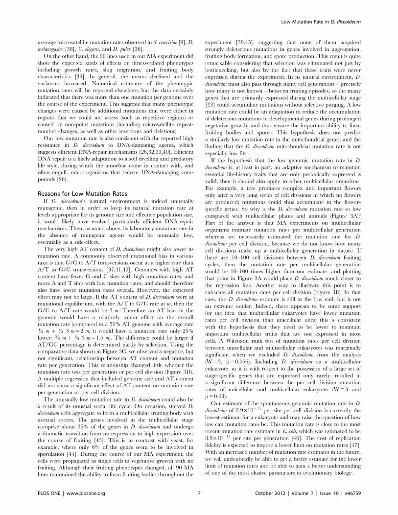
Figure 1. Life cycle of D. discoideum showing the vegetative, social, and sexual. During the vegetative cycle, single cells feed on bacteriaand divide mitotically. Upon starvation, single cells aggregate and, depending on environmental conditions, enter the social cycle or the sexual stage.During the social stage, single cells aggregate and form a multicellular fruiting body that consists of a stalk made of dead cells holding aloft a soruscontaining the fertile spores. During the sexual stage, two haploid cells fuse and ingest the other aggregating cells to form a macrocyst. This figurewas published under CC Creative Commons Attribution - Share Alike 3.0, David Brown & Joan E. Strassmann and is available at http://www.dictybase.org/Multimedia/DdLifeCycles/index.html.doi:10.1371/journal.pone.0046759.g001
Low Mutation Rate in D. discoideum
PLOS ONE | www.plosone.org 3 October 2012 | Volume 7 | Issue 10 | e46759

rather than one with sequencing errors. To estimate the
mitochondrial mutation rate, we assumed that mutations are rare
and the fate of a mutation is determined by drift. The same
procedure was used previously by Haag-Liautard et al. [7].
Therefore, the probability of fixation of a mutation at the drift-
mutation equilibrium is equal to the frequency of the mutation,
and the heteroplasmic sites contribute to the mutation rate
estimate for one MA line in proportion to their frequency as the
(sum of f(m))/(n*g), where f(m) is the frequency of a unique
mutation, n is the number of unchanged nucleotides and g is the
number of generations. Using our cutoff of 0.03, we estimate the
mitochondrial mutation rate to be 6.7661029 per nucleotide per
generation (Poisson confidence interval: 4.161029 2 9.561029).
We identified a total of 19 mitochondrial mutations (six in MA31
and 12 in MA47, both with 97% coverage of the genome; and one
in MA55 with 91% coverage). Most mutations occurred at
a frequency lower than 0.1. Only one mutation in MA47 reached
a frequency of 0.44. We did observe candidate mutations that
occurred at higher frequencies, but none of them was unique to
one MA line. Fortunately, the choice of the cutoff threshold had
relatively little effect on the estimate: for a cutoff of 0.01, the
mutation rate estimate was 1.4461028; and for a cutoff of 0.10, it
was 1.1161028.
Alternate Alignments and Modified Filtering ParametersDifferent mapping tools, methods to compile a consensus
sequence, and filtering techniques all consistently identified the
same single nuclear mutation described above. The maximum
likelihood (ML) method developed by Lynch [9,35] was unable to
estimate a mutation rate, because it exhausted all possible
mutations within two iterations. Changing our filtering parameters
had little effect on the estimated mutation rates. Lowering the
minimum coverage or increasing the maximum coverage did not
increase the number of unique mutations; but instead increased
the number of unchanged nucleotides, leading to slightly lower
mutation rate estimates. The opposite trend was observed when
we required that 100% (instead of 90%) of the bases agree with the
majority base call. Increasing the minimum-agreement require-
ment did not alter the number of mutations identified; it did,
however, reduce the number of unchanged nucleotides and thus
Figure 2. Identification of unchanged sites and unique mutations. Analysis pipeline and examples of rules used to identify mutations (red)unique to one mutation accumulation line, and unchanged nucleotides (green). Each line represents a hypothetical position in the genome and ischaracterized by the chromosome (Chr), position (Pos), and the reference base (Ref). Columns 4–6 list the majority consensus base for the three MAlines (MA31, MA47, MA55). Columns 7–9 show the number of reads covering this particular position in the three MA lines, columns 10–12 listthe fraction of reads agreeing with the majority base. We used a minimum agreement of 90%. Column 13 gives the read coverage for this position forthe self-mapping of the genome and indicates whether the position is uniquely mappable (if self-mapping coverage= 62). Columns 14–16 give theresults from our filtering criteria, with unchanged sites indicated in green, mutations in red, and confirming bases in yellow. Positions that were notcovered in all three lines were excluded from the analysis.doi:10.1371/journal.pone.0046759.g002
Low Mutation Rate in D. discoideum
PLOS ONE | www.plosone.org 4 October 2012 | Volume 7 | Issue 10 | e46759

increase the mutation rate, albeit very slightly (from 2.89610211 to
2.97610211).
Discussion
We combined an MA experiment and whole-genome sequenc-
ing and estimated the single-nucleotide mutation rates for
mitochondrial and nuclear DNA in the social amoeba D.
discoideum. The mitochondrial mutation rate of 6.7661029 (with
a Poisson confidence interval ranging from 4.161029 to
9.561029) per nucleotide per cell-generation is based on 19
unique mutations in the three genomes and is lower than the
mitochondrial mutation rates observed in other species, which
range from 1.2961028 in S. cerevisiae [9] to 17.361028 in the
asexually reproducing D. pulex [36] with C. elegans and D.
melanogaster falling in between [7,17].
We estimated the nuclear mutation rate to be 2.89610211 per
site per generation with a Poisson confidence interval ranging from
7.4610213 to 1.6610210. This estimate is based on one single
nucleotide mutation within three experimental lines. Given this
per-site mutation rate and the genome size and number of
generations, we would expect there to be one mutation on average
in each experimental genome over the 1000 generations.
Our estimate for the nuclear mutation rate is the lowest
spontaneous mutation rate reported to date for a eukaryote [8]
(Figure 3). Because there was only one mutation, the confidence
interval of our estimate spans two orders of magnitude; but even
so, the upper limit is still considerably lower than any other per-
generation mutation rate for eukaryotes [8].
Combining our new estimate with previous estimates for other
cellular organisms used by Lynch [8] shows that mutation rate
increases significantly with genome size (Figure 3A); though if we
restrict our analysis to the five estimates obtained by whole-
genome sequencing, the regression loses significance. To compare
mutation rates across taxa, we scaled all of the estimates to the
number of cell divisions instead of generations by using the
numbers of germ-line divisions per generation published in Lynch
[8]. After accounting for the number of germ-line cell divisions per
generation, we no longer observe a significant effect of genome
size on mutation rates. While D. discoideum still has the lowest point
estimate, the confidence interval includes the mutation rate
estimates for plants, D. melanogaster and Homo sapiens, while all
other single-celled eukaryotes have considerably higher mutation
rates (Figure 3B).
Given our very low nuclear mutation rate estimate, we focus on
three questions. First, are there any biases that might result in an
estimate that is far lower than the true value? Second, are there
any other data supporting a low mutation rate in D. discoideum?
Finally, are there reasons why we might expect the mutation rate
to be low in this species?
Possible BiasesSeveral factors could potentially lead to an underestimation of
the mutation rate in D. discoideum. The high AT content combined
with an unusual abundance of repeat sequences and similar short
sequences throughout the genome make it impossible to align
many short reads without ambiguity. The exclusion of such sites
could downwardly bias our estimate if point mutations are more
likely in the repeat regions. It is likely, however, that estimates in
other species would incorporate a similar bias, and we therefore
consider this an unlikely explanation for D. discoideum having
a lower rate.
Our estimate is based on strains that evolved during an MA
experiment in the laboratory under controlled, benign conditions.
The cells were never exposed to UV light, were fed non-
pathogenic bacteria, and grew on rich nutrient agar. Environ-
mental factors such as UV radiation and DNA-damaging
compounds in soil and the bacteria eaten by D. discoideum could
lead to more mutations in the wild. MA experiments for other
species, however, were also performed under benign laboratory
conditions. Nevertheless, if D. discoideum’s natural environment is
more mutagenic than those of other species, it is possible that its
mutation rate in the wild is less different from those of other
species than the estimates suggest.
The low mutation rate we observed in our three lines raises the
question of potential cross contamination of the lines during the
MA experiment, but multiple lines of evidence suggest this is very
unlikely. As outlined in the Material and Methods, we used extra
precautions to prevent any cross contamination and mixing up of
the lines. The three strains we used are distinguishable by
microsatellite mutations [34]. Mixing up the lines during the DNA
extraction and library prep is not consistent with our finding of
multiple mitochondrial mutations that were unique to one MA
line only. Our mutation rate for the mitochondria is very similar to
previous estimates for the mitochondrial mutation rates. Finally,
even in the extreme case of two of our lines actually being the same
line (a possibility excluded by the above evidence), the number of
mutable sites would be reduced by 1/3, and the estimated
mutation rate would increase by only 50%.
Finally, we should consider the possibility that our analysis was
too conservative and led to an artificially low mutation rate
compared with other studies. This seems unlikely for two reasons.
First, our approach was very similar to the approach used to
estimate the mutation rate of D. melanogaster. Second, mutation rate
estimates are much more sensitive to the number of mutations
than to the number of unchanged sites. Most of the filtering
criteria we used were identical to the ones used by Keightley et al.
[13] (three lines, same average coverage, minimum coverage of
five, 90% agreement among reads, and coverage of all three lines
at a site), with the exception that we only considered uniquely
mappable sites and relaxed the coverage requirement in confirm-
ing MA lines. Using only uniquely mappable sites will miss some
reads at sites where some but not all reads can be mapped; but
because the same criterion was applied to changed and unchanged
sites, it should not affect our estimates. Relaxing the coverage of
confirming MA lines allowed us to consider more sites (33% of the
genome). Requiring a minimum coverage of five for all lines at
a given site reduced the fraction of the genome under consider-
ation to 21%, and only slightly increased the mutation rate to
4.4610211 with a Poisson confidence interval ranging from
1.4610212 to 2.5610210, which is still considerably lower than
other eukaryotic mutation rate estimates. The relatively small
effect of changing the minimum coverage requirement also
suggests that our relaxed comparison-coverage does not account
for the 100-fold difference in mutation rates between D.
melanogaster and D. discoideum. While even smaller fractions of the
genome have been used to estimate mutation rates (e.g., roughly
15% was used for C. elegans [37]), we decided to use as much of the
genome as possible by applying the relaxed coverage requirements
for the confirming bases.
The mutation rates for S. cerevisiae [9] and C. elegans [37] are both
based on ML methods. We used the ML method developed by
Lynch, but were unable to estimate the mutation rate with only
one mutation. The ML method correctly identified the one
mutation, but failed to estimate a mutation rate. Regardless of the
method used to estimate mutation rates, the mutation rate
estimates are much more sensitive to changes in the number of
mutations than they are to changes in the number of unchanged
Low Mutation Rate in D. discoideum
PLOS ONE | www.plosone.org 5 October 2012 | Volume 7 | Issue 10 | e46759

nucleotides. We were very carful to use the same criteria to identify
mutations and unchanged nucleotides, in order to avoid over-
estimating one or the other. The results of using different
approaches, methods, and parameters suggest that the mutation
rate estimate is very robust to changes in the analysis, as we were
only able to detect one mutation overall regardless of changes in
the analysis. No other approach or method was able to identify
more mutations that could be confirmed with Sanger sequencing.
Other Data on Mutation RateOur low mutation rate estimate is consistent with low levels of
genetic variation observed in D. discoideum strains isolated from the
wild across a large geographic range [31]. Our estimate is also
consistent with the unusually low microsatellite mutation rates for
both dinucleotide and trinucleotide repeats across all 90 lines in
our MA experiment [34]. The average microsatellite mutation rate
was 1.161027 per repeat per generation, which is lower than the
Figure 3. The relationship of mutation rates and genome size or AT content. Mutation rate (as X61029 per nucleotide) per generation andmutation rate per cell division are plotted as a function of genome size (in MB) (A and B) and as a function of AT content (C and D). We observeda significant relationship between mutation rate and genome size (Log10(mutation rate) =21.0909+0.75056log10(Genome size), with R2 = 0.58 andp= 0.0024.), but not for mutation rate per cell division or mutation rates and AT content. D. discoideum is represented by the red dot; the error barsindicate the Poisson confidence interval for our nuclear mutation rate. Mutation rates for Eubacteria and Archaea are given as averages of multipleestimates and are represented by open symbols. The average mutation rate of Eubacteria does not include Buchnera aphidicola due to its unusuallyhigh mutation rate, which is characteristic for endosymbionts. Circles represent mutation rate estimates obtained from high-throughput sequencingof MA lines; estimates obtained through other methods are represented by triangles. Mutation rates for yeast are calculated as the average fromLynch [8] and Nishant et al. [15]; all other estimates are from Lynch [8].doi:10.1371/journal.pone.0046759.g003
Low Mutation Rate in D. discoideum
PLOS ONE | www.plosone.org 6 October 2012 | Volume 7 | Issue 10 | e46759
average microsatellite mutation rates observed in S. cerevisiae [9], D.
melanogaster [38], C. elegans, and D. pulex [36].
On the other hand, the 90 lines used in our MA experiment did
show the expected kinds of effects on fitness-related phenotypes
including growth rates, slug migration, and fruiting body
characteristics [39]. In general, the means declined and the
variances increased. Numerical estimates of the phenotypic
mutation rates will be reported elsewhere, but the data certainly
indicated that there was more than one mutation per genome over
the course of the experiment. This suggests that many phenotypic
changes were caused by additional mutations that were either in
regions that we could not assess (such as repetitive regions) or
caused by non-point mutations (including microsatellite repeat-
number changes, as well as other insertions and deletions).
Our low mutation rate is also consistent with the reported high
resistance in D. discoideum to DNA-damaging agents, which
suggests efficient DNA-repair mechanisms [26,32,33,40]. Efficient
DNA repair is a likely adaptation to a soil dwelling and predatory
life style, during which the amoebae come in contact with, and
often engulf, microorganisms that secrete DNA-damaging com-
pounds [26].
Reasons for Low Mutation RatesIf D. discoideum’s natural environment is indeed unusually
mutagenic, then in order to keep its natural mutation rate at
levels appropriate for its genome size and effective population size,
it would likely have evolved particularly efficient DNA-repair
mechanisms. Then, as noted above, its laboratory mutation rate in
the absence of mutagenic agents would be unusually low,
essentially as a side-effect.
The very high AT content of D. discoideum might also lower its
mutation rate. A commonly observed mutational bias in various
taxa is that G/C to A/T transversions occur at a higher rate than
A/T to G/C transversions [37,41,42]. Genomes with high AT
content have fewer G and C sites with high mutation rates, and
more A and T sites with low mutation rates, and should therefore
also have lower mutation rates overall. However, the expected
effect may not be large. If the AT content of D. discoideum were at
mutational equilibrium, with the A/T to G/C rate at m, then the
G/C to A/T rate would be 3 m. Therefore an AT bias in the
genome would have a relatively minor effect on the overall
mutation rate (compared to a 50% AT genome with average rate
K m + K 3 m= 2 m, it would have a mutation rate only 25%
lower: L m + J 3 m= 1.5 m). The difference could be larger if
AT/GC percentage is determined partly by selection. Using the
comparative data shown in Figure 3C, we observed a negative, but
not significant, relationship between AT content and mutation
rate per generation. This relationship changed little whether the
mutation rate was per generation or per cell division (Figure 3D).
A multiple regression that included genome size and AT content
did not show a significant effect of AT content on mutation rate
per generation or per cell division.
The unusually low mutation rate in D. discoideum could also be
a result of its unusual social life cycle. On occasion, starved D.
discoideum cells aggregate to form a multicellular fruiting body with
asexual spores. The genes involved in the multicellular stage
comprise about 25% of the genes in D. discoideum and undergo
a dramatic transition from no expression to high expression over
the course of fruiting [43]. This is in contrast with yeast, for
example, where only 6% of the genes seem to be involved in
sporulation [44]. During the course of our MA experiment, the
cells were propagated as single cells in vegetative growth with no
fruiting. Although their fruiting phenotypes changed, all 90 MA
lines maintained the ability to form fruiting bodies throughout the
experiment [39,45], suggesting that none of them acquired
strongly deleterious mutations in genes involved in aggregation,
fruiting body formation, and spore production. This result is quite
remarkable considering that selection was eliminated not just by
bottlenecking, but also by the fact that these traits were never
expressed during the experiment. In its natural environment, D.
discoideum must also pass through many cell generations – precisely
how many is not known – between fruiting episodes, so the many
genes that are primarily expressed during the multicellular stage
[43] could accumulate mutations without selective purging. A low
mutation rate could be an adaptation to reduce the accumulation
of deleterious mutations in developmental genes during prolonged
vegetative growth, and thus ensure the important ability to form
fruiting bodies and spores. This hypothesis does not predict
a similarly low mutation rate in the mitochondrial genes, and the
finding that the D. discoideum mitochondrial mutation rate is not
especially low fits.
If the hypothesis that the low genomic mutation rate in D.
discoideum is, at least in part, an adaptive mechanism to maintain
essential life-history traits that are only periodically expressed is
valid, then it should also apply to other multicellular organisms.
For example, a tree produces complex and important flowers
only after a very long series of cell divisions in which no flowers
are produced; mutations could thus accumulate in the flower-
specific genes. So why is the D. discoideum mutation rate so low
compared with multicellular plants and animals (Figure 3A)?
Part of the answer is that MA experiments on multicellular
organisms estimate mutation rates per multicellular generation
whereas we necessarily estimated the mutation rate for D.
discoideum per cell division, because we do not know how many
cell divisions make up a multicellular generation in nature. If
there are 10–100 cell divisions between D. discoideum fruiting
cycles, then the mutation rate per multicellular generation
would be 10–100 times higher than our estimate, and plotting
that point in Figure 3A would place D. discoideum much closer to
the regression line. Another way to illustrate this point is to
calculate all mutation rates per cell division (Figure 3B). In that
case, the D. discoideum estimate is still at the low end, but is not
an extreme outlier. Indeed, there appears to be some support
for the idea that multicellular eukaryotes have lower mutation
rates per cell division than unicellular ones; this is consistent
with the hypothesis that they need to be lower to maintain
important multicellular traits that are not expressed in most
cells. A Wilcoxon rank test of mutation rates per cell division
between unicellular and multicellular eukaryotes was marginally
significant when we excluded D. discoideum from the analysis
(W = 3, p = 0.056). Including D. discoideum as a multicellular
eukaryote, as it is with respect to the possession of a large set of
stage-specific genes that are expressed only rarely, resulted in
a significant difference between the per cell division mutation
rates of unicellular and multicellular eukaryotes (W = 3 and
p = 0.03).
Our estimate of the spontaneous genomic mutation rate in D.
discoideum of 2.9610211 per site per cell division is currently the
lowest estimate for a eukaryote and may raise the question of how
low can mutation rates be. This mutation rate is close to the most
recent mutation rate estimate in E. coli, which was estimated to be
8.9610211 per site per generation [46]. The cost of replication
fidelity is expected to impose a lower limit on mutation rates [47].
With an increased number of mutation rate estimates in the future,
we will undoubtedly be able to get a better estimate for the lower
limit of mutation rates and be able to gain a better understanding
of one of the most elusive parameters in evolutionary biology.
Low Mutation Rate in D. discoideum
PLOS ONE | www.plosone.org 7 October 2012 | Volume 7 | Issue 10 | e46759

Materials and Methods
Mutation Accumulation ExperimentWe created 90 MA lines and 10 control lines by starting each
line from a single clone of the ancestor AX4, as described in
McConnell et al. [34]. We propagated each line independently for
140 days by reducing the population size to a single cell every
other day, resulting in 70 single-cell bottlenecks and 1000
vegetative divisions or generations (with no fruiting body
formation). We grew the cells at low density on a fresh lawn of
bacteria. After two days, plaques were visible, allowing us to pick
up the cells of a randomly chosen plaque and streak them onto
a fresh lawn of bacteria. Cells in a distinct, round plaque all
originated from a single cell. We repeated this process every other
day. In order to reduce the potential for cross contamination
among the evolving lines, we used several precautions. We
transferred the 100 lines in batches of ten (i.e., 1–10, 11–20, and so
forth). The three lines that we sequenced were in separate batches
and hence very unlikely to have been mixed (note that mixing
within a batch, or with any non-sequenced line, would not bias the
estimated mutation rate – it would simply substitute one line for
another). The transfers were done by at least two people, who both
checked the identity of the line to be transferred and the new plate
identity. If there was any question about the identity of a line, the
plates were discarded and we repeated the questionable transfer
using backup plates. Every ten bottlenecks, we froze a sample of
the population. To freeze the populations, we grew each line to
high density, collected the spores after fruiting body formation,
and froze each line along with the ancestor at 280uC in KK2
buffer (14.0 mM KH2PO4 and 3.4 mM K2HPO4, pH = 6.4)
supplemented with 20% glycerol. While all lines remained in
vegetative growth during the MA experiment, they retained the
ability to form fruiting bodies throughout the experiment.
Number of Cell GenerationsWe estimated the number of cell divisions during the 48-hour
period between transfers by counting the number of cells in each
of 16 48-hour plaques of the ancestral clone and taking the
logarithm with base two. This yielded an estimate of 14.22
divisions (6 st.dev. 0.544 divisions). The rate was also estimated
for the 90 MA lines, and did not differ [39]. Multiplying by 70
transfers yields 995 cell divisions, slightly lower than the 1007
divisions reported previously [34] based on only eight replicates.
We used the round figure of 1000 in our estimates.
DNA Extraction and SequencingWe randomly picked three MA lines (MA31, MA47 and MA55)
and grew them to a density of 16107 cells/ml in HL-5 media (10 g
bactopeptone #2, 5 g yeast extract, 10 g glucose, 0.35 g
Na2HPO4, 0.35 g KH2PO4, 1 ml 100x trace elements,
pH 6.4–6.6). We collected cells from 100 ml culture and extracted
good-quality DNA using a protocol developed by Mariko Katoh-
Kurasawa as follows: we washed the cells three times with 50 ml of
KK2 buffer by centrifuging (1300 rpm for 3 minutes at 4uC),
discarding the supernatant, and re-suspending the cells in fresh
KK2 buffer. After the last wash, we re-suspended the cells in 10 ml
nucleus-isolation buffer (40 mM Tris, 1.5% Sucrose, 0.1 mM
EDTA, 6 mM MgCl2, 40 mM KCl, 5 mM DTT, and 0.4%
NP40-Alternative) to lyse the cells and extract the nuclei. After 10
minutes incubation on ice, we spun the cells at 8000 rpm for 5
minutes before carefully removing the supernatant and re-
suspending the nuclei in EDTA to a final concentration of 100–
200 mM with 450 ml of STE solution, 10 ml of 20% SDS, 25 ml
ddH2O, and 10 ml of 10 mg/ml Proteinase K, and incubating the
samples at 60 degrees for 60 minutes. We performed a phenol-
chloroform extraction and ethanol precipitation purification to
extract the DNA. We prepared the genomic DNA for sequencing
using the Illumina library-preparation kit (DNA Sample Prep Kit
# FC–102–1001). We prepared the libraries for MA31 and MA47
at the same time and the library for MA55 at a later date. We
initially sequenced MA31 and MA47 on an Illumina GA1
sequencer with 36 cycles on two lanes for each genome. We
processed the resulting reads using the Illumina GA pipeline 1.3.
After initial analyses we decided to increase the coverage and
number of MA lines so we sequenced one additional lane for both
MA31 and MA47 and three for MA55. We sequenced these lanes
with 51 cycles and processed the reads with the Illumina GA
pipeline 1.4. All reads were single-end reads, regardless of their
length. We used Firecrest for image processing and Bustard for
base calling. We used the PIQA pipeline [48] to assess the quality
of the reads and determined that the sequencing quality declined
after 31 bps. Therefore, we trimmed all the reads, regardless of
their initial length to a length of 31 bases before aligning them to
the reference sequence (Eichinger et al. 2005) (retrieved from
www.dictybase.org on May 13, 2009). All reads used in this project
are available under accession number SRA056497.
Alignment using BowtieWe mapped the reads to the reference genome with zero or
one mismatch per read using Bowtie [49] (-n1 -S –best –
solexa1.3-quals with a seed length of 28 bp). Bowtie uses
Burrows-Wheeler indexing, which results in fast alignments of
reads to larger genomes. We used SAMtools [50] to create
a pileup file (pileup -c -f). The pileup file contains information
about how the reads align to a given site in the genome. Each
row in the pileup file represents a position in the genome and
lists the chromosome, position, reference sequence, consensus
sequence, consensus quality, SNP quality (both Phred-scaled), the
root mean square mapping quality, read base for each read that
aligned to the site, and the alignment quality for each read base.
We used the pileup command to compile a consensus sequence
using a Bayesian model [50,51]. We extended the pileup file to
the full-genome length, which also included uncovered sites. In
addition to the consensus sequence compiled by the pileup
command, we used the information in the pileup file to
determine what percentage of the reads called for a particular
base, and compiled a second consensus sequence based on
a majority rule: a consensus base was called if at least 50% of the
reads agreed. If two bases were tied at 50% agreement each, we
did not make a base call and considered the position uncovered.
Because D. discoideum has a haploid genome, we did not have to
account for the possibility of heterozygosity.
Repeat Sequences and Uniquely Mappable PositionsTo reduce problems caused by repeated sequences, we limited
our analyses to uniquely mappable sites. To identify nucleotides
that can be mapped uniquely with 31-bp reads, we used the
reference sequence and created all possible 31-bp reads for both
strands and aligned the ‘fake’ reads to the reference sequence with
zero or one mismatch (seed length 28 bp) using Bowtie (-n1–best -
sam). As for the real reads, we compiled a pileup file in SAMtools
(pileup -c -f). The mapping options that we used in Bowtie allowed
the reads to map to multiple places. As a result, exactly 62 reads
covered each uniquely mappable position. Based on this self-
mapping, we determined that 59% (20,337,176 bases) of the
nuclear genome could be mapped uniquely with 31-bp reads.
Low Mutation Rate in D. discoideum
PLOS ONE | www.plosone.org 8 October 2012 | Volume 7 | Issue 10 | e46759

Identification of Mutated and Unchanged Sites in theNuclear Genome
To calculate the mutation rate, we identified all apparent
unique point mutations as well as all the sites where the reference
nucleotide was confirmed (Figure 2), being careful to apply the
same criteria to changed and unchanged sites. Candidate
mutations were characterized as single nucleotide substitutions,
where the majority base in an MA line differed from the reference
base with at least five-reads coverage and 90% agreement. In
addition, an accepted mutation had to be unique to one MA line,
because mutations are rare events and it is very unlikely that the
same mutation occurs multiple times independently. Candidate
mutations that appear in more than one MA line are much more
likely to be errors in the reference genome, differences between
our ancestor clone and the reference genome, or mutations that
occurred while we were growing up cells to start our 90 lines.
Therefore, if a candidate mutation was the majority base in any of
the other two lines, even with coverage of just one read, we
excluded it. In order to avoid bias, we implemented a parallel rule
for unchanged sites. We considered a nucleotide in one MA line
unchanged if the majority base agreed with the reference base and
if the reference base was confirmed (again with any non-zero level
of coverage and the majority rule) in the other MA lines.
Error RateTo calculate the error rate, we used an approach similar to that
of Keightley et al. [13], determining the fraction of reads covering
a base that did not agree with the majority of the reads and
calculating the average error rate for each line individually. We
calculated the overall average error rate as the average of the three
MA lines.
Mutation Rate in the Mitochondrial GenomeTo estimate the mitochondrial mutation rate, we used similar
procedures but accounted for possible heteroplasmy, because
a single D. discoideum cell contains multiple copies of the
mitochondrial genome. Following Haag-Liautard et al. [7], we
assumed that mutations in the mitochondria were maintained
through mutation-drift balance and that the likelihood of fixation
of a mutation was equal to its current frequency, so that each
heteroplasmic mutation adds to the estimated mutation rate
according to its frequency (see their equation 2). We identified sites
that could be mapped uniquely (97% of the mitochondrial
genome) and were covered by at least 400 reads, which is a tenth
of the average coverage of the mitochondria.
As in the nuclear analysis, we assumed that mutations arise
independently and therefore only accepted candidate mutations
that were called in a single MA line. Mutations that occurred in
more than one line above the cutoff minimum frequency were not
included. To calculate the mutation rate, we used equation (2) in
Haag-Liautard [7]. We summed all the frequencies of mutations in
one line and divided the sum by the number of sites meeting the
same criteria as the mutations (covered by at least 400 reads and
uniquely mappable) and the number of generations (1000). We
estimated the mutation rate for each line individually and
calculated the average mutation rate. We calculated the Poisson
confidence interval for 19 mutations.
We used two approaches to choose a cutoff frequency f for
distinguishing true mutations (often heteroplasmic) from sequenc-
ing errors. In method one, we calculated mutation rate estimates
using 10 values of f from 0.01 to 0.10 and looked for a minimum
that reflected not accepting too many sequencing errors as
mutations and not missing too many real, low-frequency
mutations. The estimates were in a relatively narrow range with
the minimum at a cutoff of f= 0.03. Second, we used our empirical
estimate of alignment/sequencing error for nuclear sites (see
Results) to assess how likely we were, at each possible cutoff, to
have sequencing errors leading to the acceptance of a false
mutation. Our empirical estimate was in the range of 0.002 per
read, and we doubled that to account for the fact that some sites
were likely to be systematically more error-prone. At that error
rate, we asked: if we have 400 reads, how low can we make the
cutoff frequency f to keep the binomial probability of having more
than 4006f erroneous reads below 1 across all our sites (i.e., lower
than 6.761026 for each of the 150000 mitochondrial sites in the
three genomes)? That answer also yielded a cutoff of f= 0.03, so
we used that for our reported estimate.
Other Mapping ToolsIn addition to the results presented here, we performed several
variations of the analysis using: 36-bp reads, a different mapping
tool, different ways to compile a consensus sequence, varying
filtering parameters, and a ML method, respectively. We initially
decided to cut all our reads to 31-bp because of the declining
quality of the reads after base 32. To align the 36-bp reads, we
used the same approach as described above. We also used
a different mapping tool, MAQ, to align the reads and compile the
majority consensus sequence. To identify mutations, we compiled
a consensus sequence based on the majority call of all the reads
aligning to one position. We used this consensus sequence to
estimate our mutation rate. We also performed the same analysis
using the consensus sequence compiled by MAQ or SAMtools
(ambiguous calls were replaced by the majority base call). Using
the consensus sequence consistently resulted in a large number of
candidate mutations that were later identified as false positives by
Sanger sequencing. We performed all of these analyses with
varying filtering parameters. We tried the ML method developed
by Lynch [9,35] to estimate the mutation rates. The low number
of mutations in our data set, however, did not work with this
method; after two iterations, all the mutations were exhausted and
the program was unable to estimate the mutation rate.
Confirmation of the SNPs using Sanger SequencingTechnology
We used Sanger sequencing to confirm the computationally
predicted unique mutation in MA31 by sequencing all MA lines
and the ancestor (using forward primer: 59-CTTTCAAGGT-
GAAGCGAATAAAACA and reverse primer: 59-ACA-
TATGCTTTGAGTGGGAGATTAC).
Acknowledgments
We thank M. Katoh-Kurasawa and E. A. Ostrowski for the DNA
extraction protocol. Z. Xu and J. Ren provided support during the library
preparation. We thank A. Martinez-Alcantara, E. Ballesteros, S. E. Kalla,
J. I. Lucas-Lledo, M. Lynch, E. A. Ostrowski, A. M. Perez, and M. Rojas
for helpful discussion and support with the analysis.
Author Contributions
Conceived and designed the experiments: GS PH YF JES DCQ.
Performed the experiments: SAF GS. Analyzed the data: GS PH SG YF
MAQ JES DCQ. Contributed reagents/materials/analysis tools: GS PH
YF JES DCQ. Wrote the paper: GS MAQ JES DCQ.
Low Mutation Rate in D. discoideum
PLOS ONE | www.plosone.org 9 October 2012 | Volume 7 | Issue 10 | e46759

References
1. Fisher RA (1930) The Genetical Theory of Natural Selection. Oxford:Clarendon Press.
2. Baer CF, Miyamoto MM, Denver DR (2007) Mutation rate variation inmulticellular eukaryotes: causes and consequences. Nature Reviews 8: 619–631.
3. Kondrashov FA, Kondrashov AS (2010) Measurements of spontaneous rates of
mutations in the recent past and the near future. Philosophical Transactions ofthe Royal Society of London Series B-Biological Sciences 365: 1169–1176.
4. Drake JW, Charlesworth B, Charlesworth D, Crow JF (1998) Rates of
spontaneous mutation. Genetics 148: 1667–1686.
5. Eyre-Walker A, Keightley PD (2007) The distribution of fitness effects. Nature
Reviews Genetics 8: 610–518.
6. Denver DR, Dolan PC, Wilhelm LJ, Sung W, Lucas-Lledo JI, et al. (2009) Agenome-wide view of Caenorhabditis elegans base-substitution mutation processes.
Proceedings of the National Academy of Sciences of the United States of
America 106: 16310–16314.
7. Haag-Liautard C, Coffey N, Houle D, Lynch M, Charlesworth B, et al. (2008)Direct Estimates of the mitochondrial DNA mutation rate in Drosophila
melanogaster. PLoS Biology 6: e204.
8. Lynch M (2010) Evolution of the mutation rate. Trends in Genetics 26: 345–
352.
9. Lynch M, Sung W, Morris K, Coffey N, Landry CR, et al. (2008) A genome-wide view of the spectrum of spontaneous mutations in yeast. Proceedings of the
National Academy of Sciences of the United States of America 105: 9272–9277.
10. Ossowski S, Schneeberger K, Lucas-Lledo JI, Warthmann N, Clark RM, et al.(2010) The rate and molecular spectrum of spontaneous mutations in Arabidopsis
thaliana. Science 327: 92–94.
11. Haag-Liautard C, Dorris M, Maside X, Macaskill S, Halligan DL, et al. (2007)
Direct estimation of per nucleotide and genomic deleterious mutation rates inDrosophila. Nature 445: 82–85.
12. Keightley PD (1999) Analysis of mutation accumulation experiments: response
to Deng, Li and Li. Genet Res, Camb 74: 87–91.
13. Keightley PD, Trivedi U, Thomson M, Oliver F, Kumar S, et al. (2009) Analysis
of the genome sequences of three Drosophila melanogaster spontaneous mutationaccumulation lines. Genome Research 19: 1195–1201.
14. Halligan DL, Keightley PD (2009) Spontaneous mutation accumulation studies
in evolutionary genetics. Annual Review of Ecology Evolution and Systematics
40.
15. Nishant KT, Wei W, Mancera E, Argueso JL, Schlattl A, et al. (2010) TheBaker’s Yeast Diploid Genome Is Remarkably Stable in Vegetative Growth and
Meiosis. PLoS Genetics 6.
16. Xu S, Schaack S, Seyfert A, Choi E, Lynch M, et al. (2012) High MutationRates in the Mitochondrial Genomes of Daphnia pulex. Molecular Biology and
Evolution 29: 763–769.
17. Denver DR, Morris K, Lynch M, Vassilieva LL, Thomas WK (2000) High
direct estimate of the mutation rate in the mitochondrial genome of Caenorhabditiselegans. Science 289: 2342–2344.
18. Queller DC, Ponte E, Bozzaro S, Strassmann JE (2003) Single-Gene Green-
beard Effects in the Social Amoeba Dictyostelium discoideum. Science 299: 105–106.
19. Strassmann JE, Queller DC (2007) Altruism among amoebas. Natural History
116: 24–29.
20. Strassmann JE, Zhu Y, Queller DC (2000) Altruism and social cheating in thesocial amoeba Dictyostelium discoideum. Nature 408: 965–967.
21. Escalante R, Vicente JJ (2000) Dictyostelium discoideum: a model system for
differentiation and patterning. The International Journal of Developmental
Biology 44: 819–835.
22. Urushihara H (2008) Developmental biology of the social amoeba: History,current knowledge and prospects. Development Growth & Differentiation 50:
S277–S281.
23. Hagele S, Kohler R, Merkert H, Schleicher M, Hacker J, et al. (2000)
Dictyostelium discoideum: a new host model system for intracellular pathogens of thegenus Legionella. Cellular Microbiology 2: 165–171.
24. Hasselbring BM, Patel MK, Schell MA (2011) Dictyostelium discoideum as a model
system for identification of Burkholderia pseudomallei virulence factors.Infection and Immunity 79: 2079–2088.
25. Annesley SJ, Fisher PR (2009) Dictyostelium discoideum - a model for many reasons.Molecular and Cellular Biochemistry 329: 73–91.
26. Kessin RH (2001) Dictyostelium: Evolution, Cell Biology, and the Development of
Multicellularity. Cambridge, UK: Cambridge University Press. 294 p.
27. Raper KB (1984) The Dictyostelids. Princeton, NJ: Princeton University Press
453 p.28. Bonner JT (1947) Evidence for the formation of aggregates by chemotaxis in the
development of the slime mold Dictyostelium discoideum. J Exp Zool 106: 1–26.29. Saxer G, Brock DA, Queller DC, Strassmann JE (2010) Cheating does not
explain selective differences at high and low relatedness in a social amoeba.
BMC Evol Biol 10: 76.30. Eichinger L, Pachebat JA, Glockner G, Rajandream MA, Sucgang R, et al.
(2005) The genome of the social amoeba Dictyostelium discoideum. Nature 435: 43–57.
31. Flowers JM, Li SI, Stathos A, Saxer G, Ostrowski EA, et al. (2010) Variation,
Sex, and Social Cooperation: Molecular Population Genetics of the SocialAmoeba Dictyostelium discoideum. PLoS Genetics 6.
32. Deering RA, Guyer RB, Stevens L, Watson-Thais TE (1996) Some repair-deficient mutants of Dictyostelium discoideum display enhanced susceptibility to
Bleomycin. Antimicrobial Agents and Chemotherapy 40: 464–467.33. Deering RA (1968) Dictyostelium discoideum: a gamma-ray resistant organism.
Science 162: 1289–1290.
34. McConnell R, Middlemist S, Scala C, Strassmann JE, Queller DC (2007) Anunusually low microsatellite mutation rate in Dictyostelium discoideum, an organism
with unusually abundant microsatellites. Genetics 177: 1499–1507.35. Lynch M (2008) Estimation of nucleotide diversity, disequilibrium coefficients,
and mutation rates from high-coverage genome-sequencing projects. Molecular
Biology and Evolution 25: 2409–2419.36. Seyfert AL, Cristescu MEA, Frisse L, Schaack S, Thomas WK, et al. (2008) The
rate and spectrum of microsatellite mutation in Caenorhabditis elegans and Daphnia
pulex. Genetics 178: 2113–2121.
37. Denver DR, Dolan PC, Wilhelm LJ, Sung W, Lucas-Lledo JI, et al. (2009) Agenome-wide view of Caenorhabditis elegans base-substitution mutation processes.
Proceedings of the National Academy of Sciences of the United States of
America 106: 16310–16314.38. Schug MD, Mackay TFC, Aquadro CF (1997) Low mutation rates of
microsatellite loci in Drosophila melanogaster. Nature Genetics 15: 99–102.39. Middlemist SA (2009) Mutational processes in Dictyostelium discoideum: how
mutations affect social behaviors and fitness. Houston: Rice University.
40. Deering RA (1994) Dictyostelium discoideum, a lower eukaryote model for thestudy of DNA repair: Implications for the role of DNA-damage chemicals in the
evolution of repair proficient cells. Adv Space Res 14: 389–393.41. Galtier N, Piganeau G, Mouchiroud D, Duret L (2001) GC-content evolution in
mammalian genomes: The biased gene conversion hypothesis. Genetics 159:907–911.
42. Hershberg R, Petrov DA (2010) Evidence That Mutation Is Universally Biased
towards AT in Bacteria. Plos Genetics 6.43. Van Driessche N, Shaw C, Katoh M, Morio T, Sucgang R, et al. (2002) A
transcriptional profile of multicellular development in Dictyostelium discoideum.Development 129: 1543–1552.
44. Deutschbauer AM, Williams RM, Chu AM, Davis RW (2002) Parallel
phenotypic analysis of sporulation and postgermination growth in Saccharomyces
cerevisiae. Proceedings of the National Academy of Sciences of the United States
of America 99: 15530–15535.45. Kuzdzal-Fick JJ, Fox SA, Strassmann JE, Queller DC (2011) High Relatedness
Is Necessary and Sufficient to Maintain Multicellularity in Dictyostelium.Science 334: 1548–1551.
46. Wielgoss S, Barrick JE, Tenaillon O, Cruveiller S, Chane-Woon-Ming B, et al.
(2011) Mutation rate inferred from synonymous substitutions in a long-termevolution experiment with Escherichia coli. Genes | Genomes | Genetics 1: 183–
186.47. Sniegowski PD, Gerrish PJ, Johnson T, Shaver A (2000) The evolution of
mutation rates: separating causes from consequences. Bioessays 22: 1057–1066.
48. Martınez-Alcantara A, Ballestros E, Feng C, Rojas M, Koshinksy H, et al. (2009)PIQA: pipeline for Illumina G1 genome analyzer data quality assessment.
Bioinformatics 25: 2428–2429.49. Langmead B, Trapnell C, Pop M, Salzberg SL (2009) Ultrafast and memory-
efficient alignment of short DNA sequences to the human genome. Genome
Biology 10: R25.50. Li H, Handsaker B, Wysoker A, Fennell T, Ruan J, et al. (2009) The Sequence
Alignment/Map format and SAMtools. Bioinformatics 25: 2078–2079.51. Li H, Ruan J, Durbin R (2008) Mapping short DNA sequencing reads and
calling variants using mapping quality scores. Genome Research 18: 1851–1858.
Low Mutation Rate in D. discoideum
PLOS ONE | www.plosone.org 10 October 2012 | Volume 7 | Issue 10 | e46759