Embed Size (px)
Citation preview

1
Annual Report 2012
WHITEHEAD INSTITUTE

2 1
inspired scientific inquiry can be all-consuming, virtually demanding a fixation on the here and now. and yet, too sharp a focus on the present may blur the overall vision. at whitehead institute, the world’s best biomedical researchers are avoiding such myopia, executing passionately each day but with an eye toward the future. driven to pursue transformational discoveries, they have their sights set.
SIGHTS SET

2 3
CONTENTS3
4
16
50
56
58
62
63
From the Director
Scientific Achievement
Principal Investigators
Whitehead Fellows
Community Evolution
Honor Roll of Donors
Financial Summary
Leadership
During 2012, we quietly celebrated Whitehead Institute’s 30th anniversary. As we approached this milestone, our faculty members immersed themselves in a year-long planning exercise whose stated objective was to establish a scientific vision—both for the Institute and for their individual laboratories.
We set out to plot our future course. We formed a special committee. We met separately. We met as a whole. We dissected policy documents from prominent research organizations and funding agencies. We hosted scientific leaders. We challenged ourselves and we extracted each other’s best thinking on the direction of our scientific enterprise. We were comprehensive, prospective, and introspective. We were also conclusive, ultimately realizing that to be at our best, we must remain true to ourselves.
Thus, we emerged from this process reinvigorated and rededicated to our founding mission: We will continue to attack biology’s most difficult questions in pursuit of discoveries that will have a disproportionate impact on improving human health. Be assured, this is far from an embrace of the status quo. Indeed, our principal investigators—as is the theme of this report—have their sights set on a number of potentially transformative initiatives. These are projects that surfaced during our visioning and are fueled by the kind of scientific audaciousness that has long distinguished our Institute.
Although unwavering in our commitment to the fundamentals, I was recently reminded of its importance by an unlikely source. Just a few months ago, Sanofi CEO Christopher Viebacher made headlines when he announced that his company would no longer expend vast resources to develop drugs for Alzheimer’s disease. In an interview during a meeting of pharmaceutical industry lobbyists, Viehbacher stated: “I think we have to do a lot more basic science work to understand what’s going on. We really, at best, partially understand the cause of the disease. It’s hard to come up with meaningful targets. Unless we’ve got better targets, we’re not really making any progress…We have to be humble in front of science.”
As we commit to fill the kind of void Viehbacher describes, I am both humbled and grateful to our faculty, staff, friends, and supporters whose collective conviction enables us to do what we’ll do.
David C. Page
from the director
WHY WE’LL DO WHAT WE DO

4 5
a whitehead institute laboratory is seldom a quiet place. scientists are apt to gather at all hours, tweaking experiments, analyzing data, and refining hypotheses generated by creativity that knows no schedule. the only regularity, it seems, is the emergence of high-impact, award-winning research and discovery.
SCIENTIFIC ACHIEVEMENT
4 5

scientific achievement
6 7
Identifying the abnormal almost always requires a thorough understanding of the normal. It’s a truism that drives develop-mental biologists to investigate the complexities of cell growth and differen-tiation and tissue and organ generation: Knowing how something goes right might help prevent it from going wrong.
Whitehead Member Terry Orr-Weaver recently discovered a mechanism in developing fruit flies that preserves tight junctions between cells comprising the blood-brain barrier, even during rapid growth. The blood-brain barrier is essential for maintaining the brain’s stable environment, preventing entry of harmful viruses and bacteria and isolating the brain’s specific hormonal and neurotransmitter activity from that in the rest of the body. In the fruit fly, the blood-brain boundary is composed of glia cells that form an envelope sealed around nerve cells. As the brain rapidly expands during development, the glial envelope must grow correspondingly to
remain intact. Until now, little had been known about how the blood-brain barrier maintains its integrity as the brain it protects develops.
The Orr-Weaver lab determined that as the larval fruit fly brain grows by cell division, it instructs subperineurial glia (SPG) cells that form the blood-brain barrier to enlarge by creating multiple copies of their genomes in a process known as polyploidization. Orr-Weaver believes this is a conserved develop-mental strategy that accommodates organ growth, noting that cell layers in the human placenta and skin may employ polyploidization to expand while maintaining a sound boundary between the fetus and its surroundings, and the body and the outside world, respectively.
Tissue and organ growth and regenera-tion are mainstays of research in Whitehead Member Peter Reddien’s lab, where planarian flatworms are under intense study for their renowned ability to regenerate any missing body
part, even as adults. The lab, however, has now expanded the planarian job description, using the worms as a model system for studying eye development and eye diseases in vertebrates, including humans.
During the past year, scientists produced an exhaustive catalog of genes active in the planarian eye. Within this catalog are genes with human homologs that are known to be involved in eye develop-ment and others that are associated with age-related macular degeneration and other retinal disorders.
“It’s exciting to get this complete list of genes in one fell swoop,” says Reddien. “This provides perhaps the most comprehensive list of genes involved in eye biology in a model system other than Drosophila.”
These planarian eyes have active genes in-volved in light detection (green), pigment synthesis (blue), and regeneration (red).
DEVELOpMENTAL bIOLOGY An eye on growth
For a cancer patient, over-expression of the notorious MYC oncogene is a very bad sign. Scientists have long known that in tumor cells, elevated levels of MYC’s protein product, c-Myc, are associated with poor clinical outcomes, including increased rates of metastasis, recurrence, and mortality. Yet decades of research producing thousands of scientific papers on the subject had failed to explain exactly how c-Myc exerts its effects across a broad range of cancer types.
The prevailing theory emerging from this massive body of research had been that in tumor cells, c-Myc affects the expression of specific genes or sets of genes—that so-called Myc target genes are selectively activated or repressed, leading to aberrant cellular behavior. Enter Whitehead Member Richard Young, whose lab has dispelled this commonly held notion by showing that elevated expression of c-Myc amplifies the activity of all expressed genes in tumor cells of
multiple cancer types, from lymphomas to lung cancers. It turns out that high levels of c-Myc send a tumor cell’s gene expression program into overdrive. Transcription increases dramatically, allowing malignant cells to overwhelm factors that might normally hamper their growth and proliferation.
This surprising finding is a simple, ele- gant explanation for how a single protein can have such profound effect in so many and varied types of cancer. Says Young: “MYC is a key driver in most major cancers, but it has been notori-ously difficult to drug. Now that we know the mechanism by which c-Myc acts, we can go after the components of that mechanism as potential drug targets.”
In the meantime, Whitehead Member David Sabatini is exploring a novel approach to delivering drugs directly into cancer cells. Researchers in Sabatini’s lab discovered that certain molecules present in high concentrations on the
surfaces of many cancer cells can be exploited to funnel lethal toxic molecules into the malignant cells. Although the lab’s initial finding was based on the study of a single toxic molecule and its transporter protein, Sabatini believes this phenomenon could be leveraged more broadly.
He notes: “Our work suggests a diff- erent strategy for cancer therapy that takes advantage of the capacity of a cancer cell to take up something toxic that a normal cell does not. By identifying transporters on the surface of cancer cells, you might be able to find a molecule specifically toxic to that cell. You really could have something much more selective to cancer cells.”
High levels of the protein c-Myc are associated with poor outcomes in cancer patients. Above, c-Myc (red) and its partner, Max (blue), interact with DNA.
CANCER hyperActivity for genes, speciAl delivery for drugs
scientific achievement
8 9
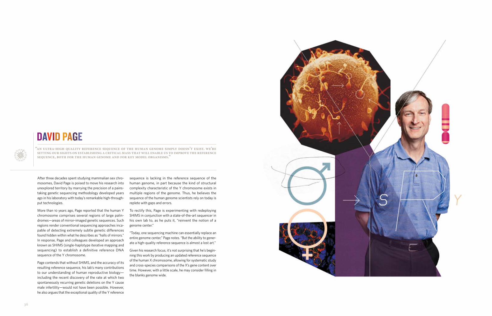
The discovery that fully differentiated adult cells could be “reprogrammed” to an embryonic stem-cell like state— creating what are known as induced pluripotent stem (iPS) cells—has revo- lutionized the field of stem cell science. Among the most appealing aspects of the discovery has been the promise of treating human disease with embry-onic-like stem cells generated without the use of embryos.
Although techniques to create iPS cells have improved since the break-through in human cells was first reported in 2007, reprogramming remains an inefficient process. By some estimates, within a given population of cells under- going reprogramming by traditional methods, no more than 1 percent actually achieve pluripotency. However, new research in the lab of Whitehead Member Rudolf Jaenisch could up the success rate markedly.
Members of the lab have identified new genetic markers that allow scientists to
predict which treated cells will success-fully become pluripotent. By examining gene expression in individual cells—rather than across a colony of cells—throughout reprogramming, researchers found four genes that are turned on quite early in the cells that would reach pluripotency. Importantly, markers once thought to be key to pluripotency were found in cells that were only partially reprogrammed. Armed with these newly discovered mark- ers, researchers can quickly disregard colonies of cells that will not become fully reprogrammed and would thus be useless for iPS cell-based therapeutics or disease studies.
Another long-term goal of stem cell science is the application of such cells in regenerative medicine. Progress in this arena requires an improved understanding of stem cell behavior at the molecular level, particularly within the environment of a living organism rather than the highly artificial environ-ment of the Petri dish.
With their renowned powers of regeneration and more than half of their genes having homologs in humans, the planarian flatworms in Whitehead Member Peter Reddien’s lab are a logical choice for this line of research. In a significant advance, the lab recently devised a method to identify potential genetic regulators of stem cells and determine if those genes affect the two main functions of stem cells: differ- entiation and renewal of the stem cell population.
In total, the lab found 10 genes impacting planarian stem cell renewal, and two genes with roles in both renewal and differentiation. Intriguingly, three of the renewal genes code for proteins that are similar to components of a protein complex known to regulate embryonic stem cell biology in higher organisms, including mammals.
A planarian pluripotent stem call can give rise to proliferating (red) and differentiat-ing (blue) cells.
STEM CELLS finding new rules And regulAtions
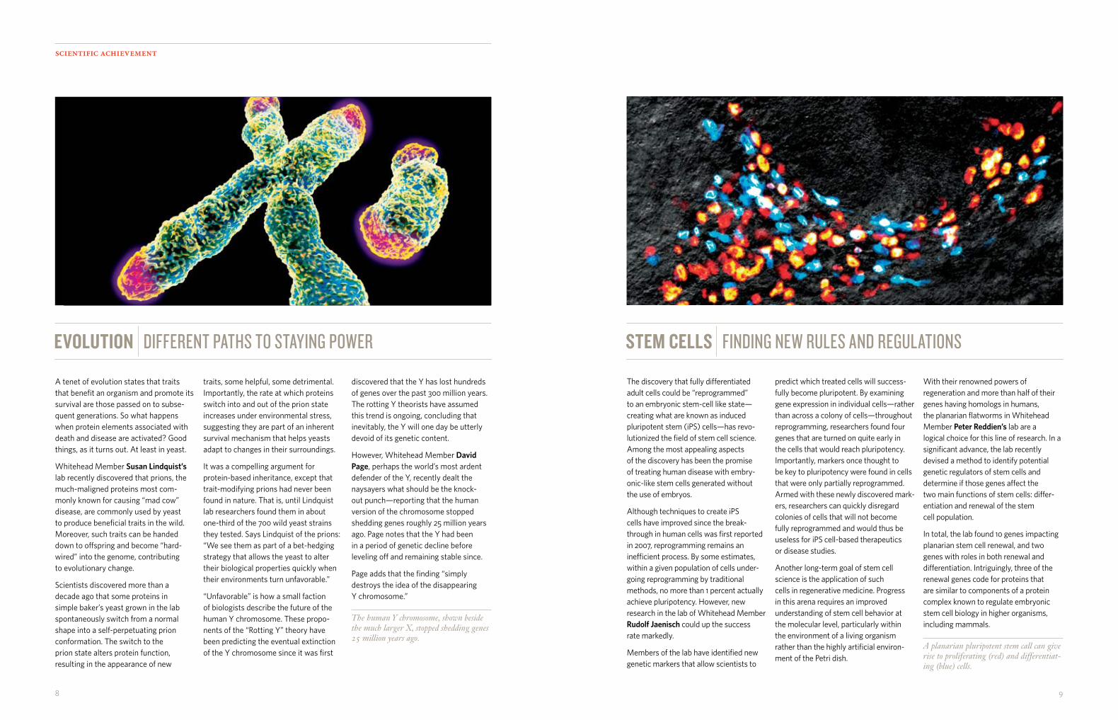
A tenet of evolution states that traits that benefit an organism and promote its survival are those passed on to subse-quent generations. So what happens when protein elements associated with death and disease are activated? Good things, as it turns out. At least in yeast.
Whitehead Member Susan Lindquist’s lab recently discovered that prions, the much-maligned proteins most com-monly known for causing “mad cow” disease, are commonly used by yeast to produce beneficial traits in the wild. Moreover, such traits can be handed down to offspring and become “hard-wired” into the genome, contributing to evolutionary change.
Scientists discovered more than a decade ago that some proteins in simple baker’s yeast grown in the lab spontaneously switch from a normal shape into a self-perpetuating prion conformation. The switch to the prion state alters protein function, resulting in the appearance of new
EVOLUTION different pAths to stAying power
traits, some helpful, some detrimental. Importantly, the rate at which proteins switch into and out of the prion state increases under environmental stress, suggesting they are part of an inherent survival mechanism that helps yeasts adapt to changes in their surroundings.
It was a compelling argument for protein-based inheritance, except that trait-modifying prions had never been found in nature. That is, until Lindquist lab researchers found them in about one-third of the 700 wild yeast strains they tested. Says Lindquist of the prions: “We see them as part of a bet-hedging strategy that allows the yeast to alter their biological properties quickly when their environments turn unfavorable.”
“Unfavorable” is how a small faction of biologists describe the future of the human Y chromosome. These propo-nents of the “Rotting Y” theory have been predicting the eventual extinction of the Y chromosome since it was first
discovered that the Y has lost hundreds of genes over the past 300 million years. The rotting Y theorists have assumed this trend is ongoing, concluding that inevitably, the Y will one day be utterly devoid of its genetic content.
However, Whitehead Member David Page, perhaps the world’s most ardent defender of the Y, recently dealt the naysayers what should be the knock- out punch—reporting that the human version of the chromosome stopped shedding genes roughly 25 million years ago. Page notes that the Y had been in a period of genetic decline before leveling off and remaining stable since.
Page adds that the finding “simply destroys the idea of the disappearing Y chromosome.”
The human Y chromosome, shown beside the much larger X, stopped shedding genes 25 million years ago.

1 110
research stories
What if you discovered that the way an entire field had happily been going about its business for many years was actually wrong? And what if your discovery had the potential to invalidate an enormous volume of work completed using the errant approach you identified?
Scientists in the lab of Whitehead Member Richard Young recently faced these very questions when they realized that assumptions typically employed in the generation and interpretation of data from global gene expression ana- lyses can lead to deeply flawed conclusions about gene activity and cell behavior. Given that global gene expression analysis is employed across a broad range of biological and biomedical research to explore fundamental cellular processes, diagnose disease, support drug development, and even tailor treatment regimens to specific maladies, the impact of the finding could be immense.
“Expression analysis is one of the most commonly used methods in modern biology,” says Young. “So we are con-cerned that flawed assumptions may affect the inter- pretation of many biological studies.”
Much of today’s interpretation of gene expression data relies on the assumption that all cells being analyzed have similar total amounts of messenger RNA (mRNA), the roughly 10 percent of a cell’s RNA that serves as a blueprint for protein synthesis. However, many cells, including aggressive cancer cells, are capable of producing many times more mRNA than other cells. Yet, traditional global gene express- ion analyses have typically ignored such differences.
“We’ve highlighted this common assumption in gene expression analysis that potentially affects many researchers,” says Tony Lee, a Young lab scientist and a corresponding author of a paper published in October 2012 in the journal Cell. “We provided a concrete example of the problem.”
Members of the Young lab uncovered the flaw while inves- tigating genes expressed in cancer cells with high levels of c-Myc, a gene regulator known to be highly expressed in aggressive cancer cells. When comparing cells with high and low c-Myc levels, they were surprised to find very different results when they employed different approaches to gene expression analysis.
Further investigation revealed striking differences in the total amounts of RNA in cells containing high levels of c-Myc and those with low levels of c-Myc, yet these differences were masked by standard experimental and analytical methods.
A FLAW IN THE SYSTEM
This visualization of a large-scale gene expression study forms a clustered, three-dimensional gene expression network. Each dot represents a gene, and links between dots occur where there is correlation between expression patterns. Genes with similar ex-pression profiles link to many other dots and form clusters shown in the same color.
“The different results we saw from different methods of gene expression analysis were shocking, and led us to reinvestigate the whole process on several platforms,” says Jakob Lovén, postdoctoral researcher in Young’s lab and a co-author of the Cell paper. “We then realized that the common assumption that cells contain similar levels of mRNA is wrong and can lead to serious misinterpretations, particularly with cancer cells that can have very different amounts of RNA.”
Knowing that disclosing this remarkable finding might send shockwaves throughout a scientific community that has come to rely so heavily on global gene expression analyses, the lab members were quick to include in their paper a solution to this potentially major problem. Young and his lab report that by using synthetically produced mRNAs, called RNA spike-ins, as standardized controls, researchers can compare experimental data and eliminate assumptions about total cell RNA amounts. The remedy applies to each of the three gene expression analysis platforms they investigated.
Although the researchers believe the use of RNA spike- ins should become the new standard for global gene expression analyses, questions are likely to persist about the interpretations of earlier research.
“There are more than 750,000 expression datasets in public databases, and because they generally lack informa-tion about the cell numbers used in the analysis, it is unclear whether they can be re-examined in order to validate the original interpretation” says David Orlando, another scientist in the Young lab. “It may be necessary to reinvesti-gate some important concepts.”

12 13
research stories
After years of investigating the cellular coping mechanism known as the heat shock response and its relationship to aggressive cancers, the lab of Whitehead Member Susan Lindquist is now deploying its considerable exper- tise to support a clinical trial of an experimental drug in a subpopulation of breast cancer patients.
The heat shock response enables cells to withstand temperature spikes and other stressors. Crammed with distorted and abnormal proteins, malignant cells are deviants that live in a tumor’s distorted landscape—a world lacking abundant nutrients and oxygen. To survive these exceptionally stressful circumstances, many cancer cells have usurped the normally beneficial heat shock response to support their existence. An integral component of this system is heat shock protein 90 (HSP90), which has been shown to help malignant cells accommodate the genetic changes and profound biological disturbances that occur in cancers.
Researchers have hypothesized that inhibiting HSP90 in patients whose tumors are estrogen receptor-positive (ER+) just might render breast cancer cells less resistant to treatment with drugs that prevent the tumors from fueling themselves with estrogen. Such is the rationale for the clinical trial, which began enrolling patients last spring. In the trial, patients with recurrent or metastatic ER+ breast cancer will be treated either with fulvestrant, an estrogen-blocking drug currently marketed by AstraZeneca as Faslodex, or with a combination of fulvestrant and ganetespib, a drug that inhibits HSP90 and is in clinical development by Massachusetts-based Synta Pharmaceuticals.
The trial’s principal investigator is Nancy Lin, the Clinical Director of the Breast Oncology Center at Dana-Farber Cancer Institute and Assistant Professor at Harvard Medical School. Lin is collaborating with Luke Whitesell, an oncolo-gist and senior research scientist in the Lindquist lab.
Breast cancer is the most common cause of cancer-related deaths in women worldwide. An estimated 40,000 women in the United States are expected to die from the disease this year. In the developed world, approximately two-thirds of all breast cancer cases are ER+. Highly dependent on their estrogen receptors for growth and survival, these tumor cells are sensitive to hormonal therapies, making drugs like tamoxifen and fulvestrant effective first-line therapies for many patients.
A HEAT SHOCk pROTEIN ON TRIAL—IN THE CLINIC
However, in patients with breast cancers that recur or metastasize after initial hormonal therapy, long-term remission is uncommon and life expectancy is about three years. Currently, these patients have few therapeutic options. By adding an HSP90 inhibitor, Lin and Whitesell hope to hobble the resistant cancer cells, making them less likely to escape the effects of fulvestrant.
Lin likens a malignant tumor to a car on a highway in a high-speed chase. As the car races down the road, police may erect a roadblock to stop it. In the case of ER+ breast cancer, that roadblock is hormonal therapy. Unfortunately, a tumor may mutate enough to find a detour around the roadblock and continue on its destructive course. Adding a heat shock protein inhibitor to the mix may thwart the cancer’s ability to find an alternate route.
“Because of the way HSP90 inhibitors work, it’s like throwing a bunch of roadblocks across a bunch of detours,” says Lin, who will be running the clinical portion of the trial. “So the idea is to try to block these alternative pathways that might allow the tumor to grow.”
“Inhibiting HSP90 in tumors may not cause them to roll over and die,” says Whitesell, who with support from the Susan G. Komen for the Cure will be analyzing blood and tissue samples from trial participants. “But it may change the biology of the cancer to make it more susceptible to other things that we already know work against the cancer, like fulvestrant.”
Sandro Santagata, a pathologist in the Lindquist lab who will also be examining patient tissue samples, says the lab’s heat shock protein experience will add immeasurably to the trial, regardless of its outcome.
“The Lindquist lab has a great basic scientific understanding of the biology of the heat shock system,” says Santagata. “That foundation will hopefully provide important insights to increase the likelihood of success for this trial and if it doesn’t work, an understanding of why. That’s always a tricky part. Figuring out the ‘why’ is always complicated, so it helps to have people like Susan and Luke who have a lot of background in the system you’re studying.”
The structure of the heat shock protein HSP90. The protein is known to help malignant cells accommodate genetic changes and profound disturbances in normal biology that occur in cancers.

14 15
HONORS AND AWARDS
Mary GehringIn August, Whitehead Member Mary Gehring was selected as a recipient of the Rosalind Franklin Young Investigator Award, which is funded by The Peter and Patricia Gruber Foundation and is administered by a joint committee appoint- ed by the Genetics Society of America and the American Society of Human Genetics. Given every three years to two recipients, this career development research award is “intended to inspire and support new generations of women in the field of genetics.” Candidates are women in the first one to three years of an independent faculty-level position whose work displays “originality and scientific creativity.” The award includes $75,000 in research funding over a three-year period.
Rudolf JaenischThe International Society for Stem Cell Research (ISSCR) named Whitehead Founding Member Rudolf Jaenisch the winner of the 2012 McEwen Award for Innovation. The award, supported by the McEwen Centre for Regenerative Medicine in Toronto, honors original thinking and groundbreaking research pertaining to stem cells or regenerative medicine that opens new avenues of exploration toward the understanding or treatment of human disease or affliction. Jaenisch was cited specifically for “his pioneering discoveries in the areas of genetic and epigenetic control of development in mice that directly impact the future potential of embryonic stem cells and induced pluripotent stem cells for therapeutic utility.” ISSCR President-elect Shinya Yamanaka, recipient of the inaugural McEwen Award in 2011, states: “Dr. Rudolf Jaenisch has always been on the cutting-edge of our field and his research has been a source of inspiration not only for myself, but has influenced the careers of some of our most esteemed colleagues.” Jaenisch received his $100,000 award in June at the ISSCR annual meeting in Yokohama, Japan.
In late October, the Franklin Institute of Philadelphia named Jaenisch one of eight 2013 Laureates—esteemed individuals honored for their pioneering achievements in science, tech- nology, and business leadership. Jaenisch is the recipient of the Benjamin Franklin Medal in Life Science for “discovering heritable controls of gene expression that are independent of the DNA sequence information. These mechanisms affect normal development and diseases, such as cancer, and suggest promising new therapies.” Established in 1824, The Franklin Institute describes its award program as the “oldest and most
comprehensive science and technology honor bestowed in the country and around the world.” Other 2013 Laureates include Subra Suresh, Director of the National Science Foundation, and Michael Dell, Chairman and CEO of computer firm Dell, Inc. The roster of past Laureates includes Albert Einstein, Thomas Edison, Marie Curie, Orville Wright, and Stephen Hawking.
Susan LindquistWhitehead Member Susan Lindquist was awarded the E. B. Wilson Medal, the American Society for Cell Biology’s (ASCB) highest honor. ACSB announced the honor in late April. Since the E.B. Wilson Medal was inaugurated in 1981, it has been given to 47 biologists, including seven Nobel Laureates. According to ASCB Medal Committee Chair Raymond Deshaies, a Professor of Biology at California Institute of Technology and a Howard Hughes Medical Institute investigator, “The committee was deeply impressed by the originality and broad significance of Dr. Lindquist’s research. In particular, the committee noted her elegant studies on the propagation of prions as well as her work on HSP90 as an ‘evolutionary capacitor’ that enables cells to accumulate genetic variation in their genomes without paying a phenotypic cost.”
Terry Orr-WeaverIn late May, the Federation of American Societies for Experimental Biology (FASEB) named Whitehead Member Terry Orr-Weaver the recipient of its 2013 Excellence in Science Award. Established in 1989, the award is given in recognition of outstanding achievement by women in biological science. Recipients are women whose career achievements have contributed significantly to further our understanding of a particular discipline by excellence in research. A FASEB selection committee chose Orr-Weaver from a field of 46 nominees. In presenting Orr-Weaver as its choice, selection committee chair Sally Moody, a professor in the Department of Anatomy and Regenerative Biology at The George Washington University School of Medicine and Health Sciences wrote: “Dr. Orr-Weaver’s scientific research has made her an internationally recognized leader in the field of DNA replication and the cell cycle.” Moody also lauded Orr-Weaver’s “service to the professional community and mentoring of students at all levels.” Orr-Weaver receives a $10,000 unrestricted research grant funded by FASEB. She is the second Whitehead Member to receive the award. Susan Lindquist was so honored in 2009.
Hidde PloeghWhitehead Institute Member Hidde Ploegh was named a recipient of a 2012 National Institutes of Health (NIH) Director’s Pioneer Award, intended to accelerate the pursuit of potentially groundbreaking research. The NIH Director’s Pioneer Award program, established in 2004, supports individual scientists of “exceptional creativity who propose pioneering—and possibly transforming approaches—to major challenges in biomedical and behavioral research.” The Pioneer program is one of three risk-taking funding initiatives of the NIH’s so-called Common Fund. In announcing the awardees, NIH Director Francis Collins stated: “The Common Fund High Risk, High Reward program provides opportunities for innovative investigators in any area of health research to take risks when the potential impact in biomedical and behavioral science is high.”
Ploegh, one of only 10 Pioneer Award recipients nationwide, is receiving up to five years of funding to support his single-domain antibody research. Ploegh plans to leverage unique protein labeling technology developed in his lab to identify the targets of single-domain antibodies in large-scale fashion and to explore how such antibodies could be used to alter intracellular activity and cellular function. He plans to apply the approach in such model organisms as Drosophila and C. elegans.
Gabriel VictoraIn late September, Whitehead Fellow Gabriel Victora was named a recipient of a 2012 National Institutes of Health (NIH) Director’s Early Independence Award, aimed at accelerating the careers of exceptionally creative junior scientists. Modeled after Whitehead Institute’s renowned Fellows Program, the Early Independence Award program was launched in 2011 to support young scientists within one year of having earned their doctoral degrees. The award enables qualified recipients to conduct independent biomedical or behavioral research by skipping the traditional postdoctoral training period. Victora, one of only 14 awardees selected nationwide, is receiving five years of funding from the NIH for his research into the way the cells of the immune system interact to generate high-affin-ity antibodies that confer protection from disease-causing viruses and bacteria.
Robert WeinbergThe Pezcoller Foundation and the American Association for Cancer Research (AACR) named Whitehead Founding Member Robert Weinberg the winner of the 2012 Pezcoller Foundation-AACR International Award for Cancer Research. Established in 1997, this annual award recognizes a scientist:
• Who has made a major scientific discovery in basic cancer research OR who has made significant contributions to translational cancer research;
• Who continues to be active in cancer research and has a record of recent, noteworthy publications; and
• Whose ongoing work holds promise for continued substantive contributions to progress in the field of cancer.
The award includes an unrestricted grant of 75,000 euros.
Richard YoungIn early May, Whitehead Member Richard Young was elected to membership in the National Academy of Sciences in recognition of distinguished and continuing achievements in original research. Election to membership in the Academy is among the highest honors that can be accorded a U.S. scientist or engineer. A pioneer in mapping the circuitry that controls cell differentiation and function, Young joined Whitehead Institute in 1984. With his election, he became the ninth Whitehead Member to hold membership in the National Academy. The others are David Bartel, Gerald Fink, Rudolf Jaenisch, Susan Lindquist, Harvey Lodish, Terry Orr-Weaver, David Page, and Robert Weinberg.
Whitehead InstituteFor the third time in four years, and the second year in a row, Whitehead Institute was named the best place in the United States for postdoctoral researchers to work. The honor was announced in late March by The Scientist magazine, which conducts an annual survey of postdocs at research institutions internationally. In 2012, more than 1,500 postdocs provided their input. In the survey’s 10 years of existence, Whitehead has ranked in the top 15 five times. In addition to its three first-place rankings, Whitehead placed third in 2010 and 14th in 2008.

16 17
if an institution can be measured by the caliber of its faculty, then whitehead institute stands very tall indeed. here, an elite collection of some of the world’s finest scientists conduct groundbreaking research while training and mentoring future generations whose contributions may one day rival their own.
pRINCIpAL INVESTIGATORS
16 17

1918
“we have our sights set on elucidating the mechanisms of microRNA action. we know that microRNAs influence the expression of most human genes, and we’re in a position to more clearly understand how they do this.”
More than one billion years ago, the single-celled ancestors of what we now identify as plants and animals diverged. Since their parting, numerous systems in the two types of organisms evolved differently, but they retained many similarities at the cellular and molecular levels—both have nuclei, replicate DNA, and create proteins using similar mechanisms. In addition, both developed ways to produce offspring by combining eggs and sperm.
Some researchers thought that they had identified another fundamental plant-animal similarity during early develop-ment: messenger RNAs (mRNAs) and proteins inherited by the fertilized egg from its mother initially take command of the embryo, with paternal and embryonic influences exerted in later development. Puzzled by inconsistencies between this conclusion and results from other labs, David Bartel’s lab set out to settle the matter. By studying embryogenesis in the plant Arabidopsis thaliana, they deter-mined that early plant development is indeed different from that of animals—plant embryos are not controlled by inherited maternal molecules, but are steered by genes from both the father and the mother from the time that the embryo is just one or two cells in size.
Working with colleagues, Bartel and his lab also recently defined and analyzed the structure of the yeast version of the Argonaute protein. Scientists had already mapped out the bacterial version of the Argonaute structure, but such a diagram of a eukaryotic structure remained elusive for close to a decade. In the highly conserved process that modulates gene expression, called RNA interference (RNAi), Argonaute binds to microRNAs or other types of very short RNAs. Argonaute then matches the microRNA to mRNAs with complementary sequences and slices the complementary mRNAs, thereby reducing the number of mRNA templates available for protein synthesis. For Bartel, solving the eukaryotic Argonaute structure not only com-pletes a goal of his field, it also will help him and others gain a better understanding of the RNAi process and pro-vide a basis for future experiments.
DAVID bARTEL

20 2 1
“we’ve set our sights on understanding how cells divide. this is a big question in biology with a lot of different aspects. how does it physically happen in cancer cells? in mouse cells? what is the regulatory framework that controls it? how do the kinetochore and spindle orientation work? because we’re guided by the passions and interests of our members, the lab will always move in new and unexpected directions.”
The equal distribution of chromosomes during mitotic cell division fascinates Iain Cheeseman. This delicate dance of paired chromosomes—23 pairs in humans—is carefully choreographed, as a misstep could turn a cell cancerous, kill it, or cause harmful mutations to be transmitted to resulting daughter cells.
Thanks to the Cheeseman lab, we have a better under-standing of how one partner—the microtubule—partici-pates in this dance. Microtubules are thin, hollow protein threads that connect paired chromosomes to one of two spindle poles and align the pairs along the cell’s midline before tearing the pairs apart. The connection between microtubule and chromosome relies on a protein complex called the kinetochore, which grips the end of the microtu-bule. A microtubule generates force by shortening and peeling back strands of its protein thread at one end (pic-ture a peeling banana). Cheeseman’s lab has found that vertebrate kinetochores take a two-pronged approach to microtubule attachment—one kinetochore component, called the Ndc80 complex, latches onto the microtubule above its peeling end, while another component, the Ska1 complex, interacts with both the microtubule’s fraying end and its intact trunk. Together these complexes firmly link
the microtubule to the kinetochore while harnessing the microtubule’s force.
Spindle poles, chromosomes, and microtubules together comprise the “mitotic spindle”. As mitosis progresses, this unit shifts within the cell. How the spindle is placed within the cell defines the size of the resulting daughter cells and their orientation relative to each other, but the signals that control this proper placement were unknown. Through careful observation, Cheeseman’s lab has determined precisely how this phenomenon occurs, solving a 50-year mystery in the process. It turns out that a molecular motor named dynein acts at the cell membrane to pull on micro-tubules (and the entire mitotic spindle) to move it toward the edge of the cell. Researchers found that when a spindle pole comes close to the cell membrane, a signal from the spindle pole knocks dynein off the cell membrane, stopping the spindle pole’s forward motion and sending dynein to the opposite side of the cell. Cycles of behavior elegantly allow the mitotic spindle to find the middle of the cell through a seesawing motion of the spindle poles and chro-mosomes that continues until the spindle and the chromo-somes reside perfectly in the cell’s center.
IAIN CHEESEMAN

2322
“i want to understand how much variation there is within a species. what creates the variation? this is not a simple question and will probably take many years to answer.”
The challenge of personalized medicine is to customize heath care to the individual patient. Rapid and cost effec-tive DNA sequencing has brought that goal much closer to reality, and soon invasive procedures such as amniocente-sis may be replaced by sequencing the DNA that the nascent embryo releases into the mother’s blood.
Whole genome sequencing has made it possible to predict rare diseases such as cystic fibrosis in the embryo. However, the ability to predict the more common heritable diseases based on genome sequencing has proven much more com-plex. For example, even identical twins with exactly the same genomes may differ in their risk for Type 2 diabetes. So, the answer cannot be solely in the genome sequence.
To identify the causes of this complex inheritance, the Fink laboratory has been analyzing the inheritance of complex traits in baker’s yeast. Yeast has a small, easily analyzed genome and can be manipulated readily in the laboratory, making analysis of complex traits more amenable. While studying complex traits in yeast, Fink found a surprising result: Strains with identical genomes had different growth properties. This result was just like that obtained with identical twins. What was going on?
“The difference in growth could not be a result of the chromosomal DNA in the nucleus, so we had to consider other possibilities,” says Fink. Other heritable information, such as viruses and mitochondria (the energy generating organelles), is transmitted through the cytoplasm and is non-nuclear. Fortunately, yeast has a facile way to test for the inheritance of this non-chromosomal information. Fink was surprised to find that the growth of yeast could be dramatically modified by the presence of a double-stranded RNA virus and the mitochondrial genome. In one case a strain was lethal with the virus and viable without it, yet both had the same nuclear genome.
Fink suspects that this non-nuclear information contributes to the inheritance of complex human diseases as well. In fact there is evidence that a complex human disorder, Crohn’s disease, may be determined by the interaction of a virus infection with a mutation in a Crohn’s disease susceptibility gene. In the absence of the virus the disease does not develop. Uncloaking the hidden sources of heritability in yeast may reveal the origins of complex diseases in humans.
GERALD FINk

2 4 25
If the genome is like a genetic cookbook filled with recipes for making all of the cells in a plant or animal, the process known as methylation is like a series of paperclips pinning certain pages together to render those recipes unreadable. Methylation tailors gene expression, allowing every cell in the organism to carry all the recipes, but permitting access only to the instructions each specific cell type needs.
To understand better how this type of tailoring, or gene silencing, operates in plants, Mary Gehring is studying methylation in the model organism Arabidopsis thaliana. In one line of research, her lab is crossing three strains of A. thaliana and examining the resultant offspring to determine which methylated marks are passed on, or “imprinted”. By comparing the results, Gehring intends to identify which genes are consistently imprinted across strains, how much variation the strains have in the inheritance of such silenced genes, and what function these genes have.
In a similar experiment, Gehring and her collaborators are comparing gene imprinting in A. thaliana to that in maize. Maize and A. thaliana diverged approximately 170-200 million years ago (for comparison, humans and mice diverged 75 million years ago), but Gehring has found that the two species have a few imprinted genes in common. These include a family of genes called the polycomb group genes, which in plants and animals code for proteins that are important for silencing the expression of other genes. Some of the other imprinted genes identified in both maize and A. thaliana maintain DNA methylation. The conservation of imprinting of certain genes across eons of evolution hints at the importance of both the genes and the imprinting process.
With the advent of inexpensive genome sequencing, Gehring is contemplating widening the scope of her research by sequencing plants and investigating the effects of imprinting throughout the plant kingdom.
“i want to understand how information—beyond what is encoded in the dna itself—is transferred between generations. how much information is being transferred, and what is the mechanism of that transfer?”
MARY GEHRING

26 27
“we know that there are signaling pathways that normal cells activate only when placed under conditions of stress. many of these same conditions are present in malignant tumors. i am convinced these normal stress pathways play a huge role in enabling cancer cells to resist therapies. we have our sights set on understanding that interplay between normal stress-adaptive pathways, tumor pathobiology, and drug resistance.”
Cancer stem cells (CSCs) differ markedly from the cancer cells that comprise the bulk of a tumor. Among their many malignant properties, CSCs can seed new tumors in envi-ronments hostile to ordinary cancer cells, resurrect old tumors, and even pave their own roadways to travel to distant parts of the body. Like the seemingly unstoppable creatures in many horror films, CSCs also survive nearly everything modern medicine can throw at them—including radiation or chemotherapies.
In an ideal world, cancer therapies could be highly selective by exploiting biological differences between normal and cancerous cells. This seemingly simple goal is, in reality, very difficult to achieve for one simple reason: cancer cells share most of their cellular functions and pathways with normal cells; only a few pathways are selectively corrupted in cancer cells to ensure their uncontrolled growth and survival. Although scientists have identified some path-ways critical only for cancer cells, few such mechanisms have been found in CSCs.
However, Piyush Gupta and his lab recently identified one mechanism crucial for CSCs—a stress response that in nor-mal cells is triggered by nutrient deprivation, low oxygen levels, or large-scale protein production. CSCs depend on this mechanism, called the unfolded protein response (UPR), to provide the proteins necessary for their migration and eventual integration into remote tissues; the UPR also may be linked to the ability of CSCs to resist chemotherapy drugs.
The protein demands of CSCs overtax the cells’ protein production machinery in the endoplasmic reticulum (ER). To eke out more production bandwidth within the ER, CSCs activate the UPR. Gupta has found that CSCs exposed to any additional ER stress either die or halt production of the proteins needed for migration. Gupta hypothesizes that pairing ER stressors with traditional chemotherapies could more effectively eliminate CSC and non-CSC populations within tumors.
pIYUSH GUpTA

28 29
“we now have an important new tool to study polygenic diseases. the technology is such that we could imagine altering syntenic regions of the mouse genome to make mice that are more genetically compatible to human patients, or we could envision growing human es cells with alterations at 20 different loci and create matching isogenic controls. it’s very powerful. we want to do this.”
The creation of mouse models to study human disease has been a mainstay of biomedical research for decades. Such models are vitally important, but as no less an authority than Rudolf Jaenisch—who transformed the field by creat-ing the first transgenic mouse in 1974—will tell you, they’re also problematic.
“The challenge for mouse models is that for some diseases, they don’t work at all,” says Jaenisch.
Generating mouse models has changed little over the years. In simple terms, scientists alter specific genes that have been associated with a given disease and then study the development and course of the disease as well as the effects of various interventions, including genetic and chemical. The whole process, in which scientists insert a piece of DNA into a mouse embryonic stem (ES) cell, inject the modified cell into an early-stage embryo, and then implant the embryo into a foster female mouse, can take years and cost tens of thousands of dollars.
The result is typically a mouse strain with a single copy of a gene “knocked out,” which is fine for the study of so-called monogenic diseases. However, many of our most prevalent, most devastating diseases are multi- genic; that is, associated with mutations in multiple genes. To address this challenge, researchers in Jaenisch’s lab recently employed a novel gene editing technique known as CRISPR to generate mice with as many as five genetic mutations in just a few weeks.
This successful experiment represented the first time that CRISPR (for “clustered regularly interspaced short palin-dromic repeat”) was used to alter multiple genes in a single step. The approach appears to be so efficient and so trac-table that it’s likely to become the gold standard for gen-erating new genetically altered animal models.
Never given to hyperbole, Jaenisch is sufficiently convinced of CRISPR’s potential to advance disease modeling that he now refers to it simply as “a game changer.”
RUDOLF JAENISCH

30 3 1
“using yeast genetics to identify a compound and its mechanism of action against the fundamental pathology of a disease illustrates the power of the system we’ve built. despite the prevalence of these horrible neurodegenerative diseases, not one disease-modifying drug exists. we’ve set our sights on changing that.”
Neurodegenerative disorders such as Huntington’s disease, Parkinson’s disease, and Alzheimer’s disease are character-ized by protein misfolding, resulting in toxic accumulations of proteins in the cells of the central nervous system. Over the years, Susan Lindquist and her lab have developed and refined a remarkable research platform in which common yeast cells become living test tubes in which to study pro-tein folding, function, trafficking, and aggregation in models of these diseases.
The approach is a hallmark of Lindquist’s research—using the simple, in this case yeast cells, to unravel the complex—and it’s begun to bear fruit, most recently in the hunt for therapeutic strategies against Parkinson’s disease. Through genetic modification, the lab has created yeast cells that overproduce the protein alpha-synuclein, whose accumula-tion is associated with Parkinson’s. In turn, the lab has conducted massive chemical screens for compounds capable of rescuing these sick cells from the pernicious effects of alpha-synuclein.
A screen of nearly 200,000 compounds identified one chemical entity that not only reversed alpha-synuclein toxicity in yeast cells, but also partially rescued neurons
in the model nematode C. elegans and in rat neurons. Significantly, the cellular pathologies observed in yeast mirrored those seen in the higher organisms and were reduced by treatment with the identified compound. Naturally, these findings raise an important question: Might they apply to humans?
In follow on research that goes a long way toward providing the answer, scientists examined neurons derived from induced pluripotent stem (iPS) cells generated from Parkinson’s patients. The cells and differentiated neurons (of a type damaged by the disease) carried an alpha-synu-clein mutation akin to those studied in the yeast, worm, and rat models. To ensure that any pathology developed in the cultured neurons could be attributed solely to the genetic defect, the researchers also derived control neurons from iPS cells in which the mutation had been corrected.
As the patient-derived neurons aged in culture, they manifested worsening dysfunction in a number of cellu- lar processes associated with alpha-synuclein toxicity. Remarkably, exposure to the compound identified via yeast screens reversed the damage in these neurons.
SUSAN LINDQUIST

32 33
“our sights continue to be set on understanding in great detail all the steps required to make red blood cells and to use these findings to treat diseases and to genetically modify red blood cells for a variety of practical applications.”
Having researched red blood cells for the past 55 years, shouldn’t Harvey Lodish be close to exhausting the subject by now? The fact that he’s not speaks volumes, about both the complexity of these cells and their importance to medicine.
One long-term focus of his lab is erythropoiesis, the pro-duction of red blood cells (RBCs), and the problems that arise—including multiple forms of anemia—when such production falls short. Several years ago, the lab purified an RBC progenitor cell known as a burst-forming unit erythroid (BFU-E), showing that it can be triggered to self-renew under conditions of stress, such as traumatic blood loss or by treatment with glucocorticoids, which are used to treat certain anemias. BFU-E self-renewal eventually leads to a roughly 60-fold increase in the number of RBCs formed from each BFU-E.
While a comprehensive understanding of the mechanisms regulating self-renewal has been elusive, the Lodish lab recently identified an RNA binding protein that is essen-tial for BFU-E self-renewal. This protein, Zfp36l2, induces degradation of messenger RNAs needed for red cell
differentiation, thus keeping these BFU-E cells in a state that allows them to produce increased numbers of red cells over time. Zfp36l2 is a potential therapeutic target to bol-ster erythropoiesis via BFU-E self-renewal.
In other research that has captured the attention of the U.S. military, the Lodish lab is genetically modifying RBCs to bind to and eliminate toxins or pathogens and possibly serve as vaccines or imaging agents. The approach takes advantage of the fact that RBCs have no nuclei or DNA, meaning that altered cells cannot form tumors.
The lab has introduced genes coding for specific surface proteins into early-stage RBC progenitors. As the RBCs approach maturity and jettison their nuclei, these proteins remain on the cell surface. Leveraging protein-tagging technology developed by colleague Hidde Ploegh, they are linking anti-toxin antibodies and several small-molecule therapeutics to these modified surface proteins to treat illness or neutralize the activity of a biological weapon. This research is currently funded by DARPA, the federal govern-ment’s Defense Advanced Research Projects Agency.
HARVEY LODISH

34 35
“we know that the regions of dna that are fragile during replication are likely fragile because of their chromatin structure and because of events occurring directly at the replication fork. we’re now in the position to pursue these two pathways to delineate mechanistically why these regions are so difficult to replicate.”
The genetic landscape within a cancer cell is pretty grim. In the process of acquiring immortality and the ability to invade the rest of the body, the genomes of cancer cells accrue multiple mutations. In fact, whole regions may be misplaced, replicated numerous times, or go missing alto-gether. Although such changes are well documented, little is known about what actually sends cancer cells down their road to ruin.
One theory suggests that genomes have fragile sections—regions of DNA through which the machinery charged with replication cannot pass. This leads to changes in DNA copy number and rearrangement of the order of the genome.
Using the fruit fly as a model organism, Terry Orr-Weaver and her lab delve into how DNA replication works, or doesn’t, in cells. They exploit the fact that the same replica-tion machinery is used in fruit fly and human cells and that fruit flies can develop metastatic tumors. Orr-Weaver lik-ens the process of DNA replication to a train chugging down railroad tracks. Usually, everything runs smoothly,
and the train arrives at its intended destination. But under certain stressful circumstances, the train derails. In the case of cells, derailment of the replication machinery can lead to the DNA damage found in cancer cells.
Thus far, Orr-Weaver has identified 30 sites in the fruit fly genome where DNA replication is apt to become stalled. By studying these sites, Orr-Weaver has determined that replication is hampered by molecular barriers lodged on the DNA track. She says she was surprised to find that in addition, an “engineer” on the replication machinery some-times appears to pull back on the throttle to slow or stall the machinery as it travels down the DNA track. Orr-Weaver is now investigating how the barricades and the braking engineer are regulated, whether these obstacles to replication do indeed lead to breaks in the DNA, and how these breaks can cause the genetic rearrangements seen in cancer cells.
TERRY ORR-WEAVER
36 37
After three decades spent studying mammalian sex chro-mosomes, David Page is poised to move his research into unexplored territory by marrying the precision of a pains-taking genetic sequencing methodology developed years ago in his laboratory with today’s remarkable high-through-put technologies.
More than 10 years ago, Page reported that the human Y chromosome comprises several regions of large palin-dromes—areas of mirror-imaged genetic sequences. Such regions render conventional sequencing approaches inca-pable of detecting extremely subtle genetic differences found hidden within what he describes as “halls of mirrors.” In response, Page and colleagues developed an approach known as SHIMS (single-haplotype iterative mapping and sequencing) to establish a definitive reference DNA sequence of the Y chromosome.
Page contends that without SHIMS, and the accuracy of its resulting reference sequence, his lab’s many contributions to our understanding of human reproductive biology—including the recent discovery of the rate at which two spontaneously recurring genetic deletions on the Y cause male infertility—would not have been possible. However, he also argues that the exceptional quality of the Y reference
sequence is lacking in the reference sequence of the human genome, in part because the kind of structural complexity characteristic of the Y chromosome exists in multiple regions of the genome. Thus, he believes the sequence of the human genome scientists rely on today is replete with gaps and errors.
To rectify this, Page is experimenting with redeploying SHIMS in conjunction with a state-of-the-art sequencer in his own lab to, as he puts it, “reinvent the notion of a genome center.”
“Today, one sequencing machine can essentially replace an entire genome center,” Page notes. “But the ability to gener-ate a high-quality reference sequence is almost a lost art.”
Given his research focus, it’s not surprising that he’s begin-ning this work by producing an updated reference sequence of the human X chromosome, allowing for systematic study and cross-species comparisons of the X’s gene content over time. However, with a little scale, he may consider filling in the blanks genome wide.
“an ultra-high quality reference sequence of the human genome simply doesn’t exist. we’re setting our sights on establishing a critical mass that will enable us to improve the reference sequence, both for the human genome and for key model organisms.”
DAVID pAGE

38 39
“we’re setting our sights on the generation of a new toolbox that allows us to interfere with protein function in living cells, without alterations in the host cell proteome per se. we intend to do this through application of camelid-derived single antibodies. these proteins have highly desirable properties that should allow us to make good progress towards this goal.”
Throughout his career-long quest to understand all the dynamics of the immune system’s response to invading pathogens, Hidde Ploegh has engineered a number of novel tools and methods to advance his cause. His latest tech-nique, however, just may be the most unexpected.
A portion of Ploegh’s lab is currently immersed in research with antibodies derived from, of all things, a group of ador-able, fuzzy alpacas. The science leverages one remarkable aspect of the alpaca immune system. It turns out that alpacas are capable of generating two kinds of antibodies: the traditional two-chain (heavy and light chains) anti- bodies found in mammals and vertebrates, and smaller, single-chain antibodies (heavy chain only). These unique antibodies, commonly referred to as VHHs, are produced by all members of the camelid family, including llamas, Bactrian camels, dromedaries, and, of course, alpacas.
While attending a lecture on VHH antibodies a few years ago, Ploegh realized that their small size—roughly 1/10 that of normal antibodies—and high thermal stability make VHHs ideal for targeting antigens residing inside cells of interest. Moreover, he recognized that VHHs are amenable
to modification via “sortagging,” a highly specific protein labeling technique developed in his lab, to track their activity within cells and to identify which antigen a single VHH binds.
To put it in simple terms, the current research plan is as follows: immunize alpacas with selected cellular material, have them generate VHHs in response, and use the VHH antibodies to probe the inner workings of cells with here-tofore unachievable precision. The project is still in its early stages, but the principle is being proven.
“We’ve been able to immunize the alpacas with complex mixtures and then here in the lab determine what the antibodies are binding to,” says Ploegh lab postdoctoral researcher Juanjo Cragnolini. “This really does allow us to engineer the smaller antibodies we need. It works so beau-tifully, it’s pretty unbelievable.”
Beyond advancing our understanding of intracellular activi-ties, these camelid antibodies are expected to have applica-tions in vaccine and therapeutic development.
HIDDE pLOEGH

40 41
“i’d like to identify the key concepts that explain how animals can regenerate from any type of wound. we’re getting some of these concepts, but we now need to explain these concepts with molecular mechanisms and understand how, together, these mechanisms bring about regeneration.”
The focus of Peter Reddien’s lab is planarians—tiny flat-worms renowned for their ability to regenerate any missing body part. Although scientists have been studying planar-ians for more than 100 years, progress has been hampered by the lack of a comprehensive genetic toolkit of the variety that has fueled research in mice, fruit flies, and other model organisms.
To rectify this imbalance, Reddien and his lab have spent the past several years developing new planarian-specific tools and reagents. The hard work has begun to pay off. Armed with their newest implements, Reddien and his lab recently set out to identify as many genes involved in regenerative processes as possible. The result was three collections of genes that play roles in wound response, eye regeneration, and regulation of planarian pluripotent stem cells.
The lab found 94 genes whose expression is affected by wounding. It is the first such catalog of genes active in this crucial initial step of regeneration. As part of the research, scientists were able to determine the function of certain
genes, including runt-1, which belongs to a family of genes also found in humans and other animals. In planarian stem cells, runt-1 is expressed at fairly low, constant levels. Its expression increases immediately after wounding but before the stem cells differentiate into specific, dedicated cell types. runt-1 appears to be particularly important in the regeneration of eyes and certain neurons, and the Reddien lab is trying to gauge its significance in the regen-eration of other cell types as well.
For Reddien, this work merely scratches the surface of what can be achieved with the three gene inventories. By systematically manipulating the genes on the lists and understanding how they impart distinct attributes to vari-ous cell types, he hopes to learn more about these major processes and establish the planarian as the preeminent model system for the study of regeneration.
pETER REDDIEN

4 2 43
“we believe we can take a comprehensive approach to identifying mechanistic links among metabolic processes, cancer-causing genetic mutations, and cell signaling pathways that could aid therapeutic development. we’ve set our sights on building a dedicated research facility to take this on.”
Of the many components an organism requires to ensure proper development, growth, and good health, nutrients would certainly be categorized as essential. It sounds simple enough, but the way an organism senses and uses those nutrients is remarkably complex, tightly regulated, and under intense scrutiny in David Sabatini’s lab.
The lab has been mapping the intricate signaling pathways that control cellular and organismal metabolism and how their deregulation can affect the aging process and such human diseases as diabetes and cancer. Among the phe-nomena being studied is caloric restriction (CR), which has been shown in animals to prolong lifespan while improving overall health, in part by improving insulin sensitivity and glucose tolerance, both of which decline with age.
Given the benefits of CR, scientists have been exploring the possibility of developing drugs that could mimic its effects. Intriguingly, the immunosuppressant rapamycin appears to do just that, albeit at a price. It turns out that rapamycin also impairs glucose tolerance and insulin sensitivity, two hallmarks of diabetes. So what’s rapamycin doing that CR is not?
Enter Sabatini, a pioneer in the study of the key growth regulatory pathway known as mechanistic target of rapamycin, or mTOR. Several years ago, the lab identified two major protein complexes within the mTOR pathway—mTORC 1 and mTORC2—and it had been thought that rapamycin was exerting its effects by inhibiting mTORC1. However, the lab recently discovered that rapamycin inhib-its both complexes. Moreover, scientists found that mTORC1 inhibition leads to prolonged lifespan, while disruption of mTORC2 adversely affects insulin sensitivity. This surpris-ing finding suggests that targeted inhibition of mTORC1 could lead us down a path toward age-defying therapies.
Because CR has also been shown to reduce the incidence of cancer, and because metabolism in cancer cells differs dramatically from that in normal cells, a portion of the Sabatini lab is increasingly focused on studying the many ways tumors are able to alter their energy production and use to ensure their own survival.
DAVID SAbATINI
4 4 45
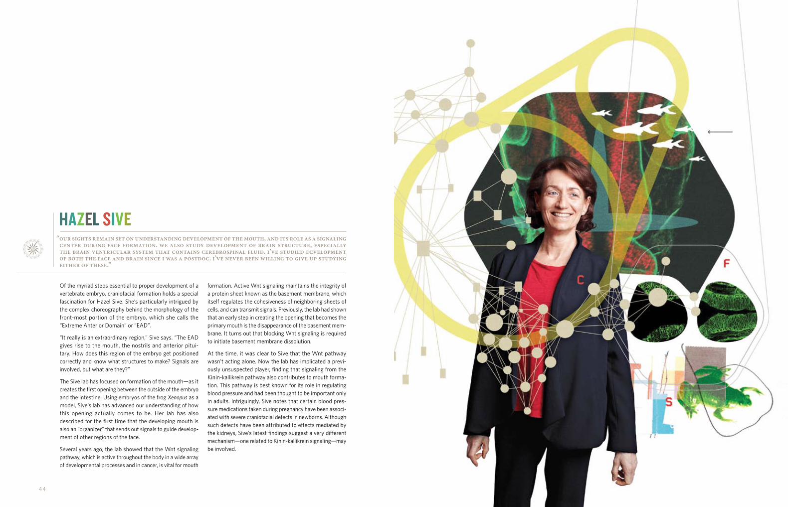
“our sights remain set on understanding development of the mouth, and its role as a signaling center during face formation. we also study development of brain structure, especially the brain ventricular system that contains cerebrospinal fluid. i’ve studied development of both the face and brain since i was a postdoc. i’ve never been willing to give up studying either of these.”
Of the myriad steps essential to proper development of a vertebrate embryo, craniofacial formation holds a special fascination for Hazel Sive. She’s particularly intrigued by the complex choreography behind the morphology of the front-most portion of the embryo, which she calls the “Extreme Anterior Domain” or “EAD”.
“It really is an extraordinary region,” Sive says. “The EAD gives rise to the mouth, the nostrils and anterior pitui- tary. How does this region of the embryo get positioned correctly and know what structures to make? Signals are involved, but what are they?”
The Sive lab has focused on formation of the mouth—as it creates the first opening between the outside of the embryo and the intestine. Using embryos of the frog Xenopus as a model, Sive’s lab has advanced our understanding of how this opening actually comes to be. Her lab has also described for the first time that the developing mouth is also an “organizer” that sends out signals to guide develop-ment of other regions of the face.
Several years ago, the lab showed that the Wnt signaling pathway, which is active throughout the body in a wide array of developmental processes and in cancer, is vital for mouth
formation. Active Wnt signaling maintains the integrity of a protein sheet known as the basement membrane, which itself regulates the cohesiveness of neighboring sheets of cells, and can transmit signals. Previously, the lab had shown that an early step in creating the opening that becomes the primary mouth is the disappearance of the basement mem-brane. It turns out that blocking Wnt signaling is required to initiate basement membrane dissolution.
At the time, it was clear to Sive that the Wnt pathway wasn’t acting alone. Now the lab has implicated a previ-ously unsuspected player, finding that signaling from the Kinin-kallikrein pathway also contributes to mouth forma-tion. This pathway is best known for its role in regulating blood pressure and had been thought to be important only in adults. Intriguingly, Sive notes that certain blood pres-sure medications taken during pregnancy have been associ-ated with severe craniofacial defects in newborns. Although such defects have been attributed to effects mediated by the kidneys, Sive’s latest findings suggest a very different mechanism—one related to Kinin-kallikrein signaling—may be involved.
HAZEL SIVE

46 47
“master regulators of metastasis are the controllers of life and death. we’re looking at the effects that they have on the biological functions of individual cells and what signals impinge on a cell to make it become epithelial or mesenchymal.”
For the past several years, Robert Weinberg has focused much of his lab on a single adversary: the cancer stem cells capable of seeding new tumors and driving metastasis.
“We’re finally figuring out what’s going on with cancer stem cells—how they’re maintained and how they operate,” he says. Unlocking these mysteries may point to new ways of vanquishing cancer stem cells (CSCs) and preventing the hundreds of thousands of deaths they cause each year.
Weinberg was the first to establish that these insidious cells are created during a process called an epithelial-mesenchymal transition (EMT) and the first to identify the genes that initiate this transformation. An EMT confers on CSCs the seeming superpowers to travel anywhere in the body, survive in foreign, hostile environments, and dodge many current radiation and chemotherapies.
By learning what makes CSCs tick, Weinberg has identified potential weaknesses that could be exploited by drugs. It turns out that CSCs actually exist in a surprisingly tenuous state, and if their equilibrium is upset by interrupting cer-tain signals within the cells or by pushing the cells to dif-ferentiate (mature), the resulting cells can become vulnerable to drugs.
Weinberg and his lab are also studying small cell surface membrane projections, called filopodia, which CSCs use to move around and anchor themselves in tissues before spawning new tumors. These tiny feet, which Weinberg has determined are another product of the EMT, are critical to the ongoing proliferation of cancer cells. In theory, ham-pering filopodia formation should make a primary tumor unable to metastasize.
As it is, tumors have various capacities for seeding new tumors and modulating levels of aggressiveness. In breast cancer, for example basal carcinomas are highly aggressive, whereas luminal carcinomas are considerably less so. A master gene regulator of the EMT process, called Zeb1, is active in basal carcinomas and other aggressive cancers, but inactive in non-aggressive tumors. The lab recently found that a key controller of Zeb1 may ultimately deter-mine a cancer’s aggressiveness. A drug that forces the master controller to turn Zeb1 off could help tame more aggressive cancers.
RObERT WEINbERG

48 49
“we’re setting our sights on identifying all of the super-enhancers, because if you look across genome-association studies, you find disease-related mutations occurring in super-enhancers. cancer is a good place to start, but ultimately we’ll go after all the major diseases.”
Our genes make us the unique individuals we are. We can’t argue with that. Or can we?
Well, Richard Young will tell you that things just aren’t that simple. In fact, Young says that only about 5% of the variety of distinguishing traits among us is attributable to our protein-coding genes. But if that’s the case, what else is happening? It’s a question Young and his lab are coming ever closer to answering definitively.
“Most of what determines susceptibility to disease, and perhaps many other differences among individuals such as appearance and behavior, lies in regulatory regions of the genome,” Young says. “That’s what we study.”
The lab recently discovered a relatively small set of power-ful gene regulators that control cell state and identity. It turns out that in healthy cells, these “super-enhancers”, as the lab calls them, control genes responsible for cellular functions and developmental transitions—such as that from embryonic stem cell to nerve cell. Cancer cells, how-ever, manage to assemble their own insidious super-enhancers to overproduce harmful oncogenes that lead to aggressive tumors.
Although the complexity of the human cellular control system is daunting—to date more than one million regula-tory elements affecting the expression of tens of thousands of genes have been identified—Young’s super-enhancer paradigm introduces a surprising degree of simplicity. It now appears that just a few hundred of these special regu-lators control most of the key genes that give each cell its unique properties and functions.
In a related finding, the lab has discovered that despite their potency, super-enhancers are particularly sensitive to changes in their environment. Having observed cancer cells’ ability to develop their own enhancers to the detri-ment of normal cellular function, Young and colleagues theorized that the super-enhancers themselves might be vulnerable to disruption, thereby serving as therapeutic targets. Thus far, a number of experimental therapies effec-tive against multiple myeloma and several other types of cancer have been shown to act on super-enhancers con-trolling known oncogenes.
RICHARD YOUNG

50 5 1
while there’s no single formula for creating impactful scientific careers, the whitehead fellows program offers a virtually foolproof recipe: take a handful of the world’s most promising young scientists, add research support, remove faculty responsibilities such as teaching, combine these ingredients in an independent lab, and share when properly seasoned. the final product rarely disappoints.
WHITEHEAD FELLOWS
50 5 1

5 2 53
As the Andria and Paul Heafy Fellow of Whitehead Institute, Yaniv Erlich arrived at the Institute determined to develop novel tools and methods for studying human genomics. Nearly three years into his tenure, he’s making good on that promise.
Most recently, his lab created an algorithm to analyze so-called short tandem repeats (STRs)—collections of repeated two to six nucleotide-long sequences (e.g., CTGCTGCTG) that are distributed throughout the genome. Because the number of repeats in STRs can mutate quickly, each person’s set of these genetic markers is different from every other person’s, making STRs ideal for creating a unique DNA fingerprint. That characteristic made STR
analysis useful in forensic endeavors, including crime scene investigations, during the late 1990s and early 2000s.
The advent of high-throughput sequencing technolo- gies, however, rendered work with STRs obsolete. That is until Erlich unveiled a program called lobSTR, a three-step system that accurately and simultaneously profiles more than 100,000 STRs from a human genome sequence in a single day.
Says Erlich: “With this tool, we provide access to tens of thousands of quickly changing markers that you couldn’t get before, and those can be used in medical genetics, population genetics, and forensics.”
YANIV ERLICH
Sebastian Lourido believes parasites just don’t get the attention they deserve. In fact, he says parasites are “understudied,” at least at the molecular level.
Accordingly, he plans to change that, beginning with a focus on single-celled parasites known as apicomplexans, a family that includes Plasmodium, which causes malaria, and Toxoplasma gondii, the pathogen behind the infection toxoplasmosis. Apicomplexans transition through a variety of stages during their lives—between replication within host cells and migration, and between different develop-mental stages—a fact that makes them all the more appeal-ing to Lourido.
“I’m fascinated by how many different fields have to come together to study one of these parasites,” he says. “It requires genetics, epidemiology, cell biology, and biochemistry.”
Lourido is currently investigating the roles a family of enzymes known as calcium-dependent protein kinases (CDPK) play in the lifecycle of Toxoplasma.
“Toxoplasma is unique, and it’s wildly successful,” Lourido says. “Roughly 25% of the world’s population is infected with it, and yet the majority of those infected are asymptomatic. Understanding Toxoplasma can tell us a lot about how para-sites subvert the immune system and could contribute to our understanding of malaria and other apicomplexans.”
SEbASTIAN LOURIDO
5 4 55
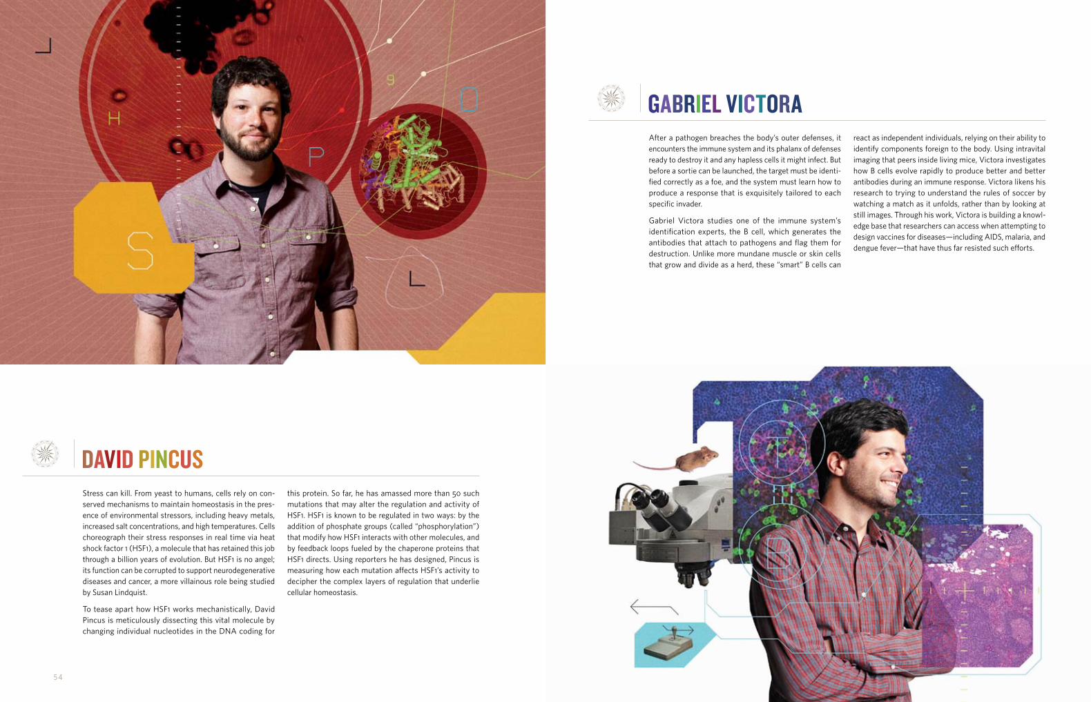
Stress can kill. From yeast to humans, cells rely on con-served mechanisms to maintain homeostasis in the pres-ence of environmental stressors, including heavy metals, increased salt concentrations, and high temperatures. Cells choreograph their stress responses in real time via heat shock factor 1 (HSF1), a molecule that has retained this job through a billion years of evolution. But HSF1 is no angel; its function can be corrupted to support neurodegenerative diseases and cancer, a more villainous role being studied by Susan Lindquist.
To tease apart how HSF1 works mechanistically, David Pincus is meticulously dissecting this vital molecule by changing individual nucleotides in the DNA coding for
this protein. So far, he has amassed more than 50 such mutations that may alter the regulation and activity of HSF1. HSF1 is known to be regulated in two ways: by the addition of phosphate groups (called “phosphorylation”) that modify how HSF1 interacts with other molecules, and by feedback loops fueled by the chaperone proteins that HSF1 directs. Using reporters he has designed, Pincus is measuring how each mutation affects HSF1’s activity to decipher the complex layers of regulation that underlie cellular homeostasis.
DAVID pINCUS
After a pathogen breaches the body’s outer defenses, it encounters the immune system and its phalanx of defenses ready to destroy it and any hapless cells it might infect. But before a sortie can be launched, the target must be identi-fied correctly as a foe, and the system must learn how to produce a response that is exquisitely tailored to each specific invader.
Gabriel Victora studies one of the immune system’s identification experts, the B cell, which generates the antibodies that attach to pathogens and flag them for destruction. Unlike more mundane muscle or skin cells that grow and divide as a herd, these “smart” B cells can
react as independent individuals, relying on their ability to identify components foreign to the body. Using intravital imaging that peers inside living mice, Victora investigates how B cells evolve rapidly to produce better and better antibodies during an immune response. Victora likens his research to trying to understand the rules of soccer by watching a match as it unfolds, rather than by looking at still images. Through his work, Victora is building a knowl-edge base that researchers can access when attempting to design vaccines for diseases—including AIDS, malaria, and dengue fever—that have thus far resisted such efforts.
GAbRIEL VICTORA

xxxxxxxxxxxx xxxxxxxxxxxx
community evolution
56 57
William F. Pounds (left), Dean Emeritus at the MIT Sloan School of Management, joined the Whitehead Board of Directors in June 2012. Whitehead Institute mourned the February passing of John Pratt, who ran the Institute’s administration for 25 years.
More than 100 high school students from across Massachusetts attended the Whitehead Spring Lecture Series, “Bridges to Bioengineering,” held during the public school vacation week in April.
Board NewsIn June, William F. Pounds was elected to the Whitehead Institute Board of Directors. Bill, as he’s known to his colleagues, is Professor Emeritus of Management and Dean Emeritus at the MIT Sloan School of Management. An expert on operations management and quantitative modeling of decision behavior, Bill was instrumental in developing the operations management curriculum at the Sloan School, where he served as Dean from 1966 to 1981. From 1981 to 1991, he was the senior advisor to the Rockefeller family. A familiar figure on the boards of a number of corporations and nonprofit organizations, Pounds holds a BS in chem- ical engineering from Carnegie Institute of Technology along with an MS in mathematical economics and a PhD from the Graduate School of Industrial Administration.
Institute NewsIn February 2012, the Whitehead Institute community was profoundly saddened by the passing of John Pratt, the Institute’s former Associate Director and a central figure in establishing what would become one of the world’s preeminent biomedical research institutions.
Whitehead Founding Director David Baltimore hired John in 1981 to run the administration of a nascent organization. Upon learning of John’s death, David stated: “John was at the heart of the development of Whitehead Institute. One of the luckiest things that ever happened to me was that I
Public OutreachWhitehead Institute has maintained a steadfast commit- ment to science education and outreach by offering programs meant to enhance science teaching and learning for the entire community. Ranging in scope from lectures and workshops for teachers and students to special events for non-scientists, Whitehead’s public programming has been showcasing cutting-edge biomedical research while introducing the Institute and its scientific prowess to new audiences for more than two decades.
During the 2011-2012 academic year, Whitehead’s Seminar Series for High School Teachers brought nearly 80 teachers to the Institute each month for lectures and working dinners with colleagues and Whitehead scientists. This year’s series, Advances in Cancer Biology and Therapeutics, explored how novel research and development efforts are changing the way we as a society think about cancer now and how we might address it in the future. Individual lecture topics included cancer metastasis, cancer vaccines, and the development of nanotechnology-based cancer therapeutics.
In April, more than 100 students attended the 2012 edition of Whitehead’s Spring Lecture Series for High School Students. The three-day program, Bridges to Bioengineering, combined expert lectures, laboratory demonstrations, informal lunchtime conversations, and a panel discussion to
tracking the comings, goings, and interactions that mark a year in the life of an institution that is never at rest...
was introduced to him. John was just terrific, and I needed him so badly because in my own background, I had never managed anything more than my own laboratory. He was so solid and had such strength. He taught me a huge lesson that I share with those who will listen: that filling that position (the head of administration) is the most important thing a new director can do.”
John would go on to dedicate himself to the Institute for 25 years, during which he collaborated not only with David Baltimore, but also with David’s successors in Directors Gerald Fink and Susan Lindquist. Along the way, he became a close friend, confidante, and mentor to a number of Whitehead faculty and administrators.
“As Director now, when I reflect—as I often do—on Whitehead’s remarkable camaraderie, collaborative spirit, its collegiality, and the regard we have for integrity as a defining virtue, I think of John,” said current Director David Page. “His words and his actions were consistently driven by his ideals, and they here high. When we think about Whitehead Institute and its commitment to excellence, we often speak of the key parts played by Jack Whitehead and David Baltimore, but it’s pretty clear there was a third person in the mix, a person who was executing on their vision and putting flesh on the bone. That person was John Pratt.”
introduce scientifically-inclined students to the myriad ways leaders in the fields of biology, engineering, and medicine are joining forces in the pursuit of breakthroughs to improve human health and welfare.
For the second straight year, Whitehead Institute participated in the Annual Massachusetts Statewide Biotechnology Job Shadow Day, sponsored by the Massachusetts Biotechnology Council (MassBio) and the Massachusetts Biotechnology Council Education Foundation (MassBioEd). Whitehead was one of 14 Massachusetts-based research institutions and biotechnology companies to spend an early June day helping to educate more than 500 Massachusetts students from 15 different high schools about potential careers in life sciences.
Based on the success of the program’s 2011 debut, the Institute reprised A Girl’s Guide to Understanding Life Science, which is supported by Whitehead Member Susan Lindquist. Held at the close of the school year, the program introduces 7th and 8th grade girls to the world of scientific exploration. This year’s participants learned how to fashion DNA molecules, first with specially designed LEGO blocks and later with a 3-D software program. Whitehead researchers also showed the girls how to extract DNA from strawberries and led them through an experiment that taught them about the science of epidemiology.

xxxxxxxxxxxx xxxxxxxxxxxx
58 59
HONOR ROLL OF DONORSmember’s circle $2,500 and above
Anonymous (2)
Lorraine and Emanuel Balkin
David and Arlene Brook
Jenifer Schweitzer Brooks
George and Judith Carmany
Neil and Martha Chayet
Mimmy and Phil Cooper
Michael and Barbara Eisenson
Garey V. Ellis and Tomora-Lutreast Ellis
Peter Feinstein and Miriam Weinstein
John Fletcher
Kenneth S. Fong
Mr. and Mrs. James W. Fordyce
G. S. Beckwith and Katharine Gilbert
Karl Handelsman
Donald and Margery Hetzel
Rob Hoyt and Molly King
Mr. and Mrs. Thomas Campbell Jackson
Robert and Anne James
Al Jones and Larissa Lezama
Paul and Barbara Joskow
Luciano Lauretti and Jane Roy
Ann R. Leven
Earl and Barbara Lewis
Brigitte D. Linz
Gordon N. Litwin and Anne Luzzatto
Mr. and Mrs. George McCabe
John C. McCormick
Fred Meyer
Skip and Lyerka Miller
Linda Mirels, Wendy Fisher and Philip Kirsh
Franny Mitchell
Michelle Morphew
Martin A. Mullins
Suzanne and Norman* Priebatsch
Marcia J. Radosevich, PhD
Joy Gail Raywid
Philip R. and Nancy L. Reilly
Alison Andrews Reyes and Carlos Reyes
Api A. Rudich
Nick Ryan
Roger Sametz, Sametz Blackstone Associates, Inc.
Alison Sander
David Schechter
Ruth Scheuer, DrPH, JD and James Scheuer, MD
Arthur and Yvette Schleifer
Samantha and Gabriel Schlumberger
Stephen Seiler and Bessie Sze Seiler
Parag Shah
Barry and Donna Slotnick
Mr. and Mrs. R. Gregg Stone
Peter and Laurie Thomsen
Mrs. Jeptha H. Wade
Robert and Mary Wright
friend’s circle $1,000 and above
Alchemy Foundation
E. S. Anderson
Thomas and Elizabeth Armstrong
Philip and Eve Cutter through the Goldhirsh Foundation
Dr. and Mrs. George Hatsopoulos
Allan and Joan Kleinman
Jerome and Judith Koch
Frank and Amy Linde
Alexandra and Matthew Murray
Lois Peerce
Jerome and Doreen Porush
Nancy L. Rose and James M. Poterba
Margaret Sand
L. Dennis and Susan R. Shapiro
Eric Summers and Liz Zobel
Mr. and Mrs. Paul Tramontano
Pamela Wasserstein
Arnold and Inge Weinberg
Robert Weinberg and Amy Shulman Weinberg
supporters Other Gifts
Barry and Erin Aprison
Jacob F. Brown, II
Adelaide de Menil
Mary Claire Delaney
William Dietrich and Rebecca Mosher
Donald and Nancy Dougherty
David and Marion Ellis
Jack and Adrienne Erickson
Julian and Judith Jadow
Douglas Kahn
Sara Kaul
Johanna L. Klein
Murph Levin
Roz Lichter
Linda F. Lynn
Larry and Sara McReynolds
Herbert and Sydelle Ornstein
Catherine Kuehn Price
Princeton University Class of 1970
Mr. and Mrs. Edward W. Probert
Ed Ross
Ron and Betsey Scharlack
Scheuer Associates Foundation, Inc.
Charles N. Schlangen
Mr. and Mrs. Lionel B. Spiro
Dennis and Cynthia Staats
founder’s circle $100,000 and above
Brit d’Arbeloff
Liliana and Hillel Bachrach
Landon and Lavinia Clay
Charles Ellis and Linda Lorimer
Andria and Paul Heafy
Gwen R. Rowe*
Susan Whitehead
director’s circle $50,000 and above
Arthur and Carol Brill
Kaia and Jonathan Goldstein
Peter S. and Kathryn S. Kim
David H. Koch
Mark and Helene Lapman
William and Helen Pounds
Peter and Renée Whitehead
Stephen and Nathalie Wong
leader’s circle $25,000 and above
Anonymous
Simon and Lori Jawitz
Jim and Cathleen Stone
Drs. Joan and Herman Suit
benefactor’s circle $10,000 and above
Anonymous (2)
Brendan O. Bradley
Ronald and Ronni Casty
Mr. and Mrs. Robert G. Dettmer
Amy and Jimmy Haber
Dr. and Mrs. Joel Kassimir
The LeBaron Foundation
Gregory E. Moore and Wynne W. Szeto
Matt and Katrina Nichols
Matt and Mauny Plum
Ellen and Leonard Polaner
Christina and Mitch Sayare
Daniel and Dianne Vapnek
patron’s circle $5,000 and above
John E. Abele
Douglas Fambrough and Kimberly Moy
Tina Santi Flaherty
Peter A. Glicklich
Leatrice and Thomas Gochberg
Richard Goldsby and Barbara Osborne
Helping Hand Foundation
Candace and W. Michael Humphreys
Harvey and Pam Lodish
David and Elizabeth Page
Dr. and Mrs. Lawrence C. Pakula
Dr. and Mrs. Robert Palmer
James and Patricia Poitras
Clayton and Julianne Rose
Eliot and Ruth Snider Charitable Trust
Harold and Anna S. Ullian Charitable Foundation, Steven A. Meyer; Robert Snider; Eliot Snider, Trustees
* Deceased
whitehead institute recognizes with deepest gratitude those individuals, organizations, foundations, and corporations who lent their support so generously in fiscal year 2012, between july 1, 2011 and june 30, 2012.

xxxxxxxxxxxx xxxxxxxxxxxx
although cutting-edge research represents whitehead institute’s immutable core, a community as vital as this is always subject to changes large and small. 2011 had its share of transitions.
60 61
American Federation for Aging Research
Applied Biosystems
Boston Children’s Hospital
Breast Cancer Alliance
The Breast Cancer Research Foundation
Celgene Corporation
Cell Signaling Technology, Inc.
Jane Coffin Childs Memorial Fund for Medical Research
Cole-Parmer
Cotswold Trust
Dana-Farber Cancer Institute
ELA Research Foundation
Ellison Medical Foundation
Eppendorf
Eurofins MWG Operon
Genewiz Inc.
Harvard Medical School
The Hope Funds for Cancer Research
Howard Hughes Medical Institute
Human Frontier Science Program
Johnson & Johnson, Inc.
The JPB Foundation
Kapa Biosystems, Inc.
The W.M. Keck Foundation
Charles A. King Trust
The David H. Koch Institute for Integrative Cancer Research at MIT
Susan G. Komen for the Cure
Labrepco, Inc.
Leica Microsystems
Leukemia and Lymphoma Society
Life Sciences Research Foundation
Life Technologies Corporation
Loewy Family Foundation, Inc.
Lonza Walkersville, Inc.
Virginia and D.K. Ludwig Fund for Cancer Research at MIT
Massachusetts General Hospital
Massachusetts Institute of Technology
The G. Harold and Leila Y. Mathers Charitable Foundation
National Niemann-Pick Disease Foundation
The Netherlands Organisation for Scientific Research
New England BioLabs, Inc.
PerkinElmer Inc.
The Pew Charitable Trusts
Qiagen, Inc.
RJG Foundation
Damon Runyon Cancer Research Foundation
Sage Scientific
Eleanor Schwartz Charitable Foundation
Searle Scholars Program
Sigma-Aldrich Co., LLC
Simons Foundation
Singapore/MIT Alliance
Richard and Susan Smith Family Foundation
The Starr Cancer Consortium
Alexander and Margaret Stewart Trust
James M. and Cathleen D. Stone Foundation
United States - Israel Binational Science Foundation
University of Cambridge
VWR International
Samuel Waxman Cancer Research Foundation
Westnet, Inc.
Helen Hay Whitney Foundation
Joan and Thomas Steitz
Dick and Betty Stone
Robert Talanian
in honor & memory of gifts
Honorary Gifts
In honor of Susan Whitehead, Michael and Barbara Eisenson
In honor of Harvey Lodish, Myra and Dick Grand
In honor of Arthur Brill, Jerome and Judith Koch
In honor of Leatrice Gochberg, Ann R. Leven
In honor of Barbara Chasen and Paul Joskow, Prof. and Mrs. Robert Mnookin
In honor of James and Ruth Scheuer’s 50th wedding anniversary, Arthur and Joan Sarnoff
In honor of James and Ruth Scheuer, Mr. and Mrs. Archer Scherl
In honor of Susan Whitehead, Peter and Laurie Thomsen
In Honor of Dr. Joel Kassimir
Boies, Schiller & Flexner LLP
Judy and Jamie Dimon
Wendy Fisher
Tina Santi Flaherty
Stuart Hayim and Terry Mitten Hayim
Mrs. Elizabeth R. Kabler
Philip Kirsh
Denise Lefrak-Calicchio
Linda Mirels
Sandra Day O’Connor
Christine Ponz
Felix and Elizabeth Rohatyn
Barry and Donna Slotnick
Barry Weisfeld
Memorial Gifts
In memory of Michele Bradley, Brendan Bradley
In memory of Michele Bradley, John E. Bradley
In memory of Jerome N. Levinson, Arthur Brill and Carol Tobin
In memory of John D. Crawford, Adrienne and Jack Erickson
In memory of Jacqueline McCarthy, Adrienne and Jack Erickson
In memory of Jack Rosenberg, Peter A. Glicklich
In memory of David L. Mitchell, Frances B. Mitchell
In memory of Dorothy Ornitz, Michele and Alan Newmark
In memory of John Pratt, David and Elizabeth Page
In memory of David Mitchell, Robert and Jessie Palmer
In memory of Dominika Ruggiero, William F. Redmond, Jr.
In memory of John Pratt, Margaret Sand
In Memory of Allan Kingsbury
Margaret H. Blackwell
Evelyn and Douglas Chamberlain
Jack and Linda Darbyshire
Chester R. Gilmore, Jr.
Laurie A. Hamilton
Carole Kingsbury Hornik and Morris Hornik
Russell and Patricia Lovaas
Gail A. Russell
Charles N. Schlangen
Mr. and Mrs. Wayne A. Simpson
In Memory of Richard H. Reuther
Rose and Robert Belzer
Bill and Sandra Chauncey
Paul and Mary Cowan
Richard and Carol Derry
Mary Jane Hargrove
Richard and Valerie Kezer
Gerald and Mary Ann Perry
Larry and Ginger Thompson
Polly W. Wimer and Susan W. Hewett-Chapman
Matching Gifts
Chevron Humankind
Goldman Sachs & Co.
Robert Wood Johnson Foundation
restricted giftsLiliana and Hillel Bachrach Fund for Stem
Cell Research at Whitehead Institute Liliana and Hillel Bachrach
Clay Fund for Auto-Immune and Infectious Diseases Study Landon and Lavinia Clay
Andria and Paul Heafy Fellow of Whitehead Institute Andria and Paul Heafy
Stem Cell and Regenerative Biology Initiative Susan Whitehead
corporate and foundation supportAdvanced Medical
Research Foundation
Aimm Therapeutics
American Association for Cancer Research
American Bioanalytical, Inc.
American Cancer Society
American Diabetes Association
Each of our donors is important to us. Please notify us of any omissions or errors.

xxxxxxxxxxxx xxxxxxxxxxxx
although cutting-edge research represents whitehead institute’s immutable core, a community as vital as this is always subject to changes large and small. 2011 had its share of transitions.
62 63
LEADERSHIp
board of directorsBrit J. d’ArbeloffJoshua S. BogerArthur W. BrillNeil L. ChayetLandon T. ClayCharles D. Ellis (Chair) Jonathan M. GoldsteinPeter M. Hecht
(through September 2012)Peter S. KimDavid H. KochRobert S. LangerMark C. LapmanDavid C. PageWilliam F. PoundsNancy L. RosePhillip A. SharpJohn J. WhiteheadPeter J. WhiteheadSusan E. Whitehead (Vice Chair)Paul L. Joskow (Emeritus)
officers of the corporationCharles D. Ellis, Board ChairSusan E. Whitehead,
Board Vice ChairDavid C. Page, President Martin A. Mullins, Vice PresidentLaura Sander, TreasurerArthur W. Brill, SecretaryKimberly S. Forness, Assistant Secretary
(through December 2012)Caroline Romano, Assistant Secretary
board of advisory scientistsBonnie BasslerRuth LehmannRichard P. LiftonMichael StrattonAlexander van Oudenaarden
directorDavid C. Page
whitehead membersDavid Bartel Iain CheesemanGerald R. FinkMary GehringPiyush Gupta Rudolf JaenischSusan L. Lindquist Harvey F. Lodish Terry L. Orr-WeaverHidde PloeghPeter ReddienDavid SabatiniHazel L. SiveRobert A. WeinbergRichard A. YoungAffiliate Member: David Gifford
whitehead fellowsYaniv ErlichSebastian LouridoDavid PincusGabriel Victora
board of associates
Mid-Atlantic RegionLeatrice E. Gochberg, Chair
New England RegionNeil L. Chayet, Chair
faculty and fellowsWhitehead principal investigators are world-class scientists working at the frontiers of biological research. Under the Institute’s close affiliation with Massachusetts Institute of Technology, Whitehead Members also are members of MIT’s Biology department or other MIT departments.
The Whitehead Fellows program allows exceptionally talented young scientists to establish independent research programs without undertaking the full range of normal faculty duties.
faculty achievementsWhitehead faculty includes the recipient of the 2011 National Medal of Science (Jaenisch), the 2010 National Medal of Science (Lindquist), the 1997 National Medal of Science (Weinberg), nine members of the National Academy of Sciences (Bartel, Fink, Jaenisch, Lindquist, Lodish, Orr-Weaver, Page, Weinberg, and Young), seven fellows of the American Academy of Arts and Sciences (Fink, Jaenisch, Lindquist, Lodish, Page, Ploegh, and Weinberg), five members of the Institute of Medicine (Fink, Jaenisch, Lindquist, Page, and Weinberg), and five Howard Hughes Medical Institute investigators (Bartel, Lindquist, Page, Reddien, and Sabatini).
FINANCIAL SUMMARY
2012 Revenues & Support 2012 Total: $76.0 million
2012 Expenditures & Disbursements 2012 Total: $70.3 million
75%
Research52.4 million
7%
Administrative Salaries and Benefits4.7 million
7%
Capitalized Plant and Debt-Related5.3 million
6%
Rental4.3 million
4%
Other General and Administrative2.8 million
1%
Utilities0.8 million
Research 53.8 million (74%)
Administrative Salaries and Benefits 4.6 million (6%)
Capitalized Plant and Debt-Related5.6 million (8%)
Rental 4.3 million (6%)
Other General and Administrative 3.6 million (5%)
Utilities 0.7 million (1%)
total$72.6 million (100%)
2011*
20%
Corporate and Foundation Support15.1 million
27%
Whitehead Support20.9 million
23%
Gifts and Other Revenue17.2 million
30%
Federal Research Grants22.8 million
Corporate and Foundation Support 14.5 million (19%)
Gifts and Other Revenue 16.7 million (21%)
Federal Research Grants 25.1 million (32%)
Whitehead Support 21.5 million (28%)
total$77.8 million (100%)
2011*
*Certain reclassifications have been made to conform to changes in the Institute’s financial statements.

Of the many pathology screens performed on a breast cancer biopsy, the test for estrogen receptor (ER) status is among the most telling. ER-positive tumors tend to respond to hormone- suppression therapy and are generally associated with more favorable out-comes, while patients with ER-negative tumors are notoriously difficult to treat and have low five-year survival rates.
In a finding that may one day help overcome treatment resistance, researchers in the lab of Whitehead Member David Sabatini recently identified a protein in a key metabolic pathway that plays a prominent role in the growth of ER-negative breast tumors. The lab developed a novel in vivo system to screen a set of 133 genes that have been linked to aggressive breast cancer, finding that a gene known as PHGDH codes for a protein that is elevated in 70% of ER-negative breast cancers. Working with human breast cancer cell
lines grown in mice, researchers found that suppressing production of the protein (which is one of three enzymes in the serine biosynthesis pathway) in cells in which it is overex-pressed led to a dramatic reduction in tumor cell growth.
“We do think this has some therapeutic relevance, where an inhibitor of this enzyme would have effects on the cells we identified that overexpress this enzyme,” says Sabatini. “We’ve provided proof of principle. Whether a drug against this protein would be valuable remains to be determined.”
Meanwhile, scientists in the lab of Whitehead Member Susan Lindquist have found that ER-positive breast cancer patients whose tumors have high levels of a cellular survival protein known as heat shock factor 1 (HSF1) experience increased mortality.
The heat shock response, which is controlled by transcription factors like
HSF1, enables cells to withstand temperature spikes and other stressors. To survive within the stressed environ-ment of a tumor, cancer cells often hijack the normally beneficial heat shock response to support their existence. By examining HSF1 levels in tissue samples from more than 1,800 patients in a large epidemiological study, Lindquist lab researchers discovered that patients whose ER-positive tumors have high levels of HSF1 had poorer outcomes, with tumors that tended to be larger and more aggressive.
“HSF1’s relationship to prognosis raises possibilities for diagnostic applications,” says Sandro Santagata, a postdoctoral researcher in the Lindquist lab. “HSF1 levels could help determine who will fare better and possibly who will have a poorer response to certain drugs.”
CANCER UPS AND DOWNS FOR NEGATIVES AND POSITIVES
scientific achievement
The human genome has a dirty, not-so-little secret: a huge portion of it is essentially unaccounted for, comprising dark matter of unknown function. In fact, according to estimates from a massive project funded by the National Human Genome Research Institute, only 10% of RNAs transcribed in a human cell go on to template functional proteins. The remainder of RNAs is lumped under the umbrella term “non-coding RNAs (ncRNAs).”
Among the varieties of ncRNAs are tiny microRNAs, which have been relatively well explored, and a group of larger ncRNAs (those longer than 200 base pairs) knowns as long intervening non-coding RNAs (lincRNAs). Located between protein protein-coding genes, lincRNAs, though abundant, have only recently come under systematic scrutiny—with surprising findings.
Researchers in the lab of Whitehead Founding Member Harvey Lodish
recently identified a lincRNA that plays a pivotal role in the production of red blood cells by preventing programmed cell death, or apoptosis, in red blood cell progenitors.
“Apoptosis, is very important, particu-larly in the hematopoietic (blood-form-ing) system, where inhibition of cell death leads to leukemias,” says Lodish. “We know a lot about the genes and proteins that regulate apoptosis, but this is the first example of a non-coding RNA that plays a role in blood cells. We would not be surprised to find this lincRNA or others like it upregulated in cancers.”
Meanwhile, a recent collaboration between the labs of Whitehead Members David Bartel and Hazel Sive revealed fascinating functional equiva-lence for lincRNAs found in humans and zebrafish. Researchers began by identifying more than 500 lincRNAs in zebrafish, 29 of which proved to have homologs in mammals. Perturbing
expression of two of the 29 caused dramatic physical effects in developing zebrafish embryos. Suppression of one resulted in abnormally large nasal regions and extremely small heads and eyes, while knockdown of the other produced embryos with malformed heads and enlarged brain ventricles. Intriguingly, injection of the human homologs of the lincRNAs led to normal cranial formation in the developing embryos.
“These studies show that zebrafish, which are frequently used to study the genetics of animal development, can also serve as a tool to uncover in systematic fashion the functions of lincRNAs,” says Bartel. “This is another case in which a phenomenon in zebrafish provides insight into what’s probably happening in humans, as has been established in many studies of protein-coding genes.”
LONG NON-CODING RNAS THESE ‘LNCS’ AREN’T MISSING AT ALL
64 65
Board News
The year began on a sad note with the January passing of Board Member Emeritus Abraham J. Siegel. Abe, as he was known, was Dean of the MIT Sloan School of Management when he joined Whitehead’s Founding Board of Directors in 1982, and he proved instrumental in steering the Institute through its formative years. Abe was revered for his keen intelligence, profound wisdom, and wonderful personality. Former Whitehead Director Gerald Fink credits Abe’s counsel and leadership on the Board with helping to guide the Institute through a number of critically important strategic initiatives, including a building expansion and the establishment of the Whitehead/MIT Center for Genome Research. Abe’s unique understanding of the culture of an academic research institution was the impetus for the establishment of the Abraham J. Siegel Fellowship, awarded annually to a graduate student training at Whitehead Institute. Abe served on Whitehead’s Board of Directors for 10 years before becoming its first Member Emeritus. Upon learning of Abe’s passing, Whitehead Founding Director David Baltimore wrote: “Abe was a great person and a great friend of Whitehead Institute. His judgment was so sound and his wisdom so deep.”
At the close of 2011, biotechnology executive Joshua Boger was elected to the Whitehead Institute Board of Directors. Joshua founded Vertex Pharmaceuticals in 1989, serving as its CEO from 1992 until his retirement in May 2009. He remains a
member of the Vertex Board of Directors. Prior to founding Vertex, he served as Senior Director of Basic Chemistry at Merck Sharp & Dohme Research Laboratories. He holds a BA in chemistry and philosophy from Wesleyan University as well as an MS and PhD in chemistry from Harvard University.
Institute News
In the fall of 2011, after five and a half years as a Whitehead Fellow, Andreas Hochwagen moved his laboratory to lower Manhattan to become an assistant professor of biology at New York University.
At NYU, Hochwagen is continuing his study of meiotic cell division, the intricately complex process he began investigat-ing as a graduate student at MIT (in the lab of former Whitehead Fellow Angelika Amon) and established himself in the field during his own stint as a Whitehead Fellow. He’s not about to stop now, and, as a result of his training, he hasn’t even had to pause.
“The transition to running my own lab has been incredibly smooth,” he says. “The Whitehead Fellows program prepared me so well for all of this. It was a fantastic opportunity to be independent and yet be included in meetings of other labs and have faculty mentors. As Thijn [former Whitehead Fellow Thijn Brummelkamp] used to say, ‘It’s the best job in the world.’”
64
Credits
director and editor Matt Fearer
associate editorNicole Giese Rura
editorial assistant Ceal Capistrano
design Hecht/Horton Partners, Inc.
Images
pages 4–5 Jasmine McCammon/Whitehead Institute; reprinted from “Zebrafish homologs of genes within 16p11.2, a genomic region associated with brain disorders, are active during brain development, and include two deletion dosage sensor genes”; Alicia Blaker-Lee, Sunny Gupta, Jasmine M. McCammon, Gianluca De Rienzo, and Hazel Sive; Dis. Model. Mech. November 2012, vol. 5 no. 6 834-851. page 6 AbsturZ at en.wikipedia page 7 Sylvan Lapan/Whitehead Institute; reprinted from “dlx and sp6-9 Control Optic Cup Regeneration in a Prototypic Eye;” Sylvain W. Lapan and Peter W. Reddien; PLoS Genet. 2011 August; 7(8): e1002226. page 8 Pasieka/Science Photo Library page 9 Dan Wagner/Whitehead Institute page 10 Anton Enright, Wellcome Images page 12 Edward Kinsman/Science Photo Library page 16–17 Sasha Targ/Whitehead Institute page 19–49 Christopher Churchill Photography/Stuart Bradford Illustration page 50–51 Rob Mathis/Whitehead Institute page 52–55 Christopher Churchill Photography/Stuart Bradford Illustration page 56 William Pounds photo courtesy of MIT Sloan School of Management; John Pratt photo by Furnald Gray Photography page 57 Justin Knight page 64 Dan Wagner/Whitehead Institute
pages 4–5To study the effects of autism-related genes on development, the structure of these axon tracts in embryonic zebrafish brains were stained with a fluorescent marker and processed to appear in various colors. The first and third rows of images show dorsal views of the hindbrain, the second and fourth rows are side views of the axon tracts.
pages 16–17In this image of a mouse popliteal lymph node, the lymph node collagen capsule (blue) surrounds germinal centers containing follicular dendritic cells (red), antigen-specific T cells (green) and B cells (cyan).
pages 50–51 When this colony of breast cancer cells was first established, every cell was marked with a unique color. As the cells divided, each mother cell passed its color marker along to its daughter cells. The result is a visual history that allows cells with the same color to be traced to the same common ancestor.
pages 64Planarian flatworms have the ability to regenerate any tissue as adults. These embryonic planarian cells are expressing smedwi-1 (red), a gene that is expressed only in proliferating cells. High levels of smedwi-1 appear yellow, and nuclein and syncytial yolk are marked blue.

66
Whitehead Institute for Biomedical Research Nine Cambridge Center Cambridge, MA 02142
whitehead.mit.edu
tinyurl.com/facebook-whitehead
twitter.com/WhiteheadInst


!["Deleuze's Encounter With Whitehead" [Chapter 2 of Whitehead book]](https://img.dokumen.tips/doc/110x75/589d92621a28ab494a8bb804/deleuzes-encounter-with-whitehead-chapter-2-of-whitehead-book.jpg)


