Embed Size (px)
Citation preview

61Crit Care & Shock 2010. Vol 13, No.2
attenuation of alcohol withdrawal symptoms has previously been evaluated. (5,6) Dexmedetomidine, a newer centrally acting alpha2-adrenergic agonist with an imidazole structure, is an active d-isomer of medetomidine, an agent that has been used as a sedative and analgesic in veterinary medicine for years. As dexmedetomidine is eight times more selective than clonidine, it is conceivable that dexmedetomidine may also be a useful agent for the treatment of substance withdrawal. (3,4)
Recently, dexmedetomidine is recognized as an alternative drug for sedation in critically ill patients. Its role in the prevention and treatment of delirium in the ICU has been demonstrated and its use in alcohol withdrawal has been reported. (2-4) Clinical trials that compared dexmedetomidine to benzodiazepine and propofol infusion showed less incidence of delirium and shorter duration of ventilator time. (3,4)
In this case report, dexmedetomidine’s array of clinical manifestations, especially sedation and anxiolysis was an advantage and its undesirable effect on the cardiovascular system was not seen. To date, its use as sedation in the ICU is getting more popular but is limited to the recommended infusion duration of less than 24 hours as approved by FDA in 1999. The concern for its withdrawal symptoms similar to other alpha2-adrenergic agonists still exists.
Conclusion
Dexmedetomidine with its pharmacokinetic advantage as an alpha agonist exerts the desired effects of sedation, analgesia, anxiolysis and sympatholysis with less respiratory depression. In this case, it offered useful and effective sedation in the management of refractory agitation in a substance abuse patient. However, more clinical evidence is needed for recommendation of its use on agitated patients in the ICU.
mg/hr to control his agitations. There was no history of medical illness but he has been sniffing recreational drugs for the past 10 years. The renal and liver profiles were within normal range but his urine was tested positive for opioids. The blood toxicology analysis was negative for benzodiazepines, paracetamol and salicylates. However levels of amphetamines, opioids, cocaine and ketamine were not included.
Three hours later, the metabolic acidosis had improved slightly; pH 7.35, bicarbonate 22.9 mmol/L and lactate 2.4 mmol/L. He had spontaneous eyes opening but with pin point pupils and not focusing, E4VtM4. He was still irritable, restless and needed restraint, with blood pressure of 140/92 mmHg and a pulse rate of 88 per min. In view of the persisting symptoms, we decided to give a loading dose of 50 µg dexmedetomidine intravenously over 10 minutes followed by 0.3 µg/kg/hr infusion. Propofol infusion was terminated. A total of 1.2 mg of intravenous naloxone at 0.4 mg incremental doses was given to reverse the opioids effect as the pupils remained pin point. Over the next six hours, he gradually became less irritable, more arousable, obeyed commands and became cooperative while his metabolic status normalized. His haemodynamics were stable throughout the night with blood pressures ranging from 125-130 mmHg systolic and 70-80 mmHg diastolic and a pulse rate of 65-78 per min. He was then extubated at 17 hours after ICU admission and dexmedetomidine infusion was tapered down and discontinued.
Discussion
All drugs of abuse act on the mesocorticolimbic dopamine system via different pathways. (4) Alpha2-adrenergic agonists have long been recognized as a potential agent for the treatment of substance withdrawal. The use of clonidine in the
References
1. Chevrolet JC, Jolliet P. Clinical review: agitation and delirium in the critically ill--significance and management. Crit Care 2007;11:214-8.
2. Bourne RS, Tahir TA, Borthwick M, Sampson EL. Drug treatment of delirium: past, present and future. J Psychosom Res 2008;65:273-82.
3. Szumita PM, Baroletti SA, Anger KE, Wechsler ME. Sedation and analgesia in the intensive care unit: evaluating the role of dexmedetomidine. Am J Health Syst Pharm 2007;64:37-44.
4. Cami J, Farre M. Drug Addiction. N Engl J Med 2003;349:975-86.
5. Lam SW, Alexander E. Dexmedetomidine
Use in Critical Care. AACN Adv Crit Care 2008;19:113-20.
6. El-Kadi AO, Sharif SI. The influence of chronic treatment with clonidine, yohimbine and idazoxan on morphine withdrawal. Psychopharmacology (Berl) 1997;132:67-73.
White lung: the effects of trauma
Rosángela L. Fernández-Medero, Mariela Rivera, Fernando Joglar, Pablo Rodríguez, Manuel Q. Canario, Jorge Pelet, William Rodríguez
Crit Care & Shock (2010) 13:61-66
Abstract
The clinical diagnosis of acute diaphragmatic injury can be challenging since signs and symptoms may be nonspecific. We present a 67 y/o male patient admitted to the Puerto Rico Trauma Center Intensive Care Unit
with blunt abdominal trauma. He was diagnosed with post-traumatic diaphragm eventration after evaluation with a follow up chest x ray. Diaphragm plication and pleurolysis were performed without complications.
From San Juan City Hospital, San Juan, Puerto Rico (Rosángela L. Fernández-Medero), Trauma Center at University District Hospital, University of Puerto Rico, Medical Sciences Campus, School of Medicine, San Juan, Puerto Rico (Mariela Rivera, Fernando Joglar, Pablo Rodríguez, Manuel Q. Canario, Jorge Pelet) and VA Caribbean Healthcare System, Pulmonary and Critical Care Medicine Section, San Juan, Puerto Rico (William Rodríguez).
Address for correspondence:
Rosángela L. Fernández-Medero
Pulmonology Training Program
PMB 463 PO Box 70344
San Juan, PR 00936-8344
Tel: 787-313-9093
Fax: 787-258-7534
Email: [email protected]
Key words: abdominal trauma, blunt trauma, critical care, diaphragm eventration, diaphragm rupture, intensive care, trauma.
Introduction
Diaphragmatic rupture occurs in 0.8-5% of patients with major blunt thoraco-abdominal trauma and up to 70% of diaphragmatic tears are missed initially. Elevation of a single hemi diaphragm can be attributed to adjacent pleural, pulmonary or subphrenic disease, or it can occur secondary to a phrenic nerve palsy. (1) Rarely, it is related to an intrinsic weakness of the diaphragm or eventration. Because diaphragmatic rupture is often associated with thoracic or abdominal injuries that require surgical treatment, the diagnosis is usually made intraoperatively in many cases. Special attention has to be given to minor changes in the diaphragm or to basal lung atelectasis or consolidation. If possible, the post-injury thorax films should always be
compared with previous chest radiographs.
Case report
A 67 y/o man with history of chronic ethanolism and smoking was transferred from another hospital to our institution after suffering multiple body traumas (MBT) as a pedestrian due to a motor vehicle accident. On arrival to the ER the patient was on mechanical ventilation, with a Glasgow Coma Scale (GCS) score of 8, a right frontal head laceration, blunt chest trauma with a flail chest and a right chest tube in place, blunt abdominal trauma, a right arm hematoma, and evidence of right lower extremity injury, including an open ankle fracture. Imaging studies revealed a left parietal brain contusion, first and second right sided rib fractures, a right lung opacity and evidence of free intra-abdominal fluid. There was evidence of anemia, metabolic acidosis, and systemic inflammatory response syndrome. He was admitted to the Trauma Intensive Care Unit with diagnosis of brain parietal contusion, flail chest, blunt abdominal trauma, and a right open ankle fracture. After IVF’s resuscitation and blood transfusions an exploratory laparotomy was done due to abdominal compartment syndrome secondary to resuscitation and blunt abdominal trauma. He was found with small bowel mesenteric contusion and a temporary

62 Crit Care & Shock 2010. Vol 13, No.2
etiologic types of eventration, congenital and acquired. Congenital eventration is characterized by muscular aplasia, different to an acquired eventration that is caused by any injury to the phrenic nerve. (3) Eventration of the diaphragm is a disorder in which all or part of the diaphragmatic muscle is replaced by fibroelastic tissue. (4,5) The diaphragm retains its continuity and attachments to the costal margin. However, the weakened hemidiaphragm is displaced into the thorax, which can compromise breathing.
Clinical diagnosis of acute diaphragmatic injuries can be challenging. Symptoms may be nonspecific and include dyspnea, chest pain, shoulder pain, and cyanosis. (6) The diagnosis of eventration is suspected in infants with respiratory distress or when a hemidiaphragm appears elevated on a frontal or lateral chest radiograph. Other radiographic findings that may be seen include shift of the mediastinum, atelectasis, and elevation of the stomach. The diagnosis is confirmed by fluoroscopy or ultrasonography. Diaphragmatic movement during breathing typically is minimal or paradoxical, rising with inspiration and falling with expiration. If surgery is required, the atrophied muscle in the eventrated area can be visualized directly. (1,7)
Proposed mechanisms for blunt injury include lateral impact, with the resulting distortion of the thoracoabdominal wall causing shearing of the diaphragm or disruption of its attachments, and direct effect on the diaphragm caused by a sudden increased in the intra-abdominal pressure as a result of a frontal impact. (8) The threefold greater frequency of diaphragmatic injuries in lateral impact motor vehicle collisions relative to frontal impact collisions should increase the radiologist’s suspicion for blunt diaphragmatic rupture (BDR) in cases of severe side impact injury. (8-10)
In the trauma patients, the diagnosis is often delayed because of attention to other serious injuries or lack of specific clinical and radiographic signs. For example, in penetrating peridiaphragmatic trauma, the possibility of a diaphragmatic laceration may not be considered, or the plain film findings may be extremely subtle or absent. Although elevation of the diaphragm can be appreciated on conventional PA and lateral chest radiography, this modality is considered as inadequate to differentiate a diaphragmatic paralysis from eventration. (11,12)
abdominal closure was done with a Bogota bag. Four days after admission, the patient progressed to severe sepsis. A right above the knee amputation was performed since he presented right leg necrosis and non viable tissue secondary to the right ankle open fracture. Re-laparotomy was done with secondary closure. The patient´s postoperative course was complicated with ventilator associated pneumonia and failure to wean requiring a tracheotomy. He completed IV antibiotics satisfactorily, the chest tube was discontinued, and he continued hemodynamically and clinically stable. On hospitalization day 22 from admission, a follow up chest radiograph showed a right basal consolidation with effusion associated to hemidiaphragm elevation, so a second chest tube was placed because since residual hemothorax was thought (Figure 1). A white lung was noted on follow up chest film. Atelectasis secondary to a mucus plug was suspected. Bronchoscopy showed viscous secretions, otherwise, no other abnormalities. In spite of above, the right white lung persisted with hemidiaphragm elevation (Figure 2). A chest CT was performed which suggested either a delayed diaphragm rupture or an eventration (Figure 3). A bedside sonogram (Figure 4) provided a good evaluation of the diaphragm, so a diagnosis of diaphragm eventration was strongly suggested, for which patient was scheduled for a thoracotomy. The right posterolateral thoracotomy findings were compatible with a right diaphragmatic eventration and right fibrothorax with an associated lung collapse (Figure 5). Diaphragm plication and pleurolysis were performed without complications, and total lung re-expansion was achieved. The patient continued with significant clinical improvement and was transferred to the ward, initiated on a rehabilitation program, and 32 days after hospitalization a tracheotomy decannulation was successfully performed.
Discussion
Blunt abdominal trauma is a leading cause of morbidity and mortality among adult and pediatric trauma victims. Blunt trauma is also a leading cause of intra-abdominal injuries. Among this diaphragmatic injuries are sometimes suspected or diagnosed. However, the clinical evaluation of these diaphragmatic ruptures are often encountered and its pathophysiology is more easily understood. (2) Eventration of the diaphragm, which is an abnormal elevation of an intact diaphragm, is another pathology. There are two distinct

63Crit Care & Shock 2010. Vol 13, No.2
etiologic types of eventration, congenital and acquired. Congenital eventration is characterized by muscular aplasia, different to an acquired eventration that is caused by any injury to the phrenic nerve. (3) Eventration of the diaphragm is a disorder in which all or part of the diaphragmatic muscle is replaced by fibroelastic tissue. (4,5) The diaphragm retains its continuity and attachments to the costal margin. However, the weakened hemidiaphragm is displaced into the thorax, which can compromise breathing.
Clinical diagnosis of acute diaphragmatic injuries can be challenging. Symptoms may be nonspecific and include dyspnea, chest pain, shoulder pain, and cyanosis. (6) The diagnosis of eventration is suspected in infants with respiratory distress or when a hemidiaphragm appears elevated on a frontal or lateral chest radiograph. Other radiographic findings that may be seen include shift of the mediastinum, atelectasis, and elevation of the stomach. The diagnosis is confirmed by fluoroscopy or ultrasonography. Diaphragmatic movement during breathing typically is minimal or paradoxical, rising with inspiration and falling with expiration. If surgery is required, the atrophied muscle in the eventrated area can be visualized directly. (1,7)
Proposed mechanisms for blunt injury include lateral impact, with the resulting distortion of the thoracoabdominal wall causing shearing of the diaphragm or disruption of its attachments, and direct effect on the diaphragm caused by a sudden increased in the intra-abdominal pressure as a result of a frontal impact. (8) The threefold greater frequency of diaphragmatic injuries in lateral impact motor vehicle collisions relative to frontal impact collisions should increase the radiologist’s suspicion for blunt diaphragmatic rupture (BDR) in cases of severe side impact injury. (8-10)
In the trauma patients, the diagnosis is often delayed because of attention to other serious injuries or lack of specific clinical and radiographic signs. For example, in penetrating peridiaphragmatic trauma, the possibility of a diaphragmatic laceration may not be considered, or the plain film findings may be extremely subtle or absent. Although elevation of the diaphragm can be appreciated on conventional PA and lateral chest radiography, this modality is considered as inadequate to differentiate a diaphragmatic paralysis from eventration. (11,12)
abdominal closure was done with a Bogota bag. Four days after admission, the patient progressed to severe sepsis. A right above the knee amputation was performed since he presented right leg necrosis and non viable tissue secondary to the right ankle open fracture. Re-laparotomy was done with secondary closure. The patient´s postoperative course was complicated with ventilator associated pneumonia and failure to wean requiring a tracheotomy. He completed IV antibiotics satisfactorily, the chest tube was discontinued, and he continued hemodynamically and clinically stable. On hospitalization day 22 from admission, a follow up chest radiograph showed a right basal consolidation with effusion associated to hemidiaphragm elevation, so a second chest tube was placed because since residual hemothorax was thought (Figure 1). A white lung was noted on follow up chest film. Atelectasis secondary to a mucus plug was suspected. Bronchoscopy showed viscous secretions, otherwise, no other abnormalities. In spite of above, the right white lung persisted with hemidiaphragm elevation (Figure 2). A chest CT was performed which suggested either a delayed diaphragm rupture or an eventration (Figure 3). A bedside sonogram (Figure 4) provided a good evaluation of the diaphragm, so a diagnosis of diaphragm eventration was strongly suggested, for which patient was scheduled for a thoracotomy. The right posterolateral thoracotomy findings were compatible with a right diaphragmatic eventration and right fibrothorax with an associated lung collapse (Figure 5). Diaphragm plication and pleurolysis were performed without complications, and total lung re-expansion was achieved. The patient continued with significant clinical improvement and was transferred to the ward, initiated on a rehabilitation program, and 32 days after hospitalization a tracheotomy decannulation was successfully performed.
Discussion
Blunt abdominal trauma is a leading cause of morbidity and mortality among adult and pediatric trauma victims. Blunt trauma is also a leading cause of intra-abdominal injuries. Among this diaphragmatic injuries are sometimes suspected or diagnosed. However, the clinical evaluation of these diaphragmatic ruptures are often encountered and its pathophysiology is more easily understood. (2) Eventration of the diaphragm, which is an abnormal elevation of an intact diaphragm, is another pathology. There are two distinct
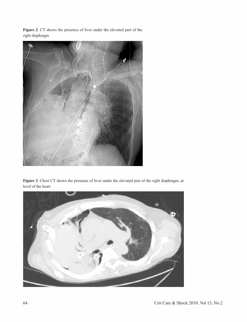
Ultrasound, not routinely considered as part of the work up diagnostic tool, may be of utility for the correct diagnosis, as in our case. The AP film may show dome-like configuration to anterior aspect of right diaphragm (Figure 1). In evaluation with a chest CT scan, CT may show the presence of liver under the elevated part of the right diaphragm (Figure 2), while ultrasound findings include herniation of liver tissue into the pleural space and distorted diaphragm image (Figure 3).
Management consisted mainly of surgical repair. Video-assisted thoracic surgery (VATS), although a less invasive approach, avoids the incision of the lower intercostal muscles, which may adversely affect ventilation, and is less painful. (11-15) Although most of the clinical experience with this intervention is in adult patients, (1-7,10) VATS has been performed successfully, for example in pediatric patients with eventration secondary to bilateral phrenic nerve paralysis. (1-7,10,11) This procedure could potentially reduce both post-operative recovery and morbidity (1,7,10,11)
Conclusions
Rupture of the diaphragm may occur after blunt trauma, as in this case. The diaphragm most frequently is ruptured on the left side, perhaps because the liver may dissipate some of the force of an abdominal blow, lessening the likelihood of rupture of the right hemidiaphragm. The manifestations of a ruptured diaphragm can be delayed, and sometimes the bowel herniates through the diaphragm very early. In cases in which surgery is not initially required, a diaphragmatic tear can be missed, especially when it is small and when there is no herniation of abdominal structures to the chest. Trauma intensive care unit specialists have to be suspicious in all cases of trauma to the lower chest, but also in patients with abdominal or pelvic trauma in whom the chest radiograph should be evaluated carefully. The diagnosis (eventration versus diaphragmatic rupture) should be considered in any patient with a persistent pleural effusion for whom a chest tube did not improve what is initially considered a “white lung”.
Figure 1. AP film showing dome-like configuration to anterior aspect of right diaphragm
64 Crit Care & Shock 2010. Vol 13, No.2
Figure 2. CT shows the presence of liver under the elevated part of the right diaphragm
Figure 3. Chest CT shows the presence of liver under the elevated part of the right diaphragm, at level of the heart

65Crit Care & Shock 2010. Vol 13, No.2
Figure 2. CT shows the presence of liver under the elevated part of the right diaphragm
Figure 3. Chest CT shows the presence of liver under the elevated part of the right diaphragm, at level of the heart
Figure 4. US shows herniation of liver tissue into the pleural space, and distorted diaphragm image
Figure 5. Right posterolateral thoracotomy with right diaphragm eventration and a right fibrothorax with lung collapse

66 Crit Care & Shock 2010. Vol 13, No.2
References
1. Sangster G, Ventura VP, Carbo A, Gates T, Garayburu J, D’Agostino H. Diaphragmatic rupture: a frequently missed injury in blunt thoracoabdominal trauma patients. Emerg Radiol 2007;13:225-30.
2. ACEP Clinical Policies Committee; Clinical Policies Subcommittee on Acute Blunt Abdominal Trauma. Clinical policy: Critical issues in the evaluation of adult patients presenting to the emergency department with acute blunt abdominal trauma. Ann Emerg Med 2004;43:278-90.
3. Raos M, Batinica S. Diaphragmatic eventration. Lijec Vjesn 2003;125:13-5.
4. Bisgard JD. Congenital eventration of the diaphragm. J Thorac Surg 1947;16:484-91.
5. Shah-Mirany J, Schmitz GL, Watson RR. Eventration of the diaphragm. Physiologic and surgical significance. Arch Surg
1968;96:844-50.6. Sliker CW. Imaging of diaphragm injuries.
Radiol Clin North Am 2006;44:199-211.7. Schumpelick V, Steinau G, Schluper I,
Prescher A. Surgical embryology and anatomy of the diaphragm with surgical applications. Surg Clin North Am [serial online] 2000 [cited 2008 Dec 19]; 80:213-39. Available from: URL:http://www.uptodate.com/patients/content/abstract.do?topicKey=~S5%2FM%2Fj02r1c2Wc4&refNum=1.
8. Kearney PA, Rouhana SW, Burney RE. Blunt rupture of the diaphragm: mechanism, diagnosis, and treatment. Ann Emerg Med 1989;18:1326-30.
9. Iochum S, Ludig T, Walter F, Sebbag H, Grosdidier G, Blum AG. Imaging of diaphragmatic injury: a diagnostic challenge? Radiographics 2002;22:S103-16.
10. Mouroux J, Venissac N, Leo F, Alifano M, Guillot F. Surgical treatment of diaphragmatic eventration using video-assisted thoracic surgery: a prospective study. Ann Thorac Surg 2005;79:308-12.
11. Shimizu M. Bilateral phrenic-nerve paralysis treated by thoracoscopic diaphragmatic plication in a neonate. Pediatr Surg Int 2003;19:79-81.
12. Verhey PT, Gosselin MV, Primack SL, Kraemer AC. Differentiating diaphragmatic paralysis and eventration. Acad Radiol 2007;14:420-5.
13. Chin EF, Lynn RB. Surgery of eventration of the diaphragm. J Thorac Surg 1956;32:6-14.
14. Deslauriers J. Eventration of the diaphragm. Chest Surg Clin N Am 1998;8:315-30.
15. Reed JA, Borden DL. Eventration of the diaphragm. Arch Surg 1935;31:30-64.


