Embed Size (px)
Citation preview
Original articleArticle original
� 2013 CEOPublished by / Edite par Elsevier Masson SAS
All rights reserved / Tous droits reserves
White defects on enamel: Diagnosis andanatomopathology: Two essential factors forproper treatment (part 1)
Taches blanches de l’�email : diagnostic etanatomopathologie : deux donn�eesindispensables pour bien les traiter (partie 1)
Maud DENISa,*,b, Anthony ATLANa,c, Elsa VENNATd, Gil TIRLETa,e,Jean-Pierre ATTALa,b
aHopital Charles-Foix, 12, rue de la R�epublique, 94200 Ivry-sur-Seine, FrancebUnit�e de recherche biomat�eriaux innovations et interfaces, hopital Charles-Foix, universit�eParis Descartes, 12, rue de la R�epublique, 94200 Ivry-sur-Seine, Francec98 bis, rue Bobillot, 75013 Paris, Franced �Ecole central Paris, laboratoire MSSMat, Grande Voie des Vignes, 92295 Chatenay-Malabrycedex, Francee234, boulevard Raspail, 75014 Paris, France
Available online: 16 April 2013 / Disponible en ligne : 16 avril 2013
SummaryEarly-stage caries (white spots), fluoroses, traumatic hypomine-ralizations and molar incisive hypomineralization (MIH) allpresent, to differing degrees, clinical symptoms involving whitemarks on the enamel. This article shows that proper diagnosisleads to better understanding of the three-dimensional aspects ofthe lesion, thereby ensuring the appropriate choice of a specifictreatment.
� 2013 CEO. Published by Elsevier Masson SAS. All rightsreserved
Key-words
·Enamel hypomineralization.
·White spot.·Fluorosis.
·Traumatic hypomineralization.International Orthodontics 2013 ; 11 : 139-165doi:10.1016/j.ortho.2013.02.014
R�esum�e
Les caries initiales (white spots), les fluoroses, les hypo-min�eralisations traumatiques et les hypomin�eralisationsmolaires incisives (MIH) pr�esentent toutes, a diff�erentsstades, un tableau clinique comportant des l�esions blanchesde l’�email. Nous montrerons dans cet article quel’�etablissement du bon diagnostic assure une meilleureappr�ehension tridimensionnelle de la l�esion garantissantd’adopter l’ad�equat traitement sp�ecifique.� 2013 CEO. Edite par Elsevier Masson SAS. Tous droitsreserves
Mots-cl�es
·Hypomin�eralisation am�elaire.
·White spot.
·Fluorose.
·Hypomin�eralisation traumatique.*Correspondence and reprints / Correspondance et tir�es a part :
Maud DENIS,, Unit�e de recherche biomat�eriaux innovations et interfaces, universit�eParis Descartes, 1, rue Maurice-Arnoux, 92120 Montrouge, France.e-mail address / Adresse e-mail : [email protected]
139
Maud DENIS et al.
·MIH.
·Erosion-infiltration.In everyday practice, the dental surgeon, under increasingpressure from patients with esthetic concerns, is more andmore often called on to treat abnormalities in tooth color.The lack of good initial training in this field unfortunatelymeans that the etiological diagnosis of such problems is oftenperceived by the physician as an awkward hurdle to overcome.This is no doubt why dentists often prefer to abstain fromproposing treatment, or sometimes even evacuate the prob-lem–and the lesion–with the bur. And yet there exists today abasic range of appropriate treatments comprising whitening,erosion/infiltration and/or micro-/mega-abrasion. However,before opting for one or other of these treatments, it is essentialto understand the etiology of the lesion. This article will firstlook at the optical mechanism which explains the whiteappearance of areas of hypomineralization in the enameland then examine one by one the various etiologies, describingprecisely the corresponding lesions and their consequences interms of type of treatment and its limitations. A second articlewill be devoted more specifically to the treatment of thesespots by erosion-infiltration.
The feature common to all whitedefects: hypomineralization
The presence of a visible white area on the tooth surface is duesolely to a defect in the enamel. In this clinical situation, thedentin is never involved. Healthy enamel is the most highlymineralized tissue in the organism, 96% of its composition (byweight) being represented by hydroxyapatite; the remaining4% are organic fluids. Conversely, in the presence of a whitespot (WS), this mineral phase is seriously diminished andreplaced by organic fluids. This is why we refer to “enamelhypomineralization”. This defect in the enamel is due to modi-fications of the chemical composition of the substrate.
The laws of optics indicate that when there is a difference inrefractive index between two phases, there will be an interfacecausing deviation of incident light rays.The refractive index of healthy enamel is the same as that ofhydroxapatite, which is, as explained above, by far the mostimportant constituent of the phase (RIhealthy enamel =RIhydroxyapatite = 1.62). There are, therefore, hardly any inter-faces in healthy enamel; the light ray passes through thesubstrate with no modification of its trajectory until it isreflected at the dentino-enamel junction (fig. 1).However, in hypomineralized enamel, the light ray encountersmultiple interfaces between organic fluids and the mineralphase, with different indices of refraction (respectively 1.33and 1.62). At each interface, the light is thus deviated and
140
·MIH.�
·Erosion-infiltration.Le chirurgien dentiste, de par la demande esth�etique crois-sante des patients, est au sein de son cabinet de plus en plusr�eguli�erement confront�e au traitement des anomalies de teintedes dents. L’absence de formation initiale claire dans cedomaine fait malheureusement que bien souvent leur diag-nostic �etiologique est percu par le praticien comme une �etaped�elicate. C’est sans doute ce qui explique que ce dernier optepr�ef�erentiellement pour l’abstention, ou parfois meme pourl’�eviction de la l�esion par fraisage. Pourtant, actuellement ilexiste un arsenal th�erapeutique « a minima » adapt�e asso-ciant l’�eclaircissement, l’�erosion-infiltration et/ou la micro-/m�ega-abrasion. Seulement, avant de se d�ecider pour l’uneou l’autre de ces th�erapeutiques, il est imp�eratif de connaıtrel’�etiologie de la l�esion. Dans le cadre de cet article, apr�es avoirexpliqu�e le m�ecanisme optique qui explique l’aspect blancha-tre des taches d’hypomin�eralisation am�elaires, nous repren-drons une par une les diff�erentes �etiologies en d�ecrivantpr�ecis�ement les l�esions correspondantes, et les con-s�equences en termes de type de traitement et limites. Unsecond article sera consacr�e plus sp�ecifiquement au traite-ment par �erosion-infiltration de ces diff�erentes taches.
Un aspect blanc, un point commun :l’hypomin�eralisation
La pr�esence de tache blanche visible a la surface des dentsr�esulte uniquement d’un d�efaut am�elaire. Dans ce tableauclinique, la dentine n’est jamais mise en cause. L’�email sain,tissu le plus min�eralis�e de l’organisme, pr�esente en poids96 % d’hydroxyapatite ; les 4 % restants repr�esentent lesfluides organiques. Inversement, en pr�esence de tacheblanche, cette phase min�erale se trouve fortement diminu�eeet remplac�ee par des fluides organiques. C’est pourquoi nousparlons d’« hypomin�eralisation am�elaire ». Ce d�efaut d’�emailest la cons�equence d’une alt�eration des taux de la compo-sition chimique du substrat.Les lois de l’optique indiquent que s’il existe une diff�erenced’indice de r�efraction entre deux phases, il y a apparition d’uneinterface provoquant la d�eviation du rayon incident.Dans le cas de l’�email sain, son indice de r�efraction est lememe que celui de l’hydroxyapatite, phase largement major-itaire comme �evoqu�e pr�ec�edemment (IR�email sain =IRhydroxyapatite = 1,62). L’�email sain pr�esente alors que peud’interface ; le rayon lumineux traverse l’�epaisseur du substratsans modification de trajectoire, et ce jusqu’a la jonctionam�elo-dentinaire avant d’etre r�efl�echie (fig. 1).Tandis que dans le cas de l’�email hypomin�eralis�e, le rayonlumineux rencontre de multiples interfaces « fluides organi-ques/min�eral » d’indice de r�efraction diff�erent (respective-ment 1,33 et 1,62), provoqu�ees par leur alternance de phase.A chaque interface, le rayon est alors d�evi�e et r�efl�echi,
International Orthodontics 2013 ; 11 : 139-165
[(Fig._1)TD$FIG]
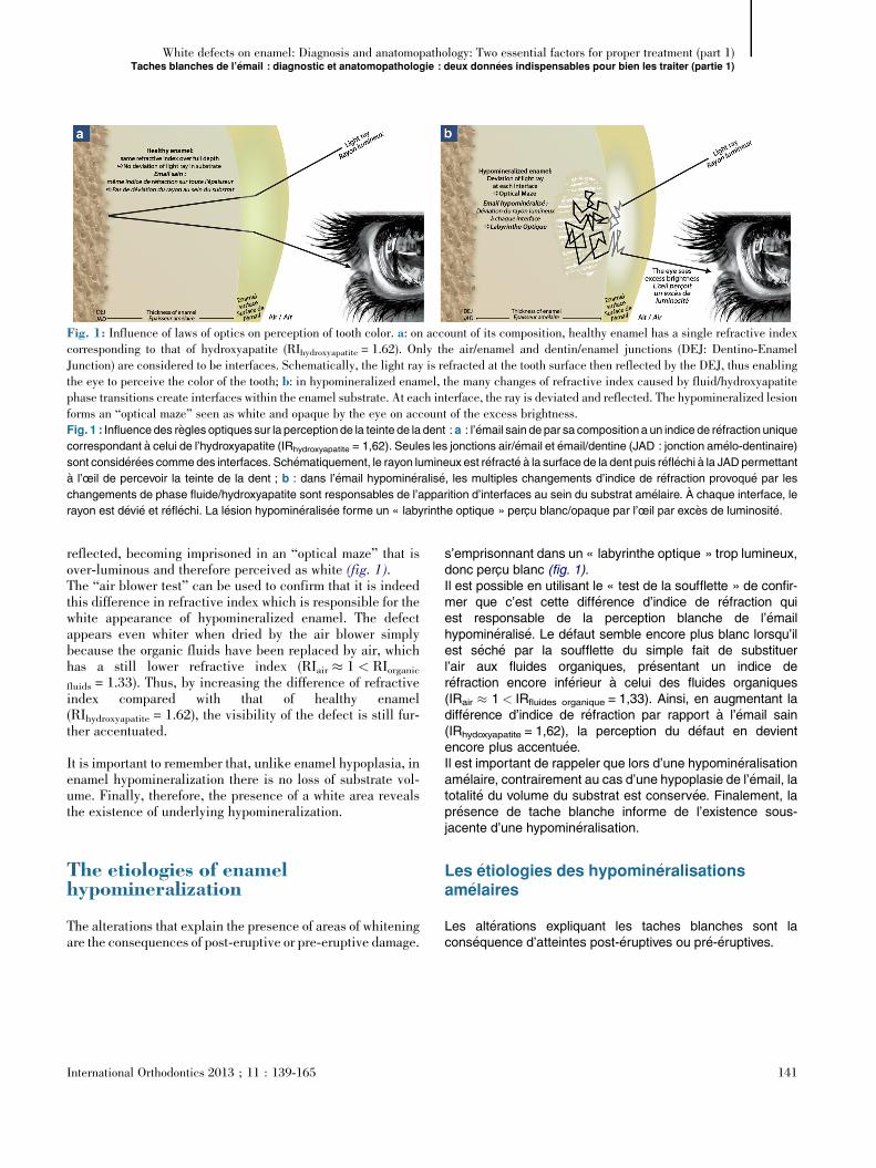
Fig. 1: Influence of laws of optics on perception of tooth color. a: on account of its composition, healthy enamel has a single refractive indexcorresponding to that of hydroxyapatite (RIhydroxyapatite = 1.62). Only the air/enamel and dentin/enamel junctions (DEJ: Dentino-EnamelJunction) are considered to be interfaces. Schematically, the light ray is refracted at the tooth surface then reflected by the DEJ, thus enablingthe eye to perceive the color of the tooth; b: in hypomineralized enamel, the many changes of refractive index caused by fluid/hydroxyapatitephase transitions create interfaces within the enamel substrate. At each interface, the ray is deviated and reflected. The hypomineralized lesionforms an “optical maze” seen as white and opaque by the eye on account of the excess brightness.Fig. 1 : Influence des r�egles optiques sur la perception de la teinte de la dent : a : l’�email sain de par sa composition a un indice de r�efraction unique
correspondant a celui de l’hydroxyapatite (IRhydroxyapatite = 1,62). Seules les jonctions air/�email et �email/dentine (JAD : jonction am�elo-dentinaire)
sont consid�er�ees commedes interfaces. Sch�ematiquement, le rayon lumineux est r�efract�e a la surface de la dent puis r�efl�echi a la JAD permettant
a l’œil de percevoir la teinte de la dent ; b : dans l’�email hypomin�eralis�e, les multiples changements d’indice de r�efraction provoqu�e par les
changements de phase fluide/hydroxyapatite sont responsables de l’apparition d’interfaces au sein du substrat am�elaire. A chaque interface, le
rayon est d�evi�e et r�efl�echi. La l�esion hypomin�eralis�ee forme un « labyrinthe optique » percu blanc/opaque par l’œil par exc�es de luminosit�e.
White defects on enamel: Diagnosis and anatomopathology: Two essential factors for proper treatment (part 1)Taches blanches de l’�email : diagnostic et anatomopathologie : deux donn�ees indispensables pour bien les traiter (partie 1)
reflected, becoming imprisoned in an “optical maze” that isover-luminous and therefore perceived as white (fig. 1).The “air blower test” can be used to confirm that it is indeedthis difference in refractive index which is responsible for thewhite appearance of hypomineralized enamel. The defectappears even whiter when dried by the air blower simplybecause the organic fluids have been replaced by air, whichhas a still lower refractive index (RIair � 1 < RIorganicfluids = 1.33). Thus, by increasing the difference of refractiveindex compared with that of healthy enamel(RIhydroxyapatite = 1.62), the visibility of the defect is still fur-ther accentuated.
It is important to remember that, unlike enamel hypoplasia, inenamel hypomineralization there is no loss of substrate vol-ume. Finally, therefore, the presence of a white area revealsthe existence of underlying hypomineralization.
The etiologies of enamelhypomineralization
The alterations that explain the presence of areas of whiteningare the consequences of post-eruptive or pre-eruptive damage.
International Orthodontics 2013 ; 11 : 139-165
s’emprisonnant dans un « labyrinthe optique » trop lumineux,donc percu blanc (fig. 1).Il est possible en utilisant le « test de la soufflette » de confir-mer que c’est cette diff�erence d’indice de r�efraction quiest responsable de la perception blanche de l’�emailhypomin�eralis�e. Le d�efaut semble encore plus blanc lorsqu’ilest s�ech�e par la soufflette du simple fait de substituerl’air aux fluides organiques, pr�esentant un indice der�efraction encore inf�erieur a celui des fluides organiques(IRair � 1 < IRfluides organique = 1,33). Ainsi, en augmentant ladiff�erence d’indice de r�efraction par rapport a l’�email sain(IRhydoxyapatite = 1,62), la perception du d�efaut en devientencore plus accentu�ee.Il est important de rappeler que lors d’une hypomin�eralisationam�elaire, contrairement au cas d’une hypoplasie de l’�email, latotalit�e du volume du substrat est conserv�ee. Finalement, lapr�esence de tache blanche informe de l’existence sous-jacente d’une hypomin�eralisation.
Les �etiologies des hypomin�eralisationsam�elaires
Les alt�erations expliquant les taches blanches sont lacons�equence d’atteintes post-�eruptives ou pr�e-�eruptives.
141
Maud DENIS et al.
Post-eruptive damage: white spots
The first stages of carious disease are characterized by hypo-mineralization without cavity formation. When this is clini-cally visible, we refer to it as white spots (WS).
Prevalence
It is important to note that WS are the most frequent patho-logies responsible for areas of whitening, with an estimatedprevalence of 24% [1]. This rises to 49.6% following multi-bracket orthodontic treatment, and the incidence of at leastone non-cavitated lesion before/after orthodontic treatment is72.9% [2]. Their treatment is all the more important in that it isthe front teeth that are preferentially involved.
Moreover, this prevalence is multifocal: it concerns at leastthree teeth in 46% of patients not treated orthodontically, andrises to 77% in those who have received orthodontic treatment[3]. These white lesions reduce by one third at 3 months and byhalf at 6 months, but once present they remain, and are stillvisible 5 years after debonding.
The frequency of appearance of these elective post-eruptivelesions, and their long-lasting nature place the dentist underan obligation to take preventive measures and ensure earlydetection so that treatment is restricted to a necessaryminimum.
Diagnosis
The diagnosis of the initial carious lesions is essentially amatter of clinical observation; at this stage, on account ofthe acellular, avascular and nerve-free nature of enamel, thereare no associated symptoms. When these lesions reach thesmooth surfaces of the front teeth, they are mainly found in thesites where bacterial plaque preferentially accumulates:either in the labial cervical third, or around the brackets inthe event of orthodontic treatment. They are opaque, mat,chalky-white areas of enamel, variable in shape and sizeand with more or less clear-cut outlines (fig. 2a). The enamelsurface is still continuous but possesses a certain degree ofroughness, often associated with accentuation of micro-ridges(perikymata). To avoid the subjective element in visual obser-vation of the initial carious lesion, the “International CariesDetection and Assessment System” (ICDAS) suggested byEkstrand (Table I) [4] is used; this also provides informationon histological involvement. Early detection tools (quantita-tive laser fluorescence [Diagnodent�], Fibre OpticTransillumination [FOTI], etc.) can also be important aidsfor their evaluation. The dentist should support clinical obser-vation by weighing up the risk factors (direct, environmentaland individual) listed by Selwitz [5] that may be involved inthe development of caries so as to confirm the diagnosis andsubsequently restore an environment favorable to oral health.
142
Atteinte post-�eruptive : white spots (WS)
Les premiers temps de la maladie carieuse se traduisent parune hypomin�eralisation non cavitaire. Lorsqu’elle est clinique-ment visible, nous l’appelons white spots (WS).
Pr�evalence
Il est important de noter que la pr�evalence des WS est la plusimportante de toutes les pathologies responsables de tacheblanche. Elle est estim�ee a 24 % [1]. Elle passe a 49,6 %a l’issu d’un traitement orthodontique par multi-attaches, avecune incidence d’aumoins une l�esion non cavitaire avant/apr�estraitement orthodontique de 72,9 % [2]. Il est d’autant plusn�ecessaire de les prendre en charge que l’atteinte des dentsdu sourire est pr�ef�erentielle.De plus, elle est multifocale ; elle concerne au moins troisdents chez 46 % des patients non trait�es orthodontiquementet passe a 77 % chez ceux ayant recu un traitement orthodon-tique [3]. Une fois install�ees, meme si ces l�esions blanches ser�eduisent d’un tiers a trois mois et de la moiti�e a six mois, ellesapparaissent encore visible cinq ans apr�es d�epose del’appareillage.La fr�equence d’apparition de ces l�esions �electives post-�eruptive associ�ee a leur p�erennit�e, conf�ere au praticien undevoir de prophylaxie et de d�etection pr�ecoce permettantl’instauration d’« intervention minimale ».
Diagnostic
Le diagnostic des l�esions carieuses initiales est principale-ment permis par l’observation clinique ; a ce stade �etantdonn�e le caract�ere acellulaire, avasculaire, non innerv�e del’�email, aucune symptomatologie n’y est associ�ee.Lorsqu’elles atteignent les surfaces lisses des dentsant�erieures, elles sont principalement retrouv�ees en regarddes sites de pr�edilection de d�epots de plaque bact�erienne ;soit dans le tiers cervical vestibulaire, soit autour des attacheslors d’un traitement orthodontique. Elles pr�esentent alors unezone d’�email mate-opaque, blanc crayeux, de taille et deforme variables dont les contours sont plus ou moins diffus(fig. 2a). L’�email de surface encore continu pr�esente une cer-taine rugosit�e souvent accompagn�ee d’une accentuation desmicroreliefs (p�erikymaties). Pour �eviter le caract�ere subjectifde l’observation visuelle d’une l�esion carieuse initiale, le« Syst�eme international de d�etection et d’�evaluation de lacarie » (ICDAS) propos�e par Ekstrand (Tableau I) [4], infor-mant simultan�ement de l’atteinte histologique, est utilis�e. Lesoutils de d�etection pr�ecoce (fluorescence laser quantitative[Diagnodent�], transillumination par fibres optiques [FOTI],etc.) peuvent aussi etre des atouts importants dans leur�evaluation. Le praticien compl�ete l’observation clinique parla pond�eration des facteurs de risques (directs, environne-mentaux et personnels) pr�esent�es par Selwitz [5] pouvant etreimpliqu�es dans le d�eveloppement carieux, afin de certifier le
International Orthodontics 2013 ; 11 : 139-165
[(Fig._2)TD$FIG]
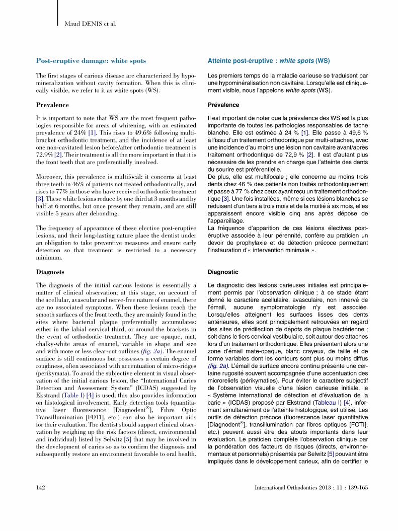
Fig. 2: Clinical appearances characteristic of the various etiologies involved in the presence of white defects on the enamel; a: whitespot. The localization of hypomineralization can correspond to a zone where bacterial plaque preferentially accumulates; here followingmulti-bracket orthodontic treatment. It appears chalky-white, mat, opaque, with variable shape and size and outlines that are more orless well-defined; b: fluorosis. Symmetrical involvement of groups of homologous teeth. In the early stages, it is revealed by thepresence of horizontal, more or less convergent white lines, accompanied or not by cloudy chalky areas that give the tooth a parchment-like appearance. Loss of substance is always the consequence of post-eruptive weakening of the crystalline lattice; c: traumatichypomineralization. Although many clinical expressions are possible, it is generally punctiform, located on the incisal coronal third ofone or several neighboring teeth; d: MIH. In the “mild” form, lesions of molars and/or incisors take the form of well-defined opaquewhite areas, limited to the occlusal half of the tooth crown. Opacities on homologous teeth are asymmetrical.Fig. 2 : Aspects cliniques caract�eristiques des diff�erentes �etiologies en cause dans la pr�esence de tache blanche de l’�email : a : white
spot. La localisation de l’hypomin�eralisation est a rapprocher avec une zone pr�ef�erentielle d’accumulation de plaque bact�erienne ; ici
suite au port d’un appareillage orthodontique par multi-attache. Elle revet un aspect mate-opaque, blanc crayeux, de taille et de forme
variables dont les contours sont plus oumoins diffus ;b : fluorose. L’atteinte est sym�etrique sur les groupesde dents homologues. Elle se
traduit dans les premiers stades par la pr�esence de lignes blanches horizontales W confluentes, accompagn�ees ou non de zones
nuageuses crayeuses conf�erant a la dent un aspect parchemin�e. La perte de substance est toujours cons�ecutive a un affaiblissement
post-�eruptif de la trame cristalline ; c : l’hypomin�eralisation traumatique. Malgr�e sa grande vari�et�e d’expression clinique, elle est
g�en�eralement ponctiforme, situ�ee sur le tiers incisale coronaire d’un ou plusieurs dents voisine ; d : MIH. L’atteinte des molaires et/
ou incisives pr�esente, a un stade « l�eger », des opacit�es blanches bien d�elimit�ees, circonscrites a la moiti�e occlusale de la couronne. Les
opacit�es sont asym�etriques sur les dents homologues.
White defects on enamel: Diagnosis and anatomopathology: Two essential factors for proper treatment (part 1)Taches blanches de l’�email : diagnostic et anatomopathologie : deux donn�ees indispensables pour bien les traiter (partie 1)
Anatomopathology
During the initial carious process, alternating phases ofdemineralization and remineralization cause preferential dis-solution and reprecipitation and, thereby, the preservation of a
International Orthodontics 2013 ; 11 : 139-165
diagnostic et de r�etablir ult�erieurement un environnement enad�equation avec la sant�e buccale.
Anatomopathologie
Lors du processus carieux initial, l’alternance de phase ded�emin�eralisation/remin�eralisation permet par dissolution/repr�ecipitation pr�ef�erentielles la pr�eservation d’une « couche
143
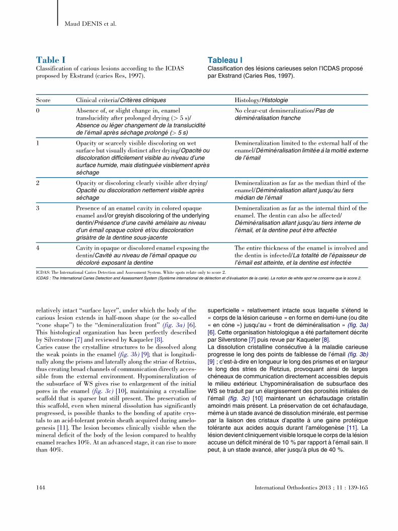
Table IClassification of carious lesions according to the ICDASproposed by Ekstrand (caries Res, 1997).
Tableau IClassification des l�esions carieuses selon l’ICDAS propos�epar Ekstrand (Caries Res, 1997).
Score Clinical criteria/Crit�eres cliniques Histology/Histologie
0 Absence of, or slight change in, enameltranslucidity after prolonged drying (> 5 s)/Absence ou l�eger changement de la translucidit�ede l’�email apr�es s�echage prolong�e (> 5 s)
No clear-cut demineralization/Pas ded�emin�eralisation franche
1 Opacity or scarcely visible discoloring on wetsurface but visually distinct after drying/Opacit�e oudiscoloration difficilement visible au niveau d’unesurface humide, mais distingu�ee visiblement apr�ess�echage
Demineralization limited to the external half of theenamel/D�emin�eralisation limit�ee a lamoiti�e externede l’�email
2 Opacity or discoloring clearly visible after drying/Opacit�e ou discoloration nettement visible apr�ess�echage
Demineralization as far as the median third of theenamel/D�emin�eralisation allant jusqu’au tiersm�edian de l’�email
3 Presence of an enamel cavity in colored opaqueenamel and/or greyish discoloring of the underlyingdentin/Pr�esence d’une cavit�e am�elaire au niveaud’un �email opaque color�e et/ou discolorationgrisatre de la dentine sous-jacente
Demineralization as far as the internal third of theenamel. The dentin can also be affected/D�emin�eralisation allant jusqu’au tiers interne del’�email, et la dentine peut etre affect�ee
4 Cavity in opaque or discolored enamel exposing thedentin/Cavit�e au niveau de l’�email opaque oud�ecolor�e exposant la dentine
The entire thickness of the enamel is involved andthe dentin is infected/La totalit�e de l’�epaisseur del’�email est atteinte, et la dentine est infect�ee
ICDAS: The International Caries Detection and Assessment System. White spots relate only to score 2.ICDAS : The International Caries Detection and Assessment System (Syst�eme international de d�etection et d’�evaluation de la carie). La notion de white spot ne concerne que le score 2.
Maud DENIS et al.
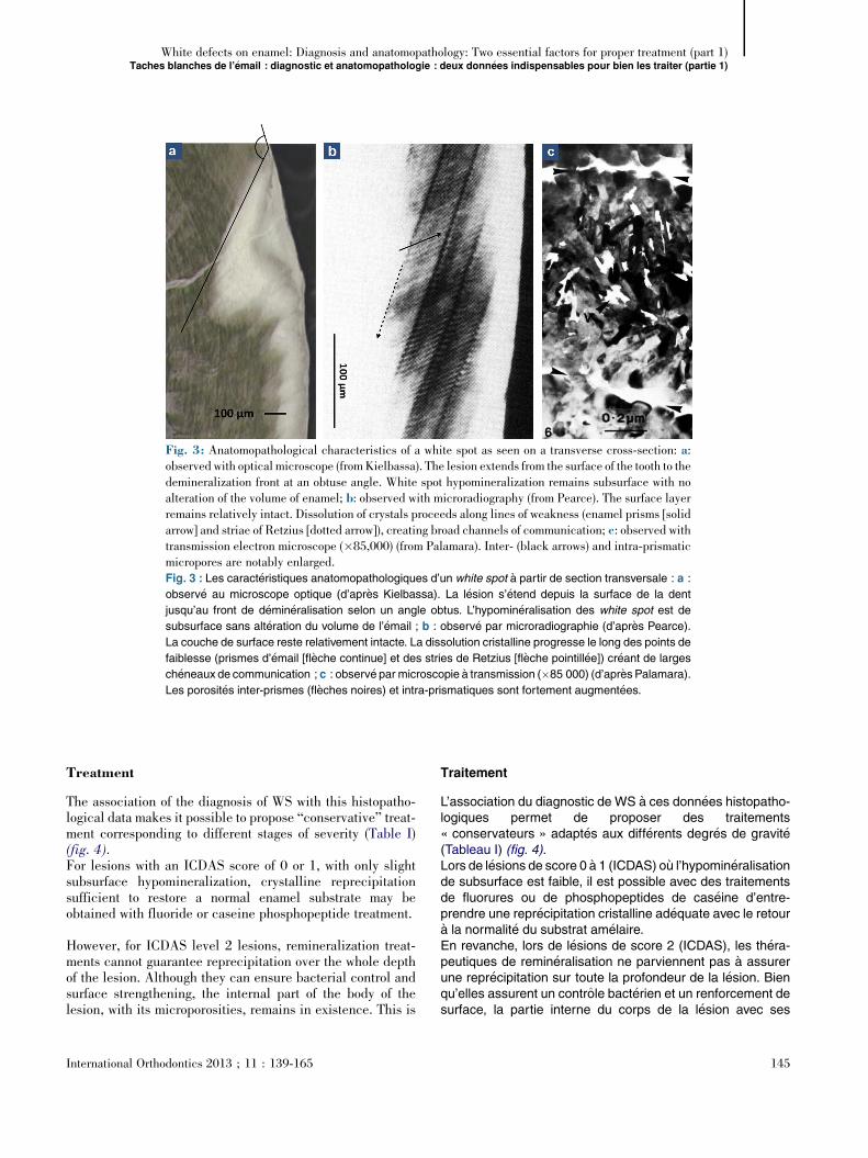
relatively intact “surface layer”, under which the body of thecarious lesion extends in half-moon shape (or the so-called“cone shape”) to the “demineralization front” (fig. 3a) [6].This histological organization has been perfectly describedby Silverstone [7] and reviewed by Kaqueler [8].Caries cause the crystalline structures to be dissolved alongthe weak points in the enamel (fig. 3b) [9]; that is longitudi-nally along the prisms and laterally along the striae of Retzius,thus creating broad channels of communication directly acces-sible from the external environment. Hypomineralization ofthe subsurface of WS gives rise to enlargement of the initialpores in the enamel (fig. 3c) [10], maintaining a crystallinescaffold that is sparser but still present. The preservation ofthis scaffold, even when mineral dissolution has significantlyprogressed, is possible thanks to the bonding of apatite crys-tals to an acid-tolerant protein sheath acquired during amelo-genesis [11]. The lesion becomes clinically visible when themineral deficit of the body of the lesion compared to healthyenamel reaches 10%. At an advanced stage, it can rise to morethan 40%.
144
superficielle » relativement intacte sous laquelle s’�etend le« corps de la l�esion carieuse » en forme en demi-lune (ou dite« en cone ») jusqu’au « front de d�emin�eralisation » (fig. 3a)[6]. Cette organisation histologique a �et�e parfaitement d�ecritepar Silverstone [7] puis revue par Kaqueler [8].La dissolution cristalline cons�ecutive a la maladie carieuseprogresse le long des points de faiblesse de l’�email (fig. 3b)[9] ; c’est-a-dire en longueur le long des prismes et en largeurle long des stries de Retzius, provoquant ainsi de largesch�eneaux de communication directement accessibles depuisle milieu ext�erieur. L’hypomin�eralisation de subsurface desWS se traduit par un �elargissement des porosit�es initiales del’�email (fig. 3c) [10] maintenant un �echafaudage cristallinamoindri mais pr�esent. La pr�eservation de cet �echafaudage,meme a un stade avanc�e de dissolution min�erale, est permisepar la liaison des cristaux d’apatite a une gaine prot�eiquetol�erante aux acides acquis durant l’am�elogen�ese [11]. Lal�esion devient cliniquement visible lorsque le corps de la l�esionaccuse un d�eficit min�eral de 10 % par rapport a l’�email sain. Ilpeut, a un stade avanc�e, aller jusqu’a plus de 40 %.
International Orthodontics 2013 ; 11 : 139-165
[(Fig._3)TD$FIG]
Fig. 3: Anatomopathological characteristics of a white spot as seen on a transverse cross-section: a:observed with optical microscope (fromKielbassa). The lesion extends from the surface of the tooth to thedemineralization front at an obtuse angle. White spot hypomineralization remains subsurface with noalteration of the volume of enamel; b: observed with microradiography (from Pearce). The surface layerremains relatively intact. Dissolution of crystals proceeds along lines of weakness (enamel prisms [solidarrow] and striae of Retzius [dotted arrow]), creating broad channels of communication; c: observed withtransmission electron microscope (�85,000) (from Palamara). Inter- (black arrows) and intra-prismaticmicropores are notably enlarged.Fig. 3 : Les caract�eristiques anatomopathologiques d’un white spot a partir de section transversale : a :
observ�e au microscope optique (d’apr�es Kielbassa). La l�esion s’�etend depuis la surface de la dent
jusqu’au front de d�emin�eralisation selon un angle obtus. L’hypomin�eralisation des white spot est de
subsurface sans alt�eration du volume de l’�email ; b : observ�e par microradiographie (d’apr�es Pearce).
La couche de surface reste relativement intacte. La dissolution cristalline progresse le long des points de
faiblesse (prismes d’�email [fl�eche continue] et des stries de Retzius [fl�eche pointill�ee]) cr�eant de larges
ch�eneaux de communication ; c : observ�e par microscopie a transmission (�85 000) (d’apr�esPalamara).
Les porosit�es inter-prismes (fl�eches noires) et intra-prismatiques sont fortement augment�ees.
White defects on enamel: Diagnosis and anatomopathology: Two essential factors for proper treatment (part 1)Taches blanches de l’�email : diagnostic et anatomopathologie : deux donn�ees indispensables pour bien les traiter (partie 1)
Treatment
The association of the diagnosis of WS with this histopatho-logical data makes it possible to propose “conservative” treat-ment corresponding to different stages of severity (Table I)(fig. 4).For lesions with an ICDAS score of 0 or 1, with only slightsubsurface hypomineralization, crystalline reprecipitationsufficient to restore a normal enamel substrate may beobtained with fluoride or caseine phosphopeptide treatment.
However, for ICDAS level 2 lesions, remineralization treat-ments cannot guarantee reprecipitation over the whole depthof the lesion. Although they can ensure bacterial control andsurface strengthening, the internal part of the body of thelesion, with its microporosities, remains in existence. This is
International Orthodontics 2013 ; 11 : 139-165
Traitement
L’association du diagnostic de WS a ces donn�ees histopatho-logiques permet de proposer des traitements« conservateurs » adapt�es aux diff�erents degr�es de gravit�e(Tableau I) (fig. 4).Lors de l�esions de score 0 a 1 (ICDAS) ou l’hypomin�eralisationde subsurface est faible, il est possible avec des traitementsde fluorures ou de phosphopeptides de cas�eine d’entre-prendre une repr�ecipitation cristalline ad�equate avec le retoura la normalit�e du substrat am�elaire.En revanche, lors de l�esions de score 2 (ICDAS), les th�era-peutiques de remin�eralisation ne parviennent pas a assurerune repr�ecipitation sur toute la profondeur de la l�esion. Bienqu’elles assurent un controle bact�erien et un renforcement desurface, la partie interne du corps de la l�esion avec ses
145

[(Fig._4)TD$FIG]
Fig. 4: What treatment for different stages of early cariouslesions? Following orthodontic treatment, the patient presentscarious lesions with ICDAS scores of 1, 2 and 3. Only scores 2and 3 are visible on this photo of teeth without air-drying. The lossof substance associated with score-3 lesions of 13, 12, 22 and 23is the result of excessive mechanical action. The dentin is notinvolved. At this stage, topical application alone of a remineraliz-ing product will not lead to disappearance of the defects in theenamel despite a return to a healthy state. Only conservativetreatment by “erosion-infiltration” can halt the progression ofcrystal dissolution by creating an impermeable barrier, and alsorestore tooth color.Fig. 4 : Quels traitements pour quel niveau d’atteinte de l�esions
carieuses initiales ? Le patient pr�esente a l’issu d’un traitement
orthodontique des l�esions carieuses de score ICDAS 1, 2 et 3.
Seules les scores 2 et 3 sont visibles sur cette photo de dents non
s�ech�ees. Les pertes de substances des l�esions de score 3 des 13,
12, 22 et 23 sont cons�ecutives a une action m�ecanique excessive.
La dentine n’est pas mise en cause. A ce degr�e d’atteinte, l’appli-
cation topique seule de produit remin�eralisant ne permettra pas la
disparition des d�efauts am�elaires malgr�e un retour a l’�etat de
sant�e. Seule, l’application d’une th�erapeutique conservatrice par
« �erosion-infiltration » stoppera la progression de la dissolution
cristalline par imperm�eabilisation, tout en restaurant la teinte des
dents.
Maud DENIS et al.
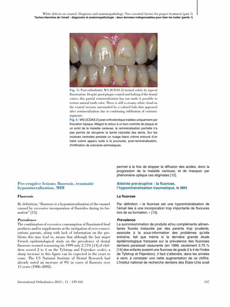
why, despite perfect etiological control, the white area persistsover time, and brown coloring may even appear followingprogressive micro-infiltration by extrinsic pigments (fig. 5).Today, for these score 2 lesions, the only genuine “minimallyinvasive” treatment available on the market, to both arrest thecaries and restore good esthetics, is “erosion-infiltration”. Theonly product currently marketed that corresponds to this prin-ciple is Icon (DMG). First of all, it uses hydrochloric acid toeliminate the relatively intact surface layer and open upaccess to the body of the lesion. Then, in a second stage, afluid resin is infiltrated into the broad channels of communi-cation. The body of the lesion is rendered watertight by meansof a resin with a refractive index (IRi-con
� = 1.44) close to thatof healthy enamel, so as to both stop the diffusion of acids, thushalting progression of the caries, and mask the discolorationsby optical means [12].
146
microporosit�es continue de persister. C’est ce qui explique,malgr�e un parfait controle �etiologique, la conservation a longterme de la tache blanche, voire meme l’apparition de colora-tion brune suite a la micro-infiltration progressive de colorantsextrins�eques (fig. 5). Aujourd’hui, pour ces l�esions de score 2,le seul traitement v�eritablement « aminima » disponible sur lemarch�e, assurant a la fois l’arret de la maladie carieuse et ler�etablissement de l’esth�etique, est l’« �erosion-infiltration ». Leseul produit commercialis�e actuellement qui r�epond a ce prin-cipe s’appelle Icon (DMG). Il �elimine dans un premier tempspar un traitement a l’acide chlorhydrique la couche de surfacerelativement intacte. C’est ce qui permet d’ouvrir l’acc�es aucorps de la l�esion et, dans un second temps, d’infiltrer avecune r�esine fluide ses larges ch�eneaux de communication.L’imperm�eabilisation du corps de la l�esion avec une r�esined’indice de r�efraction (IRi-con
� = 1,44) proche de l’�email sain,
International Orthodontics 2013 ; 11 : 139-165
[(Fig._5)TD$FIG]
Fig. 5: Post-orthodontic WS (ICDAS 2) treated solely by topicalfluorination. Despite good plaque control and halting of the dentalcaries, this partial remineralization has not made it possible torestore natural tooth color. There is still a creamy-white cloud onthe central incisors surrounded by a colored halo that appearedafter remineralization due to continuing infiltration of extrinsicpigments.Fig. 5 :WS (ICDAS 2) post-orthodontique trait�ees uniquement par
fluoration topique. Malgr�e le retour a un bon controle de plaque et
un arret de la maladie carieuse, la remin�eralisation partielle n’a
pas permis de r�ecup�erer la teinte naturelle des dents. Sur les
incisives centrales persiste un nuage blanc cr�eme entour�e d’un
halot color�e apparu suite a la poursuite, post-remin�eralisation,
d’infiltration de colorants extrins�eques.
White defects on enamel: Diagnosis and anatomopathology: Two essential factors for proper treatment (part 1)Taches blanches de l’�email : diagnostic et anatomopathologie : deux donn�ees indispensables pour bien les traiter (partie 1)
Pre-eruptive lesions: fluorosis, traumatichypomineralization, MIH
Fluorosis
By definition, “fluorosis is a hypomineralization of the enamelcaused by excessive incorporation of fluorides during its for-mation” [13].
PrevalenceThe combination of excessive consumption of fluorinated foodproducts and/or supplements at the instigation of over-consci-entious parents, along with lack of information on the pro-blems this may lead to, means that although the last majorFrench epidemiological study on the prevalence of dentalfluorosis seemed reassuring (in 1999 only 2.75% [14] of chil-dren scored 2 to 4 on the Tylstrup and Fejerskov scale), asharp increase in this figure can be expected in the years tocome. The US National Institute of Dental Research hadalready noted an increase of 9% in cases of fluorosis over15 years (1986–2002).
International Orthodontics 2013 ; 11 : 139-165
permet a la fois de stopper la diffusion des acides, donc laprogression de la maladie carieuse, et de masquer parph�enom�ene optique ces stigmates [12].
Atteinte pr�e-�eruptive : la fluorose,l’hypomin�eralisation traumatique, le MIH
La fluorose
Par d�efinition « la fluorose est une hypomin�eralisation del’�email li�ee a une incorporation trop importante de fluorureslors de sa formation » [13].
Pr�evalenceLa surconsommation de produits et/ou compl�ements alimen-taires fluor�es instaur�ee par des parents trop prudents,associ�ee a la sous-information des probl�emes qu’elleentraıne, fait que meme si la derni�ere grande �etude�epid�emiologique francaise sur la pr�evalence des fluorosesdentaire paraissait rassurante (en 1999, seulement 2,75 %[14] des enfants avaient une fluorose de grade 2 a 4 de l’indexde Tylstrup et Fejerskov), il faut s’attendre, dans les ann�eesa venir, a constater une nette augmentation de ce chiffre.L’Institut national de recherche dentaire des �Etats-Unis avait
147
[(Fig._6)TD$FIG]
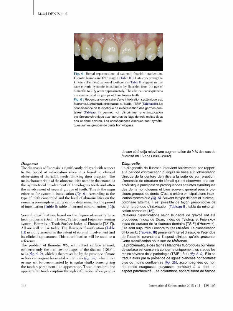
Fig. 6: Dental repercussions of systemic fluoride intoxication.Fuorotic lesions are TSIF stage 1 (Table III). Data concerning thekinetics of mineralization of tooth germs (Table II) suggest in thiscase chronic systemic intoxication by fluorides from the age of3 months to 21/2 years approximately. The clinical consequencesare symmetrical on groups of homologous teeth.Fig. 6 : R�epercussion dentaire d’une intoxication syst�emique aux
fluorures. L’atteinte fluorotique est au stade 1 TSIF (Tableau III). La
connaissance de la cin�etique de min�eralisation des germes den-
taires (Tableau II) permet, ici, d’incriminer une intoxication
syst�emique chronique aux fluorures de l’age de trois mois a deux
ans et demi environ. Les cons�equences cliniques sont sym�etri-
ques sur les groupes de dents homologues.
Maud DENIS et al.
DiagnosisThe diagnosis of fluorosis is significantly delayed with respectto the period of intoxication since it is based on clinicalobservation of the adult teeth following their eruption. Themain characteristic of the abnormalities noted in the enamel isthe symmetrical involvement of homologous teeth and oftenthe involvement of several groups of teeth. This is the maincriterion for systemic intoxication (fig. 6). According to thetype of tooth concerned and the level of abnormalities on thecrown, a presumptive dating can be determined for the periodof intoxication (Table II: table of coronal mineralization [15]).
Several classifications based on the degree of severity havebeen proposed (Dean’s Index, Tylstrup and Fejerskov scoringsystem, Horowitz’s Tooth Surface Index of Fluorosis [TSIF]).All are still in use today. The Horowitz classification (TableIII) usefully associates the extent of coronal involvement andits clinical appearance. This classification will be used as areference.The problem of fluorotic WS, with intact surface enamel,concerns only the less severe stages of the disease (TSIF 1to 4) (fig. 6–9), which is then revealed by the presence of moreor less convergent horizontal white lines (fig. 2b), which mayor may not be accompanied by irregular chalky zones givingthe tooth a parchment-like appearance. These discolorationsappear after tooth eruption through infiltration of exogenous
148
de son cot�e d�eja relev�e une augmentation de 9 % des cas defluorose en 15 ans (1986–2002).
DiagnosticLe diagnostic de fluorose intervient tardivement par rapporta la p�eriode d’intoxication puisqu’il se base sur l’observationclinique de la denture d�efinitive a la suite de son �eruption.L’anomalie de structure de l’�email qui est observ�ee, a la car-act�eristique principale de provoquer des atteintes sym�etriquesdes dents homologues et bien souvent g�en�eralis�ees a plu-sieurs groupes de dents. C’est le crit�ere principal d’une intox-ication syst�emique (fig. 6). Suivant le type de dent et le niveaucoronaire atteints, il est possible de facon pr�esomptive dedater la p�eriode d’intoxication (Tableau II : table de min�erali-sation coronaire [15]).Plusieurs classifications selon le degr�e de gravit�e ont �et�epropos�ees (index de Dean, index de Tylstrup et Fejerskov,index de surface de la fluorose dentaire [TSIF] d’Horowitz).Elle sont aujourd’hui encore toutes utilis�ees. La classificationd’Horowitz (Tableau III) pr�esente l’int�eret d’associer l’�etenduede l’atteinte coronaire a l’aspect clinique qu’elle pr�esente.Cette classification nous sert de r�ef�erence.La probl�ematique des taches blanches fluorotiques ou l’�emailde surface est conserv�e, concerne uniquement les stades lesmoins s�ev�eres de la pathologie (TSIF 1 a 4) (fig. 6–9). Elle setraduit alors par la pr�esence de lignes blanches horizontalesplus ou moins confluentes (fig. 2b), accompagn�ees ou nonde zones nuageuses crayeuses conf�erant a la dent unaspect parchemin�e. Les colorations apparaissent de facons
International Orthodontics 2013 ; 11 : 139-165
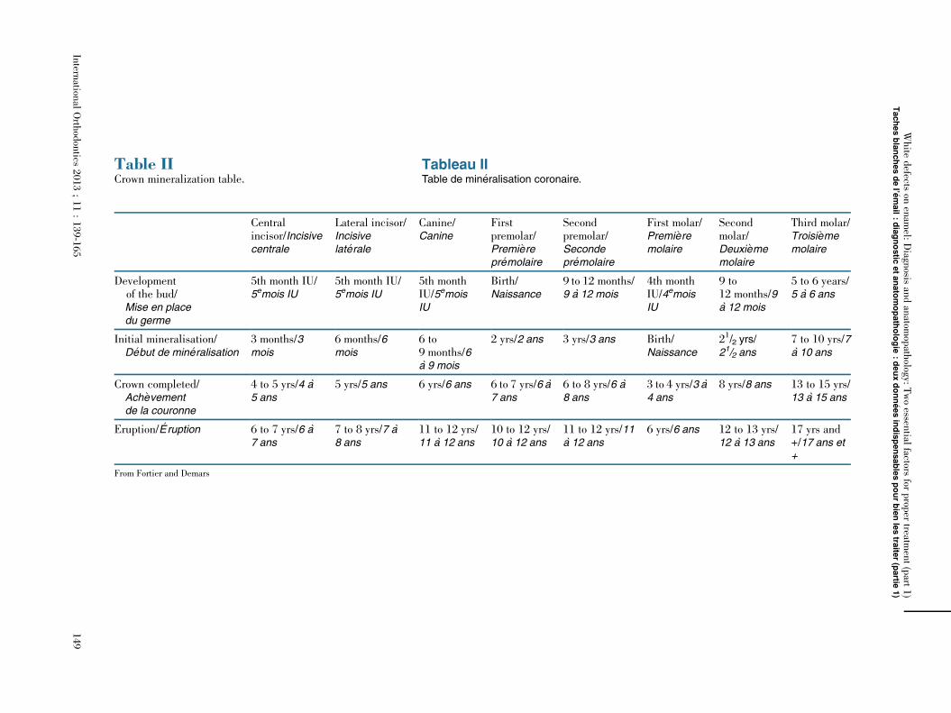
Table IICrown mineralization table.
Tableau IITable de min�eralisation coronaire.
Centralincisor/Incisivecentrale
Lateral incisor/Incisivelat�erale
Canine/Canine
Firstpremolar/Premi�erepr�emolaire
Secondpremolar/Secondepr�emolaire
First molar/Premi�eremolaire
Secondmolar/Deuxi�ememolaire
Third molar/Troisi�ememolaire
Developmentof the bud/Mise en placedu germe
5th month IU/5emois IU
5th month IU/5emois IU
5th monthIU/5emoisIU
Birth/Naissance
9 to 12 months/9 a 12 mois
4th monthIU/4emoisIU
9 to12 months/9a 12 mois
5 to 6 years/5 a 6 ans
Initial mineralisation/D�ebut de min�eralisation
3 months/3mois
6 months/6mois
6 to9 months/6a 9 mois
2 yrs/2 ans 3 yrs/3 ans Birth/Naissance
21/2 yrs/21/2 ans
7 to 10 yrs/7a 10 ans
Crown completed/Ach�evementde la couronne
4 to 5 yrs/4 a5 ans
5 yrs/5 ans 6 yrs/6 ans 6 to 7 yrs/6 a7 ans
6 to 8 yrs/6 a8 ans
3 to 4 yrs/3 a4 ans
8 yrs/8 ans 13 to 15 yrs/13 a 15 ans
Eruption/ �Eruption 6 to 7 yrs/6 a7 ans
7 to 8 yrs/7 a8 ans
11 to 12 yrs/11 a 12 ans
10 to 12 yrs/10 a 12 ans
11 to 12 yrs/11a 12 ans
6 yrs/6 ans 12 to 13 yrs/12 a 13 ans
17 yrs and+/17 ans et+
From Fortier and Demars
Intern
ational
Orth
odontics
2013;11:139-165
149
White
defects
onenam
el:Diagn
osisandanatom
opath
ology:Twoessen
tialfactors
forprop
ertreatm
ent(part
1)
Tachesblanchesdel’ �e
mail:diagnostic
etanatomopathologie
:deuxdonn�eesindispensablespourbienlestra
iter(partie
1)
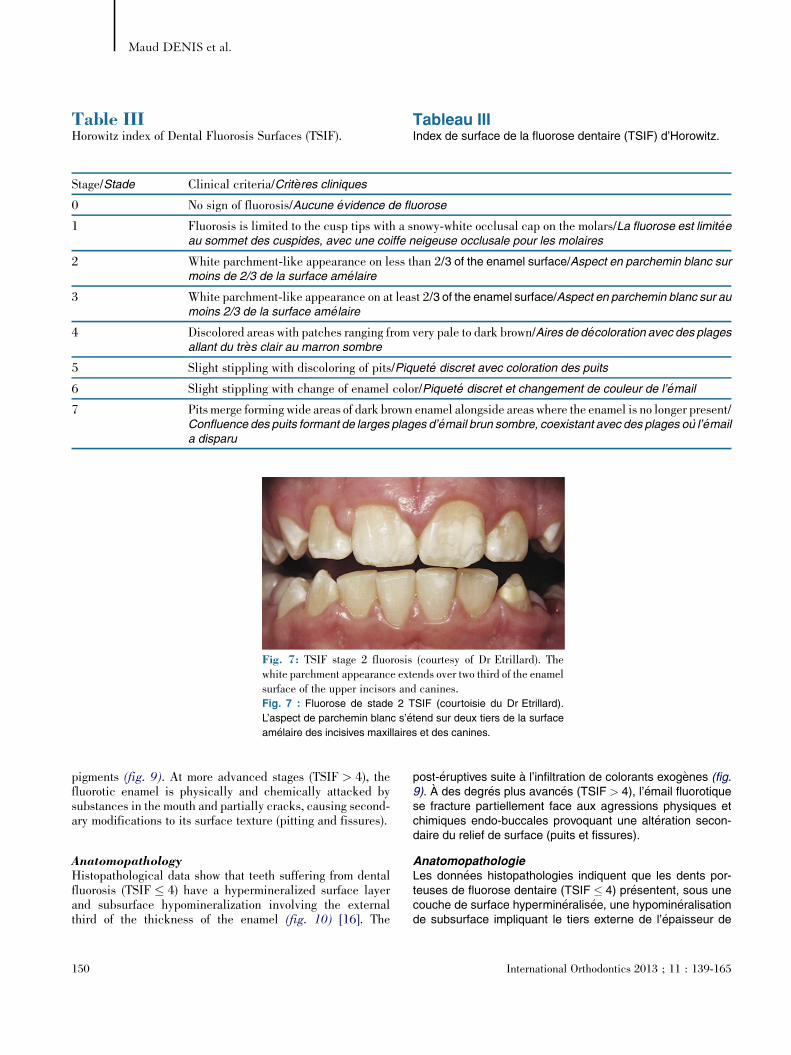
Table IIIHorowitz index of Dental Fluorosis Surfaces (TSIF).
Tableau IIIIndex de surface de la fluorose dentaire (TSIF) d’Horowitz.
Stage/Stade Clinical criteria/Crit�eres cliniques
0 No sign of fluorosis/Aucune �evidence de fluorose
1 Fluorosis is limited to the cusp tips with a snowy-white occlusal cap on the molars/La fluorose est limit�eeau sommet des cuspides, avec une coiffe neigeuse occlusale pour les molaires
2 White parchment-like appearance on less than 2/3 of the enamel surface/Aspect en parchemin blanc surmoins de 2/3 de la surface am�elaire
3 White parchment-like appearance on at least 2/3 of the enamel surface/Aspect en parchemin blanc sur aumoins 2/3 de la surface am�elaire
4 Discolored areas with patches ranging from very pale to dark brown/Aires de d�ecoloration avec des plagesallant du tr�es clair au marron sombre
5 Slight stippling with discoloring of pits/Piquet�e discret avec coloration des puits
6 Slight stippling with change of enamel color/Piquet�e discret et changement de couleur de l’�email
7 Pits merge forming wide areas of dark brown enamel alongside areas where the enamel is no longer present/Confluence des puits formant de larges plages d’�email brun sombre, coexistant avec des plages ou l’�emaila disparu
[(Fig._7)TD$FIG]
Fig. 7: TSIF stage 2 fluorosis (courtesy of Dr Etrillard). Thewhite parchment appearance extends over two third of the enamelsurface of the upper incisors and canines.Fig. 7 : Fluorose de stade 2 TSIF (courtoisie du Dr Etrillard).
L’aspect de parchemin blanc s’�etend sur deux tiers de la surface
am�elaire des incisives maxillaires et des canines.
Maud DENIS et al.
pigments (fig. 9). At more advanced stages (TSIF > 4), thefluorotic enamel is physically and chemically attacked bysubstances in the mouth and partially cracks, causing second-ary modifications to its surface texture (pitting and fissures).
AnatomopathologyHistopathological data show that teeth suffering from dentalfluorosis (TSIF � 4) have a hypermineralized surface layerand subsurface hypomineralization involving the externalthird of the thickness of the enamel (fig. 10) [16]. The
150
post-�eruptives suite a l’infiltration de colorants exog�enes (fig.9). A des degr�es plus avanc�es (TSIF > 4), l’�email fluorotiquese fracture partiellement face aux agressions physiques etchimiques endo-buccales provoquant une alt�eration secon-daire du relief de surface (puits et fissures).
AnatomopathologieLes donn�ees histopathologies indiquent que les dents por-teuses de fluorose dentaire (TSIF � 4) pr�esentent, sous unecouche de surface hypermin�eralis�ee, une hypomin�eralisationde subsurface impliquant le tiers externe de l’�epaisseur de
International Orthodontics 2013 ; 11 : 139-165

[(Fig._8)TD$FIG]
Fig. 8: TSIF stage 3 fluorosis. The white parchment appearanceextends over the whole of the enamel surface of the upper centralincisors.Fig. 8 : Fluorose de stade 3 TSIF. L’aspect de parchemin blanc
s’�etend sur la totalit�e de la surface am�elaire des incisives centrales
maxillaires.
[(Fig._9)TD$FIG]
Fig. 9: TSIF stage 4 fluorosis. Fluorotic lesions involve the wholesurface of the upper incisors. As well as the white parchment-likeappearance, the central incisors present dark-brown areas due topost-eruptive infiltration of the hypomineralized enamel by exo-genous chromophoric proteins.Fig. 9 : Fluorose de stade 4 TSIF. L’atteinte fluorotique concerne
l’ensemble de la surface des incisives maxillaires. Les incisives
centrales pr�esentent, en plus d’un aspect blanc parchemin�e, des
plages marron sombre cons�ecutives a une infiltration post-
�eruptive de l’�email hypomin�eralis�ee par les prot�eines chromo-
phores exog�enes.
White defects on enamel: Diagnosis and anatomopathology: Two essential factors for proper treatment (part 1)Taches blanches de l’�email : diagnostic et anatomopathologie : deux donn�ees indispensables pour bien les traiter (partie 1)
organization of the hypomineralization resulting from dentalfluorosis is the same as that encountered in early cariouslesions. At a macroscopic level, the mineral structure appearsunaffected. There is simply an increase in porosity along theinterprismatic spaces and the striae of Retzius (as in earlycarious lesions), due to a reduction in the number of crystalsbut not of their size [17]. Free spaces in the enamel are
International Orthodontics 2013 ; 11 : 139-165
l’�email (fig. 10) [16]. L’organisation de l’hypomin�eralisationcons�ecutive a une fluorose dentaire est la meme que cellerencontr�ee dans les l�esions carieuses initiales. A l’�echellemacroscopique, la trame min�erale n’est pas perturb�ee. Il y aseulement une augmentation des porosit�es le long desespaces inter-prismatiques et des stries de Retzius (toutcomme dans les l�esions carieuses initiales) permise par unediminution du nombre des cristaux et non de leur taille [17].
151
[(Fig._10)TD$FIG]
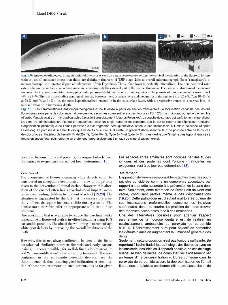
Fig. 10:Anatomopathological characteristics of fluorosis as seen on a transverse cross-section (the cervical localization of the fluorotic lesionswithout loss of substance shows that these are definitely fluoroses of TSIF stage 2/3): a: overall microradiograph (from Yanagisawa); b:microradiograph with greater degree of enlargement (from Fejerskov). The surface layer is perfectly mineralized. The demineralized zoneextends below the surface at an obtuse angle and concerns only the external part of the enamel thickness. The prismatic structure of the enamelremains intact; c: semi-quantitative mapping under polarized-light microscopy (from Fejerskov). The porosity of fluorotic enamel varies from 1+% to 25+%. There is a descending gradient of porosity between the subsurface layer and the interior of the enamel (1/4 at 25+%, 1/4 at 10+%, 1/4at 5+% and 1/4 at 1+%); i.e. the most hypomineralized enamel is in the subsurface layer, with a progressive return to a normal level ofmineralization with increasing depth.Fig. 10 : Les caract�eristiques anatomopathologiques d’une fluorose a partir de section transversale (la localisation cervicale des l�esions
fluorotiques sans perte de substance indique que nous sommes surement face a des fluoroses TSIF 2/3) : a : microradiographie d’ensemble
(d’apr�es Yanagisawa) ; b : microradiographie a plus fort grossissement (d’apr�es Fejerskov). La couche de surface est parfaitement min�eralis�ee.
La zone de d�emin�eralisation s’�etend en subsurface selon un angle obtus et ne concerne que la partie externe de l’�epaisseur am�elaire.
L’organisation prismatique de l’�email persiste ; c : cartographie semi-quantitative obtenue par microscopie a lumi�ere polaris�ee (d’apr�es
Fejerskov). La porosit�e d’un �email fluorotique va de 1+ % a 25+ %. Il existe un gradient d�ecroissant du taux de porosit�e entre de la couche
de subsurface et l’int�erieur de l’�email (1/4 de 25+ %, 1/4 de 10+ %, 1/4 de 5+ % et 1/4 de 1+ %) ; c’est-a-dire que l’�email le plus hypomin�eralis�e se
trouve en subsurface, puis retourne en profondeur progressivement a un taux de min�eralisation normal.
Maud DENIS et al.
occupied by ionic fluids and proteins, the origin of which (fromthe matrix or exogenous) has not yet been determined [18].
TreatmentThe occurrence of fluorosis causing white defects could beconsidered an acceptable compromise in view of the prioritygiven to the prevention of dental caries. However, this alter-ation of the enamel often has a psychological impact, some-times even leading children to drop out of school [19,20]. Thesituation is aggravated by the fact that the disease preferen-tially affects the upper incisors, visible during a smile. Thedentist must therefore offer an appropriate solution to theseproblems.One possibility that is available to reduce the parchment-likeappearance of fluorosed teeth is in-office bleaching using 10%carbamide peroxide. The aim of the whitening is to camouflagewhite spot defects by increasing the overall brightness of theteeth.
However, this is not always sufficient. In view of the histo-pathological similarity between fluorosis and early cariouslesions, it seems possible, for well-defined cloudy areas, toadd “erosion-infiltration” after whitening treatment. The ureacontained in the carbamide peroxide deproteinizes thefluorotic enamel, thus ensuring good infiltration. A combina-tion of these two treatments in such patients has so far given
152
Les espaces libres am�elaires sont occup�es par des fluidesioniques et des prot�eines dont l’origine (matricielles ouexog�enes) n’est a ce jour pas d�etermin�ee [18].
TraitementL’apparition de fluorose responsable de taches blanches pour-rait etre consid�er�ee comme un compromis acceptable parrapport a la priorit�e accord�ee a la pr�evention de la carie den-taire. Seulement, cette alt�eration de l’�email est souvent malv�ecue, conduisant parfois meme a des d�escolarisations[19,20]. Cette pathologie est d’autant mal tol�er�ee qu’une deses localisations pr�ef�erentielles concerne les incisivessup�erieures, dents du sourire. Le praticien doit alors trouverdes r�eponses acceptables face a ces demandes.Une des alternatives possibles pour att�enuer l’aspectparchemin�e de la fluorose dentaire est de r�ealiser un�eclaircissement ambulatoire au peroxyde de carbamidea 10 %. L’�eclaircissement aura pour objectif de camouflerles d�efauts blancs en augmentant la luminosit�e g�en�erale desdents.Seulement, cette proposition n’est pas toujours suffisante. Sereportant a la similitude histopathologie des fluoroses avec lesl�esions carieuses initiales, il apparaıt possible, en cas de plagenuageuse bien d�elimit�ee, de compl�eter l’�eclaircissement parun temps d’« �erosion-infiltration ». L’ur�ee contenue dans leperoxyde de carbamide assure la d�eprot�einisation de l’�emailfluorotique, pr�ealable a une bonne infiltration. L’association de
International Orthodontics 2013 ; 11 : 139-165
White defects on enamel: Diagnosis and anatomopathology: Two essential factors for proper treatment (part 1)Taches blanches de l’�email : diagnostic et anatomopathologie : deux donn�ees indispensables pour bien les traiter (partie 1)
perfectly satisfactory esthetic results with a clinical follow-upof 19 months [21].
Traumatic hypomineralization
Traumatic hypomineralization of a permanent tooth is a con-sequence of periodontal trauma affecting the deciduous teeth.This may involve displacements of all kinds (concussion,subluxation, luxation with or without lateral displacement,intrusion, extrusion, shedding/extraction), which represent80% of trauma to the primary dentition [22]. Whatever theseverity of this trauma, the appearance of sequellae is spo-radic. A simple shock that often passes unnoticed duringchildhood may be the cause of the occurrence of these defects.Intrusion nevertheless remains the most harmful injury [23].During the emergency consultation, the dentist can only offerparents a presumptive indication of the future risks.
Trauma confined to hard tissues and the pulp of deciduousteeth cannot in itself give rise to sequellae affecting the per-manent tooth buds. However, resulting periapical infectionscan do so.The close anatomical proximity that exists between the apexesof the anterior deciduous teeth and the germs of their perma-nent successors, that also display delayed calcification,explains this relationship. On the level of the incisors, thetwo types of dentition are separated only by a slender bonybarrier less than 3 mm thick, or sometimes even by simplefibrous connective tissue. Slight periapical inflammation canthen disturb mineralization of the underlying germ, dependingon its degree of calcification. Generally speaking, it is disrup-tion at the stage of maturation of the ameloblasts that leads tothe appearance of traumatic hypomineralization. In the eventof earlier disturbance, there may also be associatedhypoplasia.Other more serious sequellae classified by Andreasen (struc-tural abnormalities [coronal and/or radicular] and positionalabnormalities) are not mentioned here.
PrevalenceThe prevalence of this hypomineralization is estimated as5.2%. This figure is not surprising, given that one third ofchildren suffer a traumatic episode involving their deciduousteeth before the age of 5. The early years of life are a periodwhen children are learning to walk and exploring their envi-ronment. This frequently leads to premaxillary injuries.Recurrence of trauma has no impact on the occurrence ofsequellae. However, age can affect both their nature and theirseverity.
DiagnosisTraumatic hypomineralization can present a wide variety ofclinical expressions differing in shape, outline, localization
International Orthodontics 2013 ; 11 : 139-165
ces deux th�erapeutiques chez ces patients pr�esente a ce jourdes r�esultats esth�etiques enti�erement satisfaisants avec unrecul clinique de 19 mois [21].
Hypomin�eralisation traumatique
L’« hypomin�eralisation traumatique » de dent permanente estune s�equelle cons�ecutive a un traumatisme du parodonte desdents lact�eales. Il peut s’agir de toutes les formes de luxation(concussion, subluxation, luxation sans/avec d�eplacementlat�eral, intrusion, extrusion, expulsion/extraction), repr�esen-tant 80 % des traumatismes lact�eales [22]. L’apparition des�equelles est sporadique quelle que soit la gravit�e du trauma-tisme. Une simple concussion, bien souvent pass�ee inapercuependant la petite enfance, peut ainsi etre mise en cause dansl’apparition de ces d�efauts. L’intrusion reste tout de meme laplus d�el�et�ere [23]. Le praticien, lors de la consultationd’urgence, ne peut d�elivrer aux parents que des pr�esomptionsde survenue.Les traumatismes circonscrits aux tissus durs et a la pulpe desdents lact�eales ne peuvent a eux seuls entraıner de s�equellessur les germes ascensionnels. En revanche, les infections dup�eriapex qui en sont cons�ecutives le peuvent.La forte proximit�e anatomique existante entre les apex desdents lact�eales ant�erieures et les germes des dents perma-nentes successionnelles arborant, en plus, une calcificationtardive, explique cette interrelation. Dans le cas des incisives,seule une fine barri�ere osseuse de moins de 3 mmd’�epaisseur, et parfois meme un simple tissu conjonctiffibreux, s�epare les deux dentures. Une sommaire inflamma-tion du p�eriapex peut alors perturber la min�eralisation dugerme sus-jacent selon son stade de calcification. De facong�en�erale, c’est la perturbation du stade de maturation desam�eloblastes qui aboutit a l’apparition d’hypomin�eralisationtraumatique. Lors de perturbation plus pr�ecoce, une hypopla-sie peut �egalement y etre associ�ee.D’autres formes de s�equelles plus grave (anomalies structur-ales [coronaires et/ou radiculaires] et anomalies de positions)class�ees par Andreasen ne sont pas �evoqu�ees ici.
Pr�evalenceLa pr�evalence de cette hypomin�eralisation est �evalu�eea 5,2 %. Ce chiffre n’est pas �etonnant, �etant donn�e qu’un tiersdes enfants est expos�e avant l’age de cinq ans a un �episodetraumatiques des dents temporaires. Les premi�eres ann�eesde la vie sont pour l’enfant une p�eriode d’apprentissage de lamarche et de d�ecouverte de son environnement. Les bles-sures du pr�emaxillaire y sont fr�equemment associ�ees. Lar�ecurrence des traumatismes n’a pas d’effet sur la survenuedes s�equelles. En revanche, l’age en a un sur leur nature etleur gravit�e.
DiagnosticLes hypomin�eralisations traumatiques peuvent revetir unegrande vari�et�e d’expressions cliniques de part leur forme, leurcontour, leur localisation et meme leur teinte. Ce sont
153
[(Fig._11)TD$FIG]
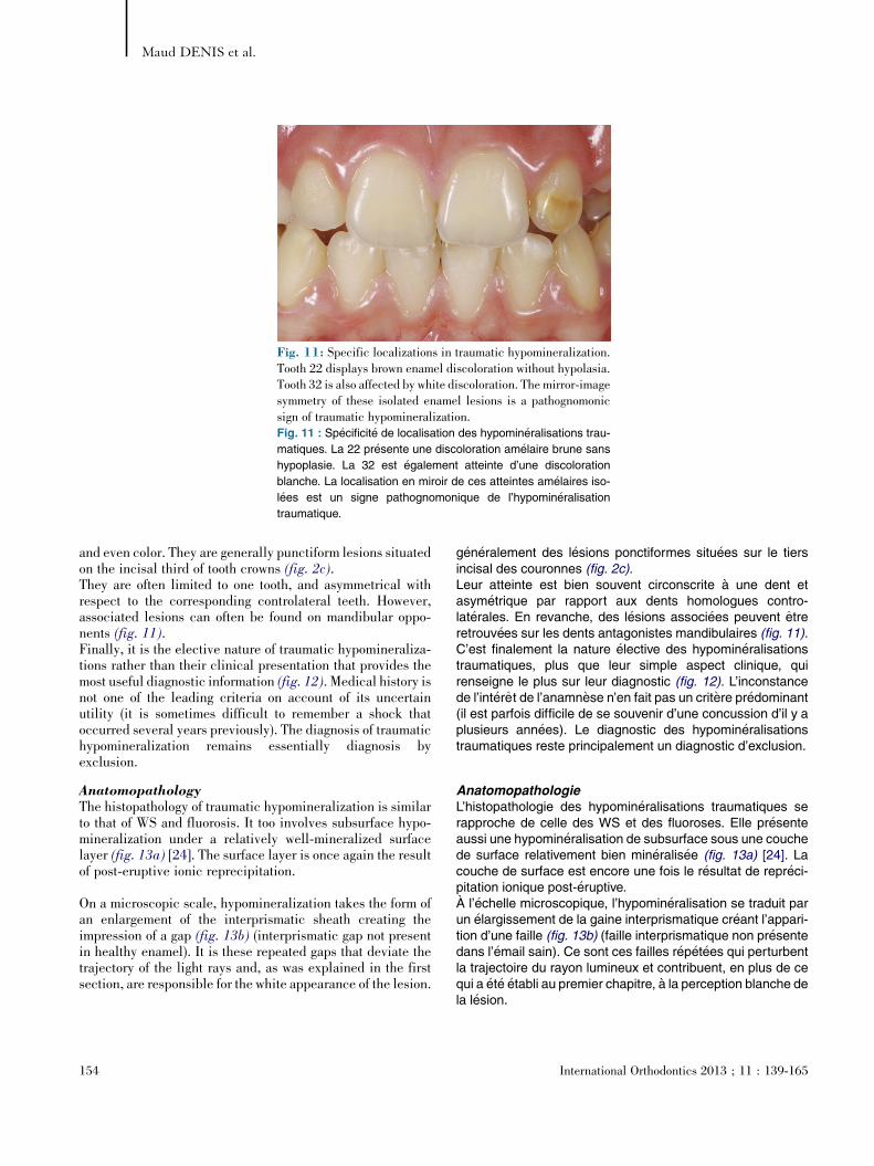
Fig. 11: Specific localizations in traumatic hypomineralization.Tooth 22 displays brown enamel discoloration without hypolasia.Tooth 32 is also affected by white discoloration. The mirror-imagesymmetry of these isolated enamel lesions is a pathognomonicsign of traumatic hypomineralization.Fig. 11 : Sp�ecificit�e de localisation des hypomin�eralisations trau-
matiques. La 22 pr�esente une discoloration am�elaire brune sans
hypoplasie. La 32 est �egalement atteinte d’une discoloration
blanche. La localisation en miroir de ces atteintes am�elaires iso-
l�ees est un signe pathognomonique de l’hypomin�eralisation
traumatique.
Maud DENIS et al.
and even color. They are generally punctiform lesions situatedon the incisal third of tooth crowns (fig. 2c).They are often limited to one tooth, and asymmetrical withrespect to the corresponding controlateral teeth. However,associated lesions can often be found on mandibular oppo-nents (fig. 11).Finally, it is the elective nature of traumatic hypomineraliza-tions rather than their clinical presentation that provides themost useful diagnostic information (fig. 12). Medical history isnot one of the leading criteria on account of its uncertainutility (it is sometimes difficult to remember a shock thatoccurred several years previously). The diagnosis of traumatichypomineralization remains essentially diagnosis byexclusion.
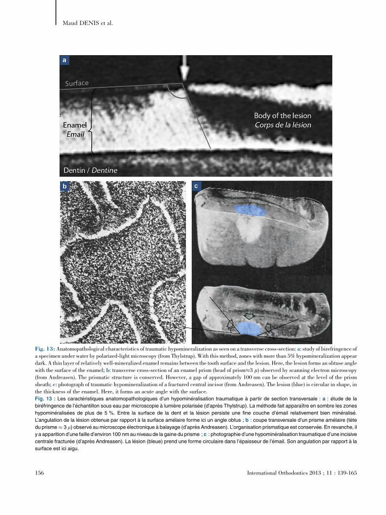
AnatomopathologyThe histopathology of traumatic hypomineralization is similarto that of WS and fluorosis. It too involves subsurface hypo-mineralization under a relatively well-mineralized surfacelayer (fig. 13a) [24]. The surface layer is once again the resultof post-eruptive ionic reprecipitation.
On a microscopic scale, hypomineralization takes the form ofan enlargement of the interprismatic sheath creating theimpression of a gap (fig. 13b) (interprismatic gap not presentin healthy enamel). It is these repeated gaps that deviate thetrajectory of the light rays and, as was explained in the firstsection, are responsible for the white appearance of the lesion.
154
g�en�eralement des l�esions ponctiformes situ�ees sur le tiersincisal des couronnes (fig. 2c).Leur atteinte est bien souvent circonscrite a une dent etasym�etrique par rapport aux dents homologues contro-lat�erales. En revanche, des l�esions associ�ees peuvent etreretrouv�ees sur les dents antagonistes mandibulaires (fig. 11).C’est finalement la nature �elective des hypomin�eralisationstraumatiques, plus que leur simple aspect clinique, quirenseigne le plus sur leur diagnostic (fig. 12). L’inconstancede l’int�eret de l’anamn�ese n’en fait pas un crit�ere pr�edominant(il est parfois difficile de se souvenir d’une concussion d’il y aplusieurs ann�ees). Le diagnostic des hypomin�eralisationstraumatiques reste principalement un diagnostic d’exclusion.
AnatomopathologieL’histopathologie des hypomin�eralisations traumatiques serapproche de celle des WS et des fluoroses. Elle pr�esenteaussi une hypomin�eralisation de subsurface sous une couchede surface relativement bien min�eralis�ee (fig. 13a) [24]. Lacouche de surface est encore une fois le r�esultat de repr�eci-pitation ionique post-�eruptive.A l’�echelle microscopique, l’hypomin�eralisation se traduit parun �elargissement de la gaine interprismatique cr�eant l’appari-tion d’une faille (fig. 13b) (faille interprismatique non pr�esentedans l’�email sain). Ce sont ces failles r�ep�et�ees qui perturbentla trajectoire du rayon lumineux et contribuent, en plus de cequi a �et�e �etabli au premier chapitre, a la perception blanche dela l�esion.
International Orthodontics 2013 ; 11 : 139-165

[(Fig._12)TD$FIG]
Fig. 12: Diagnosis of traumatic hypomineralization. The clinicalappearance of the lesion (here very similar to that of fluorosis)provides very little etiological information. It is the location that ismore useful diagnostically; isolated lesion with no controlateralsymmetry.Fig. 12 : Diagnostic d’une hypomin�eralisation traumatique.
L’aspect clinique de la l�esion (tr�es semblable ici a une fluorose)
informe peu sur son �etiologie. C’est sa localisation �elective qui
informe le plus sur son diagnostic ; l�esion isol�ee avec absence
de sym�etrie sur la controlat�erale.
White defects on enamel: Diagnosis and anatomopathology: Two essential factors for proper treatment (part 1)Taches blanches de l’�email : diagnostic et anatomopathologie : deux donn�ees indispensables pour bien les traiter (partie 1)
However, their depth and morphology are just as variable astheir expression; they depend on the circumstances of theiroccurrence. Thus, just like WS and fluoroses, they can some-times extend like drops of oil below the surface of the enamelwhich they then encounter at an obtuse angle (fig. 13a). Atother times, they may adopt a circular shape and form an acuteangle with the enamel surface (fig. 13c) [25].
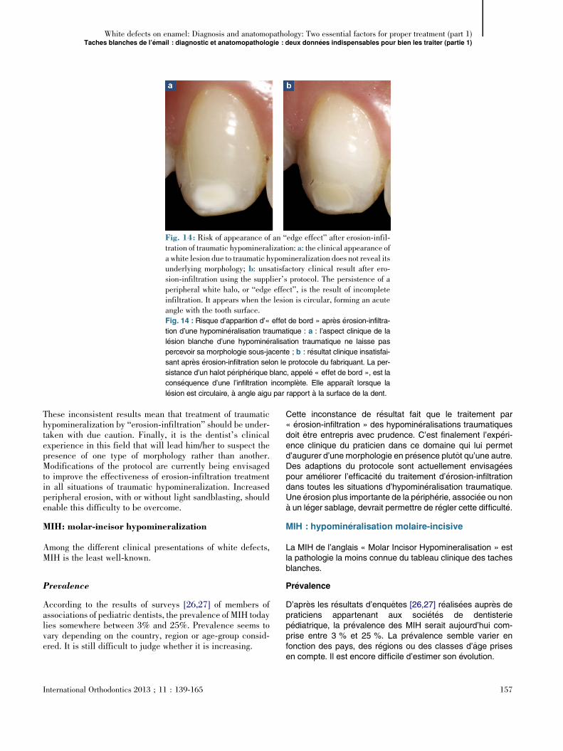
TreatmentIt is due to these inconsistent angles that the results of treat-ment of traumatic hypomineralization by erosion-infiltrationare difficult to predict.On account of the acute angle, the infiltration is often incom-plete on the margins, leading to a visual contouring of thelesion known as the “edge effect” (fig. 14). The erosion stageeliminates the thin, relatively well-mineralized surface layer,making only the central part of the “body” accessible to theresin, while on the edges of the lesion, in the acute angle,erosion alone cannot remove the peripheral lips of healthyenamel. These overhangs prevent the resin from penetratingthe porous areas on the edges of the lesion. The center of thespot disappears but a more or less homogeneous white outlineremains. The “patch” appearance produced by this “edgeeffect” is unfortunately sometimes more unsightly than thespot itself.
Conversely, in the presence of an obtuse angle (as in the caseof WS and fluorosis), the erosion stage is effective. Completeinfiltration is obtained over the whole of the hypomineralizedarea and the spot disappears totally.
International Orthodontics 2013 ; 11 : 139-165
En revanche, leur profondeur et leur morphologie sont toutesaussi variables que leur expression ; elles d�ependent descirconstances de survenue. Ainsi, parfois elle s’�etend, toutcomme les WS et les fluoroses, en goutte d’huile sous la sur-face am�elaire y pr�esentant alors un angle obtus (fig. 13a). Etd’autre fois, elle acquiert une morphologie circulaire formantdes angles aigus par rapport a la surface am�elaire (fig. 13c)[25].
TraitementCe sont ces inconstances d’angulation qui rendent aujourd’huile traitement par « �erosion-infiltration » des hypomin�eralisa-tions traumatiques peu pr�edictif.En effet, a cause de cette angulation aigu€e, l’infiltration estsouvent incompl�ete au niveau des marges produisant visuel-lement un d�etourage de la l�esion appel�e « effet de bord »(fig. 14). L’�etape d’�erosion �elimine la mince couche de surfacerelativement min�eralis�ee de la l�esion rendant accessible a lar�esine seulement la partie centrale de son « corps ». Tandisqu’au pourtour de la l�esion a angle aigu, l’�erosion ne peut a elleseule �eliminer les b�equets p�eriph�eriques d’�email sainexistants ; l’acc�es aux porosit�es n’�etant en regard de cesb�equets pas lib�er�e, la r�esine ne peut y p�en�etrer. Il se produitalors une disparition de la tache en son centre associ�ee a lapr�eservation d’un fin contour blanc plus ou moins homog�ene.L’effet pastille provoqu�e par cet « effet de bord » est malheur-eusement parfois encore plus disgracieux que la tache en elle-meme.Inversement, lors d’une angulation obtus (comme dansles cas de WS et fluorose), l’�etape d’�erosion estsuffisante. L’infiltration est compl�ete sur l’ensemble del’hypomin�eralisation ; la tache disparaıt alors compl�etement.
155
[(Fig._13)TD$FIG]
Fig. 13:Anatomopathological characteristics of traumatic hypomineralization as seen on a transverse cross-section: a: study of birefringence ofa specimen under water by polarized-light microscopy (from Thylstrup). With this method, zones with more than 5% hypomineralization appeardark. A thin layer of relatively well-mineralized enamel remains between the tooth surface and the lesion. Here, the lesion forms an obtuse anglewith the surface of the enamel; b: transverse cross-section of an enamel prism (head of prism�3 m) observed by scanning electron microscopy(from Andreasen). The prismatic structure is conserved. However, a gap of approximately 100 nm can be observed at the level of the prismsheath; c: photograph of traumatic hypomineralization of a fractured central incisor (from Andreasen). The lesion (blue) is circular in shape, inthe thickness of the enamel. Here, it forms an acute angle with the surface.Fig. 13 : Les caract�eristiques anatomopathologiques d’un hypomin�eralisation traumatique a partir de section transversale : a : �etude de la
bir�efringence de l’�echantillon sous eau par microscopie a lumi�ere polaris�ee (d’apr�es Thylstrup). La m�ethode fait apparaıtre en sombre les zones
hypomin�eralis�ees de plus de 5 %. Entre la surface de la dent et la l�esion persiste une fine couche d’�email relativement bien min�eralis�e.
L’angulation de la l�esion obtenue par rapport a la surface am�elaire forme ici un angle obtus ; b : coupe transversale d’un prisme am�elaire (tete
du prisme � 3 m) observ�e aumicroscope �electronique a balayage (d’apr�es Andreasen). L’organisation prismatique est conserv�ee. En revanche, il
y a apparition d’une faille d’environ 100 nmau niveau de la gaine du prisme ; c : photographie d’une hypomin�eralisation traumatique d’une incisive
centrale fractur�ee (d’apr�es Andreasen). La l�esion (bleue) prend une forme circulaire dans l’�epaisseur de l’�email. Son angulation par rapport a la
surface est ici aigu.
156 International Orthodontics 2013 ; 11 : 139-165
Maud DENIS et al.
[(Fig._14)TD$FIG]
Fig. 14: Risk of appearance of an “edge effect” after erosion-infil-tration of traumatic hypomineralization: a: the clinical appearance ofa white lesion due to traumatic hypomineralization does not reveal itsunderlying morphology; b: unsatisfactory clinical result after ero-sion-infiltration using the supplier’s protocol. The persistence of aperipheral white halo, or “edge effect”, is the result of incompleteinfiltration. It appears when the lesion is circular, forming an acuteangle with the tooth surface.Fig. 14 : Risque d’apparition d’« effet de bord » apr�es �erosion-infiltra-
tion d’une hypomin�eralisation traumatique : a : l’aspect clinique de la
l�esion blanche d’une hypomin�eralisation traumatique ne laisse pas
percevoir sa morphologie sous-jacente ; b : r�esultat clinique insatisfai-
sant apr�es �erosion-infiltration selon le protocole du fabriquant. La per-
sistance d’un halot p�eriph�erique blanc, appel�e « effet de bord », est la
cons�equence d’une l’infiltration incompl�ete. Elle apparaıt lorsque la
l�esion est circulaire, a angle aigu par rapport a la surface de la dent.
White defects on enamel: Diagnosis and anatomopathology: Two essential factors for proper treatment (part 1)Taches blanches de l’�email : diagnostic et anatomopathologie : deux donn�ees indispensables pour bien les traiter (partie 1)
These inconsistent results mean that treatment of traumatichypomineralization by “erosion-infiltration” should be under-taken with due caution. Finally, it is the dentist’s clinicalexperience in this field that will lead him/her to suspect thepresence of one type of morphology rather than another.Modifications of the protocol are currently being envisagedto improve the effectiveness of erosion-infiltration treatmentin all situations of traumatic hypomineralization. Increasedperipheral erosion, with or without light sandblasting, shouldenable this difficulty to be overcome.
MIH: molar-incisor hypomineralization
Among the different clinical presentations of white defects,MIH is the least well-known.
Prevalence
According to the results of surveys [26,27] of members ofassociations of pediatric dentists, the prevalence of MIH todaylies somewhere between 3% and 25%. Prevalence seems tovary depending on the country, region or age-group consid-ered. It is still difficult to judge whether it is increasing.
International Orthodontics 2013 ; 11 : 139-165
Cette inconstance de r�esultat fait que le traitement par« �erosion-infiltration » des hypomin�eralisations traumatiquesdoit etre entrepris avec prudence. C’est finalement l’exp�eri-ence clinique du praticien dans ce domaine qui lui permetd’augurer d’une morphologie en pr�esence plutot qu’une autre.Des adaptions du protocole sont actuellement envisag�eespour am�eliorer l’efficacit�e du traitement d’�erosion-infiltrationdans toutes les situations d’hypomin�eralisation traumatique.Une �erosion plus importante de la p�eriph�erie, associ�ee ou nona un l�eger sablage, devrait permettre de r�egler cette difficult�e.
MIH : hypomin�eralisation molaire-incisive
La MIH de l’anglais « Molar Incisor Hypomineralisation » estla pathologie la moins connue du tableau clinique des tachesblanches.
Pr�evalence
D’apr�es les r�esultats d’enquetes [26,27] r�ealis�ees aupr�es depraticiens appartenant aux soci�et�es de dentisteriep�ediatrique, la pr�evalence des MIH serait aujourd’hui com-prise entre 3 % et 25 %. La pr�evalence semble varier enfonction des pays, des r�egions ou des classes d’age prisesen compte. Il est encore difficile d’estimer son �evolution.
157
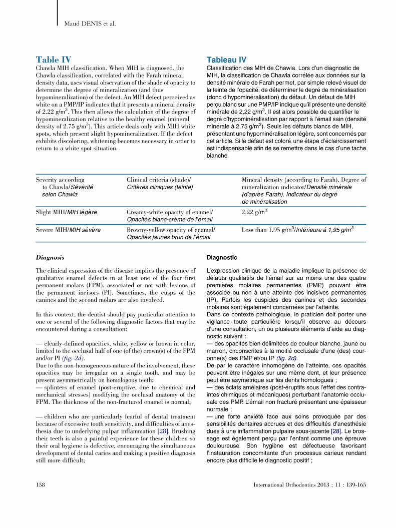
Table IVChawla MIH classification. When MIH is diagnosed, theChawla classification, correlated with the Farah mineraldensity data, uses visual observation of the shade of opacity todetermine the degree of mineralization (and thushypomineralization) of the defect. AnMIH defect perceived aswhite on a PMP/IP indicates that it presents a mineral densityof 2.22 g/m3. This then allows the calculation of the degree ofhypomineralization relative to the healthy enamel (mineraldensity of 2.75 g/m3). This article deals only with MIH whitespots, which present slight hypomineralization. If the defectexhibits discoloring, whitening becomes necessary in order toreturn to a white spot situation.
Tableau IVClassification des MIH de Chawla. Lors d’un diagnostic deMIH, la classification de Chawla corr�el�ee aux donn�ees sur ladensit�e min�erale de Farah permet, par simple relev�e visuel dela teinte de l’opacit�e, de d�eterminer le degr�e de min�eralisation(donc d’hypomin�eralisation) du d�efaut. Un d�efaut de MIHpercu blanc sur une PMP/IP indique qu’il pr�esente une densit�emin�erale de 2,22 g/m3. Il est alors possible de quantifier ledegr�e d’hypomin�eralisation par rapport a l’�email sain (densit�emin�erale a 2,75 g/m3). Seuls les d�efauts blancs de MIH,pr�esentant une hypomin�eralisation l�eg�ere, sont concern�es parcet article. Si le d�efaut est color�e, une �etape d’�eclaircissementest indispensable afin de se remettre dans le cas d’une tacheblanche.
Severity accordingto Chawla/S�ev�erit�eselon Chawla
Clinical criteria (shade)/Crit�eres cliniques (teinte)
Mineral density (according to Farah). Degree ofmineralization indicator/Densit�e min�erale(d’apr�es Farah). Indicateur du degr�ede min�eralisation
Slight MIH/MIH l�eg�ere Creamy-white opacity of enamel/Opacit�es blanc-cr�eme de l’�email
2.22 g/m3
Severe MIH/MIH s�ev�ere Browny-yellow opacity of enamel/Opacit�es jaunes brun de l’�email
Less than 1.95 g/m3/Inf�erieure a 1,95 g/m3
Maud DENIS et al.
Diagnosis
The clinical expression of the disease implies the presence ofqualitative enamel defects in at least one of the four firstpermanent molars (FPM), associated or not with lesions ofthe permanent incisors (PI). Sometimes, the cusps of thecanines and the second molars are also involved.
In this context, the dentist should pay particular attention toone or several of the following diagnostic factors that may beencountered during a consultation:
— clearly-defined opacities, white, yellow or brown in color,limited to the occlusal half of one (of the) crown(s) of the FPMand/or PI (fig. 2d).Due to the non-homogeneous nature of the involvement, theseopacities may be irregular on a single tooth, and may bepresent asymmetrically on homologous teeth;— splinters of enamel (post-eruptive, due to chemical andmechanical stresses) modifying the occlusal anatomy of theFPM. The thickness of the non-fractured enamel is normal;
— children who are particularly fearful of dental treatmentbecause of excessive tooth sensitivity, and difficulties of anes-thesia due to underlying pulpar inflammation [28]. Brushingtheir teeth is also a painful experience for these children sotheir oral hygiene is defective, encouraging the simultaneousdevelopment of dental caries and making a positive diagnosisstill more difficult;
158
Diagnostic
L’expression clinique de la maladie implique la pr�esence ded�efauts qualitatifs de l’�email sur au moins une des quatrepremi�eres molaires permanentes (PMP) pouvant etreassoci�ee ou non a une atteinte des incisives permanentes(IP). Parfois les cuspides des canines et des secondesmolaires sont �egalement concern�ees par l’atteinte.Dans ce contexte pathologique, le praticien doit porter unevigilance toute particuli�ere lorsqu’il observe au d�ecoursd’une consultation, un ou plusieurs �el�ements d’aide au diag-nostic suivant :— des opacit�es bien d�elimit�ees de couleur blanche, jaune oumarron, circonscrites a la moiti�e occlusale d’une (des) cour-onne(s) des PMP et/ou IP (fig. 2d).De par le caract�ere inhomog�ene de l’atteinte, ces opacit�espeuvent etre in�egales sur une meme dent, et leur pr�esencepeut etre asym�etrique sur les dents homologues ;— des �eclats am�elaires (post-�eruptifs sous l’effet des contra-intes chimiques et m�ecaniques) perturbant l’anatomie occlu-sale des PMP. L’�email non fractur�e pr�esentant une �epaisseurnormale ;— une forte anxi�et�e face aux soins provoqu�ee par dessensibilit�es dentaires accrues et des difficult�es d’anesth�esiedues a une inflammation pulpaire sous-jacente [28]. Le bros-sage est �egalement percu par l’enfant comme une �epreuvedouloureuse. Son hygi�ene est d�efectueuse favorisantl’instauration concomitante d’un processus carieux rendantencore plus difficile le diagnostic positif ;
International Orthodontics 2013 ; 11 : 139-165
[(Fig._15)TD$FIG]
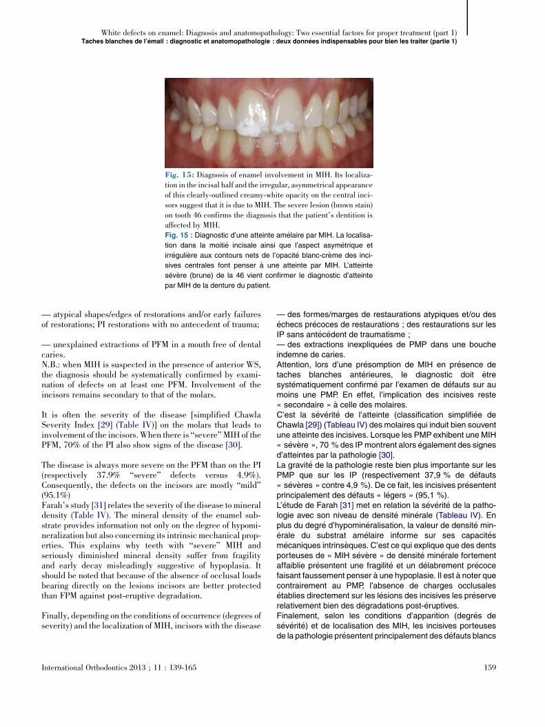
Fig. 15: Diagnosis of enamel involvement in MIH. Its localiza-tion in the incisal half and the irregular, asymmetrical appearanceof this clearly-outlined creamy-white opacity on the central inci-sors suggest that it is due to MIH. The severe lesion (brown stain)on tooth 46 confirms the diagnosis that the patient’s dentition isaffected by MIH.Fig. 15 : Diagnostic d’une atteinte am�elaire par MIH. La localisa-
tion dans la moiti�e incisale ainsi que l’aspect asym�etrique et
irr�eguli�ere aux contours nets de l’opacit�e blanc-cr�eme des inci-
sives centrales font penser a une atteinte par MIH. L’atteinte
s�ev�ere (brune) de la 46 vient confirmer le diagnostic d’atteinte
par MIH de la denture du patient.
White defects on enamel: Diagnosis and anatomopathology: Two essential factors for proper treatment (part 1)Taches blanches de l’�email : diagnostic et anatomopathologie : deux donn�ees indispensables pour bien les traiter (partie 1)
— atypical shapes/edges of restorations and/or early failuresof restorations; PI restorations with no antecedent of trauma;
— unexplained extractions of PFM in a mouth free of dentalcaries.N.B.: when MIH is suspected in the presence of anterior WS,the diagnosis should be systematically confirmed by exami-nation of defects on at least one PFM. Involvement of theincisors remains secondary to that of the molars.
It is often the severity of the disease [simplified ChawlaSeverity Index [29] (Table IV)] on the molars that leads toinvolvement of the incisors.When there is “severe”MIH of thePFM, 70% of the PI also show signs of the disease [30].
The disease is always more severe on the PFM than on the PI(respectively 37.9% “severe” defects versus 4.9%).Consequently, the defects on the incisors are mostly “mild”(95.1%)Farah’s study [31] relates the severity of the disease to mineraldensity (Table IV). The mineral density of the enamel sub-strate provides information not only on the degree of hypomi-neralization but also concerning its intrinsic mechanical prop-erties. This explains why teeth with “severe” MIH andseriously diminished mineral density suffer from fragilityand early decay misleadingly suggestive of hypoplasia. Itshould be noted that because of the absence of occlusal loadsbearing directly on the lesions incisors are better protectedthan FPM against post-eruptive degradation.
Finally, depending on the conditions of occurrence (degrees ofseverity) and the localization of MIH, incisors with the disease
International Orthodontics 2013 ; 11 : 139-165
— des formes/marges de restaurations atypiques et/ou des�echecs pr�ecoces de restaurations ; des restaurations sur lesIP sans ant�ec�edent de traumatisme ;— des extractions inexpliqu�ees de PMP dans une boucheindemne de caries.Attention, lors d’une pr�esomption de MIH en pr�esence detaches blanches ant�erieures, le diagnostic doit etresyst�ematiquement confirm�e par l’examen de d�efauts sur aumoins une PMP. En effet, l’implication des incisives reste« secondaire » a celle des molaires.C’est la s�ev�erit�e de l’atteinte (classification simplifi�ee deChawla [29]) (Tableau IV) des molaires qui induit bien souventune atteinte des incisives. Lorsque les PMPexhibent une MIH« s�ev�ere », 70 % des IPmontrent alors �egalement des signesd’atteintes par la pathologie [30].La gravit�e de la pathologie reste bien plus importante sur lesPMP que sur les IP (respectivement 37,9 % de d�efauts« s�ev�eres » contre 4,9 %). De ce fait, les incisives pr�esententprincipalement des d�efauts « l�egers » (95,1 %).L’�etude de Farah [31] met en relation la s�ev�erit�e de la patho-logie avec son niveau de densit�e min�erale (Tableau IV). Enplus du degr�e d’hypomin�eralisation, la valeur de densit�e min-�erale du substrat am�elaire informe sur ses capacit�esm�ecaniques intrins�eques. C’est ce qui explique que des dentsporteuses de « MIH s�ev�ere » de densit�e min�erale fortementaffaiblie pr�esentent une fragilit�e et un d�elabrement pr�ecocefaisant faussement penser a une hypoplasie. Il est a noter quecontrairement au PMP, l’absence de charges occlusales�etablies directement sur les l�esions des incisives les pr�eserverelativement bien des d�egradations post-�eruptives.Finalement, selon les conditions d’apparition (degr�es des�ev�erit�e) et de localisation des MIH, les incisives porteusesde la pathologie pr�esentent principalement des d�efauts blancs
159
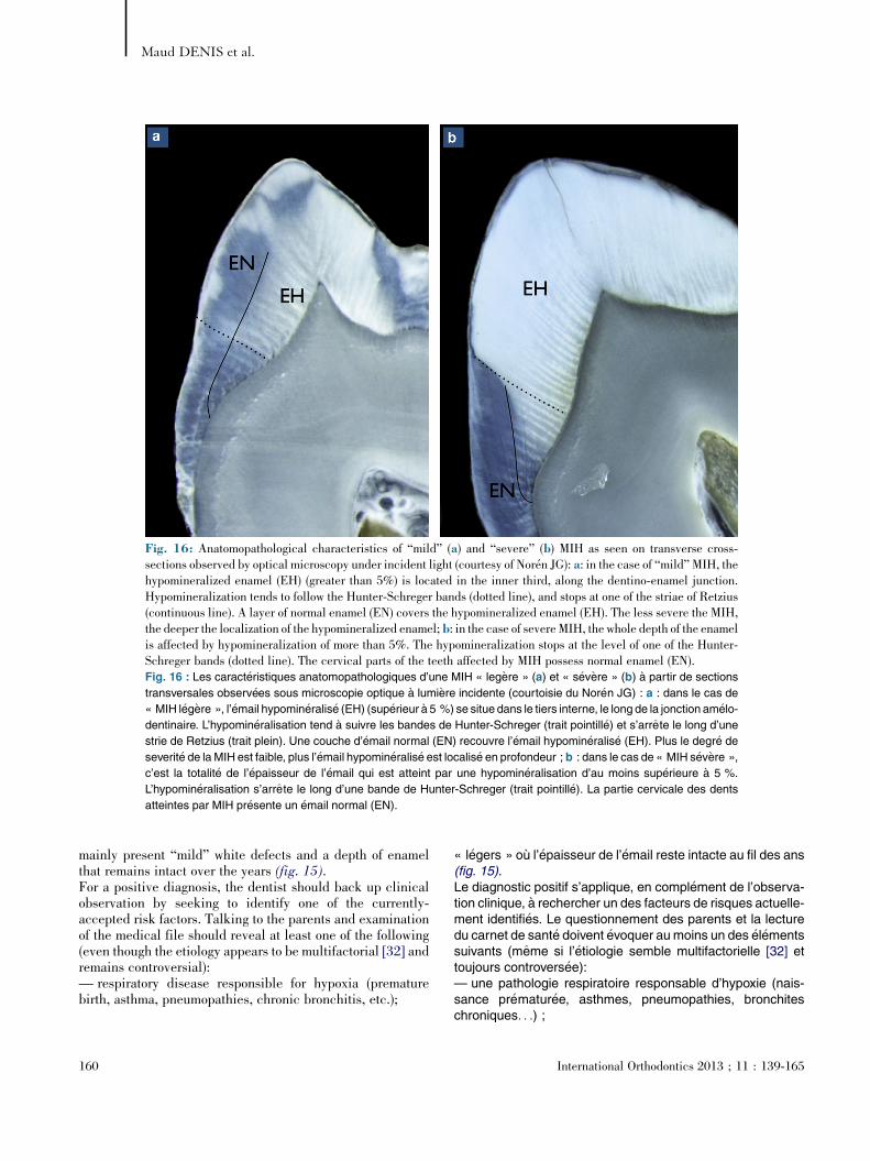
[(Fig._16)TD$FIG]
Fig. 16: Anatomopathological characteristics of “mild” (a) and “severe” (b) MIH as seen on transverse cross-sections observed by optical microscopy under incident light (courtesy of Nor�en JG): a: in the case of “mild” MIH, thehypomineralized enamel (EH) (greater than 5%) is located in the inner third, along the dentino-enamel junction.Hypomineralization tends to follow the Hunter-Schreger bands (dotted line), and stops at one of the striae of Retzius(continuous line). A layer of normal enamel (EN) covers the hypomineralized enamel (EH). The less severe the MIH,the deeper the localization of the hypomineralized enamel; b: in the case of severe MIH, the whole depth of the enamelis affected by hypomineralization of more than 5%. The hypomineralization stops at the level of one of the Hunter-Schreger bands (dotted line). The cervical parts of the teeth affected by MIH possess normal enamel (EN).Fig. 16 : Les caract�eristiques anatomopathologiques d’une MIH « leg�ere » (a) et « s�ev�ere » (b) a partir de sections
transversales observ�ees sous microscopie optique a lumi�ere incidente (courtoisie du Nor�en JG) : a : dans le cas de
« MIH l�eg�ere », l’�email hypomin�eralis�e (EH) (sup�erieur a 5 %) se situe dans le tiers interne, le long de la jonction am�elo-
dentinaire. L’hypomin�eralisation tend a suivre les bandes de Hunter-Schreger (trait pointill�e) et s’arrete le long d’une
strie de Retzius (trait plein). Une couche d’�email normal (EN) recouvre l’�email hypomin�eralis�e (EH). Plus le degr�e de
severit�e de la MIH est faible, plus l’�email hypomin�eralis�e est localis�e en profondeur ; b : dans le cas de « MIH s�ev�ere »,
c’est la totalit�e de l’�epaisseur de l’�email qui est atteint par une hypomin�eralisation d’au moins sup�erieure a 5 %.
L’hypomin�eralisation s’arrete le long d’une bande de Hunter-Schreger (trait pointill�e). La partie cervicale des dents
atteintes par MIH pr�esente un �email normal (EN).
Maud DENIS et al.
mainly present “mild” white defects and a depth of enamelthat remains intact over the years (fig. 15).For a positive diagnosis, the dentist should back up clinicalobservation by seeking to identify one of the currently-accepted risk factors. Talking to the parents and examinationof the medical file should reveal at least one of the following(even though the etiology appears to be multifactorial [32] andremains controversial):— respiratory disease responsible for hypoxia (prematurebirth, asthma, pneumopathies, chronic bronchitis, etc.);
160
« l�egers » ou l’�epaisseur de l’�email reste intacte au fil des ans(fig. 15).Le diagnostic positif s’applique, en compl�ement de l’observa-tion clinique, a rechercher un des facteurs de risques actuelle-ment identifi�es. Le questionnement des parents et la lecturedu carnet de sant�e doivent �evoquer aumoins un des �el�ementssuivants (meme si l’�etiologie semble multifactorielle [32] ettoujours controvers�ee):— une pathologie respiratoire responsable d’hypoxie (nais-sance pr�ematur�ee, asthmes, pneumopathies, bronchiteschroniques. . .) ;
International Orthodontics 2013 ; 11 : 139-165
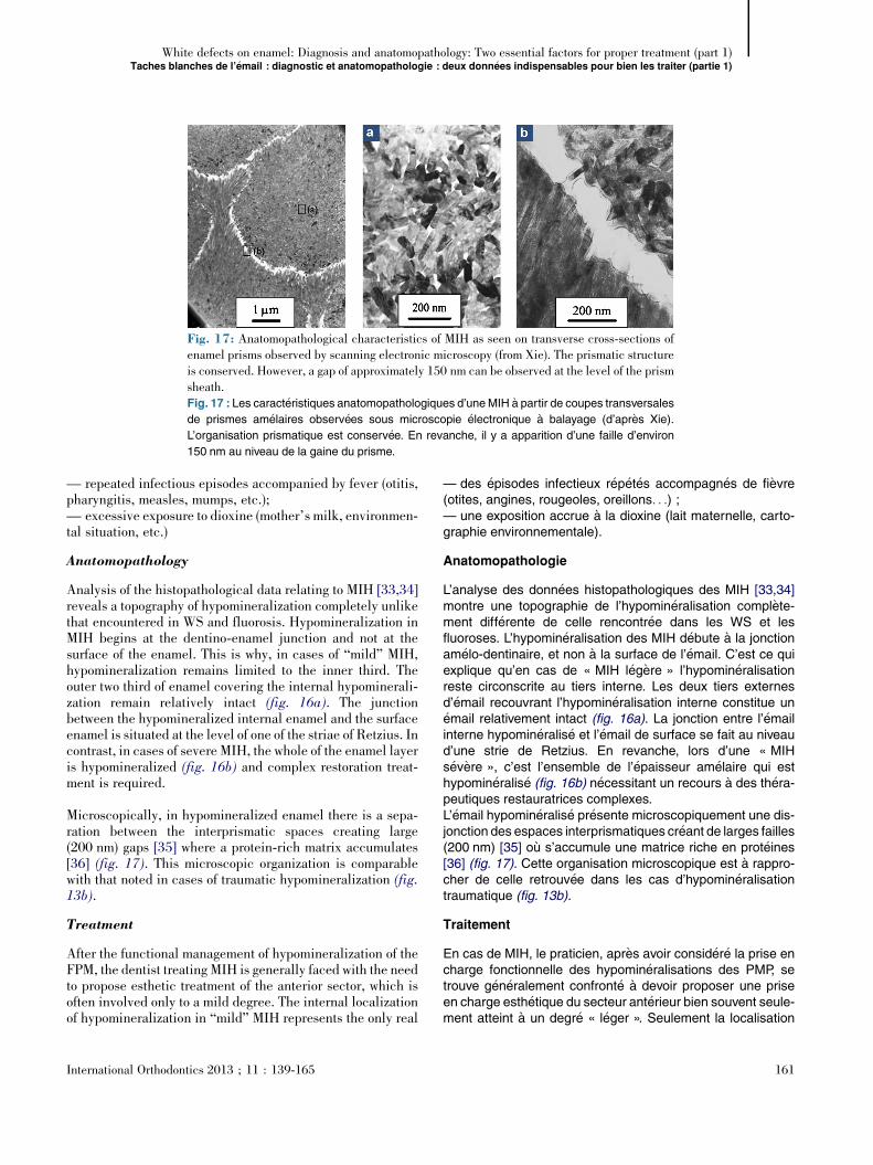
[(Fig._17)TD$FIG]
Fig. 17: Anatomopathological characteristics of MIH as seen on transverse cross-sections ofenamel prisms observed by scanning electronic microscopy (from Xie). The prismatic structureis conserved. However, a gap of approximately 150 nm can be observed at the level of the prismsheath.Fig. 17 : Les caract�eristiques anatomopathologiques d’uneMIH a partir de coupes transversales
de prismes am�elaires observ�ees sous microscopie �electronique a balayage (d’apr�es Xie).
L’organisation prismatique est conserv�ee. En revanche, il y a apparition d’une faille d’environ
150 nm au niveau de la gaine du prisme.
White defects on enamel: Diagnosis and anatomopathology: Two essential factors for proper treatment (part 1)Taches blanches de l’�email : diagnostic et anatomopathologie : deux donn�ees indispensables pour bien les traiter (partie 1)
— repeated infectious episodes accompanied by fever (otitis,pharyngitis, measles, mumps, etc.);— excessive exposure to dioxine (mother’s milk, environmen-tal situation, etc.)
Anatomopathology
Analysis of the histopathological data relating to MIH [33,34]reveals a topography of hypomineralization completely unlikethat encountered in WS and fluorosis. Hypomineralization inMIH begins at the dentino-enamel junction and not at thesurface of the enamel. This is why, in cases of “mild” MIH,hypomineralization remains limited to the inner third. Theouter two third of enamel covering the internal hypominerali-zation remain relatively intact (fig. 16a). The junctionbetween the hypomineralized internal enamel and the surfaceenamel is situated at the level of one of the striae of Retzius. Incontrast, in cases of severe MIH, the whole of the enamel layeris hypomineralized (fig. 16b) and complex restoration treat-ment is required.
Microscopically, in hypomineralized enamel there is a sepa-ration between the interprismatic spaces creating large(200 nm) gaps [35] where a protein-rich matrix accumulates[36] (fig. 17). This microscopic organization is comparablewith that noted in cases of traumatic hypomineralization (fig.13b).
Treatment
After the functional management of hypomineralization of theFPM, the dentist treating MIH is generally faced with the needto propose esthetic treatment of the anterior sector, which isoften involved only to a mild degree. The internal localizationof hypomineralization in “mild” MIH represents the only real
International Orthodontics 2013 ; 11 : 139-165
— des �episodes infectieux r�ep�et�es accompagn�es de fi�evre(otites, angines, rougeoles, oreillons. . .) ;— une exposition accrue a la dioxine (lait maternelle, carto-graphie environnementale).
Anatomopathologie
L’analyse des donn�ees histopathologiques des MIH [33,34]montre une topographie de l’hypomin�eralisation compl�ete-ment diff�erente de celle rencontr�ee dans les WS et lesfluoroses. L’hypomin�eralisation des MIH d�ebute a la jonctionam�elo-dentinaire, et non a la surface de l’�email. C’est ce quiexplique qu’en cas de « MIH l�eg�ere » l’hypomin�eralisationreste circonscrite au tiers interne. Les deux tiers externesd’�email recouvrant l’hypomin�eralisation interne constitue un�email relativement intact (fig. 16a). La jonction entre l’�emailinterne hypomin�eralis�e et l’�email de surface se fait au niveaud’une strie de Retzius. En revanche, lors d’une « MIHs�ev�ere », c’est l’ensemble de l’�epaisseur am�elaire qui esthypomin�eralis�e (fig. 16b) n�ecessitant un recours a des th�era-peutiques restauratrices complexes.L’�email hypomin�eralis�e pr�esente microscopiquement une dis-jonction des espaces interprismatiques cr�eant de larges failles(200 nm) [35] ou s’accumule une matrice riche en prot�eines[36] (fig. 17). Cette organisation microscopique est a rappro-cher de celle retrouv�ee dans les cas d’hypomin�eralisationtraumatique (fig. 13b).
Traitement
En cas de MIH, le praticien, apr�es avoir consid�er�e la prise encharge fonctionnelle des hypomin�eralisations des PMP, setrouve g�en�eralement confront�e a devoir proposer une priseen charge esth�etique du secteur ant�erieur bien souvent seule-ment atteint a un degr�e « l�eger ». Seulement la localisation
161
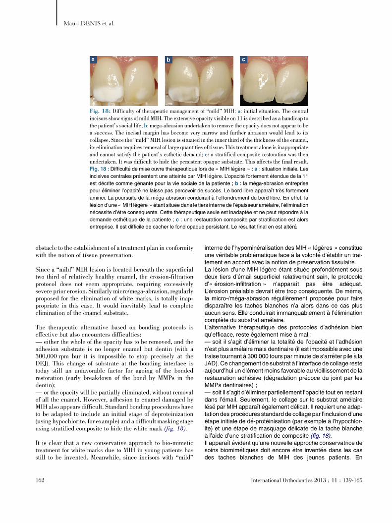
[(Fig._18)TD$FIG]
Fig. 18: Difficulty of therapeutic management of “mild” MIH: a: initial situation. The centralincisors show signs of mildMIH. The extensive opacity visible on 11 is described as a handicap tothe patient’s social life; b: mega-abrasion undertaken to remove the opacity does not appear to bea success. The incisal margin has become very narrow and further abrasion would lead to itscollapse. Since the “mild” MIH lesion is situated in the inner third of the thickness of the enamel,its elimination requires removal of large quantities of tissue. This treatment alone is inappropriateand cannot satisfy the patient’s esthetic demand; c: a stratified composite restoration was thenundertaken. It was difficult to hide the persistent opaque substrate. This affects the final result.Fig. 18 : Difficult�e de mise ouvre th�erapeutique lors de « MIH l�eg�ere » : a : situation initiale. Les
incisives centrales pr�esentent une atteinte par MIH l�eg�ere. L’opacit�e fortement �etendue de la 11
est d�ecrite comme genante pour la vie sociale de la patiente ; b : la m�ega-abrasion entreprise
pour �eliminer l’opacit�e ne laisse pas percevoir de succ�es. Le bord libre apparaıt tr�es fortement
aminci. La poursuite de la m�ega-abrasion conduirait a l’effondrement du bord libre. En effet, la
l�esion d’une « MIH l�eg�ere » �etant situ�ee dans le tiers interne de l’�epaisseur am�elaire, l’�elimination
n�ecessite d’etre cons�equente. Cette th�erapeutique seule est inadapt�ee et ne peut r�epondre a la
demande esth�etique de la patiente ; c : une restauration composite par stratification est alors
entreprise. Il est difficile de cacher le fond opaque persistant. Le r�esultat final en est alt�er�e.
Maud DENIS et al.
obstacle to the establishment of a treatment plan in conformitywith the notion of tissue preservation.
Since a “mild” MIH lesion is located beneath the superficialtwo third of relatively healthy enamel, the erosion-filtrationprotocol does not seem appropriate, requiring excessivelysevere prior erosion. Similarly micro/mega-abrasion, regularlyproposed for the elimination of white marks, is totally inap-propriate in this case. It would inevitably lead to completeelimination of the enamel substrate.
The therapeutic alternative based on bonding protocols iseffective but also encounters difficulties:— either the whole of the opacity has to be removed, and theadhesion substrate is no longer enamel but dentin (with a300,000 rpm bur it is impossible to stop precisely at theDEJ). This change of substrate at the bonding interface istoday still an unfavorable factor for ageing of the bondedrestoration (early breakdown of the bond by MMPs in thedentin);— or the opacity will be partially eliminated, without removalof all the enamel. However, adhesion to enamel damaged byMIH also appears difficult. Standard bonding procedures haveto be adapted to include an initial stage of deproteinization(using hypochlorite, for example) and a difficult masking stageusing stratified composite to hide the white mark (fig. 18).
It is clear that a new conservative approach to bio-mimetictreatment for white marks due to MIH in young patients hasstill to be invented. Meanwhile, since incisors with “mild”
162
interne de l’hypomin�eralisation des MIH « l�eg�eres » constitueune v�eritable probl�ematique face a la volont�e d’�etablir un trai-tement en accord avec la notion de pr�eservation tissulaire.La l�esion d’une MIH l�eg�ere �etant situ�ee profond�ement sousdeux tiers d’�email superficiel relativement sain, le protocoled’« �erosion-infiltration » n’apparaıt pas etre ad�equat.L’�erosion pr�ealable devrait etre trop cons�equente. De meme,la micro-/m�ega-abrasion r�eguli�erement propos�ee pour fairedisparaıtre les taches blanches n’a alors dans ce cas plusaucun sens. Elle conduirait immanquablement a l’�eliminationcompl�ete du substrat am�elaire.L’alternative th�erapeutique des protocoles d’adh�esion bienqu’efficace, reste �egalement mise a mal :— soit il s’agit d’�eliminer la totalit�e de l’opacit�e et l’adh�esionn’est plus am�elaire mais dentinaire (il est impossible avec unefraise tournant a 300 000 tours par minute de s’arreter pile a laJAD). Ce changement de substrat a l’interface de collage resteaujourd’hui un �el�ement moins favorable au vieillissement de larestauration adh�esive (d�egradation pr�ecoce du joint par lesMMPs dentinaires) ;— soit il s’agit d’�eliminer partiellement l’opacit�e tout en restantdans l’�email. Seulement, le collage sur le substrat am�elairel�es�e par MIH apparaıt �egalement d�elicat. Il requiert une adap-tation des proc�edures standard de collage par l’inclusion d’une�etape initiale de d�e-prot�einisation (par exemple a l’hypochlor-ite) et une �etape de masquage d�elicate de la tache blanchea l’aide d’une stratification de composite (fig. 18).Il apparaıt �evident qu’une nouvelle approche conservatrice desoins biomim�etiques doit encore etre invent�ee dans les casdes taches blanches de MIH des jeunes patients. En
International Orthodontics 2013 ; 11 : 139-165
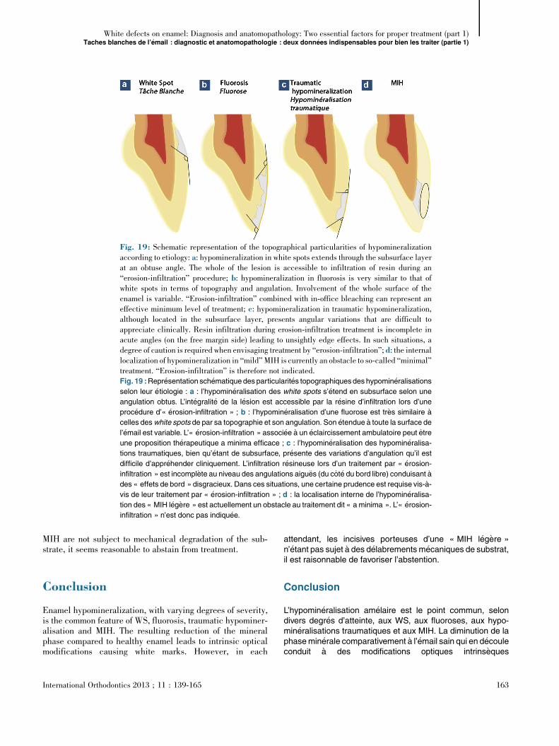
[(Fig._19)TD$FIG]
Fig. 19: Schematic representation of the topographical particularities of hypomineralizationaccording to etiology: a: hypomineralization in white spots extends through the subsurface layerat an obtuse angle. The whole of the lesion is accessible to infiltration of resin during an“erosion-infiltration” procedure; b: hypomineralization in fluorosis is very similar to that ofwhite spots in terms of topography and angulation. Involvement of the whole surface of theenamel is variable. “Erosion-infiltration” combined with in-office bleaching can represent aneffective minimum level of treatment; c: hypomineralization in traumatic hypomineralization,although located in the subsurface layer, presents angular variations that are difficult toappreciate clinically. Resin infiltration during erosion-infiltration treatment is incomplete inacute angles (on the free margin side) leading to unsightly edge effects. In such situations, adegree of caution is required when envisaging treatment by “erosion-infiltration”; d: the internallocalization of hypomineralization in “mild”MIH is currently an obstacle to so-called “minimal”treatment. “Erosion-infiltration” is therefore not indicated.Fig. 19 :Repr�esentation sch�ematiquedes particularit�es topographiques des hypomin�eralisations
selon leur �etiologie : a : l’hypomin�eralisation des white spots s’�etend en subsurface selon une
angulation obtus. L’int�egralit�e de la l�esion est accessible par la r�esine d’infiltration lors d’une
proc�edure d’« �erosion-infiltration » ; b : l’hypomin�eralisation d’une fluorose est tr�es similaire a
celles deswhite spots de par sa topographie et son angulation. Son �etendue a toute la surface de
l’�email est variable. L’« �erosion-infiltration » associ�ee a un �eclaircissement ambulatoire peut etre
une proposition th�erapeutique a minima efficace ; c : l’hypomin�eralisation des hypomin�eralisa-
tions traumatiques, bien qu’�etant de subsurface, pr�esente des variations d’angulation qu’il est
difficile d’appr�ehender cliniquement. L’infiltration r�esineuse lors d’un traitement par « �erosion-
infiltration » est incompl�ete au niveau des angulations aigu€es (du cot�e du bord libre) conduisant a
des « effets de bord » disgracieux. Dans ces situations, une certaine prudence est requise vis-a-
vis de leur traitement par « �erosion-infiltration » ; d : la localisation interne de l’hypomin�eralisa-
tion des « MIH l�eg�ere » est actuellement un obstacle au traitement dit « a minima ». L’« �erosion-
infiltration » n’est donc pas indiqu�ee.
White defects on enamel: Diagnosis and anatomopathology: Two essential factors for proper treatment (part 1)Taches blanches de l’�email : diagnostic et anatomopathologie : deux donn�ees indispensables pour bien les traiter (partie 1)
MIH are not subject to mechanical degradation of the sub-strate, it seems reasonable to abstain from treatment.
Conclusion
Enamel hypomineralization, with varying degrees of severity,is the common feature of WS, fluorosis, traumatic hypominer-alisation and MIH. The resulting reduction of the mineralphase compared to healthy enamel leads to intrinsic opticalmodifications causing white marks. However, in each
International Orthodontics 2013 ; 11 : 139-165
attendant, les incisives porteuses d’une « MIH l�eg�ere »n’�etant pas sujet a des d�elabrementsm�ecaniques de substrat,il est raisonnable de favoriser l’abstention.
Conclusion
L’hypomin�eralisation am�elaire est le point commun, selondivers degr�es d’atteinte, aux WS, aux fluoroses, aux hypo-min�eralisations traumatiques et aux MIH. La diminution de laphasemin�erale comparativement a l’�email sain qui en d�ecouleconduit a des modifications optiques intrins�eques
163
Maud DENIS et al.
pathological context this hypomineralization assumes differ-ent topographical forms (fig. 19), each requiring a specifictherapeutic approach. Prior diagnosis of these forms is obvi-ously an essential prerequisite to treatment. This leads tobetter understanding of the anatamopathology and shows thatWS and fluorosis are broadly comparable, allowing treatmentby erosion-infiltration to be indiscriminately applied.Traumatic hypomineralization is also a comparable situation,although variation of surface angles should encourage greatercaution. Finally, it is only in the case of mild MIH, wherehypomineralization is located internally, that abstention isrecommended. A forthcoming article will present all possibletreatments for these marks, along with a new modification tothe erosion-infiltration protocol making it possible to treathypomineralization resulting from MIH.
Disclosure of interest
The authors declare that they have no conflicts of interestconcerning this article.
References/R�ef�erences
1. Gorelick L, Geiger AM, Gwibanding. Am J Orthod 1982
2. Richter AE, Arruda AO, Petreated with comprehensive(5):657–64.
3. Øgaard B. Prevalence of worthodontically treated perso1989;96(5):423–7.
4. Ismail AI, Sohn W, Tellezsystem (ICDAS): an integratEpidemiol 2007;35(3):170–8
5. Selwitz RH, Ismail AI, Pitts6. Kielbassa AM, M€uller J, Ge
mally invasive dentistry: a reenamel lesions. Quintessenc
7. Silverstone LM. Structure o1973;3:100–60.
8. Kaqueler JC, Le May O. Ldentaire. �Ed. Masson, Paris
9. Pearce EIF, Nelson DGA. Mback-scattered electrons. J D
10. Palamara J, Phakey PP, Rachof white spot and brown spot
11. Wang LJ, Tang RK, Bonstenanoscale enamel dissolutio
164
responsables de taches blanches. Seulement, cette hypo-min�eralisation acquiert selon chaque pathologie diff�erentesformes topographiques (fig. 19) qui conduisent a devoir adap-ter sp�ecifiquement leur approche th�erapeutique. Leur diag-nostic pr�ealable devient �evidemment un pr�erequis indispen-sable a leur prise en charge. Les connaissancesanatomopathologies qui en d�ecoulent, informent que les WSet les fluoroses sont globalement comparables. C’est ce quiassure la possibilit�e d’appliquer indistinctement le protocoled’�erosion-infiltration. Les hypomin�eralisations traumatiquespeuvent �egalement y etre assimil�ees malgr�e la variabilit�e deson angulation de surface amenant a une plus grande pru-dence. Finalement, seule la localisation interne de l’hypo-min�eralisation des MIH l�eg�eres am�ene actuellement a uneabstention. Dans un prochain article, nous pr�esenterons tousles traitements possibles de ces taches, puis nous proposer-ons une modification originale du protocole de l’�erosion-infil-tration qui permettra de traiter les hypomin�eralisations li�eesaux MIH.
D�eclaration d’int�erets
Les auteurs d�eclarent ne pas avoir de conflits d’int�erets enrelation avec cet article.
nnett AJ. Incidence of white spot formation after bonding and;81(2):93–8.ters MC, Sohn W. Incidence of caries lesions among patientsorthodontics. Am J Orthod Dentofacial Orthop 2011;139
hite spot lesions in 19-year-olds: a study on untreated andns 5 years after treatement. Am J Orthod Dentofacial Orthop
M, et al. The international caries detection and assessmented system for mesuring dental caries. Community Dent Oral.NB. Dental caries. Lancet 2007;369:51–9.rnhardt CR. Closing the gap between oral hygiene and mini-view on the resin infiltration technique of incipient (proximal)e Int 2009;40:663–81.f carious enamel, including the early lesion. Oral Sci Rev
�esions carieuses de l’�email. Anatomie pathologique bucco-p. 41–58 1998.icrostructural features of carious human enamel imaged withent Res 1989;68(2):113–8.inger WA, Orams HJ. Ultrastructure of the intact surface zonecarious lesions in human enamel. Oral Pathol 1986;15:28-35.in T, Orme CA, Bush PJ, Nancollas GH. A new model forn. J Phys Chem 2005;109:999-1005.
International Orthodontics 2013 ; 11 : 139-165

12. Atlan A, Denis M, Tirlet G, Attal JP. L’�erosion-infiltration : un nouveau traitement destaches blanches de l’�email. Clinic 2012 (Hors s�erie l’esth�etique a la francaise):23–9 .
13. Sixou JL, Bailleul-forestier I. Recommandation sur la pr�escription des fluorures de lanaissance a l’adolescence. J Odontol Stomatol Pediatr 2004;11(3):157–68.
14. Hescot P, Roland E. La sant�e dentaire en France en 1998. UFSBD, Paris p. 126 1999.15. Fortier JP, Demars-Fremault C. Abr�eg�e de p�edodontie. Masson �edit, Paris 1987.16. Fejerskov O, Larsen MJ, Richards A, Baelum V. Dental tissue effects of fluoride. Adv Dent
Res 1994;8(1):15-31.17. Yanagisawa T, Takuma S, Fejerskov O. Ultrastructure and composition of enamel in human
dental fluorosis. Adv Dent Res 1989;3(2):203–10.18. Wright JT, Chen SC, Hall KI, Yamauchi M, et al. Protein characterization of fluorosed
human enamel. J Dent Res 1996;75(12):1936–41.19. Riordan PJ. Perceptions of dental fluorosis. J Dent Res 1993;72:1268–74.20. Marshman Z, Gibson B, Robinson PG. The impact of developmental defects of enamel on
young people in the UK. Community Dent Oral Epidemiol 2009;37:45-57.21. Tirlet G, Fron H, Attal JP. Infiltration, a new therapy for masking white spots on enamel: a
case series with 19-month follow-up. Eur J Esthet Dent 2013 .22. Von Arx T. Developmental disturbances of permanent teeth following trauma to the primary
dentition. Aust Dent J 1993;38(1):1-10.23. De Amorim LFG, Estrela C, Da Costa LRRS. Effects of traumatic dental injuries to primary
teeth on permanent teeth–a clinical follow-up study. Dent Traumatol 2011;27:117–21.24. Thylstrup A, Andreasen JO. The influence of traumatic intrusion of primary teeth on their
permanent successors in monkeys. A macroscopic, polarized ligiit and scanning electronmicroscopic study. J Oral Pathol 1977;6:296-306.
25. Andreasen JO, Sundstr€om B, Ravn JJ. The effect of traumatic injuries to primary teeth ontheir permanent successors. I. A clinical and histologic study of 117 injured permanentteeth. Scand J Dent Res 1971;79:219–83.
26. Weerheijm KL, Duggal M, Mejare I. Judgement criteria for Molar-Incisor-Hypomineralisation (MIH) in epidemiologic studies: a summary of the European meetingon MIH held in Athens, 2003. Eur Arch Paediatr Dent 2003;3:110–3.
27. Crombie FA, Manton DJ, Weerheijm KL, Kilpatrick NM. Molar-Incisor-Hypomineralisation: a survey of members of the Australian and New Zealand Society ofPaediatric Dentistry. Aust Dent J 2008;53(2):160–6.
28. Rodd HD, Boissonade FM, Day PF. Pulpal status of hypomineralised permanent molars.Pediatr Dent 2007;29:514–20.
29. Chawla N, Messer LB, Silva M. Clinical studies on molar-incisor hypomineralisation. Part2: Development of a Severity Index. Eur Arch Paediatr Dent 2008;9(4):191–9.
30. Lygidakis NA, Dimou G, Briseniou E. Molar-incisor hypomineralisation (MIH).Retrospective clinical study in Greek children. I. Prevalence and defect characteristics.Eur Arch Paediatr Dent 2008;9(4):200–6.
31. Farah R, Drummond B, Swain M, Williams S. Linking the clinical presentation of molar-incisor hypomineralisation to its mineral density. Int J Paediatr Dent 2010;20:353–60.
32. Beentjes VEVM, Weerheijm JL, Groen HM. Factors involved in the aetiology of molar-incisivor hypomineralisation (MIH). Eur J Paediatr Dent 2002;3(1):9-13.
33. J€alevik B, Nor�en JG. Enamel hypomineralisation of permanent first molars: a morphologicalstudy and survey of possible aetiological factors. Int J Paediatr Dent 2000;10(4):278–89.
34. Fearne J, Anderson P, Davis GR. 3D X-ray microscopic study of the extent of variations inenamel density in first permanent molars with idiopathic enamel hypomineralisation. BrDent J 2004;196(10):634–8.
35. Xie ZH, Kilpatrick NM, Swain MV, Munroe PR, Hoffman M. Transmission electron micro-scope characterization of molar-incisor-hypomineralisation. J Mater Sci Mater Med2008;19(10):3187–92. doi: 10.1007/s10856-008-3441-2 [Epub 2008 Apr 26].
36. Fagrell TG, Dietz W, J€alevik B, Nor�en JG. Chemical, mechanical and morphologicalproperties of hypomineralised enamel of permanent first molars. Acta Odontol Scand2010;68:215–22.
International Orthodontics 2013 ; 11 : 139-165 165
White defects on enamel: Diagnosis and anatomopathology: Two essential factors for proper treatment (part 1)Taches blanches de l’�email : diagnostic et anatomopathologie : deux donn�ees indispensables pour bien les traiter (partie 1)


![Enamel matrix derivative [Emdogain(R)] for …...[Intervention Review] Enamel matrix derivative (Emdogain®) for periodontal tissue regeneration in intrabony defects Marco Esposito1,](https://img.dokumen.tips/doc/110x75/5e7b0259219c7e285a26632d/enamel-matrix-derivative-emdogainr-for-intervention-review-enamel-matrix.jpg)