Embed Size (px)
Citation preview
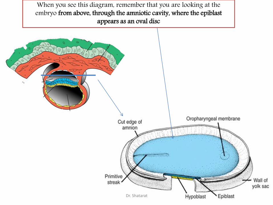
When you see this diagram, remember that you are looking at the embryo from above, through the amniotic cavity, where the epiblast
appears as an oval disc
Dr. Shatarat

Dr. Shatarat

Why the embryo needs the vascular system?
When it appears?
Where it appears?
with later contributions from neural crest
mesenchyme
Dr. Shatarat

4
Primitive streak
The Cardiac progenitor cells migrate from the Epiblast
Cranial direction on
each side of the
notochordal process
and around
the prechordal plate
Through
In a
Dr. Shatarat

into the splanchnic layer of the lateral plate mesoderm
Dr. Shatarat

into the splanchnic layer of the lateral plate mesoderm
Dr. Shatarat

The cells from both sides meet
cranially to form the
These cells will form :
• The atria
• Left ventricle
• Part of right ventricle
Primary Heart Field (PHF)
• The remainder of the right ventricle
• outflow tract (conus cordis and truncus
arteriosus)
Are derived from the
Secondary Heart Field (SHF)
Dr. Shatarat

ONE-SOMITE AND TWO-SOMITE STAGES
Dr. Shatarat

Paired endothelial strands
ANGIOBLASTIC CORDSappear in the cardiogenic mesoderm during the third week of
development
Dr. Shatarat

Dr. Shatarat

Two hearts tubs The two tubs are Fused Single heart tube is formed
These cords canalize to form two heart tubes that soon fuse as embryo
folds laterallyto form a single heart tube late in the third week
As the embryo folds laterally Dr. Shatarat

Dr. Shatarat

In addition to the cardiogenic region, other blood islands appearbilaterally, parallel and close to the midline of the embryonic shield.
These islands form a pair of longitudinal vessels, the dorsal aortae.
Dr. Shatarat

Dr. Shatarat

No known cardiac anomaly can be attributed to
the developmental phases described thus far
Dr. Shatarat

Formation of the cardiac loop
The heart is essentially
a straight tube with a caudal venosus end and cranial arterial end
It lies within the pericardial cavity
is attached posteriorly only by the dorsal mesocardium
What we have by now
The embryo now
is about 2.2 mm long
is approximately 23 days old
begins to beat
About 3 days have elapsed between the appearance of intraembryonic vasculogenesis and
the formation of the endocardial tube
Dr. Shatarat

1- Sinus venosus
2- Primitive atrium.
3- primitive ventricle.
4- Bulbus cordis (conus).
5- truncus arteriosus.
Differential growth defines five segments of the heart tube:(from caudal to cephalic or
according to direction of blood flow)
Dr. Shatarat

Dr. Shatarat

The sinus venosus represent the venous end of the heart
It receives 3 veins:
1- Common cardinal vein body wall
2- Umbilical vein from placenta
3- Vitelline vein from yolk sac
1
2
3
The tubular truncus arteriosus (TA) is
continuous cranially with the aortic sacDr. Shatarat

The arterial end of the heart is fixed by the pharyngeal arches
The venous end of the heart is fixed by the septum transversumDr. Shatarat

Dr. Shatarat

The part of the tube lying within the pericardial
cavity is made up of bulbus cordis and ventricle
the heart bends on itself
(usually bends to the right,
thus the proximal bulbus
cordis (RV) lying anterior and
to the right of the primitive
ventricle)
forming
a U-shaped
bulboventricular loop
Dr. Shatarat

As the atrium and sinus
venosus are freed from the
septum transverum they
come to lie behind and
above the ventricle and the
heart tube is now
S-shaped
Dr. Shatarat

Dr. Shatarat

Dr. Shatarat

At this stage
Dr. Shatarat

• At this stage the bulbus cordis and
ventricle are separated by a deep
bulbo-ventricular sulcus.
Dr. Shatarat

• This sulcus gradually
becomes shallower so
that the bulbus cordis
and the ventricle come
to form
one chamber which
communicates with the
truncus arteriosus.
The primary
interventricular
foramen
Dr. Shatarat

• The atrial chamber expands
so that parts of it come to
project forwards on either
side of the truncus
Dr. Shatarat

The atrial chamber expands so
that parts of it come to project
forwards on either side of the
truncus
Dr. Shatarat

As a result of these changes the exterior of the
heart assumes its definitive shape
Dr. Shatarat

Abnormalities of Cardiac Looping
Dextrocardia, in which the heart lies on
the right side of the thorax instead of the
left,
is caused because the heart loops to the
left instead of the right.
Dextrocardia may coincide with situs
inversus, a complete reversal of
asymmetry in all organs. Situs inversus,
which occurs in 1/7000 individuals,
usually is associated with normal
physiology, although there is a slight risk
of heart defects. In other cases sidedness
is random, such that some organs are
reversed and others are not
Dr. Shatarat

It is often stated that looping of the tube is the
first visual evidence of asymmetry in the
embryo, although careful examination reveals
that the atrioventricular canal has become
asymmetric prior to the start of looping.
Although the sense of laterality of the
developing organs of the body, including the
atrial appendages, develops during gastrulation,
the pathway of signalling that governs
rightward looping of the heart tube remains
unknown
. However, it is now well established that
signalling pathways including Pitx2, nodal,
lefty, and cited-2, determine the formation of
the morphologically left-sided or right-sided
features seen in organs such as the lungs, the
bronchial tree, the liver and spleen, and the
atrial appendages
Dr. Shatarat