Embed Size (px)
Citation preview

“What’s Up, Panc?”
JI GRAND ROUNDS
LAGADE-ONG
UNIVERSITY OF THE EAST RAMON MAGSAYSAY MEMORIAL
MEDICAL CENTER, INC. College of Medicine
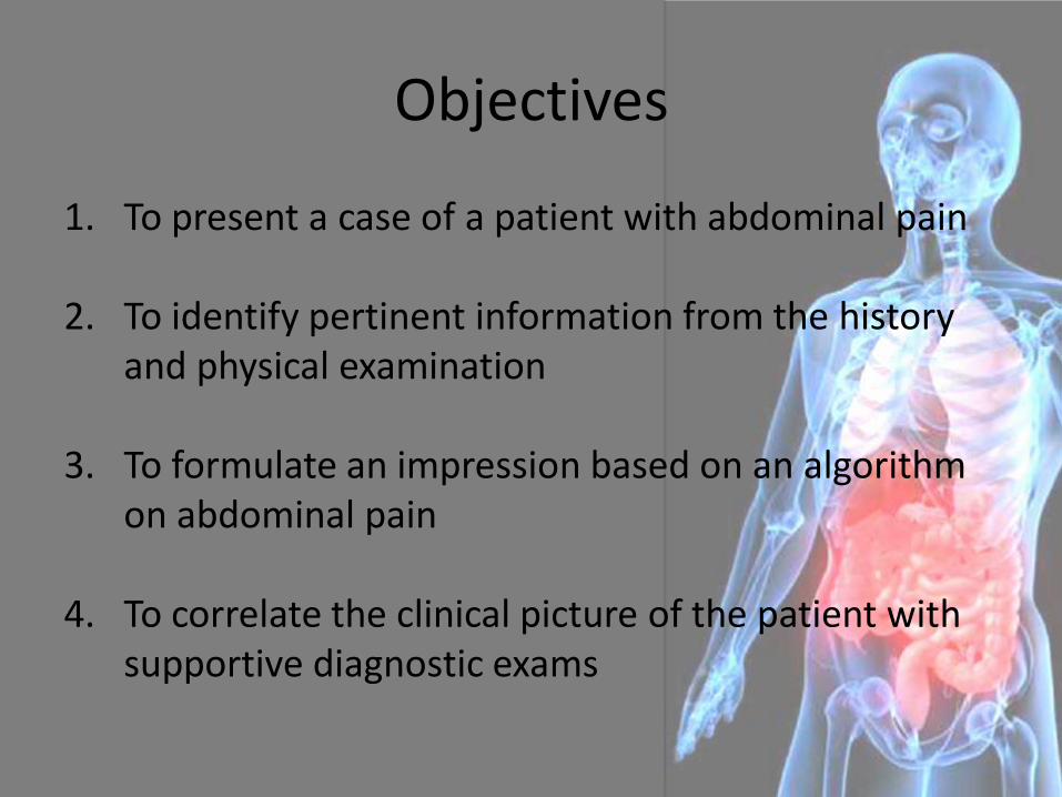
Objectives
1. To present a case of a patient with abdominal pain
2. To identify pertinent information from the history and physical examination
3. To formulate an impression based on an algorithm on abdominal pain
4. To correlate the clinical picture of the patient with supportive diagnostic exams
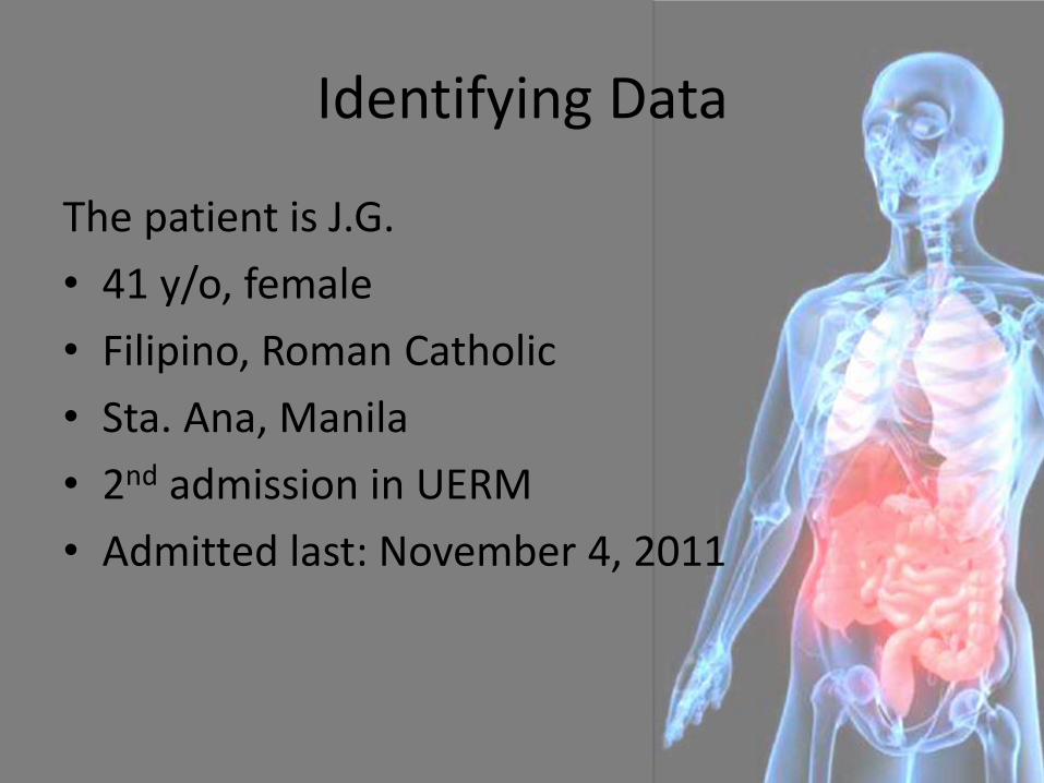
Identifying Data
The patient is J.G.
• 41 y/o, female
• Filipino, Roman Catholic
• Sta. Ana, Manila
• 2nd admission in UERM
• Admitted last: November 4, 2011
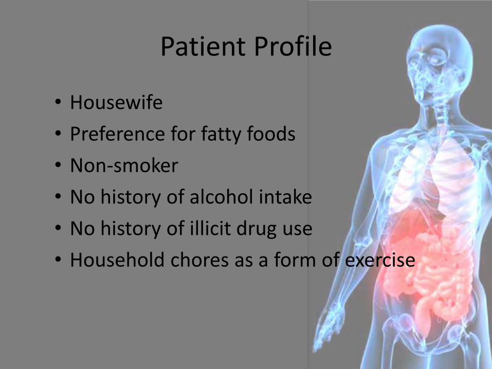
Patient Profile
• Housewife
• Preference for fatty foods
• Non-smoker
• No history of alcohol intake
• No history of illicit drug use
• Household chores as a form of exercise
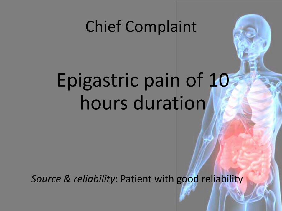
Chief Complaint
Epigastric pain of 10 hours duration
Source & reliability: Patient with good reliability

Temporal Profile
7 days 6 days 10 hours A
PRIOR TO ADMISSION
Epigastric pain with radiation to the back Vomiting Bloatedness & frequent flatulence Intake of HNBB & Ranitidine
10 9 8 7 6 5 4 3 2 1 0
Inte
nsi
ty
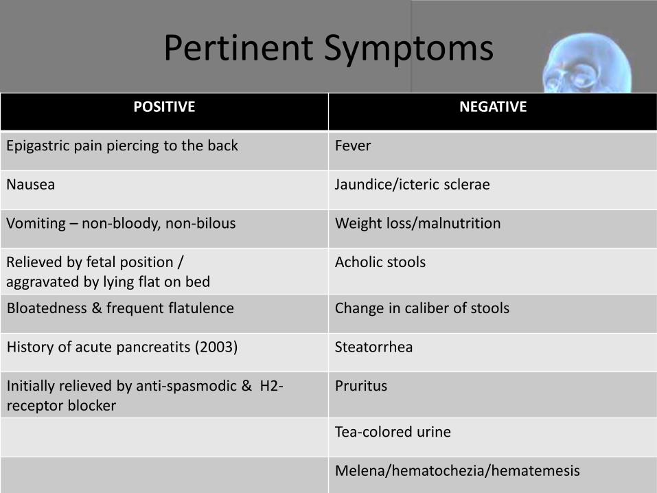
Pertinent Symptoms
POSITIVE NEGATIVE
Epigastric pain piercing to the back Fever
Nausea Jaundice/icteric sclerae
Vomiting – non-bloody, non-bilous Weight loss/malnutrition
Relieved by fetal position / aggravated by lying flat on bed
Acholic stools
Bloatedness & frequent flatulence Change in caliber of stools
History of acute pancreatits (2003) Steatorrhea
Initially relieved by anti-spasmodic & H2-receptor blocker
Pruritus
Tea-colored urine
Melena/hematochezia/hematemesis
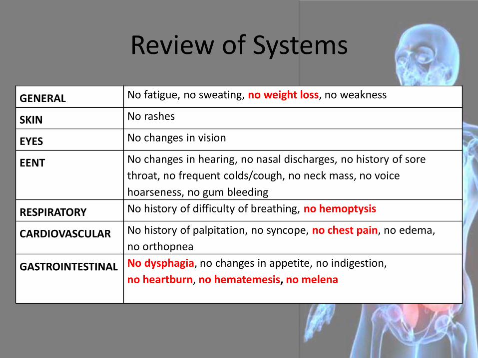
Review of Systems
GENERAL No fatigue, no sweating, no weight loss, no weakness
SKIN No rashes
EYES No changes in vision
EENT No changes in hearing, no nasal discharges, no history of sore
throat, no frequent colds/cough, no neck mass, no voice
hoarseness, no gum bleeding
RESPIRATORY No history of difficulty of breathing, no hemoptysis
CARDIOVASCULAR No history of palpitation, no syncope, no chest pain, no edema,
no orthopnea
GASTROINTESTINAL
No dysphagia, no changes in appetite, no indigestion,
no heartburn, no hematemesis, no melena
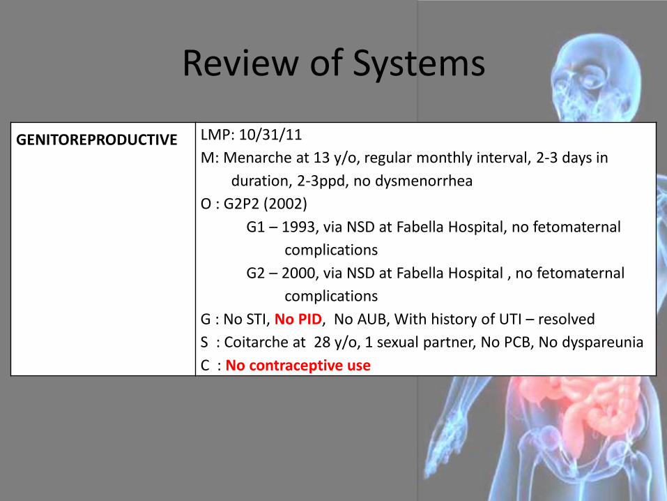
Review of Systems
GENITOREPRODUCTIVE LMP: 10/31/11
M: Menarche at 13 y/o, regular monthly interval, 2-3 days in
duration, 2-3ppd, no dysmenorrhea
O : G2P2 (2002)
G1 – 1993, via NSD at Fabella Hospital, no fetomaternal
complications
G2 – 2000, via NSD at Fabella Hospital , no fetomaternal
complications
G : No STI, No PID, No AUB, With history of UTI – resolved
S : Coitarche at 28 y/o, 1 sexual partner, No PCB, No dyspareunia
C : No contraceptive use

Review of Systems
BREAST No lump, no pain, no discharges
EXTREMITIES No cyanosis, no clubbing, no edema
HEMATOPOIETIC SYSTEM No excessive bleeding or easy bruisability
NERVOUS SYSTEM No headaches, no tremors, no head trauma
MUSCULOSKELETAL No joint stiffness, no swelling, no muscle weakness
ENDOCRINE SYSTEM No heat/cold intolerance, no polyuria, no polydyspsia,
no polyphagia
PSYCHIATRIC No behavioral changes

Past Medical History
• 2003: Acute Pancreatitis
– Admitted in UERM for 4 days
– Unrecalled medications given during admission
– Unrecalled laboratory diagnostics done
– No home medications given & was lost for follow-up
• No history of hypertension, DM, asthma, or allergy, accidents, trauma, or previous surgeries
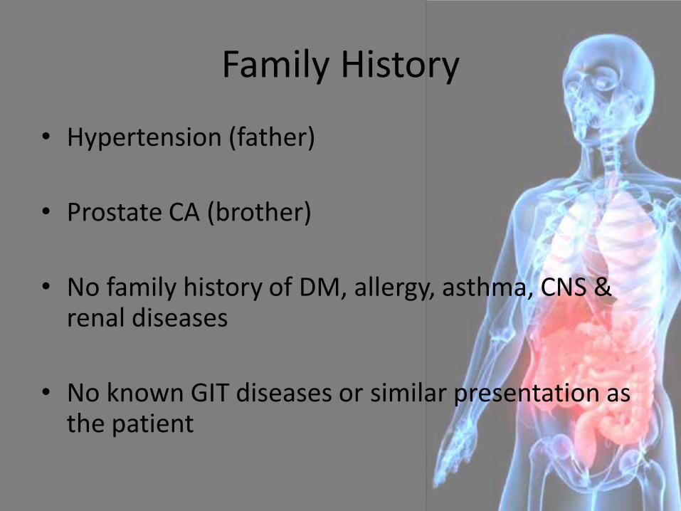
Family History
• Hypertension (father)
• Prostate CA (brother)
• No family history of DM, allergy, asthma, CNS & renal diseases
• No known GIT diseases or similar presentation as the patient

Admitting Physical Examination
General Survey Awake, alert, in pain, well-nourished
Vitals BP: 120/80 mmHg HR:72 bpm RR: 24 cpm Temp: 35.0oC Weight: 59kg / Height: 157.5cm BMI = 24
HEENT Anicteric sclerae, pink palpebral conjuntivae, 2-3mm EBRTL, full extra ocular movement, no neck vein distention, no tonsillopharyngeal congestion, no cervical lymphadenopathies

Admitting Physical Examination
Chest and Lungs
No retractions, no chest lag, equal chest expansion, resonant on all lung fields, clear breath sounds on all lung fields
Heart Adynamic precordium, normal rate and regular rhythm, distinct S1 and S2, no murmurs

Admitting Physical Examination
Abdomen Flabby, hypoactive bowel sounds, no abdominal
bruits, soft, tympanitic on all quadrants, liver span = 9cm, no splenomegaly, with direct tenderness on epigastric area on deep palpation, no Murphy’s sign, no Cullen’s sign, no Grey Turner’s sign, no fluid wave
Extremities Full range of motion, full and equal pulses, no cyanosis, no edema

Pertinent Findings • Subjective Data:
– 41 year old / female
– 1 week history of epigastric pain
– Nausea, vomiting, bloatedness and frequent flatus
– No fever, jaundice, weight loss, acholic stool, steatorrhea, pruritus, tea colored urine, and bleeding
– Previous history of Acute Pancreatitis (2003)
– No history of alcohol intake
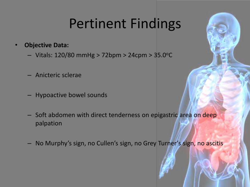
Pertinent Findings • Objective Data:
– Vitals: 120/80 mmHg > 72bpm > 24cpm > 35.0oC
– Anicteric sclerae
– Hypoactive bowel sounds
– Soft abdomen with direct tenderness on epigastric area on deep palpation
– No Murphy’s sign, no Cullen’s sign, no Grey Turner’s sign, no ascitis
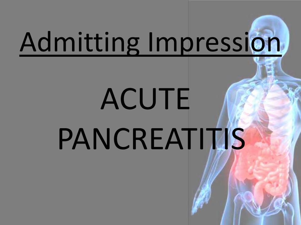
Admitting Impression
ACUTE PANCREATITIS
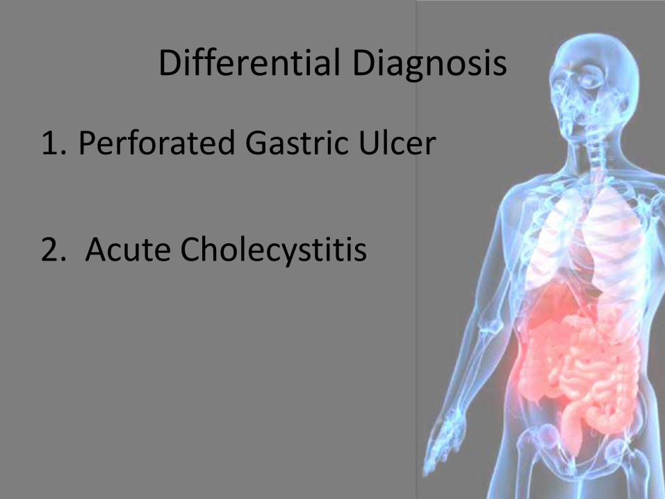
Differential Diagnosis
1. Perforated Gastric Ulcer
2. Acute Cholecystitis
APPROACH TO ABDOMINAL PAIN
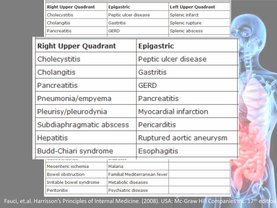
Fauci, et.al. Harrisson’s Principles of Internal Medicine. (2008). USA: Mc-Graw Hill Companies Inc. 17th edition.
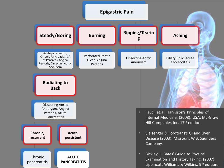
Epigastric Pain
Steady/Boring
Acute pancreatitis, Chronic Pancreatitis, CA
of Pancreas, Angina Pectoris, Dissecting Aortic
Aneurysm
Radiating to Back
Dissecting Aortic Aneurysm, Angina
Pectoris, Acute Pancreatitis
Chronic, recurrent
Chronic pancreatitis
Acute, persistent
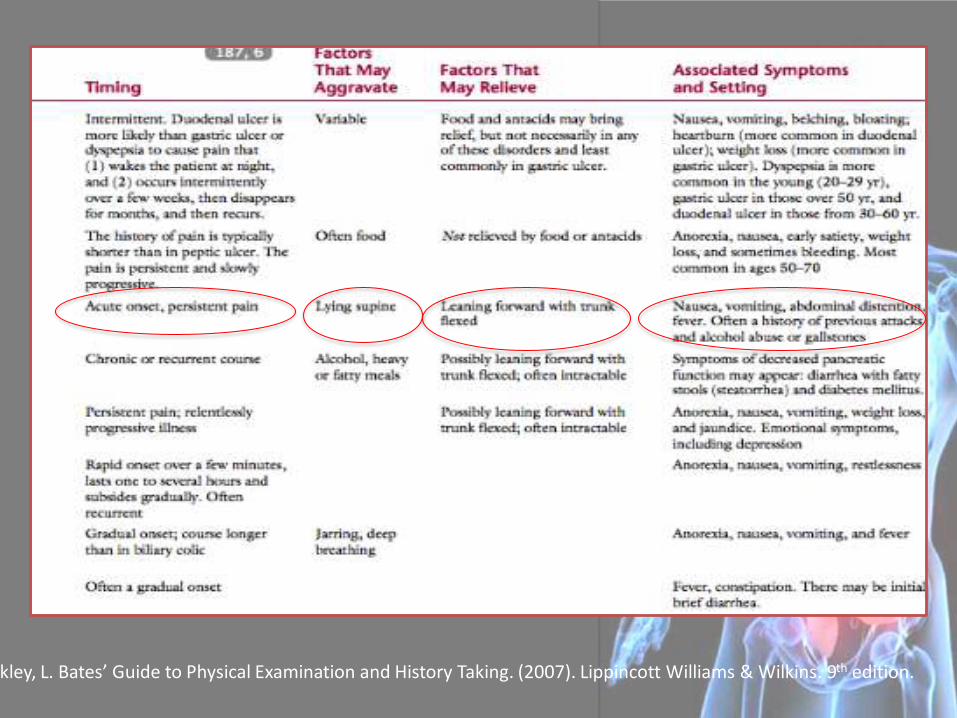
ACUTE PANCREATITIS
Burning
Perforated Peptic Ulcer, Angina
Pectoris
Ripping/Tearing
Dissecting Aortic Aneurysm
Aching
Biliary Colic, Acute Cholecystitis
• Fauci, et.al. Harrisson’s Principles of Internal Medicine. (2008). USA: Mc-Graw Hill Companies Inc. 17th edition.
• Sleisenger & Fordtrans’s GI and Liver Disease (2003). Missouri: W.B. Saunders Company.
• Bickley, L. Bates’ Guide to Physical Examination and History Taking. (2007). Lippincott Williams & Wilkins. 9th edition.
• Bickley, L. Bates’ Guide to Physical Examination and History Taking. (2007). Lippincott Williams & Wilkins. 9th edition.
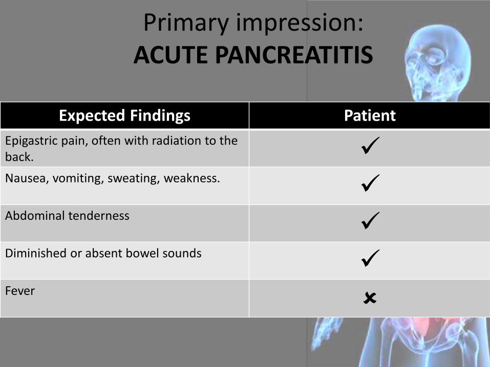
Primary impression: ACUTE PANCREATITIS
Expected Findings Patient
Epigastric pain, often with radiation to the back. Nausea, vomiting, sweating, weakness.
Abdominal tenderness
Diminished or absent bowel sounds
Fever

DIAGNOSTICS

DIFFERENTIAL DIAGNOSIS

RULE IN: Perforated Ulcer
• Sudden recurrence of severe aching colicky epigastric pain piercing through the back
• Prior episodes of tolerable epigastric pain (PRS 5-6/10) ~ 15-30 minutes
• Nausea, vomiting, and bloating
• History of Ranitidine use which provided transient relief

• No history of PUD or gastric ulcer with supporting endoscopic findings
• No history of chronic NSAID use
• No tests confirming presence of H. Pylori infection
• Pain not associated with food intake or missed meals
RULE OUT: Perforated Ulcer
• Abdominal pain (epigastric)
• Leukocytosis
• Nausea, vomiting, abdominal distention, hypoactive bowel sounds
• Risk Factors: Forty, Fat, Female, Fertile/Flatulent
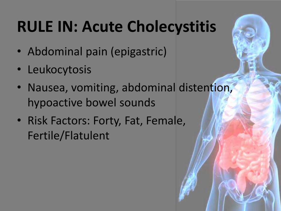
RULE IN: Acute Cholecystitis

• Absence of progression of abdominal pain from initial epigastric area towards the RUQ, right shoulder, and right interscapular area
• Absence of fever from the triad of abdominal pain, fever, and leukocytosis
• No Murphy’s sign
RULE OUT: Acute Cholecystitis

• Elevated levels of Serum AMYLASE
• Ultrasound findings of:
Fatty liver; Cholecytitis with Cholecystolithiases; Dilated CBD and intrahepatic ducts
RULE IN: Acute Cholecystitis

DISCUSSION
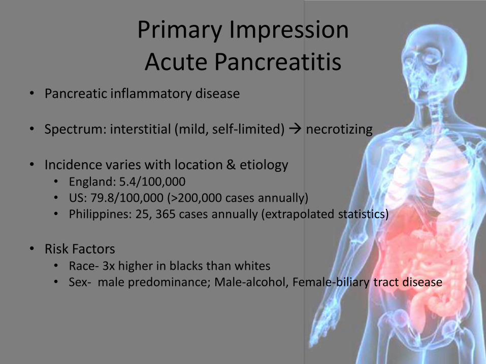
Primary Impression Acute Pancreatitis
• Pancreatic inflammatory disease
• Spectrum: interstitial (mild, self-limited) necrotizing
• Incidence varies with location & etiology • England: 5.4/100,000 • US: 79.8/100,000 (>200,000 cases annually) • Philippines: 25, 365 cases annually (extrapolated statistics)
• Risk Factors
• Race- 3x higher in blacks than whites • Sex- male predominance; Male-alcohol, Female-biliary tract disease
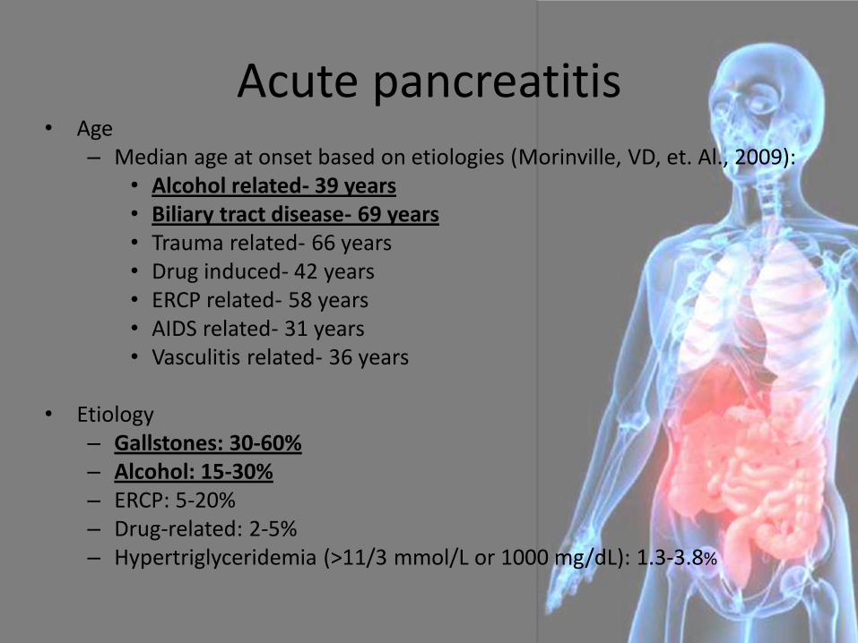
Acute pancreatitis • Age
– Median age at onset based on etiologies (Morinville, VD, et. Al., 2009): • Alcohol related- 39 years • Biliary tract disease- 69 years • Trauma related- 66 years • Drug induced- 42 years • ERCP related- 58 years • AIDS related- 31 years • Vasculitis related- 36 years
• Etiology
– Gallstones: 30-60% – Alcohol: 15-30% – ERCP: 5-20% – Drug-related: 2-5% – Hypertriglyceridemia (>11/3 mmol/L or 1000 mg/dL): 1.3-3.8%

PATHOGENESIS

MANAGEMENT

Evidence Ratings Classification
I. At least one published systematic review of multiple well-designed randomised controlled trials.
II. At least one published properly designed randomized controlled trial of appropriate size and in an appropriate clinical setting.
III. Evidence from published well-designed trials without randomization, single group prepost, cohort, time series, or matched case-controlled studies.
IV. Evidence from well-designed nonexperimental studies from more than 1 center or research group or opinon of respected authorities, based on clinical evidence, descriptive studies, or reports of expert consensus committees.
Management
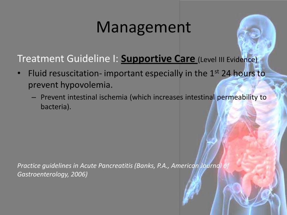
Treatment Guideline I: Supportive Care (Level III Evidence)
• Fluid resuscitation- important especially in the 1st 24 hours to prevent hypovolemia. – Prevent intestinal ischemia (which increases intestinal permeability to
bacteria).
Practice guidelines in Acute Pancreatitis (Banks, P.A., American Journal of Gastroenterology, 2006)

Management • Treatment Guideline II: Transfer to intensive care unit
(level III Evidence)
– This is imperative if there are signs that the pancreatitis is severe or going to be severe. • Organ dysfunction • Sustained hypoxemia • Hypotension
– Other danger signals to be considered: • Obesity (BMI>30) • Oliguria with urine output (<50 mL) • Tachycardia (>120 beats/minute) • Evidence of encephalopathy • Increase need of narcotic agents to counteract pain.
Management
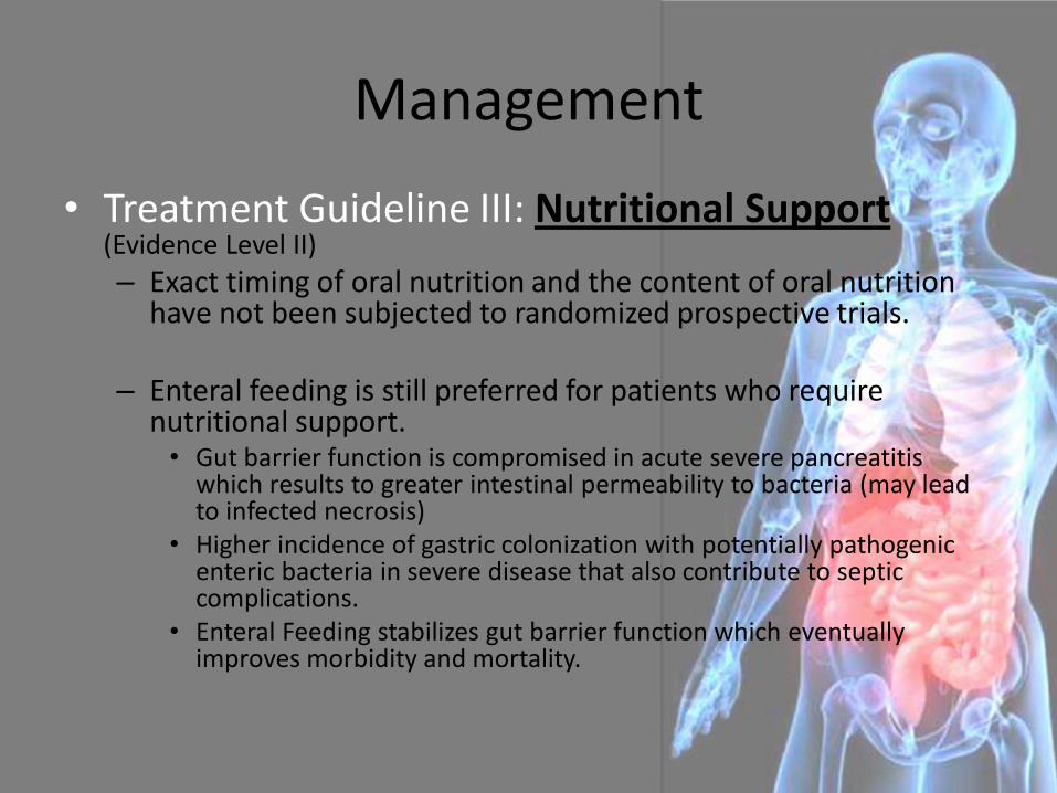
• Treatment Guideline III: Nutritional Support (Evidence Level II)
– Exact timing of oral nutrition and the content of oral nutrition have not been subjected to randomized prospective trials.
– Enteral feeding is still preferred for patients who require nutritional support. • Gut barrier function is compromised in acute severe pancreatitis
which results to greater intestinal permeability to bacteria (may lead to infected necrosis)
• Higher incidence of gastric colonization with potentially pathogenic enteric bacteria in severe disease that also contribute to septic complications.
• Enteral Feeding stabilizes gut barrier function which eventually improves morbidity and mortality.
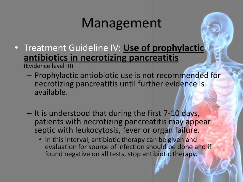
Management
• Treatment Guideline IV: Use of prophylactic antibiotics in necrotizing pancreatitis (Evidence level III)
– Prophylactic antiobiotic use is not recommended for necrotizing pancreatitis until further evidence is available.
– It is understood that during the first 7-10 days, patients with necrotizing pancreatitis may appear septic with leukocytosis, fever or organ failure. • In this interval, antibiotic therapy can be given and
evaluation for source of infection should be done and if found negative on all tests, stop antibiotic therapy.

Complications
• Acute Fluid Collections – Pancreatic ascites
– Pleural effusion
• Pseudocyst
• Intraabdominal infections (Pancreatic Abscess)
• Pancreatic necrosis (sterile or infected) 40-60%

Complications: Systemic
• Shock
• GI bleeding
• CBD obstruction
• Ileus
• Splenic infarction/ rupture
• Disseminated Intravascular Coagulation
• Subcutaneous fat necrosis
• ARDS
• Pleural effusion
• ARF
• Sudden blindness

Prognosis • Risk factors that adversely affect survival of sever acute pancreatitis
– Severe Acute Pancreatitis 1. Associated with organ failure and/or local complications such as necrosis 2. Clinical manifestations a. Obesity BMI > 30 b. Hemoconcentration (hematocrit > 44%) c. Age > 70
3. Organ failure a. Shock b. Pulmonary insufficiency (PO2 < 60) c. Renal failure (CR > 2.0 mg%) d. GI bleeding
4. Ranson criteria (not fully utilizable until 48 h) 5. Apache II score > 8 (cumbersome)

Prognosis
• Clinical Assessment and prognostication
– Used to identify patients in greatest need of aggressive medical treatment
• Mild disease= can have interstitial edema of pancreas, inflammatory infiltrate without hemorrhage or necrosis, minimal or no organ dysfunction
• Severe disease= inflammatory infiltrate is severe, necrosis of the parenchyma accompanied by evidence of severe gland dysfunction associated with multi-organ failure.

Ranson Criteria Criteria On admission Criteria After 48 hours
Patient Score Patient Score
Age (> 55y/o)
41 0 Hct fall (> 10%)
14% 1
WBC (> 16 g/L)
19.2 1 Urea rise (> 5.0 mmol/L)
x x
Glucose (>11.1 mmol/L)
8.3 0 Serum Calcium (< 2.0 mmol/L)
2.3 0
LDH (> 350 UI/L)
317 0 Pa02 (< 60 mmHg)
79 0
SGOT (> 250 UI/L)
242 0 Base deficit (BE > 4.0 mmol/L)
0.3 0
Fluid sequestration (> 6 L)
< 6 L (no
evidence of third spacing)
0
TOTAL (after 48 hours)
2
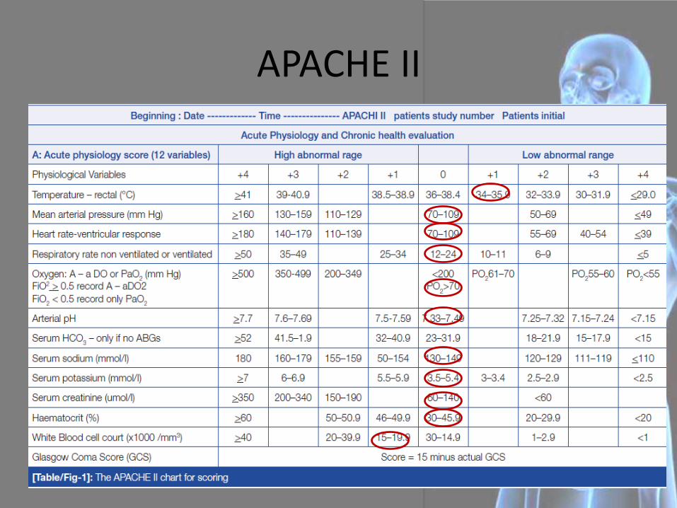
APACHE II

PATIENT’S SCORE: 2 Pts

Prognosis
10% of interstitial pancreatitis organ failure
54% of necrotizing pancreatitis organ failure
Overall mortality in acute pancreatitis : 5%
* 3% in interstitial pancreatitis
* 17% in necrotizing pancreatitis

Recent Studies
• Early endoscopic intervention vs. Early Conservative management
– In a study conducted by Oria, A. et. Al. failed to provide evidence that Early Endoscopic intervention benefits patients with acute gallstone pancreatitis and biliopancreatic obstruction.
– The persistence of main bile duct stones does not by itself contribute to worsening or persisting pancreatic inflammation. If acute cholangitis can be safely excluded, EEI is not mandatory and should not be considered a standard indication.
• ERCP on reduction of complications
• odds of having complications are reduced in predicted severe disease by Early ERCP but renders non-significant effect in predicted mild disease and for reduction of mortality in either predicted or severe disease. (Khurram et. Al, 2009)

References
• Ayub, K. et. Al., Endoscopic retrograde cholangiopancreatography in gallstone-associated acute pancreatitis, Cochrane upper Gastrointestinal and Pancreatic Diseases group, Oct 2009.
• Fauci, et.al. Harrisson’s Principles of Internal Medicine. (2008). USA: Mc-Graw Hill Companies Inc. 17th edition.
• Oria, A. , Early Endoscopic Intervention Versus Early Conservative Management in Patients With Acute Gallstone Pancreatitis and Biliopancreatic Obstruction A Randomized Clinical Trial, Annals of Surgery, 2007; 245- 1: 10-17
• Sleisenger & Fordtrans’s GI and Liver Disease (2003). Missouri: W.B. Saunders Company.
• Bickley, L. Bates’ Guide to Physical Examination and History Taking. (2007). Lippincott Williams & Wilkins. 9th edition.
• •


![[Cartilha] Cartilha PANC Viveiros Comunitários](https://img.dokumen.tips/doc/110x75/5695d0921a28ab9b02930095/cartilha-cartilha-panc-viveiros-comunitarios.jpg)