Embed Size (px)
Citation preview

Gerald Maurer
Department of CardiologyMedical University of Vienna
What the Cardiologist needs to know from Medical Images

What kinds of Cardiologists
• Plumbers
• Electricians
• Photographers
• And then there’s another kind…..
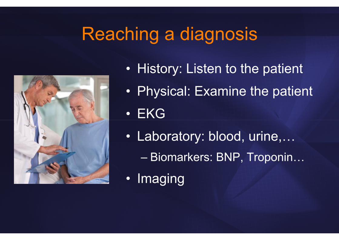
Reaching a diagnosis
• History: Listen to the patient
• Physical: Examine the patient
• EKG
• Laboratory: blood, urine,…– Biomarkers: BNP, Troponin…
• Imaging

Echo
CMRSPECT
CT PET

• Morphology and function
• Hemodynamics and flow patterns
• Tissue characteristics and metabolism
• Interventional imaging
• Prognosis and Outcome
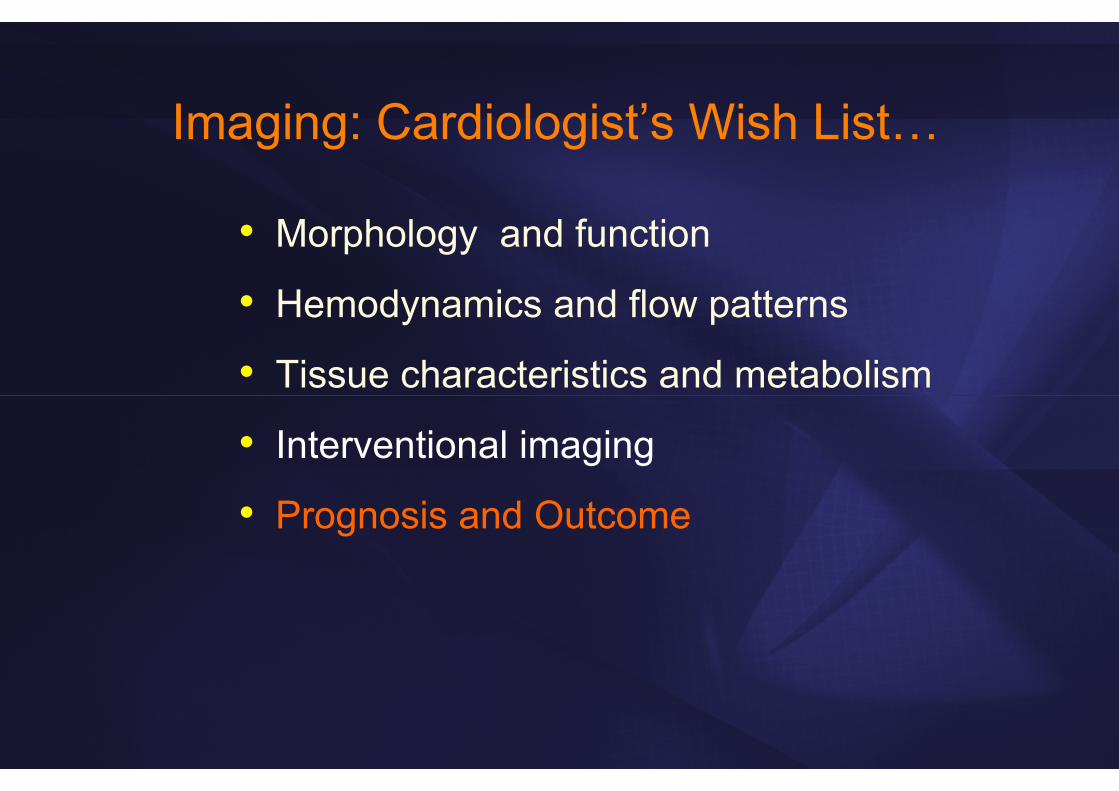
Imaging: Cardiologist’s Wish List…

• Morphology and function
• Hemodynamics and flow patterns
• Tissue characteristics and metabolism
• Interventional imaging
• Prognosis and Outcome
Imaging: Cardiologist’s Wish List…


Function

FunctionNormal AbnormalAbnormal
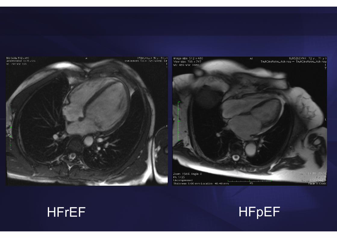
HFpEFHFrEF
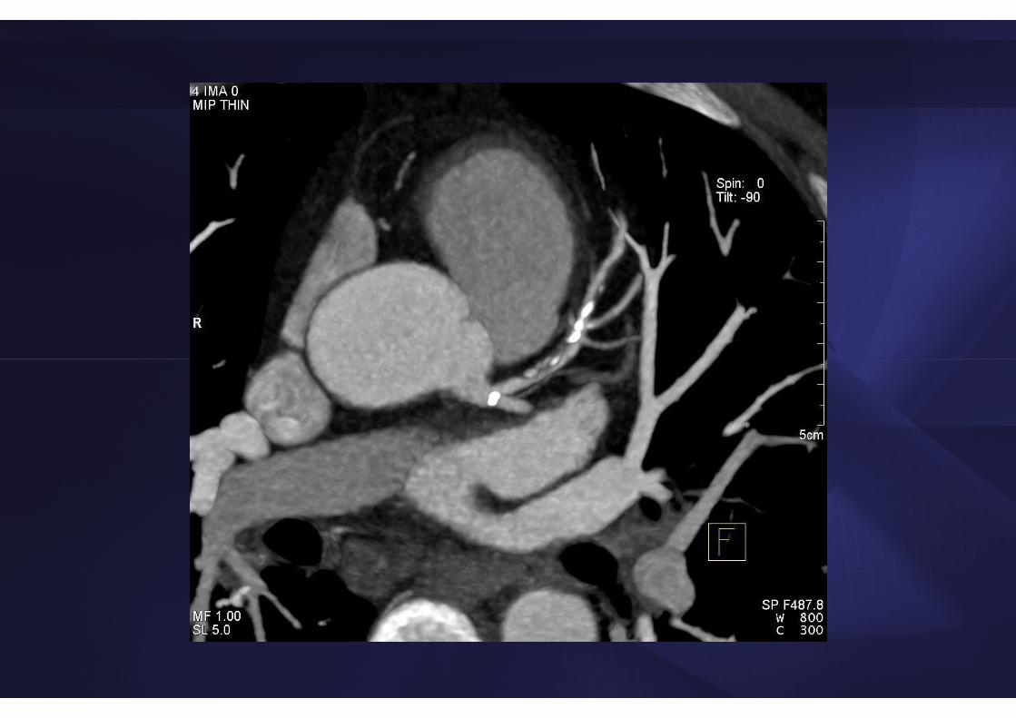


• Morphology and function
• Hemodynamics and flow patterns
• Tissue characteristics and metabolism
• Interventional imaging
• Prognosis and Outcome
Imaging: Cardiologist’s Wish List…
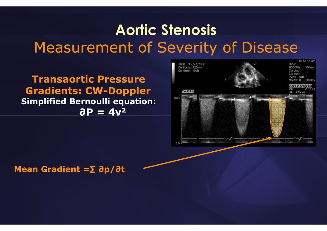
Aortic StenosisMeasurement of Severity of Disease
Transaortic Pressure Gradients: CW-Doppler
Simplified Bernoulli equation:∂P = 4v2
Mean Gradient =∑ ∂p/∂t
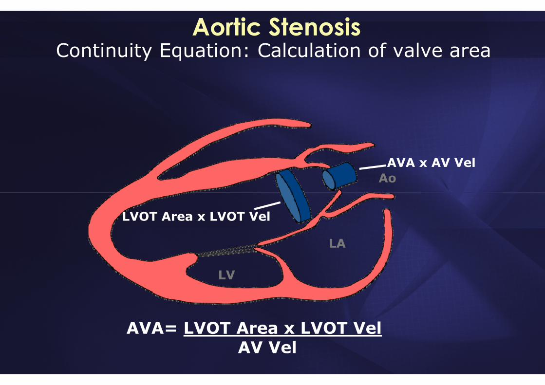
Aortic StenosisContinuity Equation: Calculation of valve area
LA
LV
Ao
LVOT Area x LVOT Vel
AVA x AV Vel
AVA= LVOT Area x LVOT VelAV Vel
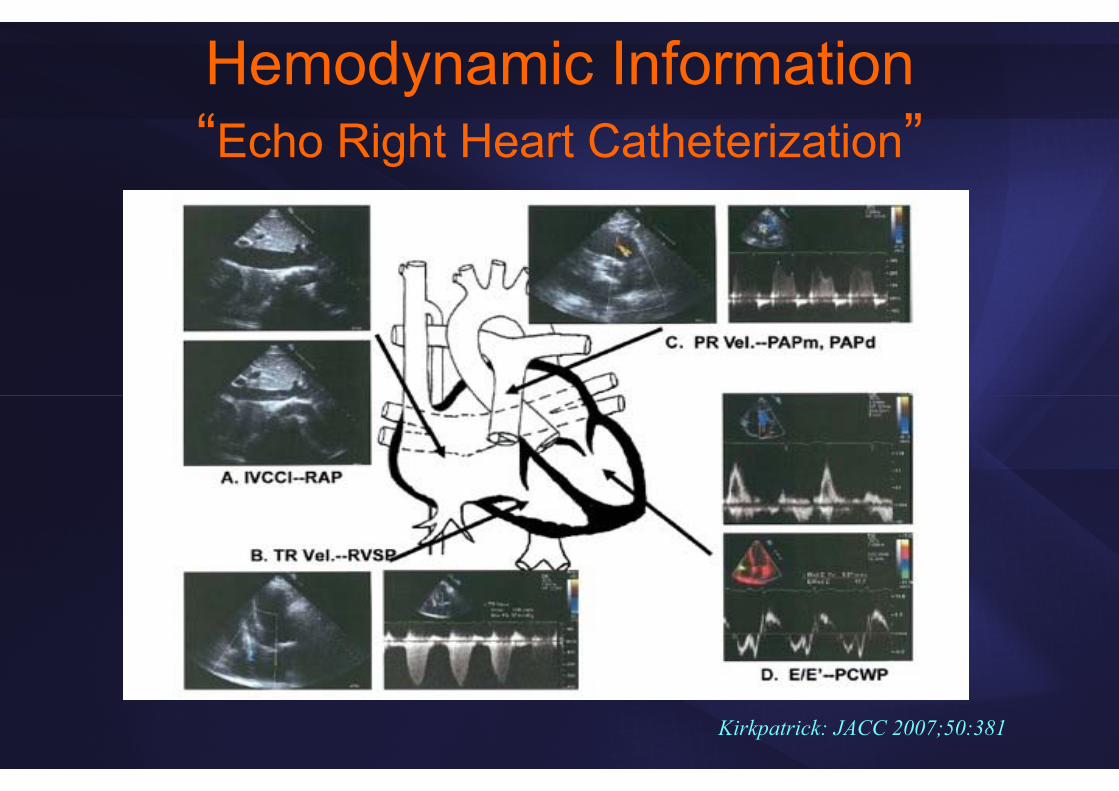
Hemodynamic Information“Echo Right Heart Catheterization”
Kirkpatrick: JACC 2007;50:381
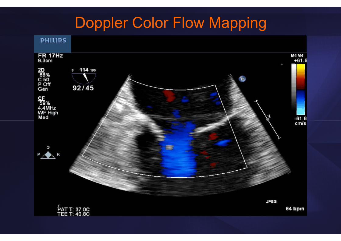
Doppler Color Flow Mapping
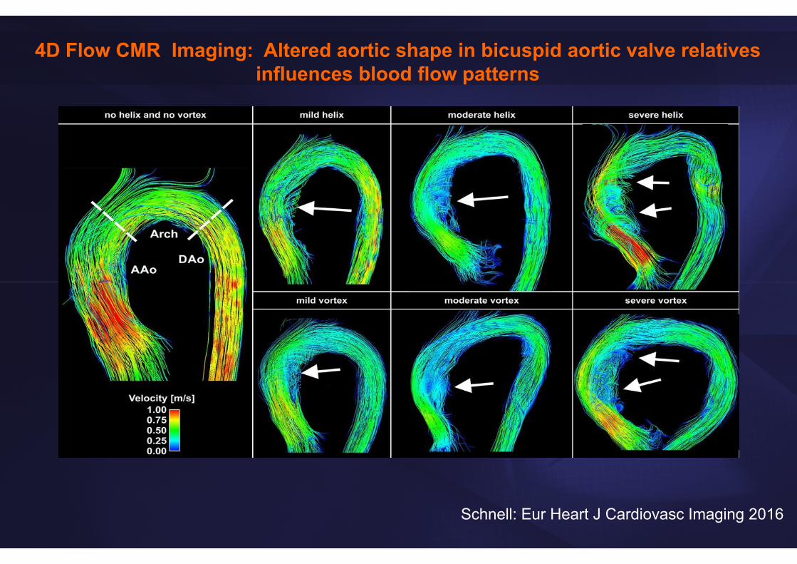
4D Flow CMR Imaging: Altered aortic shape in bicuspid aortic valve relatives influences blood flow patterns
Schnell: Eur Heart J Cardiovasc Imaging 2016
• Morphology and function
• Hemodynamics and flow patterns
• Tissue characteristics and metabolism
• Interventional imaging
• Prognosis and Outcome
Imaging: Cardiologist’s Wish List…

DMI Myocarditis
Dil CMP
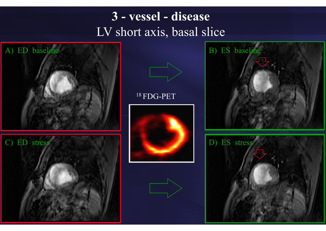
A) ED baseline
C) ED stress
B) ES baseline
D) ES stress
18 FDG-PET
3 - vessel - diseaseLV short axis, basal slice
• Morphology and function
• Hemodynamics and flow patterns
• Tissue characteristics and metabolism
• Interventional imaging
• Prognosis and Outcome
Imaging: Cardiologist’s Wish List…
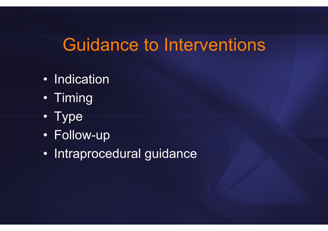
Guidance to Interventions
• Indication• Timing • Type• Follow-up• Intraprocedural guidance
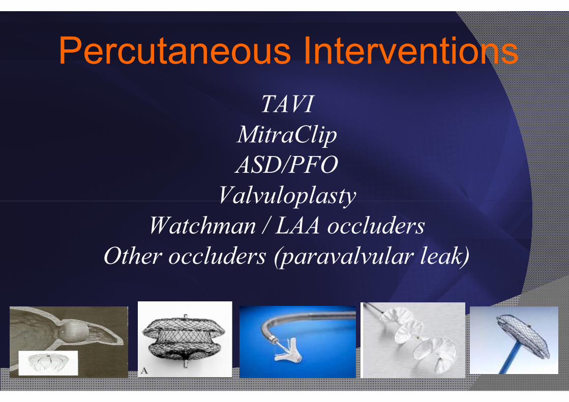
Percutaneous InterventionsTAVI
MitraClipASD/PFO
ValvuloplastyWatchman / LAA occluders
Other occluders (paravalvular leak)
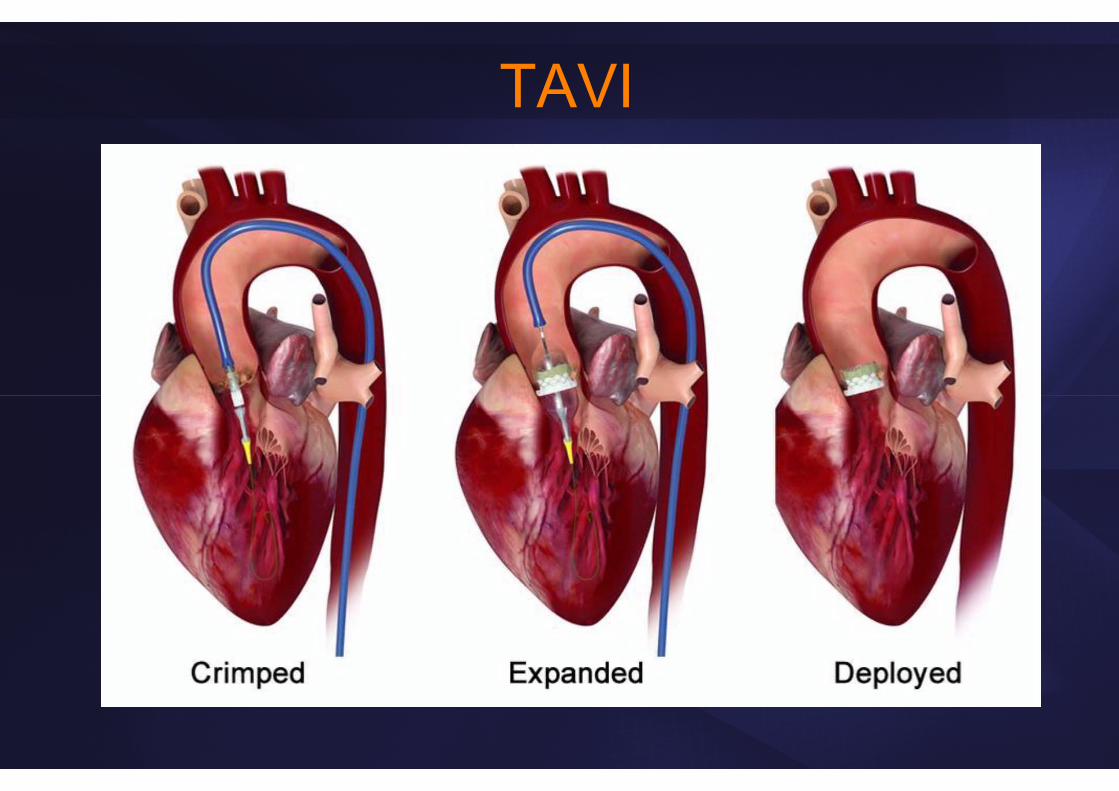
TAVI

Assessment of Vascular Access3D-CTANGIO CT
Vessel size vs. sheath size (ID vs. OD)• 18F (~21F): 6.9 mm (CoreValve)• 22F (25F): 8.3 mm (23 mm Edwards Sapien)• 24F (28F): 9.2 mm (26 mm Edwards Sapien)
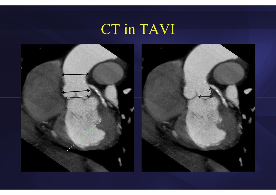
CT in TAVI
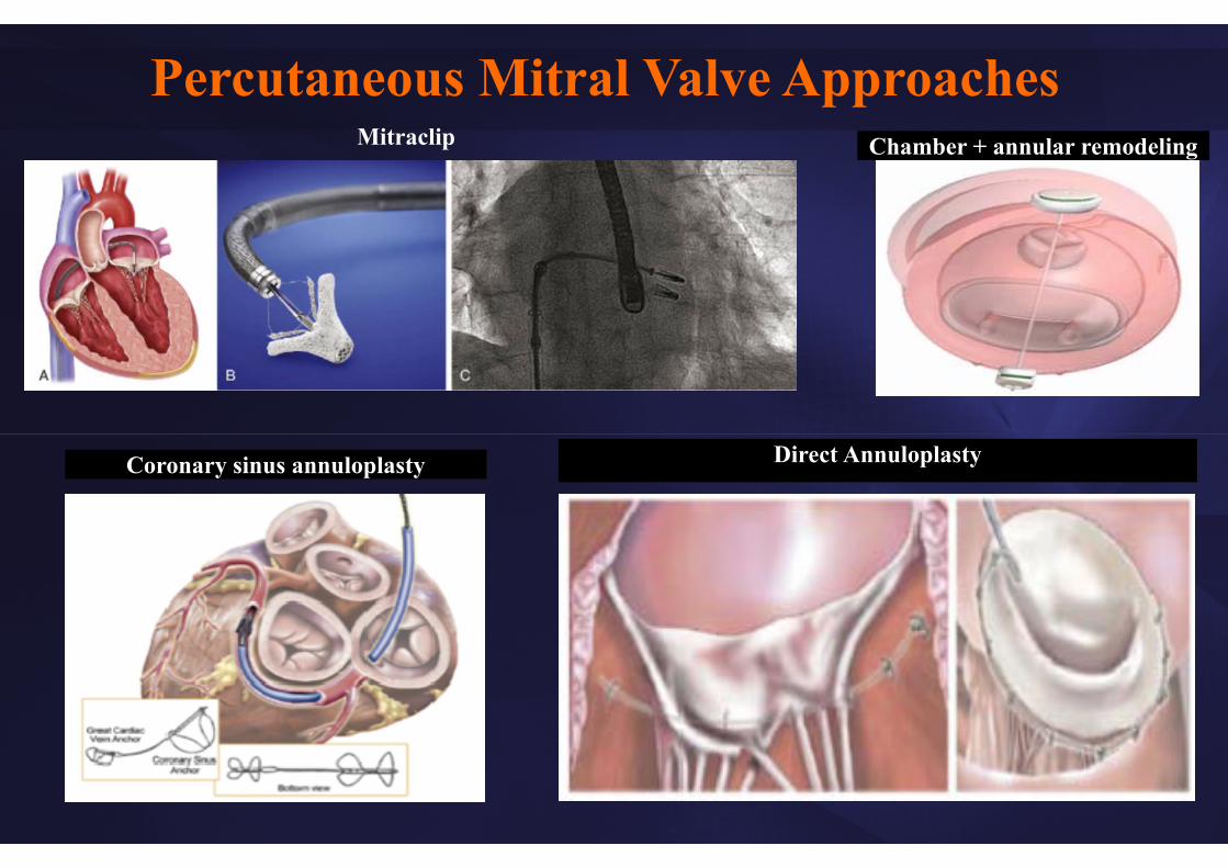
Coronary sinus annuloplasty Direct Annuloplasty
Chamber + annular remodeling
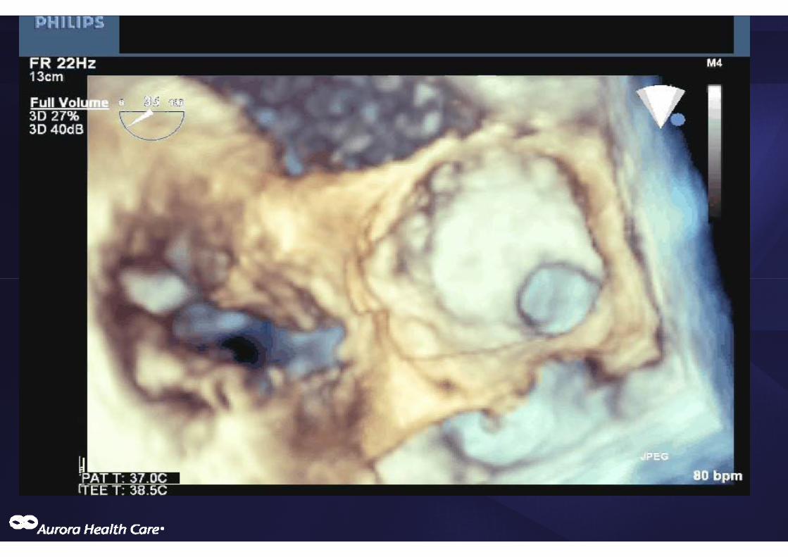
Percutaneous Mitral Valve ApproachesMitraclip

29
Mitral valve clip
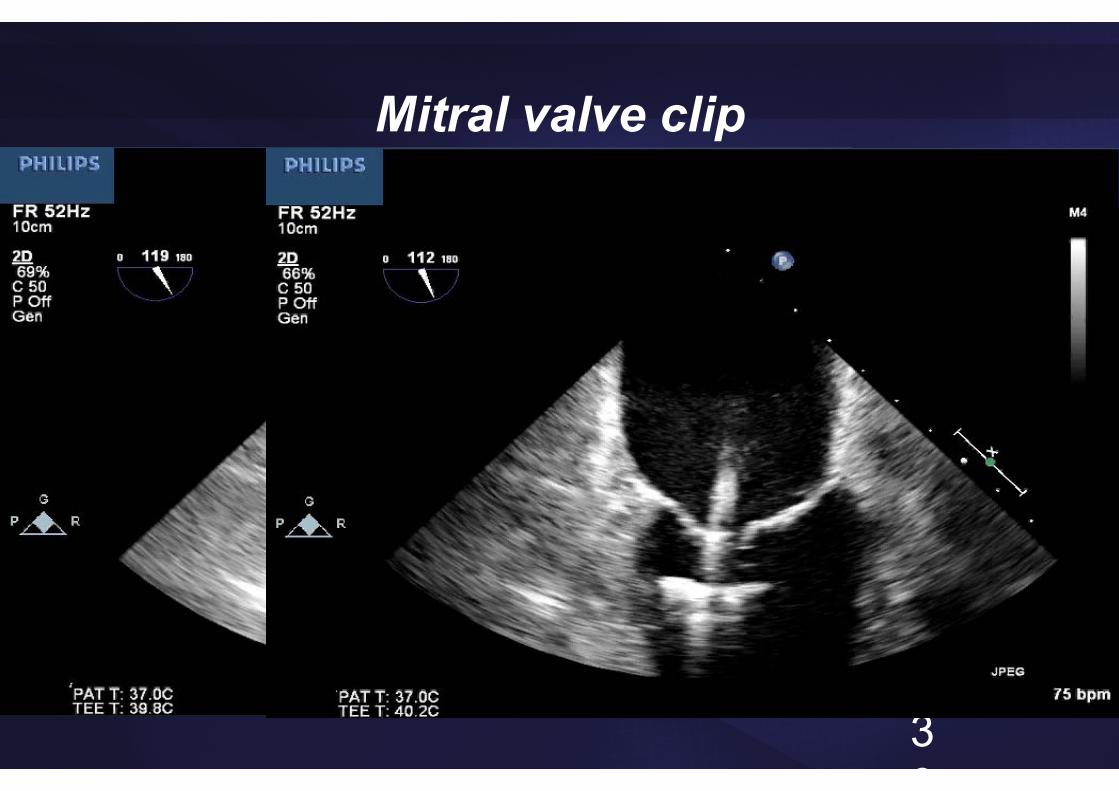
30
Mitral valve clip
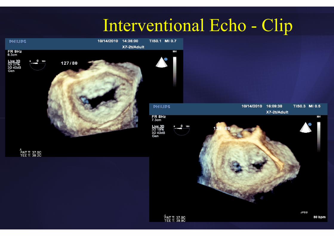
Interventional Echo - Clip

Nickenig: J Am Coll Cardiol Intv. 2016;9:2039
Transcatheter Mitral Annuloplasty Cardioband Percutaneous Mitral Repair System


Interventional MRI
McGuirt T: Rad Techn 2016;87:622
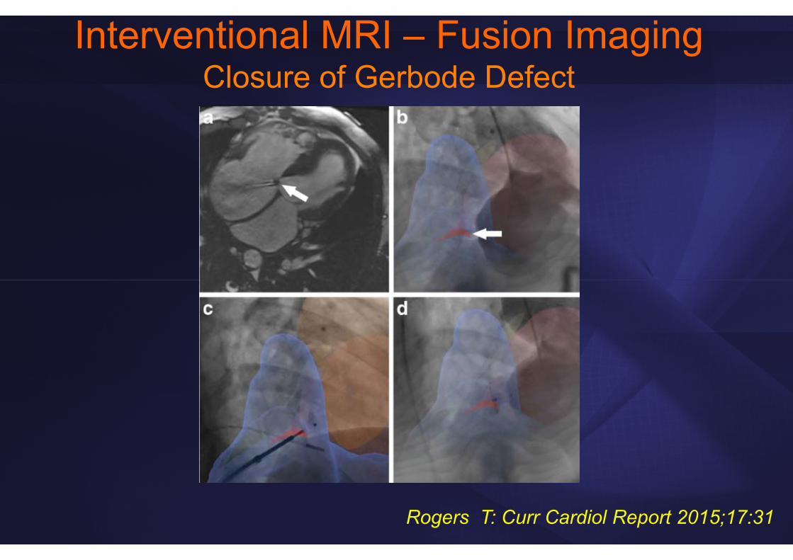
Interventional MRI – Fusion ImagingClosure of Gerbode Defect
Rogers T: Curr Cardiol Report 2015;17:31
Impact of Imaging?• Detailed morphologic, functional, hemodynamic,
metabolic and molecular information
• These tools can be used in patients but also for screening healthy populations
• Improved understanding of disease processes, risk stratification
• Basis for developing rational treatment algorithms that should improve outcome

Do they??
Concerns and Pitfalls• Sensitive technologies may detect subclinical disease that
should be left alone
• Overinterpretation
• Detection of non-target findings that may not have clinical relevance but require additional testing
• Risk from invasive or semi-invasive procedures
• Radiation exposure
• Contrast agents – adverse effects
• Cost
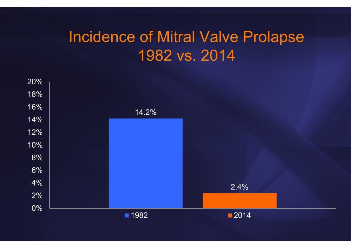
Incidence of Mitral Valve Prolapse1982 vs. 2014
14.2%
2.4%
0%2%4%6%8%
10%12%14%16%18%20%
1982 2014
• Morphology and function
• Hemodynamics and flow patterns
• Tissue characteristics and metabolism
• Interventional imaging
• Prognosis and Outcome
Imaging: Cardiologist’s Wish List…

Prognostic Information

Prognostic Information of Echo in CHFVal-Heft Trial
Wong M: JACC 2004;43:2022
n=5010 Pts.

Asymptomatic Aortic Stenosis
AV-Vel > 4 m/s
AV-Vel 3-4 m/s
AV-Vel < 3 m/s
Otto CM, et al. Circulation 95:2262, 1997
Even
t-fr
ee S
urvi
val (
%)
0 12 24 36 48 60months
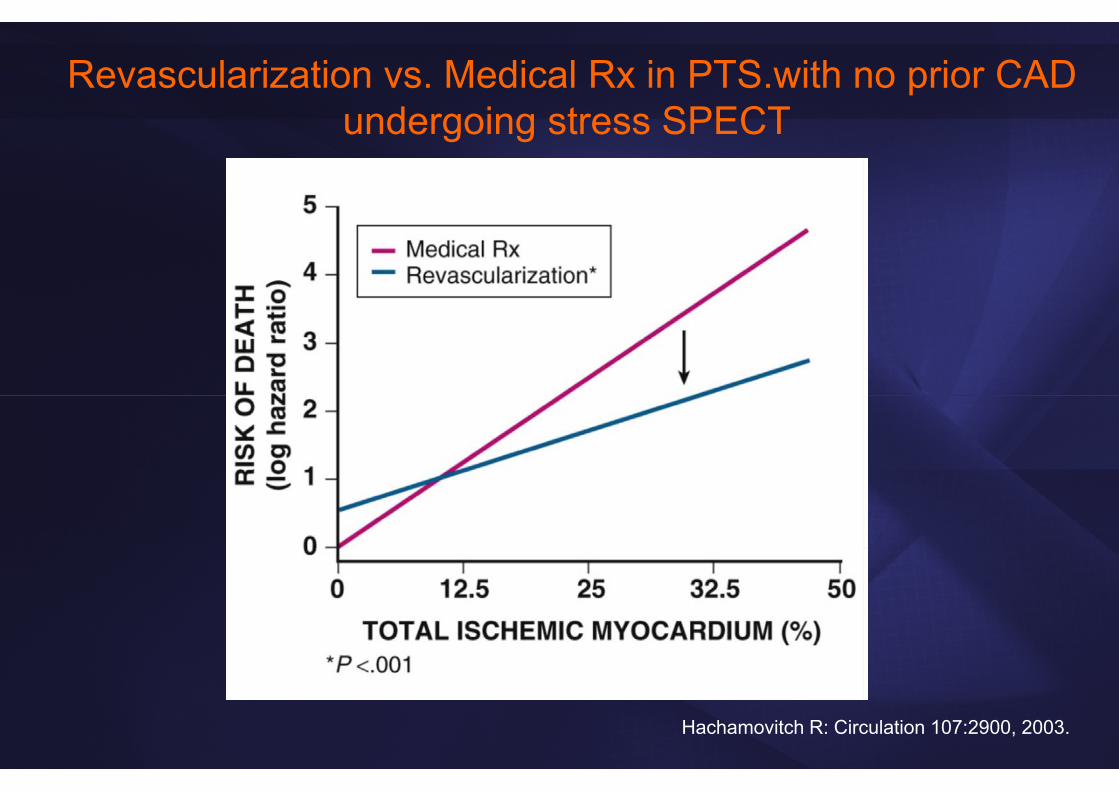
Revascularization vs. Medical Rx in PTS.with no prior CAD undergoing stress SPECT
Hachamovitch R: Circulation 107:2900, 2003.
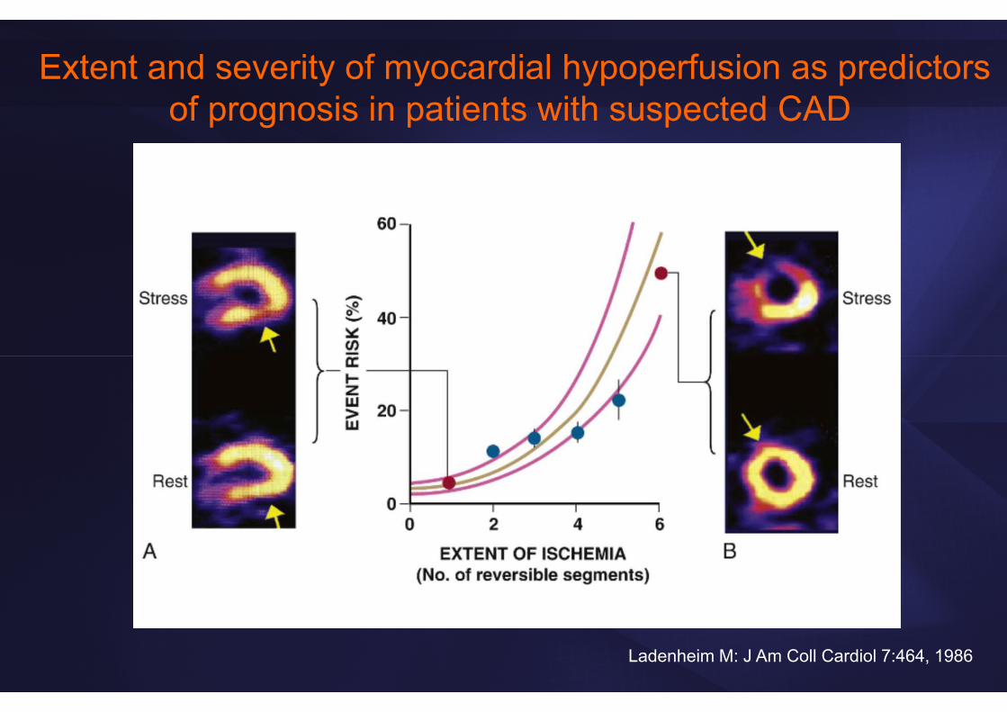
Extent and severity of myocardial hypoperfusion as predictors of prognosis in patients with suspected CAD
Ladenheim M: J Am Coll Cardiol 7:464, 1986

Differences in All-Cause Mortality Risk based on CCT Angiography Findings: CONFIRM Registry
Min JK: J Am Coll Cardiol 58:849, 2011
What about Imaging
in Ischemic Heart Disease?
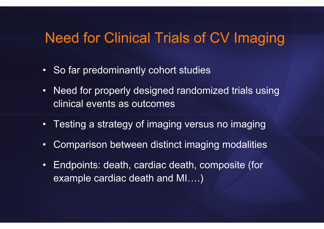
Need for Clinical Trials of CV Imaging
• So far predominantly cohort studies
• Need for properly designed randomized trials using clinical events as outcomes
• Testing a strategy of imaging versus no imaging
• Comparison between distinct imaging modalities
• Endpoints: death, cardiac death, composite (for example cardiac death and MI….)

OAT (Occluded Artery Trial)Coronary Intervention After MI
• 2166 patients randomized, SPECT in 589 – viability testing in 124
• Mild to mod. ischemia in 40% of SPECT pts
• Ischemia did NOT alter finding that an open artery did not improve outcome after MI! (however, pts with severe ischemia excluded from trial)
Hochman JS: NEJM 2006;355:2395

INSPIRE Trial• 728 pts – 205 with large total (≥20%) and ischemic
(≥10%) SPECT perfusion defects and an LVEF≥35%
• “SPECT could effectively monitor changes in scintigraphic ischemia after medical or revascularization therapy”
• Intensive medical therapy was comparable to revascularization (no identification by SPECT who would benefit from revascularization)
Mahmarian JJ: JACC 2006;48:2458
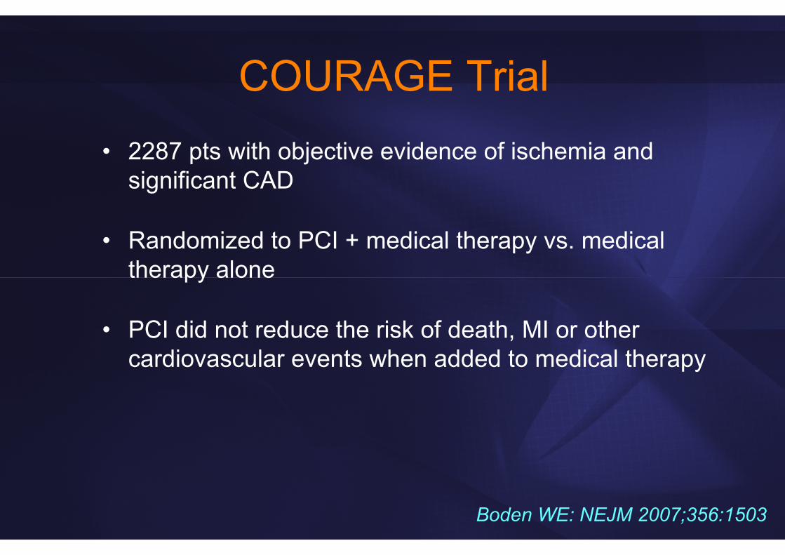
COURAGE Trial• 2287 pts with objective evidence of ischemia and
significant CAD
• Randomized to PCI + medical therapy vs. medical therapy alone
• PCI did not reduce the risk of death, MI or other cardiovascular events when added to medical therapy
Boden WE: NEJM 2007;356:1503

COURAGE Trial – Nuclear Substudy
• 314 of 2287 COURAGE pts enrolled
• Benefit of >5% reduction of ischemia (by either method), but prospective testing of this hypothesis still needed
• “…not certain that one would need imaging in clinical practice to achieve the goal of reduced symptoms”
Shaw LJ: Circulation 2008;117:1283
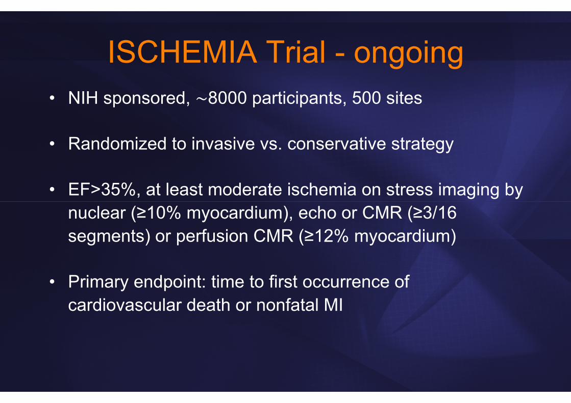
ISCHEMIA Trial - ongoing• NIH sponsored, 8000 participants, 500 sites
• Randomized to invasive vs. conservative strategy
• EF>35%, at least moderate ischemia on stress imaging by nuclear (≥10% myocardium), echo or CMR (≥3/16 segments) or perfusion CMR (≥12% myocardium)
• Primary endpoint: time to first occurrence of cardiovascular death or nonfatal MI

Metabolic Imaging: Prognosis in CAD and LV Dysfunction
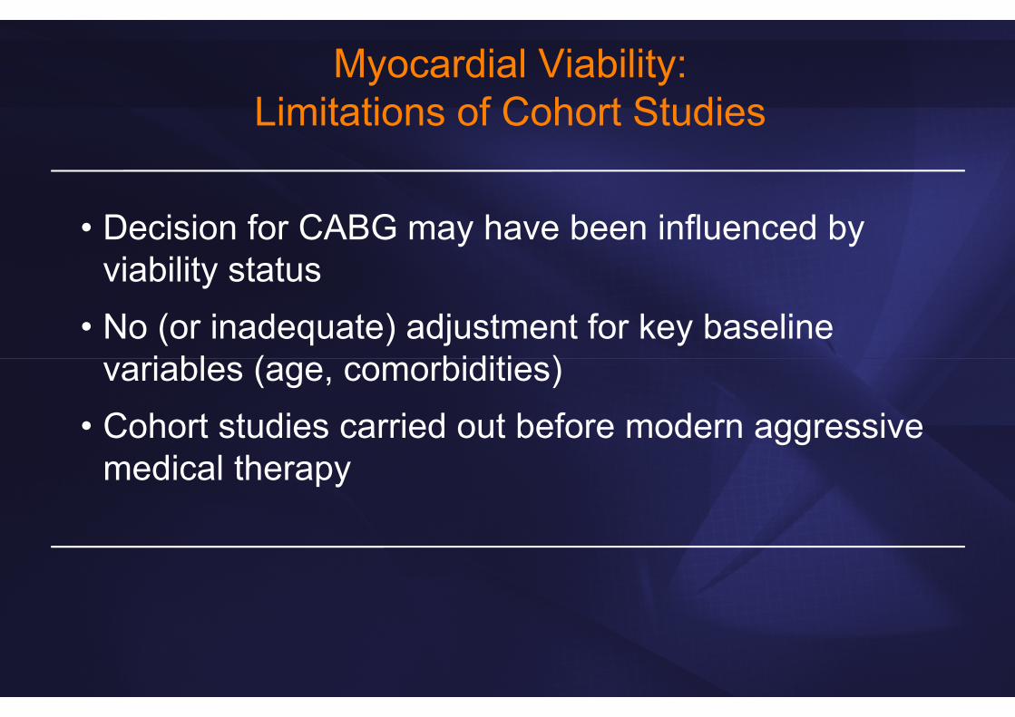
Myocardial Viability:Limitations of Cohort Studies
• Decision for CABG may have been influenced by viability status
• No (or inadequate) adjustment for key baseline variables (age, comorbidities)
• Cohort studies carried out before modern aggressive medical therapy
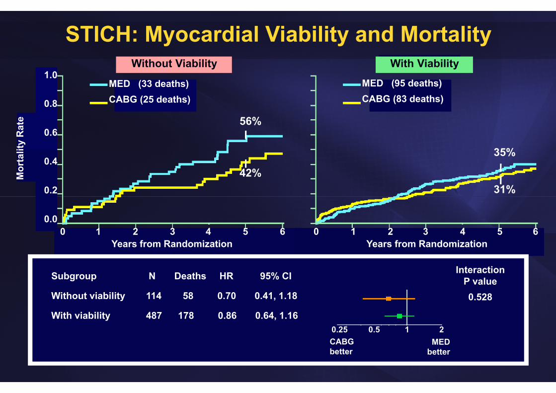
STICH: Myocardial Viability and Mortality1.0
0.8
0.6
0.4
0.2
0.0
Mor
talit
y R
ate
Years from Randomization Years from Randomization0 1 2 3 4 5 6 0 1 2 3 4 5 6
MED (33 deaths)CABG (25 deaths)
MED (95 deaths)CABG (83 deaths)
Subgroup
Without viability
With viability
N Deaths HR 95% CI
114 58 0.70 0.41, 1.18
487 178 0.86 0.64, 1.161 20.50.25
CABGbetter
MEDbetter
Without Viability With Viability
InteractionP value0.528
56%
42%
35%
31%
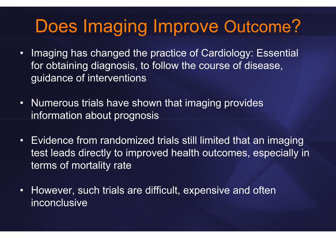
Does Imaging Improve Outcome?• Imaging has changed the practice of Cardiology: Essential
for obtaining diagnosis, to follow the course of disease, guidance of interventions
• Numerous trials have shown that imaging provides information about prognosis
• Evidence from randomized trials still limited that an imaging test leads directly to improved health outcomes, especially in terms of mortality rate
• However, such trials are difficult, expensive and often inconclusive