Embed Size (px)
Citation preview

doi:10.1093/brain/awl309 Brain (2007), 130, 10–35
REV IEW ARTICLE
What clinical disorders tell us about the neuralcontrol of saccadic eye movements
Stefano Ramat,1 R. John Leigh,2 David S. Zee3 and Lance M. Optican4
1University of Pavia, Pavia, Italy, 2Case Western Reserve University, Cleveland, OH, 3Johns Hopkins University,Baltimore and 4National Eye Institute, Bethesda, MD, USA
Correspondence to: Lance M. Optican, PhD, Laboratory of Sensorimotor Research, NEI, NIH, DHHS,Building 49, Room 2A50, Bethesda, MD 20892-4435, USAE-mail: [email protected]
Saccades are rapid eye movements that redirect the fovea from one object to another. A great deal has beenlearned about the anatomy and physiology of saccades, making them an ideal system for studying the neuralcontrol of movement. Basic research on normal eye movements has greatly increased our understanding ofsaccadic performance, anatomy and physiology, and led to a large number of control system models. Thesemodels simulate normal saccades well, but are challenged by clinical disorders because they often do notincorporate the specific anatomical and physiological substrates needed tomodel clinically important abnorm-alities. Historically, studies of saccadic abnormalities in patients have played a critical role in understanding theneural control of saccades because they provide information that complements basic research and thusrestricts hypotheses to those that are biologically plausible. This review presents four examples of clinicaldisorders (slow saccades, interrupted saccades, high-frequency saccadic oscillations and macrosaccadic oscilla-tions) that have provided insights into the neurobiology of saccades, have driven the development of newmodels, and have suggested an explanation or treatment for these disorders. We raise general questions forboth scientists and clinicians that will assist in their efforts to understand the neural control of movement,improve diagnostic criteria and develop new treatments.
Keywords: cerebellum; macrosaccadic oscillations; opsoclonus; saccadic palsy; superior colliculus
Abbreviations: cFN ¼ caudal FN; cMRF ¼ central mesencephalic reticular formation; EBN ¼ excitatory PBN;FN ¼ fastigial nucleus; FNN ¼ FN neuron; IBN ¼ inhibitory PBN; IN ¼ internuclear neuron; INC ¼ interstitial nucleusof Cajal; LLBN ¼ long-lead burst neuron; LR ¼ lateral rectus muscle; MedRF ¼ medullary reticular formation;MN ¼ motor neuron; MR ¼ medial rectus; MVN ¼ medial vestibular nucleus; NMDA ¼ n-methyl D-aspartate;NPH ¼ nucleus prepositus hypoglossi; NRTP ¼ nucleus reticularis tegmenti pontis; OPN ¼ omnipause neuron;PBN ¼ premotor burst neuron; PG ¼ pulse generator; PMT ¼ paramedian tract; PPRF ¼ paramedian pontine reticularformation; riMLF ¼ rostral interstitial nucleus of the medial longitudinal fasciculus; RIP ¼ raphe interpositus nucleus;SC ¼ superior colliculus; SCBN ¼ SC burst neurons; SCBUN ¼ SC build-up neurons; SO ¼ superior oblique;SR ¼ superior rectus; T-channel ¼ T-type Ca2+ channel; VIn ¼ sixth nerve
Received July 17, 2006. Revised October 5, 2006. Accepted October 6, 2006. Advance Access publication November 21, 2006.
IntroductionSaccades are rapid eye movements used to redirect the fovea
from one object to another. They must be fast and accurate
to support clear vision. A great deal has been learned about
the anatomy and physiology of saccades since they were first
reliably recorded over 100 years ago by Dodge and Cline
(1901; Wade and Tatler, 2005). This knowledge makes them
Published by Oxford University Press on behalf of the Guarantors of Brain, 2006.The online version of this article has been published under an open access model. Users are entitled to use, reproduce, disseminate, or display the open accessversion of this article for non-commercial purposes provided that: the original authorship is properly and fully attributed; the Journal and Oxford UniversityPress are attributed as the original place of publication with the correct citation details given; if an article is subsequently reproduced or disseminated not in itsentirety but only in part or as a derivative work this must be clearly indicated. For commercial re-use, please contact [email protected].
Dow
nloaded from https://academ
ic.oup.com/brain/article-abstract/130/1/10/347132 by guest on 14 April 2019

an ideal system for studying the neural control of
movement. Basic research on normal saccades, especially
neurophysiological recordings from identified neurons
during behaviour in monkeys, has revealed a great deal
about saccadic performance, anatomy, and physiology.
Clinical studies of patients with saccadic disorders, and
studies of the effects on saccades of brain lesions in animals,
have led to the development of quantitative hypotheses
(models) of the neural control of saccades. These models
simulate normal saccades well, but are challenged by clinical
disorders because they often do not incorporate the specific
anatomical and physiological substrates needed to model
clinically important abnormalities. The interaction between
research on animals and patients thus leads to a better
understanding of how the brain controls movements than
either alone. This interaction has made saccades one of the
best understood of all vertebrate movements.
The goal of theoretical studies of eye movements is to
develop models that realistically represent neurobiological
processes, i.e. those that are isomorphic with the brain. Such
models elucidate the neuronal mechanisms underlying
motor control (Girard and Berthoz, 2005). The purpose of
a model is to summarize knowledge, support insights, make
hypotheses explicit and quantitative, and predict or explain
new phenomena. Although each aspect of a movement can
be explained by many models, the requirement that a single
model account for as much normal and abnormal behaviour
as possible constrains the choice of models and reveals
isomorphisms that contribute to our understanding of
brain function. Thus, a key factor in modelling the neural
control of saccades was the interaction of clinical and basic
science. Nonetheless, although clinician-scientists interested
in abnormal eye movements have used saccadic models to
explain human disorders, the full impact of the basic
research effort on saccades has yet to be translated into
better biomedical care, i.e. to the bedside.
What might be impeding this translation? First,
most scientists are not familiar with clinical disorders of
human saccades, and most clinicians are not familiar
with experimental studies of saccades in animals. Second,
experimental and clinical studies often differ, in the
experimental paradigms, method of training or instruction
given to the subject, motivation or rewards, number of
subjects involved, ability to characterize the deficit, and
availability of controls. Third, clinical studies tend to be
inclusive, attempting to describe features common among
a group of patients with similar symptoms, whereas
scientific studies tend to be exclusive, focusing on one or a
few differences between experimental and control subjects.
Finally, basic scientists often study saccades in several
species (e.g. monkey or cat) that are both anatomically
and physiologically different from human patients. These
different approaches have sometimes led the two groups to
emphasize different aspects of motor control. For example,
the superior colliculus (SC, a midbrain structure that is
involved in making eye and head movements) has been the
focus of intense interest by basic scientists for over thirty
years, but discussion of the effects of SC lesions is
conspicuously absent from clinical texts.
Our goal here is to stimulate more interdisciplinary
interactions by reviewing selected disorders of human saccades
that pose specific questions for currentmodels. In each case we
describe key features of the abnormality, and discuss the issues
that the disorder poses for saccadic models.We limit the scope
of this review to brainstem and cerebellar contributions to the
generation of saccades, because these are better understood
than more cognitive functions, such as detecting and selecting
targets in a visual scene.
Characteristics of saccadesSaccades are the rapid eye movements used to voluntarily
move gaze from one target of interest to another (Fig. 1).
Human saccades follow a target jump within �250 ms, are
fast (up to �600�/s), brief (typically �30–100 ms), accurate,
and stop abruptly (i.e. with little subsequent ocular drift).
Saccades made to target jumps >10� in amplitude often
undershoot the target by �10% (Kapoula, 1985), and, after
a short latency (�150 ms), are followed by a corrective
saccade. Saccadic waveforms are characterized by the main
sequence, a set of relationships between saccade amplitude
and peak velocity (Fig. 1E), and between amplitude and
duration (Fig. 1G) (Bahill et al., 1975). The first is
logarithmically compressed for large amplitudes, whereas
the latter is linear down to <5� with a non-zero Y-intercept.
The main sequence plots are similar in monkeys and
humans, except that monkey eye movements are �30–50%
faster. Patients with certain diseases make saccades that
deviate in specific ways from the normal main-sequence
plots, making them a useful diagnostic tool. Other useful
measures of saccades are their reaction time (latency),
symmetry of their velocity waveform, and trajectory in space
or on a phase plane (discussed further below under Clinical
disorders of the saccadic system and the development of
models).
During visual search, the point of fixation is moved
between features that lie in different directions and at
different depths. Thus, saccadic movements generally have
both conjugate (both eyes rotate in the same direction,
called version) and disconjugate (the eyes rotate in opposite
directions, called vergence) components.
This review summarizes current knowledge about the
generation of saccades, and examines how the study of
different clinical disorders has led to advances in saccadic
models. First, we will review the neurobiology of saccades,
using a bottom-up approach to identify brainstem and
cerebellar components that can be incorporated into models
for saccade generation. Second, we will discuss, from a
historical perspective, how current models for the genera-
tion of saccades by the brainstem and cerebellum
were developed. Third, we will present four clinical
disorders of saccades (slow saccades, interrupted saccades,
Saccades: a review Brain (2007), 130, 10–35 11
Dow
nloaded from https://academ
ic.oup.com/brain/article-abstract/130/1/10/347132 by guest on 14 April 2019

Fig. 1 (A) Representative record of a 10� horizontal saccade made by a normal subject. In this and subsequent figures, positivevalues indicate rightward, upward or clockwise eye rotations from the subject’s viewpoint. Position (blue) and velocity (red) recordsare shown. (B) Simulated 10� horizontal saccade by model described in text. (C) Representative record of a 40� horizontal saccade madeby a normal subject; note the positive skewing of the velocity waveform. (D) Simulated 40� horizontal saccade. (E) Plot of peak velocityversus amplitude of saccades. Data points (red dots) are saccades from 10 normal subjects. These normal data are fitted with an exponentialequation of the form: Vp=Vmax (1 � e�A/C), where Vmax is the asymptotic peak velocity, A is amplitude, and C is the angle constant shapingthe exponential rise. Also plotted are the 5 and 95% prediction intervals. The plus symbols (blue) correspond to slow saccades made by apatient who had suffered brainstem ischaemia during cardiac surgery (see Fig. 5A). (F) Model simulation of peak velocity—amplitude mainsequence using required saccade amplitudes from 4� to 40�. (G) Plot of saccade duration versus amplitude. The data from 10 normalsubjects are fitted with a power equation of the form: D = D1A
n, where D is duration, D1 is the duration of a 1� saccade, A is saccadeamplitude, and n is a curvature parameter. (H) Model simulation of duration—amplitude main sequence using required saccade amplitudesfrom 4� to 40�. (Note that scales change between each row.)
12 Brain (2007), 130, 10–35 S. Ramat et al.
Dow
nloaded from https://academ
ic.oup.com/brain/article-abstract/130/1/10/347132 by guest on 14 April 2019

high-frequency saccadic oscillations and macrosaccadic
oscillations). We selected these disorders because they are
well defined and instructive about the underlying neuro-
biology, and we will discuss their implications for current
models. Finally, we will discuss how models may aid the
understanding of saccadic disorders and lead to the
development of clinically useful therapies.
Further details are documented in supplementary material
at Brain online, including videos of the clinical disorders
presented here.
Neurobiology of saccadesA sustained research effort over more than three decades,
comprising many anatomical, physiological, and behavioural
reports, has led to a better understanding of how the brain
generates saccades. Here we provide a brief description of
important brainstem and cerebellar populations of neurons
that contribute to the generation of saccades (key features of
each type of neuron are summarized in tables). A schematic
summary of the anatomy is shown in Fig. 2.
Oculomotor plantWhen discussing the neural control of eye movements, it is
helpful to start by considering the dynamics of the eye and
orbital tissues (e.g. Tenon’s capsule, fat, ligaments),
extraocular muscles and pulleys. Together, these elements
form the oculomotor plant (in engineering terms plant
refers to whatever is controlled).
Extraocular musclesMovements of each eye are controlled by six extraocular
muscles, which originate at the back or nasal side of
the orbit and travel to fibromuscular pulleys that are
formed by the fascia of the orbit (Demer, 2004). The
outer, orbital part of the muscle inserts partially on
the pulley, and the inner global part passes through the
pulley and inserts on the globe. Details of ocular movements
in three dimensions (yaw, pitch and roll) depend upon
the geometry of the muscle origins, pulleys and
insertions (Quaia and Optican, 2003a), but are not the
focus of this review. Here, we will regard the six muscles
as grouped into three agonist-antagonist pairs obeying
Fig. 2 A parasagittal section of the monkey brainstem showing the locations of PBNs. EBN for horizontal saccades lie in the PPRF; IBN forhorizontal saccades lie in the MedRF; EBN for vertical and torsional saccades lie in the rostral interstitial nucleus of the median longitudinalfasciculus (riMLF). Some vertical IBN may reside in or close to the INC. EBN and IBN project to ocular motoneurons lying in the abducensnucleus (VI), the trochlear nucleus (IV) and the oculomotor nucleus (III). OPN (indicated by a red asterisk) lie in the midline raphe of thepons between the rootlets of the abducens nerve (CN VI) and influence the activity of EBN and IBN. The mesencephalic reticular formation(MRF) may house a putative latch mechanism that keeps OPN inhibited until the saccade is over and the eye is on target (see Fig. 4C). CG:central grey; MB: mammillary body; CN III: rootlets of the oculomotor nerve; CN IV: trochlear nerve; CN VII: genu of facial nerve; ND:nucleus of Darkschewitsch; NRTP: nucleus reticularis tegmenti pontis; NPH: nucleus prepositus hypoglossi; PC: posterior commissure; TR:tractus retroflexus. (Figure courtesy of Dr Jean Buttner-Ennever).
Saccades: a review Brain (2007), 130, 10–35 13
Dow
nloaded from https://academ
ic.oup.com/brain/article-abstract/130/1/10/347132 by guest on 14 April 2019

Sherrington’s law of reciprocal innervation: lateral rectus
(LR) and medial rectus (MR); superior rectus (SR) and
inferior rectus (IR); superior oblique (SO) and inferior
oblique (IO).
Ocular motoneuronsThe brain innervates the extraocular muscles via three
cranial nerves. The abducens nerve (VIn) innervates the
ipsilateral LR, the trochlear nerve (IVn) innervates the
contralateral SO muscle, and the oculomotor nerve (IIIn)
innervates the ipsilateral MR, IR, IO and contralateral SR.
Muscles are paired in two ways. First, for each agonist there
is a corresponding antagonist muscle with almost the same
axis of action for that eye (i.e. LR–MR, SR–IR, SO–IO).
Second, muscles are yoked to move both eyes together
(e.g. left LR and right MR, left SO and right IO). Saccades
tend to follow Hering’s law, with equal innervation going
to muscles in a yoked pair. However, as noted above, when
looking between targets at different depths, different size
movements can be made in each eye. In this review, we
assume separate conjugate saccade and disconjugate ver-
gence systems (Enright, 1998; Zhou and King, 1998; Ramat
et al., 1999). Although recent evidence suggests that this is
an oversimplification, it will be sufficient for the types of
disorders discussed here.
Final common pathThe same motor neurons (MNs) and extraocular muscles
are active for all types of eye movements (e.g. saccades,
pursuit and vergence). Thus, systems generating innervation
for different movement types are said to share a final
common path. The shape of the signals entering the final
common path depends on both the desired eye movement
and the dynamic response of the eye plant. One of the first
important insights in understanding the neural control of
movement came when Robinson (1964) showed that the
dynamics of the oculomotor plant were dominated by the
viscosity of the muscles themselves. Thus, if a constant
torque were suddenly applied to the globe, the eye would
drift from its initial orientation to its final orientation with
an exponentially decaying speed (time constant �200 ms)
and the eye would reach a steady position only after about
three time constants (�600 ms). Since the eyes can make a
saccade every 200–300 ms, this has obvious disadvantages
for vision. In addition, elastic forces imposed by the orbital
tissues tend to return the eye to centre position.
Thus, all types of movement entering the final common
path have to consist of two main components: a phasic
component (pulse) giving the torque needed to overcome
the viscous drag of the orbital tissues, and a tonic component
(step) giving the torque needed to overcome the elasticity
of the orbital tissues. For saccades, the phasic part of
the innervation has a discharge rate about nine times higher
than the tonic part, so the combined innervation has a
pulse-step shape (Robinson, 1970; Quaia and Optican,
2003b). Thus, to make a saccade, the brain must compute
two different but related components of innervation. If
the pulse and step are not matched correctly, the eye will
drift (for �600 ms) after each saccade from the position
reached during the pulse to that corresponding to the step.
Final common integratorRobinson (1975) suggested that the brain could calculate the
step from the pulse by integrating it (in the mathematical
sense), because the number of spikes in the pulse was
proportional to saccade amplitude. Earlier, Skavenski and
Robinson (1973) had discovered strong evidence for a neural
integrator in the vestibular system, which introduced a phase
lag of �90� between the vestibular signal (head velocity) and
the signal encoded on ocular motoneurons (eye position). If
all the eye movement systems need to share a final common
path that is matched to the plant, they must also all share
the same integrator (otherwise, switching from, say, pursuit
to vestibular movements would cause the eye to drift).
Thus, the input to the final common path for any type of
eye movement (e.g. vestibular, pursuit, saccade) should be a
desired eye velocity.
Brain stemreticular formationThe brainstem houses the essential machinery for generating
saccades. A partial functional dichotomy exists between the
midbrain and pons. Neurons in the pons aremostly concerned
with the horizontal component of saccades, whereas the
midbrain controls vertical and torsional components.
Premotor burst neurons (PBN)Two main types of neuron are critical for saccade generation,
which we collectively refer to as PBN because they project
monosynaptically to ocular motoneurons (Table 1). The
excitatory PBNs (EBN) burst just before saccades andprobably
generate the pulse of saccadic innervation (vanGisbergen et al.,
1981). These neurons are glutamatergic (McElligott and
Spencer, 2000; Horn, 2006).
Horizontal EBN are located in the paramedian pontine
reticular formation (PPRF), below and extending rostrally
from the level of the abducens nucleus (Strassman et al.,
1986a; Horn et al., 1997). They project monosynaptically to
the ipsilateral abducens nucleus, and to a network of
neurons in the nucleus prepositus hypoglossi (NPH) and
adjacent medial vestibular nucleus (MVN) that contribute to
the final common integrator’s horizontal component.
Bilateral chemical lesions in the PPRF selectively abolish
horizontal saccades (Henn et al., 1984).
Vertical and torsional EBN are located in the midbrain, in
the rostral interstitial nuclei of the medial longitudinal
fasciculus (riMLF) (Horn and Buttner-Ennever, 1998). They
project monosynaptically to the vertical and torsional MNs,
and to the interstitial nucleus of Cajal (INC), which is an
important part of the final common integrator’s vertical and
14 Brain (2007), 130, 10–35 S. Ramat et al.
Dow
nloaded from https://academ
ic.oup.com/brain/article-abstract/130/1/10/347132 by guest on 14 April 2019

torsional components. Bilateral chemical lesions of riMLF
abolish vertical and torsional saccades (Suzuki et al., 1995).
The second group of PBN consists of inhibitory burst
neurons (IBN), which have a firing pattern almost identical
to that of EBN (Strassman et al., 1986a). Horizontal IBN lie
in the medullary reticular formation (MedRF), below
and extending caudally to the level of the abducens nucleus.
The horizontal IBN are glycinergic (McElligott and Spencer,
2000), and project to several sites, but predominantly
contralaterally to the abducens nucleus and horizontal EBN
and IBN. They suppress activity in the contralateral PBN of
the antagonist muscle, and thus may mediate Sherrington’s
law of reciprocal innervation.
Vertical and torsional IBN lie in the midbrain in the
region of the INC and riMLF (Horn, 2006). These IBN are
assumed to play a role similar to that of horizontal IBN, but
for vertical and torsional components of saccades. Note that
the vertical IBN are GABAergic (Spencer and Wang, 1996),
whereas the horizontal IBNs are glycinergic. As discussed
later, this difference may contribute to the prevalence of
horizontal saccadic oscillations, because glycine is active at
two receptors, an inhibitory (strychnine-sensitive) receptor
and a neuromodulatory (n-methyl D-aspartate; NMDA)
receptor (Miura and Optican, 2006).
Omnipause neurons (OPN)OPN (Table 2) are an important element of the saccadic
system, but their role is not fully understood. OPN lie close
to the midline in the raphe interpositus nucleus (RIP) and
their dendritic arborizations cross the midline (Buttner-
Ennever et al., 1999). They are glycinergic, and are presumed
to inhibit PBN bilaterally in both the pons and midbrain. In
an awake animal, OPN are tonically active (Strassman et al.,
1987). The OPN were first recognized because they pause
before saccades in any direction (Keller, 1974). Robinson
proposed that their function was to prevent the PBN
from firing except when a saccade was called for (Robinson,
Table 1 Characteristics of PBN
Premotor burst neurons (PBN)
* Name* Also called short-lead or medium-lead burst neurons (SLBN
or MLBN)* Consist of excitatory (EBN) and inhibitory (IBN) units
* What* EBN output is the pulse of innervation for ipsilateral saccades* IBN output inhibits contralateral EBN and IBN and ipsilateral
cMRF* Discharge rate is proportional to eye velocity
* When* PBN discharge starts 10–15 ms prior to ipsilateral saccade
start* 50% of IBN and some EBN discharge at end of contralateral
saccades* Where
* For horizontal saccades, EBN lie below and extend rostral tothe abducens nucleus in the PPRF and IBN lie below andextend caudal to abducens nucleus in the MedRF
* For vertical and torsional saccades, EBN lie in rostralinterstitial nucleus of medial longitudinal fasciculus (riMLF)and IBN lie in the region of INC and riMLF
* Afferents* LLBNs excite PBN* OPN and contralateral IBN inhibit PBN* Cerebellar FNN excites contralateral IBN and EBN
* Efferents* Horizontal motor system
& EBN (transmitter: glutamate) project monosynaptically toabducens nucleus motoneurons and internuclear neurons(which ascend in the contralateral MLF to the medial rectussubdivision of the oculomotor nucleus), and ipsilateral IBN
& IBN (glycinergic) projects monosynaptically to contralateralEBN and abducens nucleus inter- and motoneurons, and tocontralateral IBN
& EBN (uncrossed) and IBN (crossed) project to neuronscontributing to the neural integrator for horizontal eyemovements: NPH and MVN
* Vertical motor system& EBN (glutamatergic) and IBN (GABAergic) project
monosynaptically to oculomotor (III) and trochlear (IV) nuclei& EBN and IBN project to neurons contributing to the neural
integrator for vertical and torsional eye movements: INC,rostral vestibular nuclei and the cell groups of the PMT
Table 2 Characteristics of OPN
Omnipause neurons (OPN)
* Name* Called OPN because they are tonically active when awake
but pause for saccades in all directions* What
* OPN monosynaptically inhibit all PBN* Act as a switch to change from fixation to saccade mode* May also modulate strength of response of PBN by providing
glycine to NMDA receptors* When
* OPN tonic discharge stops � 20 6 10 ms before saccadeonset and restarts � 10 6 5 ms before saccade end
* Since EBN start 10–15 ms before saccade onset, OPN pausemust begin �5–10 ms before EBN burst, and restart �5 msafter the EBN burst is over
* Thus, OPNs can veto a saccade, but do not directly controlstarting and stopping it
* Where* OPN neurons all lie in nucleus raphe interpositus (RIP) in the
midline pons* OPN dendritic arborizations cross the midline
* Afferents* OPN receive bilateral excitatory inputs from rostral pole of
SC (fixation and small saccade zone)* Inhibitory inputs from LLBN* Inputs from frontal and supplementary eye fields, LLBN and
cFN.* OPN also receive an inhibitory input during the course of the
saccade, presumably from some type of saccade-relatedburst neuron
* Efferents* OPN (glycinergic) project monosynaptically to both
horizontal and vertical EBN and IBN* OPN also project to spinal cord (C1–C4), perhaps to
coordinate gaze control
Saccades: a review Brain (2007), 130, 10–35 15
Dow
nloaded from https://academ
ic.oup.com/brain/article-abstract/130/1/10/347132 by guest on 14 April 2019

1975). Later experiments showed that OPN performed
a more complicated function than just gating saccades.
Kaneko and colleagues made chemical lesions of the RIP,
and found that saccades became slower, but had normal
latency and accuracy (Kaneko, 1996; Soetedjo et al., 2002).
Miura and Optican pointed out that this could be expla-
ined if OPN were not acting as a gate, but rather as a
neuromodulator to increase the responsiveness of saccade-
related neurons (Miura and Optican, 2006). This role is
necessary because, without it, neurons in the saccadic system
would have very high gains and would be susceptible to
oscillations. By increasing the gain of these neurons only
before saccades, OPN could prepare the system to make a
fast movement without running the risk of becoming
unstable. This is also consistent with the finding that OPN
are glycinergic, because their pre-saccade activity releases
the agonist co-factor for NMDA channels on PBN. They do
not cause a saccade, however, because the action of the
glycine at the inhibitory strychnine-sensitive channels is
assumed to dominate its effect at the NMDA channels.
Excitatory input to the OPN arrives from the SC and
vestibular nuclei (Strassman et al., 1987; Langer and Kaneko,
1990). The OPN pause �16 ms before saccades, and resume
�15 ms before the end of saccades. Inhibitory input seems
Fig. 3 Early models of the saccadic system. (A) A local negative feedback loop model (Robinson, 1975), which assumes that efferencecopy of eye position, uu, corrects retinal error, e, and recreates the central percept of the spatial location of the target, uuT . This signal actsas the input to a bang–bang control system consisting of a high-gain saturating amplifier, PG, followed by an integrator, NI. SJ1, 2, 3 aresumming junctions; suppressor switch S is normally open, unless the OR-gate (red) is activated by a trigger signal, Trig, or the PG output.MLF carries the pre-emphasis signal (or burst). The orbital dynamics are in Laplace notation and Te is the time constant of the ocular motorplant. (B) Model to account for saccadic oscillations by a high-gain pulse generator with a negative feedback loop (Ashe et al., 1991).The input signal to the EBN is desired change in eye position (ED). A trigger signal (Trig) inhibits the OPN (red), which also receive abias signal (TONE). The output of the EBN is a pulse of innervation, which projects to ocular motoneurons (OMN), to the common ocularmotor integrator (NI) that generates a step of innervation, and to the IBN. RI is the re-settable integrator with a delay (blue) interposedbetween its output (an internal estimate of current eye position) and an inhibitory synapse on the EBN. The IBN inhibit OPN duringthe saccade (NB: IBN to OPN projections have not been found).
16 Brain (2007), 130, 10–35 S. Ramat et al.
Dow
nloaded from https://academ
ic.oup.com/brain/article-abstract/130/1/10/347132 by guest on 14 April 2019

to show two time courses, a fast one associated with a trigger
that shuts off the OPN to initiate a saccade, and a slower
one that prevents the OPN from turning on again until
the saccade is over (Yoshida et al., 1999).
Latch-circuit neuronsThe original local feedback model of the saccadic system
(Robinson, 1975; Zee et al., 1976) was the first that could
generate the pulse of innervation needed tomake a saccade and
stop automatically (Fig. 3A). The main element of the local
feedback model was a saturating high-gain amplifier (pulse
generator; PG). A gate was needed to prevent the high-gain
amplifier inside the feedback loop from oscillating. In the
original model, the pulse generator neuron was inhibited by a
switch cell (S in Fig. 3A), which gated the input to prevent the
oscillations. To initiate a saccade, a trigger signal (Trig) of
short, fixed duration would inhibit the switch cell, and the
pulse generator would begin firing. As the movement
progressed, the trigger input died away, but the activity of
the pulse generator neuron was fed back, through an OR-gate,
to keep the switch cell off until the movement was over. This
gate was called a latch because once it was closed, it kept itself
closed (through feedback) until the end of the saccade. When
the pulse generator neuron and the trigger were both off, the
suppressor cell (S) became active because of a static input
Robinson called ‘tone’.
Subsequent research into the brainstem cell types associated
the pulse generator cells with the excitatory medium-lead
burst neurons (EBN), and the suppressor cells with the OPNs.
This still leaves four unanswered questions about the
suppressor mechanism. (i) Why are the OPNs usually active
(i.e. where does the tone come from)? (ii) What shuts them
down before the saccade (what is the trigger)? (iii) What holds
them off during the saccade? And, (iv) what reinitiates their
activity at the end of a saccade?
We refer to the suppressor mechanism as a latch circuit,
because a latch remembers its input even after the input has
been removed. This requires some form of feedback to create
two stable states, on and off, that are controlled by two
inputs, usually called set and reset. Robinson’s original gate
circuit acts as a latch circuit because it feeds back the output
of the pulse generator to the suppressor cell. However, it
only has the set input, which is the trigger. The circuit
unlatches automatically when the motor error estimate is
reduced to the dead-zone (which stops the burst cell
activity). The problem of making accurate saccades still
comes down to how and when to restart the OPNs. A more
recent hypothesis has been proposed that separates the
feedback control of gaze from the latch circuit (Lefevre et al.,
1998). The saccade normally ends when the ipsilateral
cerebellum fires the contralateral brainstem IBNs, choking
off (i.e. interrupting, stopping) the drive signal to the
MNs. By making the latch circuit independent of motor
error, the effect of time jitter on movement accuracy is
significantly reduced. Accuracy can be maintained by the
cerebellar circuit, and stability can be guaranteed as long as
the OPNs reactivate soon after. A more detailed hypothesis
for a latch circuit is developed below in A model of the latch
circuit.
Long-lead burst neurons (LLBN)LLBN (Table 3) are found in the brainstem and receive
input from the SC and cortical areas responsible for
saccades, such as the frontal eye field, parietal eye field
and supplementary eye field (Scudder et al., 1996a, b). Some
of the LLBN probably project to the premotor burst
neurons, whereas others [in the nucleus reticularis tegmenti
pontis (NRTP)] project to the cerebellum. Scudder (1988)
proposed a saccade model that used the LLBN as a
re-settable integrator in the feed-forward path. The re-
settable integrator was thought to be distinct from the
common final integrator and to apply only to saccadic
signals (see below, in Conceptual evolution of saccadic
models). However, later evidence suggests that the re-
settable integrator does not really exist, and that the
cerebellum replaces its function (Quaia et al., 1999). Thus,
LLBN seem to act as a summing junction for combining
saccade commands from different areas involved in the
preparation for a saccade.
Eye position (tonic) neurons and the neuralintegrator for eye movementsTonic (and burst–tonic) neurons carry a signal related to the
step component of MNs. The step is assumed to be obtained
by integrating (in the mathematical sense) velocity signals
from PBN that generate the pulse of innervation (this is
an oversimplification, but sufficient for our needs here).
Neurons that carry an eye position, or step, signal for
horizontal saccades lie in the medulla and pons in the
NPH nuclei (McCrea and Horn, 2006), and adjacent MVN.
Neurons that carry a vertical and torsional eye position
(step) signal are present in and around the INC. In addition,
the cell groups of the paramedian tract (PMT) also probably
contribute to the neural integrator (Buttner-Ennever and
Horn, 1996).
The mechanism that performs the integration is not
well understood, although models that integrate because
of reciprocal innervation (positive or negative) across the
midline have been developed (Cannon and Robinson, 1985;
Cova and Galiana, 1996; Arnold and Robinson, 1997), and
recent work has cast light on how networks of neurons could
perform this mathematical operation (Aksay et al., 2005).
Curiously, when the cerebellum is lesioned the time
constant of this integrator drops from �20 s to �2 s
(Carpenter, 1972), but when the commissures connecting
the left and right halves of the integrator are cut, the time
constant drops below �300 ms (i.e. only the time constant
of the oculomotor plant itself is left). Thus, the integration
of ocular motor signals depends on both cerebellar and
Saccades: a review Brain (2007), 130, 10–35 17
Dow
nloaded from https://academ
ic.oup.com/brain/article-abstract/130/1/10/347132 by guest on 14 April 2019

brainstem circuits, but their respective roles remain
enigmatic.
Superior colliculus (SC)One influence on PBN and OPN is the SC, a multi-layered
structure in the midbrain. The SC is laid out in a retinotopic
map (Robinson, 1972), with small contralateral movements
related to activity in the rostral SC, and large contralateral
movements related to caudal SC. This is true with the head
fixed (eye only saccades) or free (gaze saccades, where gaze =
eye + head). Neurons in the superficial layer are related to
visual events, but neurons in the intermediate and deep layers
are related to both visual and motor events. Schiller et al.
(1980) showed that saccades remained accurate after SC
ablation, and thus that it was not necessary for generating the
pulse for saccades. Later it was also shown that ablations of the
cerebellum caused persistent deficits in saccade amplitude
(e.g. Optican and Robinson, 1980). Despite these early
observations, until the early 1990s the SC was widely assumed
to control saccadic amplitude, direction and trajectory. Many
studies have since shown that themajor role of the SC seems to
be in selecting a target for foveation—either with saccadic or
pursuit movements (Krauzlis, 2004)—initiating the move-
ment (Bell et al., 2005) and contributing to its speed (Hanes
et al., 2005), but does not determine amplitude, duration,
direction or trajectory of the saccade.
The intermediate and deep layers of the SC contain two
major types of neurons that are laid out in a retinotopic
map. One type shows a target related build-up in activity
(SCBUN), and a burst just before the saccade. The other
bursts just before the saccade, but has no prelude of activity
(SCBN). Electrophysiological studies show that both SCBN
and SCBUN project from the SC to the brainstem (Munoz
and Wurtz, 1993, 1995a). The SC projection is not uniform.
The rostral half of the SC sends more fibres to the RIP
(region of the OPN) than does the caudal half. The SC
neurons project mostly to LLBN, and probably do not have a
functional projection to PBN (Gandhi and Keller, 1997).
The level of activity in the prelude of SCBUN corresponds
to the likelihood that the target at that location will be the
goal of a saccade (Basso and Wurtz, 1997). SC activity is also
a function of retinal error, whether for a saccade or a pursuit
movement (Krauzlis et al., 2004).
In every experiment where the saccade goal is not the
same as the visual target, such as adapted saccades
(FitzGibbon et al., 1986), saccades to moving targets
(Keller et al., 1996b), and strongly curved saccades (Port
and Wurtz, 2003), the locus of activity on the SC corres-
ponds to the visual target, and not the ensuing movement.
Ablations of the SC significantly increase saccade latency and
reduce peak speed, but do not affect accuracy (Hanes et al.,
2005). Thus, the most parsimonious interpretation of SC
function is that it contributes to identifying the target
(in retinotopic coordinates) that is to be foveated, generates
a trigger signal to shut down the OPN, and sends a fixed-
direction drive to LLBNs to begin the saccade.
Central mesencephalic reticularformation (cMRF)The cMRF has strong, topographically organized, reciprocal
connections with the SC (Cohen and Buttner-Ennever, 1984;
Chen and May, 2000) and the region of the OPNs (Langer
and Kaneko, 1983, 1990), and projects to the EBN and IBN
in the brainstem (Buttner-Ennever et al., 1999). Cells in the
cMRF that are related to saccades discharge for contraversive
movements, and can be divided into two groups (Waitzman
et al., 1996). One group has a low background rate of
firing (<10 spikes/s), and the other, larger, group has a high
background rate of firing (>10 spikes/s). Both groups start
to burst �30 ms before the saccade. Some cells have an
abrupt, or clipped, end to the burst, at the end of the
saccade. Indeed, burst duration in �31% of cells in cMRF
are correlated with saccade duration, �48% with saccade
amplitude, and �58% with saccade velocity; some cells
respond to two of the three metrics (Cromer and Waitzman,
2006). Lesions in the cMRF result in hypermetric contra-
lateral saccades, reduced latency, and macrosaccadic square-
wave jerks (Waitzman et al., 2000). These results suggest that
the cMRF is important for saccade function at both the
initiation and termination stages. One possible model of
their contribution is described below.
CerebellumA second influence on PBN and OPN is the cerebellum,
which plays an important role in steering and stopping
saccades, thus determining their accuracy. The posterior
pole of the midline cerebellum has been most intensively
studied. This region is divided into a cortical part (vermis
and paravermis) and the underlying deep cerebellar nuclei
(fastigial, interpositus and dentate). All relevant signals
Table 3 Characteristics of LLBN
Long-lead burst neurons (LLBN)
* Name* Long-lead because discharge starts long (hundreds of ms)
before saccades* What
* May provide information on the target location inretinotopic coordinates to the PBN and cerebellum
* May also provide a trigger signal through PBN to shut downOPN
* When* LLBN discharge >40 ms prior to saccade onset, with slow
build-up before burst, just like build-up neurons in SC* Where
* Distributed throughout the brainstem reticular formation,especially in the cMRF and NRTP
* Afferents* SC, supplementary, and perhaps other, cortical eye fields,
cerebellar FNN* Efferents
* LLBN (transmitter unknown) project to EBN, IBN, OPN
18 Brain (2007), 130, 10–35 S. Ramat et al.
Dow
nloaded from https://academ
ic.oup.com/brain/article-abstract/130/1/10/347132 by guest on 14 April 2019

(e.g. eye position and velocity, target position and velocity)
needed to control saccades arrive in the cerebellar cortex on
mossy fibres. Climbing fibres bring a signal from the inferior
olive to the cerebellar cortex. Climbing fibre activity is
related to the distance to the target (motor error) at the
beginning and end of a movement (Kitazawa et al., 1998).
The climbing fibre activity is assumed to be necessary to
learn proper motor control, but its role is not well
understood.
Dorsal vermis Purkinje cells (PC)Stimulation in many parts of the cerebellum (lobes V–VII of
the vermis, and the hemispheres, crus I and II) can evoke a
saccade (Ron and Robinson, 1973). Stimulation of vermal
lobule V evokes saccades that range from upward to
horizontal, while stimulation of lobules VI and VII evokes
saccades that range from horizontal to downward. The
amplitude of the elicited saccade, and the amount of post-
saccadic drift, depend upon the initial position of the eye in
the orbit. Purkinje cells that discharge in relation to saccades
are located in a more restricted region, lobes VI(c)–VII,
called the oculomotor vermis (Noda and Fujikado, 1987).
The oculomotor vermis projects to the caudal part of the
fastigial nuclei. Purkinje cells in the dorsal vermis discharge
�15 ms before saccades in a preferred direction (Ohtsuka
and Noda, 1995). Stimulation of the vermis produces
saccades with an ipsilateral component (Ron and Robinson,
1973). With currents near threshold, a topographic
organization is evident (Noda and Fujikado, 1987).
Fastigial nucleus neurons (FNN)The caudal part of the fastigial nucleus (cFN) is responsible
for sending saccade commands to the region of the PBN in
the contralateral brainstem. FNN fire tonically with a low
rate, and burst near the time of a saccade (Ohtsuka and
Noda, 1991; Fuchs et al., 1993). FNN fire for saccades in
all directions, but electrical stimulation in the cFN elicits
contralateral saccades (Noda et al., 1988). The latency of
the burst is a function of the direction of the movement.
FNN burst before saccade start for contralateral movements,
but burst near saccade end for ipsilateral movements. Thus,
the same neurons are firing for ipsi- and contralateral
saccades, with the only difference being that they fire later
for ipsilateral saccades. This issue is addressed further in the
section Cerebellar models.
Cerebrum and basal gangliaMany areas of the cerebral cortex (e.g. frontal eye fields,
supplementary eye fields, lateral intraparietal cortex) are
known to be involved in identifying and selecting targets for
saccades (Leigh and Kennard, 2004). The basal ganglia are
involved in selecting and preventing movements, and in
reward (Hikosaka et al., 2000). The substantia nigra pars
reticulata tonically inhibits the intermediate layers of the SC.
This tonic activity can be suppressed by inhibition from the
caudate nucleus, allowing a saccade. As important as they
are for selecting the target for saccades, these areas do not
play a role in generating the saccadic pulse itself. Thus, a
discussion of their function is beyond the scope of this
review.
Models for saccadesConceptual evolution of saccadic modelsIn this section we present a brief history of saccadic models,
beginning with the first model that was physiologically
plausible. Robinson proposed that the innervation needed
to make a saccade had to consist of two components: a pulse
to overcome the viscous drag in the orbit, allowing for a fast
movement, and a step to overcome the elastic restoring force
in the orbit. Robinson proposed that the step could be
computed as a function of the pulse, which simplified the
problem of generating saccadic innervation to that of gene-
rating the pulse (Robinson, 1973). Furthermore, extraocular
muscle proprioception, unlike skeletal muscle spindles, does
not participate in a stretch reflex (Keller and Robinson,
1971). Thus, it was assumed that saccades were ballistic, i.e.
their innervation was programmed before the saccade, and
simply ‘played out’ during the movement.
Shortly after this, Zee et al. (1976) showed that the slow
refixations made by patients with spinocerebellar disease
were actually very slow saccades (see section Slow saccades).
Indeed, these movements were so slow that visual
re-afference returned in time to influence the saccadic
system. They found that if the target jumped to a new
location after the patient began a saccade, the eye would turn
around in mid-flight and go to the new target. This
demonstrated that the saccadic system was not ballistic and
led Zee and Robinson (1979) to propose that the pulse of
innervation was generated during the movement by an
internal, or local, feedback loop based on an efference copy
of eye position. This efference copy was obtained by
integrating the pulse that came from the brainstem burst
neurons (Zee et al., 1976). This model immediately found
wide acceptance because it generated both normal and slow
saccades that stopped automatically (i.e. without pre-
programming), and included a role for the recently
discovered burst and pause neurons in the pons. An impor-
tant refinement of the original local feedback loop model
was made by Zee and Robinson in 1979, stimulated by an
attempt to explain microsaccadic oscillations (see Section on
high-frequency saccadic oscillations). In developing that
model they used a new non-linear function to represent the
activity of the burst neurons for a given motor error which is
still widely used today (van Gisbergen et al., 1981). To
explain the small amplitude, high-frequency oscillations
observed in some patients, they added a delay in the local
feedback loop around the high-gain amplifier representing
the burst neurons (Zee and Robinson, 1979; Ashe et al.,
1991; Fig. 3B).
The original version of the model had an efference copy
of the eye position signal which was fed back to a
Saccades: a review Brain (2007), 130, 10–35 19
Dow
nloaded from https://academ
ic.oup.com/brain/article-abstract/130/1/10/347132 by guest on 14 April 2019

comparator that computed the instantaneous, or dynamic
motor error: the difference between desired eye position and
current eye position. The efference copy was also used to
reconstruct an internal estimate of the target’s position in
spatial coordinates. However, all saccade-related neurons
found to date encode retinotopic error and change in eye
position, not position in space. When Jurgens et al. (1981)
found evidence for a separate neural integrator for saccades,
they modified the local feedback loop model so that an
efference copy of eye velocity was fed to a re-settable
integrator that was reset to zero before each saccade. The
output of this re-settable integrator was an efference copy of
instantaneous eye displacement, which could thus be
compared to a desired eye displacement signal in retinotopic
coordinates. No reconstruction of target position in space
was needed.
The Jurgens–Robinson model is the one from which most
others have descended. Saccadic models evolved slowly after
the local feedback loop was introduced. Optican and Miles
(1985) introduced a third component of innervation (the
slide) that was necessary to explain the transition between the
pulse and the step. This led to the current concept of saccadic
innervation consisting of a pulse–slide–step, with the slide and
the step being automatically computed from the pulse
generated by the burst neurons. Based on experimental data
from the cat (Munoz et al., 1991), activity in the SC during the
execution of a saccade was modelled as a moving hill (Droulez
and Berthoz, 1991; Lefevre et al., 1994), which was considered
to represent the tracking of the progress of the fovea toward the
intended target (i.e. the dynamic motor error). This and other
work (Waitzman et al., 1988) suggested that the saccadic
feedback loop might be closed within the SC itself. However,
subsequent efforts to show an error-related spread inmonkeys
have failed (Munoz and Wurtz, 1995b; Aizawa and Wurtz,
1998; Anderson et al., 1998).
More recently, Guitton and colleagues have found that in a
head-free animal, the locus of activity in the SC corresponds
to the retinotopic location of the target, not instantaneous
motor error (Bergeron and Guitton, 2002; Bergeron et al.,
2003). Furthermore, if multiple small steps are needed to
acquire the target, the activity on the SC jumps from one
locus to the next, always encoding the eccentricity of the
target for the next saccade (Matsuo et al., 2004). Thus,
despite evidence that the SC receives some feedback about
gaze movements, it does not seem to be responsible for
calculating motor error during a movement.
The major problem with the Jurgens–Robinson model is
that it was designed from the top down to perform an eye
tracking task, and thus is not isomorphic to the brain. Many
structures in the brain that are clearly related to saccades,
from the cerebral cortex to the SC to the cerebellum, were
not included in that model, and there is no obvious way to
fit them into it. The problem of incorporating anatomical
and physiological evidence into a model was addressed
directly by Optican and colleagues, who took descriptions of
known cell activity in different brain areas and combined
them (i.e. from the bottom-up) to form a working saccadic
system (Lefevre et al., 1998; Quaia et al., 1999). This
neuromimetic model was able to make visually guided
saccades, and provided a structure for the addition of more
cell types and brain areas to account for other types of
saccadic behaviour. More importantly, its isomorphic design
made it completely different, in both structure and signals,
from the classic engineering design of target tracking systems
(Optican and Quaia, 2001). The neuromimetic model has
many significant structural differences from the Jurgens–
Robinson model, but maintains its key element, the local
feedback loop for the control of saccade trajectory (Fig. 3).
However, it has no re-settable integrator, no efference copy
of eye displacement, and no motor error signal. Instead, all
their functions are performed by distributed networks in
the brainstem and cerebellum (Optican and Quaia, 2002).
The performance of that model is very good when making
normal saccades. However, it contained no mechanism for
restarting the OPNs, and could not explain hypometria of
saccades after lesions of contralateral FN. Future models will
have to resolve these problems.
Simple saccade model that can be appliedto clinical disordersRecently we developed a model of brainstem and cerebellar
control of horizontal saccades that we applied to the clinical
disorders that we discuss in the next section. This model was
derived from the neuromimetic model of Optican’s group. It
included many anatomical details of the brainstem circuitry
that were not known when the Jurgens–Robinson model
was proposed. The brainstem circuit was based on the
anatomical findings by Strassman et al. (1986a), and first
represented in a saccade model of the local feedback loop
proposed by Scudder (1988). The newmodel includes two new
feedback loops, the shortest being one between the IBNs on
both sides. Although the IBNs are inhibitory, their reciprocal
connection creates a positive-feedback loop. The second
positive-feedback loop involves EBNs and IBNs on both sides.
The IBNs are prone to oscillate, because of their high
sensitivity, close proximity (which gives rise to only a short
delay) and reciprocal negative connections (which result in
positive feedback). In his model Scudder avoided oscillations
by assuming that the strength of these connections was quite
low, although there is no direct evidence about their strengths.
Our new mathematical model (Ramat et al., 2005) of the
saccadic brainstem mechanism tries to reproduce the salient
characteristics and projections of the different populations
of neurons that are responsible for producing horizontal
saccades from the OPN to the abducens nuclei (VI). Thus,
the model represents the OPN, the bilateral EBN and IBN,
and the VI nucleus including both the motor and inter-
nuclear neurons (INs; Fig 4A). Each population of neurons
is modelled as a single element with lumped parameters. All
neurons are modelled with a similar structure representing
the membrane as a high-pass filter showing adaptation,
20 Brain (2007), 130, 10–35 S. Ramat et al.
Dow
nloaded from https://academ
ic.oup.com/brain/article-abstract/130/1/10/347132 by guest on 14 April 2019

followed by a non-linear saturation and a small delay
(0.8 ms). The model for the EBN neurons is shown in
Fig. 4B, where the output non-linearity is represented by the
‘Burst’ block implementing the soft saturation in Eq. 1 (Zee
and Robinson, 1979).
BðeÞ ¼�Bmð1� e�ðe�e0Þ/bÞ‚ e > e00‚ e<e0
ð1Þ
The same structure, although with independent para-
meters, was used to represent the IBN.
The consequence of the membrane adaptation property
is that these neurons show post-inhibitory rebound: at the
offset of inhibition there is a rebound in the membrane
potential that carries it to positive values, thus allowing the
cell to fire one or more action potentials spontaneously.
Post-inhibitory rebound is a property of some neuron types
that, at the offset of hyperpolarization, produce a discharge
mediated by low-threshold Ca2+ channels (Perez-Reyes,
2003). This implies that neurons showing this property may
fire a train of action potentials when they are abruptly
disinhibited, even without an input actively driving the cell.
Fig. 4 (A) A brainstem neural network model for generation of horizontal saccades (Ramat et al., 2005). Projections with flat endingsare inhibitory, the others excitatory. Saccades require reciprocal innervation to the medial and lateral recti (ML and LR) of both eyes.The LR is driven by the ipsilateral abducens nucleus (VI n) MNs. The VI n also contains an IN that sends its axon via the medial longitudinalfasciculus to the contralateral III n, which drives the MR of the other eye. EBN thus provide the drive to the ipsilateral MN and IN.The EBN also project to the ipsilateral IBN. The IBN inhibit the contralateral MN and IN. Thus, an EBN/IBN pair provides reciprocalinnervation to the muscles. The IBN also inhibit the contralateral EBN and IBN. A consequence of this cross-coupling is that theEBN/IBN pairs on both sides form a short-latency, positive-feedback loop. When OPN are active, they prevent this loop fromoscillating. (B) Burst neuron model allowing post-inhibitory rebound and adaptation. The cell membrane contains a low-pass filter(time constant mTc), and a high-pass filter in a positive feed-forward path. The gain (aGain) and time constant (aTc) of this second high-passfilter determines the amount of post-inhibitory rebound. Burst: output non-linearity of the burst neurons; IN: input signal; K: aconstant. (C) A latch-circuit model (Rucker et al., 2005). The key to the latch model is that the SC $ cMRF connections form apositive-feedback loop that keeps them firing during the saccade. The output of this loop goes to latch cells (blue), which inhibitthe OPN (see text for details).
Saccades: a review Brain (2007), 130, 10–35 21
Dow
nloaded from https://academ
ic.oup.com/brain/article-abstract/130/1/10/347132 by guest on 14 April 2019
Post-inhibitory rebound in the saccadic burst neurons was
first introduced by Enderle and Engelken (1995) who sug-
gested that it is the only mechanism needed for generating
saccades. A later model combined the post-inhibitory
rebound with excitatory input from LLBNs to generate
saccades (Miura and Optican, 2006).
The circuitry that moves the eye horizontally is
represented in black in Fig. 4A, including the lateral and
medial rectus muscles, the adducting MNs in the III nuclei,
the abducting MNs and IN in the abducens nuclei, and their
projections. The input driving saccades is provided by the
ipsilateral EBN (shown in blue in Fig. 4A), which project to
both the abducens MN and abducens IN, the latter in turn
projecting across the midline to the contralateral IIIn. The
IBN, shown in red in Fig. 4A, are excited by the ipsilateral
EBN and inhibit the contralateral VIn and PBN.
The interconnections between burst neurons form two
positive-feedback loops: one short loop is formed by the IBN
neurons reciprocally inhibiting each other on the two sides of
the midline; the other involves both EBN and IBN bilateral
groups. This longer loop is provided by the excitatory
projection of EBN onto ipsilateral IBN, which in turn inhibit
the contralateral EBN which excite the ipsilateral IBN, which
cross back to inhibit the originally firing EBN. This organi-
zation, which was anatomically identified (Strassman et al.,
1986a), implements Sherrington’s law of reciprocal innerva-
tion (contraction of agonist while ensuring the inhibition of
antagonist muscles), and favours the fast build-up of EBN
discharge (Strassman et al., 1986a).
When a desired eye position signal coding for a rightward
saccade excites the right EBN, it projects both to the ipsi-
lateral VI nucleus and to the ipsilateral IBN. The excitation
of ipsilateral IBN inhibits the contralateral VI, the
contralateral EBN and the contralateral IBN. Therefore the
inhibitory action of this contralateral (left) IBN group is
reduced, both directly via the IBN–IBN projection and
indirectly since the inhibition of the (left) EBN in turn
reduces the (left) IBN excitation. The left IBN’s inhibition
disinhibits the right IBN, thus closing the short positive-
feedback loop from IBN to IBN, and disinhibits the right
EBN, thus closing the second positive-feedback loop
involving EBN and IBN bilaterally.
The OPN (shown in green in Fig. 4A) project to all four
groups of burst neurons, tonically inhibiting them except
during saccades in all directions (Keller, 1974), blinks (Hepp
et al., 1989, Mays and Morrisse, 1995), and to some extent
during vergence (Busettini and Mays, 2003).
In addition to the described structures and connections,
the suggested mathematical model includes a ‘desired eye
position’ input to the saccadic mechanism, turning off the
OPN when a saccade is programmed, and a local feedback
loop responsible for turning off the saccades once the eyes
are on target (Ramat et al., 2005).
In humans, the mechanism generating saccades is poten-
tially unstable due to the high-gain of the output non-
linearity of the burst neurons and to the positive-feedback
loops coupling EBN–IBN and IBN–IBN. If the burst neurons
are not inhibited by the OPN and are not driven to produce
a saccade, such latent instability may lead to high-frequency,
conjugate oscillations of the eyes composed of back-to-back
saccades occurring without intervening periods of steady
fixation (see section High-frequency saccadic oscillations).
Such a condition may occur during blinks, saccade-vergence
interactions, and orthogonally directed saccades.
In fact, OPN pause for saccades in any direction and thus
release all populations of PBN, although the saccade may
have only one component, e.g. the OPN shut off for a vertical
saccade, disinhibiting both the vertical and horizontal PBN.
The vertical EBN will be driven to produce a vertical
saccade, while the horizontal EBN will not receive any dri-
ving input. Yet, the offset of the OPN hyperpolarization will
produce post-inhibitory rebound in both horizontal IBN
and EBN on both sides of the midline, causing these cells to
simultaneously fire a few action potentials. Any imbalance in
the circuit will allow one side to prevail and a periodic
oscillation will ensue. Suppose that the right EBN produces a
few spikes from post-inhibitory rebound. They will drive the
right IBN which will inhibit the left EBN and the eyes will
move to the right. Because of the fast decay (adaptation) of
the post-inhibitory rebound in the right EBN, the left EBN
will be disinhibited and will in turn show a post-inhibitory
rebound, driving the left IBN, in turn inhibiting the right
EBN and moving the eyes to the left. As the post-inhibitory
rebound in the left EBN is extinguished, the process repeats
itself until the OPN are turned back on after the vertical
saccade is over.
Cerebellar modelsAll fibres coming to the cerebellar cortex also deliver a
branch to the corresponding deep nuclei (e.g. both vermis
and FN). Currently, there are two types of cerebellar models.
The first type learns (because of association with climbing
fibre activity) to activate an output fibre at some time after
an input fibre discharges. These models are always 1D, and
cannot be extended to deal with multidimensional problems.
The second type is pre-wired to perform certain hypothetical
functions, such as initializing the locus of activity on the
vermis and updating that locus based on velocity feedback
information (Optican and Quaia, 2002). These models are
helpful in understanding control of saccade trajectory, but
do not provide insights into how the cerebellum could deve-
lop a network with such a function.
The mossy fibre inputs to the cerebellum carry signals
from the brainstem LLBNs in the NRTP. The NRTP receives
inputs from the SC and the FN itself. Neurons in the cFN
discharge about 8 ms prior to onset of saccades with
contralateral components, but generally towards the end of
saccades with ipsilateral components. A major mystery of
the cerebellum is how does the output from the ipsilateral
FN fire later than its inputs, i.e. near the end of the saccade?
One interpretation (Lefevre et al., 1998; Quaia et al., 1999)
22 Brain (2007), 130, 10–35 S. Ramat et al.
Dow
nloaded from https://academ
ic.oup.com/brain/article-abstract/130/1/10/347132 by guest on 14 April 2019

is that the cerebellum, through the FNN, controls the
trajectory and endpoint of a saccade. The difference in
latency between ipsi- and contralateral FNNs breaks the
symmetry between the projections from the cFN to the PBN
in the brainstem. For example, for a rightward saccade, the
left FNN excite both the IBN and EBN on the right side
(Fig. 4C). The IBN on the right then inhibit the EBN and
IBN on the left side, allowing the eye to move to the right. At
the end of the movement the right-side FNN fire, exciting
both the IBN and EBN on the left side. However, the IBN
on the right side are already inhibiting the PBN on the left. If
we assume that the FNN excitation to the IBN, but not
the EBN, overcomes the inhibition from the right IBN, then
the IBN on the left will shut down the EBN on the right,
stopping the saccade.
Optican and colleagues have proposed a theory to account
for this experimental result (Lefevre et al., 1998; Quaia
et al., 1999; Optican and Quaia, 2002; Optican, 2005). In this
theory the role of the cerebellum in movement control is
2-fold. First, it recognizes from the sensory, motor and
behavioural context what its contribution to the movement
should be (determined by learning from experience).
Second, it integrates velocity feedback information to obtain
instantaneous displacement information (thus performing a
role equivalent to the resetting integrator in the Jurgens–
Robinson model) and modifies the saccade drive signal to
compensate for errors. The integration in their model is
performed by updating the locus of activity in the cerebellar
cortex, giving the appearance of a wave of activity that
spreads from the contralateral to the ipsilateral side during a
saccade. This causes the difference in latency of the ipsi- and
contralateral FNN observed during eye and gaze saccades
(Optican, 2005). No evidence for this wave has been found
yet, but the wave idea is not central to the cerebellar hypo-
thesis. Any mechanism, such as a population of neurons
with varying thresholds, that can perform the integration
with an appropriate delay on both sides of the midline
would be sufficient.
A model of the latch circuitHaving described features of models for PBN, OPN, SC and
cerebellum, we are now in a position to return to the latch
circuit for saccades. A schematic diagram of a proposed latch
circuit model (Rucker et al., 2005) for a rightward saccade
is shown in Fig. 4C (the model is symmetric, but many
elements and pathways have not been drawn to emphasize
crossover of activity from one side to the other during the
movement). This new proposal differs from previous latch
circuits because it is bistable, requiring both a set and reset
command. The set command turns on reciprocal excitation
between a subset of SC and cMRF neurons, and the reset
command breaks that positive-feedback loop. We thus need
two types of neurons in the SC, one which serves as part of
the latch circuit, and one that sends information about the
selected target to the brainstem. In Fig. 4C, the ‘When’ and
‘Where’ cells are two hypothetical sub-classes of SCBN. They
correspond to two groups of SC cells, one that loses activity
(blue versus red trace for ‘When’ cells), and one that does
not (red and blue trace overlap for ‘Where’ cells), after OPN
lesions (Soetedjo et al., 2002). Correspondingly, cMRF cells
can be divided into two groups, one that receives ‘When’ cell
activity and participates in the latch (which may be the
cMRF cells related to saccade duration, presumably those
having a low background rate), and one that relays target
information to the brainstem, which may be the cMRF cells
related to saccade amplitude and/or velocity, presumably
those having a high background rate (Waitzman et al., 1996;
Cromer and Waitzman, 2006). The SC fix cells and the high
background cMRF cells excite the OPNs, keeping them on
between saccades.
In this model, the rapid offset of the OPN before a saccade
is caused by a burst in the EBN-like latch cell (Keller and
Missal, 2003), which is driven by the low background cMRF
cells. The key to the latch model is that the SC$cMRF
connections form a positive-feedback loop that keeps them
firing during the saccade, with no other input. The output of
this loop goes to latch cells, which inhibit the OPN (through
a GABAergic interneuron, shown in red). That loop conti-
nues firing until the ipsilateral (right side for a rightward
saccade) fastigial output of cerebellum fires the contralateral
(left) IBN cells, which are the choke for the ipsilateral (right)
EBNs. In addition, those (left) IBN send a unilateral projec-
tion to the cMRF on the same side (Strassman et al., 1986b).
This inhibits the low background cMRF cells, breaking the
positive feedback loop and acting as the reset signal for the
latch. These cells would correspond to the duration-related,
clipped cells found in the cMRF (Cromer and Waitzman,
2006). In the model, the rapid onset of the OPNs is caused
by excitatory inputs from the SC (FIX) and cMRF (high
background).
Clinical disorders of the saccadic system andthe development of modelsSlow saccadesClinical features of slow saccadesSeveral clinical disorders cause a marked slowing of saccades
that can easily be detected at the bedside. Slow saccades are
caused by diseases affecting the extraocular muscles or their
cranial nerve supply; by brainstem stroke; and by a number
of genetic and degenerative diseases, such as the spinocere-
bellar ataxias (SCAs), Huntington’s disease, and progressive
supranuclear palsy (PSP), which affect neurons throughout
the brainstem and cerebellum (Leigh and Zee, 2006).
The first example (Fig. 5A) is of a patient who developed
slow saccades following cardiac surgery (Tomsak et al.,
2002). Both horizontal and vertical saccades were very slow,
and vestibular quick phases were essentially absent. Other
types of eye movement, including smooth pursuit and
the vestibulo-ocular reflex, were normal. The precise cause
Saccades: a review Brain (2007), 130, 10–35 23
Dow
nloaded from https://academ
ic.oup.com/brain/article-abstract/130/1/10/347132 by guest on 14 April 2019

of the deficit in this patient is unknown. However, a
similarly affected patient died of a post-operative infection,
and subsequent examination of his brainstem showed
neuronal loss and gliosis mainly confined to the paramedian
pons, in an area that included both OPN and the EBN
for horizontal saccades; the midbrain was spared (Hanson
et al., 1986).
This example indicates one difficulty posed by clinical
studies. The videos and eye movement recordings from these
two patients with saccadic palsy following cardiac surgery
were similar, but only one patient had a post-mortem study
providing anatomical details of the lesion. Based on the
clinical similarity of the behavioural findings, one would like
to infer that patients who develop selective saccadic palsy
following cardiac surgery have the same lesion. However,
only by comparing many studies, including neuropatho-
logical findings, can we make a reasonable inference about
cause and effect. In fact, patients who develop saccadic palsy
following cardiac surgery do show some differences; for
example, sometimes only vertical saccades are slow (Tomsak
et al., 2002). Models aid such comparisons between different
individuals because they can summarize the results of each
patient studied in a consistent and quantitative way.
Selective slowing of either horizontal or vertical saccades
is also described in other conditions. Thus, Niemann-Pick
type C disease, a genetic disorder in which sphingolipid is
Fig. 5 (A) Representative example of a slow horizontal saccade in a 54-year-old man. His saccadic disorder developed followingcardiac valve replacement surgery, presumably due to brainstem ischaemia. Main-sequence plots compare his saccades with normalsubjects in Fig. 1E. Note that several saccadic pulses are apparent as multiple peaks within the velocity waveform. (B) Simulation ofa slow saccade (same colours as for A). Note that the simulated slow saccade is faster and briefer than the example in A, and showsonly two velocity peaks (C) Comparison of trajectories of oblique saccades made to and from four target positions starting at primaryposition in a patient with Niemann-Pick type C disease, who showed a selective slowing of vertical saccades (Rottach et al., 1997).Arrowheads indicate the direction of the eye movement. The trajectory of the target jump is shown as a dotted line. The trajectoriesof the patient’s saccades are strongly curved, reflecting the initial, faster, horizontal component and the later, slower, verticalcomponent. (D) Time plot comparing horizontal and vertical components of an oblique saccade made by the patient withNiemann-Pick type C disease. Horizontal oscillations occurred after the horizontal component had ended, but while the verticalcomponent was still ongoing.
24 Brain (2007), 130, 10–35 S. Ramat et al.
Dow
nloaded from https://academ
ic.oup.com/brain/article-abstract/130/1/10/347132 by guest on 14 April 2019

deposited mainly in the midbrain, affecting the riMLF
(Solomon et al., 2005), causes selective slowing of vertical
saccades (Rottach et al., 1997); horizontal saccades may lie
within the normal range for the amplitude—peak velocity
relationship. Consequently, when such patients are asked to
make diagonal saccades, the trajectories of these movements
are strongly curved (Fig. 5C). Furthermore, after the hori-
zontal component is completed the eye oscillates horizon-
tally until the slower vertical component is completed
(Fig. 5D). The significance of these oscillations is discussed
below. Selective slowing of horizontal saccades occurs in
SCA2; these patients also show curved trajectories of
saccades made to diagonal target displacements (Leigh and
Zee, 2006).
Questions posed by slow saccadesSlow saccades raise many interesting questions: how do
current hypotheses account for slow saccades and what are
the implications of slow saccades for these models? Why do
midbrain lesions slow only the vertical component of
saccades, whereas paramedian pontine lesions either slow
horizontal saccades selectively or slow both horizontal and
vertical saccades? What is the mechanism that slows saccades
in these disorders? The utility of models comes in their
ability to provide a framework for answering these questions.
Sometimes, this requires modifying the model, creating a
synergy between basic and clinical research.
Influence of slow saccades on modelsAs noted in the section Conceptual evolution of saccadic
models, an early example of translation from ‘bedside to
bench’ in the saccadic system was a study of patients with
slow voluntary gaze-shifting eye movements (Zee et al.,
1976); their nystagmus quick phases were also either slow or
absent. One compelling observation was that these patients’
eye movements could turn around in mid-flight to follow a
target that jumped away and then back. At first it was not
known what type of movements these were, although two
types of eye movements were suspected, smooth pursuit and
saccades. The speed of smooth pursuit movements is
proportional to target speed, and thus has no characteristic
main-sequence relationship for speed versus amplitude.
Furthermore, most subjects cannot voluntarily generate
pursuit movements in the absence of a moving target. How,
then, could these patients generate voluntary pursuit to a
jumping target? In contrast, saccades can be generated
voluntarily, even when there is no target. Also, saccade
speeds are characteristically related to saccade amplitude by
a main-sequence relationship that is shared with the quick
phases of vestibular nystagmus. However, at that time
saccades were believed to be ballistic and thus not responsive
to jumps in the target occurring after the saccade was
‘launched’.
It was finally concluded that these slow movements were
abnormally slow saccades because they were voluntary, had a
characteristic, if slow, relationship between amplitude and
peak velocity (Fig. 1E), slowing was different in different
directions, and quick phases in the dark were affected in the
same way (Zee et al., 1976). Thus, these movements were
like normal saccades but ‘worse’. If they were pursuit move-
ments, the lesion would have made them ‘better’ than
normal, because they could now be made to a jumping
target and in the dark during nystagmus.
This slow saccade hypothesis has subsequently received
several lines of support. First, slow saccades have been
produced experimentally by injecting lidocaine into the
PPRF (Barton et al., 2003), which is known to be related to
saccade, but not pursuit, generation. Second, it has been
confirmed that patients can make a continuous saccade that
turns around in mid-flight to follow a jumping target
(MacAskill et al., 2000). Even normal subjects can generate
saccades that turn around in mid-flight, if the target jumps
are large enough (�60�) and timed correctly (Becker and
Jurgens, 1979). At the time, this finding was quite a surprise,
because it called into question current models of the saccadic
system as a ballistic, sampled-data system (i.e. visual
information was only available at certain times). Instead, it
suggested that visual informationwas available at all times, and
that saccades were under continuous control (i.e. not pre-
programmed). These arguments led to the local feedback loop
model of saccades (Robinson, 1975; Zee et al., 1976).
One drawback of the seminal model of Zee et al. was its
prediction that the brain kept track of target position in
space, whereas all known neurophysiological evidence only
supported target position in retinotopic coordinates.
Another study of saccade-slowing provided an alternative
hypothesis. Jurgens et al. (1981) found that wide variations
in saccade velocity, even those produced by administering
the drug diazepam, were compensated by the local feedback
loop. The effect of diazepam on the time constants of the
ocular drifts led them to propose that the feedback loop
should operate on changes in eye position, rather than
eye position itself, which required a second, re-settable
integrator in the local feedback loop. This version of the
model eliminated the internal signal related to target
position in space, which is a good thing, because in over
30 years no such signal has ever been discovered.
One potentially confusing issue is why either experimental
inactivation (Kaneko, 1996; Soetedjo et al., 2002) or clinical
lesions (Hanson et al., 1986) involving OPN cause slowing
of both horizontal and vertical saccades. As noted above
in the section Brainstem reticular formation, OPN are
glycinergic, and inhibit the PBN during steady fixation
through strychnine-sensitive glycine receptors. However,
glycine can also act as a neuromodulator, enhancing the
effect of glutamatergic NMDA channels (Ahmadi et al.,
2003; Miura and Optican, 2006), thereby amplifying the
input signal from LLBN. Furthermore, PBN may show post-
inhibitory rebound depolarization at OPN offset, which
could account for a large part of the saccadic acceleration
(Ahmadi et al., 2003; Miura and Optican, 2006). Therefore,
Saccades: a review Brain (2007), 130, 10–35 25
Dow
nloaded from https://academ
ic.oup.com/brain/article-abstract/130/1/10/347132 by guest on 14 April 2019

when OPN lesions remove glycine from PBN dendrites and
eliminate post-inhibitory rebound, the remaining trigger
signal from LLBN produces only modest eye acceleration,
resulting in a slow saccade. Thus, in the recently suggested
neuromimetic model, saccade-slowing can be explained
based on the membrane properties of the burst neurons,
considering that the lack of disinhibition from the OPNs
removes the additional currents from T and NMDA
channels (Miura and Optican, 2006).
Use of our model to simulate slow saccades brings out
another interesting point about slow saccades. Note how the
velocity waveform of the patient’s slow saccade (Fig. 5A)
appears as a series of velocity peaks, rather than a single peak
with a smooth waveform. This behaviour can be simulated
(Fig. 5B) by turning off the OPN but delaying the change of
eye position command by 80 ms. Thus, the post-inhibitory
rebound depolarization due to cessation of omnipause
neuron discharge occurred too early. This simple attempt to
simulate slow saccades raises further issues that require study
with experiments and models.
Answers from modelsSlow saccades were discovered at the bedside, and were
critical to discovering that the pulse of innervation needed
to make a saccade is generated by a local feedback loop. That
model of the saccadic system has been so well studied that
it can now provide many answers which may be of benefit
clinically. First, the model explains why saccades are slow,
either because EBN are directly affected, or because OPN
are lost, indirectly slowing EBN firing. Second, the model
explains why some lesions affect only vertical or horizontal
movements, whereas others affect both. Lesions in the
midbrain may destroy EBN that provide the vertical saccadic
drive. In contrast, lesions in the pons may either destroy
EBN that provide the horizontal saccadic drive, or destroy
the OPN, removing the glycine needed to make fast
Fig. 6 (A) Representative record of horizontal saccade made by a patient with late-onset Tay-Sachs disease. The velocity record (green)shows saccades with transient decelerations during which eye velocity declines (indicated by asterisk), but not to zero, and then increasesagain. Thus these saccades appear to stutter. Clinically, only minor changes in saccades can be noticed. (B) Model simulation of aninterrupted saccade, using the model shown in Fig. 4C (Rucker et al., 2005). (C) A phase plane of the same response shown in A, with eyevelocity plotted against eye position. The asterisks correspond to points at which velocity abruptly decreased, but did not go to zero.
26 Brain (2007), 130, 10–35 S. Ramat et al.
Dow
nloaded from https://academ
ic.oup.com/brain/article-abstract/130/1/10/347132 by guest on 14 April 2019

horizontal and vertical saccades. Thus, the model can be of
use in diagnosis to localize the site of the lesion based on the
distribution of saccade deficits.
Premature termination of saccadesClinical features of ‘stuttering’ or transientlydecelerated saccadesA second clinical abnormality of saccades is a premature,
transient deceleration (but not enough to stop the eye)
followed by the successful resumption of the original
movement. Normal subjects make a single saccade of
�90–100% of the eccentricity of the target. These saccades
have smooth velocity profiles that are bell-shaped for small
movements, but progressively positively skewed for larger
movements (Fig. 1C). Occasionally, normal subjects show
transient decelerations during large saccades (Abel et al.,
1987), but the effect is much more marked in disease states.
In late-onset Tay-Sachs disease (LOTS) (Rucker et al.,
2004) the velocity waveform shows one or more transient
decelerations (Fig. 6A), although the effect is difficult to
discern at the bedside. This disorder diffusely affects
brainstem and cerebellar regions important in the control
of saccades. In the mouse model of LOTS, cerebellar granule
and Purkinje cells are mainly affected but there is also some
involvement of brainstem nuclei that are less well described
(Jeyakumar et al., 2002).
The velocity waveforms shown in Fig. 6A suggest the
presence of two or more saccadic pulses. Analysis of such
saccades has proved difficult using the conventional appr-
oach of defining saccade start and end on the basis of a
velocity threshold, because these transient decelerations may
not reduce eye speed <50�/s. An alternative approach is to
use peak eye velocity as a measure of the saccadic pulse size,
and the period between peak acceleration and peak
deceleration as a measure of saccadic pulse width. Another
method is to use phase planes (i.e. plotting eye velocity
versus eye position; cf. Fig. 6C). Patients with LOTS show
smaller pulse widths, and greater decelerations than controls,
implying that the saccadic burst has been prematurely
terminated (Rucker et al., 2004). These velocity waveforms
are reminiscent of the effects of experimental electrical
stimulation of OPN in monkeys, which can also briefly
interrupt a saccade (Munoz and Wurtz, 1993; Keller et al.,
1996a; Gandhi and Keller, 1999).
Questions posed by stuttering saccadesWhat causes the premature, transient decelerations
of saccades in LOTS? Where is the mechanism that stalls
saccades in mid-flight? Is therapy possible for stuttering
saccades in LOTS?
Influence of stuttering saccades on modelsThere has been little transfer of these clinical results to the
‘bench’. In the local feedback loop hypothesis (see above) it
was implicit that saccades would end when the error signal
went to zero and the OPN restarted. However, this turns out
not to be the case because, at least in cats, the eye can stop
even if the OPN are still off (Pare and Guitton, 1998). Thus,
study of prematurely decelerated saccades is critical to
improving our understanding of how saccades end. When
the anatomy of the brain defects is better known, hopefully it
will provide some insight into the latch circuit and the
source of the tonic input to the OPN.
Answers from modelsAbrupt, partial decelerations indicate that the output of the
EBN neurons is suddenly decreased. EBN output could be
decreased if the excitatory input to the EBN were reduced.
This explanation seems unlikely, because after the decelera-
tion the saccade continues to the original target. If the EBN
lost their input, another mechanism would have to restore it.
Instead, our understanding of the saccadic system from basic
research suggests two different ways to interfere with EBN
output by changing their inhibitory inputs.
The original local feedback model needed a ‘latch circuit’
to hold off OPN until the saccade was completed (Robinson,
1975). The electrophysiological basis for such a circuit only
became evident with the report by Keller (1974) of neurons
in the PPRF that had the required properties of latch
neurons (see Table 4). A prototype of a possible latch circuit
is sketched out in Fig. 4C and described in more detail in the
section Latch-circuit neurons. Interference with this latch
circuit could cause the OPN to turn back on prematurely
(Rucker et al., 2004). The OPN would inhibit the EBN, and
cause the eye to decelerate. However, this is not a simple
explanation for the stuttering saccades seen in disease,
because another mechanism would be needed to shut off
the OPN again, and allow resumption of the saccade to the
original target. A more significant problem with this hypo-
thesis is that the OPN project to many places in the brain and
spinal cord, not just to the EBN in the pons and midbrain.
Thus, prematurely restarting the OPN might simply reset
the saccadic system and stop the saccade altogether.
Another possibility is that the choke signal, from deep
cerebellar nuclei through the IBN, could turn on prema-
turely (Rucker et al., 2005). All the burst and tonic signals
needed to make the contralateral cerebellar output at the
start of a saccade arrive on the mossy fibres, which project
bilaterally to the deep cerebellar nuclei. Thus, one can view
the role of the cerebellum in the neuromimetic model as
holding off the output of the ipsilateral cerebellum (going to
the antagonist IBN) until the end of the saccade. For
example, for horizontal saccades this requires that the
Purkinje cells in the ipsilateral vermis inhibit the ipsilateral
fastigial nuclei. Loss or dysfunction of cerebellar Purkinje
cells might allow the fastigial cells to start the antagonist IBN
prematurely. These IBN would inhibit the agonist EBN and
IBN, but might not restart the OPN. Because there is still a
large drive to the agonist PBN, it would be possible for them
Saccades: a review Brain (2007), 130, 10–35 27
Dow
nloaded from https://academ
ic.oup.com/brain/article-abstract/130/1/10/347132 by guest on 14 April 2019
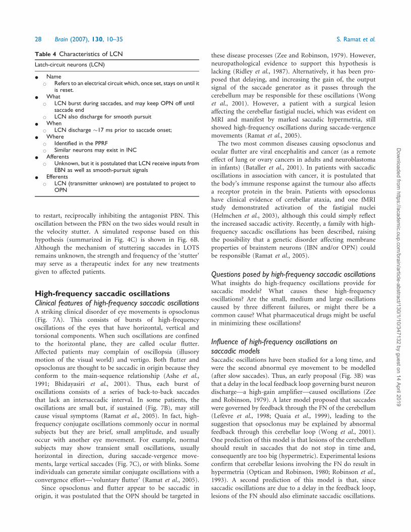
to restart, reciprocally inhibiting the antagonist PBN. This
oscillation between the PBN on the two sides would result in
the velocity stutter. A simulated response based on this
hypothesis (summarized in Fig. 4C) is shown in Fig. 6B.
Although the mechanism of stuttering saccades in LOTS
remains unknown, the strength and frequency of the ‘stutter’
may serve as a therapeutic index for any new treatments
given to affected patients.
High-frequency saccadic oscillationsClinical features of high-frequency saccadic oscillationsA striking clinical disorder of eye movements is opsoclonus
(Fig. 7A). This consists of bursts of high-frequency
oscillations of the eyes that have horizontal, vertical and
torsional components. When such oscillations are confined
to the horizontal plane, they are called ocular flutter.
Affected patients may complain of oscillopsia (illusory
motion of the visual world) and vertigo. Both flutter and
opsoclonus are thought to be saccadic in origin because they
conform to the main-sequence relationship (Ashe et al.,
1991; Bhidayasiri et al., 2001). Thus, each burst of
oscillations consists of a series of back-to-back saccades
that lack an intersaccadic interval. In some patients, the
oscillations are small but, if sustained (Fig. 7B), may still
cause visual symptoms (Ramat et al., 2005). In fact, high-
frequency conjugate oscillations commonly occur in normal
subjects but they are brief, small amplitude, and usually
occur with another eye movement. For example, normal
subjects may show transient small oscillations, usually
horizontal in direction, during saccade-vergence move-
ments, large vertical saccades (Fig. 7C), or with blinks. Some
individuals can generate similar conjugate oscillations with a
convergence effort—‘voluntary flutter’ (Ramat et al., 2005).
Since opsoclonus and flutter appear to be saccadic in
origin, it was postulated that the OPN should be targeted in
these disease processes (Zee and Robinson, 1979). However,
neuropathological evidence to support this hypothesis is
lacking (Ridley et al., 1987). Alternatively, it has been pro-
posed that delaying, and increasing the gain of, the output
signal of the saccade generator as it passes through the
cerebellum may be responsible for these oscillations (Wong
et al., 2001). However, a patient with a surgical lesion
affecting the cerebellar fastigial nuclei, which was evident on
MRI and manifest by marked saccadic hypermetria, still
showed high-frequency oscillations during saccade-vergence
movements (Ramat et al., 2005).
The two most common diseases causing opsoclonus and
ocular flutter are viral encephalitis and cancer (as a remote
effect of lung or ovary cancers in adults and neuroblastoma
in infants) (Bataller et al., 2001). In patients with saccadic
oscillations in association with cancer, it is postulated that
the body’s immune response against the tumour also affects
a receptor protein in the brain. Patients with opsoclonus
have clinical evidence of cerebellar ataxia, and one fMRI
study demonstrated activation of the fastigial nuclei
(Helmchen et al., 2003), although this could simply reflect
the increased saccadic activity. Recently, a family with high-
frequency saccadic oscillations has been described, raising
the possibility that a genetic disorder affecting membrane
properties of brainstem neurons (IBN and/or OPN) could
be responsible (Ramat et al., 2005).
Questions posed by high-frequency saccadic oscillationsWhat insights do high-frequency oscillations provide for
saccadic models? What causes these high-frequency
oscillations? Are the small, medium and large oscillations
caused by three different failures, or might there be a
common cause? What pharmaceutical drugs might be useful
in minimizing these oscillations?
Influence of high-frequency oscillations onsaccadic modelsSaccadic oscillations have been studied for a long time, and
were the second abnormal eye movement to be modelled
(after slow saccades). Thus, an early proposal (Fig. 3B) was
that a delay in the local feedback loop governing burst neuron
discharge—a high-gain amplifier—caused oscillations (Zee
and Robinson, 1979). A later model proposed that saccades
were governed by feedback through the FN of the cerebellum
(Lefevre et al., 1998; Quaia et al., 1999), leading to the
suggestion that opsoclonus may be explained by abnormal
feedback through this cerebellar loop (Wong et al., 2001).
One prediction of this model is that lesions of the cerebellum
should result in saccades that do not stop in time and,
consequently are too big (hypermetric). Experimental lesions
confirm that cerebellar lesions involving the FN do result in
hypermetria (Optican and Robinson, 1980; Robinson et al.,
1993). A second prediction of this model is that, since
saccadic oscillations are due to a delay in the feedback loop,
lesions of the FN should also eliminate saccadic oscillations.
Table 4 Characteristics of LCN
Latch-circuit neurons (LCN)
* Name* Refers to an electrical circuit which, once set, stays on until it
is reset.* What
* LCN burst during saccades, and may keep OPN off untilsaccade end
* LCN also discharge for smooth pursuit* When
* LCN discharge �17 ms prior to saccade onset;* Where
* Identified in the PPRF* Similar neurons may exist in INC
* Afferents* Unknown, but it is postulated that LCN receive inputs from
EBN as well as smooth-pursuit signals* Efferents
* LCN (transmitter unknown) are postulated to project toOPN
28 Brain (2007), 130, 10–35 S. Ramat et al.
Dow
nloaded from https://academ
ic.oup.com/brain/article-abstract/130/1/10/347132 by guest on 14 April 2019
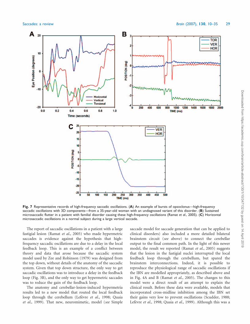
The report of saccadic oscillations in a patient with a large
fastigial lesion (Ramat et al., 2005) who made hypermetric
saccades is evidence against the hypothesis that high-
frequency saccadic oscillations are due to a delay in the local
feedback loop. This is an example of a conflict between
theory and data that arose because the saccadic system
model used by Zee and Robinson (1979) was designed from
the top down, without details of the anatomy of the saccadic
system. Given that top down structure, the only way to get
saccadic oscillations was to introduce a delay in the feedback
loop (Fig. 3B), and the only way to get hypermetric saccades
was to reduce the gain of the feedback loop.
The anatomy and cerebellar-lesion-induced hypermetria
results led to a new model that routed the local feedback
loop through the cerebellum (Lefevre et al., 1998; Quaia
et al., 1999). That new, neuromimetic, model (see Simple
saccade model for saccade generation that can be applied to
clinical disorders) also included a more detailed bilateral
brainstem circuit (see above) to connect the cerebellar
output to the final common path. In the light of this newer
model, the result we reported (Ramat et al., 2005) suggests
that the lesion in the fastigial nuclei interrupted the local
feedback loop through the cerebellum, but spared the
brainstem interconnections. Indeed, it is possible to
reproduce the physiological range of saccadic oscillations if
the IBN are modelled appropriately, as described above and
in Fig. 4A and B (Ramat et al., 2005). The changes to this
model were a direct result of an attempt to explain the
clinical result. Before these data were available, models that
incorporated cross-midline inhibition among the IBN set
their gains very low to prevent oscillations (Scudder, 1988;
Lefevre et al., 1998; Quaia et al., 1999). Although this was a
Fig. 7 Representative records of high-frequency saccadic oscillations. (A) An example of bursts of opsoclonus—high-frequencysaccadic oscillations with 3D components—from a 35-year-old woman with an undiagnosed variant of this disorder. (B) Sustainedmicrosaccadic flutter in a patient with familial disorder causing these high-frequency oscillations (Ramat et al., 2005). (C) Horizontalmicrosaccadic oscillations in a normal subject during a large vertical saccade.
Saccades: a review Brain (2007), 130, 10–35 29
Dow
nloaded from https://academ
ic.oup.com/brain/article-abstract/130/1/10/347132 by guest on 14 April 2019

reasonable assumption, study of the clinical case showed that
it appears to have been incorrect.
Answers from modelsThis example illustrates the potential synergy between bench
and bedside. The clinical study showed that there were
severe problems with the original theory of what caused
saccadic oscillations. Neither the range of oscillation freq-
uencies nor the effects of cerebellar lesions could be fully
explained. Worse, the assumption that cross-midline gains
in the brainstem were very low may have misled researchers
about the importance of those circuits in saccadic behaviour.
The new hypothesis, which combines results from basic and
clinical research, both accounts for the full range of
frequencies seen in human eye movements and explains
why the cerebellar lesion spared the oscillations. Further-
more, it suggests that these oscillations are caused by a
very specific group of cells in the brainstem (IBN–IBN)
with reciprocal inhibition. Importantly, the new hypothesis
emphasizes that the specific properties of the neurons
involved (e.g. post-inhibitory rebound), and not just the
topology of the circuit, may be key to explaining
experimental and clinical findings.
This interpretation allows us to answer the questions
raised above. First, high-frequency oscillations are caused by
reciprocal inhibition of PBN across the midline, creating a
positive-feedback loop. The frequency and amplitude of the
oscillations are not controlled by the delay in the loop, but
rather by membrane properties (adaptation time constants
and gains) of the IBN. Second, all sizes of oscillations can be
evoked from the same brainstem positive-feedback circuit by
changing the parameters (especially membrane adaptation
time constants and gains). Thus, we conclude that all of
these disorders have a common substrate. As mentioned
above, the horizontal IBN are known to use glycine as an
inhibitory transmitter (McElligott and Spencer, 2000), and
thus may also be modulating the excitatory NMDA receptor
(Miura and Optican, 2006). This suggests that treatment
with a drug that blocks the glycine co-agonist site on NMDA
receptors of IBNs, or lowers the sensitivity of their excitatory
receptors (i.e. glutamate receptors such as AMPA and
NMDA), or blocks the T-type calcium channel, might
reduce saccadic oscillations.
Macrosaccadic oscillationsClinical features of macrosaccadic oscillationsMacrosaccadic oscillations consist of horizontal saccades
that occur in runs, spontaneously building up and then
decreasing in amplitude, with intersaccadic intervals of
�200 ms (Fig. 8). They are different from small ‘square-
wave jerks’ shown by normal subjects, which take the eye
<2� away from the fixation point, and return it within
200 ms (Abadi and Gowen, 2004). Macrosaccadic oscilla-
tions are also distinct from flutter and opsoclonus (discussed
in the previous section) because they have a normal
intersaccadic interval of �200 ms. Macrosaccadic oscilla-
tions are usually induced by a gaze shift, but may occur
during attempted fixation. They overshoot the visual target
in both directions, thus leading to oscillations around the
fixation point. Usually, macrosaccadic oscillations are
mainly horizontal, but they may have vertical and torsional
components.
Macrosaccadic oscillations occur in patients with destruc-
tive lesions affecting the deep cerebellar nuclei, including the
FN (Selhorst et al., 1976). They also occur with genetic
cerebellar ataxias (Swartz et al., 2003), but rarely with
brainstem lesions (Averbuch-Heller et al., 1996). In some
affected patients, saccades are faster than in controls (Swartz
et al., 2003). Patients with a genetic cerebellar degeneration
may also have slowed conduction in peripheral nerves,
suggesting that conduction along parallel fibres in the
cerebellum may also be slowed (Swartz et al., 2003).
In monkeys, hypermetric saccades can be induced by
pharmacologically inactivating the fastigial nuclei (Robinson
et al., 1993). However, irrepressible saccades are not a
prominent feature with bilateral FN inactivation. After the
initial hypermetric saccade, corrective saccades decline in
gain and fixation behaviour is characterized by an offset
rather than by sustained oscillations about the visual target.
In monkeys, irrepressible saccades can be produced by
microinjection into the caudal SC of the GABA-antagonist
bicuculline (Hikosaka and Wurtz, 1985) or the rostral pole
of the SC with the GABA-agonist muscimol (Munoz and
Wurtz, 1993). Similar large saccadic oscillations are reported
after inactivation of the adjacent cMRF (Waitzman et al.,
2000). A destructive lesion affecting the rostral pole of the SC
in one monkey also produced irrepressible saccades (Carasig
et al., 2005). In contrast, in human patients paramedian
midbrain disease is not reported to cause irrepressible
saccades; in fact saccadic palsy (small and slow movements)
is reported (Zackon and Sharpe, 1984).
Questions posed by macrosaccadic oscillationsWhat causes macrosaccadic oscillations? Why does attempted
fixation not stop the oscillations? Why are horizontal
saccades affected more than vertical saccades? Why are
macrosaccadic oscillations not encountered clinically with
lesions affecting the mesencephalic reticular formation or SC?
Do models suggest any therapeutic measures?
Influence of macrosaccadic oscillations on modelsThe earliest saccade models were simple negative feedback
loops that compared target eccentricity with eye position.
Even such simple models would generate macrosaccadic
oscillations if the gain of the feed-forward mechanism were
increased (Selhorst et al., 1976). If the feed-forward gain is
>1, a saccade will overshoot the target, causing a new retinal
error in the opposite direction. The new error will evoke
another saccade, which will also overshoot. Although this
model has the correct behaviour, it does not help us
30 Brain (2007), 130, 10–35 S. Ramat et al.
Dow
nloaded from https://academ
ic.oup.com/brain/article-abstract/130/1/10/347132 by guest on 14 April 2019
understand what parts of the brain are involved. More
modern ideas suggest that lesions of the midline cerebellum
cause hypermetria because they delay the onset of the choke
that stops saccades. Thus, the saccades overshoot the target,
which is equivalent in some ways to an increase in the feed-
forward gain. However, it is not exactly the same, because
simply increasing the gain would make all saccades bigger by
a constant factor, which is not the case, as large saccades are
not magnified as much as small ones. All of these models
generate macrosaccadic oscillations when triggered by a gaze
shift. No mechanism has been suggested for how they can
start during fixation, although microsaccades, which occur
spontaneously (Martinez-Conde et al., 2004), might trigger
larger saccadic oscillations.
Answers from modelsMacrosaccadic oscillations provide a good example of a well-
studied saccadic disorder for which the theory is incomplete.
Studies have shown that excess activity in the caudal SC or
inadequate activity in the rostral SC cause irrepressible
saccades. But it is not clear that the SC is involved in many
clinical cases of macrosaccadic oscillations. Hypermetria
after FN lesions suggests the oscillations are not due to a
gain change, but to a delay in choking off the saccadic drive
signals. Thus it is a feedback, not a feed-forward, failure.
Understanding why the oscillations start up and die off
during fixation will require a new model that includes a
more detailed mechanism for fixation. The propensity for
macrosaccadic oscillations to occur in the horizontal plane
may relate to the different neurotransmitter profiles of
inhibitory burst neurons for horizontal or vertical saccades,
which is discussed in the section Premotor burst neurons
(PBN). Drugs that reduce saccadic gain might be effective in
suppressing macrosaccadic oscillations.
ConclusionWe have considered four examples of well-defined disorders
of saccades caused by disease in humans. Each example has
provided insights into the brainstem, midbrain and
cerebellar machinery that generates normal saccades in
healthy humans. We have suggested how such abnormal
behaviours are helpful in identifying those hypotheses for
saccades that are biologically plausible. Conversely, current
models for normal saccade generation can provide insights
into some of these disorders. Here we discuss ways that
models of saccades can aid topological diagnosis, point to
underlying molecular mechanisms that suggest novel
therapies, and guide future studies.
Implication of saccadic models fortopological diagnosisAlthough the quantitative predictions of a hypothesis are
best evaluated by measuring differences between model
simulations and experimental data, qualitative predictions of
Fig. 8 Examples of horizontal saccades made by a normal subject (blue line) and a patient with a spinocerebellar degeneration (red line)in response to a 25� target jump (dotted line) from centre position to right 25� and back. After a reaction time, the normal subjectmakes an initial saccade that is hypometric, followed by a subsequent correction. A similar pattern occurs after the target returns tocentre. The patient shows hypermetria, overshooting the target when it jumps to the right, and especially when it jumps back to centre,following which there are macrosaccadic oscillations about the point of fixation. Positive values correspond to rightward movements.
Saccades: a review Brain (2007), 130, 10–35 31
Dow
nloaded from https://academ
ic.oup.com/brain/article-abstract/130/1/10/347132 by guest on 14 April 2019
hypotheses can be tested at the beside. As improbable as it
may sound, a simple sketch of a model drawn in a pocket
notebook can guide accurate diagnosis. Thus, saccades that
are slow and yet accurate can be appreciated because a
negative feedback loop with maintained burst neuron
discharge, even though it is reduced, continues until the
eye is on target. Saccades that are prematurely terminated
can be viewed as a malfunction of a latch circuit that turns
on OPN too early, or causes IBN stuttering, in the middle of
a saccade, causing early and increased deceleration. High-
frequency saccadic oscillations can be considered in terms of
the intrinsic instability of a brainstem network in which
some inhibitory burst neurons reciprocally inhibit IBN on
the opposite side of the brainstem. This leads to a positive-
feedback loop that will oscillate if started by post-inhibitory
rebound unless held in check by OPN. Lower-frequency
macrosaccadic oscillations can be conceived in terms of
increased saccadic gain arising because the ‘stop’ signal
arrives too late to land the eye on target, making them
hypermetric.
In fact, disorders of saccades are potentially very useful for
topological diagnosis in common disorders affecting the
brainstem. One example is the relative slowing of adducting
versus abducting saccades in internuclear ophthalmoplegia
in multiple sclerosis, which attacks central, but not peri-
pheral, myelin. Thus, the saccadic pulse signal to medial
rectus motoneurons is low-pass filtered due to loss of central
myelin on axons of abducens INs (which run in the medial
longitudinal fasciculus) but not on the axons of abducens
motoneurons (which run outside of the CNS) (Leigh and
Zee, 2006).
What we hope to have demonstrated is that conceptualiza-
tion of mechanisms for saccade generation takes such
interpretation to a level that can be applied to a range of
dynamic disorders. In fact, we suggest that neurologists are
always in the ‘clinical lab’, and are well placed to make
observations that can be used to test current saccadic models.
Implication of saccadic models forunderstanding disease mechanismsand developing new therapiesBesides providing the means to localize a disease process to
a circuit or population of neurons, careful analysis of
disorders of saccades can suggest the nature of the disorder
at the level of neuronal membranes, or even molecules such
as proteins that are determined by genes. Thus, the degree of
slowing of saccades in SCA2 has been related to the number
of trinucleotide repeats (Seifried et al., 2005). Further,
saccadic hypermetria in recessive spinocerebellar ataxia with
saccadic intrusions (SACSI) coexists with slowing of axonal
transmission in peripheral nerves, suggesting that delayed
conduction of the ‘stop’ signal on unmyelinated parallel
fibres of the cerebellum might lead to saccadic hypermetria
(Swartz et al., 2003). Membrane properties are as important
as circuits, and thus new classes of agents that have effects on
neuron membranes require consideration in the treatment
of abnormal eye movements. For example, genetic disorders
of P/Q calcium channels, such as the episodic ataxias, can
produce abnormalities of eye movements (Stahl and James,
2005) that are amenable to therapy with drugs such as 4-
aminopyridine (a potassium channel blocker) (Strupp et al.,
2004). We have also pre-sented preliminary evidence that
high-frequency saccadic oscillations could be due to an
abnormality of the membrane properties of inhibitory burst
neurons (Ramat et al., 2005). Another mechanism only
recently proposed is that normally the inhibitory neuro-
transmitter glycine inhibits burst neuron discharge but,
paradoxically, glycine may also act as a neuromodulator at
NMDA receptors to increase the effects of glutamate on burst
neurons and thereby generate the sharp acceleration that
characterizes saccades (Miura and Optican, 2006). Drugs that
block this co-agonist action of glycine, or that block the
glutamate site at excitatory synapses, should be considered as
possible therapies for high-frequency oscillations.
Implication of saccadic models fordirecting new researchAlthough this review deals only with brainstem and
cerebellar control of saccades, these movements have been
used to investigate higher functions such as memory,
decision-making and reward (Itoh et al., 2003, Leigh and
Kennard, 2004, Nachev et al., 2004). Thus, analysis of
saccade behaviour can be used to characterize many
disorders affecting all levels of the brain, including genetic
disorders such as the SCAs, degenerative disorders, such as
parkinsonism, and conditions causing dementia and
psychiatric illnesses, including schizophrenia, and atten-
tional disorders (Leigh and Zee, 2006). Several different
approaches, including functional imaging and transcranial
magnetic stimulation, are being used in combination with
novel behavioural tests in patients with discrete brain lesions
to understand better the mechanisms of higher-level control
of saccades (Muri, 2005).
What lies ahead? There are still a number of disorders
of saccades that are not understood at all, e.g. small saccadic
intrusions—‘square-wave jerks’ (Abadi and Gowen, 2004).
An understanding of the saccadic system at the level of
the biophysical properties of cell membranes and the
genetics of neurotransmitters and receptors has only just
begun to be explored. This may have an important impact
on patient care, because of the constant development of new
drugs that may act at various receptors. Closer interaction
between basic and clinical researchers is essential if rapid
progress is to be made in understanding mechanisms and
disorders of neural control, and improving their diagnosis
and treatment.
Supplementary materialSupplementary material is available at Brain Online.
32 Brain (2007), 130, 10–35 S. Ramat et al.
Dow
nloaded from https://academ
ic.oup.com/brain/article-abstract/130/1/10/347132 by guest on 14 April 2019

AcknowledgementsThis study was supported by the Office of Research and
Development, Medical Research Service, Department of
Veterans Affairs; National Eye Institute grants EY01849,
EY06717 and EY08060; the Evenor Armington Fund; FIRB
grant RBNE01FJ4J; Intramural Division of the National Eye
Institute, NIH, DHHS.
References
Abadi RV, Gowen E. Characteristics of saccadic intrusions. Vision Res 2004;
44: 2675–90.
Abel LA,Traccis BT,TroostBT,Dell’OssoLF. Saccadic trajectories changewith
amplitude, not time. Neuro-ophthalmology 1987; 7: 309–14.
Ahmadi S, Muth-Selbach U, Lauterbach A, Lipfert P, Neuhuber WL,
Zeilhofer HU. Facilitation of spinal NMDA receptor currents by spillover of
synaptic released glycine. Science 2003; 300: 2094–7.
Aizawa H, Wurtz RH. Reversible inactivation of monkey superior
colliculus. I. Curvature of saccadic trajectory. J Neurophysiol 1998;
79: 2082–96.
Aksay E, Baker R, Seung HS, Tank DW. Visual adaptation of the the
oculomotor horizontal velocity-to-position integrator. Proc Natl Acad Sci
USA 2005; 23: 10852–8.
Anderson RW, Das S, Keller EL. Estimation of spatiotemporal neural
activity using radial basis function networks. J Comput Neurosci 1998;
5: 421–41.
Arnold DB, Robinson DA. The oculomotor integrator: testing of a neural
network model. Exp Brain Res 1997; 113: 57–74.
Ashe J, Hain TC, Zee DS, Schatz NJ. Microsaccadic flutter. Brain 1991;
114: 461–72.
Averbuch-Heller L, Kori AA, Rottach K, Dell’Osso LF, Remler BF, Leigh RJ.
Dysfunction of pontine omnipause neurons causes impaired fixation:
macrosaccadic oscillations with a unilateral pontine lesion. Neuro-
ophthalmology 1996; 16: 99–106.
Bahill AT, Clark MR, Stark L. The main sequence, a tool for studying human
eye movements. Math Biosci 1975; 24: 191–204.
Barton EJ, Nelson JS, Gandhi NJ, Sparks DL. Effects of partial lidocaine
inactivation of the paramedian pontine reticular formation on saccades of
macaques. J Neurophysiol 2003; 90: 372–86.
Basso MA, Wurtz RH. Modulation of neuronal activity by target uncertainty.
Nature 1997; 389: 66–9.
Bataller L, Graus F, Saiz A, Vilchez J. Clinical outcome in adult onset
idiopathic or paraneoplastic opsoclonus-myoclonus. Brain 2001;
124: 437–43.
Becker W, Jurgens R. An analysis of the saccadic system by means of double
step stimuli. Vision Res 1979; 19: 967–83.
Bell AH, Meredith MA, van Opstal AJ, Munoz DP. Crossmodal integration in
the primate superior colliculus underlying the preparation and initiation of
saccadic eye movements. J Neurophysiol 2005; 93: 3659–73.
Bergeron A, Guitton D. In multiple-step gaze shifts: omnipause (OPNs) and
collicular fixation neurons encode gaze position error: OPNs gate saccades.
J Neurophysiol 2002; 88: 1726–42.
Bergeron A, Matsuo S, Guitton D. Superior colliculus encodes distance to
target, not saccade amplitude, in multi-step gaze shifts. Nat Neurosci 2003;
6: 404–13.
Bhidayasiri R, Somers JT, Kim JI, Ramat S, Nayak S, Bokil HS, et al. Ocular
oscillations induced by shifts of the direction and depth of visual fixation.
Ann Neurol 2001; 49: 24–8.
Busettini C, Mays LE. Pontine omnipause activity during conjugate and
disconjugate eye movements in macaques. J Neurophysiol 2003;
90: 3838–53.
Buttner-Ennever JA, Horn AKE. Pathways from cell groups of the paramedian
tracts to the floccular region. Ann NY Acad Sci 1996; 781: 532–40.
Buttner-Ennever JA, Horn AK, Henn V, Cohen B. Projections from the
superior colliculus motor map to omnipause neurons in monkey. J Comp
Neurol 1999; 413: 55–67.
Cannon SC, RobinsonDA. An improved neural-networkmodel for the neural
integrator of the oculomotor system: more realistic neuron behavior. Biol
Cybern 1985; 53: 93–108.
Carasig D, Paul K, Fucito M, Ramcharan E, Gnadt JW. Irrepressible saccades
from a tectal lesion in a Rhesus monkey. Vision Res 2005; 46: 1161–9.
Carpenter RHS. Cerebellectomy and the transfer function of the vestibulo-
ocular reflex in the decerebrate cat. Proc R Soc Lond B Biol Sci 1972;
181: 353–74.
Chen B, May PJ. The feedback circuit connecting the superior colliculus and
central mesencephalic reticular formation: a direct morphological
demonstration. Exp Brain Res 2000; 131: 10–21.
Cohen B, Buttner-Ennever J. Projections from the superior colliculus to
a region of the central mesencephalic reticular formation (cMRF)
associated with horizontal saccadic eye movements. Exp Brain Res 1984;
57: 167–76.
Cova AC, Galiana HL. A bilateral model integrating vergence and the
vestibulo-ocular reflex. Exp Brain Res 1996; 107: 435–52.
Cromer JA, Waitzman DM. Neurones associated with saccade metrics in the
monkey central mesencephalic reticular formation. J Physiol (Lond) 2006;
570: 507–23.
Demer JL. Pivotal role of orbital connective tissues in binocular alignment and
strabismus. Invest Ophthalmol Vis Sci 2004; 45: 729–38.
Dodge R, Cline TS. The angle velocity of eye movements. Psychol Rev 1901;
8: 145–57.
Droulez J, Berthoz A. A neural network model of sensoritopic maps with
predictive short-term memory properties. Proc Natl Acad Sci USA 1991;
88: 9653–7.
Enderle JD, Engelken EJ. Simulation of oculomotor post-inhibitory rebound
burst firing using a Hodgkin-Huxley model of a neuron. Biomed Sci
Instrum 1995; 31: 53–8.
Enright JT. Monocularly programmed human saccades during vergence
changes? J Physiol (Lond) 1998; 512: 235–50.
FitzGibbon EJ, GoldbergME, SegravesMA. Short term saccadic adaptation in
the monkey. In: Keller E, Zee DS, editors. Adaptive processes in the visual
and oculomotor systems. Oxford: Pergamon; 1986. p. 329–33.
Fuchs AF, Robinson FR, Straube A. Role of the caudal fastigial nucleus in
saccade generation: I Neuronal discharge patterns. J Neurophysiol 1993;
70: 1723–40.
Gandhi NJ, Keller EL. Spatial distribution and discharge characteristics of
superior colliculus neurons antidromically activated from the omnipause
region in monkey. J Neurophysiol 1997; 78: 2221–5.
Gandhi NJ, Keller EL. Comparison of saccades perturbed by stimulation of
the rostral superior colliculus, the caudal superior colliculus and the
omnipause neuron region. J Neurophysiol 1999; 82: 3236–53.
Girard B, Berthoz A. From brainstem to cortex: computational models of
saccade generation circuitry. Prog Neurobiol 2005; 77: 215–51.
Hanes DP, Smith MK, Optican LM, Wurtz RH. Recovery of saccadic
dysmetria following localized lesions in monkey superior colliculus. Exp
Brain Res 2005; 160: 325.
Hanson MR, Hamid MA, Tomsak RL, Chou SS, Leigh RJ. Selective saccadic
palsy caused by pontine lesions: clinical, physiological and pathological
correlations. Ann Neurol 1986; 20: 209–17.
Helmchen C, Rambold H, Sprenger A, Erdmann C, Binkofski F. Cerebellar
activation in opsoclonus: an fMRI study. Neurology 2003; 61: 412–5.
Henn V, Lang W, Hepp K, Reisine H. Experimental gaze palsies in monkeys
and their relation to human pathology. Brain 1984; 107: 619–36.
Hepp K, Henn V, Vilis T, Cohen B. Brainstem regions related to saccade
generation. In: Wurtz RH, Goldberg ME, editors. The neurobiology of
saccadic eye movements. Amsterdam: Elsevier; 1989. p. 105–212.
Hikosaka O, Wurtz RH. Modification of saccadic eye movements by GABA-
related substances I. Effect of muscimol and bicuculline inmonkey superior
colliculus. J Neurophysiol 1985; 53: 266–91.
Hikosaka O, Takikawa Y, Kawagoe R. Role of the basal ganglia in the control
of purposive saccadic eye movements. Physiol Rev 2000; 80: 953–78.
Horn AK. The reticular formation. In: Butttner-Ennever JA, editor.
Neuroanatomy of the oculomotor system. (Progress in Brain Research,
Volume 151). New York: Elsevier; 2006. p. 33–79.
Saccades: a review Brain (2007), 130, 10–35 33
Dow
nloaded from https://academ
ic.oup.com/brain/article-abstract/130/1/10/347132 by guest on 14 April 2019
Horn AKE, Buttner-Ennever JA. Premotor neurons for vertical eye-
movements in the rostral mesencephalon of monkey and man: the
histological identification by parvalbumin immunostaining. J Comp
Neurol 1998; 392: 413–27.
Horn AKE, Buttner-Ennever JA, Suzuki Y, Henn V. Histological identifica-
tion of premotor neurons for horizontal saccades in monkey and man by
parvalbumin immunostaining. J Comp Neurol 1997; 359: 350–63.
Itoh H, Nakahara H, Hikosaka O, Kawagoe R, Takikawa Y, Aihara K.
Correlation of primate caudate neural activity and saccade parameters in
reward-oriented behavior. J Neurophysiol 2003; 89: 1774–83.
Jeyakumar M, Smith D, Eliott-Smith E, Cortina_Borja M, Reinkensmeier G,
Butters TD, et al. An inducible mouse model of late onset Tay-Sachs
disease. Neurobiol Dis 2002; 10: 201–10.
Jurgens R, BeckerW, Kornhuber HH. Natural and drug-induced variations of
velocity and duration of human saccadic eye movements: evidence for a
control of the neural pulse generator by local feedback. Biol Cybern 1981;
39: 87–96.
Kaneko CRS. Effect of ibotenic acid lesions of the omnipause neurons on
saccadic eye movements in Rhesus macaques. J Neurophysiol 1996;
75: 2229–42.
Kapoula Z. Evidence for a range effect in the saccadic system. Vision Res 1985;
25: 1155–7.
Keller EL. Participation of medial pontine reticular formation in eye
movement generation in monkey. J Neurophysiol 1974; 37: 316–32.
Keller EL, Missal M. Shared brainstem pathways for saccades and smooth-
pursuit eye movements. Ann NY Acad Sci 2003; 1004: 29–39.
Keller EL, Robinson DA. Absence of a stretch reflex in extraocular muscles of
the monkey. J Neurophysiol 1971; 34: 908–19.
Keller EL, Gandhi NJ, Shieh JM. Endpoint accuracy in saccades interrupted by
stimulation in the omnipause region in monkeys. Visual Neurosci 1996a;
13: 1059–67.
Keller EL,GandhiNJ,WierPT.Dischargeof superior collicularneuronsduring
saccades made to moving targets. J Neurophysiol 1996b; 76: 3573–7.
Kitazawa S, Kimura T, Yin PB. Cerebellar complex spikes encode both
destinations and errors in arm movements. Nature 1998; 392: 494–7.
Krauzlis RJ. Recasting the smooth pursuit eye movement system.
J Neurophysiol 2004; 91: 591–603.
Krauzlis RJ, Liston D, Carello CD. Target selection and the superior colliculus:
goals, choices and hypotheses. Vision Res 2004; 44: 1445–51.
Langer TP, Kaneko CR. Efferent projections of the cat oculomotor reticular
omnipause neuron region: an autoradiographic study. J Comp Neurol
1983; 217: 288–306.
Langer TP, Kaneko CRS. Brainstem afferents to the oculomotor omnipause
neurons in monkey. J Comp Neurol 1990; 295: 413–27.
Lefevre P, Missal M, Galiana HL. Modeling slow correcting gaze movements.
J Vestib Res 1994; 4: 371–81.
Lefevre P, Quaia C, Optican LM. Distributed model of control of saccades by
superior colliculus and cerebellum. Neural Netw 1998; 11: 1175–90.
Leigh RJ, Kennard C. Using saccades as a research tool in the clinical
neurosciences. Brain 2004; 127: 460–77.
Leigh RJ, Zee DS. The neurology of eye movements (Book/DVD). Fourth
edition. New York: Oxford University Press; 2006.
MacAskill MR, Anderson TJ, Jones RD. Suppression of displacement in
severely slowed saccades. Vision Res 2000; 40: 3405–13.
Martinez-Conde S, Macknik SL, Hubel DH. The role of fixational eye
movements in visual perception. Nat Rev Neurosci 2004; 5: 229–40.
Matsuo S, Bergeron A, Guitton D. Evidence for gaze feedback to the cat
superior colliculus: discharges reflect gaze trajectory perturbations.
J Neurosci 2004; 24: 2760–73.
Mays LE, Morrisse DW. Electrical stimulation of the pontine omnipause area
inhibits eye blink. J Am Optom Assoc 1995; 66: 419–22.
McCrea RA, Horn AK. Nucleus prepositus. In: Buttner-Ennever JA, editor.
Neuroanatomy of the oculomotor system. (Progress in Brain Research,
Volume 151). New York: Elsevier; 2006. p. 205–30.
McElligott JG, Spencer RF. Neuropharmacological aspects of the vestibulo-
ocular reflex. In: Anderson JH, Beitz AJ, editors. Neurochemistry of the
vestibular system. Boca Raton: CRC Press; 2000. p. 199–222.
Miura K, Optican LM. Membrane channel properties of premotor excitatory
burst neurons may underlie saccade slowing after lesions of omnipause
neurons J Comput Neurosci 2006; 20: 25–41.
Munoz DP, Wurtz RH. Fixation cells in monkey superior colliculus II.
Reversible activation and deactivation. J Neurophysiol 1993; 70:
576–89.
Munoz DP,Wurtz RH. Saccade-related activity in monkey superior colliculus
I. Characteristics of burst and buildup cells. J Neurophysiol 1995a;
73: 2313–33.
Munoz DP,Wurtz RH. Saccade-related activity in monkey superior colliculus
II. Spread of activity during saccades. J Neurophysiol 1995b; 73: 2334–48.
Munoz DP, Pelisson D, Guitton D. Movement of neural activity on the
superior colliculus motor map during gaze shifts. Science 1991;
251: 1358–60.
Muri RM. MRI and fMRI analysis of oculomotor function. In: Buttner-
Ennever JA, editor. Neuroanatomy of the oculomotor system. (Progress in
Brain Research, Volume 151). New York: Elsevier; 2005. p. 509–32.
Nachev P, Rees G, Parton A, Kennard C, Husain M. Volition and conflict in
human medial frontal cortex. Curr Biol 2004; 15: 122–8.
Noda H, Fujikado T. Topography of the oculomotor area of the cerebellar
vermis in macaques as determined by microstimulation. J Neurophysiol
1987; 58: 359–78.
Noda H, Murakami S, Yamada J, Tamaki Y, Aso T. Saccadic eye movements
evoked by microstimulation of the fastigial nucleus of macaque monkeys.
J Neurophysiol 1988; 60: 1036–52.
Ohtsuka K, Noda H. Saccadic burst neurons in the oculomotor region of the
fastigial nucleus of macaque monkeys. J Neurophysiol 1991; 65: 1422–34.
Ohtsuka K, Noda H. Discharge properties of Purkinje cells in the oculomotor
vermis during visually guided saccades in the macaque monkey.
J Neurophysiol 1995; 74: 1828–40.
Optican LM. Sensorimotor transformation for visually guided saccades. Ann
NY Acad Sci 2005; 1039: 132–48.
Optican LM, Miles FA. Visually induced adaptive changes in primate saccadic
oculomotor control signals. J Neurophysiol 1985; 54: 940–58.
Optican LM, Quaia C. From sensory space to motor commands: lessons
from saccades. In: Proceedings of the 23rd IEEE EMBC Conference,
Istanbul, 25–28 Oct. 2001. p. 820–3.
Optican LM, Quaia C. Distributed model of collicular and cerebellar function
during saccades. Ann NY Acad Sci 2002; 956: 164–77.
Optican LM, Robinson DA. Cerebellar-dependent adaptive control of
primate saccadic system. J Neurophysiol 1980; 44: 1058–76.
Pare M, Guitton D. Brain stem omnipause neurons and the control of
combined eye-head gaze saccades in the alert cat. J Neurophysiol 1998;
79: 3060–76.
Perez-Reyes E. Molecular physiology of low-voltage-activated T-type calcium
channels. Physiol Rev 2003; 83: 117–61.
Port NL, Wurtz RH. Sequential activity of simultaneously recorded neurons
in the superior colliculus during curved saccades. J Neurophysiol 2003;
90: 1887–903.
Quaia C, Optican LM. Dynamic eye plant models and the control of eye
movements. Strabismus 2003a; 11: 17–31.
Quaia C, Optican LM. Three-dimensional rotations of the eye. In:
Kaufman PL, Alm A, editors. Adler’s physiology of the eye: clinical
application. New York: Mosby; 2003b. p. 818–29.
Quaia C, Lefevre P, Optican LM. Model of the control of saccades by superior
colliculus and cerebellum. J Neurophysiol 1999; 82: 999–1018.
Ramat S, Das VE, Somers JT, Leigh RJ. Tests of two hypotheses to account for
different-sized saccades during disjunctive gaze shifts. Exp Brain Res 1999;
129: 500–10.
Ramat S, Leigh RJ, Zee DS, Optican LM. Ocular oscillations generated by
coupling of brainstem excitatory and inhibitory saccadic burst neurons.
Exp Brain Res 2005; 160: 89–106.
Ridley A, Kennard C, Scholtz CL, Buttner-Ennever JA, Summers B,
Turnbull A. Omnipause neurons in two cases of opsoclonus associated
with oat cell carcinoma of the lung. Brain 1987; 110: 1699–709.
Robinson DA. The mechanics of human saccadic eye movement. J Physiol
(Lond) 1964; 174: 245–64.
34 Brain (2007), 130, 10–35 S. Ramat et al.
Dow
nloaded from https://academ
ic.oup.com/brain/article-abstract/130/1/10/347132 by guest on 14 April 2019
Robinson DA. Oculomotor unit behavior in the monkey. J Neurophysiol
1970; 33: 393–403.
Robinson DA. Eye movements evoked by collicular stimulation in the alert
monkey. Vision Res 1972; 12: 1795–808.
Robinson DA. Models of the saccadic eye movement control system.
Kybernetik 1973; 14: 71–83.
RobinsonDA. Oculomotor control signals. In: Lennerstrand F, Bach-y-Rita P,
editors. Basic mechanisms of ocular motility and their clinical implications.
Oxford: Pergamon Press; 1975. p. 337–74.
Robinson FR, Straube A, Fuchs AF. Role of the caudal fastigial nucleus in
saccade generation II. Effects of muscimol inactivation. J Neurophysiol
1993; 70: 1741–58.
Ron S, Robinson DA. Eye movements evoked by cerebellar stimulation in the
alert monkey. J Neurophysiol 1973; 36: 1004–22.
Rottach KG, von Maydell RD, Das VE Zivotofsky AZ, Discenna AO,
Gordon JL, et al. Evidence for independent feedback control of horizontal
and vertical saccades from Niemann-Pick type C disease. Vision Res 1997;
37: 3627–3638.
Rucker JC, Shapiro BE, Han YH, Kumar AN, Garbutt S, Keller EL, et al.
Neuro-ophthalmology of late-onset Tay-Sachs disease (LOTS). Neurology
2004; 63: 1918–26.
Rucker JC, Leigh RJ, Keller EL, Optican LM. Mechanisms of interrupted
saccades differ in normal subjects and late-onset Tay-Sachs patients. Soc
Neurosci Abstr 2005. p. 859.13.
Schiller PH, True SD, Conway JL. Deficits in eye movements following frontal
eye-fieldandsuperiorcolliculusablations. JNeurophysiol1980;44:1175–89.
Scudder CA. A new local feedback model of the saccadic burst generator.
J Neurophysiol 1988; 59: 1455–75.
Scudder CA, Moschovakis AK, Karabelas AB, Highstein SM. Anatomy and
physiology of saccadic long-lead burst neurons recorded in the alert
squirrel monkey 2. Pontine neurons. J Neurophysiol 1996a; 76: 353–70.
Scudder CA, Moschovakis AK, Karabelas AB, Highstein SM. Anatomy and
physiology of saccadic long-lead burst neurons recorded in the alert
squirrel monkey 1. Descending projections from the mesencephalon.
J Neurophysiol 1996b; 76: 332–52.
Seifried C, Velazquez-Perez L, Santos-Falcon N, Abele M, Ziemann U,
Almaguer LE, et al. Saccade velocity as a surrogate disease marker in
spinocerebellar ataxia type 2. Ann NY Acad Sci 2005; 1039: 524–7.
Selhorst JB, Stark L, Ochs AL, Hoyt WF. Disorders in cerebellar ocular motor
control II. Macrosaccadic oscillations, an oculographic, control system and
clinico-anatomic analysis. Brain 1976; 99: 509–22.
Skavenski AA, Robinson DA. Role of abducens neurons in vestibuloocular
reflex. J Neurophysiol 1973; 36: 724–38.
Soetedjo R, Kaneko CR, Fuchs AF. Evidence that the superior colliculus
participates in the feedback control of saccadic eye movements.
J Neurophysiol 2002; 87: 679–95.
Solomon D, Winkelman AC, Zee DS, Gray L, Buttner-Ennever J. Niemann-
Pick type C disease in two affected sisters: ocular motor recordings and
brain-stem neuropathology. Ann NY Acad Sci 2005; 1039: 436–45.
Spencer RF, Wang SF. Immunohistochemical localization of neurotransmit-
ters utilized by neurons in the rostral interstitial nucleus of the medial
longitudinal fasciculus (riMLF) that project to the oculomotor and
trochlear nuclei in the cat. J Comp Neurol 1996; 366: 134–48.
Stahl JS, James RA. Neural integrator function inmurine CACNA1Amutants.
Ann NY Acad Sci 2005; 1039: 580–2.
Strassman A, Highstein SM,McCrea RA. Anatomy and physiology of saccadic
burst neurons in the alert squirrel monkey I. Excitatory burst neurons.
J Comp Neurol 1986a; 249: 337–57.
Strassman A, Highstein SM,McCrea RA. Anatomy and physiology of saccadic
burst neurons in the alert squirrel monkey II. Inhibitory burst neurons.
J Comp Neurol 1986b; 249: 358–80.
StrassmanA, Evinger LC,McCrea RA, Baker RG,Highstein SM. Anatomy and
physiology of intracellularly labelled omnipause neurons in the cat and
squirrel monkey. Exp Brain Res 1987; 67: 436–40.
StruppM, Kalla R, DichgansM, Freilinger T, Glasauer S, Brandt T. Treatment
of episodic ataxia type 2 with the potassium channel blocker
4-aminopyridine. Neurology 2004; 62: 1623–5.
Suzuki Y, Buttner-Ennever JA, Straumann D, Hepp K, Hess BJM,
Henn V. Deficits in torsional and vertical rapid eye movements and
shift of Listing’s plane after uni- and bilateral lesions of the rostral
interstitial nucleus of the medial longitudinal fasciculus. Exp Brain Res
1995; 106: 215–32.
Swartz BE, Li S, Bespalova IN, Burmeister M, Dulaney E, Robinson FR, et al.
Pathogenesis of clinical signs in recessive cerebellar ataxia with saccadic
intrusions and sensorimotor neuropathy (SCASI). Ann Neurol 2003;
54: 824–8.
Tomsak RL, Volpe BT, Stahl JS, Leigh RJ. Saccadic palsy after cardiac
surgery: visual disability, rehabilitation. Ann NY Acad Sci 2002; 956: 430–3.
van Gisbergen JAM, Robinson DA, Gielen S. A quantitative analysis of
generation of saccadic eye movements by burst neurons. J Neurophysiol
1981; 45: 417–42.
Wade NJ, Tatler BW. The moving tablet of the eye. Oxford: University Press;
2005.
Waitzman DM, Ma TP, Optican LM, Wurtz RH. Superior colliculus
neurons provide the saccadic motor error signal. Exp Brain Res 1988;
72: 649–52.
Waitzman DM, Silakov VL, Cohen B. Central mesencephalic reticular
formation (cMRF) neurons discharging before and during eye movements.
J Neurophysiol 1996; 75: 1546–72.
Waitzman DM, Silakov VL, Palma-Bowles S, Ayers AS. Effects of reversible
inactivation of the primate mesencephalic reticular formation I. Hyper-
metric goal-directed saccades. J Neurophysiol 2000; 83: 2260–84.
Wong AM, Musallam S, Tomlinson RD, Shannon P, Sharpe JA. Opsoclonus
in three dimensions: oculographic, neuropathologic and modelling
correlates. J Neurol Sci 2001; 189: 71–81.
Yoshida K, Iwamoto Y, Chimoto S, Shimazu H. Saccade-related inhibitory
input to pontine omnipause neurons: an intracellular study in alert cats.
J Neurophysiol 1999; 82: 1198–208.
Zackon DH, Sharpe JA. Midbrain paresis of horizontal gaze. Ann Neurol
1984; 16: 495–504.
Zee DS, Robinson DA. A hypothetical explanation of saccadic oscillations.
Ann Neurol 1979; 5: 405–14.
Zee DS, Optican LM, Cook JD, Robinson DA, Engel WK. Slow saccades in
spinocerebellar degeneration. Arch Neurol 1976; 33: 243–51.
ZhouW, KingWM. Premotor commands encodemonocular eyemovements.
Nature 1998; 393: 692–5.
Saccades: a review Brain (2007), 130, 10–35 35
Dow
nloaded from https://academ
ic.oup.com/brain/article-abstract/130/1/10/347132 by guest on 14 April 2019