Embed Size (px)
Citation preview

WORLD
NEWSGASTROENTEROLOGY
Vol. 9, Issue 2, 2004
ISSN 1567 7753
Offi cial Newsletter of the World Gastroenterology Organisation (OMGE / WGO) and the World Organisation of Digestive Endoscopy (OMED)
Modern Management of Echinococcosis
Does the COX-1/COX-2 Concept Still Hold?
GI Medicine on the Frontiers: Iran
WGO/OMGE-OMED Training Centers
WGO/OMGE Position Statement :Colorectal Cancer Screening and Surveillance
Global Guidelines for the 21st Century
PULL OUT

WGO/OMGE and OMED Supporters of WGN
© 2004WGO/OMGE – OMED / Marathon International
All rights reserved. No part of this publication may be reproduced, stored in a retrieval system or transmitted in any form or by any means, electronic, mechanical, photocopying, recording or otherwise, without the prior permission of the copyright owner.
No responsibility is assumed by the Publisher for any injury and/or damage to persons or property as a matter of products liability, through negligence or otherwise, or from any use or operation of any methods, products, instructions or ideas contained in the material herein. Because of rapid advances in the medical science, the Publisher recommends that independent verifi cation of diagnoses and drug dosages should be made.
The World Gastroenterology Newsletter is published by Marathon International B.V. in association with WGO/OMGE and OMED.
Marathon International.
Printed in the Netherlands

EDITORIAL BOARD
EDITOR-IN-CHIEFJ.D. Waye (USA)
SENIOR EDITORSM. Classen (Germany)A. Montori (Italy)
MANAGING EDITORB. Barbieri (Germany)
PUBLISHERK. Foley (The Netherlands)
EDITORIAL COMMITTEEH. D. Allescher (Germany)H. Cohen (Uruguay)G. Costamagna (Italy)M. Crespi (Italy)Angelita Habr-Gama (Brazil)M.J.G. Farthing (UK)M. Rizzetto (Italy)T. Rösch (Germany)Esmat A. Sheba (Egypt)C. Tzeuton (Cameroon)C. Yurdaydin (Turkey)Qi-lian Zhang (China)
INTERNET SECTION EDITORSJ. Krabshuis (France)A. Grassi (Italy)
INTERNET SITESwww.worldgastroenterology.orgwww.omed.org
EDITORIAL OFFICE ADDRESSMedconnectBruennsteinstrasse 10 81541 MunichGermanyTel.: + 49 89 4141 92 40Fax: + 49 89 4141 92 45E-mail: [email protected]
PUBLISHING/ADVERTISING ADDRESSMarathon InternationalWesterdijk 1R1621 LC HoornThe NetherlandsTel.: +31 229 211 980Fax: +31 229 211 241
E D I T O R I A L 5
Message from the Editor-in-Chief — Jerry Waye
Impact of Screening on Surgery and Early Detection of Gastrointestinal Cancer — Alberto Montori
C O N G R E S S N E W S 9
A Memorable Experience Awaits You at the WCOG 2005, Montreal! — Hugh Chaun
S C I E N T I F I C N E W S 1 1
Role of Genetic Factors in the Pathogenesis of Alcoholic Liver Disease —
J. Petrasek, M. Jirsa, J. Sperl, F. Stickel, D. Schuppan, J. Spicak
Modern Management of Echinococcosis — Hans G. Schipper
Critical Appraisal of Laparoscopic Bile Duct Exploration — Bertrand Millat
Does the COX-1/COX-2 Concept Still Hold? — Christopher Hawkey
Molecular Biology for the Gastroenterologist — Peter Ferenci
G A S T R O I N T E S T I N A L M E D I C I N E O N T H E F R O N T I E R S 2 3
Upper Gastrointestinal Cancer in Iran — A. Pourshams, R. Malekzadeh
E D U C A T I O N A N D T R A I N I N G 2 7
OMGE/OMED Training Centers — Jim Toouli
The European Endoscopy Training Center in Rome — Guido Costamagna
D I G E S T I V E C A N C E R A W A R E N E S S C A M P A I G N 3 3
Digestive Cancer Series: Taxonomy for Neoplastic Lesions of the Digestive Mucosa — René Lambert
International Digestive Cancer Alliance Meeting during Digestive Disease Week 2004 — Sidney J. Winawer, Meinhard Classen, Paul Rozen
OMED Colorectal Cancer Screening Committee — Paul Rozen, Sidney J. Winawer
Ten Rules for Cancer Prevention — Attilio Giacosa, Massimo CrespiO
W G O / O M G E I N S I G H T 4 1
Treasurer’s Report — J.E. Geenen
Global Guidelines for the 21st Century – Focus on “Evidence” or Focus on “Need”?
ASNEMGE–WGO/OMGE European School of Gastroenterology Launched
O B I T U A R Y 4 5
Elbio Zeballos — Henry Cohen
G A S T R O E N T E R O L O G Y O N T H E I N T E R N E T 4 7
PubMed/Medline: What Every Gastroenterologist Needs to Know — Justus Krabshuis
N E W S F R O M T H E I N D U S T R Y 5 1
3
C O N T E N T S


The major European Conference for Gastroenterol-ogy, UEGW, is right around the corner. It will be held in Prague, Czech Republic from 25 to 29 September, 2004. The topics and presentations are outstanding, and you will miss out on a great educational opportu-nity if you do not attend. Start preparing now for the quadrennial meeting of the World Congress of Gastro-enterology which will be held in Montreal next year from September 10–14, 2005. Be sure to make time in your schedule to include this exceptional international conference. Mark your calendars, make hotel arrange-ments, and secure the air fl ights since thousands of doctors will fl ock to this gem of a city in affordable Canada to learn, to meet friends, to be updated, to socialize, and to have all of the progress in the entire fi eld of gastroenterology over the past 4 years detailed by the world’s leading experts. There will be unprec-edented hours of live endoscopy broadcast daily to the convention facility.
Since this issue precedes UEGW, we have high-lighted several of the presentations at the combined EAGE, ISDS, EDS and EAES Postgraduate Course to be held on September 25 and 26, 2004.
OMGE/WGO and OMED are continuing their inter-national leadership in all fi elds of gastroenterology, and especially in training and education. New learn-ing centers are being established and certifi ed. IDCA continues its focus on worldwide digestive cancers,
Message from the Editor-in-ChiefJerry Waye
E D I T O R I A L
and the OMED cancer screen-ing committee continues its multidisciplinary activities. Our special insert in this issue is concerned with colorectal can-cer screening as a part of the Digestive Cancer Awareness Campaign. There is a special report on mucosal neoplastic nomenclature.
In this age of information explosion, our resident librar-ian presents special pointers on extracting information from Pub-Med. This issue also inau-gurates a new series of articles on “Gastroenterology on the Frontier”. The fi rst article in this series is from Iran, where special problems and cancers are encoun-tered. I request anyone who knows of “frontier-style”gastroenterology be in touch with WGN so that we may track down the broad variety of gastroenterology that is being practiced outside of the mainstream of medicine.
■
Jerome D. Waye, MDClinical Professor of MedicineMt. Sinai Medical CenterNew York, USAE-mail: [email protected]
.The city of Prague, venue of UEGW 2004.
5


7
In the last two decades, gastroenterologists have been focusing their efforts on the screening and early detection of gastrointestinal cancers, with considerable success. Discussions at the meetings of the Combined Screening Committee of the Organisation Mondiale d’Endoscopie Digestive (OMED) and the International Digestive Cancer Alliance (IDCA) have been making a valuable contribution to answering the following ques-tions:● Which areas of screening require quality assurance?● What methodology should be used?● What further action is required?● What impact have the screening campaigns had
on surgeons’ performance and on the outcome for patients?
The work of the OMED Screening Committee, chaired by Paul Rozen, in collaboration with the International Digestive Cancer Alliance (IDCA) chaired by Meinhard Classen and Sidney Winawer, is sure to be helpful in defi ning appropriate current indications for oncologic surgery. Surgeons today need to be directly involved in the screening process, accepting that in the future, their role in decision-making for the patient will extend well beyond the operating room. Surgeons need to
Message from the Senior Editor
Impact on Surgery of Screening and Early Detection of Gastrointestinal Cancer
Alberto Montori
be aware of current devel-opments in their own and other fi elds, maintaining their enthusiasm and commitment and fully participating in every aspect of patient care. As teachers, they should prepare students for the profession in the best possible way, and as surgeons they should try to cure patients by prevention and early detection, as well as by radical intervention when necessary. All of this can be achieved with com-bined efforts based on a multidisciplinary approach.
I conclude with the old maxim: the competent sur-geon knows how to operate; the good surgeon knows when to operate; and the best surgeon knows whennot to operate.
■
Alberto Montori, M.D., F.A.C.S., OMED Senior EditorChairman and Professor of Surgery,“La Sapienza” UniversityViale Regina Elena 32400161 Rome, ItalyE-mail: [email protected]
Information about advertisingWorld Gastroenterology News is currently distributed twice a year
to 48 000 gastroenterologists worldwide.
If you would like information about advertising in this publication please contact Marathon International, Westerdijk 1R, 1621 LC Hoorn, The Netherlands, tel.: +31 229 211 980, fax: +31 229 211 241
For information regarding editorial content, please contact Bridget Barbieri at Medconnect, Bruennsteinstrasse 10, 81541 Munich, Germany, tel: +49 89 4141 92 40 or telefax: +49 89 4141 92 45, e-mail: [email protected]
Association Management Company
E D I T O R I A L

Yourmeeting
place in 2005
In Canadian forests, the maple is considered a valuable
tree. It is harvested for its hard, resilient wood and,
mainly in Quebec, for its sap from which a
delicious syrup is obtained.
The collection of the sap, followed by its
transformation into syrup by boiling, is a
very old custom inherited from native
Indians who were the first to
recognize it as a source of
energy and nutrition. In early
spring, they would pierce
the tree trunk with a
tomahawk, placing a
wood chip under the
hole to channel
the maple water
into a bark
receptacle.SEPTEMBER 10-14, 2005
A TASTE OF CANADA:OUR UNIQUE MAPLE SYRUP
VISIT our stand at UEGW Prague to enjoy pancakes and maple syrup

The meeting in Montreal 10–14 September, 2005 promises to be a milestone in the history of the World Congress of Gastroenter-ology. It will embrace a unique program that will address the expanding horizons of global goals in Gastroenterology in the 21st century. 2005 WCOG will be an event not to be missed, and Can-ada eagerly anticipates welcoming all gastroenterologists, hepatolo-gists, pediatric gastroenterologists, and gastrointestinal surgeons, pathologists, basic scientists, and GI nurse assistants and research assistants world-wide to Montreal. Of the many outstanding aspects of the program in Montreal that will enrich your educational and personal experiences, the following are just a few highlights.
THREE FULL DAYS OF LIVE
ENDOSCOPY transmits directly from Toronto and Hong Kong that will demonstrate state-of-the-art techniques in diagnostic and thera-peutic endoscopy by experts from around the world.
The SCIENTIF IC PROGRAM whichwill deliver CURRENT EVIDENCE
and CUTTING-EDGE RESEARCH
applicable to clinical practice by world-renowned speakers, on all important topics of gastroenterol-ogy and hepatology world-wide. These days will include CANCER OF
THE GI TRACT , its global epidemiol-ogy, genetic environmental issues; ABDOMINAL PAIN , its international perspective, mechanisms of pain generation, and new drug thera-pies; HEPATOLOGY , with presenta-tion of new concepts in pathogen-
esis and approaches to treatment; INFECTION AND THE GI TRACT will include a global perspective of the GI complications of AIDS.
The GASTROINTESTINAL SURGICAL
PROGRAM will discuss controversies in the management of infl am-matory bowel disease, recent advances in anorectal disorders, oesophageal surgery, surgery for obesity, and frontiers of transplan-tation.
You can also attend POSTGRADU-
ATE COURSES of your choice over the week-end prior to the main scientifi c congress. These will include the AGA Course on EVI-
DENCE-BASED GASTROENTEROL-
OGY: Translating the Evidence into Practice; INTERACTIVE COURSE ON
UPPER AND LOWER INTESTINAL DIS-
EASE AND LIVER DISEASE , organized by the Sociedad Interamericana de Endoscopia Digestiva and the Interamerican Society of Gastroen-terology; EMERGING TECHNOLOGIES
AND CURRENT PRACTICE IN DIGES-
TIVE ENDOSCOPY , presentation en français, organized jointly by the Association de Gastroenterologie et d’Endoscopie du Quebec and the Société Française d’EndoscopieDigestive; FRONTIERS IN MINIMALLY
INVASIVE THERAPIES FOR DIGESTIVE
DISEASES: EVIDENCE AND TECH-
NIQUES , organized by the Canadian Surgeon Forum; and INTESTINAL
FAILURE, FUNCTION FOODS AND GI
DISEASE AND HEALTH , organized by the Canadian Society for Clini-cal Nutrition. In addition, there will be a post-meeting conference on CONTROVERSIES IN THE DIAGNOSIS
AND THERAPY OF INFLAMMATORY
A Memorable Experience Awaits You At the 2005 WCOG, Montreal!Hugh Chaun, Co-chair, Press & Congress News Canadian Organizing Committee, 2005 WCOG
BOWEL DISEASE , organized by Yale University, to be held in Stowe, Ver-mont, U.S.A.
The meeting will offer an excel-lent opportunity to enjoy the splendours of MONTREAL , a unique North-American city with a rich European heritage, vibrant, con-temporary, multicultural, safe, easily accessible, and most affordable. Its many restaurants are recognized as some of the best in this continent, and September is the ideal month weather-wise to be in Montreal.
Montreal is the home of the OSLER LIBRARY , Canada’s national library for the history of medi-cine. McGill University was the alma mater of SIR WILLIAM OSLER
(1848–1919), Canada’s most illus-trious physician, often regarded as the greatest physician of the past century. He wrote more than 1500 papers and books, many on gas-trointestinal disorders. He referred to DYSPEPSIA as “the besetting malady”. He postulated that “morepeople are killed by over-eating and drinking than by the sword…”Although the sword is history, not much has changed in this regard in the last hundred years!
As a further invitation to all our colleagues in every continent to come and share an unforgettable experience in Montreal in Sep-tember, 2005, it is appropriate to refl ect on one of William Osler’ssayings – “Medicine is the only world-wide profession, following everywhere the same methods, actuated by the same ambitions and pursuing the same ends.”
■
9
C O N G R E S S N E W S


11
Role of Genetic Factors in the Pathogenesis of Alcoholic Liver DiseaseJ. Petrasek, M. Jirsa, J. Sperl, F. Stickel, D. Schuppan, J. Spicak
BackgroundExcessive alcohol intake leads to
liver disease, which can range from simple steatosis to steatohepatitis, fi brosis, and/or cirrhosis. While steatosis develops in most regular drinkers, the vast majority do not develop more severe liver dam-age; the reasons for this are largely unknown, but may include both environmental and genetic factors.
Earlier data from retrospec-tive epidemiological studies are confl icting, raising doubts over the relationship between alcohol consumption and the risk of liver disease. In contrast, recent pro-spective studies provide evidence for both a threshold effect and a dose–response relationship, sug-gesting that the minimum alcohol quantity that confers a measur-able risk of developing alcoholic cirrhosis is 30 g ethanol per day and that the cumulative lifetime alcohol intake leading to cirrhosis is 100 kg of pure ethanol. In addi-tion, men and women who con-sume more than 80 g/d of ethanol have an equal risk for cirrhosis. According to these data, less than 5% of heavy drinkers develop cir-rhosis, which contrasts with the previously published incidence rate of 20%.
Evidence for a Genetic Predisposition to Advanced Forms of Alcoholic Liver Disease
The most compelling evidence for a genetic predisposition to alco-holic liver disease comes from two
large studies, by Hrubec et al. and Reed et al., showing that genetic factors contribute about 50% of all the risk variables for devel-opment of end-organ damage.
The epidemio-logical data have stimulated investi-gations based on a “candidate gene”strategy to search for genes and polymorphisms involved in a predisposition to alcohol-induced liver damage. The projects are based on the prin-ciple of allelic association studies, comparing relative distributions of genotypes between patients with alcoholic liver disease and drinkers (control individuals) without liver disease. Unfortunately, associations between single gene variants and alcohol-induced liver damage need to be judged with caution, due to the polygenic background of alco-holic liver disease.
Candidate Genes for a Predisposition to Alcoholic Liver Disease
Genes encoding enzymes in ethanol metabolism. Differ-ences in ethanol metabolism and elimination are, in part, genetically determined. Two major degrada-tion systems catalyze ethanol elimination. Low blood levels of ethanol are degraded by alco-hol dehydrogenase (ADH) and subsequently by aldehyde dehy-drogenase (ALDH), producing acetaldehyde and acetate, respec-
Julius Spicak
tively. Chronic ingestion of larger amounts of ethanol induces the microsomal cytochrome P450 2E1 (CYP2E1) system, which converts ethanol to acetaldehyde. CYP2E1 activity may increase up to 10-fold, thereby increasing the amount of toxic acetaldehyde and reactive oxygen species. Both ADH and ALDH reduce the beta oxidation rate and induce lipid accumula-tion. The resulting hepatic steatosis increases the risk of lipid peroxi-dation, which is an independent risk factor for the development of severe forms of alcoholic liver disease.
Numerous functional polymor-phisms in genes encoding ALD, ALDH and CYP2E1 have been studied. According to in-vitro stud-ies, the presence of the ADH1B*2 and ALD1C*1 allele increases the catalytic activity of alcohol dehy-drogenase, resulting in higher acetaldehyde production, but these fi ndings could not be reproduced in subsequent studies. ALDH2*2 polymorphism, present in 50% of the Asian population, reduces the rate of acetaldehyde breakdown
S C I E N T I F I C N E W S
Jan Petrasek
Note: This paper will be presented at the core meeting of the UEGW in Prague, September 2004.

12
by aldehyde dehydrogenase and elevates the serum level of acetal-dehyde, causing fl ushing, tremor, and sweating and thus discourag-ing individuals from further alcohol drinking.
Genes affecting endotoxin-derived infl ammation in alco-holic liver disease. According to both in-vitro and in-vivo studies, it is postulated that ethanol acti-vates Kupffer cells by promoting leakage of bacterial lipopolysac-charide (endotoxin) from the intestine into the portal circulation. Activated Kupffer cells produce large amounts of tumor necrosis factor-alpha (TNF-�) and other proinfl ammatory cytokines such as interleukin-1 (IL-1) and IL-6. Key organelles in inducing hepa-tocyte necrosis and apoptosis are the mitochondria. Chronic ethanol exposure promotes mitochondrial oxidative stress, decreases adenos-ine triphosphate (ATP) synthesis, and initiates apoptosis. Numer-ous allelic variants of the TNF-�gene have a direct impact on TNF-� expression and on the tis-sue concentration of TNF-�. Single nucleotide polymorphisms at posi-tions -308, -238 result in increased TNF-� synthesis, while polymor-phism -863 decreases TNF-� gene expression. Despite the importance of these experimentally proved fi ndings, none of these polymor-phisms convincingly infl uence the risk of alcoholic liver disease.
Genes involved in hepatic fi brogenesis. Activation of stellate cells is the crucial event initiating hepatic fi brogenesis. Activation of these cells is induced by several mechanisms, the most important being transforming growth factor-beta (TGF-�), which is secreted by numerous cell types. Activated stel-late cells transform into contractile myofi broblasts, which synthesize excess collagen. According to
experimental studies, polymor-phisms within the TGF-� gene could have an impact on TGF-�expression and thus infl uence the risk of alcoholic liver disease. However, no association with liver fi brosis has been proved in alco-holic liver disease.
Conclusions and PerspectivesIn summary, genetic factors
may explain the broad spectrum of interindividual responses to chronic ethanol exposure. In the previous 15 years, it has been suggested that numerous candidate genes and polymorphisms may act as genetic factors capable of predis-posing ethanol-exposed individu-als to chronic forms of alcoholic liver disease. In-vitro studies of the candidate genes proved that some of these polymorphisms involve
functional differences that could possibly lead to biological effects. However, so far, most of the poly-morphisms tested have not been shown to have any signifi cance in human studies.
Future studies should aim to identify a panel of candidate genetic variations. Identifying these would make it possible to clarify the pathogenic pathways of chronic liver disease and contribute to new approaches to prevention and treatment strategies.
■
Corresponding AuthorProfessor Julius SpicákClinic of Hepatogastroenterology Institute of Clinical & Experimental
Medicine Vídenská 1958/9 14021 Praha 4 Krc Czech Republic E-mail: [email protected]
Modern Management of Echinococcosis
Hans G. Schipper
IntroductionEchinococcosis is a zoonotic
infection transmitted by dogs in livestock-raising areas that inciden-tally affects humans. Worldwide, infection with the larval stage of the dog tapeworm Echinococcus granulosus is the one that occurs most frequently. This relatively benign parasitic disease is char-acterized by slowly growing cysts, most commonly in the liver and less frequently in lungs and rarely elsewhere in the body. Develop-ing countries with poor hygiene in which sheep and cattle are raised are high-risk areas for acquiring cystic echinococcosis.
Infection with the larvae of the fox tapeworm Echinococcus
multilocularisoccurs in fewer areas. Echinococcus multiloculariscauses a slowly progressive liver necrosis that tends to metastasize and behave like a malignant disease. Colder climates and mountain and forest areas inhabited by foxes are the primary risk areas for acquiring alveolar echinococcosis. Infection with Echinococcus vogeli (the jag-uar tapeworm) and Echinococcusoligarthrus (the puma tapeworm) is rare and occurs only in South America.
This paper will focus on cystic echinococcosis of the liver, the
S C I E N T I F I C N E W S

13
most relevant type of echinococco-sis in the world.
Pathogenesis and EtiologyDogs are the defi nitive host
of Echinococcus granulosus and harbor the tapeworm in the small intestine. Sheep and cattle are intermediate hosts and become feco-orally infected by Echinococcus eggs shed into the environment in the feces of infected dogs. Echinococcus eggs hatch in the intestinal mucosa of the intermediate host and trans-form into oncospheres, which penetrate the bowel wall. Via the portal circulation, the liver is reached, in which slowly expand-ing cysts develop. The intermediate host responds to the presence of the parasitic cyst by surrounding it with fi brous tissue. This com-plex of parasitic cyst plus fi brous capsule is called an Echinococcuscyst or hydatid cyst. The parasite’slife-cycle is closed when dogs are infected by viable cyst-containing organs from slaughtered livestock. In the intestine of the dog, proto-scoleces develop into adult tape-worms.
Humans are dead-end hosts who become infected by Echinococcus eggs feco-orally. Children playing with infected dogs may become infected early in life.
EpidemiologyEchinococcosis is prevalent
throughout the world. High preva-lence rates are found in parts of southern and eastern Europe, the former USSR, Middle East, northern and eastern Africa, north-western Kenya (Turkana), southern Sudan, Ethiopia, Eritrea, north-western and eastern China, and South America. Sporadic infections occur in North America, the south-western United States, Central America, northern South America, South Africa, Aus-
S C I E N T I F I C N E W S
tralia, New Zealand, and northern Europe.
DiagnosisThe diagnosis is based on (1)
history and geography; (2) imag-ing; and (3) serology. Parasitology of cystic contents confi rms the diagnosis. Most cysts occur in the liver (52–77%), and they are found less frequently in the lungs (8.5–44%) or elsewhere in the body (15–19%).
ComplicationsAnaphylactic shock, cyst infec-
tion, and rupture into the biliary tree are the most severe complications.
Anaphylactic shock due to spontaneous or traumatic cyst rupture, or during surgery, is a rare and severe complication. Seeding of cyst contents into the perito-neal cavity is a serious secondary complication of cyst rupture. Cysts may become infected following bacteremia or via communicat-
ing bile ducts, especially when endoscopic retrograde cholangio-pancreatography (ERCP) has been performed. Rupture into the biliary tree typically occurs in larger cysts containing multiple daughter cysts. Less threatening complications are related to the mass effect of the cyst, and include compression of the bile ducts and portal or hepatic veins. This may result in obstructive jaundice, postobstructive cholangi-tis, and impaired blood fl ow in the portal and hepatic veins. Treatment is primarily directed at resolving the mass effect, by percutaneous or surgical intervention.
ImagingUltrasonography is the pre-
ferred diagnostic tool for hepatic hydatid cysts. It is easily available, cost-effective, and is used to clas-sify cysts and assess their viability (Figure 1).
Computed tomography (CT) is usually the next step after an ultra-
Figure 1. Ultrasound showing a univesicular cyst with a “double membrane” and one
daughter cyst.
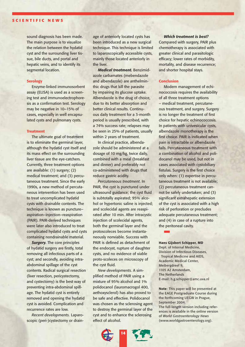
Which treatment is best?Compared with surgery, PAIR plus chemotherapy is associated with greater clinical and parasitologic effi cacy; lower rates of morbidity, mortality, and disease recurrence; and shorter hospital stays.
ConclusionModern management of echi-
nococcosis requires the availability of all three treatment options – medical treatment, percutane-ous treatment, and surgery. Surgery is no longer the treatment of fi rst choice for hepatic echinococcosis. In patients with univesicular cysts, albendazole monotherapy is the fi rst choice. PAIR is indicated when pain is intractable or albendazole fails. Percutaneous treatment with a combination of alcohol and poli-docanol may be used, but not in cases associated with cystobiliary fi stulas. Surgery is the fi rst choice only when: (1) expertise in percu-taneous treatment is not available; (2) percutaneous treatment can-not be safely undertaken; and (3) signifi cant extrahepatic extension of the cyst is associated with a high risk of perforation or precludes adequate percutaneous treatment; and (4) in case of a rupture into the peritoneal cavity.
■
Hans Gijsbert Schipper, MDDept. of Internal Medicine, Division of Infectious Diseases,
Tropical Medicine and AIDS, Academic Medical Center, Meibergdreef 9, 1105 AZ Amsterdam, The NetherlandsE-mail: [email protected]
Note: This paper will be presented at the EAGE Postgraduate Course during the forthcoming UEGW in Prague, September 2004. The full-length version including refer-ences is available in the online version of World Gastroenterology News(www.worldgastroenterology.org).
S C I E N T I F I C N E W S
sound diagnosis has been made. The main purpose is to visualize the relation between the hydatid cyst and the surrounding liver tis-sue, bile ducts, and portal and hepatic veins, and to identify its segmental location.
SerologyEnzyme-linked immunosorbent
assay (ELISA) is used as a screen-ing test and immunoelectrophore-sis as a confi rmation test. Serology may be negative in 10–15% of cases, especially in well encapsu-lated cysts and pulmonary cysts.
TreatmentThe ultimate goal of treatment
is to eliminate the germinal layer, although the hydatid cyst itself and its mass effect on the surrounding liver tissue are the eye-catchers. Currently, three treatment options are available: (1) surgery; (2) medical treatment; and (3) percu-taneous treatment. Since the early 1990s, a new method of percuta-neous intervention has been used to treat uncomplicated hydatid cysts with drainable contents. The technique is known as puncture–aspiration–injection–reaspiration(PAIR). PAIR-derived techniques were later also introduced to treat complicated hydatid cysts and cysts containing nondrainable material.
Surgery. The core principles of hydatid surgery are fi rstly, total removing all infectious parts of a cyst; and secondly, avoiding intra-abdominal spillage of the cyst contents. Radical surgical resection (liver resection, pericystectomy, and cystectomy) is the best way of preventing intra-abdominal spill-age. The hydatid cyst is entirely removed and opening the hydatid cyst is avoided. Complication and recurrence rates are low.
Recent developments. Laparo-scopic (peri-)cystectomy or drain-
age of anteriorly located cysts has been introduced as a new surgical technique. This technique is limited to laparoscopically accessible cysts, mainly those located anteriorly in the liver.
Medical treatment. Benzimid-azole carbamates (mebendazole and albendazole) are anthelmin-thic drugs that kill the parasite by impairing its glucose uptake. Albendazole is the drug of choice, due to its better absorption and better clinical results. Continu-ous daily treatment for a 3-month period is usually prescribed, with a 74% success rate; relapses may be seen in 25% of patients, usually within 2 years of treatment.
In clinical practice, albenda-zole should be administered at a dosage of 10 mg/kg, twice daily, combined with a meal (breakfast and dinner) and preferably not co-administered with drugs that reduce gastric acidity.
Percutaneous treatment. In PAIR, the cyst is punctured under ultrasound guidance; the cyst fl uid is subtotally aspirated; 95% alco-hol or hypertonic saline is injected; and scolecidal agents are reaspi-rated after 10 min. After intracystic injection of scolecidal agents, both the germinal layer and the protoscoleces become instanta-neously nonviable. Success with PAIR is defi ned as detachment of the endocyst, rupture of daughter cysts, and no evidence of viable proto-scoleces on microscopy of the cyst fl uid.
New developments. A sim-plifi ed method of PAIR using a mixture of 95% alcohol and 1% polidocanol (lauromacrogol 400, aethoxysclerol) has also proved to be safe and effective. Polidocanol was chosen as the sclerosing agent to destroy the germinal layer of the cyst and to enhance the sclerosing effect of alcohol.
14

15
Laparoscopic exploration of the common bile duct (CBD) is carried out for the diagnosis or the treat-ment of CBD stones. CBD stones demonstrated by laparoscopic intraoperative cholangiography (IOC) or laparoscopic ultrasonog-raphy (LUS) are extracted either through the cystic duct or through choledochotomy. An alternative for the treatment of CBD stones is to perform an endoscopic sphinc-terotomy either before, during, or after laparoscopic cholecystectomy. The aim of this review is to evalu-ate these different techniques.
Intraoperative Cholangiography (IOC)
Two prospective randomized studies have assessed the value of routine IOC during laparoscopic cholecystectomy. In the fi rst pro-spective trial, by Soper and Dun-nigan [1], relevant information was obtained in eight of 56 patients, and it altered the intraoperative management in four. In the sec-ond trial, by Nies et al. [2], IOC
was unsuccessful in 27 patients (19.6%), most often because of failure to intubate the cystic duct. Unsuspected cystic or CBD stones were diagnosed by IOC in three of 138 patients.
Cystic duct cholangiography is clearly better than cholecysto-cholangiography, and fl uoroscopic imaging should be the standard for IOC. Until now, no specifi c clinically signifi cant complications directly attributable to laparoscopic IOC have been reported. Expected suc-cess rates for laparoscopic IOC are in the range of 90–100%. Inability to cannulate a narrow cystic duct is the main cause of failure. The success rate is higher in a group with selective indications for IOC in comparison with a group with-out such indications – 98% versus 93%, respectively.
In addition to screening for potential asymptomatic CBD stones, the second important attri-bute of IOC is that it provides ade-quate defi nition of the ductal anat-omy. The disastrous consequences
and enormous costs of bile duct injuries have to be taken into account when evaluating the cost-effectiveness of IOC. The rate of bile duct inju-ries associated with laparoscopic cholecystectomy is approximately 0.6%, and this can be reduced with IOC. In all cases associated with an increased risk of CBD injury, and particularly during a physician’s fi rst 20–30 cases of laparoscopic cho-lecystectomy, IOC should be man-datory. Ability to perform an IOC, not just removing the gallbladder, should be one of the requirements for adequate training in laparo-scopic biliary surgery.
Laparoscopic Ultrasonography (LUS)
Several studies of LUS have been published suggesting that LUS is superior to IOC. LUS is per-formed with a higher success rate, in a shorter time, and with better specifi city, but with less precision with regard to the delineation of the biliary tree anatomy. LUS is of little, if any, help in diagnosing or preventing bile duct injuries, whereas IOC is better than LUS for delineating the entire biliary tree, from the intrahepatic tree to the pancreatic portion of the CBD.
LUS is operator-dependent, and there is evidently a learning curve – 20–40 examinations, which may be diffi cult to obtain in small-vol-ume centers in which the 10% prevalence of CBD stones means that the learning curve may take 1–2 years to complete.
Critical Appraisal of Laparoscopic Bile Duct Exploration
Bertrand Millat
Table 1. Criteria for routine intraoperative cholangiography
Preoperative factors
Endoscopic retrograde cholangiography ± sphincterotomy
Ultrasound fi ndings
Common bile duct size over 6 mm
Choledocholithiasis
History of jaundice or pancreatitis
Elevated bilirubin, alkaline phosphatase, transaminases
Intraoperative factors
Unclear anatomy
Conversion to open cholecystectomy
Dilated cystic duct over 4 mm
S C I E N T I F I C N E W S

16
Laparoscopic Extraction of Common Bile Duct Stones
Once stones have been detected during laparoscopic IOC, laparoscopic extraction of them is a logical extension of the pro-cedure. Laparoscopic exploration of the CBD can be performed either through the cystic duct or by laparoscopic choledochotomy, and both procedures are feasible and safe. Endoscopic sphincterotomy (ES) is commonly offered preop-eratively as the alternative to sur-gery for CBD stones. ES is indicated in patients with severe cholangitis for urgent drainage of infected bile, and in patients with retained stones after cholecystectomy. In open conventional surgery, con-trolled studies have not shown that ES, performed either prior to sur-gery or in patients with gallbladder in situ, was superior to single-step surgical management. The conclu-sions reached in these randomized trials have not been extrapolated to laparoscopic biliary surgery.
Data gathered from randomized trials have demonstrated that ES, as an additional procedure to sur-gery, does not improve the clinical results in patients who are fi t for primary single-stage surgical treat-ment, whether performed laparo-scopically or not. Severe cholangitis is an unquestionable indication for urgent endoscopic drainage, regardless of whether or not the CBD can be cleared of associated stones.
All surgeons undertaking lapa-roscopic cholecystectomy must be able to perform an IOC. When IOC demonstrates CBD stones, appropriate treatment is decided on according to the available equip-ment and skills. Transcystic clear-ance of CBD stones may be suc-cessful. In the case of large stones (more than 20 mm in diameter) or other potential diffi culties as regards postoperative ES, such as a periampullary diverticulum, conver-sion to open surgery is indicated when the stone cannot be removed
during laparoscopy. In other cases, the available data do not allow any formal conclusions regarding the choice between advanced laparo-scopic biliary explorations and post-operative ES. In a decision analysis by Erickson and Carlson [3], assess-ing different approaches to using endoscopic retrograde cholangiog-raphy (ERC) in patients undergo-ing laparoscopic cholecystectomy, postoperative ERC was associated with lower costs and less morbid-ity, but laparoscopic CBD explora-tion was not included in the study design. Finally before embarking on a more invasive laparoscopic CBD exploration policy for small stones that cannot be retrieved using the transcystic approach, surgeons must remember that asymptom-atic migration does exist, even if the defi nitive fate of small CBD stones is not at present known. The potential safety afforded by temporary biliary drainage still has to be balanced with its unavoidable morbidity.
Does the COX-1/COX-2 Concept Still Hold?Christopher Hawkey
Recognitionthat nonsteroi-dal anti-infl am-matory drugs (NSAIDs) were inhibitors of prostaglandinsynthesis was
critical to understanding both their therapeutic activity and gastroin-testinal pathology. In the stomach and duodenum, inhibition of pros-taglandin synthesis undermines defensive mechanisms such as mucosal blood fl ow and mucus and bicarbonate secretion, and leads to the development of micro-
erosions that ultimately deepen to become ulcers as a consequence of acid peptic attack. Subsequently, it has become clear that prosta-glandin synthesis derives from two distinct but similar cyclooxygenase enzymes. The constitutive cyclo-oxygenase – cyclooxygenase-1 (COX-1) – is expressed in many tis-sues, including the gastrointestinal tract. The inducible cyclooxygen-ase – cyclooxygenase-2 (COX-2) –becomes highly expressed under the infl uence of many factors such as cytokines and growth factors in tissue injury, infl ammation, and malignant transformation.
The COX-1/COX-2 hypothesis was that drugs that inhibit COX-2 would have the same therapeutic activity as NSAIDs, but without their adverse effects.
Therapeutic ActivityNumerous clinical trials have
shown that COX-2 inhibitors relieve pain and infl ammation in arthritis in a dose-dependent fashion, with maximum effects that are usu-ally not signifi cantly different from those of NSAIDs. Thus, although some have suggested – contrary to the COX-1/COX-2 hypothesis – that COX-1 may contribute to
S C I E N T I F I C N E W S

SC
IE
NT
IF
IC
NE
WS
17
symptoms in arthritis, this effect does not appear to be suffi ciently great to have a measurable clinical impact.
Toxicity of COX-2 Inhibitors and NSAIDs
Remarkably, acute studies have shown that even at very high doses (up to ten times the thera-peutic dosage), COX-2 inhibitors cause no demonstrable muco-sal injury in healthy volunteers. Medium-term endoscopy stud-ies in patients show substantial reductions in ulceration in com-parison with NSAIDs. In some,
but not all, studies, the levels of ulceration have been similar to those observed with a placebo. In outcome studies, the incidence of clinically signifi cant ulcers or ulcer complications is reduced in comparison with NSAIDs. In most studies, the reduction has been between 50% and 60% rather than the 75–85% that might be inferred from the fourfold to fi ve-fold increase in ulcer complications caused by NSAIDs.
DyspepsiaThe original COX-1/COX-2
hypothesis did not relate to dys-pepsia. Nevertheless, high levels of dyspepsia with NSAIDs are suf-fi ciently general that this may be a class-based (and therefore mecha-nism-based) effect. This argument is reinforced by data from COX-2 inhibitors, which quite consistently appear to be associated with levels of dyspepsia that are greater than those with a placebo but lower than those with NSAIDs.
Thrombotic EventsWhen patients present with
NSAID-associated ulcer bleed-ing, it may be because the NSAID causes ulcer formation or impairs hemostasis, leading to bleeding. Equally, since vascular prostacyclin is recognized to be largely derived from the COX-2 enzyme, it is pos-sible that COX-2 inhibitors could induce thrombosis. This issue came to a head in the Vioxx Gastrointes-tinal Outcomes Research (VIGOR) study, in which patients receiving naproxen were reported to have fewer thrombotic cardiovascular events than those receiving rofe-coxib. This could be attributed either to a harmful effect of rofe-coxib or of naproxen. Recent data suggest that the latter is the domi-nant factor, although the former may not be absent.
References[1] Soper NJ, Dunnegan DL. Routine versus selective intra-operative chol-
angiography during laparoscopic cholecystectomy. World J Surg 1992; 16: 1133–40.
[2] Nies C, Bauknecht F, Groth C, et al. Intraoperative Cholangiographie als Routinemethod? Eine prospektive, kontrollierte, randomisierte Studie. Chirurg 1997; 68: 892–7.
[3] Erickson RA, Carlson B. The role of endoscopic retrograde cholangio-pancreatography in patients with laparoscopic cholecystectomies. Gas-troenterology 1995; 109: 252–63.
■
Bertrand Millat, MDDept. of Visceral Surgery, Saint Eloi University Hospital, 80, avenue Augustin Fliche,34295 Montpellier cedex 5, FranceE-mail: [email protected]
Note: This paper will be presented at the EAGE Postgraduate Course during the forthcoming UEGW in Prague, September 2004. The full-length version including refer-ences is available in the online version of World Gastroenterology News (www.worldgastroenterology.org).
CancerSince COX-2 is induced in gastro-
intestinal cancers, a development of the COX-1/COX-2 hypothesis predicts that COX-2 inhibitors may prevent or reverse gastrointestinal malignancy without harmful effects on normal mucosa. This has been shown in animal studies and in genetic models in humans. How-ever, aspirin is also effective, but is a COX-1 inhibitor. Understanding the way in which aspirin prevents or reverses premalignant and malignant processes in the gastro-intestinal tract will contribute to our understanding of oncogenesis.
Practical PrescribingThe COX-1/COX-2 hypothesis
does not discount the infl uence of additional factors, and in fact acknowledges that NSAID ulcers have prostaglandin-dependent and acid peptic-dependent com-ponents. This would lead one to hypothesize that acid suppression and COX-2 inhibitor substitution might be complementary strategies in preventing ulcer disease. Recent data show that acid suppression very effectively reduces ulcer devel-opment and dyspepsia in patients receiving COX-2 inhibitors who are at high risk of these two gastroin-testinal problems.■
Professor C.J. Hawkey Division of Gastroenterology University Hospital School of Medical and Surgical
Sciences NG7 2UH Nottingham United Kingdom E-mail: [email protected]
Note: This paper will be presented at the EAGE Postgraduate Course during the forthcoming UEGW in Prague, September 2004. The full-length version including refer-ences is available in the online version of World Gastroenterology News(www.worldgastroenterology.org).
S C I E N T I F I C N E W S

Cholangio-Pancreaticoscopes
Autofluorescence-Bronchoscopy
Gastroscopes
Duodenoscopes
Naso-Pharyngo-Laryngoscopes
Cystoscopes
Colonoscopes
Ureteroscopes
Choledocho-Nephroscopes
Enteroscopes
Sigmoidoscopes
Cholangio-Pancreaticoscopes
Zoom Colono- and Gastroscopes
Ultrasound-Endoscopes
Autofluorescence-Bronchoscopy
Intubation-Endoscopes
Bronchoscopes
for your precious momentsfor your precious moments
details areeverythingdetails areeverything
Many consider the new Pentax 70K/80K series to be the best video systemavailable. Superior and patented hygiene features, enlarged instrument chan-nels and our world class Digital Signal Processing (DSP) technology are justsome of the reasons why. Contact Pentax for a new perspective on your nextendoscopic procedure: Telephone +49-40-56192·0; Fax +49-40-5604213;E-mail: [email protected] or Internet: www.pentax-endoscopy.com
19
S C I E N T I F I C N E W S
Molecular Biology for the Gastroenterologist
Peter Ferenci
An increasing number of diseases of the digestive organs are being recognized as having a genetic background (Table 1). The era of genetics began with the obser-vations by Gregor Mendel that changes in the color of fl owers and the shape of their seeds followed a clear pattern over the years. The fundamental rules of inheritance that he established were thus based on easily recognizable signs. His work preceded the discovery that DNA is the carrier of genetic information.
An observed trait is referred to as a phenotype; the genetic infor-mation defi ning the phenotype is called the genotype. As advances were made in understanding the functioning of DNA, phenotypic genetics was replaced by molecu-lar genetics. In contrast to phe-notypic genetics, which assumes that gene products are either fully functional or devoid of function as
a result of a mutation, molecular genetics describes variations in the base sequence of the genes. These changes are not always associated with impaired func-tions of the gene product, and do not necessarily imply the presence of phenotypic disease. It is these fundamental differences from phe-notype-based genetics that defi ne the role of molecular genetics in clinical medicine.
Defi nitionsA few basic defi nitions are
needed to clarify the implications of molecular genetics.
What constitutes a normal gene? A normal gene is defi ned by the base sequence that is observed in the majority of healthy individu-als in a given population, and it is known as the wild type. Base variations in the wild-type gene in healthy individuals are termed DNA polymorphisms. These alternative
forms of a gene or a genetic marker are referred to as alleles. In other instances, allelic variants may refl ect mutations in a gene that clearly alter its function.
What is a mutation? A muta-tion is a base sequence that differs from the wild type in a patient who presents with a phenotypic disorder. The sequence is never observed in healthy individuals.
The functional consequences of a mutation are manifold. Muta-tions can be broadly classifi ed as either gain-of-function mutations or loss-of-function mu tations.Gain-of-function mutations are typically dominant. Inactivating mutations are usually recessive, and an affected individual is homo-zygous or compound heterozygous (i.e., carrying two different mutant alleles) for the disease-causing mutations. Mutations may result in the complete absence of gene products (“null” mutations), or in proteins devoid of any function. Such mutations are associated with severe diseases occurring at birth or in early childhood. Other mutations result in less pronounced functional consequences and milder disease that presents later in life. A change in a single amino acid may affect the tertiary structure, the assembly, inactivation, secretion, or conforma-tional stability of the gene product.
Tools for Molecular-Genetic Analysis
Molecular genetics requires the visualization of differences in the DNA sequence. DNA polymor-
Table 1. Selected genetic diseases in gastroenterology and hepatology.
Diseases Gene symbolCholestatic liver diseases Byler disease, Summerskill–Walshe syndrome FIC1 (now ATP8B1) Progressive familial intrahepatic cholestasis 2 ABCB11 Progressive familial intrahepatic cholestasis -3 ABCB4 Dubin–Johnson syndrome ABCB2 (now TAP1)Hepatic storage diseases Wilson’s disease ATP7B Hemochromatosis HFEColon cancer Familial polyposis coli APC Hereditary nonpolyposis colon cancer MSH2, MLH1 Peutz–Jeghers syndrome STK11Idiopathic pancreatitis Cystic fi brosis CFTR Hereditary pancreatitis Trypsinogen, SPINKCrohn’s disease NOD2 (now CARD15)

20
phisms in coding regions (exons) or noncoding regions of the genes are inherited in accordance with the Mendelian rules. The value of highly variable DNA sequences as genetic markers rests on straightforward principles. Every person carries two copies of each chromosome, except for the sex chromosomes. To be useful for analyzing the transmis-sion of the two chromosomes in a family, the DNA copies at the poly-morphic site in the person under study have be different in the two chromosomes. The chromosomal sites at which the DNA sequences can have many alternative forms are thus ideal sites for genetic markers. In the human genome, the sites that have the properties most favorable for such extensive variation include a repetition of the same short DNA sequence a
variable number of times (known as tandem-repeat sequences or microsatellites).
There are several methods of assessing variations in the DNA sequence. Restriction fragment length polymorphism (RFLP) analysis detects variations in the size of DNA fragments obtained after digestion with restriction enzymes.
Direct mutation analysis. Direct sequencing. New technolo-gies allow automated sequence analysis of large portions of a gene to detect point mutations, dele-tions, inversions, and other changes in the nucleotide sequence. How-ever, direct sequencing of the whole sequence does not yet play a role in clinical medicine.
Polymerase chain reaction (PCR)-based detection of known
mutations. The simplest approach takes advantage of the base-sequence specifi city of restriction endonucleases. A mutation will prevent the enzyme from cutting at that point in the sequence; conversely, a mutation may result in the creation of a new enzyme-recognition site and lead to cutting where it nor mally should not occur. The pattern of digestion is indica-tive of the presence or absence of a particular mutation. The specifi c-ity of the PCR reaction itself allows direct detection of mutations.
Interpretation of Test ResultsMolecular-genetic analysis can
yield three possible fi ndings: the individual being tested can be a homozygous carrier of the muta-tion, a heterozygous carrier of it, or does not carry the mutation at all.
S C I E N T I F I C N E W S
Blackwell Publishing is proud to sponsorThe American Journal of Gastroenterology Lecture
The use of Anesthesiain Endoscopy-
A Critical Examination
BOOKS:
Phone: 1-800-216-2522 • Fax: 802-864-7626e-mail: [email protected]
JOURNALS:
Phone: 1-800-835-6770 • Fax: 781-388-8232e-mail: [email protected]
www.blackwellgastroenterology.com
As the world’s largest independent society publisher, Blackwellpublishes over 700 journals with more than 550 academic andprofessional societies. We partner with leading societies to advanceknowledge in Gastroenterology, Hepatology and Endoscopy.
Attend the debate on November 1, 2004 from 2:40 to 3:20 p.m. at the
69th Annual Scientific Meeting of theAmerican College of Gastroenterology
in Orlando, Florida
To register for the annual meeting, visit www.acg.gi.org
Journal Publishing Partners

21
S C I E N T I F I C N E W S
Homozygous mutation carrier.Genotypic diagnosis in a healthy individual raises the question of whether person being tested will ever develop the disease. In most hereditary diseases, the penetrance of a disease is not complete.
Heterozygous mutation car-rier. Compound heterozygotescarry two different disease-causing genes. In most inherited diseases, multiple different mutations of the affected gene are present (more than 800 in cystic fi brosis, for example). The problem is to dif-ferentiate a “true” heterozygote (carrying a wild-type allele) from a compound heterozygote. The most important question is whether the individual being tested is (and will remain) free of a disease or not. According to Mendelian rules, individuals carrying a wild-type and a disease-causing gene with auto-somal-recessive inheritance (true heterozygotes) are healthy. This statement is only valid if the other gene not carrying the mutation is also functionally intact. The gene that does not have an established mutation may have a different (dis-ease-causing) one. Unfortunately, this is not an exception, but a gen-eral rule.
Haploinsuffi ciency . Mutation in a single allele can result in a situa-tion in which one normal allele is not suffi cient for a normal pheno-type. This phenomenon applies, for example, to the expression of rate-limiting enzymes in heme synthesis that cause the porphyrias. Mutation in a single allele can also result in loss of function due to a dominant-negative effect.
Loss of heterozygosity. Individu-als with a normal and an abnormal gene without any apparent disease may undergo somatic mutations of the normal gene later in life. Such events may result in overt dysfunc-tion of the gene product in the
affected cells. This loss of heterozy-gosity is assumed to be an impor-tant event in carcinogenesis.
Individuals not carrying the mutation. A negative fi nding does not exclude phenotypic disease, since other mutations of the gene may be present. In addition, gene defects may be due to mutation of other genes.
Target Populations for Molecular-Genetic Testing
Patients with symptomatic phenotypic disease. In patients with hereditary diseases, DNA analysis strengthens the fi nal diag-nosis. In diseases that have only a few mutations (such as HFE-asso-ciated hemochromatosis), muta-tion analysis can replace invasive diagnostic tests. In patients with a transferrin saturation index > 45%, testing for common HFE mutations allows a direct diagnosis of heredi-tary hemochromatosis.
Mutation analysis is important for differentiating between various genetic diseases with similar phe-notypic symptoms, as in patients with primary fi ndings of iron over-loading. At least four independent genetic diseases are now known to result in the accumulation of iron in various organs.
Family screening. Mutation analysis is the state-of-the-art approach for screening the family of index patients and can replace other diagnostic tests to identify individuals at risk of developing the disease. A negative test result in a relative of a patient with a disease-related mutation indicates a low risk of the disease.
Population screening. Muta-tion analysis has not yet been tested for detecting presymptom-atic disease in the general popula-tion. Apart from the diffi culties in interpreting test results mentioned, there are also several factors
that limit the use of genetic tests for population screening. Firstly, screening is only appropriate if a validated treatment is available for asymptomatic individuals. Secondly, other screening strate-gies may be more cost-effective or straightforward than mutation analysis. For colorectal screening, DNA-based mutation analysis is not capable of replacing endos-copy, as a colonoscopy examina-tion is needed whether or not a mutation is present. In addition, the development of cancer can be prevented by endoscopic polypec-tomy. Endoscopy combined with testing for fecal occult blood will therefore continue to be the stan-dard approach for the foreseeable future.
Disease association studies.The rapid growth of human genet-ics is creating countless opportuni-ties for studies of disease associa-tion. Given the number of poten-tially identifi able genetic markers and the multitude of clinical outcomes to which these may be linked, the testing and validation of statistical hypotheses in genetic epidemiology is a challenge on an unprecedented scale.
■
Prof. Peter Ferenci, M.D.Dept. of Internal Medicine IV,
Gastroenterology and Hepatology, University of Vienna General Hospital,
Waehringer Guertel 18–20, A-1090 Vienna, AustriaTel. +43-1-40400-4741, fax +43-1-40400-4735E-mail: [email protected]
Note: This paper will be presented at the EAGE Postgraduate Course during the forthcoming UEGW in Prague, September 2004. The full-length version including refer-ences is available in the online version of World Gastroenterology News (www.worldgastroenterology.org).

Absolute control.
The new double-balloonmethod for examining theentire small intestine.
A leap forward — the new double-balloonendoscope allows endoscopy of the entiresmall intestine to be performed for the firsttime with all diagnostic and therapeuticresources. Examine the whole area withoutsurgery, and with optimum image quality andmaximum comfort — for both doctor andpatient. Fujinon. To see more is to know more.
www.fujinon.de Medical TV CCTV Machine Vision Binoculars
FUJINON (EUROPE) GMBH, HALSKESTRASSE 4, 47877 WILLICH, GERMANY
TEL.: +49 (0) 21 54/9 24-0, FAX: +49 (0) 21 54/9 24-2 90, www.fujinon.de
FUJI PHOTO OPTICAL CO. LTD., 1-324 UETAKE, KITAKU, SAITAMA-CITY, SAITAMA 331-9624, JAPAN
TEL.:+81 (0) 48/6 68/21 52, FAX:+81 (0) 48/6 51/85 17, www.fujinon.co.jp
is Turkish and also speaks Farsi, the offi cial Iranian language, in addi-tion to their mother tongue.
Acceptance of medical research among the general population has always been high in Ardabil. In 1968, the fi rst cancer registry pro-gram was launched on the basis of a collaborative agreement between the Health Institutes of the Univer-sity of Tehran and the International Agency for Research on Cancer (IARC) in order to study esophageal cancer in the area of the Caspian littoral, from Ardabil province in the west to Golestan province in the east. Studies conducted at that time calculated the annual age-adjusted incidence rate of esophageal cancer (mainly esophageal squamous-cell cancer) to be over 100 per 100 000 in the Gonbad district in Golestan province – among the highest rates in the world. A subsequent collab-orative case–control study revealed a pattern of very low consumption of fresh fruit and vegetables in north-eastern Iran.
BackgroundAfter cardiovascular disease and
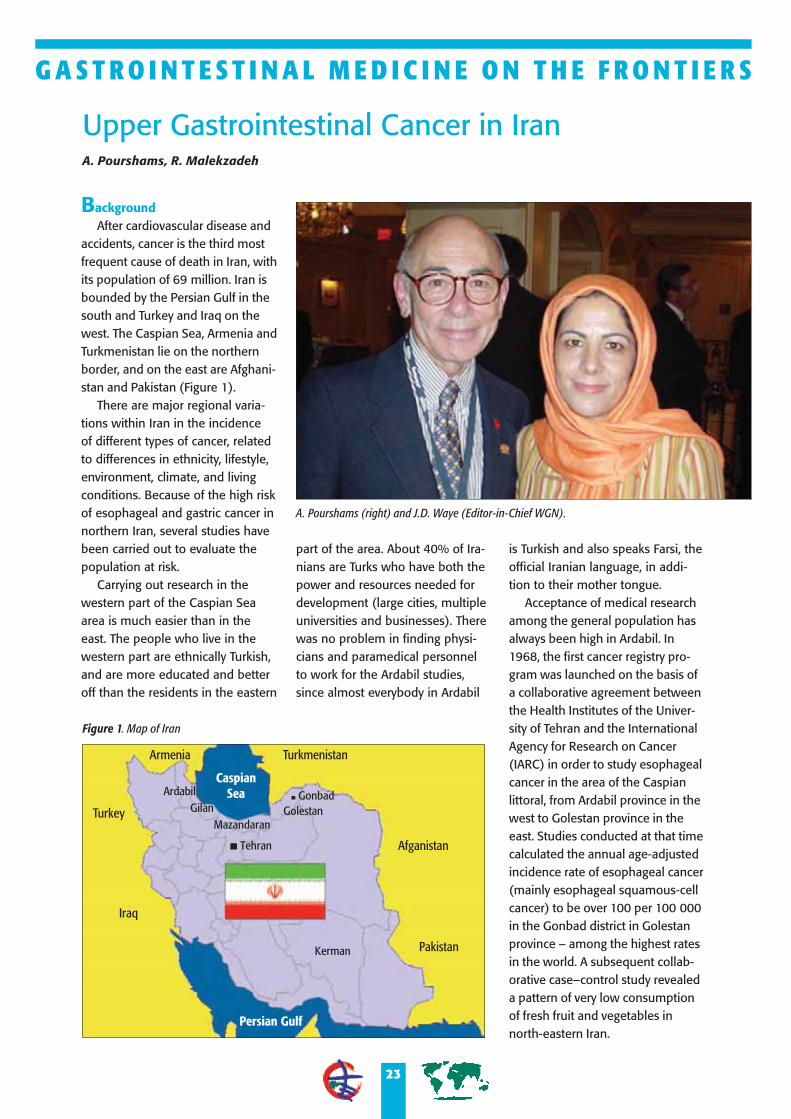
accidents, cancer is the third most frequent cause of death in Iran, with its population of 69 million. Iran is bounded by the Persian Gulf in the south and Turkey and Iraq on the west. The Caspian Sea, Armenia and Turkmenistan lie on the northern border, and on the east are Afghani-stan and Pakistan (Figure 1).
There are major regional varia-tions within Iran in the incidence of different types of cancer, related to differences in ethnicity, lifestyle, environment, climate, and living conditions. Because of the high risk of esophageal and gastric cancer in northern Iran, several studies have been carried out to evaluate the population at risk.
Carrying out research in the western part of the Caspian Sea area is much easier than in the east. The people who live in the western part are ethnically Turkish, and are more educated and better off than the residents in the eastern
Upper Gastrointestinal Cancer in IranA. Pourshams, R. Malekzadeh
part of the area. About 40% of Ira-nians are Turks who have both the power and resources needed for development (large cities, multiple universities and businesses). There was no problem in fi nding physi-cians and paramedical personnel to work for the Ardabil studies, since almost everybody in Ardabil
G A S T R O I N T E S T I N A L M E D I C I N E O N T H E F R O N T I E R S
Figure 1. Map of Iran
Armenia
CaspianSea
Pakistan
Afganistan
Iraq
Turkey
Turkmenistan
Persian Gulf
Ardabil
Kerman
MazandaranGolestan
■ GonbadGilan
■ Tehran
A. Pourshams (right) and J.D. Waye (Editor-in-Chief WGN).
23

The situation in the north-east part of the country, near Turkmeni-stan, is considerably different from that in the west (Ardabil). Most Turkmen live in northern Golestan province, and they constitute less than 2% of the population of Iran. In their appearance (especially their eyes), ethnicity, and social habits, they are different from Persians (who make up 50–55% of Iranians). The main traditional occupation of Turkmen has been in animal husbandry. They tradition-ally lived in temporary houses and tents in the hills, mountains, and plains in the border region between Iran and Turkmenistan, and they had several disputes with the cen-tral governments before taking up obligatory permanent residence in north-eastern Iran about 70 years ago. Because of the differences in cultures, Turkmen have not had strong relationships with the Per-sians (intermarriage, for example) until the last few years.
There were many problems in conducting the studies in the north-eastern Turkman area, including: a) The fact that more than 90% of
women and about 50% of men over 40 years of age in the vil-lages are illiterate and cannot speak Farsi, so that gathering data is possible only through face-to-face interviews together with Turkmen interpreters.
b) Finding enough educated Turk-men able to assist in the studies is a major problem; Persians cannot speak the Turkmen lan-guage and are not a suitable substitute.
c) Most Turkmen are aware that survival in those with esopha-geal cancer is poor, and did not initially accept any interven-tion for people with the signs and symptoms of esophageal cancer; however, acceptance is improving.
d) Gonbad does not have units for chemotherapy or radiotherapy, and does not have specialist cancer surgery units, so that cancer patients need to travel to other provinces. Unfortunately, there are no fl ights or fast trains between Gonbad and Tehran or other provinces that do have cancer care facilities.
e) Some of the roads are unpaved in the Gonbad district.
f) The weather is not tolerable dur-ing the summer in the Gonbad district (very hot and humid), and there is a risk of huge fl oods every spring.
The initial collaborative studies stopped at the time of the politi-cal changes that took place in Iran in 1978. Recently, The Digestive Disease Research Center (DDRC) at Tehran’s University of Medical Sciences, the IARC, and the United States National Cancer Institute have started studying upper gastro-intestinal cancers in the northern Iranian plain again. The Turkmen were resistant to endoscopy when the case–control study started in 2002. To motivate them to par-ticipate in the studies, the DDRC established a very well-equipped endoscopy unit in Gonbad 3 years ago (with the latest video endo-scopes from Olympus and Pen-tax), along with a gastrointestinal pathology laboratory, providing free diagnosis, treatment, and management of all patients with upper gastrointestinal cancer. This included payment for surgery, chemoradiotherapy, dilation pro-cedures, and stenting. Once the results were seen, acceptance of the treatment improved rapidly.
Recent Studies on Upper Gastrointestinal Cancers in Northern Iran
North-west. Gastric adeno-carcinoma is the most common
gastrointestinal malignancy in Iran. Ardabil has the highest incidence of gastric adenocarcinoma. According to an active cancer surveillance pro-gram conducted in Ardabil (1996–1999), gastric adenocarcinoma rep-resents 31% of all malignancies in the region, with incidences of 49.1 and 25.4 per 100 000 per year for men and women, respectively. Half of these gastric cancers are located in the cardia. In 2000, upper endo-scopic screening was carried out in 1011 randomly selected rural and urban residents of Ardabil, with mean age of 53 years. Urease test-ing or histology for Helicobacter pylori was positive in 89% of those tested, and 95% had chronic gas-tritis. H. pylori, which is known to contribute to the development of gastric adenocarcinoma, is com-mon in Ardabil. No dysplasia or esophageal cancer was found in a recent population-based study with chromoendoscopy screening program in 504 asymptomatic ran-domly selected adults in Ardabil.
North-east. Case–control study.A referral clinic for upper gastroin-testinal diseases was established by the DDRC in Golestan province in August 2001. Among the initial 682 patients seen at the clinic, 370 were confi rmed histologically as having cancer, including 60% with esophageal squamous-cell cancer, 6% with esophageal adenocarci-noma, 16% with gastric cardia ade-nocarcinoma, and 16% with non-cardia gastric adenocarcinoma. The proportional occurrence of these four main upper gastrointestinal cancers was similar to that seen in Linxian, China, another area with a high incidence of esophageal squamous-cell cancer, and was markedly different from the current proportions in Western countries. Negligible alcohol consumption and cigarette smoking in these patients suggest that the high rates
G A S T R O I N T E S T I N A L M E D I C I N E O N T H E F R O N T I E R S
24

of esophageal squamous-cell can-cer seen in north-eastern Iran are associated with other risk factors.
Cohort study. In 2002, a cohort study was initiated in Gonbad dis-trict to evaluate the genetic and environmental risk factors of upper gastrointestinal cancers, mainly esophageal carcinoma. A total of 1359 (704 rural, 645 urban) inhabitants aged between 35 and 80 were selected randomly and invited to participate in the study. An active follow-up examination was carried out after 12 months. Cigarette smoking and opium and alcohol use were reported by13.8%, 10.3%, and 3.7%, respec-tively. The mean temperature of ingested tea was 57.4ºC. Two sub-jects developed esophageal squa-mous-cell cancer. The data show that the incidence of esophageal carcinoma is still high in the region, but that the pattern of causes of death is similar to that in other parts of Iran.
Active surveillance. Although Iran’s National Cancer Registry program is still in its initial stages, the DDRC carried out active surveil-lance for cancers in the Caspian littoral and Kerman province (in the center of the country) in 1999 (Table 1). The data show that the cancer burden relative to each
geal carcinoma. However, none of these four alleles had a high enough prevalence in Turkmen to explain the high rates of the disease in that group. Three of the four alleles were less frequent among Turkmen than in some Asian popu-lations with lower risks of esopha-geal cancer. The authors concluded that it is unlikely that variations in these polymorphic genes are major contributors to the high incidence of esophageal carcinoma among Turkmen in Iran.
Because P53 mutations are the most frequent mutations in cancer, and may provide clues on the etio-logical mechanisms of esophageal squamous-cell cancer, P53 was analyzed in pathology samples from 98 Iranians with esophageal squamous-cell cancer, and muta-tions in 50% of the patients were found. The P53 mutation pattern in Iran was signifi cantly different from that observed in esophageal squamous-cell cancer in high-incidence areas of China and Western Europe. These results are consistent with the hypothesis that several factors are involved in P53 mutagenesis in Iran, including a background of chronic infl amma-tory stress, as was shown by Crespi and colleagues in the 1970s.
Ongoing StudiesCase studies on esophageal
and gastric cancers are ongoing in Ardabil and Golestan provinces. A cancer registry in the northern plain has been initiated by the DDRC, and reports will soon be forthcoming.
■
Corresponding Author:A. Pourshams, M.D.Digestive Disease Research CenterTehran University of Medical Sciences,TehranIranE-mail: [email protected]
organ is similar in all areas of the Caspian littoral, but quite different from that found in the central part of Iran.
Ecological studies. A study was carried out to assess the hypoth-esis that the high rates of esopha-geal carcinoma in Golestan and the high rates of gastric cancer in Ardabil may be partly attributable to selenium defi ciency. The fi nd-ings suggest that selenium defi -ciency is not a major contributor to the high incidence of esophageal cancer seen in north-eastern Iran, although it may play a role in the high incidence of gastric cancer in Ardabil province.
Genetic studies. The frequen-cies of polymorphisms in 10 genes that have been hypothesized to have a role in the risk of esophageal carcinoma were compared among three Iranian ethnic groups with highly varying rates of the disease. These three groups included high-risk Turkmen, medium-risk Turks, and low-risk Zoroastrian Persians. Compared to Zoroastrians, Turk-men had a higher frequency of four alleles that are thought to favor car-cinogenesis (CYP1A1 m1, CYP1A1 m2, CYP2A6*9, and ADH2*1);these results were consistent with an infl uence of these allele variants on the population risk of esopha-
Table 1. The relative frequency of cancers by province (1999–2002).
Ardabil Gilan Mazandaran Golestan KermanStomach 31.4%
Stomach 19.0%
Stomach 16.3%
Esophagus 31.0%
Breast 10.2%
Esophagus 13.1%
Breast 9.9%
Esophagus 12.9%
Stomach 16.8%
Stomach 8.8%
Colorectal 4.8%
Colorectal 9.2%
Breast 9.6%
Breast 8.8%
Colorectal 6.7%
Lung 4.7%
Esophagus 8.7%
Colorectal 7.1%
Colorectal 5.3%
Blood 5.6%
Blood 4.4%
Bladder 7.9%
Blood 5.3%
Bladder 3.4%
Lung 5.0%
Other cancers 41.6%
Other cancers 45.3%
Other cancers 48.8%
Other cancers 44.7%
Other cancers 63.7%
G A S T R O I N T E S T I N A L M E D I C I N E O N T H E F R O N T I E R S
25


WGO/OMGE-OMED Training CentersJim Toouli (Co-Chairman, Joint Education and Training Committee)
The Organisation Mondiale de Gastro-Entérologie/World Gastro-enterology Organization (OMGE/WGO), along with its partner orga-nization, the Organisation Mon-diale d’Endoscopie Digestive/World Organization of Digestive Endos-copy (OMED), has developed train-ing centers in different regions of the world with the aim of deliver-ing education and training in all aspects of gastroenterology. These centers have been developed over the last few years, bringing together the ideas of numerous people who have served on the executive and/or education com-mittees of the organizations and our colleagues in many parts of the world. They refl ect the major mission of the organizations, which is to foster the progress of our profession through educa-tion and training with a global perspective.
There are two types of train-ing center. The fi rst type, of which there are fi ve, are situated in the vicinity of developing countries, and their major aim is to train gas-troenterologists in the developing world. The second type, of which there is only one at this stage, is aimed at providing more advanced training in gastroenterology.
The centers have evolved gradu-ally, with most of the activity hap-pening in the last 2–5 years. The fi rst center was inaugurated in Soweto, South Africa, and was sup-ported initially by the Munich Gas-troenterology Foundation through Professor Meinhard Classen. The center was the “brainchild” of Professors Issy Segal and Classen, and the former also became its fi rst Director. The World Organiza-tion of Gastroenterology became associated with the Soweto center as a result of Professor Classen’s
involvement. Professor Segal brought his ideas regarding the training center concept to the early meetings of the newly formed combined Education and Training Committee. These ideas merged with those being developed by the chairs and committee members through other contacts, and as a result the committee took on the task of enlarging and expanding the training center concept. The WGO/OMGE executive embraced the ideas with enthusiasm and there then evolved guidelines for setting up centers in areas of the world where it was felt a need existed, where trusted colleagues would be our local representatives, and where we would be wel-comed, but also where our limited budget might provide a nidus for attracting further funds that would make the centers viable in the long term. An underlying principle has been the development of centers in association with the local gov-ernment authorities and relevant gastroenterology groups (e.g., the local gastroenterology society). In addition, each of the centers has a designated member of the WGO/OMGE executive assigned to over-see negotiations with the relevant authorities, as well as two mem-bers of the Education and Training Committee who plan its activities. (One of these two is usually the director of the center, who is co-opted on to the education commit-tee as a full member.)
Five centers have now devel-oped, including Soweto. They are all different, but have the common aim of bringing education and training in gastroenterology to our colleagues in the developing world. Apart from Soweto, the other four centers are situated in Rabat (Morocco), Karachi (Pakistan), Cairo (Egypt), and La Paz (Bolivia). The centers in Morocco, Pakistan,
E D U C A T I O N A N D T R A I N I N G
Spread of WGO/OMGE-OMED endorsed Training Centers around the world. (●● Center for advanced
endoscopic training.)
●●CairoEgypt
●●Rabat
Morocco
●●Soweto
South Africa
●●La Paz/Bolivia
●●Santiago
Chile
●●KarachiPakistan
27

View of Baragwanath hospital (the largest
in the southern hemisphere) in Soweto,
South Africa.
the world (e.g., Afghanistan). The center in Karachi can serve as the medium for disseminating valu-able programs from any of our other centers or any departments in the world that may wish to contribute to the overall program. A curriculum is being established, and we believe that the future potential of this resource will be enormous.
The Cairo center is the most recent to be inaugurated and is a joint venture with the Egyptian Ministries of Higher Education and Health and the Theodor Bilharz Institute in Cairo. In addition, the involvement of the African and Middle East Association of Gas-troenterology (AMAGE) has been important in setting up this facility. The opening ceremony and the opening meeting were highlighted in the last issue of WGN. With aims similar to those of the other centers, this center is targeting the training needs of our colleagues in the Middle East and in particular the Arab world. The fi rst training program appropriately focused on the assessment and manage-ment of portal hypertension and
the center has been placed in a renovated ward of the main teach-ing hospital in Rabat. An ambitious program has evolved, and already a number of workshops have taken place. In addition to our own involvement, our colleagues from ASNEMGE (the European/Mediterranean Gastroenterology Association – Association des Sociétés Nationales Européenneset Mediterranéennes de Gastroen-térologie) have provided fi nancial support. It is planned that trainees from the French-speaking countries of Africa will be trained at this cen-ter by our colleagues from Rabat, along with international colleagues who will visit periodically to pro-vide a global perspective.
The Karachi center has devel-oped in partnership with the Aga Khan University and its department of gastroenterology. The depart-ment and university are highly developed institutions with pro-grams at the cutting edge of medi-cine. The aims here are to develop an electronic web-based teaching program that may be deliver-able to more remote regions of
and Egypt are functioning and have taught a number of trainees on a variety of topics. The La Paz center will be inaugurated early in 2005. Each of the centers has developed differently from the others and their initial activities have been quite diverse.
In Soweto, trainees come from the surrounding countries and include gastroenterology nurses, family practitioners receiving basic training, and physicians obtaining specialist training in gastroenter-ology. Soweto aims to reach out to the developing countries in sub-Saharan Africa, where there is a large need for training in gastroenterology. The activities of the Soweto center have been embraced by the South African Gastroenterology Society, and as a result, much-needed support is provided for the center’s educa-tional activities by our South Afri-can colleagues.
The Rabat Center has evolved to address the needs of Franco-phone Africa. In conjunction with the Moroccan Ministry of Health,
28
E D U C A T I O N A N D T R A I N I N G

The team at the Soweto Center with representatives of WGO/OMGE and AMAGE.
attracted trainees from the Middle East and throughout Africa.
It is the aim of the education committee, which oversees the centers’ activities, that – while always recognizing the diversity of the various centers – a common basic philosophy should evolve for all of the centers. A subcommittee has therefore been charged with developing this philosophy and the necessary guidelines for imple-menting it. Our vision for these training centers is that they will become centers of major infl uence in the training of colleagues in the developing world. As fi nances per-mit, we would hope to expand the numbers to other regions of the world.
The second type of center aims to address areas of excellence in gastroenterology and will be rec-ognized for providing training at the cutting edge of our specialty. However, in keeping with the phi-losophy of an internationally rec-ognized center of excellence, these centers need to be able to offer hands-on experience for trainees from all countries.
The fi rst of these centers to be recognized is a center of excellence in delivering of endoscopic training and is to be designated The WGO/OMGE-OMED Advanced Center in Endoscopy Training. The fi rst center recognized is in Santiago, Chile, and will be inaugurated in August. It is hoped that other centers, not only in endoscopy but also in the other areas of gastroenterology (e.g., hepatology, luminal gastro-enterology, gastrointestinal surgery, liver transplantation, minimal-access surgery) will be recognized. We invite colleagues to consider
whether their center may qualify for this prestigious designation.
The members of the executives of the WGO/OMGE and OMED, as well as the committee members of the combined education and training committee, have great aspirations for the potential of the various training centers. We antici-pate that they will become the focus of many of the activities of our societies and have a signifi cant impact in leading the training and maintenance of standards in gas-troenterology on a global basis. On behalf of the organizations, I invite input from the wider gastroen-terology community on how best to advance our vision for these centers. In so doing, I also wish to acknowledge the support provided to the organizations by our indus-try partners, without whom none of this activity would be possible.
Whilst there is no direct fi nancial support from the biomedical indus-try for any of the centers, it is the income that is derived from the industry’s support for the World Congresses that is used to under-pin these important educational endeavors. With the anticipated success of the next World Congress in Montreal next year, we hope to not only expand the activities at the existing centers but also to expand the number of centers around the world.
■
James Toouli, MDCo-Chairman, WGO/OMGE-OMED
Education and Training CommitteeFlinders Medical Centre, Department of General and Digestive
Surgery, Bedford Park, SA 5042, Adelaide, AustraliaE-mail: [email protected]
29
E D U C A T I O N A N D T R A I N I N G

The EETC is located in the center of the univer-sity campus, close to the animal laboratory, in a fully renovated building (Figure 1) on two fl oors, with an area of approxi-mately 500 square meters. The heart of the EETC is on the basement level, where there is an operating room (Figure 2) with fi ve worksta-tions fully equipped with state-of-the-art Olympus video endoscopes and Erbe electrosurgical generators with argon plasma coagulation (APC), where trainees can carry out a variety of therapeutic endoscopic procedures on animal models, with appropriate guidance from tutors. Various endoscopic procedures can be carried out in the isolated swine upper or lower gastrointesti-nal tract using the Erlangen model – including polypectomy, muco-sectomy, hemostatic procedures, APC treatments, self-expandable metal stent insertion, antirefl ux procedures, and sphincterotomy. The EETC is also fully certifi ed and authorized to use live animals under general anesthesia for spe-cial procedures. The presence of a veterinary doctor specialized in animal care ensures that all ethical requirements are fully respected. The development of other models for teaching gastrointestinal endos-copy is also being currently investi-gated at the EETC.
On the fi rst fl oor, there is an auditorium for 20–25 people, with up-to-date audiovisual equipment and a permanent link to an endos-copy suite at Gemelli University
hospital to allow live display of endoscopic procedures conducted by local staff or by invited guest teachers. The center includes a small library and meeting room and another room equipped with four personal computer worksta-tions for electronic interactive teaching, as well as a Simbionix electronic simulator for upper and lower gastrointestinal endoscopy and ERCP (Figure 3). Uniquely, the EETC includes facilities in which every step in the teaching process that should precede hands-on experience with patients can be replicated.
Other facilities associated with the center include a low-cost hotel on campus and a nearby apartment that can accommodate foreign trainees free of charge for periods of up to a year, thanks to a Wilson-Cook educational grant.
The EETC started its activities in July 2003. During the past year, there have been 34 courses last-ing 2–4 days, including hands-on sessions, with more than 500 par-ticipating physicians. A further six courses for nurses at the hospital, on preoperative and postoperative assessment of patients undergoing therapeutic gastrointestinal endos-
The European Endoscopy Training Center in RomeGuido Costamagna
The European Endoscopy Train-ing Center (EETC; www.eetc.it) – a comprehensive center exclusively devoted to education and train-ing in gastrointestinal endoscopy, based at the Catholic University School of Medicine in Rome, Italy – has now completed its fi rst year. The EETC is at present the only per-manent establishment of its kind in Europe. The idea of creating a cen-ter in Rome for teaching gastroin-testinal endoscopy developed out of long-term experience in training activities over the past 15–20 years at the well-established Digestive Endoscopy Unit in Rome’s Gemelli University Hospital. Between 1987 and 2003, nearly 200 endosco-pists (111 from Italy, 16 from other European countries, and 52 from outside Europe) attended train-ing courses ranging from a month (observation) to a year (hands-on) at the unit. The EETC project was successfully implemented thanks to collaboration between the Catholic University in Rome and fi ve leading industrial companies involved in gastrointestinal endos-copy: Bard, Boston Scientifi c, Erbe Elektromedizin, Olympus, and Wilson-Cook.
Prof. Costamagna teaching EETC trainees
30
E D U C A T I O N A N D T R A I N I N G

apy, and treatment of acute and chronic pancreatitis.
The EETC has been already rec-ognized as an offi cial training cen-
Figure 3. Trainees have the opportunity
to work on personal computer worksta-
tions for electronic interactive teaching as
well as a computer simulator.
Figure 2. The operating room features
fi ve fully equipped endoscopic worksta-
tions.
Figure 1. Newly renovated premises of
the EETC.
copy, were also given by the unit’sendoscopy nurses. New courses being prepared include capsule endoscopy, esophageal endother-
ter for the European Society of Gas-trointestinal Endoscopy (ESGE) and is currently applying for similar rec-ognition by the OMED-WGO/OMGE Education and Training Committee and by the gastroenterology branch of the Union Européenne des Médecins Spécialistes/EuropeanUnion of Medical Specialists (UEMS). All of the staff at the EETC are looking forward to expanding the center’s activities further and welcoming physicians from all over the world.
■
Guido Costamagna, M.D., F.A.C.G.Scientifi c Director of the EETC,
Professor of SurgeryPoliclinico A. Gemelli Unità Operativa di Endoscopia
Digestiva L. go A. Gemelli 8 00168 Rome Italy E-mail: [email protected]
1
2
3
31
E D U C A T I O N A N D T R A I N I N G


The International Digestive Cancer Alliance
D I G E S T I V E C A N C E R A W A R E N E S S C A M P A I G N
Taxonomy: “the systematic
distinguishing, ordering, and naming
of type groups within a subject fi eld”
(Merriam–Webster 3rd Unabridged
Dictionary, 2003).
Superfi cial versus AdvancedNeoplastic Lesions������������������ ����� ����
����������������������superfi cial������������ ����������������
�������� �������������������
��������������������������
advanced������������������������������� �� ���������� ������
����������������������� ���������
���������������������������������
����������������������������
���������
The Relevance of Classifi cationsThe secondary prevention of
gastrointestinal cancer relies on detecting superfi cial neoplasia at a presymptomatic stage. At this stage, the probability of a complete cure is very high. Endoscopy is the gold standard for detection: pro-truding or excavated lesions are easily found, because they alter the surface contours. Nonprotruding lesions may be suspected on the basis of changes in color or in the network of superfi cial capillaries.
The endoscopic appearance or morphology of superfi cial lesions
affects the choice of treatment between three options: doing nothing, for a nonneoplastic lesion; carrying out endoscopic mucosec-tomy; or referring the patient for radical surgery.
The Paris Classifi cation of Gross Morphology
In 1926, Borrmann classifi ed the gross morphology of gastric cancer into types 1, 2, 3, and 4 relative to protrusion, excavation, or infi ltration. The same categories were later adopted in Japan for the esophagus and the large intes-tine, and a type 5 was added for unclassifi able advanced cancer. The Japanese experts also added type 0 for the endoscopic description of superfi cial lesions – i.e., the precur-sors of advanced cancer (Table 1). Based on these descriptions, a consensus Paris classifi cation was published in December 2003.
Large neoplasms are readily identifi ed by endoscopy, but the most important aspect of cancer prevention is the observation and classifi cation of superfi cial lesions.
Endoscopy can discover “super-fi cial” neoplastic lesions by identify-ing a slight elevation or depression, discolored areas, or irregularities in the vascular network. Various dyes can be applied to the lesions to enhance their surface characteris-tics, and endoscopic magnifi cation is opening up new perspectives for analyzing the surface (e.g., the pit pattern in the large intestine) and the capillary network, as seen through the trans-lucent unstained epithelium.
The endo-scopic description of “superfi cial”neoplastic lesions (type 0) is based
Digestive Cancer Series (Co-Editors: Sidney Winawer and Meinhard Classen)
Taxonomy for Neoplastic Lesions of the Digestive MucosaRené Lambert
Table 1. Macroscopic classifi cation of neoplasia in the gastrointestinal mucosa (Paris
Consensus Classifi cation).
Superfi cial cancerType 0 Superfi cial protruding or nonprotruding lesionsAdvanced cancerType 1 Protruding carcinoma, attached with a wide baseType 2 Ulcerated carcinoma with sharp and raised marginsType 3 Ulcerated carcinoma without defi nite limitsType 4 Nonulcerated, diffusely infi ltrating carcinomaType 5 Unclassifi able advanced carcinoma
33

on three criteria: size; morphology (Table 2); and location in the gas-trointestinal tract. Type 0 is divided into categories: protruding lesions are termed 0-I and divided into two subtypes, pedunculated (Ip) or sessile (Is). Nonprotruding and nonexcavated lesions are termed 0-II, and these are divided into three subtypes: slightly elevated (IIa), completely fl at (IIb), or slightly depressed (IIc). The distinc-tion between small sessile protrud-ing lesions (Is) and slightly ele-vated nonprotruding lesions (IIa) can be made more easily by plac-ing a closed forceps biopsy device (2.5 mm in diameter) next to the lesion and using it as a calibrating gauge. The lesion is called IIa when its elevation is less than the diam-eter of the forceps (< 2.5 mm) and Is when it is more elevated than the forceps. Excavated lesions (ulcers) are classifi ed as 0-III.
The morphologic classifi cation of superfi cial lesions has a prog-nostic value at all sites, and the
risk of invasion into the submu-cosa varies with the subtype. Depressed 0-IIc lesions deserve special atten-tion, because
the risk of invasion is greater and is relatively independent of the size. A suspicion of deep invasion can be further confi rmed if the lesion fails to lift after injection of saline into the submucosa. Types that com-bine the IIa and IIc patterns share the same prognosis as depressed lesions.
There are wide variations along the gastrointestinal tract in the relative proportions of each subtype of superfi cial neoplastic lesion that are observed. Overall, the nonprotruding subtypes are more frequent, reaching 84% in the esophagus (squamous epithe-lium), 97% in the distal stomach, and 50% in the large intestine. Depressed (IIc) lesions are fre-quent in the squamous epithelium of the esophagus (45%), but rela-tively rare in Barrett’s esophagus or at the esophagogastric junction, and are very frequent (78%) in the distal stomach. In the large intes-tine, most nonprotruding lesions
are slightly elevated (IIa), with a very low risk of progression to can-cer. On the other hand, depressed (IIc) lesions are signifi cant precur-sors of advanced cancer, in spite of being rare (no more than 5% of all superfi cial neoplastic lesions).
The Vienna Histopathological Classifi cation
There is worldwide agreement among pathologists in the inter-pretation (diagnosis, histological pattern, grading of differentiation) of invasive gastrointestinal cancer. On the other hand, there has been wide variation in the classifi cation of superfi cial neoplastic lesions: in Western countries, a polypoid neoplastic lesion of the columnar epithelium in the gastrointestinal tract is called “adenoma,” and a fl at neoplastic area is called “dys-plasia.” In Asian countries, these lesions are referred to as polypoid, fl at, and depressed adenomas. Similarly, noninvasive lesions with
Table 2. Subtypes of type 0 superfi cial
neoplastic lesions (Paris Consensus
Classifi cation).
ProtrudingPedunculated IpSessile IsNonprotrudingSlightly elevated IIaCompletely fl at IIbSlightly depressed IIcElevated and depressed IIa + IIc
IIc + IIaExcavatedUlcer III
Table 3. Histological classifi cation of superfi cial neoplastic lesions in the esophagus, stomach, and large intestine (revised Vienna Classifi cation).
Group 1 Negative for intramucosal (or intraepithelial) neoplasiaGroup 2 Indefi nite for intramucosal (or intraepithelial) neoplasiaGroup 3 Low-grade intramucosal (or intraepithelial) neoplasia
(this is equivalent to adenoma or dysplasia)
Group 4 High-grade intramucosal neoplasia 4-1 Adenoma/dysplasia4-2 Noninvasive carcinoma4-3 Suspicious for invasive carcinoma4-4 Intramucosal carcinoma with invasion of the lamina propria
Group 5 Submucosal carcinoma
Table 4. Staging of superfi cial tumors in the TNM classifi cation.
Esophagus Stage 0 Tis N0 M0Squamous-cell cancer Stage I T1 N0 M0Adenocarcinoma Stage IIb T1 N1 M0Stomach Stage 0 Tis N0 M0Carcinoma Stage IA T1 N0 M0
Stage IB T1 N1 M0Colon and rectum Stage 0 Tis N0 M0Carcinoma Stage I T1 N0 M0
34
D I G E S T I V E C A N C E R A W A R E N E S S C A M P A I G N

high-grade intraepithelial neopla-sia have been called intramucosal carcinoma in Japan, but high-grade dysplasia in Western countries.
Most of the divergence in semantics has now disappeared with the adoption of the Vienna consensus classifi cation, which applies to both squamous and columnar epithelium. In the Vienna classifi cation (Table 3), the term “intraepithelial neoplasia” replaces both of the terms “adenoma” and “dysplasia.” Noninvasive neoplastic lesions are classifi ed as low-grade or high-grade intraepithelial neo-plasia. Noninvasive intramucosal carcinoma, also called “carcinomain situ,” is in group 4-2; invasive intramucosal carcinoma is in group 4-4. Lesions termed “intramucosalcarcinoma” in the East and “high-grade dysplasia” in the West have therefore now become subdivi-sions of the same group. Carci-noma invading the submucosa is in group 5 of the classifi cation. Confi rmed superfi cial neoplastic lesions are therefore classifi ed into groups 3, 4, or 5.
The TNM Classifi cationTumor staging for cancer reg-
istries uses the TNM classifi cation (Table 4). The TNM applies only to cases that have been microscopi-cally confi rmed as malignant and combines categories selected in the T (tumor), N (nodes), and M (metastases) components in all stages.
Advanced carcinomas are clas-sifi ed as T2 (invasion of the mus-cularis propria), T3 (invasion of the serosa), or T4 (invasion into adjacent structures). Superfi cial malignant lesions are classifi ed as T1m or T1sm. For carcinomas, an “in situ” category (Tis) is used for high-grade intramucosal neoplasia when there is no invasion of the basal membrane – i.e., no inva-
sion into the lamina propria. In the esophagus and stomach, intramu-cosal lesions are Tis (noninvasive) or T1m (invasive). In the large intestine, all intramucosal lesions (invasive or noninvasive) are Tis, because there is no risk of lym-phatic propagation.
Superfi cial malignant lesions are classifi ed into stages I or II, but Tis lesions are always classifi ed as stage 0. Advanced tumors are classifi ed into stages I to IV. When the tumor is staged clinically, each component has to be defi ned as clinical or pathological. For exam-ple, after an endoscopic mucosec-tomy, pT1, cN0, cM0 means that the primary tumor is confi rmed at histology and that positive lymph nodes and metastases were absent clinically. When the classifi cation is based on postoperative fi ndings, it is called pTNM. The TNM classi-fi cation used in cancer registries is also used for clinical trials in cancer treatment.
The WHO Classifi cation of Neoplastic Lesions
The WHO International Classifi -cation of Diseases (ICD) includes a specifi c classifi cation for oncology (ICD-0). The double coding system, for topography and morphology, is also used in tumor registries, and it describes premalignant and malig-nant lesions.
The coding for topography is based on four alphanumeric sym-bols – the fi rst three refer to the viscera (C15, esophagus; C16, stomach; C18, colon; C20, rectum), and the fourth is for the subsite. An adenocarcinoma is coded C15.5 in the lower third of esophagus, C16.3 in the gastric antrum, and C18.7 in the sigmoid, with the fourth digit indicating the visceral sector.
The coding for histology has six numbers – the fi rst fi ve for the type of carcinoma, and the sixth for the
behavior of the tumor (2 for benign, 3 for malignant). An adenocarci-noma in the esophagus, stomach, or large intestine would be classi-fi ed with the code 81140/3.
An Inseparable Couple: the Clinician and the Pathologist
Assessment of invasion into the submucosa based on surgical speci-mens is easy, as the full thickness of the gastrointestinal wall is available for examination. A semiquantitative evaluation into superfi cial invasion (sm1) or deep invasion (sm2) is frequently used. In specimens from endoscopic mucosectomy, the submucosa is not complete, and a quantitative micrometric measure of the depth of invasion from the lower limit of the muscularis muco-sae is preferred. Japanese patholo-gists have established empirical cut-off limits for the legitimacy of endoscope treatment: 200 µ in the esophagus, 500 µ in the stomach and 1000 µ in the large bowel. This micrometric measure actually deserves generalization for compar-ing outcomes after either surgical or endoscopic treatment. Assessment of the completeness of a resec-tion often requires input from the clinician, which can be combined with the microscopic view from the pathologist.
■
René Lambert, MDInternational Agency for Research on
Cancer, 150, cours Albert Thomas,69372 Lyon cedex 08, FranceE-mail: [email protected]
35
D I G E S T I V E C A N C E R A W A R E N E S S C A M P A I G N

International Digestive Cancer Alliance Meeting during Digestive Disease Week 2004Sidney J. Winawer, Meinhard Classen, Paul Rozen
In the four years since the launching of the Interna-tional Digestive Cancer Alliance (IDCA) in Rome in May 2000, many meetings have been held around the world and others are being planned. A tradition has been established of holding an annual meeting dur-ing the Digestive Disease Week (DDW) conference in the United States, taking advantage of the presence of many American and international members. The IDCA meeting held during Digestive Disease Week (DDW) in New Orleans (Sunday 16 May 2004) was opened by a brief review of the evolution of IDCA and its mis-sion and activities since inception. The meeting was especially exciting; being attended by 97 invited IDCA members from 36 countries and representation of Industry.
National Colorectal Cancer Screening Programs: Successful Strategies for Unique Challenges and Barriers – Key Lessons
The aims of the meeting were: fi rstly, to present successful strategies and key lessons from several national programs; secondly, to communicate infor-mation to members about expertise, outcome models, and the resources available to assist national screen-ing programs; thirdly, to review the relative benefi ts of primary and secondary colorectal cancer (CRC) prevention; and fourthly, to initiate a discussion on gastric cancer, its epidemiology and potential for prevention.
Presentations of various pilot and screening pro-grams on CRC already established were made by professors Finlay Macrae (Australia); Benjamin Wong (Hong Kong); Robert J.C. Steele (United Kingdom); Julius Spicak (Czech Republic); Dai Ming Fan (China); Roque Sáenz (Chile); and Urs Marbet (Switzerland). CRC is on the rise in Australia, where pilot programs using immunochemical tests have been initiated. To date, 38 228 people have taken part in these, and 95% of those with positive fi ndings (8.7–12.5% of those screened), have been referred for colonoscopy. In Hong Kong, CRC is also on the increase, but so far the government has not been willing to initiate a screening program. The IDCA has agreed to help develop a prevention model with outcomes that will help make a positive case for prevention to the health authorities there. Large-scale pilot studies have already been started in the United Kingdom, using guaiac-based fecal occult blood testing (FOBT) to detect early-stage cancer, but several issues still need to be clarifi ed in connection with compliance, improve-ments in the test, pathology, and surgery. Eastern Europe has many programs in progress, almost all using FOBT, except for Poland, where a large colonos-copy screening trial is in progress involving approxi-mately 40 centers throughout the country. China has been studying various approaches to CRC screening, including guaiac-based FOBT immunochemical test-ing and exfoliative cytology. The incidence of CRC has been rising very rapidly in Chile, doubling in the last 5 years and rising 83% in the last 20 years. There are many problems there in attempting to initiate a screening program, including costs and colonoscopy capacity. There is currently no national screening program in Switzerland, although they are studying population preferences for fl exible sigmoidoscopy, FOBT, and colonoscopy, as well as the quality of the examinations, acceptance, and the outcome of each examination.
International Assistance to National Screening Programs: Expertise, Outcome Models, Resources
The second phase of the meeting provided a forum for presenting the resources available through inter-national organizations involved in the prevention of
36
D I G E S T I V E C A N C E R A W A R E N E S S C A M P A I G N

colorectal cancer. Presentations were made by Professors René Lambert (International Agency for Research on Cancer), Ann Zauber (IDCA), Carolyn Aldigé (Cancer Research and Preven-tion Foundation), Robert Smith (Union Internationale Contre le Cancer/American Cancer Society), Marion Nadel (Centers
for Disease Control, USA), Anthony Axon (European Society of Gastrointestinal Endoscopy), Christa Maar (Burda Foundation), Colm O’Morain (United European Gastroenterology Federation Public Affairs Commit-tee), and Mark Pochapin (Weill Medical College, Cornell University, Monahan Center, New York). It was clear that considerable resources are available internationally that can be helpful to the national programs. The Inter-national Agency for Research on Cancer (IARC), with its new Director, Professor Peter Boyle, has a strong interest in cancer prevention worldwide. The IARC has substantial resource materials available, includ-ing the Globocan program (http://www-dep.iarc.fr/globocan/globocan.html), which provides the latest data on cancer incidence, mortality, and other statistics worldwide.
The IDCA model is to use the Globocan data in its mathematical model, as well as country-specifi c addi-tional data to help IDCA members in each country make the case for cancer prevention to health min-isters, and then help organize awareness meetings, followed by pilot campaigns. The Cancer Research and Prevention Foundation (CRPF) has developed a number of creative methods of increasing CRC aware-ness, including the “Dialogue for Action” meeting that was replicated recently in Berlin, “remember to be screened” bracelets, the Colossal Colon model tour in U.S. cities, developing partner groups to promote screening, and having the U.S. Congress designate March as CRC Awareness Month. The American Can-cer Society (ACS) has developed a round table that brings together many disciplines from the profes-sional and lay community to work together on CRC prevention, and is working closely with the Union Internationale Contre le Cancer (UICC). A world con-gress on cancer prevention is being planned. The Centers for Disease Control (CDC) have initiated several campaigns in the USA, including “Screen for Life” (http://www.cdc.gov/cancer/screenforlife)and “Call to Action” (http://www.cdc.gov/cancer/colorctl/calltoaction) and has recently supported, along with the ACS, an International Colorectal Cancer
Screening Network that will focus on universal out-come measures and quality indicators for screening programs. An inaugural meeting was held in London in 2004.
The European Society of Gastrointestinal Endos-copy (ESGE) has mounted several initiatives, includ-ing meetings, journals, and training programs, and together with the United European Gastroenterology Federation (UEGF) it has recently published a review of CRC screening (Endoscopy 2004; 36: 348–66). The UEGF Public Affairs Committee has recently completed a European survey of colorectal cancer prevention practices. It was quite clear that both in Europe and the United States, the media and role models are criti-cal for achieving increased public awareness. Many barriers to screening perceived by the public can be broken down through active involvement by the media, and screening awareness and screening activi-ties can be increased.
Relative Benefi ts of Primary and Secondary Colorectal Cancer Prevention
The IDCA meeting concluded with a review by Pro-fessor Graeme Young (Australia) of the relative bene-fi ts of primary and secondary prevention of CRC, and a review by Professor Richard Hunt (Canada) of the epi-demiology and potential for preventing gastric cancer. The IDCA considered it important to include these top-ics in the meeting, to emphasize both primary preven-tion and the importance of other cancers in addition to CRC. The IDCA has a strong interest in primary preven-tion as well as screening and has a broad interest in all types of digestive cancers in addition to colorectal cancer. The latter two aspects are to be included in the next IDCA campaign, concerned with reducing the burden of gastric cancer through Helicobacter pylorieradication in a model project in China. A workgroup, conference, and protocol are currently being planned for October 2004 in China.
The meeting closed with a brief summary of future IDCA activities, including active involvement in China, meetings in Brazil and Rome, and the World Congress in Montreal in 2005.
■
Corresponding AuthorProfessor Sidney J. WinawerCo-Chair IDCA Memorial Sloan Kettering Cancer Center 1275 York St. NY 10021 New York USA E-mail: [email protected]
37
D I G E S T I V E C A N C E R A W A R E N E S S C A M P A I G N

OMED Colorectal Cancer Screening Committee MeetingPaul Rozen, Sidney J. Winawer
The annual meeting of the Colorectal Cancer Screening Committee of the Organisation Mondiale d’Endoscopie Digestive/World Organization of Digestive Endoscopy (OMED/WGO) was held jointly with the International Diges-tive Cancer Alliance (IDCA) during Digestive Disease Week in New Orleans on 15 May 2004.
Session 1 (Chaired by Dr. M. Crespi, Italy, and Dr. J. Mandel, USA)
This session addressed colorec-tal cancer (CRC) epidemiology and screening modeling. Dr. Mandel reported a declining CRC incidence and mortality in the white popula-tion in the United States. Dr. R. Lambert (France) reviewed Interna-tional Agency for Research on Can-cer (IARC) data on the high and ris-ing CRC incidence and mortality in “Westernizing” and “aging” coun-tries. Dr. A. Zauber (USA) discussed CRC screening modeling using the MISCAN model, and reported that fl exible sigmoidoscopy with fecal occult blood testing (FOBT) saved more life-years with lowest costs.
Session 2 (Chaired by Dr. P. Rozen, Israel, and Dr. J. Terdiman, USA)
This session considered nonen-doscopic screening methodologies.
Automated quan-titative immuno-chemical FOBTs for fecal hemo-globin, without dietary restric-tions, should provide better
specifi city than offi ce-developed guaiac FOBTs. Dr. G. Castiglione (Italy) uses the latex agglutination test (OC-SENSOR). Biennial one-day testing increased screenee compliance, the overall positivity was 5%, and the predictive positive rate for signifi cant neoplasia was 29%. Dr. H. Saito (Japan) com-pared HemSp with the automated MagStream Sp. The sensitivities of both tests were 90% for CRC, but MagStream’s specifi city was signifi -cantly lower. The automated FOBT is convenient, but its specifi city needs improvement. Dr. G.P. Young (Australia) compared InSure (an automated immunochemical test) and the guaiac, offi ce-developed HemoccultSensa test. InSure has simplifi ed sample collection, which improved compliance; predictive positive rates and specifi cities for neoplasia were similar. Two-day InSure, with 96% specifi city, detected 100% of CRCs and 60–70% of adenomas. Dr. D. Ahlquist (USA) reviewed fecal DNA test-ing; this requires collection, rapid transfer to a central laboratory, and freezing. A colonoscopy-controlled comparison showed a sensitivity of 51.6% and a specifi city of 94.4% for invasive CRC, and a sensitivity of 15.1% for advanced adenomas. Dr. J.H. Bond (USA) reviewed com-puted-tomographic colonography; the best results were from an experienced single institution. The limitations of this method are its long learning curve, radiation expo-sure, costs, availability, a decreased sensitivity for lesions smaller than 1 cm, and the inability to take biopsies or carry out polypectomy.
Session 3 (Chaired by Dr. M. Classen, Germany, and Dr. M. Korman, Australia)
This session addressed endo-scopic screening programs. Dr. R.S. Bresalier (USA) updated the U.S. Flexible Sigmoidoscopy PLCO screening trial. Three to 5 years after fl exible sigmoidoscopy, 3.1% of the patients had distal neoplasia after a negative baseline examination. Advanced distal adenoma, male sex, and age over 70 were risk fac-tors for advanced proximal neopla-sia. Nonphysicians had performed 70% of the 56 000 examinations. Dr. W. Schmiegal (Germany) addressed the issue of quality con-trol in Germany’s national colonos-copy screening program, including 320 000 examinations. Total colo-noscopy was achieved in 98.8% of the examinations, 18% of which were without sedation. Adenomas were found in 30% of cases and cancers (Dukes A and B) in 0.54%. Dr. M. Crespi (Italy) noted that Italy provides free screening colonos-copy, but there is no centralized data management and because of low uptake, a public awareness campaign has now been initiated. Dr. J. Regula (Poland) stated that Poland now has a national single-colonoscopy screening program with recruitment by general practi-tioners. A total of 30 000 persons participated, 77% without seda-tion; 90% of the examinations were completed to the cecum, and signifi cant neoplasia was detected in 5.1% of cases. Dr. D. Lieberman (USA) observed that the Medicare program allows screening colo-noscopy every 10 years; quality and resource availability is being evaluated. Interval lesions occurred 3–5 years after baseline neoplasia, cancer in 0.7–1.1% of cases, and advanced neoplasia in 6–11% – underlining the importance of high-quality examinations.
38
D I G E S T I V E C A N C E R A W A R E N E S S C A M P A I G N

Ten Rules for CancerPreventionAttilio Giacosa, Massimo Crespi (United European Gastroenterology
Federation Public Affairs Committee)
Colorectal cancer (CRC) is the leading cancer in nonsmokers (of both sexes) in North America, Australasia, and western Europe. The major risk factors for CRC are genetic and dietary. Evidence regarding genetic polymorphisms, which may infl uence the metabo-lism of nutrients, is thought to be important in the etiology of CRC and colorectal adenomatous pol-yps. At present, the strongest evi-dence of gene–nutrient interaction in relation to CRC is for folate and genetic variants associated with dif-ferences in metabolism of folate. InEuropean studies, signifi cant trends have been found for increased CRC risk proportional to the intake of bread and pasta, cakes and des-serts, and refi ned sugar. Regard-less of the specifi c food source of calories, caloric excess and its consequences can be regarded as a well-established risk factor for colorectal cancer.
Diets high in calories tend to be high in fat (especially animal fat) and refi ned carbohydrates, but low in fi ber and vegetable intake. The consequent glycemic overload produces a compensatory increase in blood insulin and insulin-like growth factors, which in turn are thought to stimulate colorectal cell turnover and increase the suscepti-bility of cells to malignant transfor-mation.
Most vegetables are inversely associated with CRC risk. A reduced risk has been particularly associated with raw, green, and cruciferous vegetables, but less so for fruit consumption. Among mac-ronutrients, a high intake of starch and saturated fat appears to lead to an increased risk. High intakes of polyunsaturatedfatty acids (chiefl y
Session 4: Panel Discussions (Chaired by Dr. S.J. Winawer, USA)
The fi rst panel session addressed the question of interval neoplasia, occurring during surveil-lance of the colon. Dr. D.O. Faigel (USA) noted that “new” fi ndings could represent polyps that were missed initially due to inadequate preparation and examination. Dr. R. Lambert (France) stated that with the low sensitivity of the guaiac test, biennial FOBTs can be false-negative for CRC, especially as some CRCs start as fl at adeno-mas. Dr. J. Mandel (USA) observed that FOBTs have to be prepared in adequate numbers and repeatedly. Dr. Y. Sano (Japan) reported that after polypectomy, 7% of patients were found to have advanced lesions at the follow-up examina-tion; 34% were fl at lesions and 5% were depressed. The panel’s con-clusions were that the use of sensi-tive FOBTs should be promoted, as well as good bowel preparation, high-quality and complete colono-scopic examinations, and follow-up adjusted to the quality of the initial examination and fi ndings.
The second panel session focused on “population CRC screening and/or case-fi nding.” Dr. M. Crespi (Italy) stated that many countries have no population CRC screening, and case fi nding should therefore be supported. Dr. R.J. Steele (UK) reported that a United Kingdom national FOBT screening program will probably be intro-duced, but he considered that this did not negate case-fi nding. Dr. B. Wong (Hong Kong) observed that there is a high incidence of CRC in Hong Kong, but that screening is not a public health priority, so that case-fi nding is therefore encour-aged there. Dr. G.P. Young (Austra-lia) considered that a national CRC screening program is best, but that case fi nding should also be avail-
able. In conclusion, it was agreed that both population screening and case-fi nding have their place.
ConclusionsThis annual meeting allowed an
international exchange of experi-ence and ideas on how to perform and promote CRC screening. It emphasized the development and promotion of high-quality screen-ing methodologies. Modeling will help decide on appropriate screen-ing policies for diverse settings. Population CRC screening should be promoted and integrated into public health policy. Case-fi nding
is acceptable and is needed when public health policy has not yet included population CRC screening.
A full report is also to be pub-lished and will be available from the authors or OMED.
■
Corresponding AuthorProf. Paul Rozen, Chairman,
OMED Screening CommitteeDept. of Gastroenterology, Tel Aviv Medical Center, 6 Weizmann Street, Tel Aviv 64239, IsraelTel.: +972-3-6974869Fax: +972-3-6974622E-mail: [email protected]
39
D I G E S T I V E C A N C E R A W A R E N E S S C A M P A I G N

● Maintain a high level of physical exercise: at least 30 minutes a day of vigorous activity are suggested (but the type of activity can vary with age).
● Avoid excess weight, preferably by increasing physical activity.● Eat plenty of fi ber-rich food and whole grains and less food con-
taining sugars and products with white fl our.● Aim to eat at least fi ve portions a day of fruits and vegetables.● Moderate the intake of animal fat (i.e., dairy fat and fatty meat) by
choosing light dairy products and removing visible fat from meat and skin from chicken. Fat derived from “added fat” during cooking can be replaced by vegetable oils and possibly extra-virgin olive oil.
● Remember that fi sh and beans are attractive alternatives to meat. Fish oil is also good for preventing some other types of cancer (such as breast cancer) and cardiovascular diseases.
● Drink plenty of water. For alcoholic beverages, have less than two glasses a day of wine or beer.
● The selection and storage of food are very important: look mainly for local and seasonal fresh or frozen food.
● Healthy cooking requires lowering the amounts of added cooking oils and fats, using low temperatures and short cooking times.
● Whatever you do, do not smoke!
UEGF Ten Rules for Healthy Lifestyle and Cancer Prevention
derived from olive oil and seed oils) show a marginal inverse asso-ciation with CRC. While fi sh con-sumption has been thought to be inversely associated with CRC risk, the correlation with meat intake is still debated.
While excess weight is associ-ated with increased CRC risk (more so in men than women), physical exercise is inversely correlated to colon cancer. In addition, the body mass index (BMI) is directly associ-ated with colon adenomas (with a stronger association for larger ade-nomas). Analysis of the available observations shows that increasing levels of physical activity are associ-ated with an approximately 40% reduction in the risk of colon can-cer, independent of BMI.
Taking all this into consideration, the Public Affairs Committee of the United European Gastroenterology Federation (UEGF) has developed “Ten Rules” on healthy diet and lifestyle for preventing colorectal cancer. These guidelines, which have been widely distributed through the national gastroenterol-ogy societies, also apply to other types of cancer (e.g., prostate, breast) and degenerative diseases in general.
■
Corresponding Author Prof. Massimo Crespi“Regina Elena” National Cancer
InstituteViale Regina Elena 29100161 Rome, ItalyE-mail: [email protected]
40
D I G E S T I V E C A N C E R A W A R E N E S S C A M P A I G N

41
W G O / O M G E I N S I G H T
Treasurer’s ReportJ.E. Geenen
The Organisation Mondiale de Gastro-Entérologie/World Gastro-enterology Organization (OMGE/WGO) had a very busy year. We successfully launched another training center in Cairo, Egypt. With this facility, we now have four func-tioning centers, with another two planned to open in 2005.
The “Train the Trainers” pro-gram is continuing to be a suc-cessful endeavor for WGO/OMGE. The feedback received from the program held in New Zealand in 2003 shows that WGO/OMGE is fulfi lling its aim of ensuring that endoscopists from all parts of the world are able to participate, learn, and take their new-found knowl-edge and skills back to their own countries.
Plans for the next World Con-gress of Gastroenterology (WCOG), to be held on 10–14 September 2005 in Montreal, are progress-ing well, and it is anticipated that the event will meet or exceed our expectations.
The above are a few examples of our accomplishments since 2003. The fi nances for managing these initiatives (as well as many others) are outlined below.
Total revenue for 2003 was
$624,702.39. Funds received from members’ dues amounted to $61,125.94; funds received from the biomedical industry, mainly from our Concordat members, amounted to $280,000. Income from interest and dividends totaled $173,616.70; refund on consult-ing fee amounted to $6.673,75 and gain on investments was $103,286.00.
WGO/OMGE’s total expenses for 2003 were $837,411.76. The expenses have been broken down into three categories: meetings; educational activities; and sup-porting services. Meeting expenses totaled $295,238.02. Educational activities, which include preparing guidelines and publications, run-ning training centers, the Train the Trainers course, and expenses for the partner societies OMED and IDCA, totaled $330,900.90. Sup-porting service expenses totaled $211,272.84. Supporting services include Medconnect, administra-tive services, professional fees, and Marathon publications.
The difference between expenses and revenue was $202,709.37.
Total expenses are continu-ing to increase. The 2004 budget
is $1 million, and the budget for 2005 is projected as $1.1 million. Hopefully we will be able to con-tinue to support all of our educa-tional programs.
Concordat Contribution UpdateThe annual Concordat contribu-
tions have been raised to $30,000 per member company. The current members are:● AstraZeneca● Boston Scientifi c● Altana● Fujinon● Janssen● Olympus● Pentax● Takeda● Novartis● Axcan
WGO/OMGE thanks the Concordat members for their ongoing sup-port.
■
Joseph E. Geenen, M.D., WGO/OMGE Treasurer
Suite 1030, 2801 W. Kinnickinnic River Parkway,Milwaukee, WI 53215, USAFax: +414-908-6504E-mail: [email protected]
Figure 1. WGO/OMGE 2003 Total Expense Comparison. Figure 2. WGO/OMGE 2003 Components of Revenue.
����������� ����������
��������������������� ���������
����� ������� ������������
!� ������"�� #��$����������� �����%
&��������'(����������� %�%��������
��)*����&���%� �����������
+ ������(�����)�������� �%�������%��
,�-��� .�/����#�����0��
% %�����������
/������ ��� ����������%�
"�� #� ,������1��%� �����
�


43
An Invitation to a Special Symposium
Global Guidelines for the 21st century – Focus on “Evidence” or Focus on “Need”?
the limited resources in the developing world. Is it necessary and ethical, therefore, to recommend and
implement “minimum” guidelines?● How can we improve on the implementation of
guidelines? Is it reasonable to expect that global guidelines can change health policy?
● Should the biomedical industry be involved in the guideline-making process? If so, in what ways?
● Is the evidence-based approach always desirable or feasible?These questions will serve as the framework for the
symposium, which will take the form of a half-day meet-ing with an invited faculty. Special invitations to attend the conference will be extended to all 93 member societ-ies of the World Gastroenterology Organization and all the collaborating organizations.
The core team of renowned gastroenterologists who will chair the fi ve symposium sessions is: Prof. M. Fried (Switzerland, Chair); Prof. E. Quigley (Ireland); Prof. G.N.J. Tytgat (The Netherlands); Prof. M. Farthing (United Kingdom); Prof. R. Hunt (Canada); and Prof. D. Bjorkman (United States). Each session will feature one advocate and one opponent of a motion. These chaired sessions and a subsequent round-table discussion will then form the basis for an WGO/OMGE position statement. For a full program with details of all the speakers for and against each motion, and the names of the moderators and commentators, please consult the symposium web site (www.worldgastroenterology.org).
This unique consensus conference will bring together key leaders involved in producing, disseminating, imple-menting clinical guidelines. Much can be learned from other medical disciplines, as well as from organizations whose task it is to monitor implementation and assess needs. With a strong belief in professional solidarity, and mindful of its global commitment to the 93 national gas-troenterology societies affi liated to it, WGO/OMGE hopes to ensure it will continue to represent gastroenterologists in every part of the world.
■
Global Guidelines Conference Core TeamChair: Prof. Michael FriedWGO/OMGE Executive SecretariatC/o Medconnect, Bruennsteinstr. 10, 81541 Munich, GermanyE-mail: [email protected]
W G O / O M G E I N S I G H T
��� !���"�#$��%$&
�������'(��������
When the World Congress of Gastro-enterology (WCOG) opens in Montreal in September 2005, the Organisation
Mondiale de Gastro-Entérologie/World Gastroenterol-ogy Organization (OMGE/WGO) will be able to look back at 4 years of successful publication of guidelines. Almost twenty guidelines will have been produced by then, and the guidelines are being quickly translated into French, Spanish, Russian, Chinese, and Arabic – in line with WGO/OMGE’s global commitments. But are global guidelines truly possible? Do they have to be based strictly on evidence, or should they also take local needs and available resources into account?
WGO/OMGE’s unique global position makes devel-oping guidelines a special challenge. In addition to the challenges experienced by all guideline developers – for example, the complex issues involved in methodological rigor, including a strictly evidence-based approach with graded recommendations whenever possible – WGO/OMGE is faced with unique challenges involved in pro-duction, dissemination, and implementation. No other body involved in gastroenterology has WGO/OMGE’s global commitments, and no one else needs to take into account the gap between resource-poor and resource-rich regions in the same way.
For WGO/OMGE, the quality of guidelines cannot be based strictly on issues of evidence alone. Sometimes there is no evidence base, and even when there is evi-dence, it may require the use of equipment and tech-nologies that are not available throughout the world. The gap between resource-poor and resource-rich regions is expanding, rather than narrowing. The increasing sophis-tication of medical technology saves extra lives in the highly resourced West, but the use of intermediate tech-nology and the provision of better infrastructure could save millions of lives in the rest of the world. Should these differences in resources be taken into account when guidelines are being compiled?
As no one else has addressed these issues systemati-cally from a medical and clinical point of view, WGO/OMGE has decided to call upon experts throughout the world to participate in a unique symposium as part of WCOG 2005 (www.wcog2005.org), focusing on the fol-lowing fi ve questions:● Are global guidelines desirable and feasible?● Most clinical guidelines do not take into account

44
ASNEMGE–WGO/OMGE European School of Gastroenterology LaunchedIn a joint venture, the Association des Sociétés Nationales Euro-péennes et Mediterranéennes de Gastroentérologie (ASNEMGE) and Organisation Mondiale de Gastro-Entérologie/World Gastroenterol-ogy Organization (OMGE/WGO) held the fi rst European School of Gastroenterology in Dubrovnik, Croatia, on 11–13 June 2004.
The event involved a new edu-cational approach, combining state-of-the-art lectures (covering gastroesophageal refl ux disease, malabsorption, infl ammatory bowel diseases, chronic liver dis-eases, and colon cancer screening) in the morning with practical train-ing in the afternoon. Each of the courses given (abdominal ultraso-nography; endoscopy training with
the Erlangen EASIE model; liver disease; how to write a scientifi c paper) was limited to a maximum of 10 participants.
With Professor G. Tytgat (WGO, Amsterdam) and Profes-sor P. Ferenci (ASNEMGE, Vienna) as chairmen, the faculty included Professors R. Caprili (Rome), M. Carneiro de Moura (Lisbon), C. Gasche (Vienna), J. Hochberger (Hildesheim), R. Hultcrantz (Stock-holm), L. Greiner (Wuppertal), K. McColl (Glasgow), F. Megraud (Bordeaux), H. Mönnikes (Ber-lin), R. Stockbrügger (Maastricht), B. Vucelic (Zagreb), and C. Yur-daydin (Ankara). The participants were young gastroenterologists from 12 European countries, enjoying a unique opportunity to
work with faculty members at this level.
The European School of Gastro-enterology will be repeated in the fi rst half of June 2005 and will be announced both by ASNEMGE and OMGE/WGO through the national societies. Participants will be selected by ASNEMGE and OMGE/WGO.
The organizers and participants are grateful to Altana Pharma for their generous support for this high-level educational event (with six Council on Medical Education cred-its), and to Doris Möstl (ASNEMGE) and Heike Dietrich (ALTANA) for their organizational support and Boris Vucelic for his invaluable work as the local organizer.
■
W G O / O M G E I N S I G H T
Participants and
faculty of the launch
of the ASNEMGE–
WGO/OMGE
European School of
Gastroenterology.

45
Elbio Zeballos Henry Cohen
Professor Elbio Zeballos died of heart failure in Montevideo in April 2004 at the age of 61.
The Uruguayan gastroenterology community has lost a respected leader, as well as an outstand-ing scholar, physician, and teacher.
Dr. Zeballos was known among his peers for his thoughtfulness and calmness, and for his friendly approach. He was always willing to express encouragement and provide valuable advice to those around him, and he never failed to express his convictions and defend his prin-ciples when necessary. He was deeply commit-ted to the university’s medical school and served for 12 years as head of the Gastroenterology Department in Montevideo’s Hospital de Clíni-cas. His outstanding teaching and unique style were widely recognized.
He served as Secretary-General of the National Academy of Medicine and as President of the Uruguayan Society of Gastroenterology (1984 to 1986), completing his fi nal term with an extremely successful national conference. As President of the most important association for the specialty in the Americas, the Asociación Interamericana de Gastroenterologia (AIGE) in 1999 to 2001, he led major advances for the society and for gastroenterology in the region. It was thanks to his exceptional work that Uruguay
was able to organize the Pan-American Congress in October 2003, at which he was elected to act as Honorary President, allowing his pupils to chair the meeting under his guidance.
In the scientifi c fi eld, he played an active role in the development of diagnostic lapa-roscopy. His interest in celiac disease brought him prominence as a recognized expert on the subject, and shortly before his death, the World Gastroenterology Organization acknowledged his expertise by appointing him as a coordina-tor for international guidelines on the condi-tion. In addition to being a natural teacher and educator, he also published and contributed to dozens of scientifi c papers. His last work, Semiología Gastroenterológica, is already a classic in the fi eld. As a keen scholar and his-torian, he was the author of several papers on the development of gastroenterology in Uruguay and South America.
In addition to his professional achievements, he was also interested in art and was himself an outstanding painter. His paintings merged aesthetic sensitivity with a fi ne sense of humor, delicate irony, and tremendous creativity.
His life-long companion Rosario and fam-ily were, above all, the most important focus of his life, and he was proud to see both of his children becoming successful members of the medical profession themselves.
■
O B I T U A R Y
Elbio Zeballos (left) with
colleague and friend Henry Cohen.


47
G A S T R O E N T E R O L O G Y O N T H E I N T E R N E T
PubMed/Medline: What Every Gastroenterologist Needs to KnowJustus Krabshuis
Earlier this year, I asked an eminent New York gastroenterologist/endoscopist a few short questions about the average U.S. gastroenterologist’s awareness of PubMed/Medline. It’s one man’s view, admittedly – but surely his answers below are not far from the truth?• Do gastroenterologists use PubMed? Yes.• Do gastroenterologists use MeSH? I doubt that
most use MeSH.• Do gastroenterologists use Boolean logic in
searches? Most know about it, but are not sure how to do it.
• Do they use the Preview/Index features? I don’t think so.
• Do they combine free text and indexing? Defi nitely not.
• Are they aware of the limitations of MeSH (e.g., creation date issues; indexing policies)? Defi nitely not.
• Are they aware of the limitations of Medline vis-à-vis other databases in terms of currency, scope, and coverage? Not at all.
Let’s assume the above views are true, and let’s also assume that it matters.
Searching PubMedPubMed searching is easy: just enter search terms
in the query box and press the Enter key, or click Go. The Features bar directly underneath the query box provides access to additional search options. The PubMed query box and Features bar are available from every screen, so you don’t need to return to the home page to enter a new search.
You can enter one or more terms (e.g., “gastro-esophageal refl ux disease”) in the query box, and PubMed automatically combines (ANDs) signifi cant terms together using automatic term mapping. The terms are searched in various fi elds of the citation. If your search includes the Boolean operators AND, OR, NOT, they have to be in upper case:
Gerd OR GordGerd AND childrenGerd NOT animalsOnce you click Go, PubMed will display your search
results. The query box displays your search terms as you entered them.
You can modify your current search by adding or eliminating terms in the query box or in Details. If you applied Limits, the check box next to Limits will be marked, and a listing of your limit selections will be displayed. To turn off the existing limits, click on the check box to remove the check before running your next search.
It is very useful to be able to limit the search to “title” and to “time.”
Example:1 Type “how to fi nd the evidence you need” in the
query box.2 Click on “Limits” on the Features bar.3 Select “Title” from the drop-down menu that starts
with “All fi elds.”4 Click “Go”The four hits together provide you with a small com-prehensive vade-mecum for searching Medline.
Searching for JournalsPubMed (www.pubmed.org) is a service provided
by the U.S. National Library of Medicine (NLM) and includes over 14 million citations for biomedical articles.
You can search by the full journal title – e.g., molecular biology of the cell; the Medline abbrevia-tion – e.g., mol biol cell; the International Standard Serial Number (ISSN) – e.g., 1059-1524; or the variant title as it appears in the NLM catalog. See the Journals Database there for the full journal titles.
Example:1 Click on “Journals database” in the left-hand bar.2 Type in “gastroenterology” in the search box, and
click “go”3 From the 41 journals with titles including the word
“gastroenterology,” click the journal you are inter-ested in – e.g., “China national journal of new gastroenterology = Chung-kuo hsin hsiao hua ping hsueh tsa chih.”
Searching for BooksDid you know you can search ‘books’ in PubMed/
Medline? Books on the “Bookshelf” are also linked to terms in PubMed abstracts: when viewing an abstract, select the “Books” link to see phrases within the abstract hyperlinked to book sections. Click on ‘books’

48
in the drop-down menu just to the left of the search box, as illustrated below. Then type in your search – e.g. “Endoscopic Retrograde Cholangiopancreatog-raphy” and click “Go.” Note the 138 items in the HSTAT book series.
Searching for ArticlesLooking for an article? Use the “Single citation
matcher”! Try and remember as many details of the article as possible.
Example: Let’s say you remember that Professor Jerry Waye
wrote an article about colonoscopy – you can’t remem-ber the journal or the title, but you do remember that the word “painless” was in the title. This is easy … go to “Single citation matcher” in the left-hand naviga-tion bar. Fill in as many details as you can remember – e.g., the author name, WAYE J, and fi ll in the word you remember, e.g. “painless”; click “Search” and voilà!
Searching for AuthorsTo search by an author’s name, enter the name in
the format of last name plus initials (with no punctua-tion) – e.g., Waye JD, Tytgat GN. PubMed automatically truncates the author’s name to account for varying initials and designations such as “Jr.” or “2nd.” A name entered using this format will prompt a search in the author fi eld. If only the author’s last name is entered, PubMed searches the name in All Fields, except when the author name is found in the MeSH transla-tion table (e.g., Yang will search as Yin-Yang [MeSH] or Yang [Text Word].) To search for an author in the author fi eld when only the last name is available, qualify the author name with the author search fi eld tag [au] – e.g., tytgat [au].
Note:• Use double quotes around the author’s name with
the author search fi eld tag [au] to turn off the auto-matic truncation, e.g., “smith j” [au].
• Use the [au] search tag if the author name is also a subject term – e.g., moran a [au]. The unqualifi ed phrase, moran a, will search as “moran A”[Substance Name] OR moran a [Text Word].
So, you want to search for articles written by Professor J.D. Waye , the Editor of WGN.
Example:1 Waye JD2 Click “go”
Searching for a Topic – GERD or GORD?The key problem here is how to make sure you
fi nd all relevant articles about gastro-(o)esophageal refl ux disease – unless you only want key articles such as reviews, perhaps . If you search for “gord” you will not fi nd “gerd,” and vice versa. But if all articles dealing with “gerd” and “gord” are indexed with the term “GERD,” then you will fi nd all articles about gas-troesophageal refl ux disease, whatever the spelling and description used by the author, if you search for articles indexed with the indexing term “GERD.”
This is the fi rst of three reasons why you should use: indexing. The second reason has to do with what is called “explosion.” If you search for “proton-pump inhibitors” (PPIs), you will only fi nd articles where these words occur together – usually adjacent to each other in that order. But surely you want to fi nd litera-ture dealing with ‘esomeprazole’ (drugs are usually indexed with their INN generic name – so Nexium is indexed as “esomeprazole,” for example – yet another reason for using indexing ) and other PPIs. In other words, you need an indexing system that assigns “nar-rower” terms like esomeprazole to broader terms such as “proton-pump inhibitors.” This indexing system is called Medical Subject Headings (MeSH). Find MeSH terms by clicking on “MeSH Database” in the left-hand side bar. Then maybe spend a few moments viewing the animated tutorial – this will pay off later.
Clinical QueriesPubMed/Medline does provide some help by offer-
ing a choice between “clinical queries” and “systematic reviews” – the two poles of the study-type evidence continuum. If you click “clinical queries” in the left-hand bar, you can choose between the options below:Category: therapy – diagnosis – etiology – prognosisEmphasis: sensitive search (broad) – specifi c search
(narrow)If you click “systematic reviews,” it limits retrieval
to articles indexed with the MeSH term “systematic reviews” and/or indexed with “systematic review” as a document type.
G A S T R O E N T E R O L O G Y O N T H E I N T E R N E T

49
Example:1 Click “Clinical Queries” in the left-hand bar on the
home page under “PubMed Services.”2 Mark : “Systematic Reviews.”3 Type “dysphagia” in the subject search box at the
bottom of the screen.4 Click “Go”And sure enough – the Cochrane Reviews come rolling out.
Complex Evidence-Based SearchesYou are a very busy clinician; instead of mastering
the intricacies of deep thesaurus- driven evidence-based multifi le searches, why not invest some time instead in telling a research librarian what you want, and then get him or her to do the job. Physicians are physicians and not knowledge managers.
Finally, remember that WGO/OMGE runs a free help desk for “remote” gastroenterologists searching for clinical or research information. E-mail WGO/OMGE’s “Ask a Librarian” service at www.omge.org.
Happy searching – and thanks to the American tax-payer for providing us all with PubMed.
References and Further Reading1 PubMed/Medline: www.pubmed.org2 Where to get help/NLM Publications on PubMed: • PubMed Help Manual • Tutorial • NLM PubMed Training Manuals • NLM Technical Bulletin- Articles about PubMed3 Evidence-based emergency medicine. How to
fi nd evidence when you need it, part1: databases, search programs, and strategies:
Corrall CJ, Wyer PC, Zick LS, Bockrath CR. Ann Emerg Med 2002; 39: 302–6.
4 How to fi nd evidence when you need it, part 2: a clinician’s guide to Medline: the basics:
Gallagher PE, Allen TY, Wyer PC. Ann Emerg Med 2002; 39: 436–40.
5 How to fi nd evidence when you need it, part 3: a clinician’s guide to Medline: tricks and special skills.
Gallagher PE, Allen TY, Wyer PC. Ann Emerg Med 2002; 39: 547–51.
6 How to fi nd evidence when you need it, part 4: Matching clinical questions to appropriate data-bases:
Wyer PC, Allen TY, Corrall CJ. Ann Emerg Med 2003; 42: 136–49.
7 WGO/OMGE’s “Ask a Librarian” desk: www.omge.org; click on “global guidelines” and then on “Ask a librarian” in the left-hand navigation bar.■
Drs Justus H. Krabshuis Highland Data ‘Les Charleix’ 24390 Tourtoirac , France E-mail: [email protected]
G A S T R O E N T E R O L O G Y O N T H E I N T E R N E T


51
ALTANAPharma
GERD is More than Just Heartburn
Gastroesophageal refl ux disease (GERD) has a highly heteroge-neous symptom profi le, character-ized by numerous abdominal and chest symptoms. Although heart-burn and acid regurgitation are fre-quently defi ned as the most promi-nent symptoms, it is acknowledged that GERD is also associated with extraesophageal manifestations – respiratory complaints, as well as dyspepsia.
GERD Symptom Assessment – ReQuest™ Leads the Way
The measurement of GERD symptoms has traditionally relied on a variety of symptom scales that are not GERD-specifi c. In addi-tion, there is a lack of standardiza-tion in defi ning and assessing the broader symptom spectrum. This limits the scope for changing to alternative treatment options that might be more suitable to treat the condition. Consequently, there is a pressing need for a tool capable of assessing symptoms accurately and evaluating the daily treatment response in patients with GERD.
ALTANA Pharma is leading the fi eld in developing ReQuest™ – a state-of-the-art refl ux question-naire. It is a brief and reliable patient-administered symptom scale, sensitive and specifi c for GERD, that can precisely defi ne
symptomatic healing. ReQuest™ was recently validated in erosive GERD and in GERD patients [1–3].
Pantoprazole: Fast, Sustained Symptomatic Relief
The fi rst study using ReQuest™ to evaluate the time to symptom relief was conducted by Mönnikes et al. in symptomatic patients with endoscopy-negative refl ux disease who received either pantoprazole 20 mg or esomeprazole 20 mg for 28 days. The results showed rapid and sustained relief of a wide range of GERD symptoms, and estab-lished that pantoprazole 20 mg is at least as effective as esomepra-zole 20 mg with regard to the time needed to achieve initial and sus-tained symptom relief [4].
Proton-Pump Inhibitors (PPIs) are Equipotent on a Milligram-for-Milligram Basis
The results of the study by Mönnikes et al. confi rm those of previous studies showing that pan-toprazole and esomeprazole are therapeutically equivalent at a dos-age of 40 mg in patients with ero-sive GERD (Los Angeles grades B/C) with regard to healing of esopha-geal lesions (88% patients healed in each group), and that pantopra-zole provides signifi cantly faster fi rst-time relief from daytime and night-time GERD-related symptoms than esomeprazole (approximately 2 days earlier) [5,6] In addition, pan-toprazole 40 mg and esomeprazole 40 mg have an equivalent effect on intraesophageal pH in patients with symptomatic GERD [7].
Pantoprazole the PPI of ChoiceIn clinical studies, pantoprazole
has been shown to provide fast, sustained, and excellent daytime and night-time symptom relief – which, combined with high heal-ing rates, makes pantoprazole the ideal PPI for treating the overall spectrum of symptomatic GERD.
References[1] Bardhan KD, Stanghellini) V, Arm- strong D, Berghöfer P, Gatz G,
Mönnikes H. Evaluation of GERD symptoms during therapy, Part 1: Development of the new GERD questionnaire ReQuestTM. Digestion 2004; 69: 229–37.
[2] Monnikes H, Bardhan KD, Stang-hellini V, Berghöfer P, Bethke TD, Armstrong D. Evaluation of GERD symptoms during therapy, Part II. Psychometric Evaluation and Validation of the New Questionnaire ReQuestTM in Erosive GERD. Diges-tion 2004; 67: 238–44.
[3] Bardhan KD, Stanghellini V, Arm-strong D, Berghofer P, Gatz G, Mon-nikes H. International validation of ReQuestTM in patients with endos-copy negative gastroesophageal refl ux disease. Aliment Pharmacol Ther 2004, in press.
[4] Mönnikes H, Bardhan KD, Pfaffen-berger B, Gatz G, Hein J, Stanghellini V. Pantoprazole 20 mg is at least as effective as esomeprazole 20 mg for fi rst and sustained symptom relief in patients with endoscopic-nega-tive gastroesophageal refl ux disease (ENGERD) [abstract]. Gut 2003; 52 (Suppl VI): A127.
[5] Gillessen A, Beil W, Modlin IM, Gatz G, Hole U. 40 mg pantoprazole and 40 mg esomeprazole are equivalent in the healing of esophageal lesions and relief from gastroesophageal refl ux disease-related symptoms. J Clin Gastroenterol 2004; 38: 332–40.
[6] Scholten T, Gatz G, Hole U. Once-daily pantoprazole 40 mg and
NEWS FROM THE INDUSTRY

52
Scheme: Dr. A. May, Wiesbaden,Germany
Fujinon
The double balloon enteroscope.
In collaboration with Dr. H. Yama-moto, Jichi Medical School, Japan, Fujinon has now developed a new double-balloon enteroscope (DBE) system that allows detailed exami-nation and treatment throughout the whole small intestine.
The PrincipleThe examination procedure
starts with the insertion of an enteroscope, which has a balloon attached to its tip, into a sliding overtube that has another balloon attached to the tip of the tube. The endoscope is fi rst inserted into the stomach with both bal-loons defl ated. When the scope
reaches the stomach, the overtube is advanced along the endoscope until the tip of the overtube reaches the stomach. With the overtube being held by an assistant to pre-vent it from being withdrawn, the scope is then inserted further until it reaches the descending duodenum. The balloon on the endoscope is then infl ated so that it maintains a stable position within the intestinal lumen. The overtube is advanced along the endoscope until the overtube tip enters the duodenum. The balloon on the overtube is then infl ated to hold a stable position in the intestine. With both balloons infl ated, the endoscope is gently withdrawn together with the over-tube to straighten it. The balloon on the endoscope tip is then defl ated, and the endoscope is advanced along the overtube, which is held
The anterograde route.
esomeprazole 40 mg have equiva- lent overall effi cacy in relieving
GERD-related symptoms. Aliment Pharmacol Ther 2003; 18: 587–94.
[7] Simon B, Muller P, Pascu O, Gatz G, Sander P, Huber R, et al. Intra-oesophageal pH profi les and phar-macokinetics of pantoprazole and esomeprazole: a crossover study in patients with gastro-oesophageal
N E W S F R O M T H E I N D U S T R Y
by the assistant, until the distal end of the scope reaches the liga-ment of Treitz.
At this point, the overtube tip is fi xed in a stable position in the duode-num, so that the scope can advance further without becom-ing looped again in the stomach. After the balloon at the tip of the scope has been infl ated and fi xed in a stable posi-tion in the intes-tine, the balloon on the overtube tip is defl ated so that the overtube can be advanced again along the scope, up to the balloon at the distal end. The overtube balloon is then infl ated again and fi xed in a stable position in the intestine, and the scope bal-loon is defl ated so that the scope
The two balloons (the tip of an enteroscope and an overtube).
refl ux disease. Eur J Gastroenterol Hepatol 2003; 15: 791–9.
Contact details:Silke HorbachALTANA Pharma AGByk-Gulden-Str. 278467 KonstanzE-mail:
[email protected]: www.altanapharma.com
The retrograde route.
Double Balloon Enteroscopy Method.

53
N E W S F R O M T H E I N D U S T R Y
Olympus
A Revolutionary System that Simplifi es and Enhances the Effi ciency of Therapeutic ERCP
The Olympus V-System is a com-plete system that integrates endo-scopes and endotherapy devices. The revolutionary design simplifi es and improves the effi ciency of therapeutic endoscopic retrograde cholangiopancreatography (ERCP). With an array of unique features, the Olympus V-System allows either the physician or the assistant to manip-ulate the guidewire. It also makes it easier to exchange catheters and enhances cannulation capability.
The Innovative OLYMPUS V-System Design Lets You Proceed with Confi dence and Effi ciency
When a guidewire slips out of position, it can be extremely
frustrating. With the new Linear-GuideV™, unwanted movement is a thing of the past. Olympus V-Sys-tem scopes feature a revolutionary V-Groove in the V-Scope forceps elevator that allows LinearGuideV™ to be securely locked in place with-out any special attachment when extended 13 cm from the distal end of the scope. Approaching the biliary or pancreatic ducts via the papilla can be accomplished quickly and easily without worrying about the guidewire slippage.
Unique Features of Endotherapy Products in the Olympus V-System
V-Marking. This indicates when to raise and lower the V-Groove forceps elevator in the Olympus V-System ERCP Scope elevator. The exclusive V-Marking is located on the proximal side of the sheath. When this marking reaches the instrument channel port on the scope’s control section, it indicates
that the device tip has reached the distal end of the scope, and the Olympus V-System ERCP Scope elevator can be lowered. When withdrawing the device from the scope, the same marking indicates when to raise the elevator to lock the guidewire.
C-Hook. Now endoscopists have the option to manipulate guidewires and devices. The convenient C-Hook allows the device handle to be attached to the endoscope’s control section, so that it is within easy reach for the endoscopist. With the device handle immediately to hand, the endoscopist can maneuver the guidewire, inject contrast media, and manipulate the handle – while still keeping hold of the scope con-trol section.
V-Sheath – device control by the endoscopist or the assistant. The C-Hook gives the endoscopist
can advance. These procedures are repeated so that the balloons can be advanced and alternately fi xed in stable positions in deeper and deeper locations. If complex loops form, the overtube can be gently withdrawn together with the scope with both of the balloons infl ated.
Contact details:Fujinon (Europe) GmbH Halskestrasse 4, 47877 Willich, GermanyTel.: +49-(0)2154/924-0Fax: +49-(0)2154/924-290Url: www.fujinon.deFuji Photo Optical Co. Ltd.1-324 Uetake, Kitaku, Saitama-City, Saitama 331-9624, JapanTel.: +81-(0)48/668/2152Fax: +81-(0)48/651/8517Url: www.fujinon.co.jp
54
TakedaLansoprazole: Wide Range of Indications and Administration Options
Eighteen years of clinical experience and availability in 98 countries throughout the world have shown that lansoprazole is effective and well-tolerated in the management of acid-related disor-ders. The range of indications and modes of administration for lanso-prazole are continuing to expand (approved indications vary from country to country; please refer to local prescribing information for all formulations mentioned here).
Lansoprazole Fast-Disintegrating Tablet (LFDT): First and Only Orally Disintregating PPI
LFDT is an easy-to-take tablet that has been micro-engineered so that it disintegrates quickly in the mouth, usually in less than 60 sec-
with a variety of administration options suitable for children and teenagers. The availability of the drug to help this larger group of patients further reinforces its leadership position in the PPI market. Use of lansoprazole in patients aged 1–17 is supported by evidence from adequate and well-controlled studies of lanso-prazole in adults, and additional clinical, pharmacokinetic, phar-macodynamic and safety studies have been conducted in pediatric patients (Figure 1).
New Intravenous Administration Option for Lansoprazole
Lansoprazole injection has been approved for the treatment of erosive esophagitis in hospitalized patients in the United States. When patients are unable to take oral formulations, intravenous injection of lansoprazole is indicated as an alternative for short-term treatment (up to 7 days) of all grades of ero-
onds, allowing a patient to take the medication with or without water. The tablet is strawberry-fl avored. Patients simply place the tablet on the tongue and allow it to disin-tegrate until the particles can be swallowed. LFDT is bioequivalent to lansoprazole capsules and has identical indications and recom-mended dosages (approved indica-tions vary from country to country). For children and elderly or other patients who have diffi culty in swal-lowing tablets, two other ways of administering the drug – via either an oral syringe or a nasogastric tube – are available in the United States.
Now Approved for Treatment of Acid Refl ux Disease in Children Aged 1–17
Lansoprazole is now available in the United States for short-term treatment of symptomatic gastro-esophageal refl ux disease (GERD) in children aged 1–17 years and in those with erosive esophagitis,
N E W S F R O M T H E I N D U S T R Y
complete control of the device, but device control can also be passed to the assistant if preferred. The unique device design allows the guidewire sheath and injection sheath/handle to be separated. The V-Sheath, with a forked sheath design, allows either the endosco-
pist or the assistant to control the device.
Procedure for replacing deviceswith the Olympus V-System:1. Confi rm the position of the V-
Marking on the V-System Endo-therapy accessory.
2. When the V-Marking is com-pletely visible above the instrument channel port the guidewire may be locked in the V-Groove.
3. Completely remove the device leaving the guidewire in place.
Olympus V System Product Line-Up• V-System ERCP Scope• LinearGuideV™ (guidewire)• CleverCut 3V™ (triple-lumen
sphincterotome)• CleverCut 2V™ (double-lumen
sphincterotome)
• FlowerBasket V™ (retrieval bas-ket)
• TetraCatch V™ (retrieval basket)• Multi3V™ (triple-lumen extrac-
tion balloon)• StarTip V™ (cannulas)• X-Press V™ (cannulas)• Biliary Stent on QuickPlace
V™ (biliary stent and insertion device)
• DoubleLayer™ Stent V on Quick-PlaceV™ (biliary stent and inser-tion device)
– And more new products will be launched in the future.
Contact details:Sales and Marketing Dept.Medical Systems GroupOLYMPUS CORPORATIONShinjuku Monolith3-1 Nishi-Shinjuku 2-chomeShinjuku-ku, Tokyo, 163-0914Japan

55
sive esophagitis in the U.S., and it will become available worldwide (Table 1).
NSAID UlcersThe risk of gastrointestinal
events induced by nonsteroidal anti-infl ammatory drugs (NSAIDs) is two to four times greater in patients with a history of ulcer disease or
complications. Clini-cians and carers need to balance the risks, benefi ts, and costs of NSAIDs, COX-2 inhibi-tors, and prophylactic PPIs. Clinically detailed, evidence-based, and economically sensitive guidelines can help rationalize decision-making involving dif-ferent NSAID treatment strategies in patients with varying degrees of gastrointestinal and cardiovascular risk (Fig-ure 2). (Lansoprazole is the only PPI in the United States that is indicated for healing and reducing the risk of recurrence of gas-tric ulcers associated with NSAIDs in chronic NSAID users, and the indications have been approved world-wide [3]).
References[1] Fiedorek S, Tolia V, Huang B, Stolle J, Lee C, et al. Gastroenterology
2004;126 (Suppl 2): 509.[2] Kovacs TO. Intravenous (IV) and oral
Lansoprazole are equivalent in sup-pressing stimulated acid output in patients with erosive esophagitis. Presented at DDW, Orlando 2003.
[3] Dubois RW, Melmed GY, Henning JM, Laine L. Guidelines for the appropri-ate use of non-steroidal anti-infl am-
Table 1. Antisecretory effect of intravenous lansoprazole 30 mg and oral lansoprazole 30 mg in patients with erosive esophagitis [2].
Parameter Lansoprazole delayed-release capsules (oral)
Intravenouslansoprazole
Intravenousplacebo
Maximal acid output (mEq/h; median, interquartile range)
7.16(4.65, 10.05)
7.64 †(4.47, 10.29)
26.90 **(22.02, 30.37)
Basal acid output (mEq/h; median, interquartile range)
0.77(0.24, 1.66)
0.51(0.09, 1.35)
3.19 *(1.87, 8.94)
* Signifi cantly different from intravenous lansoprazole at P = 0.005.** Signifi cantly different from intravenous lansoprazole at P < 0.001.† Equivalent to oral lansoprazole: signifi cant at P < 0.04 for rejecting the hypothesis that intravenous lansoprazole is inferior to
oral lansoprazole (Wilcoxon’s signed-rank test).
Figure 2. Ulcer prevention in long-term users of traditional
NSAIDs: comparison of lansoprazole, misoprostol, and placebo [4].
������������� ����������� ����������������
������������ ������� ����������������� ��� ��������� !"#$�%� �
2 �������,�)
������3#����0������
4�5
&�� �����6-�"7�� �8�49��:�5
� � � ��
���
&��� ������������'$
��
%�
��
� (��� ������)���*��'#$
&��� ������������'$ �������
Figure 1. Median percentage of days with symptoms of
gastroesophageal refl ux [1].
+!���������������,�����, ���� �� �����������-�����
2����� �)������������� �0�������9��:���������� ��0�������;��:�
���
� ��2������ ����-�"�)
�
"������������.-/�0
"��������1��2���� �����345$�.-670
"��������1��2�� ��������2������.-)�0
matory drugs, cyclo-oxygenase-2-specifi c inhibitors and proton pump inhibitors in patients requiring chronic anti-infl ammatory therapy. Aliment Pharmacol Ther 2004;19:197–208.
[4] Graham DY, Agrawal NM, Campbell DR, Haber MM, Collis C, Lukasik NL,et al. Ulcer prevention in long-term users of nonsteroidal anti-infl ammatory drugs: results of a dou-ble-blind, randomized, multicenter, active- and placebo-controlled study of misoprostol vs lansoprazole. NSAID-Associated Gastric Ulcer Prevention Study Group. Arch Intern Med 2002;162:169–75. TAP data on fi le.
Visit our homepage:www.takeda.co.jp/index-e.html
Takeda Satellite Symposium and Booth at the 12th United European Gastroenterology Week (UEGW), Prague, 2004:“Experience or evidence? New Challenges in Acid-Related Disorders”Time and date:Tuesday 28 September 2004,
18:00 h.Venue:Congress Hall, Congress
Center.Panel:Prof. James W. Freston
(chairman),Prof. Jean P. Galmiche, Prof. Chris Hawkey, Prof. Brendan Delaney, Prof. Fabio Baldi.
N E W S F R O M T H E I N D U S T R Y