Embed Size (px)
Citation preview

Week 4

Chapter 56

Composed of axial (skull, thorax, vertebral column) and appendicular (upper and lower extremities) skeletons.
Ligaments: connect bones to bones
Tendons: connect muscles to bones

Connective tissue (bone and cartilage) are made up of:◦ Living cells
◦ Non-living intracellular protein fibers
◦ Shapeless ground substance
◦ Intracellular fibers:
Collagen: inelastic, fibrous, high tensile strength, white
Elastic fibers: contain elastin-able to repeatedly stretch then return to normal shape and length. Ligaments contain a lot of elastic fibers

Firm but flexible connective tissue
Weight bearing capacity exceeded only by bone
Embryonic skeleton is mostly cartilage then replaced by bone
Chondrocytes are cartilage cells
Does not contain blood vessels or nerves
65-80% water weight in a gel matrix◦ Allows diffusion of gases, nutrients and wastes

Elastic cartilage: contains some elastin (ear)
Hyaline cartilage: pure cartilage, white (fetal skeleton, joint surfaces, costochondraljunctions)◦ Most surfaces are covered by perichondrium
(fibrous connective tissue)
Fibrocartilage: intermediate between hyaline cartilage and dense connective tissue (intervertebral disks)

Connective tissue which is strong but compressible and light
Intracellular matrix contains◦ Organic matter (1/3): cells, vessels, nerves
◦ Inorganic matter (2/3): hyroxyapatite-insoluble structure of calcium salts
◦ May also take up lead and other heavy metals and the antibiotic tetracycline in newly formed bones

www.cda-adc.ca/.../issue-2/110/fortin-1.GIF

Two types of mature bone:
1. Cancellous (spongy): interior of bones◦ Trabeculae
◦ Filled with red or yellow bone marrow
◦ Compressible
2. Compact (cortical): outer shell of bones◦ More rigid
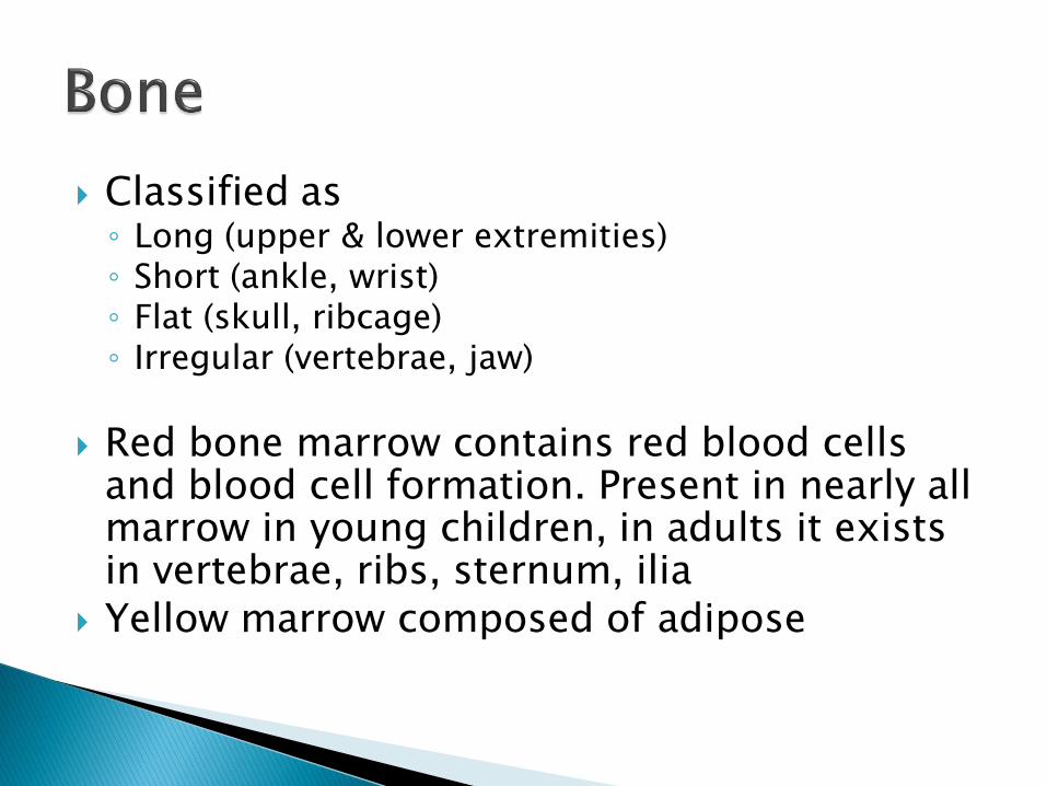
Classified as◦ Long (upper & lower extremities)◦ Short (ankle, wrist)◦ Flat (skull, ribcage)◦ Irregular (vertebrae, jaw)
Red bone marrow contains red blood cells and blood cell formation. Present in nearly all marrow in young children, in adults it exists in vertebrae, ribs, sternum, ilia
Yellow marrow composed of adipose

Long bone anatomy
Diaphysis: shaft◦ Compact bone with
marrow in the medullarycavity
Epiphysis: the ends
Metaphysis: part of the shaft that fans out as it approaches the epiphysis, contains bony trabeculae with cartilage

Osteogenic cells◦ Undifferentiated cells that differentiate into
osteoblasts in normal growth, fractures, injuries
Osteoblasts◦ Bone building cells, occurs in 2 stages:
◦ 1. Ossification: formation of osteoid (collagen and proteins)
◦ 2. Calcification: calcium deposited into osteoid
◦ Alkaline phosphatase: the enzyme that is released by osteoblasts to raise calcium & phosphate
Level is high in fractures and other conditions

Osteocytes◦ Maintain the bone matrix
◦ Lie in lakes of fluid called lacuna and connected with passageways called canaliculi
◦ Arranged in layers called lamellae
Osteoclasts◦ Function in bone resorption
◦ Produced in bone marrow
◦ Have receptors for PTH, calcitonin & other factors

Periosteum: the outer covering of bones, except at articulations◦ Outer fibrous layer
◦ Inner layer of osteogenic cells
Endosteum: the membrane that lines the spaces of spongy bone◦ Osteogenic cells important for bone remodeling

A bone tumor lifting the tibialperiosteum
http://podiatry.files.wordpress.com/2007/03/cbfig_1.jpg

Parathyroid Hormone ◦ Regulates calcium and phosphate levels in blood◦ Secreted by parathyroid glands (2 pairs on the thyroid
gland)◦ When calcium levels fall, negative feedback mechanism
causes release of PTH which increases calcium level and shuts off hormone secretion
◦ Increases serum calcium◦ Releases calcium from bone (resorption)◦ Decreases bone formation◦ Increases intestinal absorption of calcium by activating
Vitamin D ◦ Decreases calcium excretion in kidney

Calcitonin◦ Secreted by parafollicular thyroid cells
◦ Released when serum calcium rises
◦ Inhibits resorption to decrease calcium release from bone
◦ Inhibits osteoclast activity
◦ Increases renal excretion of calcium and phosphate
◦ Probably active in the management of dietary calcium

Vitamin D: obtained from diet (ergocalciferol, vitamin D2) or from skin production when exposed to UV light (cholecalciferol, vitamin D3)
Ergocalciferol is converted into cholecalciferolwhich is processed in the liver into 25-hydroxyvitamin D3 which is transported to the kidneys and converted into 1, 25 dihydroxyvitaminD3 (most potent) and 24, 25 dihydroxyvitamin D3
Adequate sunlight exposure should be sufficient

1, 25 (OH)2D3 works with PTH to regulate calcium and phosphate and regulates bone formation and mineralization◦ Increases intestinal absorption of calcium
◦ Increase in osteoclast number and activity
◦ Increased osteoblast differentiation
◦ Deficiencies lead to rickets in children and osteomalacia in adults (softening of the bones)
24, 25 dihydroxyvitamin D3 increases bone formation

Connective tissue structures
Tendons: muscle to bone◦ Aponeuroses-flat sheets of connective tissue as in
abdominal muscles
◦ Some tendons surrounded by tendon sheaths
Ligaments: bone to bone
Collagen fibers, limited blood supply
Fibrocartilage: the gradual transition of tendons or ligaments onto bone

Two classes of joints: synarthroses and diarthroses
Synarthroses: no joint cavity, very little movement◦ Synostoses: nonmovable with dense connective
tissue (skull)
◦ Synchondroses: bones connected by hyaline cartilage, little movement (ribs & sternum)
◦ Syndesmoses: fibrous disk and joined by ligaments, provide some movement (spine)

Diarthroses: freely movable joints but still with a wide range of motion: sacroiliac joints to shoulders
Surfaces covered by cartilage and held together by a strong fibrous joint capsule ◦ Outer layer is fibrous
◦ Inner layer is the synovium that secretes fluid that is normally clear/pale yellow

Blood supply: vessels enter near the joint capsule and synovial membrane has a rich blood supply (so bleeding into the fluid can occur with injury)
Nerve supply: from the same nerve trunks that supply the muscles that move the joints (reason for referred pain)
Pain fibers present in joint capsule and ligaments, sensitive to stretching and twisting

Bursae: Closed fluid-filled sacs in the synovial membrane that prevent friction on tendons (see Figure 56-7)
Menisci: fibrocartilagenousstructures that develop from an articular disk that lies between articular cartilage surfaces
www.eorthopod.com/images/ContentImages/knee

Chapter 57

MVAs are the #1 killer of adults <45 years
Motorcycle accidents common in young men
Children: falls, bicycle accidents & sports injuries
Falls are most common in adults >65 years◦ 30% in this age group have at least one fall each
year

Can be acute injuries to soft tissues (sprains or strains) or bones (fractures)
Or can be chronic, overuse injuries (stress fractures or tendinitis)
Can be prevented by training, safety equipment, warm-up/cool-down, hydration and proper nutrition

Contusion: (a bruise) direct trauma against a hard object, overlying skin intact
Hematoma: an area of local hemorrhage, infection is a possibility◦ Treat with elevation, cold, possible
aspiration
Laceration: disruption in the continuity of skin, treat with closure◦ Puncture wounds can be
contaminated with tetanus or anaerobic bacteria http://www.more-mtb.org/galleries/Ouchie2.jpg

Usually from overloading or forcible twisting or stretching
Strains: a stretching injury to a muscle or musculotendinous unit ◦ Most common in lumbar & cervical regions
◦ Can be muscle, ligament, fascial injuries
Sprains: a ligamentous injury◦ Pain and swelling subside slower than a strain
◦ Ankle is most common, knee, elbow, wrist
◦ Can cause an avulsion fracture

Avulsion fracture of calcaneous
radpod.org/.../2007/05/calcaneal_avulsion.jpg

Healing: need to regain tensile strength◦ Fibroblasts from the inner tendon sheath or from
connective tissue capillaries produce collagen
◦ Full tensile strength restored in 6-8 weeks
Treatment:◦ Elevation and cold initially
◦ Compression to reduce swelling & provide support
◦ Gradual return to exercise and rehab

Separation of bones with loss of articulation due to disruption of holding ligaments◦ Subluxation is a partial dislocation where there is
still partial contact
Congenital dislocations can occur in hip, knee Traumatic: due to high forces, can be
recurrent Pathologic: can be due to infection,
rheumatoid arthritis, paralysis Can be reduced spontaneously, manually or
surgically

Small pieces of bone or cartilage in a joint space
Can occur from trauma or worn cartilage
Common in knee, hip, ankle, elbow
Can cause joint to catch and lock
Treated with arthroscopy

Anatomy◦ 3 bones: scapula, clavicle, humerus
◦ 3 joints: acromioclavicular, glenohumeral, sternoclavicular
◦ Rotator cuff: supraspinatous, infraspinatous, teresminor and subscapularis
Rotator cuff injuries can be due to acute injury or with overuse. ◦ Tendinitis, bursitis, impingement, frozen shoulder

Injuries can occur to tendons, ligaments, patella or menisci
Often occur during twisting or compression Knee injuries always increase the risk for
osteoarthritis later in life Meniscal tears can be treated conservatively
or with surgery Patellar subluxation or dislocation-
conservative treatment first Chondromalacia- usually on underside of
patella, pain with climbing stairs or sitting

www.wheelessonline.com/image9/i1/patd1.jpg
Patellar Dislocation

Normal (smooth) Chondromalacia
www.emedx.com/emedx/diagnosis_information/

The most common bone lesion
Can be from acute injury, chronic stress or pathologic
Characterized by location, type of fracture

Healing occurs in stages:1. Hematoma formation: first 48-72 hours, initiates
cellular events to start healing
2. Cellular proliferation: periosteum, endosteum and medullary canal. Osteoblasts multiply
3. Callus formation: cartilage forms first, then calcifies. Occurs in 3rd and 4th weeks
4. Ossification: final layers of bone are placed, cast can be removed
5. Remodeling: resorption of the bony callus by osteoclasts

Treated with immobilization◦ Splints
◦ Casts
◦ Traction
◦ External fixation
◦ Internal fixation (plates, wires, screws)
Complications◦ Malunion
◦ Delayed union
◦ Nonunion

Complications:◦ Fracture blisters: usually on ankle, elbow, foot, knee and
caused by separation of epidermis
◦ Compartment syndrome: increased pressure in a limited space because of inelastic fascia. Neurologic symptoms occur, treatment should occur quickly to avoid ischemia. Treated with fasciotomy.
◦ Reflex sympathetic dystrophy: severe pain and autonomic nervous system dysfunction characterized by temperature changes and hyperhydrosis in the area
◦ Fat embolism: long bone fractures or major trauma, fat droplets lodge in lung causing respiratory failure, cerebral dysfunction, petechial rash

www.steadyhealth.com/.../Image/thumb_RSS.gif
Reflex Sympathetic Dystrophy

Acute or chronic bone infection
Hematogenous: most often caused by Staphylococcus aureus◦ Bacteria reaches bone through bloodstream
◦ Usually have chronic infection elsewhere (urinary tract, skin, IV drug users)
◦ Fever, chills, pain,
◦ X-ray findings may be delayed, bone scan will show earlier
◦ Treatment based on cultures and requires IV antibiotics at first, surgery may be required

Contiguous Spread:◦ Infection occurs from an adjacent site like an open wound
(puncture wound, open fracture, diabetic ulcer)◦ Can occur in any bone◦ Recurrent, persistent fever and poor healing◦ Diagnosed through imaging, biopsy◦ Treated with antibiotics and possible surgery
Chronic osteomyelitis: when acute infection persists beyond 6-8 weeks◦ Dead bone separates from living bone◦ May not have fever, chills or abnormal white blood cell
count◦ IV therapy needed for at least 6 weeks, surgery usually
needed
Tuberculosis can cause bone infection

www.gentili.net/.../large/left_foot_-2.jpg

Death of a segment of bone
Due to interruption of blood supply
Causes: trauma, fracture, surgery, sickle cell disease, alcoholism, corticosteroids (higher risk with longer duration and higher doses)
Treatment ranges from rest and anti-inflammatories to joint replacement
Chapter 58

Toeing-in and toeing-out
Bowlegs
Knock-knees
Flatfoot
Can start in utero, usually correct during normal growth

Osteogenesis imperfecta◦ The most common hereditary bone disease
◦ Usually autosomal dominant
Developmental dysplasia of the hip◦ Can cause instability, subluxation, dislocation
◦ Checked on newborn exams
◦ Early diagnosis is important
◦ Treated with harnessing, traction, casting
◦ Multifactorial inheritance

Congential clubfoot◦ Multifactorial inheritance
◦ One or both feet involved
◦ Increased risk with family history and maternal smoking
◦ Treated with manipulations, casting, surgery

Legg-Calvé-Perthes Disease◦ Osteonecrosis of the proximal femoral epiphysis◦ Ages 2-13, mostly boys◦ Pain in groin, hip, thigh or knee or painless limp◦ Treatment ranges from observation to bracing to
surgery
Osgood-Schlatter Disease◦ Microfractures where patellar tendon inserts on
tibial tubercle◦ Pain in front of knee◦ Worse with running, jumping, biking, stair climbing◦ Treat with rest, braces, cold, anti-inflammatories

Legg-Calvé-Perthes Disease
www.wheelessonline.com/images/bennf2.jpg

Osgood-Schlatter Disease
www.zadeh.co.uk/.../osgood-schlatter_1.jpg

Slipped Capital Femoral Epiphysis◦ Most common disorder of the hip in adolescents
◦ Femoral epiphysis unites at 14-16 years of age and slippage can occur before this
◦ Boys affected more than girls
◦ Children often overweight
◦ Knee pain, pain with walking, stiffness
◦ Treated with rest, traction, surgery

orthopedics.seattlechildrens.org/assets

Lateral deviation of the spine that can include rotation or deformity of the vertebrae
More common in girls
Most are minor curves
Postural scoliosis corrects with exercise
Structural scoliosis is fixed and can be◦ Congenital
◦ Neuromuscular
◦ Idiopathic (adolescent is the most common type)
Right curve most common

Less than 10 degrees is normal variant, more than 40 degrees is severe
Can cause shoulder height discrepancy, scapular differences, clothes fitting differently. Pain usually only if severe.
Diagnosed through screening ages 10-16, x-ray, CT, MRI
Early age and larger curves will tend to progress
Conservative treatment with <20 degrees
Bracing for 30-40 degree curves and surgery if more than 40 degrees

www.spine-surgeon.org/Photos/Scoliosis.gif

Osteopenia: reduced bone mass
Osteoporosis: loss of bone with deterioration of bone architecture and increased fragility
Most often due to aging◦ Endocrine disorders of malignancy also causes
Maximal bone mass occurs at age 30
Increase in rate of bone loss after menopause with a women’s lifetime risk of fracture 1 in 3
Risks: female, white, small frame, family history, postmenopausal, smoker, excessive alcohol or caffeine, low calcium intake, sedentary lifestyle

Imbalance in bone formation and resorption◦ Decreased osteoblast activity and increased osteoclast
activity
Estrogen deficiency◦ Testosterone deficiency in men (not as severe)
Secondary causes: ◦ Endocrine (hyperthyroidism, hyperparathyroidism)
◦ Cancer (multiple myeloma increases osteoclasts)
◦ Malabsorption (anorexia, cystic fibrosis)
◦ Alcoholism
◦ Corticosteroids
◦ Prolonged medication use (anti-convulsants, steroids)

Manifested by:◦ Thin outer cortex
◦ Loss of trabeculae
Painless until fracture occurs
Vertebral compression fracture◦ Wedging and collapse of vertebrae lead to kyphosis
and loss of height
Hip fracture
Once a fracture has occurred, risk of a second fracture is much greater

www.isbe.man.ac.uk/~mgr/fracsoln.jpg
www.nlm.nih.gov/.../ency/fullsize/18026.jpg
Osteoporotic Fractures

Diagnosis with bone mineral density (BMD) scan which scans hip and lumbar spine
Prevention is important:◦ Regular weight bearing exercise
◦ Calcium and vitamin D intake
Treatment: both of the above and possibly◦ Estrogen
◦ Calcitonin
◦ Bisphosphonates: most effective, inhibit osteoclastactivity
◦ Prevention of falls

Softening of the bones without loss of bone matrix
Causes: inadequate calcium absorption, reduced vitamin D action ◦ Can occur in renal failure due to inability of the kidney
to activate vitamin D
Symptoms: bone pain, fractures, muscle weakness
Diagnosed through labs, x-rays Treated with correcting the underlying cause and
adequate calcium & vitamin D Rickets (children): dietary (non-fortified milks)
and inadequate sun exposure, can be

Progressive disorder with excessive bone destruction and structural changes of long bones, spine, pelvis and skull
The second most common bone disorder Mid-adulthood at onset with increased risk
with increasing age Cause unknown (?viral) Increased osteoclast activity with rapid bone
resorption and irregular bone formation resulting in thick coarse bone with rough and pitted outer surface

Can be mild or severe Many people may be asymptomatic Skull: headaches, tinnitus, hearing loss Spine: kyphosis Bowing of tibia and femur Pathologic fractures (femur, spine, pelvis) Cardiovascular disease is the most common
cause of death in those with advanced disease. Caused by increased blood flow to affected tissues causing high-output cardiac failure
Osteogenic sarcomas occur in 5-10% of severe cases (femur, pelvis, humerus, tibia)

Diagnosed on x-ray and through labs and sometimes bone biopsy (if there is a concern for malignancy)
Treatment: ◦ Reduce pain
◦ Suppress with calcitonin, bisphosphonates (most effective)
◦ Adequate calcium and vitamin D

myweb.lsbu.ac.uk/.../456-842-1641250.jpg
uwmsk.org/static/residentprojects/paget8511.jpg
Paget Disease of the Bone

Characteristics of intracellular fibers, cartilage and bone
Bone cells and their purposes
Hormonal control of bone formation
Know table 56-2 (Actions of PTH, Calcitoninand Vitamin D
Types of joints, blood and nerve supplies
What are bursae and menisci?
Define different types of soft tissue injuries
Difference between strains and sprains (sites, complication of sprains)
Causes of dislocations
Common knee injuries
Common shoulder injuries
Stages of fracture healing, complications of fractures
Types and causes of osteomyelitis and risks for osteomyelitis
Corticosteroids can cause osteonecrosis

Name hereditary skeletal disorders
Know causes, associated risk factor and symptoms of juvenile disorders
Define scoliosis◦ Idiopathic most common and more in girls
◦ When is it treated
Osteoporosis-causes and risks, location of fractures
Define osteomalacia
Paget- symptoms, cellular changes, bone changes, sites, cardiovascular changes, sarcomas