Embed Size (px)
Citation preview

Type Simple branched tubularSite Numerous and closely packed
Shape Numerous & longArrangement Straight, parallel & perpendicular to the surface
Parts Duct pit – ¼ of coriumIsthmus – between duct & neckNeck – upper partBody – main partBottom – lower part
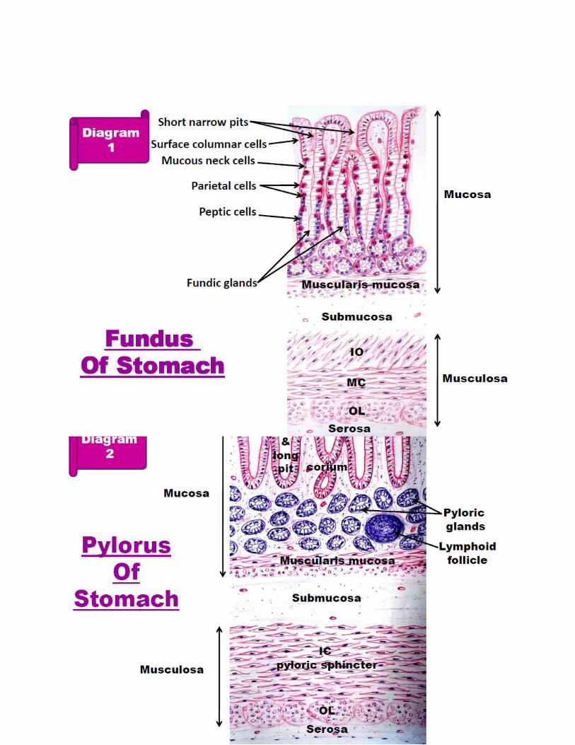
Histology of stomachFundic gasric glands
Cell lining the fundic glands
Mucous Gastric Barrier:1. A thick film of mucous which protects the stomach against enzymes & HCl.2. Mucous is secreted by the surface epithelium and the mucous neck cells
Pylorus of the stomach
Surface mucous secreting cells
a. Site: cover the surface & line the duct (pit)Function: secrete a protective film of mucous w’ protect stomach against enzyme and HCl
Mucous neck cells b. Site: neck of glandsc. Function: secrete acidic mucous, may be a transient stage in development of peptic cells from
stem cellPeptic cells d. Site: in body and bottom
e. Function: secretes pepsinogen, renin enzymes and lipaseParietal cell f. Sites: mainly in neck and body , few in isthmus and rare in bottom
g. Position: on basal membrane but not reach the lumen this is why its name is parietal cell
Entero-endocrine cells. h. Definition: Modified cells which secrete hormones. Members of the neuroendocrine (APUD) system. fundic glands have 1 type; Enterochromaffin (E.C.) cells.
i. Function: Some secrete serotonin. Others secrete endorphin. Secretion passes from capillaries of corium to blood stream.
Undifferentiated columnar cells
a. Site: In the isthmus of the glands.b. Function: renewal of cells (every 2-6 days) of fundic glands
Caveolated cells a. Rare.b. Large.c. Columnar.d. Long microvillie. Tubular invaginations (caveolae).

Characteristics of pyloric glands: 1. Deeper gastric pits. (1/2 the mucosa)2. Shorter glands. (1/2 the mucosa)3. Wider lumina.4. Widely separated.5. branched & coiled.so, cut transversely & obliquely6. secretory parts lined by mucous cells. 7. Some parietal cells at pyloric sphincter.8. Small number of entero-endocrine cells (3 types)
a. EC cells: (seretonin and endorphin)b. G cells: (gastrin)c. D cells: (somatostatin)
Differences between fundus & pylorus
Fundus PylorusMucosa Thick
More foldedThinLess folded
Duct Short (1/4 corium)Narrow
Long (1/2 corium)Wide
Gland Simple branched tubular Coiled, more branchedNumber More numerous Less numerousLength Long (3/4 corium) Short (1/2 corium)
Arrangement ParallelPerpendicular to surfaceStraightCut in one plane
Not parallelNot perpendicular to surfaceCoiledCut in various plane
Gland cells All types No peptic & oxyntic cellsEntero-endocrine
cellsOnly EC cell 3 types
(EC, G & D cells)Corium Some lymphocytic infiltration
No lymph nodulesMore lymphocytic infiltrationLymph nodules are present
Musculosa Thin (3 layers)1. Inner oblique2. Middle circular3. Outer longitudinal
Thick (2 layers)1. Inner circular2. Outer longitudinal
Histology of pancreas
General features General structures
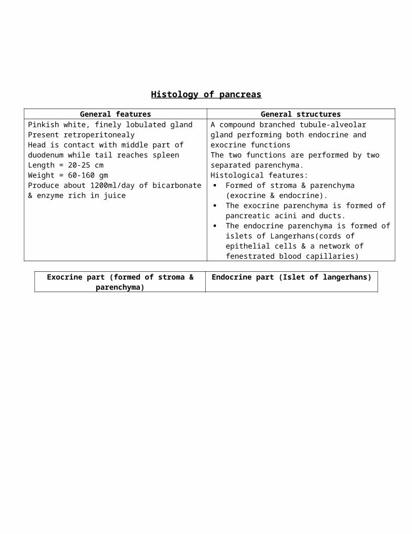
Pinkish white, finely lobulated glandPresent retroperitonealyHead is contact with middle part of duodenum while tail reaches spleenLength = 20-25 cmWeight = 60-160 gmProduce about 1200ml/day of bicarbonate & enzyme rich in juice
A compound branched tubule-alveolar gland performing both endocrine and exocrine functionsThe two functions are performed by two separated parenchyma.Histological features: Formed of stroma & parenchyma (exocrine &
endocrine). The exocrine parenchyma is formed of pancreatic acini
and ducts. The endocrine parenchyma is formed of islets of
Langerhans(cords of epithelial cells & a network of fenestrated blood capillaries)
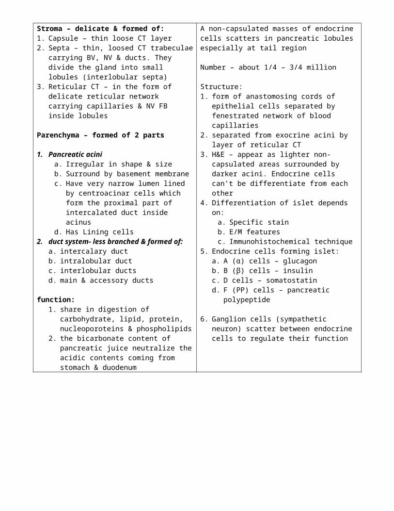
Exocrine part (formed of stroma & parenchyma) Endocrine part (Islet of langerhans)Stroma – delicate & formed of:1. Capsule – thin loose CT layer2. Septa – thin, loosed CT trabeculae carrying BV, NV
& ducts. They divide the gland into small lobules (interlobular septa)
3. Reticular CT – in the form of delicate reticular network carrying capillaries & NV FB inside lobules
Parenchyma – formed of 2 parts
1. Pancreatic acinia. Irregular in shape & sizeb. Surround by basement membranec. Have very narrow lumen lined by
centroacinar cells which form the proximal part of intercalated duct inside acinus
d. Has Lining cells2. duct system- less branched & formed of:
a. intercalary ductb. intralobular ductc. interlobular ductsd. main & accessory ducts
function:1. share in digestion of carbohydrate, lipid,
protein, nucleoporoteins & phospholipids2. the bicarbonate content of pancreatic juice
neutralize the acidic contents coming from stomach & duodenum
A non-capsulated masses of endocrine cells scatters in pancreatic lobules especially at tail region
Number – about 1/4 – 3/4 million
Structure:1. form of anastomosing cords of epithelial cells
separated by fenestrated network of blood capillaries
2. separated from exocrine acini by layer of reticular CT
3. H&E – appear as lighter non-capsulated areas surrounded by darker acini. Endocrine cells can’t be differentiate from each other
4. Differentiation of islet depends on:a. Specific stainb. E/M featuresc. Immunohistochemical technique
5. Endocrine cells forming islet:a. A (α) cells – glucagonb. B (β) cells – insulinc. D cells – somatostatind. F (PP) cells – pancreatic polypeptide
6. Ganglion cells (sympathetic neuron) scatter between endocrine cells to regulate their function
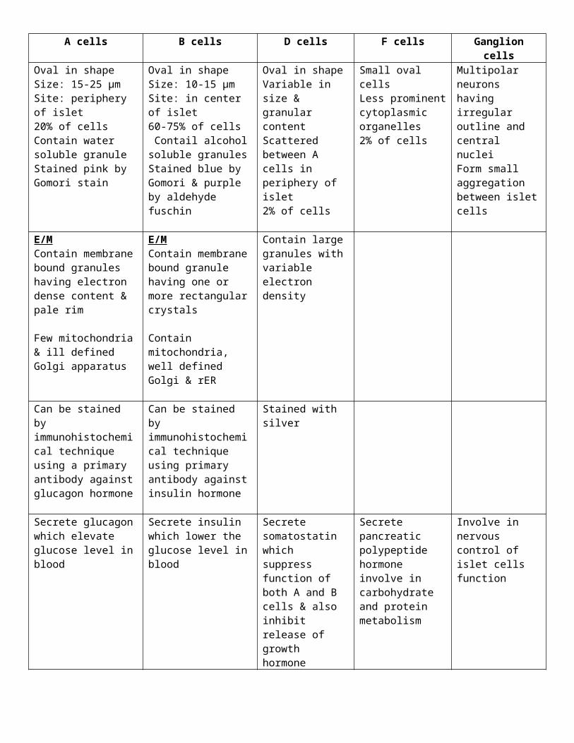
A cells B cells D cells F cells Ganglion cellsOval in shapeSize: 15-25 µmSite: periphery of islet20% of cellsContain water soluble granuleStained pink by Gomori stain
Oval in shapeSize: 10-15 µmSite: in center of islet60-75% of cells Contail alcohol soluble granulesStained blue by Gomori & purple by aldehyde fuschin
Oval in shapeVariable in size & granular contentScattered between A cells in periphery of islet2% of cells
Small oval cellsLess prominent cytoplasmic organelles2% of cells
Multipolar neurons having irregular outline and central nucleiForm small aggregation between islet cells
E/MContain membrane bound granules having electron dense content & pale rim
Few mitochondria & ill defined Golgi apparatus
E/MContain membrane bound granule having one or more rectangular crystals
Contain mitochondria, well defined Golgi & rER
Contain large granules with variable electron density
Can be stained by immunohistochemical technique using a primary antibody against glucagon hormone
Can be stained by immunohistochemical technique using primary antibody against insulin hormone
Stained with silver
Secrete glucagon which elevate glucose level in blood
Secrete insulin which lower the glucose level in blood
Secrete somatostatin which suppress function of both A and B cells & also inhibit release of growth hormone
Secrete pancreatic polypeptide hormone involve in carbohydrate and protein metabolism
Involve in nervous control of islet cells function

Histology of small intestine
Epithelium of mucosa: covers intestinal villi & line the intestinal crypts
Intestinal villi Intestinal crypts = crypt of Lieberkuhn1. Finger like projection extending from wall into lumen of
intestine2. Length = 0.5-1.5 mm3. Structures: Each villous is formed of core of CT derived from
lamina propria covered the epithelium;a. Villous epithelium: covered by 3 types of cells
Simple columnar absorbing cells (90%) Goblet cells (9.5%) Entero-endocrine cells (0.5%)
b. Villous core Formed of CT containing BV, NV & lymphatic Villi contain large lymph vessels called
CENTRAL LACTAELS4. Shape:
a. Duodenum – broad, leaf like shapeb. Jejunum – tongue shapec. Ileum – long, slender but short or absent over peyer’s
patches
1. Simple tubular glands extending from base of villi to muscularis mucosa
2. Length: 100-200 nm3. Cells lining the epithelium:
a. Stem cellsb. Paneth cellsc. Columnar absorbing cellsd. Goblet cellse. Entero-endocrine cellsf. Caveolate cellsg. M- cells
Lining epithelium of intestinal crypts
Stem cells Paneth cells Cavolate cells M cellsSite Base of crypt between
paneth cellsBase of crypt of small intestine (absent in large intestine)
Rare cells Membrane like cells = Microfold cells
L/M Shape: short columnar cell
Nucleus: oval & basal Cytoplasm: deep
basophilic
Shape: columnar or triangular cells with narrow apex
Nucleus: basal, rounded & pale
Cytoplasm: basal basophilia
Apical acidophilia
Large columnar cell Dome shape cells with basal cavity
E/M Rich in rER & polyribosome
Rich in rER, & well developed Golgi
Apical part show large electron dense of zymogen granules
Free border shows large microvilli & deep tubular invagination
Short microvilli Basement membrane
is discontinuous to facilitate transport between M cell & lamina propria
Function Renewal of the cells Secrete lysozyme enzyme which has antibacterial effect
Act as receptors Transport of intraluminal antigen

Villious epithelium covering cells
Simple columnar absorbing cells Goblet cells Entero-endocrine cellsSite Cover villi & upper part of intestinal
cryptscover intestinal villi & upper part of intestinal crypts
Cover intestinal villi and line the intestinal cryptsIt is a part of APUD cell which secrete intestinal hormones
L/M Shape: tall columnar cells Nuclei: basal, oval nuclei Cytoplasm: basophilic Free border: acidophilic striated
Shape: goblet like cells Apical part: expand and filled with mucin
granules Basal part: thin constricted & contain
nucleus and cell organelles Nucleus: flat, basal & deeply stained H&E: apical part of goblet cells appear,
vacuolated or foamy due to dissolved mucin granules
Oligomucous cells: Small less differentiated cells Contain few mucous globules Accumulation of more mucous globules
leads to expansion of apical part & become mature goblet cells
Shape: columnar cell with narrow apex
Nucleus: rounded, basal & open face
Cytoplasm: basal granules (stain with argentaphin granules)
E/M Brush border = striated border Microvilli are about 3000
microvilli/cell Microvilli are covered with thick
cell coat, very rich in alkaline phosphate enzyme needed for absorption
Cytoplasm Contain numerous rER,
mitochondria, prominent Golgi apparatus & few ribosomes
Basal part: show electron dense granules which are variable in size and shape
Apical part: long microvilli which act as chemoreceptor
Cytoplasm: numerous rER, well developed Golgi apparatus
Function 1. Absorption of useful substances2. Secretion of lactase, sucrose &
isomaltase enzymes
1. Mucous secreted lubricate passage of intestinal content
2. Secrete acid glycoprotein which prevent bacterial invasion
1. D-cells: somatostatin2. S-cells: secretin3. E-G cells: glucagon like substance4. E-cells: endorphin5. CCK cells: choly-cystokinine6. M-cells: motilin7. N- cells: neurotensin

Histology of large intestine
Colon

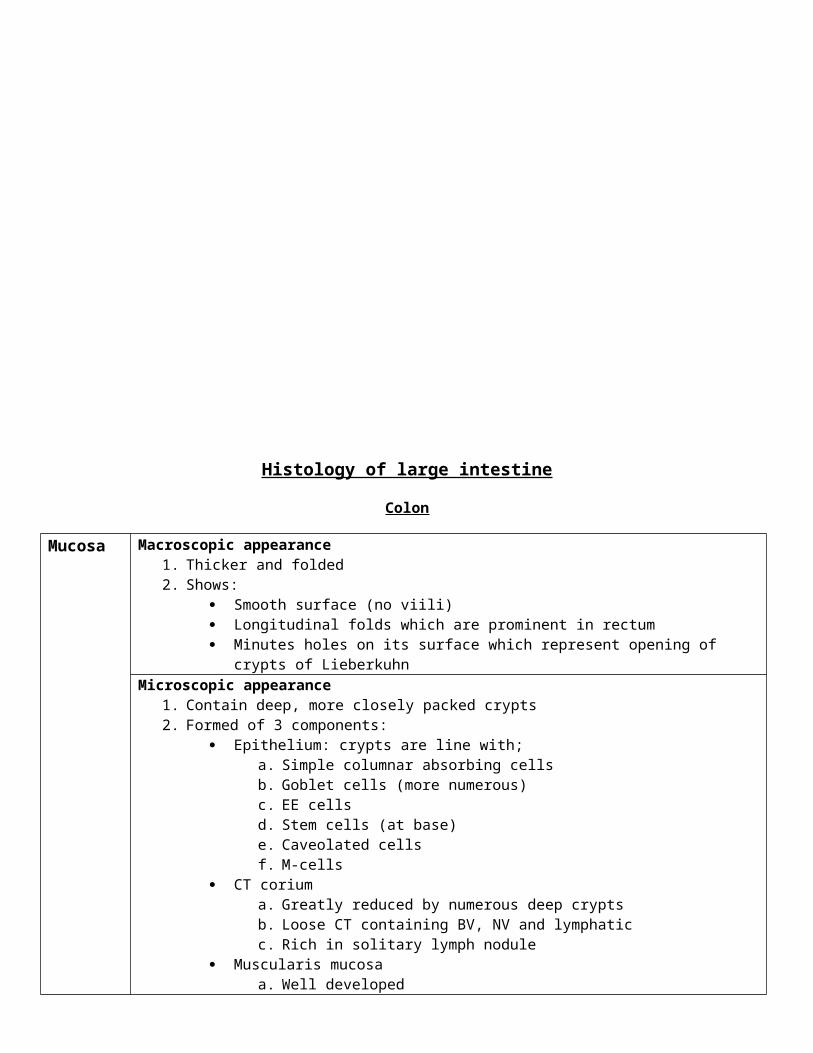
Mucosa Macroscopic appearance1. Thicker and folded2. Shows:
Smooth surface (no viili) Longitudinal folds which are prominent in rectum Minutes holes on its surface which represent opening of crypts of Lieberkuhn
Microscopic appearance1. Contain deep, more closely packed crypts2. Formed of 3 components:
Epithelium: crypts are line with;a. Simple columnar absorbing cellsb. Goblet cells (more numerous)c. EE cellsd. Stem cells (at base)e. Caveolated cellsf. M-cells
CT coriuma. Greatly reduced by numerous deep cryptsb. Loose CT containing BV, NV and lymphaticc. Rich in solitary lymph nodule
Muscularis mucosaa. Well developedb. Formed of 2 layers of SMF:
i. Inner circularii. Outer longitudinal
Submucosa
1. Formed of loose CT containing BV, lymphatic and NV2. No gland but lymphocytic infiltration was found due to abundant bacteria in large intestine
Musculosa 1. Formed of 2 layers of SMF Inner circular layer Outer longitudinal
a. Not continuousb. Breaks up into 3 bands (Taenia coli)
Serosa 1. Formed of loose CT containing BV & NV2. Covered with simple squamous mesothelial cells with flat nuclei3. Adipose CT accumulates under peritoneum in pedunculated masses which hang out from serosa
into peritoneal cavity and called appendices epiploicae
Rectum
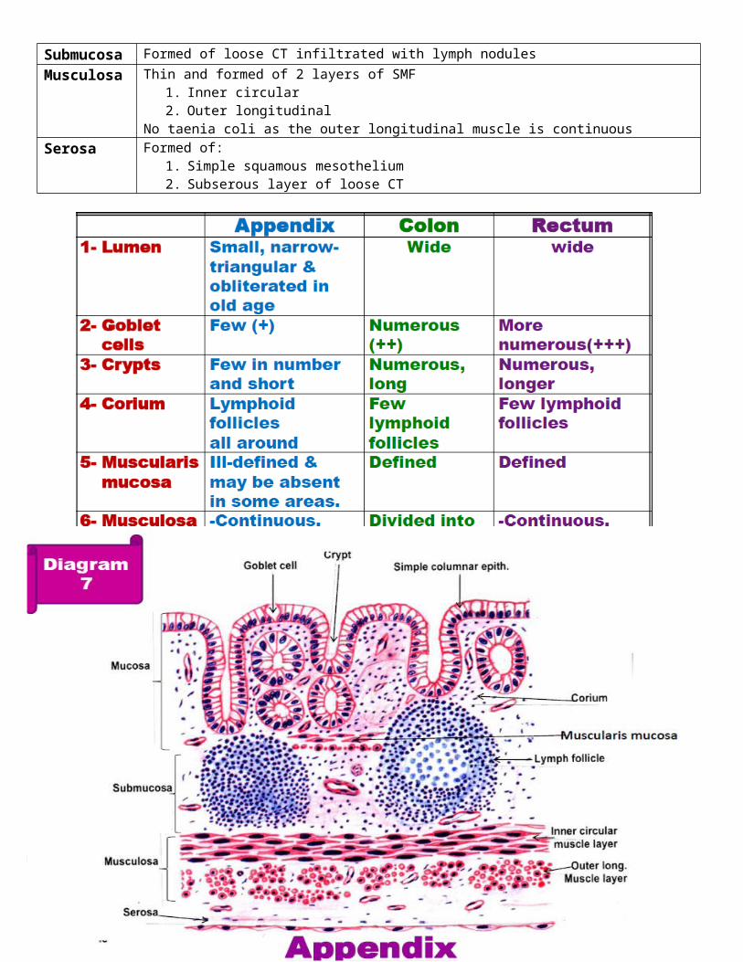
The rectum is similar to large intestine, but it is characterized by
Numerous goblet cells Longer crypts Numerous solitary lymphoid follicles A complete uninterrupted longit muscle layer (taenia coli) Adventitia partially replaces serosa
Appendix
Macroscopic appearance1. It has lumen, which is small and narrow in cross section
2. May be obliterated in old age3. Its wall is thickened by extensive lymphocytic infiltration
Microscopic appearanceMucosa It shows crypts only but no villi
Crypts are fewer in number, narrow, shorter.1. Epithelium:
a. Stem cellsb. Absorptive columnar cellsc. Goblet cells (fewer)d. EE cellse. M-cellsf. Caveolated cells
2. Corium: Packed with lymph follicles and diffuse lymphoid tissue forming circular layer3. Muscularis mucosa: ill defined and may be absent in some areas
Submucosa Formed of loose CT infiltrated with lymph nodulesMusculosa Thin and formed of 2 layers of SMF
1. Inner circular2. Outer longitudinal
No taenia coli as the outer longitudinal muscle is continuousSerosa Formed of:
1. Simple squamous mesothelium2. Subserous layer of loose CT

Histology of thyroid glands
Structures
1. Stromaa. Covered by 2 capsules
Outer fascial sheath which is a part of pretracheal fascia Inner delicate CT capsule which is frimly adherent to gland
b. Fine fibrous septa extend from capsule and divide the gland into incomplete lobulesc. Reticular fibers
2. Parenchymaa. Consist of thyroid follicles & interfollicular cells
Thyroid follicles
1. They are the structural and functional unit of gland2. Number: 30 million 3. In section, follicles have variable diameter (0.02 – 0.9 mm)4. May be rounded or oval in shape5. Lined with low cuboidal epithelium6. Follicles contain colloid (gelatinous substance) in their lumen7. Colloid:
a. Homogenous acidophilic material formed of thyroglobulin T3 & T4)b. Stain intensely with PAS and is eosinophilic with H&E
8. Morphologic appearance of follicles varies according to region of gland and its functional activity9. Cells lining follicles:
Follicular cells Parafollicular cells Majority (98%) LM: cubical secretory cells with basophilic
cytoplasm & central rounded nucleus EM:
i. Supra-nuclear Golgi apparatus, well developed rER, lysomsome, mitochondria & fine droplet of colloid
ii. Free border reveal short microvilli projecting in the lumen
iii. Cells are adherent to each other by tight junctional complexes
Function: synthesis & release thyroid hormone
Other name: C cells/ light cells/ clear cells Developed from 5th pharyngeal pouch Minority (2%) Larger and paler Rounded & oval in shape Do not abut n the lumen of follicle Enclosed between follicular cells and
basement membrane surrounding follicles EM: small rER, long mitochondria, abundant
spherical secretory granules Function: secrete calcitonin (lowers blood
calcium level by inhibiting bone resorption
Inter-follicular cells
1. Masses of cells present in between follicles2. Represent tangentially cut follicles3. Consist of follicular cells and para – follicular cells

Histology of liver
Hepatic stromaCapsule of Glisson Interlobular septa Portal canal/tract Reticular fiber
Formed of CT Thick at porta hepatis
giving of CT septa dividing liver into lobes & lobules
CT partitions dividing liver into lobules
Thin & incomplete in human
Thick & complete in pig
Triangular masses of CT present at some angles between hepatic lobules
Carries 4 structuresi. Branch of hepatic artery
ii. Branch of portal veiniii. Bile ductsiv. Lymphatic
Network of RF between parenchyma of hepatic lobules
Extend from periphery towards centre where it condensed around central vein
Hepatic parenchymaClassical hepatic lobule Portal lobule Liver acini
Mass of liver cells which drain its blood into central vein
Shape: hexagonal Structure:
Stroma Parenchyma1. Interlobular CT
septa2. Reticular fiber3. Portal canal
1. Hepatocytes2. Bile canaliculi3. Blood sinusoids &
perisinusoidal space of Disse
Mass of liver cells which drain their secretion into bile duct
Triangular in shape
Diamond mass from 2 adjacent classic hepatic lobules
Surround a central vascular core
Subdivided into 3 zone
Cells lining hepatic blood sinusoids
Endothelial cells Kupffer cellsStructures: Simple squamous cells which form a non-
continuous wall to blood sinusoid Peripheral portions of cells have fenestrations
of variable size & shape, arranged in groups called sieve plates & not covered with diaphragm
Function: Allow free passage of chylomicrons & plasma
from blood sinusoid to space of Disse
Site: on surface of endothelial cells with their processes extending into lumen & between underlying endothelial cells
L/M: large stellate cell. Could be stained by vitally with trypan blue
Nucleus: irregular with prominent nucleolus Cytoplasm:
i. Clear vacuoles & phagosomesii. Inclusion; lipochrome pigment, erythrocytes &
iron containing pigment Function:
i. Most important phagocytic cells of reticulo-endothelial system
ii. Secrete proteins related to immunologic process

Space of Disse
Structures & contents Functions: Long microvilli of hepatocyte project into space of
Disse Plasma Network of reticular fiber Adipocyte (Ito cells): fat storing cells Pit cells:
i. Found in liver of rodentsii. Small cells with short pseudopodia, but
they are not phagocytic Occasional un-myelinated nerve fibre
Facilitates exchange of metabolites between blood plasma and cells
Prevent collapse of blood sinusoid. Due to:i. Microvilli
ii. Adipocytesiii. Reticular fiberiv. Hydrostatic pressure in sinusoid is equal in
space of Dissev. Microfilament & microtubules
Lipocytes or adipoctyes
Duct system
Bile canaliculi Minute canals within hepatic plates between adjacent rows of cells No special lining, bounded by cell membranes of hepatocytes E/M:
Junctional complexes hold cell membranes of hepatocytes around the lumen Small microvilli projecting from hepatocytes Pass within the liver plates towards the periphery of the lobule where they open into canals of
Herin and preductules
Canal of Hering Short canals at the periphery of hepatic lobule Lined partially with hepatocytes and partially with cubical cells Collect bile from bile canaliculi Drain bile into bile ductules
Preductules Small canals at the periphery of hepatic lobule Lined with simple cubical epithelium Collct bile from bile canaliculi Drain bile into bile ductules
Bile ductules Small canals in portal canals Lined with simple cubical epithelium Collect bile from preductules & canal of Hering Drain into bile ducts
Bile ducts Collections of bile ductules Present in portal tracts Lined with simple cubical epithelium Drain into larger intrahepatic ducts
Intrahepatic ducts
Collection of bile ducts Lined by simple columnar epithelium
Extra hepatic ducts
Lined with simple columnar epithelium Surrounded by CT and smooth muscle fibers
Common bile duct
Epithelium: simple columnar Corium: contains mucus tubule – alveolar glands Smooth muscle: present in the wall

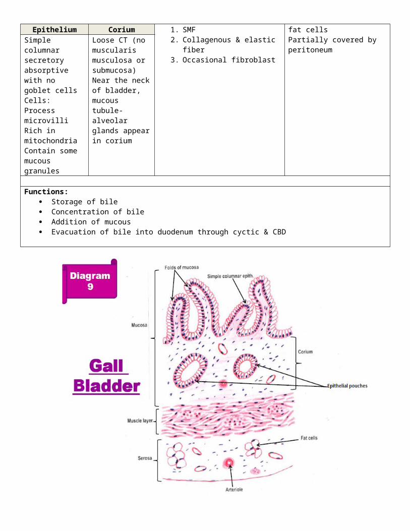
Histology of gall bladder
Structures:Mucosa Muscle coat Perimuscular coatHighly foldedConsist of:
ThinConsist of:
1. SMF2. Collagenous & elastic fiber3. Occasional fibroblast
Thick fibrous CT layerContain BV, NV, L & fat cellsPartially covered by peritoneumEpithelium Corium
Simple columnar secretory absorptive with no goblet cellsCells:Process microvilliRich in mitochondriaContain some mucous granules
Loose CT (no muscularis musculosa or submucosa)Near the neck of bladder, mucous tubule-alveolar glands appear in corium
Functions: Storage of bile Concentration of bile Addition of mucous Evacuation of bile into duodenum through cyctic & CBD

Histology of kidney
Renal CorpusleMost active part of nephron, Diameter = 150-250 µm
Bowman’s capsule Glomerulus1. A double walled cup shape capsule2. Has 2 poles3. Has 2 layersPodocytes:
a. Large flatten modified epithelial cells with oval nuclei
b. Separated from basement membrane (BM) of glomerular capillary by subpodocytic space
c. Major process extend from cell bodyd. Minor process arise from deep surface of cell body
& major process to reach capillary BM and terminate on it by feet like structures
e. Function: involve in synthesis of glomerular BM
1. A tuft of capillaries formed about 50 tortous capillary loops arising from afferent arteriole at vascular pole (non-filtered blood) & drain into efferent arteriole (filtered blood)
Mesangial cells:a. Present between glomerular blood capillariesb. Stellate in shape, with small dark nucleus,
basophilic cytoplasm rich in lysosomesc. Functions:
Responsible for continuous turnover of basement membrane by addition of new B.M. material to outside and removal of old one from inside by phagocytosis.
Cleaning the basal lamina by phagocytosis of particulate material that accumulates during the filtration process.
May be considered as continuation of the juxta-glomerular cells & secrete certain hormone
Supportive function, as they secrete collagen, chondroitin sulphate and fibronectin, which provide additional support to glomerular basement membrane.
Juxtaglomerular complexA secretory complex structure present at vascular pole of renal corpuscle
Function: regulation of blood pressure & secretion erythropoietinMacula densa Juxtaglomerular cells (JG cells) Lacis cells (polar cushion)A special type of cells that line a part of D.C.T. which fits between afferent arteriole (A.A.) & efferent arteriole (E.A.).
Some changes occur in these cells: The cells increase in number and
become crowded. The cells become columnar. The nuclei are deeply stainedand
close together The basement membrane of these
cells is lost. Golgi apparatus is present between
the nucleus and base of the cell.
Modified smooth muscle cells secretory in function.
Present in the media of afferent arteriole.
They are large with rounded nuclei.
The cytoplasm contains secretory granules (reninhormone) stained with PAS.
The internal elastic lamina of A.A. is lost, so J.G. cells are in close contact with the blood in the lumen of A. A.
J. G. cells also lie in close contact with cells of macula densa which have lost their B.M.
Groups of small cubical cells, with pale nuclei.
Present between A.A., E.A. & macula densa.
They may be supporting mesangial cells.

PCT DCTCharacteristics 1. About 15 mm long
2. More convulated3. Large diameter = 60 μm4. Narrow lumen
1. About 5 mm2. Less convoluted3. Small diameter = 30-50 μm4. Wide lumen
Lining cells:1. Number2. Shape3. Nucleus4. Side border5. Luminal border6. Base of cell7. Cytoplasm
1. 3-5 cells2. Cubical shape3. Basal & rounded4. Not clear5. Show brush appearance due to microvilli6. Basal mitochondrial striation7. Deeply acidophilic & granular
1. 5-8 cells2. Less cubical3. Central & rounded4. Clear5. Microvilli are few & short6. Basal mitochondrial striation7. Pale acidophilic & non-granular
Function 1. Reabsorption of water & sodium2. Reabsorption of glucose, amino acids,
plasma protein3. Excretion of some metabolites
1. Reabsorption of more water & sodium
Loop of HenleCharacteristics 1. U shaped tube which connects PCT with DCT
2. 2 types: Nephron with long loops – they are juxtra-medullary nephron (near cortex) Nephron with short loops (cortical nephron (in cortex proper)
Histological structures
Formed of 2 limbs:1. Descending limb
Thick part lined by simple cubical cells Thick part lined by simple sq. epithelium
2. Ascending limb Thin part lined by simple sq. epithelium Thick part lined by simple cubical cells
Function 1. Excretion of NaCl into surround tissue fluid of medullary pyramids rendering it hypertonic2. This hypertonic medium allows passage of water from the collecting tubules to outside.3. The urine in collecting tubules becomes hypertonic.
Collecting tubulesCharacteristics 1. Present in medullary rays.
2. Each collecting tubule in the cortex drains about5-10 nephrons.3. In the medulla about 6-8 collecting tubules open into a large duct called duct of Bellini which
opens at the summit of medullary pyramid.4. Collecting tubule is lined with cubical cells with clear pale basophilic cytoplasm.5. Duct of Bellini is lined with columnar cells with clear basophilic cytoplasm.
Function 1. Conduction of urine.2. Reabsorption of water from the urine due to the surrounding hypertonic tissue fluid under the
effect of A.D.H.

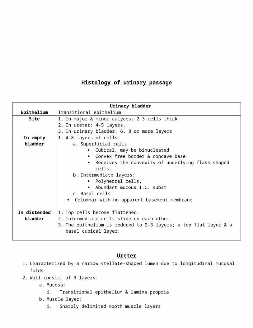
Histology of urinary passage
Urinary bladderEpithelium Transitional epithelium
Site 1. In major & minor calyces: 2-3 cells thick2. In ureter: 4-5 layers.3. In urinary bladder: 6, 8 or more layers
In empty bladder 1. 4-8 layers of cells:a. Superficial cells
Cubical, may be binucleated Convex free border & concave base. Receives the convexity of underlying flask-shaped cells.
b. Intermediate layers: Polyhedral cells, Abundant mucous I.C. subst
c. Basal cells: Columnar with no apparent basement membrane
In distended bladder
1. Top cells become flattened.2. Intermediate cells slide on each other.3. The epithelium is reduced to 2-3 layers; a top flat layer & a basal cubical layer.
Ureter1. Characterized by a narrow stellate-shaped lumen due to longitudinal mucosal folds2. Wall consist of 3 layers:
a. Mucosa:i. Transitional epithelium & lamina propria
b. Muscle layer:i. Sharply delimited mooth muscle layers
ii. Inner longitudinal & outer circulariii. In lower 1/3 – third outer longitudinal layer
c. Adventitia:i. Fibro-elastic tissue with BV & NV
ii. Merges with surrounding tissue

Male urethraThe male urethra is long (20cm) twisted tube, that conducts urine from the urinary bladder (& seminal fluid from male genitalia) to outside of the bodyGlands open in the
course of the urethra
1. Prostatic gland.2. Glands of Littré.3. Bulbo-urethral gland (Cowper’s Gland)
Parts 1. Prostatica. 3-4 cm long.b. Arise from neck of the bladder and passes through the prostate.c. V- shaped in cross section.d. Both ejaculatory duct & prostatic duct open into it.e. Epithelium: It is lined with:
i. Transitional epithelium.ii. Pseudostratified columnar ® distally.
f. Surrounded by:i. I.L. layer of smooth muscle fibers.
ii. O.C. layer of smooth muscle fibersiii. Condenses to form the Internal Sphincter of U.B.
2. Membranousa. Very short (1.5 cm).b. Lies within urogenital diaphragm.c. Extends from apex of prostate to root of penis.d. Lined with stratified columnar epithelium.e. Surrounded by striated muscle which forms the external Sphincter of UBf. Cowper’s glands are present outside its wall & open in penile urethra.
3. Penilea. The longest part (15 cm).b. Passes through corpus spongiosum.c. Its proximal part is dilated - the bulb of urethra.d. Its distal part is dilated to form - “fossa navicularis”.e. Lined with:
i. Proximally: Stratif. Colum. Epith.ii. Distally: Stratif. Squam. Epith.
f. The lamina propria of the urethra contains:i. Highly vascular C.T. rich in elastic fibers.
ii. I.L. & O.C. of smooth ms.fibsiii. Mucus secreting glands of Littrè (Intramucosal & extramucosal)
Female urethraLength: 2-6 cmOpening: in vestibule anterior to vaginal opening
Epithelium Lined with transitional epithelium at neck of bladder, changes into pesudostriatified columnar, then striatified squamous at its termination
Corium CT containing:1. I.L & O.C smooth muscle fibre2. Small mucus secreting glands which open into the lumen3. At external orifice, striated muscle form external sphincter
The connective tissue of the corium blends with that of vagina

Prostate1. Type: Compound tubulo-alveolar gland.
2. Site: Surrounds the V-shaped first part of male urethra.3. Stroma
Capsule:a. Thick fibroelastic tissueb. Rich in SMF & BV
Trabeculae:a. Thick CT septab. Extend from capsule to
urethrac. Divide the gland into lobulesd. Rich in SMF which condense
around urethra to form internal urethral sphincter
Reticular fiber:a. A network of reticular fibersb. Support the parenchyma
4. Parenchymaa. The ejaculatory duct divides the gland into 3 lobes.b. Each lobe is subdivided into lobules.c. Each lobule contains 3 types of acini which are: Mucosal acini, Submucosal acini & Main acinid. The acini are irregular in outline & variable in size.e. Lining epithelium of prostatic acini: pseudostratified columnar, columnar or cubical depending on gland activity.
Mucosal acini: The smallest type Lie in periurethral tissue Open by small ducts in
prostatic urethra. In old age, they enlarge ®
urinary obstruction.
Submucosal acini: Lie outer to mucosal acini Larger in size than mucosal
acini. Open into the urethral sinus
by 2 separate ducts.
Main acini: Lie at the periphery of the
lobule. The largest acini. Their ducts open into the
posterior margin of urethra.
(prostatic adenoma BPH)


![Minor Works Agreement - spb.sa.gov.au C Minor... · Web viewMINOR WORKS AGREEMENT FOR [#1 INSERT THE NAME OF CONSTRUCTION PROJECT] ... a word in the singular includes the plural and](https://img.dokumen.tips/doc/110x75/5ad9089b7f8b9a86378b65c4/minor-works-agreement-spbsagovau-c-minorweb-viewminor-works-agreement-for.jpg)

