Embed Size (px)
Citation preview

ORTHOPEDICS
- Fracture:o Discontinuity of bone (cortex)
- When discussing fracture, we often mention:o Location (which bone, which 1/3rd +/- which part of bone [i.e.
epiphysis, diaphysis, metaphysis or the growth plate/physis])o Integrity of skin overlying (open fracture or closed fracture)o Fracture pattern (transverse, oblique, butterfly, segmental, spiral,
comminuted, intra-articular, avulsion, compression/impacted, torus, greenstick, pathologic, stress)
o Alignment (displaced, distracted, angulated [varus = away; valgus = towards]
- Types of fractures: o Open fractures:
Bone has protruded through the soft tissue and caused a break in the skin
Concern = infection Orthopedic emergency Grading (Gustilo-Anderson classification):
I – IIIC Depends on size of laceration, tissue loss/devitalization
and major vascular injury that requires repair (3C) Initial management involves:
ABCs; ATLS (primary and secondary survey) Pain control (morphine, fentanyl) IV prophylactic antibiotics (e.g. cefazolin +/-
gentamicin if high grade) Tetanus coverage (Td vaccine and TIG) Lavage wound with sterile irrigation and sterile
dressing Important investigations (X-Ray, trauma labs, ECG,
CXR, consent) Surgical debridement OPEN REDUCTION and fixation (upper limb usually
internal)
o Complete fracture: the fracture line goes across the whole width of bone (vs. incomplete, where the fracture line does not completely pass through; such as in greenstick fracture)
o Transverse fracture: fracture line perpendicular to long axis of bone (i.e. line across)
o Oblique fracture: fracture line is not straight
COMPONENTS OF CLINICAL MEDICINE:1. DIAGNOSIS
a. Hxb. P/Ec. Investigations: lab and imaging
2. MANAGEMENT (conservative, medical, surgical)
3. OUTCOME (F/U, complications, prognosis, screening)

o Spiral fracture: rotational force to bone results in a complex, S-shaped fracture around whole bone; in pediatric patients, suspicious of child abuse
o Segmental fracture: 2 complete fractures resulting in a segment in between
o Comminuted fracture: >2 fracture fragments (tiny pieces, most likely from a gun shot)
o Butterfly fracture: two fracture lines that unite, resulting a triangular segment in between; it is a result of a BENDING force
o Intra-articular fracture: difficult to get to, may need to do open surgery
o Avulsion fracture: forceful pull on the insertion of tendon on bone results in breaking a piece of it
o Pathological fracture: Fracture that occurs in weak bones as a result of a
pathological disease MC = osteoporosis; other causes: metastatic bone disease,
primary bone cancers, multiple myeloma, osteomalacia, rickets, scurvy, osteogenesis imperfecta, bone infections
Must Rx underlying condition!
o STRESS fracture: Fracture that occurs in NORMAL bones as a result of
REPETITIVE stress with inadequate healing time in between Typical in young adults in military training (march fracture),
ballerinas, sport players, hard repeated physical activities Pain is usually on activity Sites: metatarsals, calcaneus, tibia The microfractures that develop may not be visible in the
first week or two on XRAY (actually best seen on bone scan, but very unnecessary)
Rx: temporary limitation of weight bearing or toning down physical activity
o Pediatric age group: Because of the collagenous/cartilaginous nature of immature
bone, they develop certain types of fractures Make sure to note the growing plate; fractures at the plate
risks growth arrest If everything is in order, pediatric patients are expected to
have full recovery sometimes only by closed reduction (unless inapplicable)
Torus fracture: “buckle” fracture of one cortex (impaction injury of childhood; the lateral ends of fracture appears bulging

Greenstick fracture: incomplete and angulated fracture of long bones; the fracture line can be transverse but the ends are still in continuity (think of a twig, which you can snap but it will still not separate)
Pipe fracture
o ALIGNMENT: Displaced = not in anatomic alignment (typically, distal
fragment in relation to proximal fragment) Distracted = fracture fragments are separated by a gap Impacted = fracture fragments are compressed into each other Angulated = direction of fracture apex is varus or valgus
Varus = distal segment is going towards midline (genu varus = bowleg)
Valgus = distal segment is going away from midline (genu valgum = knock-knees)
- Approach to fractures:o Clinical assessment:
ABCs (ATLS protocol of primary and secondary survey) AMPLE history (Allergies, Medications, PMH, Last meal,
Events surrounding injury): Obtain mechanism of injury and any related
conditions that occurred to this area before P/E:
Look (deformity, note whether open or closed fracture) Feel (maximal tenderness, pulses distal to injury,
sensation) Move (avoid ROM or moving injured area, but maybe
ask if they move fingers/toes, etc.)o Analgesia (strongest ones!)o Imaging (X-ray):
Rule of 2s: Two views (at least) AP and lateral (in other cases,
swimmer’s view, etc.) Two joints (one above and below) Two sides Two radiologists’ opinions Two times (one before and another after reduction)
o Management: Reduction
Closed reduction (IV sedation, apply traction, reverse the mechanism)
Open reduction (indications = NO CAST: nonunion, open fracture, compromised blood flow/neurovascular

tissue, articular surface malalignment [intraarticular fractures], salter-Harris type 3, 4, 5 fractures (see later), trauma pt who needs early ambulation)
AFTER: Assess NV status and do a post-reduction x-ray Fixation
Internal fixation (screws, plates, pinning, nails, rods) External fixation (splints, casts, traction or external
fixation devices) Follow up in 1-2 weeks to evaluate bone healing (do a X-Ray)
For stress fractures, scaphoid fracture this is also the time to diagnose it radiologically
Rehabilitation
- Salter-Harris fracture types:o Fractures around the physis in pediatric patients are important
because if it does involve the physis, it might lead to growth arrest o Type I: transverse right through the physis o Type II: transverse involving the physis which then diverts away
from physis (to the metaphysis)o Type III: transverse goes through the physis and then into the
epiphysis (cuts off a piece of bone)o Type IV: fracture goes across physis from metaphysis to epiphysiso Type V: axial force crushes physeal plate (compression/impaction
fracture)o Ways to remember it:
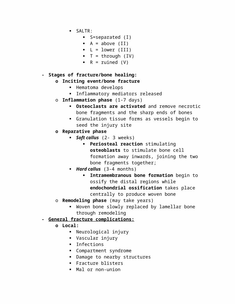
SALTR: S=separated (I) A = above (II) L = lower (III) T = through (IV) R = ruined (V)
- Stages of fracture/bone healing:o Inciting event/bone fracture
Hematoma develops Inflammatory mediators released
o Inflammation phase (1-7 days) Osteoclasts are activated and remove necrotic bone
fragments and the sharp ends of bones Granulation tissue forms as vessels begin to seed the injury site
o Reparative phase Soft callus (2- 3 weeks)
Periosteal reaction stimulating osteoblasts to stimulate bone cell formation away inwards, joining the two bone fragments together;

Hard callus (3-4 months) Intramembranous bone formation begin to ossify the
distal regions while endochondrial ossification takes place centrally to produce woven bone
o Remodeling phase (may take years) Woven bone slowly replaced by lamellar bone through
remodeling- General fracture complications:
o Local: Neurological injury Vascular injury Infections Compartment syndrome Damage to nearby structures Fracture blisters Mal or non-union AVN (e.g. scaphoid, femoral neck) Osteomyelitis Post-traumatic OA
o Systemic: Sepsis, DVT, PE, ARDS (fat embolism), …
- Major orthopedic emergencies: o Open fractures (explained above)o Vascular injuries (“hard signs”)o Compartment syndromeo Neural injuries (particularly of the spine)o Bone infections (osteomyelitis and septic arthritis)o Hip dislocation and fracture (risk of avascular necrosis)o Exsanguinating pelvic fracture (note: open pelvic fractures have a
50% MR!)
- Indications for open reduction (“NO CAST”):o Non-uniono Open fractureso Compromised blood supplyo Articular surface malalignment o Salter-Harris grade III, IV and V fractureso Trauma pt. who needs early ambulation
- Compartment syndrome: o Increased pressure within a extremity compartment that
compromises the circulation and function of the tissue (along with the neurovasculature) in the closed space (and distal to it)
o Always suspect in fractures or damage to an extremity, which all contain compartments
The complications are drastic (irreversible tissue death may occur within hours – may lose the limb!)
The affected limb may appear to be doing well early on There may be no obvious signs until it is too late
o Causes: Fractures (tibial shaft, supracondylar, forearm fractures) Crush injuries
Can cause rhabdomyolysis (hyperK [arrhythmias], hyperPO4 [hypocalcemia], AKI and acidosis, +ve urine blood dipstick but no RBCs on microscopy)
Can occur secondary to compressive forces or due to electrical burns/electrocution
IV hydration, alkalization of urine (bicarbonate), and RRT (hemodialysis)
Burn injuries (especially if circumferential) Trauma Vascular injuries (post-thrombectomy/embolectomy) Drug overdose with prolonged limb compression Iatrogenic (tight cast, poor positioning during surgery)
o Clinical presentation: 5Ps (of which, PAIN is the HALLMARK finding)
Pain (deep, poorly localized, out of proportion to injury or findings, not responding to Rx, increased with passive stretch of muscle)
Parasthesia (of the distribution of nerve compressed in that compartment)
Pallor (distal hypocirculation) Paralysis (later, when ischemia established) Pulselessness ( pulses may be present) These signs occur LATE!
Compartment may be tense on palpationo Diagnosis:
CLINICAL DIAGNOSIS (high risk patient with pain out of proportion to injury)
Pressure monitors if clinical exam unreliable (unconscious patient, children):
>30 mm Hg ~ compartment syndrome likely, needs intervention
TRICK:If an inpatient develops what appears to be a positive Homan’s sign and has leg pain, but there is no edema, don’t think DVT, think of compartment syndrome!
o Management: As for trauma (ABCs) Elevate limb to level of heart Fasciotomy within 4 hours (decompress ALL the
compartments of the affected limb: NOT just the suspected affected one)
In 48 -72 hours – wound closure, necrotic tissue debridemento Complications:
Myonecrosis volkmann’s contracture and deformities, loss of sensation, need for amputation
Rhabdomyolysis AKI, hyperkalemia, hypocalcemia
BONE INFECTIONS:
- Acute osteomyelitis:o Infection of bone, particularly at the metaphysis of long bones (this
is where blood flow is sluggish and bacteria can have time to seed bone tissue)
o Medical emergency (requires early Dx and Rx)o Routes of infection: hematogenous and direct (e.g. open fractures)o Risk factors:
Sepsis Recent trauma or surgery Immunocompromised DM IVDU TB +ve
o Causative organisms: S. aureus (MCC) Streptococci Sickle cell disease pt: MC is still S. aureus, but specific to them
is salmonella Pseudomonas (especially if immunocompromised) Polymicrobial (diabetic foot ulcer)
o Locations: It can occur anywhere Tibia and femur (MC site) Vertebral bodies (TB osteomyelitis = Pott disease)
o Presentation: Symptoms: fever and pain (over affected area) Signs: erythema, swelling and tenderness over affected
area (in chronic cases, there is a draining sinus) with reduced ROM
o DDx:

Septic arthritis and other arthritides Skin infections (e.g. cellulitis) Bone tumors (Ewing’s sarcoma in pediatric case, especially
since they tend to have fever)
o Investigations: CBC (leukocytosis) ESR, CRP (elevated) Blood culture Needle aspiration of infected bone (Most important)
Gram stain, C&S Imaging:
BEST = MRI, 2nd best = Bone scan Easiest and quickest = US X-Ray may NOT show signs until 10 days or more
(first sign is soft tissue swelling; periosteal elevation; lytic bone lesion)
Others (e.g. blood glucose levels in DM)
o Medical Management: Analgesia Empirical IV antibiotics (start right after getting specimen)
4-6 weeks (initial improvement should be noted) Anti-staph penicillin (cloxacillin) or vancomycin AND
3rd generation cephalosporin or aminoglycoside (e.g. vancomycin/cloxacillin + gentamicin)
Changed appropriately when C&S results availableo Surgical management:
No initial response to antibiotic therapy warrants this Surgical debridement of necrotic bone Open drainage for Brodie’s abscess (on x-ray: lytic lesion
surrounded by sclerotic bone)o Complications:
Sepsis (if not the primary cause) Bone destruction, pathological fractures and spread to
adjacent structures and joints (septic arthritis) Chronic osteomyelitis:
Sequestrum (necrotic bone) is walled off by periosteal reactive bone formation (involcrum)
This can lead to a cloaca that can drain into the skin resulting in a SINUS tract (chronic sinus tract presence increases the risk of carcinoma of the tract epithelium)
Decreased response to Abx (cannot penetrate) Cause is usually polymicrobial
Imaging often shows irregular patchy areas of radiolucency with radiopaque sequestrum within
Management is surgical: extensive debridement of all necrotic tissue, bone reconstruction, antibiotic beads are inserted into wound
- Acute infectious arthritis (“Septic Arthritis”)o Emergency condition that warrants immediate investigation and
treatment Any arthritis (especially monoarticular) is septic arthritis
until proven otherwise (you must do arthrocentesis)o Routes of spread: hematogenous or direct (and contiguous)o Causes:
Bacterial: S. aureus (MCC) N. gonorrheae (sexually active young adults; can be
migratory oligo or polyarthritis) Pseudomonas Salmonella spp
Viral: Parvovirus B19, HBV and HCV, Rubella Typically a self-limiting polyarthritis
o Risk factors: Sepsis DM and immunocompromised gonorrheae Prosthetic joints IVDU Joint damage (e.g. RA, trauma)
o Clinical features: Arthritis symptoms: joint pain, fever, limited activity on joint Arthritis signs: joint swelling, erythema, tenderness, reduced
ROM Patient may appear toxic Typically MONOARTICULAR (unless gonococcal, which can be
polyarticular and migratory) Joints affected: knee > hip >elbow
o Investigations: CBC (leukocytosis, high PMN) ESR, CRP elevated Blood culture (in the case of gonoccocemia, it may be –ve, so
swab mucosal surfaces for culture) Joint aspiration (Arthrocentesis):
3 Cs: count, culture and crystals Gram stain and culture

Cell count (WBC count, typically >50,000, PMN >90%) Crystals (in older adults, r/o gout and pseudogout) Glucose can be measured (typically low)
Imaging (if difficult to aspirate, for chronic or for complications)
X-ray not great early on (soft tissue swelling, joint space widening)
Late radiological findings include reduced joint space
o Management: IV empirical antibiotics:
4-6 weeks, change according to culture; if initial gram stain shows no organisms, still give!
Typically: anti-staph penicillins or vancomycin If suspecting gonococcal cause: ceftriaxone If immunocompromised: add anti-pseudomonal
antibiotics CRP can be used to monitor response
Therapeutic arthrocentesis (serially) and joint drainage may be necessary
UPPER LIMB:
Anterior dislocation of the shoulder (MC type)- Younger patients, playing sports or acute trauma
o More joint laxity- Increased recurrence risk (weaker joint ligaments); complications include
rotator cuff tear- S and S:
o Paino Shoulder appears unusual, asymmetrical, box-shaped; loss of
contour of shoulder, humeral head will be prominento Typical position: UL held abducted and slightly externally rotated
(“stuck” in the position in which blow resulted in anterior dislocation)o Decreased shoulder ROM
- Physical examination: o Look, feel, move, special testso Neurovascular and muscle action assessment necessary:
Check for radial and brachial pulses Check for axillary nerve (sensation over deltoid area
“regimental patch” and abduction of arm by deltoid muscle) Movements beyond the deformity (passive and active
movements) as well as distal sensation
- How to proceed?
YOU CAN’T ALWAYS GET WHAT YOU WANTRemember that the glenohumeral joint has given up stability for mobility while the hip joint has given up mobility for stability, making the shoulder joint more liable to dislocation.

o Shoulder X-ray (must be rules of 2: AP and lateral view also axillary view, joint above and below, two opinions)
May see hill-sachs deformity (compression fracture of posterolateral humeral head)
- Management:o Closed reduction with IV sedation and muscle relaxation
(Stimson’s longitudinal traction downwards by weight and will spontaneously reduce in 15 minutes OR manually by hennipen or various other techniques where you lift arm to 90 degrees and then externally rotate and adduct until reduced)
Post reduction x-rays and NVS assessment o Sling immobilization for 3 weeks (Allow for proper healing of
ligaments to prevent recurrences)o Recurrence rate is high (<20 years old ~ 65-95%)
Posterior dislocation of shoulder joint (1-5%)- Causes (discoordinated muscle contractions):
o Seizures (grand-mal/tonic clonic)o Electrical burns/shock
- Presentation:o UL typically held internally rotated and adducted
- Imaging:o Shoulder XR, must be 2 views, because if one view it can appear
normal- Management:
o Closed reduction, typically by longitudinal traction (stimson technique)
Inferior dislocation (luxatio erecta) – very rare- Result of hyperabduction (which causes detachment of rotator cuff)- Patient will be in severe pain, with arm held up high (180 degrees) and
appears shorter; humeral head may be felt along lateral chest wall- Closed reduction (complicated technique to do it) +/- surgical repair of
rotator cuff muscles
Frozen shoulder (Adhesive capsulitis) - Progressive pain and stiffness of shoulder that resolves spontaneously
after 18 months- Mechanism:
o Primary adhesive capsulitis Idiopathic; associated with DM Resolves spontaneously
o Secondary adhesive capsulitis Due to prolonged immobilization or following trauma
- Clinically:
o Painful phase (6-9 months): gradual onset of diffuse paino Stiff phase (4 – 9 months): decreased ROM that impacts functioningo Thawing (melting) phase (5- 26 months): gradual return of motion
- Investigations: X-ray usually normal- Management:
o Freezing phase: physiotherapy, NSAIDs, steroid injectiono Thawing phase: early physio +/- arthroscopy for debridement
HUMERAL FRACTURE:- Common locations:
o Surgical neck fractureo Midshaft fractureo Supracondylar fractureo Medial epicondyle fracture
- For all fractures:o Diagnosis is by X-RAY
MUST HAVE AT LEAST TWO DIFFERENT VIEWS, INCLUDING NEARBY JOINTS, TWO OPINIONS
o Comment on: Site of fracture (upper 1/3rd, middle 1/3rd, distal 1/3rd) Closed (simple) or open fracture (open fracture is an
orthopedic emergency) Type of fracture (transverse, oblique, spiral, segmental,
comminuted, greenstick, stress fracture, pathological fracture, torus fracture, avulsion fracture, impacted fracture, longitudinal fracture)
Alignment (displacement, rotation or angulation – all relative to the proximal segment unless otherwise stated)
- Surgical neck fracture:o Typically in adults (young adults and elderly)o Cause:
Direct traumao Presentation:
Pain, swelling, decreased ROM, ecchymoses over the upper arm and chest (related to damage to surrounding vasculature)
o Assessment: Axillary nerve injury (just like anterior dislocation)
Deltoid (arm abduction) and regimental patch sensation
Posterior humeral circumflex artery injury bleeding Radial pulse
o Dx: Best initial step = upper arm XR (again, AP, L, Y)
o Management:
Depends of fracture severity and degree of displacement Closed reduction if necessary, splinting or ORIF
- Mid-shaft fracture: o Fracture of diaphysis of the humerus o Usual cause is direct traumao S&S: pain, swelling, decreased ROMo Dx: best initial diagnostic step is upper arm XR
When you see a spiral fracture type in peds case, with parent saying he fell down or something sign of child abuse
o Assessment: Injury to RADIAL nerve running in spiral groove
Wrist drop, loss of digital extension, loss of sensation of dorsum of hand (especially laterally)
Note that radial nerve also palsied in axilla [higher up] (crutch palsy, Saturday night palsy) resulting in above + triceps action weak; or lower down (proximal radial head injury – only affects digital extension and sensation?)
Injury to deep brachial arteryo Management:
80 – 90% closed reduction and splinting Complicated cases (e.g. communicated: open reduction and
internal fixation
- Supracondylar fracture of the humerus:o Typically in pediatric cases o Fall with hyperextended arm at elbow; FOOSH o Arm held close to them, painful to toucho Neurovascular exam is usually abnormal:
Brachial artery (check peripheral pulses: ulnar and radial) Comment on pulse, perfusion status (warm, pink, or
pale and cold), capillary refill Median nerve injury Look for signs and symptoms of compartment syndrome
(swelling and 5Ps)o Investigations:
Best initial = XR If distal pulses lost (hard sign) surgically explore
o Management: If not displaced: long arm cast for 4 -6 weeks If vascular compromise or displaced (in adults):
Open reduction and internal fixation (may resolve blocked blood flow, if not, vascular surgery necessary)
Casting

Must follow up in 1 week with XR to confirm fracture is still in good position
o Complications: Median nerve palsy Tear or entrapment of brachial artery or compression Compartment syndrome (need emergency fasciotomy) Volkmann contracture (2nd to reduced perfusion or
circulation which can cause necrosis of flexor muscles, resulting in fibrosis permanent flexion contracture of hand and wrist; it occur 2nd to compartment syndrome)
- Medial epicondyle fracture:o Avulsion fracture of medial epicondyle o Less dramatic presentation, but it is the location of origin of anterior
forearm flexor compartment muscles o Causes: FOOSH, pitchers (who throw things overhanded)o Pain on medial elbow (point tenderness over medial epicondyle)o Injury to ulnar nerve possible:
Abduction and adduction of fingers, ulnar deviation of wrist Sensation over medial 1.5 digits and medial palm of the hand
o Management: Depends of fracture severity and degree of displacement Closed reduction and splinting or ORIF
FOREARM FRACTURES:
- Monteggia’s fracture:o Proximal 1/3rd ULNAR fracture AND dislocated radial heado Causes: FOOSHo Presentation:
Pain and swelling at the elbow Decreased ROM at elbow Radial head may be palpable
o Neurovascular structures involved: Radian nerve (and related posterior interosseous n.)
o Best initial diagnostic step = XR (as always)o Management:
Pediatrics ~ closed reduction and casting Adults ~ open reduction and internal fixation Open fracture (all age) ~ ORIF
- Nursemaid’s elbow (slipped radial head):o Exclusively pediatric condition (lax annular ligament)o Subluxation or dislocation of the head of the radiuso Cause:

Pulling forearm of a kid (e.g. pulling arm when crossing road or sidewalk); children have lax annular ligament
o Presentation: Pain and tenderness over elbow without significant history
of trauma Patient resists supination, arm will be guarded
o Diagnosis is typically clinical o Management:
Closed reduction (supinate then flex) If it doesn’t work, then proceed to imaging to R/O fracture
- Galeazzi’s fracture:o Often compared to monteggia’s fracture, but here:
Distal 1/3rd fracture of RADIUS AND disruption of the distal RADIOULNAR joint
o Cause: FOOSHo Presentation:
Pain, swelling and deformity at wrist jointo Diagnosis: best initial step = XRo Management:
Same as monteggia’s fracture: Peds ~ closed reduction and casting Adults ~ ORIF Open fracture ~ ORIF
- Colles’ fracture:o Fracture of distal radius (within 1 inch of DRUJ) with displacement
of the DISTAL segment DORSALLY – “dinner fork” appearanceo Cause: FOOSH (with wrist extended)o Presentation:
Pain, swelling, tenderness over the wrist Dinner fork deformity
o Best initial step: XR (2 views at least)o Management:
Depends on degree of dislocation (closed reduction and splinting ORIF)
- Smith’s fracture:o Often compared to Colles’ (“reverse” colles fracture)o Distal radial fracture with displacement of the DISTAL segment
VENTRALLYo Cause:
Fall on FLEXED wrist (rare)
o Presentation, imaging and management is the same as Colles
- Carpal Tunnel Syndromeo Carpal bones:
Proximal 4 (L->M): scaphoid, lunate, triquetral, pisiform Distal 4: trapezium, trapezoid, capitate, hamate SO LONG TO PINKY, HERE COMES THE THUMB
o Carpal tunnel: Flexor retinaculum (roof) Walls and floor: carpal bones Contents: median nerve (before entering, gives off palmar
cutaneous branch)o Etiology:
Increased intracarpal pressure Very common disorder; multifactorial F>M; middle aged patients Repetitive wrist activity (no causative factor found, only
correlation) – secretaries, office workers Conditions related to it:
Obesity Pregnancy Rheumatoid arthritis Acromegaly hypothyroidism DM Amyloidosis, MM, sarcoidosis
o Presentation: Numbness, paresthesia and pain over the median nerve
distribution (lateral 3.5 fingers, thenar eminence may be atrophied/wasting late in disease; weakness of gripping motion) – symptoms often worse at nighttime
Special tests: Compression test; Tinel’s sign at the wrist; Phalen test – all reproduce the symptoms
o DIAGNOSIS IS CLINICAL Investigations are done if very late in disease (e.g NCS and
EMG) or suspecting an etiology Do EMG before surgical steps are taken
o Management: Initial Rx: wrist splinting with the wrist in the neutral position
(or partially hyperextended), particularly at night – 1 month

If persistent, continue splinting for 1 – 2 months, but add symptomatic Rx like local steroid injection (if this fails, oral steroids may be used); NSAIDS are NOT recommended
Surgical Rx if all else fails (decompression surgery: median nerve release; endoscopically or open)
HAND
- Scaphoid fracture:o Most common carpal bone fractured (most common carpal bone
displaced is lunate)o Mechanism:
FOOSH (typically fractures through waist) Problem: proximal bone blood supply is received from distal
vessel (AVN of proximal fragment can occur)o Clinical features:
Exquisite tenderness in the anatomical snuffbox Pain with resisted pronation
o Investigations: X-ray: AP, lateral, scaphoid views with wrist extension and
ulnar deviation Fracture may not be radiologically evident until 2
weeks If patient has wrist pain + anatomical snuffbox
tenderness and negative x-ray Rx as scaphoid fracture until 2 weeks
o Treatment: If not displaced long-arm thumb spica cast for 4 weeks,
then short arm cast If displaced operative management (ORIF)
o Complications: MC = non-union/malunion AVN of proximal fragment Delayed union
- DeQuervain’s tenosynovitis:o Overuse injury/inflammation of the extensor pollicis tendon sheatho Typical picture: new mother (carries baby a lot)o Others: RAo Presentation:
Aching pain along surface of 1st metacarpal (lateral border of thumb)
Aggravated by turning motions Snapping/catching when moving thumb

Special test: finkelstein’s test (thumb in closed fist and ulnar deviation, reproduces pain)
o Diagnosis = CLINICAL o Management: Steroid injection
- Trigger finger:o Inflammatory nodule develops in the flexor tendon that may lodge in
the tendon sheath in flexion (can’t extend)o F>M; RD associatedo Presentation:
Sudden locking of the finger when trying to extend finger from flexed position, typically at night or after inactivity
Nodule maybe palpable over affected finger(s)o Diagnosis is CLINICALo Management:
Best initial Rx is steroid injection towards metacarpal head on the affected finger(s)
If this fails, surgery involving OP procedure of cutting overlying sheath
- Jersey Finger (can’t flex it):o Damage to flexor tendon as a result of rapid hyperextensiono Tackle sports; person putting flexed finger in shirt collar and person
moves fast, injures the holder’s flexor tendon o Presentation:
Pain with flexion of finger, resistance of flexiono Diagnosis is CLINICALo Rx:
Best initial management is SPLINTING of affected finger to allow tendon to heal
- Mallet finger (can’t extend it):o Damage to the EXTENSOR tendon as a result of force in flexiono Typical scenario is from flexed finger being hit by a thrown ball
(e.g. basket ball)o Presentation:
The affected joint (usually DIP) is held in a flexed position (looks like a mallet) despite other fingers all extended (inability to extend at the affected joint)
Pain and point tenderness at injury siteo Diagnosis is CLINICALo Management:

Best initial management is splinting of the affected finger
- Felon:o Abscess of the finger pado History:
50% have history of cut in distal finger or splinter, etc. May develop 2nd to paronychia
o Presentation: Severe, throbbing pain, edema, erythema of the affected finger
o Diagnosis: Diagnosis is clinical Imaging may be necessary for surgical needs
o Rx: Emergency incision and draining (NOW!)
MCC is S. aureus May lose the fingertip
Oral antibiotics
- Dupuytren’s contracture:o Chronic, fibrosing disorder (of palmar fascia) resulting in hand
contraction at resting positiono Typically in older men, Scandinavian origino May occur 2nd to liver failure o Presentation:
Can’t lay hand flat on table (fixed flexion deformity) May see pits and feel nodules in palm
o Diagnosis is clinicalo Management is surgery
- Other fractures:o Bennett’s fracture
Fracture-dislocation of the base of the first METACARPAL (thumb) with disruption of the carpometacarpal joint
o Boxer’s fracture Fracture of the metacarpal NECK CLASSICALLY of the 5th metacarpal (little finger)
o Game keeper’s thumb (Skier’s fracture) FOOSH Damage to thumb ulnar collateral ligament
LOWER LIMB
- Pelvic fracture:o Causes:

Ground level fall (MCC) Older and/or osteoporotic patient may develop
significant pelvic fracture with ground level falls High energy trauma (RTA), high level fall and any fall in the
elderly significant fracture that may be a major surgical emergency
o Mortality rate can range from 5 – 50% depending on comorbidities and complications such as:
Advanced age Open fracture Additional injuries (e.g. other fractures or soft tissue damage)
MCC of death related to pelvic fracture is HEMORRHAGE
Comorbid medical conditionso Types:
A: stable avulsion fracture (e.g. iliac wing fracture) B: open book (PS and sacrum; rotationally unstable) C: unstable vertical fracture (rotationally and vertically
unstable; typically a combination of fractures)o Presentation:
Pain, inability to bear weight, limb-length discrepancy Limb is held in external rotation
o Best initial step in management is ABCs (Trauma/ATLS protocols) Why? Well it’s a trauma case, also, pelvic fractures can damage
large vessels resulting in hemorrhage and hypovolemic shock So 2 large IV bore needles should be placed and fluids started
o Secondary survey: physical examination General inspection (e.g. short limb, externally rotated,
bruising, lacerations, vascular injury [ecchymoses, hematoma, swelling])
Manual palpation (gently to prevent further damage) Neurological examination (sciatic nerve distribution: check
for foot drop (L5), perianal sensation (S1-S4), saddle anesthesia (S3-S5))
Urogenital examination: PR for prostate (ballotable prostate) and rectal tone, bimanual examination in women
Urethral transection may occur and this is a CI for foley’s catheter, and would require confirmation using retrograde urethrogram (RUG)
Open fractures can be seen in vagina!o Investigations:
Initial trauma labs and investigations CBC (Hb is focus), BT and Cx Electrolyte panel, BUN and Cr, LFTs, lipase and amylase,
coagulation profile

Urinalysis (if feasible) Imaging (after FAST): CXR, AXR, pelvic XR
Best initial step in diagnosing pelvic fracture = imaging Traditionally pelvic x-ray CT scan is better in significant injuries to see extent of
local damage Look at pubic symphysis, sacroiliac joints, sacral wing
fractures, ischial fracture
o Management: ABCs (always first) Pelvic binder/sheeting Definitive Rx = ORIF (better than external fixation) +/- laparotomy if FAST/DPL positive DVT prophylaxis is very important Analgesia Hemodynamic monitoring (Hct)
o Complications: Vascular injury (venous > arterial) Genitourinary injury Neurologic injury Postop complications more likely (UTI, wound infection, DVT)
HIP - Femoral fractures:
o Femoral neck fracture (also head)o Intertrochanteric fractureo Femoral shaft fracture
- Femoral neck fracture:o Etiology:
Ground level fall in elderly, osteoporotic patient High velocity injury (e.g. RTA)
o A big deal because they don’t heal well (intracapsular, synovial fluid doesn’t help with callus formation) and because they may disrupt the blood supply to the femoral head (resulting in AVN of femoral head)
o Presentation: Pain, if displaced, can be super severe External rotation and limb shortening Inability to bear weight
o Investigations: Best initial diagnostic test is XR of entire femur
Disruption of Shenton’s line Altered neck-shaft angle

Multiple views: AP, cross-table lateral view CT may be obtained for purposes of surgery Garden’s classification of femoral neck fractures:
Type I: incomplete fracture Type II: complete fracture but not displaced Type III: complete fracture, partially displaced Type IV: complete and fully displaced
Other lab workup and investigation for surgery CBC Cx+BT, BUN&Cr, electrolytes, blood glucose,
coagulation profile, LFTs, cardio assessment o Management:
Depends on type and age Generally:
I and II only require internal fixation to prevent displacement
III, IV: ORIF in the young , and total hip arthroplasty in the elderly (and the extreme ages: hemi)
Note: total arthroplasty = femoral head + acetabulum replaced; hemi = only femoral head
DVT prophylaxis (high risk!!) – LMWH (SQ) Complications:
AVN of femoral head (disruption of the medial femoral circumflex arteries around the neck of the femur, that provides blood supply medially to the head of femur) – causes include: femoral neck fracture, chronic systemic steroid use, slipped capital femoral epiphysis, legg-calve-perthes disease, RA, SCD, SLE
Non-union Dislocation of prosthesis DVT
- Intertrochanteric fractureo EXTRACAPSULAR femur fractureo Causes: same as femoral neck fractureo Difference: less likely to cause AVN!o Presentation: same as femoral neck fractureo Investigations: sameo Management:
ORIF with pinning using dynamic hip screw or IM nail (no need for total hip replacement unlike many cases of fracture of neck of femur!)
Prognosis related to how quickly a safe surgery can be performed and often lower if done within 48 hours
So PLEASE:

Femoral neck fracture, depends on age and type, but in elderly with type III and IV, total hip arthroplasty may be needed; in intertrochanteric fracture, ORIF with dynamic hip screw is feasible
- Femoral shaft fractureo Diaphysis fracture only occurs if there is REALLY high velocity or
force injury – the force required to break the femur here should definitely be enough to cause injury elsewhere – so LOOK FOR OTHER INJURIES!
o Cause: RTA (or in elderly or osteoporotic pt, it might be mild trauma) Pathological fracture (lytic bone lesion; mets)
o Presentation: Severe thigh pain, tenderness, swelling, inability to bear weight
or walk; may be OPEN fracture P/E: assess neurovascular structures properly before
surgery Risk of damage to femoral vessels including deep
femoral artery (very dangerous hemorrhaging!) Risk of damage to nerves (sciatic and femoral)
Inpatient status: altered mental status, petechiae, dyspnea suspect fat embolism
o Investigations: Best initial investigation is XR (APPLY RULE OF TWOS
HEAVILY HERE, AS OTHER BONES AND JOINTS MIGHT BE INVOLVED)
Coagulation profile, ABG o Management:
ORIF with IM rod fixation Watch out for complications such as fat embolism, DVT/PE,
neurovascular compromise (e.g. massive hemorrhaging)
o Complications: FAT EMBOLISM (SYNDROME) Triad:
Altered mental status/confused Dyspnea (ARDS) Petechiae (may be on skin, but look at mucous
membranes such as conjunctiva and oral mucosa) The most dependable sign/hallmark finding is HYPOXEMIA
(PaO2 <60 mm Hg) Preceded by an asymptomatic latent period (12-24 hours)
and may occur early (1 day) or later (2 – 3 days after trauma) Might find fat globules in blood samples and in urine

Difficult to manage (supportive care especially mechanical ventilation), but may be prevented by early reduction
May also be caused by burns, severe infections, and many other conditions, but very commonly due to LONG BONE FRACTURES
- Hip dislocation:o MC = POSTERIOR hip dislocation
Others: ANTERIOR and CENTRAL hip fracture dislocation (pushed into acetabulum)
o Posterior hip dislocation: MC type Mechanism usually = dashboard injury as in RTA
Force to knee when hip flexed (anteriorly directed) Presentation:
Limb is shortened, ADDUCTED, INTERNALLY rotated (vs. fracture of neck of femur)
Management: Closed reduction (under conscious sedation or GA) ORIF if unstable, intra-articular fragments or posterior
wall fracture Must do post-reduction CT
Complications: Post-traumatic OA, AVN of femoral head, fracture of
femoral head, neck or shaft, sciatic nerve palsy, DVT
KNEE- Ligaments to be aware of:
o Medial collateral ligament (thick, attached to medial meniscus and joint capsule)
o Lateral collateral ligament (thin, attaches to head of fibula, not attached to meniscus or capsule)
o Anterior cruciate ligament (ACL; prevent anterior displacement of tibia while femur is fixed; weaker and thinner than PCL)
o Posterior cruciate ligament (PCL; prevent posterior displacement of tibia while femur is fixed)
o Note: the menisci are fibrocartilaginous structures (not ligaments)- History questions:
o In addition to pain, limited activity and history of trauma and joint involvement, ask about:
Locking (mechanical block to extension): result of loose body in joint

Painful clicking (which is audible; meniscus tear) Easy giving way (instability, can’t stabilize, flexes)
- Physical examination:o Look, feel, move, special tests
Look for what? Erythema, swelling, deformity, scars, symmetry (compare both joints)
Feel for what? Warmth, tenderness, bony landmarks/prominences, JOINT LINE (medial and lateral), posterior knee
Movements: active then passive (look at ROM, should be 140 degrees flexion, with maximum -10 degrees hyperextension; typically flexion, extension, some medial and lat rotation)
Special tests: Patellar tap (for massive joint effusion) Fluid displacement test (for minimal joint effusion) Patellar apprehension test (for patellar dislocation) Valgus stress test (for medial collateral lig) Varus stress test (for lateral collateral lig) Anterior drawer sign/test (for ACL tear) – Lachman too Posterior drawer sign/test (for PCL tear) –Lachman too McMurray and Apley grind test for meniscal injury Special note: at the hip joint, you do Thomas test for
fixed flexion deformity
- Medial collateral ligament injury:o Lateral blow to knee will stretch medial collateral ligamento Swelling and point tenderness over medial joint lineo Positive valgus stress testo Diagnosis is CLINICAL (Most ACCURATE test in diagnosis is MRI)o May be a part of the unhappy (O Donoghue) triad which results
from a lateral knee blow: MCL injury, medial meniscus injury, ACL tear
o Management: Isolated injury – immobilization with hinged cast Severe injury – surgical repair
- Lateral collateral ligament injury:o Much less common than MCL injuryo Medial blowo Presentation:
Same as MCL but on lateral side; Positive varus stress test
o Investigation and management: same as MCL

- ACL injury:o Typically from NON-contact sports (e.g. football!)
Sudden stopping or pivoting Blow to posterior knee when fixed May be part of Unhappy triad
o Presentation: Swelling and pain of the knee after hearing a popping noise
and knee giving away Positive anterior drawer test; signs of effusion
o Diagnosis (requires IMAGING) MRI is most accurate test
o Management: Non-athletes: immobilization (2-4 weeks, with early ROM) Athletes or wants to get back to sports soon: surgical repair
Tissue sources for ACL reconstruction includes hamstring, middle 1/3rd of patellar tendon, cadaveric allograft
- PCL injury:o Less common than ACL injury; history of anterior blowo Same as ACL except posterior drawer sign +ve
- Meniscus injury:o Twisting force on knee when it is partially flexedo Presentation:
Protracted knee pain, popping and clicking Difficulty in weight-bearing, flexing knee and twisting motion P/E: tenderness over joint line of affected side, +ve
McMurray o Diagnosis: MRI o Management:
Non-operative trial (NSAIDs, ROM and strengthening) Arthroscopic repair of meniscus (if non-op trial fails, or if
joint is locked)
- Patellar dislocation- Bumper fracture (fracture of lateral tibial plateau)
- Tibial shaft fractureo Key:
Most common long bone and open fracture Easy to injure (e.g. RTA, falling, sports) High risk of compartment syndrome
o Clinical picture and investigations are as all fractureso Management:

If non-displaced or not open closed reduction (if necessary), below knee cast (watch out for COMPARTMENT syndrome and common fibular nerve palsy at around fibular head with the CAST)
If open or displaced initial management (for open) and ORIF (using IM nails, plates and screws)
- Achilles tendon ruptureo Achilles tendon: two heads of gastrocnemius and soleus muscle
produce a tendon that inserts into the calcaneus; the function is mainly plantarflexion
o Cause: Sudden strong dorsiflexion (athletes, old patients, new
activity) Seen in stop-and-go sports (squash, tennis, basketball)
o Risk factor: Previous rupture (high recurrence risk!) Fluoroquinolone use
o Most common site is 2-6 cm from its insertion (where the blood supply is poorest)
o Clinical picture: Snap in lower calf, acute pain, limping with difficulty in
plantarflexion (feels like someone is kicking you in the back) P/E:
Cannot make out the Achilles tendon, palpable gap Hyperdorsiflexion sign Thompson’s test: no slight passive plantarflexion when
compressing calf muscles o Diagnosis is CLINICAL
US may be used X-Ray maybe be ordered to R/O other pathology
o Management: Average patient: cast foot in plantar flexion (to relax tendon) –
8-12 weeks Needs quick recovery: surgical repair, then casting (6-8 wk) Complications:
Sural nerve injury Infection Re-rupture (risk reduced if surgically corrected)
- Ankle fracture: o Pattern of fracture is important and varies according to the direction
of force and the position of the foot when the impact occurs The articulation between tibia and fibula here is known as the
inferior tibiofibular joint (syndesmosis)

The ligaments of the proximal foot are: Deltoid (medial) ligament running from medial
malleolus (tibia) to the talus Calcaneofibular ligament running from lateral
malleolus (fibula) to the calcaneus (part of lateral collateral ligament of ankle joint)
Spring ligament (broad and thick) which runs from the calcaneus (the sustentaculum tali) to the navicular bone; blends with the deltoid ligament proximally; important for medial longitudinal arch of foot and for transmitting body weight in foot
The ligaments (especially medially) can be so strong that a force will instead cause an avulsion fracture of the bone attached
Logic = superflexion on one side will cause a ligament/fracture because of stretch on the other side
Inversion injury will cause lateral malleolus avulsion fracture
Always examine (and investigate) proximally and distally for other bone injuries (such as a Jones fracture and a Maisonneuve fracture)
BIGGEST DDx = ANKLE SPRAIN A fracture will most probably have a more severe
picture, however it can be difficult to differentiate acutely
Ottowa ankle rules were developed to minimize unnecessary x-rays of ankle: in this rule you need Hx of pain in malleolar area AND tenderness over them OR inability to bear weight until ED presentation (i.e. not just at the time when the injury occurred)
o Types (Danis-Weber): Type A (infra-syndesmotic)
Inversion injury (avulsion of lateral malleolus and tearing of lateral collateral ligament)
Type B (trans-syndesmotic) External rotation and eversion injury (medial and
lateral malleoli are fractured) Type C (supra-syndesmotic)
Pure external rotation (avulsion of medial malleolus +/- tearing of deltoid ligament)
If a fibular fracture here occurs too, it will occur above the inferior tibiofibular joint, where it will be called a Maisonneuve fracture if it occurs in the PROXIMAL fibula
Other terms:

Bimalleolar fractures (e.g. Pott fracture or Duputyren’s fracture, which occurs when force causes sole to face laterally, causing a damage to deltoid and medial malleolus + fracture of lateral malleolus or fibula above syndesmosis) – it is basically type C
Trimalleolar (is bimalleolar + posterior tibia also fractured)
o Investigations: Ankle x-ray: views are AP, lateral and mortise view (slight
internal rotation)o Management:
Not displaced or dislocated below knee cast If displaced, joint dislocation, open fracture, type B and C,
trimalleolar ORIF Complications:
Post-traumatic arthritis
- LIGAMENTOUS ANKLE INJURY (E.g. SPRAINED/TWISTED ANKLE)o SOFT TISSUE INJURY
In orthopedics, we generally distinguish extremity swelling as soft tissue problem or a bone problem
A soft tissue problem includes the skin, synvoium (e.g. ganglion cyst), nerves and blood vessels, muscles, fat, etc.
- Ankle sprain: o Inappropriate foot inversion lateral ligament injury (MC;
>90%)o Inappropriate foot eversion Deltoid (medial) ligament injury
It is so strong that it usually avulses medial malleoluso History:
Twisting of ankle or falling when it is bent Sharp pain, especially during and immediately after, less so
latero P/E:
Ankle swelling and tenderness that is more ANTERIORLY (vs. posteriorly, this is important for Ottowa ankle)
If it is torn, ankle joint instability (dislocation) occurs: Ankle anterior drawer test/sign +ve Ecchymoses may be seen
o CLINICAL GRADING: Grade I (“microscopic” tear)
No instability of ankle joint Grade II (“macroscopic” tear)
Some laxity/looseness but no dislocation Grade III (“complete” tear)

Dislocation possible: ecchymosis seen, ankle drawer sign
o Investigation: BIG DDx = ankle fracture DON’T DO X-RAY IF OTTOWA INDEX NOT FULFILLED
o Management: First aid/initial mgmt/Grade I = R.I.C.E. or P.R.I.C.E.
R = Rest (especially initial 24 – 48 hours) I = Ice (for 15-20 minutes on and off, don’t exceed,
cover ice with towel or something) C = Compression (Elastic bandage, not too tight
[should able to move muscles], not too loose) E = Elevation P = Protection/Pulse/Pain-relievers
Grade II = strap ankle in dorsiflexion (4-6 wk), physiotherapy Grade III = below knee walking cast (4-6 wk), physiotherapy,
surgical intervention if chronic symptomatic instability develops
- Plantar fasciitis (heel spur syndrome):o 5th MC foot/ankle injuryo Pathophysiology
The deep fascia, particularly in the plantar surface near the heel, develops microtears secondary to overuse, resulting in inflammation and pain
Patient may be an athlete, diabetic or overweight, or have highly arched or low arched feet – these all contribute to stress
History: Pain develops over calcaneus plantar surface,
especially after period of rest P/E: Tenderness when palpating near the calcaneus or passive
dorsiflexion of foot DIAGNOSIS IS CLINICAL
Since it is soft tissue damage, MRI would be the best tool to visualize it, however if an imaging were to be done (e.g. to R/O fractures in the foot), you may see a reactive/inflammatory BONY SPUR (Exostosis) at the site of insertion of the fascia – it is NOT what causes the pain!
Big DDx: morton’s neuroma (but here, pain is reproduced when the tarsal heads are compressed), Achilles tendinitis (NOT rupture, and here pain is reproduced by palpating the Achilles tendon), tarsal tunnel syndrome
Management:

RICE/PRICE Pain control, stretching programs, physiotherapy Surgical management if all else fails: endoscopic release
of fascia; NO NEED TO REMOVE BONY SPUR (not source of problem!)
- Pes cavus (highly arched foot)- Pes planus (flat foot)- Hallux valgus (bunions)- Metatarsal fractures:
o Avulsion of base of 5th metatarsal ORIF if displaced
o Jones fracture (midshaft of 5th MT) Below knee non weight bearing cast (6 weeks) ORIF if athlete
o March fractures (typically shaft of 2nd and 3rd MT) Avoid vigorous sports
o Lisfranc fracture (tarso-MT fracture-dislocation)
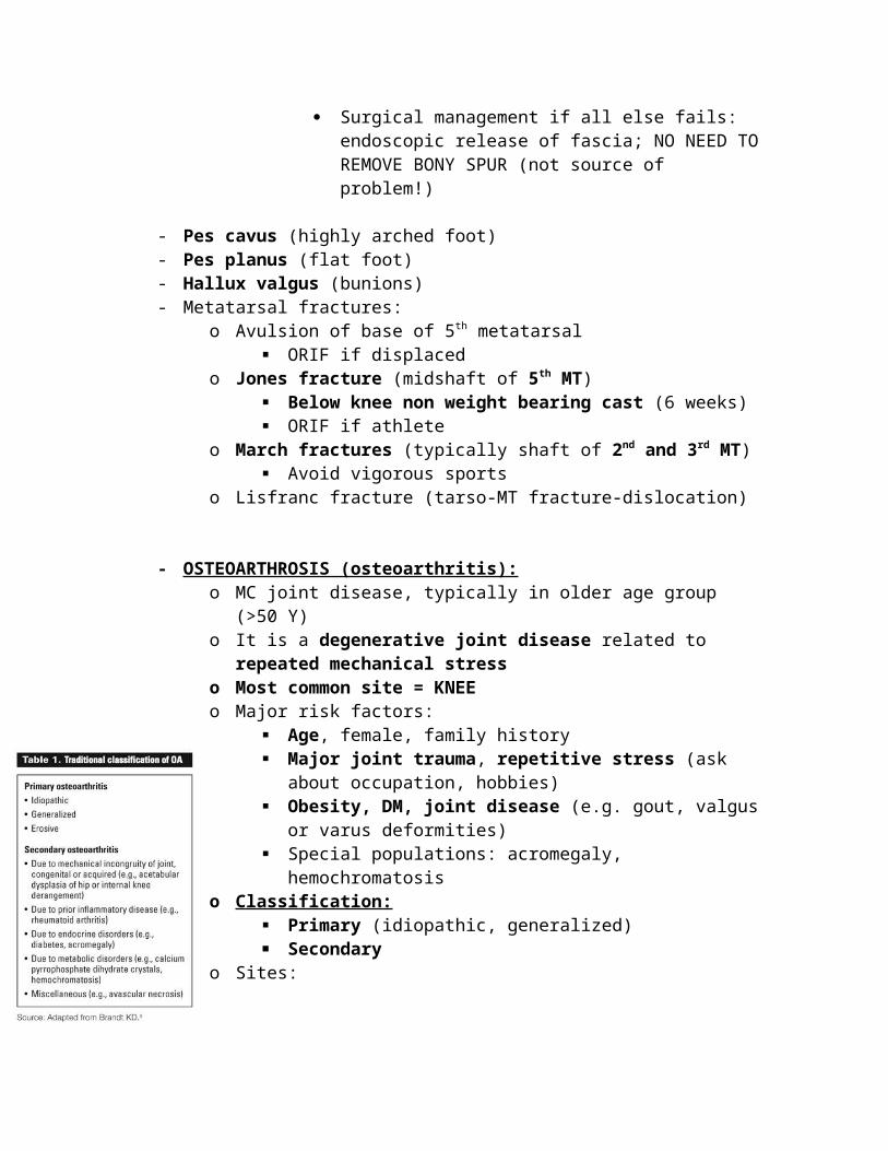
- OSTEOARTHROSIS (osteoarthritis): o MC joint disease, typically in older age group (>50 Y)o It is a degenerative joint disease related to repeated mechanical
stresso Most common site = KNEEo Major risk factors:
Age, female, family history Major joint trauma, repetitive stress (ask about occupation,
hobbies) Obesity, DM, joint disease (e.g. gout, valgus or varus
deformities) Special populations: acromegaly, hemochromatosis
o Classification: Primary (idiopathic, generalized) Secondary
o Sites: Knee and hip > spine (spondylosis, spinal stenosis) & small
joints of hand (PIP and DIP)o Clinical features:
SLOW, CHRONIC, PROGRESSFUL picture Joint pain (typically monoarticular or oligoarticular, big joints,
asymmetric) that worsens with use over the day (vs RA) Background pain at rest Joint gelling and stiffness, but <1/2 hour upon wakening In chronic cases with hand involvement:
PIP osteophyte manifest as Bouchard nodes DIP osteophyte manifest as Heberden nodes
In chronic cases of knee involvement: Depending on which side of knee joint is affected more,
they may develop a valgus or varus deformity at knee Joint mice may cause locking of knee/popping noise
In spinal involvement (See spondylosis)o Physical examination findings:
Crepitation at joint movements If acute inflammation is present, you may find joint effusion
o Investigations: Clinical diagnosis Investigations are done to R/O DDx and for assessing severity
of disease CBC (look at WBC), CRP and ESR If effusion present, you may need to do arthrocentesis
(especially to R/O septic arthritis) IMAGING: classically an x-ray (but joint US may be
useful if really inflamed)
X-RAY FINDINGS: Loss of joint space (asymmetric), which represents
damage to the articular cartilage Osteophytes (typically lateral, do not cross over the
joint) Subarticular sclerosis (bone reaction to repeated
damage by friction of bone on bone) Subchondral bone cysts (synovial fluid leaking into
bone) Joint mice (loose bodies)
o Management: GOALS:
Reduce pain Maintain mobility Prevent deformity
Non-surgical (lifestyle and medical) Lifestyle changes (correct posture, encourage
exercise and physiotherapy, strengthen periarticular muscle, weight loss if obese if possible)
NSAIDS with PPI for pain No real consensus on intra-articular injection of
hyaluronic acid, GA and chondroitin sulfate Surgical (last case scenario):
Joint replacement (e.g. knee arthroplasty)

DISORDERS OF THE SPINE:
- LBP = LOWER BACK PAIN = LUMBAGO:o M>F; 4/5 people at one point of their lifeo Most people DON’T have a systemic disease, but you must always R/O
systemic diseaseo DDx includes:
Bony: Lumbar arthropathy/spondylosis Vertebral disc herniation Infectious causes (e.g. Pott disease) Metastatic disease (e.g. prostate cancer) Multiple myeloma (especially when lying down, when
sleeping) Inflammatory diseases (e.g. ankylosing spondylitis)
Soft tissue: Renal disease, aortic aneurysm, …
o Make sure you: Ask about urinary, GI and neurological symptoms and
constitutional symptoms Do an appropriate MSK exam (assess degree of limitation):
Look (gait when walking in, obvious kyphosis or lordosis, dimple in lower back)
Feel (spinal processes, scoliosis) Move (forward flexion, extension, lateral flexion,
rotations) If required, special tests (see later)
Do a full neurological exam with special tests for femoral and sciatic nerve root compression (e.g. femoral nerve stretch test, straight leg test respectively) and gait assessment
Do a PR exam to assess for perianal anesthesia, rectal tone and to palpate the prostate
- Spondylosis:o Degeneration of the vertebral interfaces particularly at:
Zygapophyseal joints (facet arthropathy) Vertebral discs (leading to disc herniation) Spinal canal (leading to spinal stenosis)
o The clinical picture is that of: Back pain (typically lumbar) Radiculopathy (compressed nerve root) Myelopathy (compressed spinal cord)
o Lumbar facet arthropathy Lower back pain, no neurological symptoms Clinical diagnosis, Rx may indicate diagnosis
Imaging is to R/O bad DDx Intra-articular steroid/local anesthetic injection under
guidanceo Lumbar radiculopathy (most commonly at L5>L4, S1)
Impingement of nerve root due to osteophyte or disc herniation
L5 site is most common, patient is usually older Sciatic nerve roots (L4-S3) Femoral nerve roots (L2-L4)
One of the causes of sciatica (sciatic nerve root impingement): Osteophytes, disc herniation, lumbar spine stenosis,
piriformis syndrome Radiculopathy symptoms:
Radiating pain along supply of nerve Pain is usually aching, burning in character or
associated with numbness P/E:
Look, feel, move, special tests We assess for root: sensation, power/muscle bulk,
reflexes Special tests: straight leg raise test +ve (sciatic nerve
root compression), femoral nerve stretch test L4: diminished patellar reflex (L3, L4), no feet
involvement L5: involves medial leg and weakened dorsiflexion S1: involves back of leg and sole of foot and weakened
plantarflexion; diminished ankle reflex (S1, S2) S2, S3, S4: GI/GU/perianal involvement
INVESTIGATIONS: Best initial diagnostic step is MRI But in reality, we really need to correlate with clinical
history because you might find a lot of things that don’t need Rx
Management: Depends on extent of involvement, but ranges from
conservative to surgicalo Spinal stenosis:
Typically at CERVICAL and LUMBAR spine You will get MYELOPATHY (typically bilateral symptoms) Moving will make Sx worse (scratching like) Best diagnostic investigation = MRI
- Spondylolysis:o Stress fracture or defect at the pars interarticularis of the vertebral
arch
o MC site is L5, but can occur anywhere, beware in the cervical spineo Typically a result of sporting injury in young age groupo If displaced = spondylolithesis (very dangerous)o X-ray finding (best initial step!!!):
AP, lateral and oblique view Scotty dog collar sign on OBLIQUE view radiograph
o Management is usually conservative Bracing is by Boston brace (Anti-lordotic brace) In severe complicated cases (of spondylolithesis and lysis),
surgical management may include laminectomy
PEDIATRIC ORTHOPEDIC CONDITIONS:
- Things to note:o They develop some interesting type of fractures (e.g. greenstick
fracture, torus fracture, …)o They have much thicker, more active periosteum, which means
they can heal more easily than in adults: most of the time, unless complicated, closed reduction and splinting may be enough to Rx
o You see a physis (Growth plate) = KID! This area is WEAK and prone to FRACTURES Also, this area is often mistaken for a fracture (because it
looks separated!) – ALWAYS KEEP YOUR PATIENT’S AGE IN MIND WHEN READING X-RAYS!
Intra-articular fractures can damage the physis leading to GROWTH ARREST! This is why we always pay attention to them and we have a Salter-Harris classification
o Child abuse is a possibility but your threshold should be lowered based on the history
i.e. the history of how this fracture occurred doesn’t match with your understanding of how fractures occur
In general look for: Multiple different fractures at different stages of
healing Fractures in locations that are unlikely to get injured by
an infant that doesn’t walk! Spiral fractures (from twisting injuries)
- Developmental dysplasia of the hip (DDH): o Used to be called congenital hip dislocation, but it can be more or
less than just a dislocation and might not be present at birth (it isn’t necessarily congenital)
o So it is a hip disorder as a result of abnormal hip development and positioning secondary to:
Laxed ligaments (thought to be due to excess relaxin?)

Muscular underdevelopment Abnormal acetabulum roof
o The hip can display signs of subluxation or frank dislocation Dislocated femoral head that is out Dislocated head but within the socket Head subluxates out of joint when provoked Dysplastic acetabulum that is more shallow and vertical than
normalo Epidemiology/RF (The Fs)
F emale > males LeFt hip > right hip (because left body faces sacrum in utero) F irst-born child (primigravid mothers) > others F RANK breech presentation
o History: Take time to ask about family history of hip problems when
young (e.g. the father, mother and other children if she is multiparous)
o Presentation: Typically found during neonatal head to toe examination,
when screening for DDH by doing the relocation test (Ortolani test)
If this is a neonate, symptoms unlikely P/E:
Look, feel, move, special tests Inspection: increased skin folds over affected hip,
asymmetric appearance, shortened affected limb/limb-length discrepancy, limb has a limited abduction position
In older patients who can walk, you may observe a trendelenberg gait, and if bilateral, you may see a waddling gait with lumbar hyperlordosis
Move: Limited abduction of flexed hip (reduced ROM) Special tests include Ortolani (Relocation) and
Barlow (Dislocation) tests and Galeazzi’s sign We typically start with the relocation test because if
the hip is already displaced, we can reduce it by abducting it; if a clunk is felt (not heard) then you had a dislocated hip
We do Barlow to test for a dislocatable hip (it is not dislocated on presentation, but we suspect DDH), in which we adduct the hips and push posteriorly to elicit dislocation
Galeazzi’s sign is seen in older children (>1 years old): here we compare the level of knees held next to each other when knee is flexed when patient is supine;
if they are uneven, the lower is the side that is affected
o Diagnosis and investigation: Diagnosis is CLINICAL In the first 3-4 months of life, we use ULTRASOUND for
diagnostic imaging, however: We don’t need to do an US today if this is a neonate
(but instead in a week or two in a visit) because many cases are incidental as the body is recovering from delivery and may not be found in follow up examination; however we tell them about things they can do in the meantime and what to expect
We don’t use radiographs early mainly because bones are not calcified well enough yet (takes 4- 6 months) and because they fail to capture the cartilages (appear radiolucent); radiation exposure is not our main fear now
So in the first 3 months US, then do a radiograph after 3 months
Radiological findings: Disrupted shenton’s line (uses upper rami of pelvis) Femoral neck above Hilgenreiner’s line (horizontal
line through triradiate cartilage of acetabulum) or beyond Perkin’s line
Femoral head ossification center NOT in lower inner quadrant (based on intersection of Hilgenreiner’s horizontal line and Perkin’s vertical line)
Increased acetabular index (>25 degrees) by drawing a line from the horizontal line following the roof of acetabulum
o Management: Parent education about condition and possible outcomes
Until an ultrasound can be obtained, parents are told NOT to wrap infant traditionally (avoid SWADDLING: which is basically wrapping them like a mummy)
< 6 months of age = Pavlik harness Splinting/immobilizing in the reduced position
(abducted hip position) Good because it is a DYNAMIC splint (allows mobility;
it is known that for joints and periarticular bone to develop properly they actually need to colliding and active)
Bad because it will be kept for months or up to a year!

Complications of improper placement and use: skin ulcerations, femoral nerve palsy, AVN of femoral head if kept in max abduction
> 6 months of age = closed reduction and hip spica A hip spica is plaster cast that immobilizes the joint
completely in the reduced position It is ADYNAMIC (think of the cons of that!) In children who have contractures of the adductor
longus or iliopsoas muscle might need to have them released by surgery
> 18 months of age = SURGICAL Open reduction Femoral shortening (with osteotomy) Pelvic osteotomy (reconstruct the roof/shelf using
part of iliac crest)o Complications:
Hip and back pain Early hip arthritis Limb length discrepancy Pelvic inequality (tilting) Early lumbar spine degeneration
- Congenital talipes equinovarus (CTEV):o AKA: CLUBFOOT (don’t confuse this with rocker-bottom feet)o Congenital foot deformity
It is often NOT ALONE Patients will may have other MSK problems in LL and back
such as dysraphism (unfused vertebral bodies), DDH and knee deformity
Always check the neck, back, hip and knees Also check the neck for? Congenital torticollis
o The name says it all: Congenital: present at birth Talipes: foot Equino (like horse hoof when flexed): plantarflexed Varus: foot is facing MEDIALLY (inverted) End result is foot that is plantarflexed and inverted
o Etiology: Divided into:
Intrinsic causes (neurologic, muscular, CTD) Extrinsic cause (IUGR) Idiopathic
It is a FIXED deformity that is NOT limited to the foot! BELOW KNEE disease
o Presentation:
Happy DDxPositional clubfoot:Unlike CTEV, which is a fixed bony deformity that must be corrected, positional clubfoot is reversible and occurs because of positioning in utero. It has a NORMAL ROM. It has a good prognosis and is managed conservatively with a taught program of stretching, massaging, etc. They will NOT fulfill the criteria of CAVE.

Present at birth and needs evaluation Do a full head to toe examination with emphasis on look, feel,
move and special tests of the NECK, BACK, LOWER LIMB On P/E:
Small foot Small heel and deep medial crease Pes cavus (arched midfoot) Abnormally thin calves
Diagnosis of clubfoot needs 4 components (“CAVE” deformity): Cavus Forefoot Adductus Hindfoot Varus Hindfoot Equinus
o Management: Usually using PONSETI TECHNIQUE:
Correct varus deformity GRADUALLY (weekly visits for special stretching manipulations which are then casted or patient wears specialized shoes (foot braces) until deformity is corrected in about 4 – 6 weeks)
Correct equinus deformity ACUTELY at the END if necessary by cutting the Achilles tendon– heel-cord release (tenotomy)
May need to wear foot brace for a period after correction
Mild recurrences are COMMON Affected foot is permanently smaller than normal foot +/-
calf muscle atrophy
- Legg-Calve-Perthes disease (Coxa Plana):o Idiopathic AVN of femoral head (infrequently bilateral)
Results in abnormal growth of physis o Age of presentation: 4 – 8 years oldo M>F (classic trivia question: DDH F>M; LCPD M>F)o Unknown etiologyo Risk factors:
Delayed bone age (89%) Family history Low birth weight or abnormal pregnancy/delivery Miscellaneous: second hand smoking, ADHD
o Presentation: Insidious onset Parents complain that their child has a limp or an abnormal
gait (+/- pain)

They may have knee, hip, groin or thigh pain P/E:
L, F, M, ST Trendelenburg gait or antalgic gait (if in pain) Reduced hip ROM (stiff hip) Flexion contracture (do thomas test) Limb length discrepancy (late)
o Differential diagnosis: Inflammatory disease:
Infections: septic arthritis, osteomyelitis Rheuma: Juvenile rheumatoid arthritis (polyarticular, so
unlikely) Bony disease:
Slipped capital femoral epiphysis Neoplastic (primary bone tumors)
o Investigations: Hip X-ray findings are diagnostic:
May be negative early , so if you highly suspect it you may do an MRI or bone scan
Collapse of femoral head o Management:
Debatable, but generally we want to preserve joint function so:
Physiotherapy = agreed upon Brace = controversial Operative management (femoral or pelvic osteotomy) =
unlikely to be needed Risk of developing early onset OA and its related
complications
- SLIPPED CAPITAL FEMORAL EPIPHYSIS (SCFE) o Pronounced “skif-fee”o Type I salter-harris epiphyseal injury at hip joint
The growth plate, being weak, can fracture and cause the rest of the femur to be displaced
o Age group: adolescent (MCC of hip disorder in adolescent)o Risk factors:
Obesity (most important risk factor) Male Hypothyroidism (bilateral!)
o Mechanism: Hypertrophied (but weak) growth plate at the time of puberty
under mechanical stress (obesity) receives an
environmental trigger (trauma) may cause acute slipping of head or develop over time
25% have bilateral involvemento Clinical features:
ACUTE picture Sudden, severe pain at hip joint Limping
CHRONIC picture Groin, anterior thigh (+/- knee) pain
P/E: Tenderness over joint capsule Restricted internal rotation and abduction Trendelenburg sign over affected side Whitman’s sign (obligate external rotation during
PASSIVE flexion at hip joint!)o Investigations:
Pelvic (hip) x-ray: AP, lateral, frog-leg view Obvious displacement (“slipping”) of the head, usually
posterior and MEDIALLY Disruption of KLIEN’S LINE (A line which runs from
the neck of femur straight on should go through part of the head of femur)
Widened appearance of affected growth plate (because its separated)
o Management: Principles:
Prevent further slippage Prevent limb asymmetry because of growth plate
damage Operative management is typical:
Mild/moderate: internal fixation with pins in current position (prevent slippage from happening)
Severe slip: ORIF or pin physis (in intention to destroy the growth plate) without reducing and do osteotomy after to equilibrate
Why destroy growth plate? To prevent future slips Complications:
AVN Chondrolysis (loss of joint space) Premature OA
- Osgood schlatter disease: o Inflammation of patellar ligament at its insertion in the tibial
tuberosity Minor avulsions there due to repetitive tensile stress

Occurs during adolescence when growth of bones and strengthening of tendon occurs
o M>Fo Age group: boys 12 – 15 years old, playing lots of sportso Clinically:
Pain below the knee especially on jumping and kneeling, limiting sporting activities
On P/E: Tenderness over the tibial tuberosity Ask them to touch their toes, they will feel pain and
resist
o Investigations: X-ray may show fragmentation of tibial tuberosity and
ossifications in the patellar tendono Management:
Benign, self-limiting condition, but it does not resolve until growth halts
Conservative management: Restrict vigorous activities Isometric strengthening exercises are ok NSAIDs for pain
- Scoliosis: o Lateral curvature of spine with vertebral rotationo Age group: 10 – 14o Females > maleso Etiology:
IDIOPATHIC (MC) Congenital Neuromuscular (secondary to myopathies or dystrophies) Postural (non-structural scoliosis: muscle spasm, limb length
discrepancies) Others (neoplastic, traumatic)
o Clinical features: May have back pain If brought by parents, it is usually because of deformity of back
Asymmetric pelvis, creased flanks, prominent scapula P/E:
Spine: look, feel, move, special tests Forward flexion will alleviate non-structural scoliosis
and make structural ones more obvious Asymmetric shoulder height or rib humping when
bending forward suggest structural scoliosis Look for signs of underlying conditions
o Diagnosis: X-ray: measure curvature for Cobb angle, can guide
progression and managemento Management:
<25 degrees serial assessment >25 degrees bracing can help stop progression >45 degrees respiratory and cosmetic problems result, so
surgical Rx is sought for
FUCKING BONE TUMORS:
- Introduction:o The 3 germ layers:
Ectoderm skin, CNS Endoderm endothelial lining and organs Mesoderm mesenchymal tissue Tumors of mesenchymal tissues are -oma and if malignant are
called sarcomas, but NOT in all cases: Most bony tumors have their own name Blood is mesenchymal, we don’t call it sarcoma! We call
cancer of the blood leukemia! Tumors of the soft tissue may be more common than that of
bone Skin cancer and lipomas are definitely more common However, muscle cancers are less common (e.g.
rhabdomyosarcoma)o The most common bone tumor/cancer in adults is METASTATIC
Common sources include MM > breast, prostate, lung, thyroid
However, the age group will be older (>30s) vs primary bone tumors
Multiple myeloma can present with lytic bone lesions and related disease
o Primary bone tumors are rarer and occur in a YOUNGER age group (<30s)
We characterize them by their: Age group (e.g. Ewing more in kids) Location (e.g. epiphysis, metaphysis, diaphysis,
medulla) Histologic type (bone forming, cartilage forming,
osteoclastoma, bone marrow tumor) Aggressiveness (benign active/benign
aggressive/malignant) – WHO classification Most common site is DISTAL FEMUR/PROXIMAL TIBIA
Most common benign tumor is osteochondroma MC malignant tumor is metastatic (MM) MC bone tumor in kids are benign (osteochondroma)
and the MC adult bone tumor is metastatico Tumor-LIKE conditions:
These produce lytic bone lesions Aneurysmal bone cysts (ABCs), simple bone cysts (SBCs),
eosinophilic granuloma (EG), Fibrous dysplasia (FD), non-ossifying fibroma (NOF)
- Clinical features to be aware of:o Non-specific
Local pain, swelling (for weeks to months) Can often mimic any bone/joint disease (e.g. fever in
Ewing’s sarcoma picture can be Dx as osteomyelitis) Minor trauma leads to fracture (pathological fractures)
This can be the presentationo Red flags:
Persistent skeletal pain Localized tenderness Spontaneous fracture Enlarging mass/soft tissue swelling
o P/E: Make sure you do a full examination, in order to take a hint of
where the primary cancer could be (e.g. pt with hemoptysis, weight loss, bone pain… lung cancer with mets to bone)
- Investigations:o Routine x-ray initially
Although “classical” findings may indicate which type of cancer it is, the diagnosis is ONLY confirmed by BIOPSY
Location Size Lytic or lucent Extent of involvement (soft tissue swelling, pathologic
fractures) LOCATION DDx (also depends on age):
Diaphysis: Ewing’s sarcoma, eosinophilic granuloma (EG), spontaneous bone cyst, fibrous dysplasia, non-ossifying fibroma
Metaphysis: osteosarcoma, echondroma, osteochondroma, osteomyelitis
Epiphysis: giant cell tumor, chondroblastoma, bone infections
X-ray findings of BENIGN lesions:
Minimal to no periosteal reaction (no codman triangle, sunburst spicules, not laminated/onion-skinning)
Solid cortex is raised Well developing endosteal reaction with good bone
formation and intraosseous calcifications
X-ray findings of MALIGNANT lesions: Acute periosteal reaction (which is non-specific)
signs such as raised periosteum, codman triangle, sunbursting, laminations/onion-skinning
Larger size (>1 cm), irregular margins, varied bone formation, soft tissue involvement
o Other investigations to do when suspecting cancer: Lab investigations: CBC, BONE PROFILE (ALP, Ca2+, PO4, Vit
D, PTH levels), RFTs and LFTs Imaging:
MRI and bone scan Chest CT (lung cancer or mets)
Bone biopsy
HISTOLOGICAL TYPES- BENIGN ACTIVE TUMORS
o There are many things included here, but below are the important ones to know
We divide them into bone forming, cartilage forming, but some are a combination
o Cartilage forming: Osteochondroma
MC benign bone tumor 2nd and 3rd decade of life Bony tumor with cartilage cap Exostosis particularly at physeal area Growing continues until skeletal maturity established Rx typically observation; may remove if symptomatic
Echondroma Hyaline cartilage tumor 2nd and 3rd decade of life Usually asymptomatic/incidental finding
o Bone forming: Osteoma
Surfaces of facial bones Associated with gardner syndrome (FAP)
Osteoid osteoma

Young adult, classically very painful bone pain relieved by NSAIDs (aspirin classically)
Tumor occurs in diaphyseal cortex, and is lytic with a rim of reactive bone
Osteoblastoma Compared to osteoid osteoma, they are larger, involve
the vertebra and are painful but not relieved by aspirin
- BENIGN AGGRESSIVE TUMORSo Giant cell tumors (GCS)
Osteoclastoma (arise from osteoclasts) Although not usually malignant, it is very aggressive and
involves adjacent structures quickly May be malignant in some cases (only differentiated by
biopsy) Occur after skeletal maturity established Presents with pain, tenderness and swelling, reduced ROM of
nearby affected joint Prefers the epiphysis Radiographically it is a large lytic lesion with a soap-bubble
appearance, adjacent bone and soft tissue involvement 15% recurrence risk when removed Rx requires wide local excision of expendable bones
- MALIGNANT TUMORS:o Most common malignant tumor types for age:
<1 – neuroblastoma 1-10 – Ewing’s sarcoma 10 – 30 – osteosarcoma 30 – 40 – fibrosarcoma, periosteal osteosarcoma, malignant >40 – metastatic carcinoma, MM, chondrosarcoma
o Enneking staging of bone tumors: Uses grade, size, mets (not N) I low grade, intracompartmental or extracompartmental II high grade, intracompartmental or extracompartmental III mets
o Osteosarcoma: Malignant bone tumor most frequently diagnosed in 2nd decade
of life, but has bimodal distribution (teenagers and elderly) Risk factors:
Radiation exposure (elderly) History of Paget’s disease (elderly) Familial retinoblastoma (RB!)

Prefers to develop on the metaphysis of long bones (particularly at femur-tibia area)
Patients may develop local symptoms or pathological fractures
Radiologically: periosteal reaction (codman and sunburst spicules), often very large and can cross growth plate
Metastasis invariably occurs w/o Rx (commonly to the lung) Labs and imaging:
Standard labs (as above) Bone scan (R/O bone mets) Chest CT (R/O lung mets)
Management: Surgical resection (limb salvage >>>> amputation)
o Limb salvage surgery: removing tumor with wide margin of normal tissue while preserving NVS and distal extremity; joints/bone are replaced with metal prosthesis or bone graft
Adjuvant chemotherapy Neo-adjuvant chemotherapy if large (shrinks tumor) Prognosis is generally good if not mets (we use 10-year
survival; 90% 10YS if low grade)o Ewing’s sarcoma:
Malignant tumor of cells of neurectodermal origin (PNET) Genetic component: t(11;22) Most common malignant primary bone tumor in kids <20
years old May mimic bone infections (chronic osteomyelitis/septic
arthritis is big DDx) Bone pain, swelling, erythema, reduced ROM + fever,
raised CRP and WBC! Prefers to involve the MEDULLA in the DIAPHYSIS Radiologically produces a lytic bone lesion in shaft with
characteristic periosteal reaction in the form of lamination/onion-skinning
Although it is aggressive and loves to spread early, it responds well to chemotherapy +/- resection
- Multiple myeloma: o Proliferation of neoplastic plasma cells
This impacts normal blood precursor cells, the bone itself, high Ig levels without proper functioning (recurrent infections), hyperviscosity leading to CNS features, and affecting the kidney leading to renal failure (RTA type II [Fanconi], amyloidosis, nephrocalcinosis, etc.)
o MC primary malignant bone tumor in adults (43%)
o Typical age group is >40 years old; M>Fo Presentation:
Localized bone pain (especially in lower back, and when supine)
Pathological fractures Constitutional: weight loss, weakness, anorexia Others: headaches, blurring of vision, recurrent infections May display S&S of hypercalcemia
o Investigations: Labs:
CBC: anemia, thrombocytopenia, high serum immunoglobulin levels (IgG MC)
ESR and CRP are raised Bone profile: hypercalcemia, high ALP Urinalysis: Bence jones protein (free light chain
paraprotein) RFTs (BUN, Cr, CLcr, electrolytes) Serum and urine protein electrophoresis (look for M
spike, representing paraprotein/M protein – albumin is no longer the most predominant serum protein!)
Others: peripheral smear (rouleaux), beta-2-microglobulin levels (useful for staging)
Imaging: If presents with bone pain, you may do an x-ray of
affected area (e.g. lumbar spine x-ray) – typically will see a lytic bone lesion
Skeletal survey, MRI if spine involved (rule out CNS involvement)
CT-guided biopsy of affected site/bone marrow biopsyo Diagnostic criteria of symptomatic myeloma, which requires
THESE: “CRAB”:
Hypercalcemia, renal failure (no other relatable cause), anemia (<10 g/dL), bone lesions (lytic, patho fracture)
Serum and/or urine protein electrophoresis demonstrating monoclonal protein (e.g. M protein)
Bone marrow studies showing plasmacytomas (clonal expansion of plasma cells >10%)
o Management: Symptomatic
Bisphosphonates (pamidronate) to prevent fractures RBC transfusions/EPO for anemia
Chemotherapy >65 ~ Melphalan + prednisone

<65 ~ Bortezomib + prednisone Bortezomib works by blocking proteases, so that certain
enzymes that work to degrade cancer cell proteins Melphalan (classical agent) is an alkylating agent
Others: radiotherapy, debulking/reparative surgery POOR PROGNOSIS 5 year survival is 30%