Embed Size (px)
Citation preview

Page 1/25
Reactional Ultrasonic Systems And MicrowaveIrradiation For Pretreatment Of Agro-IndustrialWaste To Increase Enzymatic ActivityFabiane Fernanda Czapela
Laboratory of Microbiology and Bioprocesses, Universidade Federal da Fronteira Sul - UFFS, RodoviaERS 135 - km 72, nº 200, Erechim, RS, Brazil. Zip code 99700-970Simone Kubeneck
Laboratory of Microbiology and Bioprocesses, Universidade Federal da Fronteira Sul - UFFS, RodoviaERS 135 - km 72, nº 200, Erechim, RS, Brazil. Zip code 99700-970Karina Paula Preczeski
Laboratory of Microbiology and Bioprocesses, Universidade Federal da Fronteira Sul - UFFS, RodoviaERS 135 - km 72, nº 200, Erechim, RS, Brazil. Zip code 99700-970Caroline Dalastra
Laboratory of Microbiology and Bioprocesses, Universidade Federal da Fronteira Sul - UFFS, RodoviaERS 135 - km 72, nº 200, Erechim, RS, Brazil. Zip code 99700-970Thamarys Scapini
Laboratory of Microbiology and Bioprocesses, Universidade Federal da Fronteira Sul - UFFS, RodoviaERS 135 - km 72, nº 200, Erechim, RS, Brazil. Zip code 99700-970Charline Bonatto
Laboratory of Microbiology and Bioprocesses, Universidade Federal da Fronteira Sul - UFFS, RodoviaERS 135 - km 72, nº 200, Erechim, RS, Brazil. Zip code 99700-970Fábio Spitza Stefanski
Laboratory of Microbiology and Bioprocesses, Universidade Federal da Fronteira Sul - UFFS, RodoviaERS 135 - km 72, nº 200, Erechim, RS, Brazil. Zip code 99700-970Aline Frumi Camargo
Laboratory of Microbiology and Bioprocesses, Universidade Federal da Fronteira Sul - UFFS, RodoviaERS 135 - km 72, nº 200, Erechim, RS, Brazil. Zip code 99700-970Jessica Zanivan
Laboratory of Microbiology and Bioprocesses, Universidade Federal da Fronteira Sul - UFFS, RodoviaERS 135 - km 72, nº 200, Erechim, RS, Brazil. Zip code 99700-970Altemir José Mossi
Laboratory of Microbiology and Bioprocesses, Universidade Federal da Fronteira Sul - UFFS, RodoviaERS 135 - km 72, nº 200, Erechim, RS, Brazil. Zip code 99700-970Gislaine Fongaro

Page 2/25
Department of Microbiology, Immunology and Parasitology, Universidade Federal de Florianópolis -UFSC, Engenheiro Agronômico Andrei Cristian Ferreira St., Trindade, Florianópolis, SC, Brazil. Zip code88040-900Helen Treichel ( [email protected] )
Universidade Federal da Fronteira Sul https://orcid.org/0000-0002-3810-3000
Research
Keywords: Reaction system, Keratin residues, Biotechnological processes
Posted Date: April 23rd, 2020
DOI: https://doi.org/10.21203/rs.3.rs-22357/v1
License: This work is licensed under a Creative Commons Attribution 4.0 International License. Read Full License
Version of Record: A version of this preprint was published on September 26th, 2020. See the publishedversion at https://doi.org/10.1186/s40643-020-00338-2.

Page 3/25
AbstractKeratinous residues are generated in large quantities in industrial processes and because of this they aredi�cult to manage because the presence of keratin. Keratinases are emerging as promising alternativesin technological processes. The objective of this work was to determine the potential of pretreatment ofagro-industrial residues using an ultrasonic probe, and to evaluate activity enhancement strategies byanalyzing the behavior of enzymatic activity of keratinases exposed to various reaction systems as wellas to enzymatic concentration. We tested the application of enzymatic extracts in the degradation ofkeratinous residues. Submerged fermentation was performed with swine hair pretreated with and withoutultrasonic probe. The enzymatic extracts were exposed to reaction systems and the enzymaticconcentration technique to measure the increments of enzymatic activity. We found that the ultrasonicprobe increased reaction rates, enabling higher enzyme production in shorter time periods. The ultrasonicprobe reached 38.4% of relative activity and the enzyme concentration, increased the enzymatic activityin 53.5%. We observed potential degradation of chicken feathers with homemade enzyme extracts. Takentogether, our �ndings suggest that the results obtained in this work are promising, despite the fact thatthe data in this area are scarce.
1. IntroductionKeratinous waste is generated in large quantities; management is hampered by its resilient structure(Onifade et al, 1998; Łaba et al. 2015). These residues consist of �brous proteins such as keratins thatare resistant to physical, chemical and enzymatic actions (Korniłłowicz-Kowalska and Bohacz 2011).When found in their solid state, the structure of the keratin increases its stiffness because of cysteinebonds (Barone et al. 2006), as well as by α and β-keratin chains and disul�de bonds (Onifade et al. 1998).Α α-keratin can be found in materials such as hair, fur, nails, hooves and animal horns, while β-keratin ispresent in scales, bird feathers and beaks (Korniłłowicz-Kowalska and Bohacz 2011; Mazotto et al. 2011;Ire and Onyenama 2017).
One way to promote the degradation of these residues is via biotechnological processes, allowingdegradation through the action of speci�c enzymes such as keratinases (Gupta and Ramnani 2006;Brandelli 2008; Brandelli, Daroit and Riffel 2010; Gegeckas et al. 2018). Keratinases (EC 3.4.21/24/99.11)are a class of proteases with the speci�c potential to hydrolyze keratin, giving them a prominent role overother proteases (Brandelli, Daroit and Riffel 2010; Gegeckas et al. 2018). These enzymes haveapplications such as biotechnology (Okoroma et al. 2012; Mazotto et al. 2013; Paul et al. 2014; Brandelli,Sala and Kalil 2015) and waste degradation (Fang et al. 2013a; Yusuf et al. 2016; Su et al. 2017; Abdel-Fattah et al. 2018; Thankaswamy et al. 2018). They can be obtained commercially or from submergedfermentation (FS) or solid-state fermentation (FES) through microorganisms (Mazotto et al. 2013) suchas fungi (Kushwaha 1983; Santos et al. 1996), considered good producers of keratinases; they can alsobe isolated from keratinous waste disposal sites (Kaul and Sumbali 1997; Riffel and Brandelli 2006).When obtained in this way, they are designated “homemade” and can be used in crude or concentrated

Page 4/25
form, a technique that combines the interaction of the enzyme with salts and solvents so as to separateout interferents (Preczeski et al. 2018).
Because of the biotechnological importance of keratinases, several studies have been performed toimprove the activity and evaluate the conformational change of enzymes in general, when exposed todifferent reaction systems, including ultrasonic probe and bath and microwave (Ma et al. 2011; Jin et al.2015; Mazinani, DeLong and Yan 2015). Simple and e�cient heating technologies increase enzymeactivity and conformational change (Ma et al. 2011); residue pretreatment techniques using an ultrasonicprobe (Azmi, Idris and Yusof 2018), makes the structures more accessible to microbial and enzymaticattack (Yusof and Ashokkumar 2015).
The present study aimed to evaluate the potential for pretreatment of agro-industrial residues using anultrasonic probe and to investigate the behavior of keratinases exposed to the following reactionsystems: ultrasonic probe, ultrasonic bath and microwave and enzymatic concentration technique. Inaddition to comparing the activity of homemade enzymes with commercial enzymes and �nally, weapplied enzymatic extracts for the degradation of keratinous residues such as swine hair and chickenfeathers.
2. Materials And Methods
2.1 Keratin substratesSwine hair and chicken feathers were obtained from a food agroindustry in Rio Grande do Sul, Brazil.Residues were stored at -4 °C until use. Prior to use, the residues were washed with water and detergent,and immersed in 70% alcohol for 1 h, followed by drying at 70 ± 2 °C for 16 h (Adapted Călin et al. 2017).Chicken feathers were used in the degradation tests only.
2.2 Swine hair pretreatment by ultrasonic probeSwine hair (1 g) underwent pretreatment using an ultrasonic probe, comparing the micropoint andmacropoint. The variables used were power (100%), pulse (3) and exposure time (15 min) using distilledwater as a liquid medium (Adapted from Azmi, Idris and Yusof 2018). The tests were performed with theECO-SONICS Ultrasonic Probe/Cell Disrupter Sonicator, 20 KHz ultrasonic frequency and 550 W ultrasonicpower, equipped with a 4 mm titanium micropoint and 13 mm diameter titanium macropoint.
The sonicated content was �ltered on �lter paper and the solid fraction was subjected to a microbial loadreduction process using 70% alcohol and heating at 70 °C ± 2 °C for 16 h before starting fermentation.
2.3 Submerged fermentationSubmerged fermentation was performed with swine hair. To this end, 10 g L− 1 of pretreated and notpretreated ultrasonic probe residue was poured into 50 mM Tris HCl buffer (pH 8.5) with the addition of106 spores mL− 1 of Fusarium oxysporum. The fermentative medium was incubated at 150 rpm and

Page 5/25
28 °C for 9 days (Preczeski et al. 2020). The fungus used was isolated from soil-borne chicken featherresidues and identi�ed by the Next Generation Sequencing (NGS) method.
At the end of the fermentation, the fermented medium was �ltered and the retained solid fraction wasdried to quantify the degradation percentage of the agro-industrial residue. The �ltrate was used tomeasure keratinolytic activity and exposure in different reaction systems. Keratinolytic activity wasquanti�ed at 3, 6 and 9 days and the percentage of residue degradation after 9 days.
2.4 Keratinase assayKeratinolytic activity was measured using 0.013 g azoqueratin as substrate (SIGMA-ALDRICH K8500 α-K),3.2 mL 50 mM Tris HCl buffer (pH 8.5) and 0.8 mL enzyme extract. The reaction took place in anultrathermostatic bath at 50 °C for 1 h, after which 0.8 mL of 10% trichloroacetic acid (TCA) was added tostop the reaction, reading at 595 nm spectrophotometer (Adapted Bressollier et al. 1999).
One unit of keratinase activity was de�ned as the amount of enzyme required to produce a 0.01absorbance increase under the described assay conditions. Enzyme activity was determined before andafter exposure to reaction systems. Relative activity (%) was also calculated for activity incrementpurposes.
2.5 Enzyme activity increase
2.5.1 Exposure of enzyme extracts to reaction systemsEnzymatic extracts produced using swine hair as substrate pretreated with ultrasonic probe withmacropoint and not pretreated were exposed to ultrasonic probe, ultrasonic bath and microwave usingthe experimental design methodology, aiming to evaluate the behavior of the enzymes. Each reactionsystem was evaluated using central composite design (CCD) based on previously de�ned independentvariables, according to reaction interest and system possibilities. The response variable was relativeactivity (%), resulting from the variation between keratinolytic activities of the extract before and afterexposure to reaction systems.
2.5.1.1 Ultrasonic probeA 20 mL aliquot of the crude enzyme extract was exposed to the ECO-SONICS Cell Disruptor Probe (20KHz and 550 W) equipped with a 4-mm diameter titanium micropoint. The behavior of keratinases wasevaluated under different conditions of exposure time (2 to 10 minutes), power (40–80%) and pulse (1 to3) following experimental design.
2.5.1.2 Ultrasonic bathIn this reaction system, 2 mL aliquots of the crude enzyme extract were exposed to the ultrasonic bath,varying the temperature (°C), the power (%) and the exposure time (min) of the enzyme extract accordingto experimental design. The equipment used was the UNIQUE Ultrasonic Washer, model USC-1800 A,

Page 6/25
frequency US 40 KHz and maximum power of 137 W (0.42 W cm² (−1)). The tests were performed withultrasonic bath temperature ranging from 30 to 80 ºC, ultrasonic power ranging from 0 to 100% and withexposure time of 10 to 40 minutes, following experimental design (Mulinari et al. 2017).
2.5.1.3 MicrowaveAliquots of 3 mL of crude enzyme extract were exposed to microwave, varying conditions according toexperimental design. Exposure of the enzyme to microwaves was evaluated in the time range of 5 to 15minutes, using temperatures between 30 and 80 °C, according to the methodology described by Mazinani,Delong and Yan, (2015). The equipment used was the ANTON PAAR Monowave 100 Microwave Reactorwhich has unpulsed 500 W output power (ANTON PAAR).
2.5.2 Enzymatic concentrationTo test the most economically viable enzymatic activity increment methods, the enzyme concentrationtechnique was performed. Enzyme extracts obtained from the fermentation process were concentratedusing 0.5 mol L− 1 NaCl and 50% (v v− 1) acetone. The tests were performed in an ice bath at 4 °C. Aftercentrifugation at 9500 x g, 4 °C and 20 min, the supernatant phase was discarded and the precipitate wasresuspended with 50 mM Tris HCl buffer (pH 8.5) (Preczeski et al. 2018).
The concentrated enzymatic extract (homemade) was exposed to reaction systems under the conditionsof greatest activity increase in experimental designs and applied to the degradation of swine hair andchicken feathers, aiming to evaluate the degradation potential of keratinous residues.
2.6 Commercial enzymes and reaction systemsThe activity of the homemade enzyme extract was compared with that of the commercial enzyme K4519SIGMA with initial enzymatic activity of 2812.5 U mL− 1. K4519 SIGMA keratinase was diluted in 5 mMphosphate buffer (pH 7.0). The enzyme was exposed to the optimal enzymatic increment conditionsobtained in the experimental designs for the ultrasonic probe, ultrasonic bath and microwave and wassubsequently used for the degradation of keratinous residues.
2.7 Evaluation of keratin residue degradationSwine hair and chicken feather degradation was evaluated using crude and concentrated enzymeextracts, both with and without exposure to reaction systems. The degradation potential of agro-industrialresidues was also evaluated using the commercial enzyme extract K4519 SIGMA before and afterexposure to reaction systems.
One whole feather or 0.1 g of swine hair was used for each 8 mL of enzyme extract. Assays wereincubated at 28 °C for 28 days (Scott and Untereiner 2004), and the result was evaluated based on visibleresidue degradation.
2.8 Characterization of homemade keratinase enzymes

Page 7/25
For the purpose of obtaining the optimal pH and temperature of the enzyme used, a characterization wascarried out evaluating different pH conditions, ranging from 5 to 9 and temperature, from 37 °C to 70 °C.The buffers used were: 50 mM citrate buffer pH 5.0 and 5.7; 50 mM sodium phosphate buffer pH 7.1, pH8.4 and pH 9.0; 50 mM Tris HCl buffer pH 8.5. The characterization was carried out in anultrathermostatic bath and the activity measurement followed that speci�ed in item 2.4.
2.9 Statistical analysisThe experiments were statistically evaluated using CCD and the results of the enzymatic activity wereanalyzed using Protimiza Experimental Design software (Rodrigues and Iemma, 2014), using 95%con�dence analysis of variance (p < 0.05). Results were presented as mean ± standard deviation oftriplicate experiments. Statistical differences between data sets were veri�ed using ANOVA and Tukeytest.
3. Results And Discussion
3.1 Swine hair pretreatment by ultrasonic probeThe fermentative process to obtain the enzymatic extract was performed using swine hair as thesubstrate that had been pretreated with or without the ultrasonic probe. Enzymatic activities anddegradation percentages were evaluated (Table 1).
Table 1
Keratinolytic activity after pretreatment in an ultrasonic probe and without pretreatment at 3, 6 and 9days, as well as the percentage of degradation obtained at the end of the fermentation process.
Swine hair Enzymatic activity (U mL− 1) Degradation(%)
Pretreatment by ultrasonicprobe
Micropoint 3 days 6 days 9 days
38.8 ± 5.3a
81.0 ± 5.2a
55.7 ± 3.0a
31.8 ± 1.1 a
Macropoint 3 days 6 days 9 days
50.8 ± 5.3b
39.5 ± 4.8b
43.8 ± 1.1b
45.5 ± 0.8 b
Without pretreatment 3 days 6 days 9 days
25.0 ± 5.6a
82.2 ± 9.4a
75.2 ± 3.2c
36.3 ± 4.0 a
*Equal lowercase letters do not differ from each other by the Tukey test on the column.

Page 8/25
The keratinolytic activity obtained on the third day of the fermentation in the swine hair pretreated withthe ultrasonic probe macropoint was 50.8 ± 5.3 U mL− 1. From this result, it can be inferred that the greaterenergy dissipation caused by the macropoint resulted in more relevant data of enzymatic activity,because it has a larger diameter than that of the micropoint, which achieved enzymatic activity (38.8 ± 5.3 U mL− 1) similar to that obtained with the crude residue (25.0 ± 5.6 U mL− 1).
We found that the micropoint did not have the same impact on structure in terms of enzymatic activityfor the 3rd day, because the ultrasonic system can be in�uenced by several factors, including energydissipation, ultrasonic intensity, temperature, exposure time, and diameter (Lippert et al. 2018), all ofwhich may have resulted in lower erosion e�ciency of the structure when using the micropoint than whenusing the macropoint.
The highest keratinolytic activity was observed for swine hair without pretreatment and with micropointpretreatment at 6 days of fermentation: 82.2 ± 9.4 U mL− 1 and 81.0 ± 5.2 U mL− 1, respectively. Regardingthe degradation percentage, the highest degradation occurred in the pretreatment test with ultrasonicprobe with macropoint (45.5% ± 0.8%), and it can be inferred that the abrasion and erosion in thestructure of swine hair, which resulted in the highest enzymatic activity in three days for the same assay,also presented the highest dissolution of structural chains that make up the residue in question, resultingin the largest mass loss in this experiment.
The ultrasonic probe increases reaction rates, especially hydrolysis processes (Zou et al. 2016).Ultrasonic systems facilitate structural degradation via propagation of ultrasonic waves that causebubble cavitation, causing temperature rise, favoring the formation of rapidly reacting radicals (Wang1981; Manasseh et al. 2010; Yusof and Ashokkumar 2015), improving the enzymatic attack on thesubstrate (Yang and Fang 2015). The cavitation process is the fundamental factor for the e�ciency ofultrasound pretreatment, because the explosion of bubbles near the surface of the material causesabrasion and erosion, reducing the surface area of the structure and, consequently, improving theprocesses. hydrolysis and enzyme production (McClements 1995; Barton Bullock and Weir 1996; Masonet al. 2011). Similar observations were reported by Jin et al. (2015), who veri�ed changes in the kineticcharacteristics between proteins and substrates in ultrasonic systems, observing the increase inhydrolysis rate, improving the a�nity between enzyme and substrate.
Pretreatment with an ultrasonic probe can be considered viable with the use of the macropoint when theobjective of the process is enzyme production in shorter times. Visualizing the system in search of ane�cient residue degradation and relevant enzymatic activities, the system with swine hair withoutpretreatment presented greater viability in relation to the higher enzymatic productions of the otherexperiments, considering that in six days there was an enzymatic production of approximately 38%greater than with the pretreated macropoint residue in three days, and only a 10% difference in residuedegradation from the same assay. Comparing the data from the pretreated residue with micropoint, therewas no signi�cant difference between the data, suggesting that using the residue without pretreatment ismore promising, in addition to ensuring lower energy expenditure.

Page 9/25
3.2 Enzyme activity increase
3.2.1 Exposure of enzyme extracts to reaction systemsExposure to reaction systems was performed to evaluate the behavior of keratinases enzymes in variousreaction systems. The ultrasonic probe, ultrasonic bath and microwave were evaluated. Two enzymeextracts were exposed to the reaction systems: a) enzymatic extract from FS carried out with swine hairpretreated with ultrasonic probe with macropoint; b) enzymatic extract from FS carried out with swinehair without pretreatment.
3.2.1.1 Ultrasonic ProbeThe enzymatic extracts were exposed to an ultrasonic probe using the micropoint. This was studiedbased on an experimental matrix (Table 2), which presents the results obtained for the independentvariables of exposure time, power and pulse, and evaluated relative activity in response.
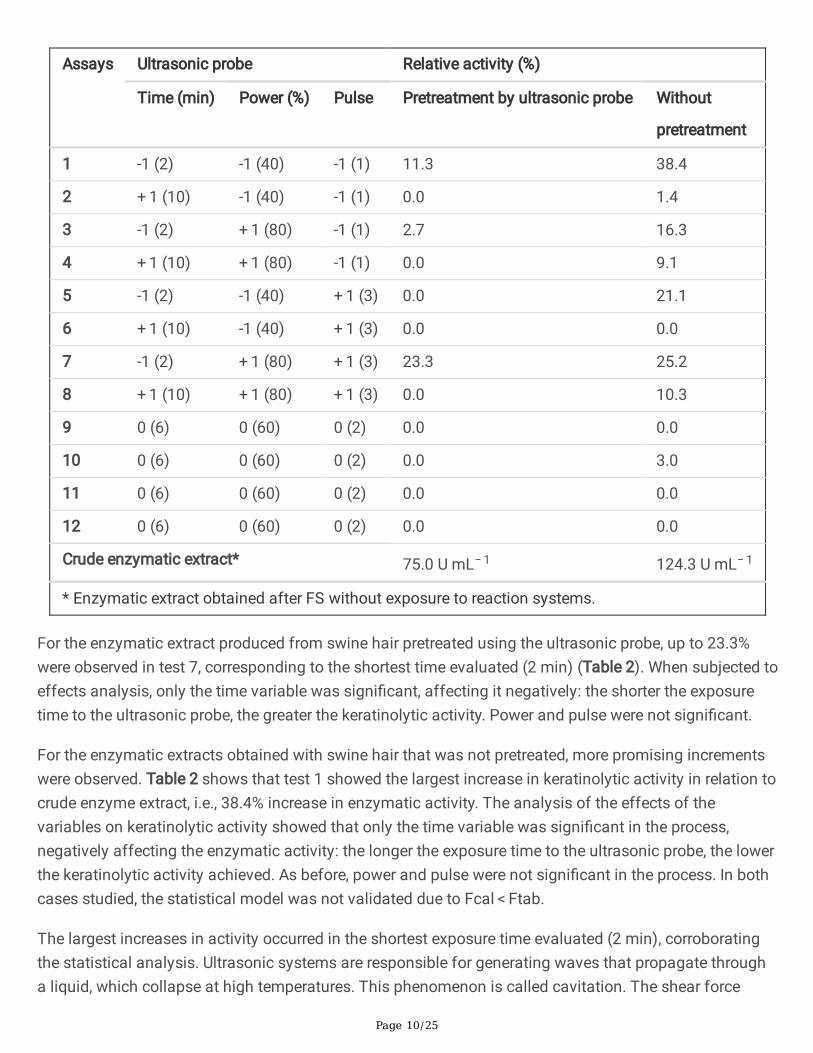
Table 2
Experimental planning matrix (real and coded values) and relative activity results (%) for the conditionsevaluated in the reaction system ultrasonic probe with enzymatic extracts produced from swine hairpretreated by ultrasonic probe and without pretreatment.
Page 10/25
Assays Ultrasonic probe Relative activity (%)
Time (min) Power (%) Pulse Pretreatment by ultrasonic probe Without
pretreatment
1 -1 (2) -1 (40) -1 (1) 11.3 38.4
2 + 1 (10) -1 (40) -1 (1) 0.0 1.4
3 -1 (2) + 1 (80) -1 (1) 2.7 16.3
4 + 1 (10) + 1 (80) -1 (1) 0.0 9.1
5 -1 (2) -1 (40) + 1 (3) 0.0 21.1
6 + 1 (10) -1 (40) + 1 (3) 0.0 0.0
7 -1 (2) + 1 (80) + 1 (3) 23.3 25.2
8 + 1 (10) + 1 (80) + 1 (3) 0.0 10.3
9 0 (6) 0 (60) 0 (2) 0.0 0.0
10 0 (6) 0 (60) 0 (2) 0.0 3.0
11 0 (6) 0 (60) 0 (2) 0.0 0.0
12 0 (6) 0 (60) 0 (2) 0.0 0.0
Crude enzymatic extract* 75.0 U mL− 1 124.3 U mL− 1
* Enzymatic extract obtained after FS without exposure to reaction systems.
For the enzymatic extract produced from swine hair pretreated using the ultrasonic probe, up to 23.3%were observed in test 7, corresponding to the shortest time evaluated (2 min) (Table 2). When subjected toeffects analysis, only the time variable was signi�cant, affecting it negatively: the shorter the exposuretime to the ultrasonic probe, the greater the keratinolytic activity. Power and pulse were not signi�cant.
For the enzymatic extracts obtained with swine hair that was not pretreated, more promising incrementswere observed. Table 2 shows that test 1 showed the largest increase in keratinolytic activity in relation tocrude enzyme extract, i.e., 38.4% increase in enzymatic activity. The analysis of the effects of thevariables on keratinolytic activity showed that only the time variable was signi�cant in the process,negatively affecting the enzymatic activity: the longer the exposure time to the ultrasonic probe, the lowerthe keratinolytic activity achieved. As before, power and pulse were not signi�cant in the process. In bothcases studied, the statistical model was not validated due to Fcal < Ftab.
The largest increases in activity occurred in the shortest exposure time evaluated (2 min), corroboratingthe statistical analysis. Ultrasonic systems are responsible for generating waves that propagate througha liquid, which collapse at high temperatures. This phenomenon is called cavitation. The shear force

Page 11/25
resulting from the explosion of these microbubbles causes rapid heat and mass transfer, promotingincreased enzymatic activity (Jin et al. 2015). Thus, the increase in activity may be a result of the shorterexposure time to the ultrasonic probe that favors the decomposition of interfering molecules and thechange in enzymatic speci�city, making enzymes more easily accessible to the reaction andconsequently increasing their activity, providing an ideal environment for the reaction between enzymeand substrate (McClements 1995; Jin et al. 2015). The enzyme also remains more regular and �exiblebecause thermodynamic parameters such as Ea, ΔH and ΔS are reduced with the use of ultrasound,causing an improvement in its activity and improving the operational stability of enzymes (Ma et al.2011; Wang, Chen and Zhu 2013).
In assays where the exposure time was longer (from 6 to 10 min), the activity was lower and, in somecases, lower than the activity obtained in the enzyme extract without exposure to the ultrasonic probe,possibly due to the enzymatic denaturation caused by the longer exposure time to the ultrasonic probe(Kapturowska, Stolarzewicz and Krzyczkowska 2012). Denaturation of enzymes can occur due to excesspressure, temperature or even shear force generated during the cavitation phenomenon in the ultrasonicsystem (Grintsevich et al. 2001; Potapovich, Eremin and Metelitza 2003).
It is important to note that the use of ultrasonic systems is e�cient in reducing reaction time, promotingincreased enzymatic activity in many cases. On the other hand, depending on the conditions studied,there may be an enzymatic denaturation and consequently a decrease in enzyme activity.
3.2.1.2 Ultrasonic bathThe greatest increase in enzymatic activity obtained for the ultrasonic bath occurred in the enzymaticextract from swine hair pretreated with ultrasonic probe (30.2%), with a temperature of 80 °C during 40minutes of exposure and 0% power, corresponding to test 6.
Analysis of effects on keratinolytic activity showed that temperature and power were important. Thetemperature variable had signi�cant positive effects; that is, the higher the temperature, the higher thekeratinolytic activity. The power variable affected the enzyme activity negatively; the lower the power, thehigher the activity. Time had no in�uence on enzymatic activity. Because Fcal > Ftab, the statistical modelwas validated.
For enzymatic extract produced from swine hair without pretreatment, only test 3, corresponding to atemperature of 30 °C during 10 minutes and 100% power, provided an increase of keratinolytic activity inrelation to the crude enzyme, which was 5.9%. However, when the results were subjected to effectsanalysis, no variable was signi�cant. For this enzyme extract studied, the model was not statisticallyvalidated due to Fcal < Ftab.
In the enzymatic extracts evaluated, different behaviors of the enzyme regarding temperature wereobserved. For the pretreated residues, the enzyme extract increased the activity at a temperature of 80 °Cand for the untreated residues, the only activity increase was at the temperature of 30 °C, the mildestwithin the study range. This shows that the enzymes produced during submerged fermentation with

Page 12/25
pretreated and untreated residues are different in the mode of action compared to the ultrasonic bathreaction system.
The behavior observed for ultrasonic bath may be related to the synergistic effect of ultrasonic waveswith temperature. Several studies show increased activity of enzymes exposed to ultrasonic bath (Leaeset al. 2013; Mulinari et al. 2017). According to the literature, temperature considerably in�uences theincrease or decrease of enzymatic activity. With a rise in temperature the activity may be increased, butdepending on the structure of the enzyme, if there is a very signi�cant rise in temperature, denaturationmay occur (Resa et al. 2009). This behavior was described in the studies by Wang et al. (2011) andOvsianko et al. (2005), where enzymatic denaturation occurred due to the effect of ultrasonic sonicationcausing an increase in temperature, also stimulating the effect of cavitation.
Ultrasonic bath alters the behavior of exposed enzymes. Thus, the use of ultrasonic systems can promoteincreases in enzymatic activity, possibly due to conformational changes in protein structure, in addition toconsiderably reducing the reaction time (Leaes et al. 2013) and also the decrease in activity bydenaturing (Resa et al. 2009).
3.2.1.3 MicrowaveFor the enzymatic extract produced from swine hair pretreated using the ultrasonic probe, test 2 showedan increase of activity, 15.1% at 80 °C for 5 minutes. Performing statistical analysis, we noted that thetemperature variable was signi�cantly positive: the higher the temperature, the higher the keratinolyticactivity achieved. Time had no in�uence on enzymatic activity. The model was statistically validated byFcal > Ftab.
For the enzymatic extract obtained from swine hair without pretreatment, we found that no assay showedan increase of keratinolytic activity. However, performing the statistical analysis, we noted that the timevariable was signi�cantly negative; that is, the longer the microwave exposure time, the lower thekeratinolytic activity achieved. The temperature had no in�uence on enzymatic activity.
The increase in activity obtained for enzymatic extract produced from swine hair pretreated withultrasonic probe was because the microwaves provided an increase in enzymatic activity and an increasein reaction e�ciency due to instantaneous overheating. Causing tremendous agitation of molecules thatinduces an increase in energy collisions, also increasing reaction and conversion rates (Ma et al. 2011;Mazinani, DeLong and Yan 2015).
The mechanism of microwave operation occurs by the interaction of the electromagnetic �eld withmatter. This causes a movement of ions, which in turn cause heat generation by two mechanisms, dipolerotation and ion conduction. In chemical reactions, microwaves cause molecular friction due to thepolarization of molecules. This process is responsible for increasing the friction of these molecules,consequently increasing the temperature and the reaction rate (Lopes et al. 2015). In this manner,microwave irradiation can cause conformational changes in the exposed structure, leading to increased

Page 13/25
enzyme activity or even damage such as enzymatic denaturation (Leonelli and Mason 2010; Lopes et al.2015; Mazinani, DeLong and Yan 2015).
Some authors point out that the use of microwaves can cause changes in thermodynamic properties,providing increased enzymatic activity (Mazinani, DeLong and Yan 2015; Golunski et al. 2017). These�ndings suggest that conformational changes in the structure of enzymes occur when exposed tomicrowaves, possibly due to cleavages that occur during the process and the formation of hot spots byinstant heating.
On the other hand, high temperatures and exposure times cause enzymatic denaturation. Possibly thisoccurred in the assays with enzymatic extract produced from untreated swine hair, where mildtemperatures of 30 °C were su�cient to cause decreased enzyme activity.
Temperature is one of the factors that most in�uence the behavior of enzymes exposed to microwaveirradiation. This causes collisions between molecules to increase, and this causes an increase in energy,causing faster reaction rates. However, at very high temperatures, the reaction rate is reduced due to thedenaturation of enzymes, caused by heat breakage and also the breakdown of ionic and hydrogen bonds,stabilizing the protein structure (Yadav and Borkar 2009; Khan and Rathod 2018).
3.2.1.4 Larger increments of enzymatic activityGiven the results obtained for the three reaction systems evaluated, the ultrasonic probe showed greaterpotential for increased enzymatic activity compared to the other reaction systems (Fig. 1).
The largest increase was obtained using the ultrasonic probe (38.4%) with the enzymatic extractproduced from swine hair without pretreatment. Ultrasonic systems also showed good results ofincreased enzymatic activity for the enzymatic extract produced from swine hair pretreated by ultrasonicprobe, with the increment values being 23.3% for ultrasonic probe and 30.2% for ultrasonic bath. Inaddition, these values were reached under mild conditions of time, power and pulse, making the processmore viable.
3.2.2 Enzymatic concentrationThe enzyme concentration technique was performed to verify the possibility of increasing enzymaticactivity from an economically viable and simple operation method. Enzyme extracts produced fromswine hair without pretreatment were used and the enzyme concentration technique was performed in thepresence of NaCl and acetone.
The homemade extract showed an enzymatic activity of 159.3 U mL− 1 after the concentration technique,increasing the activity value by 53.5% when compared to the crude enzyme extract (103.8 U mL− 1). Theconcentrated enzymatic extract (homemade) was exposed to reaction systems under the conditions ofgreatest activity increase in experimental designs, where it presented stability against the ultrasonic

Page 14/25
probe, ultrasonic bath and microwave, possibly due to the absence of interferents that were separatedduring enzymatic concentration.
After exposure, the concentrated enzymatic extract (homemade) was applied in the degradation of swinehair and chicken feathers, aiming to evaluate the degradation potential of keratinous residues.
3.3 Commercial enzymes and reaction systemsThe commercial enzyme K4519 SIGMA was exposed to ultrasonic probe, ultrasonic bath and microwaveunder the conditions of higher relative activity of homemade enzyme, in order to compare with theobtained data.
The enzyme K4519 SIGMA keratinase had initial activity of 2812.5 U mL− 1. When subjected to anultrasonic probe and bath, it showed an 11.1% increase in enzymatic activity. In microwave irradiation,there was a reduction in activity. There was high stability because it is a highly pure enzyme, with littlevariation when subjected to reaction systems. The activity increase observed for the enzyme K4519SIGMA keratinase was smaller than that obtained for the homemade enzyme when subjected toultrasonic probe (38.4%). This behavior is expected because it is a pure enzyme with high stability.
It should be noted that the production of the homemade enzyme is low-cost because it uses a highlyavailable residue, being an inhomogeneous residue with large variations, in addition to the fact ofdisposal problems secondary to di�cult degradation. The produced enzyme gave good values ofenzymatic activity and activity increases when subjected to reaction systems, becoming competitiveagainst commercial enzymes.
3.4 Evaluation of keratin residue degradationCrude and concentrated enzymatic extracts (homemade) without and with exposure to reaction systemswere evaluated for degradation potential of keratinous residues. We also compared enzymatic extractwith commercial enzyme K4519 SIGMA. Enzyme extracts were incubated at 28 °C for 28 days and theresult was presented based on visible residue degradation and enzymatic activity.
We found that, for the crude enzyme extract and after exposure to reaction systems, the values forkeratinolytic activity of the assays containing swine hair and chicken feathers showed small decreases inrelation to the initial value for enzymatic activity over the 28 days, however, remaining stable over time,suggesting that the enzyme was active and showing very promising enzyme activity. For the enzymeextract in concentrated form and after exposure to reaction systems, signi�cant increases in keratinolyticactivity were observed over the 28 days of study (Table 3).
Table 3
Measure of keratinolytic activity at initial time and after 28 days for homemade enzymatic extract crudeand concentrated and enzymatic extracts after exposure to ultrasonic reaction systems and microwaveirradiation containing swine hair and chicken feathers.

Page 15/25
CRUDE Enzyme Extracts (homemade) Swine hair Chicken feathers
Assays 0° day 28° day 0° day 28° day
Enzymatic Activity
(U mL− 1)
Enzymatic Activity
(U mL− 1)
Ultrasonic Probe 172.0 137.0 172.0 133.0
Ultrasonic Bath 153.0 124.0 153.0 115.5
Microwave 96.0 106.5 96.0 141.0
Crude Extract 171.0 151.5 171.0 121.5
CONCENTRATED Enzyme Extracts (homemade) Swine hair Chicken feathers
Assays 0° day 28° day 0° day 28° day
Enzymatic Activity
(U mL− 1)
Enzymatic Activity
(U mL− 1)
Ultrasonic Probe 159.5 231.0 159.5 192.5
Ultrasonic Bath 141.8 202.0 141.8 181.5
Microwave 149.0 229.5 149.0 191.5
Crude Extract 103.8 - 103.8 -
Concentrated Extract 159.3 - 159.3 -
Almost total degradation of chicken feathers was observed in the crude enzyme extract and enzymeextracts after exposure to reaction systems (Fig. 2), while for swine hair, there was no degradation in anyof the enzyme extracts evaluated. Possibly the enzyme could not access the resistant structure of swinehair. One factor that may explain the ease with which chicken feathers were degraded is the less rigidstructure compared to swine hair. Because they are part of the keratin family, swine hair exhibits astructure that is extremely resistant to physical, chemical and also enzymatic actions (Choińska-Pulit,Łaba and Rodziewicz 2019).
The degradation potential of chicken feathers containing the commercial enzyme extract K4519 SIGMAwas also evaluated. We found that, during the 28 days of incubation, the commercial enzyme was notable to degrade the chicken feathers, thereby favoring the homemade enzyme extract (Fig. 2).
The results obtained for the degradation test containing the homemade extracts in crude form and afterexposure to the reaction systems are promising as evidenced by the degradation potential on keratinousresidues. The commercial enzyme K4519 SIGMA is expensive, highly concentrated and stable;nevertheless, it did not degrade the chicken feathers used in the tests.

Page 16/25
Swine hair and chicken feathers are part of the group of hard keratins owing to their high concentrationsof cysteine, rendering them very resistant (Korniłłowicz-Kowalska and Bohacz 2011). However, α-keratinpresent in the swine hair structure has large amounts of sulfur, which confers highly resistant mechanicalproperties (Holkar et al. 2018). This explains why chicken feathers are more easily degraded than swinehair.
In addition, the keratin structure formed by disul�de bonds ensures stability and resistance to enzymaticdegradation. Kunert (1989) found that the reduction of disul�de bonds considerably in�uenced thedegradation of keratin structure, ensuring e�cient keratin hydrolysis. This suggests that the homemadeenzyme extract acted on disul�de bonds and reduced the cystine concentration present in the keratinstructure, as well as converting the sulfur molecule into smaller structures (Jaouadi et al. 2013).
However, the exact process of degradation involves a series of mechanisms of action that occur in 3stages. Mechanical keratinolysis occurs by the action of mycelium-producing fungi, involving thepenetration of the mycelium into the keratin residue. Then, the hydrolysis of the disul�de bonds occurs,responsible for ensure the resistance and stability of the keratin structure. Finally, proteolysis occursinvolving the action of the enzyme itself (Kunert 1989; Onifade et al. 1998).
For the concentrated enzymatic extract, no residue degradation was observed in any of the evaluatedextracts. In the tests where there were high values of keratinolytic activity, the degradation percentageswere low or almost nonexistent. This agrees with the study by Riffel and Brandelli (2006), where highvalues for keratinolytic activity were veri�ed; nevertheless, the puri�ed enzyme was not able to degradechicken feathers. The authors suggest that in these cases, enzymes such as disul�de reductases andproteases need to act so that feather degradation occurs (Fang et al. 2013b).
The presence of inhibitors may explain why there was no residue degradation (Hamiche et al. 2019).Despite the fact that the enzyme is concentrated and the amount of impurities is reduced, it has nopotential for degradation of residues (Riffel and Brandelli 2006).
3.5 Characterization of homemade keratinase enzymesThe characterization of homemade keratinases was carried out by evaluating different pH andtemperature conditions in an ultrathermostatic bath for 1 h. The highest enzyme activity was achieved atpH 8.5 (50 mM Tris HCl buffer) and 50 °C temperature.
Most of keratinases producing microorganisms operate in the neutral and alkaline pH range, and thedegradation of keratin is favored in alkaline pH, with the modi�cation of cysteine residues and makingthem more accessible for the action of the enzyme. The optimum temperature is in the range of 28 °C to50 °C, reaching 70 °C depending on the microorganism. In addition, most keratinases have good stabilityover wide pH and temperature ranges (Anbu et al. 2005).
4. Conclusion

Page 17/25
Biotechnological processes involving the biodegradation of keratinolytic materials by action ofmicroorganisms and enzymes are promising alternatives to improve the degradability of these materials,thereby adding value to the material. Ultrasonic and microwave reaction systems are viable alternativesfor evaluating the behavior of keratinases and the potential for pretreatment of keratinous residues.
The pretreatment in an ultrasonic probe using the macropoint causes greater energy dissipation due tothe larger diameter in relation to the micropoint, thus increasing the reaction rates and facilitating thedegradation process of the exposed structures due to the propagation of ultrasonic waves, resulting ingreater weight loss. Thus, the use of the ultrasonic probe as a pretreatment can be quite useful whenlooking for greater enzyme production in a shorter time. For longer processes, the use of waste withoutpretreatment becomes more promising, also decreasing energy expenditure, since the enzymatic activityachieved over time is greater.
Regarding the evaluation of the behavior of keratinases in relation to reaction systems, differentbehaviors were observed from enzymatic extracts, showing that it is possible to produce enzymes withdifferent characteristics and behaviors. The ultrasonic probe proved to be more promising, generating thegreatest increase in enzymatic activity. The enzymatic concentration showed a signi�cant increase inactivity, showing that simple and low-cost techniques can be e�cient.
It was found that the crude enzymatic extract has the potential to degrade chicken feathers, while theother extracts used did not show potential for degradation, thus making it a very interesting result.
DeclarationsEthics approval and consent to participate
Not applicable
Consent for publication
All authors agreed with this publication.
Availability of data and materials
The datasets generated for this study are available on request to the corresponding author.
Competing interests
There are not competing interests
Funding
CAPES, CNPq and FAPERGS
Authors' contributions

Page 18/25
FFC, SK, KPP, CD, TS, CB, FSS, AFC and JZ: experimental procedures, results discussion, and datatreatment AJM, GF, HT: research coordinators
Acknowledgments
The authors are grateful to the Fundação de Amparo à Pesquisa do Estado do Rio Grande do Sul -FAPERGS, to the Coordenação de Aperfeiçoamento de Pessoal de Nível Superior - CAPES; to the ConselhoNacional de Desenvolvimento Cientí�co e Tecnológico - CNPq; and to the research group at Laboratóriode Microbiologia e Bioprocessos - LAMIBI da UFFS campus Erechim.
References1. Abdel-Fattah A M, El-Gamal M S, Ismail S A, Emran M A, Hashem A M (2018) Biodegradation of
feather waste by keratinase produced from newly isolated Bacillus licheniformis J Genet EngBiotechnol 16: 311-318. doi: 10.1016/j.jgeb.2018.05.005
2. Anbu P, Gopinath S C B, Hilda A, Lakshmi priya T, Annadurai G (2005) Puri�cation of keratinase frompoultry farm isolate-Scopulariopsis brevicaulis and statistical optimization of enzyme activity.Enzyme Microb Technol 36: 639-647. doi: 10.1016/j.enzmictec.2004.07.019
3. ANTON PAAR. Reator de Micro-ondas Monowave 100 ANTON PAAR. Available in: https://www.anton-paar.com/br-pt/produtos/detalhes/sintese-assistida-por-micro-ondas-monowave-400200/. Accessed25 May 2018.
4. Azmi N A, Idris A, Yusof N S M (2018) Ultrasonic technology for value added products from featherkeratin. Ultrason Sonochem 47: 99-107. doi: 10.1016/j.ultsonch.2018.04.016
5. Barone J R, Schmidt W F, Gregoire N T (2006) Extrusion of Feather Keratin. J Appl Polym Sci 100:1432-1442. doi: 10.1002/app.23501
�. Barton S, Bullock C, Weir D (1996) The effects of ultrasound on the activities of some glycosidaseenzymes of industrial importance. Enzyme Microb Technol 18: 190-194. doi: 10.1016/0141-0229(95)00092-5
7. Brandelli A (2008) Bacterial Keratinases: Useful Enzymes for Bioprocessing Agroindustrial Wastesand Beyond. Food Bioproc Tech 1: 105-116. doi: 10.1007/s11947-007-0025-y
�. Brandelli A, Daroit D J, Riffel A (2010) Biochemical features of microbial keratinases and theirproduction and applications. Appl Environ Microbiol 85: 1735-1750. doi: 10.1007/s00253-009-2398-5
9. Brandelli A, Sala L, Kalil S J (2015) Microbial enzymes for bioconversion of poultry waste into added-value products. Food Res Int 73: 3-12. doi: 10.1016/j.foodres.2015.01.015

Page 19/25
10. Bressollier P, Letourneau F, Urdaci M, Verneuil B (1999) Puri�cation and Characterization of aKeratinolytic Serine Proteinase from Streptomyces albido�avus. Appl Environ Microbiol 65: 2570-2578. doi: 10.1128/AEM.65.6.2570-2576.1999
11. Călin M, Constantinescu-Aruxandei D, Alexandrescu E, Răut I, Doni M B, Arsene M L, Oancea F, Jecu L,Lazăr V (2017) Degradation of keratin substrates by keratinolytic fungi. Electron J Biotechn 28: 101-112. doi: 10.1016/j.ejbt.2017.05.007
12. Choińska-Pulit A, Łaba W, Rodziewicz A (2019) Enhancement of pig bristles waste bioconversion byinoculum of keratinolytic bacteria during composting. Waste Manage 84: 269-276. doi:10.1016/j.wasman.2018.11.052
13. ECO-SONICS. Desruptor de células. Available in:https://www.ecosonics.com.br/produto/20/desruptor-de-celulas. Accessed 02 June 2018.
14. Fang Z, Zhang J, Liu B, Du G, Chen J (2013a) Biodegradation of wool waste and keratinaseproduction in scale-up fermenter with different strategies by Stenotrophomonas maltophilia BBE11-1. Bioresour Technol 140: 286-291. doi: 10.1016/j.biortech.2013.04.091
15. Fang Z, Zhang J, Liu C, Du G, Chen J (2013b) Biochemical characterization of three keratinolyticenzymes from Stenotrophomonas maltophilia BBE11-1 for biodegrading keratin wastes. IntBiodeterior Biodegrad 82: 166-172. doi: 10.1016/j.ibiod.2013.03.008
1�. Gegeckas A, Šimkutė A, Gudiukaitė R, Čitavičius D J (2018) Characterization and application ofkeratinolytic paptidases from Bacillus Int J Biol Macromol 113: 1206-1213. doi:10.1016/j.ijbiomac.2018.03.046
17. Golunski S M, Scapini T, Modkovski T A, Marques C T, Camargo A F, Preczeski K P, Dalla Rosa C,Baldissarelli D P, Mulinari J, Venturin B, Vargas G D L P, Buffon J G, Mossi A J, Treichel H (2017)Commercial and Noncommercial Peroxidases Activity under Ultrasound and Microwave Treatment: aPretreatment to Improve Wastewater Treatment. J Braz Chem Soc 28: 1890-1895. doi:10.21577/0103-5053.20170023
1�. Grintsevich E E, Adzerikho I E, Mrochek A G, Metelitza D I (2001) Polydisul�des of SubstitutedPhenols as Effective Protectors of Peroxidase against Inactivation by Ultrasonic Cavitation.Biochemistry 66: 740-746. doi: 10.1023/a:1010256511200
19. Gupta R, Ramnani P (2006) Microbial keratinases and their prospective applications: an overview.Appl Environ Microbiol 70: 21-33. doi: 10.1007/s00253-005-0239-8
20. Hamiche S, Mechri S, Khelouia L, Annane R, El Hattab M, Badis A, Jaouadi B (2019) Puri�cation andbiochemical characterization of two keratinases from Bacillus amyloliquefaciens S13 isolated frommarine brown alga Zonaria tournefortii with potential keratin-biodegradation and hide-unhairingactivities. Int J Biol Macromol 122: 758-769. doi: 10.1016/j.ijbiomac.2018.10.174

Page 20/25
21. Holkar C R, Jain S S, Jadhav A J, Pinjari D V (2018) Valorization of keratin waste. Process SafEnviron 115: 85-98. doi: 10.1016/j.psep.2017.08.045
22. Ire F S, Onyenama A C (2017) Effects of Some Cultural Conditions on Keratinase Production byBacillus licheniformis Strain NBRC 14206. J Adv Biol Biotechnol 13: 1-13. doi:10.9734/JABB/2017/32726
23. Jaouadi N Z, Rekik H, Badis A, Trabelsi S, Belhoul M, Yahiaoui A B, Aicha H B, Toumi A, Bejar S,Jaouadi B (2013) Biochemical and Molecular Characterization of a Serine Keratinase fromBrevibacillus brevis US575 with Promising Keratin-Biodegradation and Hide-Dehairing Activities.PLoS One 8: 1-17. doi: 10.1371/journal.pone.0076722
24. Jin J, Ma H, Qu W, Wang K, Zhou C, He R, Luo L, Owusu J (2015) Effects of multi-frequency powerultrasound on the enzymolysis of corn gluten meal: Kinetics and thermodynamics study. UltrasonSonochem 27: 46-53. doi: 10.1016/j.ultsonch.2015.04.031
25. Kapturowska A U, Stolarzewicz I A, Krzyczkowska J (2012) Studies on the lipolytic activity ofsonicated enzymes from Yarrowia lipolytica. Ultrason Sonochem 19: 186-191. doi:10.1016/j.ultsonch.2011.06.015
2�. Kaul S, Sumbali G (1997) Keratinolysis by poultry farm soil fungi. Mycopathologia 139: 137-140. doi:10.1023/a:1006896030739
27. Khan N R, Rathod V K (2018) Microwave assisted enzymatic synthesis of speciality esters: A mini -review. Process Biochem 75: 89-98. doi: 10.1016/j.procbio.2018.08.019
2�. Korniłłowicz-Kowalska T, Bohacz J (2011) Biodegradation of keratin waste: Theory and practicalaspects. Waste Manage 31: 1689-1701. doi: 10.1016/j.wasman.2011.03.024
29. Kunert K (1989) Biochemical mechanism of keratin degradation by the actinomycete Streptomycesfradiae and the fungus Microsporum gypseum: A comparison. J Basic Microbiol 29: 597-604. doi:1002/jobm.3620290909
30. Kushwaha R K S (1983) The In Vitro Degradation of Peacock Feathers by Some Fungi. Mykosen 26:324-326. doi: 10.1111/j.1439-0507.1983.tb03218.x
31. Łaba W, Kopeć W, Chorążyk D, Kancelista A (2015) Biodegradation of pretreated pig bristles byBacillus cereus Int Biodeterior Biodegrad 100: 116-123. doi: 10.1016/j.ibiod.2015.02.024
32. Leaes E X, Lima D, Miklasevicius L, Ramon A P, Dal Prá V, Bassaco M M, Terra L M, Mazutti M A(2013) Effect of ultrasound-assisted irradiation on the activities of α-amylase and amyloglucosidase.Biocatal Agric Biotechnol 2: 21-25. doi: 10.1016/j.bcab.2012.08.003

Page 21/25
33. Leonelli C, Mason T J (2010) Microwave and ultrasonic processing: Now a realistic option forindustry. Chem Eng Process 49: 885-900. doi: 10.1016/j.cep.2010.05.006
34. Lippert T, Bandelin J, Musch A, Drewes J E, Koch K (2018) Energy-positive sewage sludge pre-treatment with a novel ultrasonic �atbed reactor at low energy input. Bioresour Technol 264: 298-305. doi: 10.1016/j.biortech.2018.05.073.
35. Lopes L C, Barreto M T M, Gonçalves K M, Alvarez H M, Heredia M F, Souza R O M A, Cordeiro Y,Dariva C, Fricks A T (2015) Stability and structural changes of horseradish peroxidase: Microwaveversus conventional heating treatment. Enzyme Microb Technol 69: 10-18. doi:10.1016/j.enzmictec.2014.11.002
3�. Ma H, Huang L, Jia J, He R, Luo L, Zhu W (2011) Effect of energy-gathered ultrasound on Alcalase.Ultrason Sonochem 18: 419-424. doi: 10.1016/j.ultsonch.2010.07.014
37. Manasseh R, Tho P, Ooi A, Petrovic-Duran K, Zhu Y (2010) Cavitation microstreaming and materialtransport around microbubbles. Phys Procedia 3: 427-432. doi: 10.1016/j.phpro.2010.01.056
3�. Mason T J, Cobley A J, Graves J E, Morgan D (2011) New evidence for the inverse dependence ofmechanical and chemical effects on the frequency of ultrasound. Ultrason Sonochem 18: 226-230.doi: 10.1016/j.ultsonch.2010.05.008
39. Mazinani S A, DeLong B, Yan H (2015) Microwave radiation accelerates trypsin-catalyzed peptidehydrolysis at constant bulk temperature. Tetrahedron Lett 56: 5804-5807. doi:10.1016/j.tetlet.2015.09.003
40. Mazotto A M, Melo A C N, Macrae A, Rosado A S, Peixoto R, Cedrola S M L, Couri S, Zingali R B, VillaA L V, Rabinovitch L, Chaves J Q, Vermelho A B (2011) Biodegradation of feather waste byextracellular keratinases and gelatinases from Bacillus World J Microbiol Biotechnol 27: 1355-1365.doi: 10.1007/s11274-010-0586-1
41. Mazotto A M, Couri S, Damaso M C T, Vermelho A B (2013) Degradation of feather waste byAspergillus niger keratinases: Comparison of submerged and solid-state fermentation. Int BiodeteriorBiodegrad 85: 189-195. doi: 10.1016/j.ibiod.2013.07.003
42. McClements D J (1995) Advances in the application of ultrasound in food analysis and processing.Trends Food Sci Tech 6: 293-299. doi: 1016/S0924-2244(00)89139-6
43. Mulinari J, Venturin B, Sbardelotto M, Dall Agnol A, Scapini T, Camargo A F, Baldissarelli D P,Modkovski T A, Rossetto V, Dalla Rosa C, Reichert Jr. F W, Golunski S M, Vargas G D L P, Dalla RosaC, Mossi A J, Treichel H (2017) Ultrasound-assisted hydrolysis of waste cooking oil catalyzed byhomemade Ultrason Sonochem 35: 313-318. doi: 10.1016/j.ultsonch.2016.10.007

Page 22/25
44. Okoroma E A, Garelick H, Abiola O O, Purchase D (2012) Identi�cation and characterisation of aBacillus licheniformis strain with profound keratinase activity for degradation of melanised feather.Int Biodeterior Biodegrad 74: 54-60. doi: 10.1016/j.ibiod.2012.07.013
45. Onifade A A, Al-Sane N A, Al-Musallam A A, Al-Zarban S (1998) A Review: Potentials forbiotechnological applications of keratin-degrading microorganisms and their enzymes for nutritionalimprovement of feathers and other keratins as livestock feed resources. Bioresour Technol 66: 1-11.doi: 1016/S0960-8524(98)00033-9
4�. Ovsianko S L, Chernyavsky E A, Minchenya V T, Adzerikho I E, Shkumatov V M (2005) Effect ofultrasound on activation of serine proteases precursors. Ultrason Sonochem 12: 219-223. doi:10.1016/j.ultsonch.2003.10.012
47. Paul T, Das A, Mandal A, Halder S K, Jana A, Maity C, DasMohapatra P K, Pati B R, Mondal K C(2014) An e�cient cloth cleaning property of a crude keratinase combined with detergent: Towardsindustrial viewpoint. J Clean Prod 66: 672-684. doi: 10.1016/j.jclepro.2013.10.054
4�. Potapovich M V, Eremin A N, Metelitza D I (2003) Kinetics of Catalase Inactivation Induced byUltrasonic Cavitation. Appl Biochem Micro 39: 140-146. doi: 10.1023/A:1022577611056
49. Preczeski K P, Kamanski A B, Scapini T, Camargo A F, Modkoski T A, Rossetto V, Venturin B, MulinariJ, Golunski S M, Mossi A J, Treichel H (2018) E�cient and low-cost alternative of lipaseconcentrating aiming at the application in the treatment of waste cooking oils. Bioprocess BiosystEng 41: 851-857. doi: 10.1007/s00449-018-1919-y
50. Preczeski K P, Dalastra C, Czapela F F, Kubeneck S, Scapini T, Camargo A F, Zanivan J, Bonatto C,Stefanski F S, Venturin B, Fongaro G, Treichel H (2020) Fusarium oxysporum and Aspergillus askeratinase producers using swine hair from agroindustrial residues. Fronti Bioeng Biotech. doi:10.3389/fbioe.2020.00071
51. Resa P, Elvira L, Sierra C, Espinosa F M (2009) Ultrasonic velocity assay of extracellular invertase inliving yeasts. Anal Biochem 384: 68-73. doi: 10.1016/j.ab.2008.09.025
52. Riffel A, Brandelli A (2006) Keratinolytic bacteria isolated from feather waste. Braz J Microbiol 37:395-399. doi: 10.1590/S1517-83822006000300036
53. Rodrigues M I, Iemma A F (2014) Protimiza Experimental Design. Available in: http://experimental-design.protimiza.com.br/. Accessed 10 January 2019.
54. Santos R M D B, Firmino A A P, Sá C M, Felix C R (1996) Keratinolytic Activity of Aspergillusfumigatus Curr Microbiol 33: 364-370. doi: 10.1007/s002849900129
55. Scott J A, Untereiner W A (2004) Determination of keratin keratin degradation by fungi using keratinazure. Med Mycol J 42: 239-246. doi: 10.1080/13693780310001644680

Page 23/25
5�. SIGMA-ALDRICH. K4519 SIGMA Keratinase. Available in:https://www.sigmaaldrich.com/catalog/product/sigma/k4519?lang=pt®ion=BR>. Accessed 28May 2018.
57. Su C, Gong J, Zhang R, Tao L, Dou W, Zhang D, Li H, Lu Z, Xu Z, Shi J (2017) A novel alkalinesurfactant-stable keratinase with superior feather-degrading potential based on library screeningstrategy. Int J Biol Macromol 95: 404-411. doi: 10.1016/j.ijbiomac.2016.11.045
5�. Thankaswamy S R, Sundaramoorthy S, Palanivel S, Ramudu K N (2018) Improved microbialdegradation of animal hair waste from leather industry using Brevibacterium luteolum (MTCC 5982).J Clean Prod 189: 701-708. doi: 10.1016/j.jclepro.2018.04.095
59. Wang L C (1981) Soybean Protein Agglomeration: Promotion by Ultrasonic Treatment. J Agr FoodChem 29: 177-180. doi: 10.1021/jf00103a044
�0. Wang J, Yanping C, Sun B, Wang C, Mo Y (2011) Effect of ultrasound on the activity of alliinase fromfresh garlic. Ultrason Sonochem 18: 534-540. doi: 10.1016/j.ultsonch.2010.09.008
�1. Wang F, Chen Z, Zhu H (2013) An e�cient enzymatic modi�cation of lily polysaccharide in ionicliquid under ultrasonic irradiation. Biochem Eng J 79: 25-28. doi: 10.1016/j.bej.2013.06.020
�2. Yadav G D, Borkar I V (2009) Kinetic and Mechanistic Investigation of Microwave-Assisted LipaseCatalyzed Synthesis of Citronellyl Acetate. Ind Eng Chem Res 48: 7915-7922. doi:10.1021/ie800591c
�3. Yang C, Fang T J (2015) Kinetics for enzymatic hydrolysis of rice hulls by the ultrasonic pretreatmentwith a bio-based basic ionic liquid. Biochem Eng J 100: 23-29. doi: 10.1016/j.bej.2015.04.012
�4. Yusof N S M, Ashokkumar M (2015) Sonochemical Synthesis of Gold Nanoparticles by Using HighIntensity Focused Ultrasound. ChemPhysChem 16: 775-781. doi: 10.1002/cphc.201402697
�5. Yusuf I, Ahmad S A, Phang L Y, Syed M A, Shamaan N A, Khalil K A, Dahalan F A, Shukor M Y (2016)Keratinase production and biodegradation of polluted secondary chicken feather wastes by a newlyisolated multi heavy metal tolerant bacterium-Alcaligenes AQ05-001. J Environ Manage 183: 182-195. doi: 10.1016/j.jenvman.2016.08.059
��. Zou S, Wang X, Chen Y, Wan H, Feng Y (2016) Enhancement of biogas production in anaerobic co-digestion by ultrasonic pretreatment. Energ Convers Manage 112: 226-235. doi:10.1016/j.enconman.2015.12.087
Figures

Page 24/25
Figure 1
Larger increments of enzymatic activity of enzymatic extracts obtained from swine hair pretreated usingthe ultrasonic probe and without pretreatment subjected to the reaction systems ultrasonic probe,ultrasonic bath and microwave.

Page 25/25
Figure 2
Total degradation of chicken feathers observed in crude enzymatic extracts and after exposure toreaction systems. A) Control; B) Crude; C) Concentrated; D) K4519 SIGMA commercial enzyme.
Supplementary Files
This is a list of supplementary �les associated with this preprint. Click to download.
GraphicalAbstract.docx