Embed Size (px)
Citation preview

WARM UP
How would you justify the scientific claim that organisms share many conserved core processes and features that evolved and are widely distributed among organisms today?

The following structural evidence supports the relatedness of all eukaryotes: the cytoskeleton, linear chromosomes, endomembrane system & organelles (ex. mitochondria & chloroplasts).
All have the same structure and function despite whether found in animal, plant, fungal, or protist cells. This means these structures and the genes that code for them existed in the common ancestor to all eukaryotes.
Eukaryotes evolved due to endosymbiosis of various prokaryotes.

Ch 7 continued

CELL COATING
• The exterior of animal cell membranes have short chains of carbohydrates bound to proteins or lipids:
1) carb – proteins = glycoproteins.2) carb – lipids = glycolipids.• Called glycocalyx or extracellular matrix (ECM)
FUNCTIONS OF THE glycocalyx / ECM:1) recognition sites (cell to cell for tissue formation)- to hold cells together.2) identification markers (ex. MHC markers allow immune system cells to
distinguish self from foreign cells)3) communication (hormone messenger receptors)

Glycocalyx/cell coating/ ECM

How are cells connected?

CELL TO CELL ADHESION1) Intercellular matrix (ECMs of adjacent cells)
a) Collagen- the most abundant glycoprotein, protein fibers that bind cells togetherb) Elastin- protein fiber that binds cells together
2) Cell junctionsa) desmosomes = anchoring junctions (plaques & fibers) “rivets”, fasten cells together in strong sheets (keratin- intermediate filament)b) tight junctions = proteins that tie cells together, leaving no space between the cells- cells fused (ie. intestines)c) communication junctions (2 kinds) allow flow of salt ions, sugars, amino acids- cytoplasmic channels between adjacent cells. (ie. heart muscle cells, cells of embryo)
– gap junction (animal cells) membrane channels that allow passage of material between cells.
– Plasmodesmata (plant cells) openings in the cell wall where adjacent membranes contact each other.

desmosome (anchoring junction)
(plaques & fibers) “rivets”
fasten cells together in strong sheets.
keratin- intermediate filament.

tight junction
tight junctions = proteins that tie cells together, leaving no space between the cells- cells fused (ie. intestines)

Gap (communicating junction)communication junctions (2 kinds) allow flow of salt ions, sugars, amino
acids- cytoplasmic channels between adjacent cells.
ie. heart muscle cells, cells of embryo1) gap junction (animal cells)
membrane channels that allow passage of material between cells.
2) Plasmodesmata (plant cells) openings in the cell wall where adjacent membranes contact each other.

Figure 7.30 Intercellular junctions in animal tissues

The End

Growth, reproduction and dynamic homeostasis require that cells create and maintain internal environments that are different from their external environments.
Q: What structural component accomplishes this?

A: cell membrane
membrane structure & functionChapter 8

I. Cell membranes are selectively permeable due to their structure.
a. Cell membranes separate the internal environment of the cell from the external environment.
b. Selective permeability is a direct consequence of membrane structure, as described by the fluid mosaic model.
c. Cell walls provide a structural boundary, as well as a permeability barrier for some substances to the internal environments.

How is the structure of a cell membrane similar to an
oreo cookie????
How is it different????? What property does this give cell membranes?

Phospholipids give the membrane both hydrophilic and hydrophobic properties (amphipathic).
The hydrophilic phosphate portions of the phospholipids are oriented toward the aqueous external or internal environments,
while the hydrophobic fatty acid portions face each other within the interior of the membrane itself.

The membrane has a hydrophobic region sandwiched between two
hydrophillic ones… just like the oreo.• Phospholipids are
amphipathic molecules- have both a nonpolar and polar region.
• Small uncharged particles can diffuse through the phospholipid bilayer…
ex. O2, CO2, N2
• But charged molecules (ex. H20) are repelled.
How do they get in???

• The plasma membrane is filled with a group of proteins called membrane proteins. • Some of these membrane proteins show only contact surfaces with either the inside or the outside of the cell (peripheral proteins).• and some of them stick out at both ends (transmembrane proteins) providing hydrophillic passageways for solutes repelled by the bilayer’s
hydrophobic tails. • MISCONCEPTION: the lipid bilayer is just that, a lipid bilayer.• The presence of proteins & the assortment of these proteins differs from cell to cell making each “selectively” permeable to polar.•
.

I. Cell membranes are selectively permeable due to their structure.
Cell membranes consist of a structural framework of phospholipid molecules, embedded proteins, cholesterol, glycoproteins and glycolipids.

Singer & Nicolson’sFluid Mosaic Model
• Proteins molecules “bobbing” in fluid bilayer of phospholipids.
• FLUID MOSAIC MODELphospholipid bilayer = groutproteins=tiles
• Hydrophilic regions of protein protrude into water
• Hydrophobic regions embedded in nonaqueous environment of “tails”

cholesterol 1.reduces fluidity of phospholipids2.prevents solidification

Figure 8.5 Evidence for the drifting of membrane proteins
Embedded proteins can be hydrophilic, with charged and polar side groups, or hydrophobic, with nonpolar side groups…
or both.
“fluid”mosaic
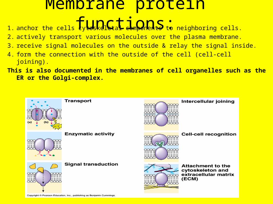
Membrane protein functions:1. anchor the cells cytoskeletal components to neighboring cells.
2. actively transport various molecules over the plasma membrane.
3. receive signal molecules on the outside & relay the signal inside.
4. form the connection with the outside of the cell (cell-cell joining).
This is also documented in the membranes of cell organelles such as the ER or the Golgi-complex.

TRAFFIC ACROSS MEMBRANES
• plasma membrane edge of life 8 nm thick
• Primary function-separates life from its nonliving environment and regulates interactions.
• Selective permeability- characteristic due to protein composition of cell membrane.
Growth and dynamic homeostasis are maintained by the constant movement of molecules across membranes.

plasma membranes are: SELECTIVELY PERMEABLE- allow some materials to cross
based on transmembrane proteins present.
easy time crossing:
• N2, O2 & CO2
• Hydrophobic molecules
difficulty crossing (needs specific protein tunnel):
• Polar/Hydrophillic molecules
ex. (glucose & H2O)
• Ions
ex. (Na+, K+, Ca 2+, Cl-)

RECAP
• Small, uncharged polar molecules and small nonpolar molecules, such as N2, freely pass across the membrane.
• Hydrophilic substances such as large polar molecules and ions move across the membrane through embedded channel and transport proteins.
• Water moves across membranes and through channel proteins called aquaporins.

transport proteins = permeases
• provide a hydrophilic channel across a membrane
• are selective for a particular solute
• Examples: 1. water channel (aquaporin)2. glucose not fructose

Permeases are transport proteins
Integral membrane proteins that facilitate the transport of a specific molecule into or out of a cell.
Similar to enzymes:- specific “substrate”- binding site- can be saturated- can be inhibited by mimics- do not catalyze a chemical
reaction… just a physical process.

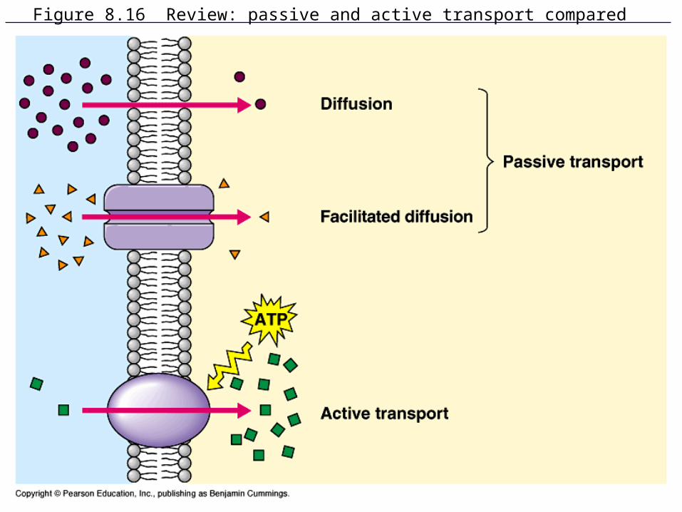
Figure 8.16 Review: passive and active transport compared

Passive transport does not require the input of metabolic energy; the net movement of molecules is from high concentration to low concentration.
1. Passive transport plays a primary role in the import of resources and the export of wastes.
2. Membrane proteins play a role in facilitated diffusion of charged and polar molecules through a membrane.– Glucose transport– Na+ transport– K+ transport

PASSIVE TRANSPORT
• NO ENERGY (ATP) REQUIRED • diffusion of a substance across a biological membrane.• Molecules travel DOWN their concentration gradient
(difference)• [HIGH] --> [LOW]• Like walking down stairs
3 TYPES:1. Diffusion2. Osmosis3. Facilitated diffusion
Transport Proteins & Gated Membrane Channels
Growth and dynamic homeostasis are maintained by the constant movement of molecules across membranes.

Figure 8.10 The diffusion of solutes across membranes

1) diffusion
• kinetic energy/thermal energy- molecules spread out- space.
• atoms or molecules move down a concentration gradient.
• from areas of high to low concentration
• “passive transport” is diffusion across a membrane (no ATP)
• ie. CO2, O2
Influenced by the temperature of the environment… kinetic energy.

2. OSMOSIS = the diffusion of water molecules across a selectively permeable membrane
where will water move?1) to the side with less water.2) to the side with more dissolved
solute.3) From the hypotonic side of the
membrane to the hypertonic one.osmotic solutions that a cell may
encounter:1. Hypertonic = solution with
higher [solute]*2. Isotonic = solution with the
same [solute]3. Hypotonic = solution with lower
[solute] *[solute] = solute concentration

if the environment is: / then inside the cell it is:1. Hypotonic2. Hypertonic3. Isotonic
1. Hypertonic2. Hypotonic3. Isotonic

Figure 8.13 The contractile vacuole of Paramecium: an evolutionary adaptation for osmoregulation
A PARAMECIUM’S ENVIRONMENT IS ___________

Figure 8.13 The contractile vacuole of Paramecium: an evolutionary adaptation for osmoregulation
A PARAMECIUM’S ENVIRONMENT IS HYPOTONICSO IT NEEDS TO PUMP OUT THE WATER OR BURST

factors that influence osmosis:1) osmotic concentration- refers to the concentration of solutes (dissolved
substances) in the water.• Water will flow from the side with the low osmotic concentration to the side of
high concentration.• The solutes will flow from the side with high concentration to the side with low
osmotic concentration.2) Osmotic Potential- tendency of water to move from one region to another. • Measure of the potential of water molecules to move between regions of differing
concentrations across a water-permeable membrane.• Water moves from the area of greater osmotic potential to the area of lower
osmotic potential.• Water moves from a hypotonic solution (more water, less solutes) to a
hypertonic solution (less water, more solutes) across a semi permeable membrane.

1. channel proteins provide hydrophylic passagewaysie. Water passes through “aquaporins”
ion channels2. carrier proteins undergo a subtle change in shape that
translocates the solute-binding site across the membraneie. glucose, amino acids, nucleotides
3. Facilitated Diffusion (Transport) 2 kinds:

4. Gated Membrane ChannelsA stimulus (electrical or chemical) causes these channel proteins to open or close.
ex. Stimulation of a nerve cell by a neurotransmitter (ligand) causes gated sodium channels to open.

II. active transport
requires energy (ATP) to move molecules “uphill” against their concentration gradient
[low] --> [high]EXAMPLES:
1. Na+/K+ Pump
2. Proton (H+ ) Pump
3. Cooperative channels/Cotransport
4. Bulk Flow
5. Endo/Exocytosis

Active transport requires free energy to move molecules from regions of low concentration to
regions of high concentration.
1. Active transport is a process where free energy (often provided by ATP) is used by proteins embedded in the membrane to “move” molecules and/or ions across the membrane and to establish and maintain concentration gradients.
2. Membrane proteins are necessary for active transport.

sodium potassium pump• Animal cells utilize the
Na+/K+ pump• 3 Na+ OUT but only 2 K+ IN• Results in more positive ions
outside than inside cell• maintains voltage (electrical
potential energy) across the membrane- membrane potential of -50 to -200 millivolts (the inside of cell is negative) which in turn creates an electrochemical gradient
• Muscle and nerve cells.


2. The Proton Pump
Electrogenic pump
(voltage generating) for:
- plants
- bacteria &
- fungi

3. Cooperative Ion ChannelsCOTRANSPORTATP driven pump stores
energy by concentrating a substance on one side of the membrane.
As the substance diffuses back it escorts another substance against it’s concentration gradient.
ex. Na+ and glucoseH+ and sucrose, amino acids, other nutrients

4.Bulk Movement or Bulk Flow
• The collective movement of substances in the same direction in response to a force or pressure over great distances.
ex. Plants rely on the bulk flow of water (based on osmosis) to carry move sugar from the leaves to non-photosynthesizing parts of the plant.
Ex. bulk flow of blood through your blood vessels.

The processes of endocytosis and exocytosis move large molecules from the external
environment to the internal environment and vice versa, respectively.
1. In exocytosis, internal vesicles fuse with the plasma membrane to secrete large macromolecules out of the cell.
2. In endocytosis, the cell takes in macromolecules and particulate matter by forming new vesicles derived from the plasma membrane.

ENDOCYTOSIS• Cell takes in
macromolecules and particulate matter
• By forming new vesicles derived from the plasma membrane.

Figure 8.19 The three types of endocytosis in animal cells

exocytosis
Large molecules (proteins & polysaccharides) are transported to the cell membrane in transport Vesicles from the Golgi App.
Membranes fuse and contents ofthe vesicle spill out of the cell.
ex. Pancreas cells secrete insulinNeurons secrete neurotransmitters

Cell membranes are selectively permeable due to their structure.
Cell walls provide a structural boundary, as well as a permeability barrier for some substances to the internal environments.
1.Plant cell walls are made of cellulose and are external to the cell membrane.
2. Other examples are cells walls of prokaryotes and fungi (chitin).
THE END.

SUMMARY: Plasma MembraneFibers of Extra cellular Matrix:CarbbohydratesCollagenGlycolipidsPhospholipid BilayerCholesterolPROTEINS:PeripheralIntegralTransmembrane
1)Peripheral- not bound to bilayer Are loosely bound to surface of membrane or integral proteins.2)Integral- penetrate the hydrophobic core Due to nonpolar amino acids in alpha helices.3)Transmembrane proteins tunnel all the way through the PLBLAct as PERMEASES.
FLUID MOSAIC MODEL- SELECTIVELY PERMEABLE