Embed Size (px)
Citation preview

Volume 31, Issue 2, 2009
In This Issue:Using the Human Response to Illness Model to assess altered level of consciousness in patients with subdural hematomas
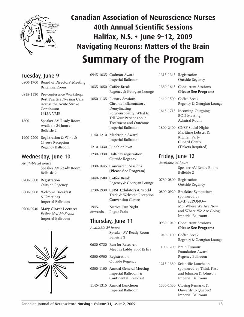
Canadian Association of Neuroscience Nurses 40th Annual Scientific Sessions
A pediatric experience with endoscopic third ventriculostomy for hydrocephalus
Back to life again—Patients’ experiences of hope three to four years after a spinal cord injury—A longitudinal study

2 Volume 31, Issue 2, 2009 • Canadian Journal of Neuroscience Nursing
ExecutivePresident/Présidente: Janice NesbittVice-President and Secretary/Vice-président et secretaire:Karen WaterhousePast-President/Présidente sortante:Debra BeveridgeTreasurer/Trésorière: Mark Bonin
CouncillorsBritish Columbia: Cindy HartleyAlberta South: Sonia RothenmundAlberta North: Kim WortonSaskatchewan: Doris NewmeyerManitoba: Wendy McDiarmidOntario West: Mary Ann ReganOntario Central: Trish HaycockOntario East: Aline MayerQuébec: Cindy McCartney(co-councillor)Sandra Berube (co-councillor)New Brunswick/PEI: Shelley PaulNova Scotia: Linsey Morrow
Newfoundland and Labrador:Nancy King
PortfoliosLegislation and Bylaws/Conseillèredes politiques: Debra BeveridgeArchivist: Geraldine FitzgeraldMembership/Abonnement:Jeanne EvansProfessional Practice/Practiqueprofessionnelle: Deb HoltomResearch/Recherche: Andrea FisherTranslation/Traduction:Suzanne MandinoProgram Liaison/Programme:Rosa SourialScientific Liaison/Scientifique:Sharron Runions
CommitteesProgram Chair—Halifax 2009:Heather StoylesScientific Chair—Halifax 2009:Debra White
Communications andMarketing (Web): Sharron RunionsCommunications and Marketing(Subscriptions): Lynn PrattEditor—Canadian Journalof Neuroscience Nursing/Rédactrice en chef—Le Journalcanadien des infirmières et infirmiersen sciences neurologiques:Sonia Poochikian-Sarkissian
RepresentativesWFNN Representative:Wilma KoopmanThink First: Sheryl FinniganCanadian Association of Brain TumourCoalition (CABTO): Janice NesbittNational Stroke Leadership Group:Rosa SourialCanadian Council on Donationand Transplantation (CCDT): VacantCanadian Nerve and Brain Coalition:Darlene Schindel
Board of directors, committee chairpersons and associatedorganization representatives — Conseil d’administration, responsablesdes comités et représentants des groupes associés 2008–2009

Editor/Rédactrice en chefSonia Poochikian-Sarkissian, University Health Network,399 Bathurst St., MP 5-311, Toronto, ON M5T 2S8; [email protected]
Peer Reviewers/RéviseuresSharon Bishop, Regina, SKJennifer Boyd, Toronto, ONAndrea Fisher, Ottawa, ONDebbie Holtom, Gananoque, ONSandra Ireland, Hamilton, ONLynn Joseph, Nepean, ONWilma Koopman, London, ONJoanna Pierazzo, Ancaster, ONMina D. Singh, Toronto, ONNancy Thornton, Calgary, AB
Yearly subscriptions are included with a membership inCANN/ACIISN ($75.00 Member, $65.00 Associate) or they may bepurchased separately. L’abonnement annuel à le Journal canadien desinfirmières et infirmiers en sciences neurologiques est inclus dans lesfrais d’inscription à l’ACIISN (75,00 $ pour les membres actifs et65,00 $ pour les membres associés).
For subscriptions, contact/Pour inscriptions :Lynn Pratt, 6357 Lumberman Way, Ottawa, ON K1C 1V6;[email protected]
• Canada: $65.00 (CAN)• United States: $65.00 (US)• International: $70.00 (US)• Single Copy: $15.00 (CAN)
Make cheques payable to: Canadian Association of NeuroscienceNurses/Les chèques doivent être émis à : L’Association canadienne desinfirmières et infirmiers en sciences neurologiques (ACIISN) : c/oLynn Pratt, 6357 Lumberman Way, Ottawa, ON K1C 1V6.
Canadian Journal ofNeuroscience NursingThe Canadian Journal ofNeuroscience Nursing is the peer-reviewed journal of the CanadianAssociation of NeuroscienceNurses (CANN)/Associationcanadienne des infirmières etinfirmiers en sciences neu-rologiques (ACIISN). The journalis published quarterly. We wel-come the submission of originalmanuscripts in the areas of prac-tice, research, theory, education,and policy, which are of interest tothe neuroscience nursing com-munity. The views, statements,and opinions expressed in thearticles, editorials, and advertise-ments are those of the authors oradvertisers. They do not neces-sarily represent the views andpolicies of CANN/ACIISN and
the editors and publishers dis-claim any responsibility orassumption of liability for thesematerials. The Canadian Journalof Neuroscience Nursing isindexed in the Cumulative Indexto Nursing and Allied HealthLiterature, International NursingIndex (INI) and Nursing CitationIndex ISSN # 1913-7176.
Mission statementThe Canadian Association ofNeuroscience Nurses (CANN)sets standards of practice andpromotes continuing profes-sional education and research.Members collaborate with indi-viduals, families, interdiscipli-nary teams and communities toprevent illness and to improvehealth outcomes for peoplewith, or at risk for, neurologicaldisorders.
CopyrightNo part of this publication can be reproduced, stored in a retrievalsystem or transmitted, in any form or by any means, without theprior written consent of the publisher or a licence from theCanadian Copyright Licensing Agency (Access Copyright). For anAccess Copyright licence, visit www.accesscopyright.ca or calltoll-free to 1-800-893-5777.
The Canadian Association of Neuroscience Nurses gratefullyacknowledges the funding provided to the Canadian Journal ofNeuroscience Nursing by the Social Sciences and HumanitiesResearch Council under the Aid to Transfer Journals program,to support the operation and expansion of the journal.
Le Journal canadiendes infirmières etinfirmiers en sciencesneurologiquesLe Journal canadien des infir-mières et infirmiers en sciencesneurologiques est le journal deL’Association canadienne desinfirmières et infirmiers en sci-ences neurologiques (ACIISN).Cette publication est revisée parses propres membres. Le journalest publié quatre fois par année.Nous acceptons les manuscritsoriginaux se rapportant à la pra-tique du nursing, de la recherche,de la théorie, de l’éducation, et del’éthique professionnelle, tousdes sujets qui suscitent l’intérêtde l’ensemble du personnel enneurosciences.
Les opinions, les points de vue, etles énoncés exprimés dans les arti-cles, éditoriaux et affiches public-itaires sont ceux des auteurs etcommerçants. Ils ne reflètent pasnécessairement les idées et les
politiques de l’ACIISN. L’éditeuret la maison d’édition n’acceptentaucune responsabilité reliée aucontenu du matériel publié dansle journal. Le Journal canadiendes infirmières et infirmiers ensciences neurologiques est rat-tachée au Cumulative Index toNursing and Allied HealthLiterature, International NursingIndex (INI) and Nursing CitationIndex ISSN # 1913-7176.
Énoncé de missionL’Association canadienne desinfirmières et infirmiers en sci-ences neurologiques (ACIISN)établit les standards de pratiquede la profession et fait la promo-tion de l’éducation permanenteet de la recherche. Les membrescollaborent avec les individus, lesfamilles, les équipes multidisci-plinaires et la communauté engénéral dans le but de prévenirles maladies neurologiques etd’améliorer la santé des gens quien sont atteints ou qui sont àrisque d’en souffrir.
Canadian Journal of Neuroscience NursingLe Journal canadien des infirmières et infirmiers en sciences neurologiques
Volume 31, Issue 2, 2009

4 Volume 31, Issue 2, 2009 • Canadian Journal of Neuroscience Nursing
The previous issue of the CanadianJournal of Neuroscience Nursing(CJNN) included the abstracts of theCanadian Association of NeuroscienceNurses (CANN) 40th Annual Conferenceto be held in Halifax, Nova Scotia, June9–12, 2009. I’m sure you all had thepleasure of reviewing these abstracts,
which always give us a tremendous sense of pride andaccomplishment. They also provide us with the deepcommitment and the professional knowledge of neurosciencenurses across Canada, who have the great capacity tocontinue being active participants in the well-being ofneuroscience patient populations throughout the world. Hopeyou can attend and enjoy the conference.
On this occasion, we would like to acknowledge ourcolleagues who organized the conference in Halifax, byensuring an environment of knowledge sharing, celebratingaccomplishments, promoting the spirit of enquiry, socialactivities and networking opportunities.
I would also encourage all conference presenters to considertransforming their work into a manuscript for publication,and making the added contribution to the existing body ofknowledge in neuroscience nursing.
You may have noticed thatCJNN has continued the publicationof French translated manuscript abstracts, together with otherCANN information, such as, Neuroscience Nursing Standards,award guidelines, etc. We hope to continue the bilingualpublications through the assistance and support provided bythe translators. In addition, the occasional publication of the“Clinical Corner” and the “Word Search” sections are addedfeatures to the journal, as suggested by our CANN membersthrough the CJNN Survey completed in 2008.
We welcome your feedback and suggestions.
Thank you for your support and wish you all an enjoyablesummer.
Respectfully,Sonia Poochikian-Sarkissian, PhD, APN, CNN(C)Editor, CJNN
Editorial: Neuroscience nurses lead the way…
The Canadian Journal of Neuroscience Nursing is published by Pappin Communications /La Journal canadienne des infirmières et infirmiers en sciences neurologiques est publié par Pappin Communications
The Victoria Centre, 84 Isabella Street, Pembroke, Ontario K8A 5S5, e-mail: [email protected] Editor: Bruce Pappin; Layout and Design: Sherri KellerAdvertising space is available/Disponibilité d’espaces pour messages publicitairesFor information, contact Heather Coughlin, Advertising Manager, Pappin Communications,The Victoria Centre, 84 Isabella Street, Pembroke, Ontario, K8A 5S5; telephone: 613-735-0952; fax: 613-735-7983E-mail: [email protected] or visit our website at www.pappin.com
Le dernier numéro du Journal canadien des infirmières etinfirmiers en sciences neurologiques (JCIISN) comprenaitles résumés de la 40ième conférence annuelle de l’Associationcanadienne des infirmières et infirmiers en sciencesneurologiques (ACIISN) qui se tiendra à Halifax enNouvelle-Écosse du 9 au 12 juin, 2009. Je suis certaine quevous avez tous eu le plaisir de lire ces résumés, qui nousdonnent un sentiment de fierté et d’accomplissement. Ils nousapportent aussi l’engagement profond et la connaissance desinfirmiers et infirmières en neurosciences à travers le Canadaqui ont une grande capacité à continuer à être des participantsactifs dans le bien-être de la population des patientsneurologiques à travers le monde. J’espère que vous pourrezparticiper et aimerez la conférence.
Nous profitons de cette occasion pour manifester notregratitude envers nos collègues qui ont organisé la conférenceà Halifax, en assurant un environnement propice à l’échangede connaissance, la célébration des accomplissements, lapromotion de l’esprit de curiosité, les activités sociales, et lesoccasions de réseautage.
J’aimerais encourager tous les conférenciers à considérer detransformer leur travail en un manuscrit pour publication, etfaire une contribution additionnelle au corps de connaissanceen sciences neurologiques.
Vous avez peut-être remarqué que le JCIISN a continué lapublication de résumés de manuscrits, et d’autre information del’ACIISN, telle que : les Standards de pratique des infirmières etinfirmiers en neurosciences, les lignes directrices pour les prix,etc. Nous espérons continuer les publications bilingues, grâce àl’assistance et le support des traductrices. De plus, la publicationdes sections “Clinical Corner” et du mot-croisé sont desnouvelles sections ajoutées au journal, tel que suggéré par lesmembres de l’ACIISN par le biais du sondage complété en 2008.
Votre rétroaction et suggestions sont les bienvenues.
Merci pour votre encouragement et je vous souhaite à tous unagréable été.
Respectueusement,Sonia Poochikian-Sarkissian, PhD, APN, CNN(C)Éditrice, JCIISN/CJNN
Éditorial : Les infirmières en neuroscience mènent la voie…

Canadian Journal of Neuroscience Nursing • Volume 31, Issue 2, 2009 5
We want yourNeuroscience Nursing News!Please send your stories, clinical ethical issues,or other information for CJNN content toDr. Sonia Poochikian-Sarkissian, Editor, [email protected]
Special thanks andwords of appreciationFrom the Editor to theCJNN Peer Reviewers and Translators!
We have relied on their assistance, valuable feedback andexpertise in publishing high-quality manuscripts in theCanadian Journal of Neuroscience Nursing.
Publish your manuscriptin the Canadian Journalof Neuroscience NursingWe welcome the submission of original manuscripts inthe areas of research, theory, practice, policy andeducation, which are of interest to the neurosciencenursing community.
CJNN Author’s Award: Authors who have published inthe CJNN will have the chance to win one of two prizes!
Upcoming CANNAnnual General Meetings& Scientific SessionsPlan to attend the following CANN Conferences:• Halifax, NS, June 9–12, 2009• Quebec City, QC, June 8–11, 2010
Cranial nerve word searchCan you find the cranial nerves in this puzzle? Circle all cranial nerve names that you find. The words can be up, down orbackwards. There are also a few cranial nerves missing from this puzzle. Do you know which ones are missing?
Retrieved from: http://faculty.washington.edu/chudler/findit3.html
AbducensFacialHypoglossalOculomotorOlfactoryOpticTrigeminalTrochlearVagus
L A N I M E G I R T J H Y Z H
C A M M R U J T A N Y N F Q F
O I S P Y A B D U C E N S M T
G C H S R P E T I S I Y Y M Z
H R O T O M O L U C O T P Q N
Z M A T T L A C H V Y C P H C
X V S F C A G W M C Q P W O Y
J S U G A V G O L B O F U Z B
Y L Y Y F C D I P B I R X A E
O H R G L K I Q P Y O U T P W
P B H E O Q W A M K H G U H P
H C L S A F E E L J G I U P C

6 Volume 31, Issue 2, 2009 • Canadian Journal of Neuroscience Nursing
AbstractHead injuries are the leading cause of trauma mortality andaccount for nearly half of all deaths related to trauma injuries.Patients who present with subdural hematomas are at risk forinitial damage to the brain, as well as for subsequent braindamage related to re-bleed, ischemia or cerebral edema. Theseinjuries can be acute or chronic in nature, and may bemanifested in the patient as an altered level of consciousness.Skilled nursing assessment of altered level of consciousnessleads to early nursing and medical intervention, which, inturn, can improve patient outcomes.
In this paper, a critical review of the literature will focus onaltered level of consciousness in patients presenting with a sub-dural hematoma. The Human Response to Illness Model will beutilized as a framework for this review. Accordingly, the physio-logical, pathophysiological, behavioural, and experiential per-spectives of altered level of consciousness will be examined. Thus,a comprehensive understanding of this human response andrationale for evidence-based interventions will be established.
BackgroundTraumatic brain injuries (TBI) are the leading cause of traumamortality, accounting for half of all trauma deaths (Tallon,Ackroyd-Stolarz, Karim, & Clarke, 2008). These injuries aremost common in young men and the elderly, and are oftenrelated to motor vehicle accidents, physical violence, or falls(Abelson-Mitchell, 2007). In 2004, a total of 16,811 headinjuries were reported in Canada, of which 68% of thesevictims were males (Canadian Institute for HealthInformation [CIHI], 2006). One classification of TBI issubdural hematomas (SDH), which are usually the result ofblunt trauma, but can also occur spontaneously in someindividuals. SDH accounts for approximately 30% of all headinjuries (Tallon et al., 2008). Depending on the size andlocation of the lesion, patients can present with focal deficits,which can cause cerebral ischemia and advance to become athreat to the patient’s life (Josephson, 2004).
Despite advances in the care of patients with TBI, patientspresenting with a diagnosis of acute SDH experience amortality rate of 60% to 90%, as a result of the primary injuryand/or secondary cerebral damage (Hlatky, Valadka,Goodman, & Robertson, 2004). While individual patientpresentations and responses can vary, altered level ofconsciousness (LOC) is a key response to monitor for signs ofchange in the patient’s clinical status. Although the incidenceof head injury has decreased by more than 35% over the pastdecade, there continues to be a need for specialized care for
those suffering a TBI (CIHI, 2006). Neuroscience nurses arein key positions to detect early changes, which can lead toearly interventions, prevent secondary injury, and improvepatient outcomes (Ammons, 1990).
Nursing care plans must be based on evidence-basedinterventions that optimize the functioning of the individualpatient and promote well-being. Holistic nursing care requiresthe nurse to have a solid knowledge base of the humanresponses to actual or potential health problems (Mitchell,Gallucci, & Fought, 1991). It is important for the nurse to beaware of the normal physiological functioning and potentialpathophysiology of altered LOC in patients with head injuriesto be able to establish appropriate interventions. On a reviewof current literature, there is a gap in research of assessment ofpatients with subdural hematomas and altered LOC using any
Using the Human Response to Illness Modelto assess altered level of consciousnessin patients with subdural hematomasBy Janice Nesbitt and Jo-Ann V. Sawatzky
L’utilisation du modèle deRéponse humaine à la maladiepour évaluer l’altération du niveaude conscience chez les patientsavec un hématome sous-dural
RésuméLes traumatismes crâniens sont la cause principale demortalité reliée à un trauma et représentent presque lamoitié de tous les décès reliés aux blessures par trauma. Lespatients avec un hématome sous-dural sont à risque d’undommage cérébral initial et aussi à risque de dommagecérébral subséquent lié au re-saignement, à l’ischémie, ou àun œdème cérébral. Ces blessures peuvent être de natureaigues ou chroniques et peuvent se manifester chez lepatient par une altération du niveau de conscience. Uneévaluation habile du niveau de conscience par l’infirmière,mène à une intervention infirmière ou médicale qui peutaméliorer les résultats cliniques du patient.
Dans cet article, une recension des écrits misera surl’altération du niveau de conscience chez des patients quiprésentent un hématome sous-dural. Le modèle de Réponsehumaine à la maladie « Human Response to IllnessModel » sera utilisé comme cadre théorique. Lesperspectives suivantes seront examinées : physiologique,physiopathologique, comportementale, et expérientielle.Ainsi, la compréhension en profondeur de cette réponsehumaine et la justification pour des interventions baséessur des faits probants seront établis.

Canadian Journal of Neuroscience Nursing • Volume 31, Issue 2, 2009 7
type of specific nursing framework. The Human Response toIllness (HRTI) Model is an effective tool to accomplish thisgoal (Mitchell et al., 1991; Figure 1). This model provides aframework that enables the nurse to examine thephysiological, pathophysiological, behavioural, andexperiential perspectives of a specific illness response. Thepurpose of this paper is to discuss each of these perspectives inrelation to the response of altered LOC in patients with SDH.Based on this comprehensive assessment, appropriateevidence-based nursing interventions will be noted as well.
Human Response to Illness ModelPhysiological perspectiveThe first component of the HRTI Model is the normalphysiological response (Mitchell et al., 1991). Thus, within thecontext of altered LOC, normal physiological functioning isexamined in terms of consciousness. Consciousness andnormal cerebral functioning are the result of physicalinteractions of brain tissue, nerve function, cerebral spinalfluid (CSF) and circulation. Cerebral perfusion, oxygenationand metabolic demands all impact the effectiveness of thiscomplex component of normal human functioning. Theaction of each of these factors fluctuates with changes in anindividual’s health status and activity, and adjusts on acontinual basis to maintain adaptive homeostasis (Reilly &Bullock, 2005). While the central nervous system (CNS)consists of the cerebral cortex, cerebellum, and spinal cord,the following review of physiology focuses on the function ofthe brain in relation to altered LOC in SDH.
A state of consciousness is dependent on an intact system ofcomplex neuronal connections within the cerebral cortex andbrainstem. If all of the components of the brain are in a state ofequilibrium, the cerebral cortex and the Reticular ActivatingSystem (RAS) are able to maintain a state of consciousness(McLeod, 2004). The RAS initiates a message in the pons,which then passes through the midbrain and thalamus, andmoves though the complex neuron pathways within thecerebral cortex to enable an appropriate response. This resultsin the state of consciousness, which includes being awake andaware. Due to the complex nature of this process, thesepathways are easily disrupted. Any change in volume (e.g., asubdural hematoma), or imbalance in metabolic activity canlead to an altered LOC (Reilly & Bullock, 2005).
Adequate perfusion of cerebral tissues is central to maintaininga normal LOC (Reilly & Bullock, 2005). The brain receivesapproximately 750 millilitres of blood per minute, which is theequivalent of 15% to 20% of the body’s total cardiac output. Theactual amount of circulating blood varies, depending on themetabolic needs of the body and brain (Hickey, 2003). Thecerebral arteries ultimately form a complicated capillary bedthat supplies nutrients to the gray matter of the cortex. Theveins drain blood back to the systemic circulation (Reilly &Bullock). Cerebral perfusion pressure (CPP), which isresponsible for the amount of oxygen and nutrients receivedfrom the brain tissues, is controlled by autoregulation of brainprocesses. Autoregulation controls the vasodilatation orvasoconstriction of blood vessels and, thus, is crucial tomaintaining an adaptive perfusion pressure (Josephson, 2004).
There are several key differences between the vasculature ofthe brain and the rest of the body. The muscle layers of thecerebral vessels are thinner in nature and, therefore, are morevulnerable to injury (Hickey, 2003). In addition, the cerebralveins have no valves and possess a unique pathway ofperfusion. Unlike systemic veins, which follow the pathwaysof arteries, cerebral veins function more independently. Theveins branch off the arteries and follow the path of the duralsinuses, which are fed by emissary and bridging veins.Emissary veins connect the extracranial veins to the duralsinuses, while bridging veins merge cerebral veins with thedural sinuses. Bridging veins are the most common sites ofsubdural hemorrhage, causing the formation of a subduralhematoma (Hickey; Reilly & Bullock, 2005).
Cerebral spinal fluid (CSF) is also an important component ofthe CNS. It is a protein- and glucose-rich, clear yellow fluidthat circulates around the brain and spinal cord in thesubarachnoid space. CSF is produced in the choroid plexus ata rate of approximately 25 millilitres per hour, for a total ofabout 500 millilitres in a 24-hour period. CSF is thenabsorbed by the arachnoid villi in the subarachnoid space.Normally, circulating CSF amounts to approximately 125–150millilitres, most of which are in the cerebral ventricles, andeasily visualized on diagnostic images (Hickey, 2003). The“modified Munro-Kellie hypothesis” (McLeod, 2004, p. 355)explains that homeostasis is maintained throughautoregulation of CSF, with the brain being able to increase ordecrease the amount of CSF to maintain intracranial pressurebetween 5mmHg and 15mmHg. This mechanism providesone explanation for gradual and vague changes in LOC.
REGULATORY
PHYSIOLOGIC
EXPERIENTIAL
PATHOPHYSIOLOGIC
BEHAVIOURAL
INTERNAL ENVIRONMENT
Figure 1. External Environment
Figure 1: HumanResponse to IllnessModel. Relationships ofthe four perspectives on human responses to the internal andexternal environment. The arrows indicate that responseswithin each perspective are capable of transmitting andreceiving information from all other perspectives. Thebehavioural responses are the ones that allow humans tocommunicate other responses to the external environment,including other human beings. We are indebted to MarthaTyler, RN, MN RRT, for helping us visualize these relationships.

8 Volume 31, Issue 2, 2009 • Canadian Journal of Neuroscience Nursing
Finally, the cerebral cortex is covered with three connectivetissue layers (i.e., pia mater, the arachnoid, and the duramater), referred to as the meninges (Hickey, 2003). The duraprovides protection for the outer layer of the cortex and thebrain stem and nerves as they exit the foramen magnum at thebase of the skull. The subdural space, which is the site ofsubdural hematomas, lies between the dura mater andarachnoid layer, and is supported with a venous vasculature(Reilly & Bullock, 2005). Disruption to the CNS system as aconsequence of an SDH can result in the pathophysiologicalresponse of altered LOC.
Pathophysiological perspectiveThe pathophysiological perspective is the second componentof the HRTI Model. This response occurs when the normallyfunctioning system is disrupted (Mitchell et al., 1991). AlteredLOC is a pathophysiological response of a variety of CNSdisorders, including SDH. With the cerebral cortex being aclosed system comprising tissue (80%), blood (10%), andcerebral spinal fluid (CSF, 10%), it is a fragile balance offunction. An increase in any of these three major braincomponents can lead to physiological changes, includingalteration in LOC (McLeod, 2004). As well, increasedpressure within the cortex will result in herniation throughthe foramen magnum, direct pressure on the RAS, and aconsequent altered LOC (Wijdicks, 2006).
Subdural hematomas are usually the consequence of blunttrauma to the head, resulting in disruption of the venoussystem, with venous blood draining into the subdural space(Hickey, 2003). The increase in blood within the subduralspace displaces brain tissue and causes increased pressurewithin the cerebral cortex. As subdural hematomas resultfrom a venous source, they can be slower in nature, and so thebrain is able to compensate during the initial stages. With thesubdural space covering the entire surface of the brain, theaffected area can become quite large before any deficits aredetected, and when the brain does begin to decompensate,neurological deficits can be significant. Once the hematomareaches a certain volume, the brain can no longeraccommodate, and changes in intracranial pressure occur.The brain can begin to swell and exert pressure on the brainstem and RAS (Reilly & Bullock, 2005). This will result inimpaired LOC and overall neurological function, which canpotentially cause permanent brain damage. Without earlyintervention, the bleeding will persist and the pressure on thebrain will continue to increase. The subsequent increasingintracranial pressure and edema exerts pressure on thehealthy brain tissue, causing cerebral ischemia. Poorer long-term clinical outcomes will result if early symptoms of alteredLOC are not noted and managed (McLeod, 2004).
With or without medical intervention, the risk of secondaryinjury is significant. Cerebral edema, hydrocephalus and re-bleed are all potential secondary complications that can arisefrom a subdural hematoma and lead to altered LOC (Reilly &Bullock, 2005). The presence of blood in the meninges canirritate the brain, leading to cerebral edema. Depending onthe size and location of the bleeding and clot formation,obstructive hydrocephalus can result, with an excessive
accumulation of CSF in the ventricles. Finally, with any type ofcerebral hemorrhage, the patient is always at risk for bleedingagain during the recovery process (Hickey, 2003; Reilly &Bullock). Thus, any one of these three complications can leadto cerebral ischemia and increased Intracranial Pressure(ICP). If severe, these complications can ultimately lead touncal herniation and fatal outcomes (Wijdicks, 2002).
Behavioural perspectiveThe behavioural perspective includes measurements of theresponse that are observable and objective in nature (Mitchellet al., 1991). The Glasgow Coma Scale (GCS) is a universallyrecognized, gold standard, objective measure for LOCassessment. This evidence-based standardized tool is widelyused to measure patient’s responsiveness and to monitortrends in responsiveness. The GCS measures the twocomponents of consciousness: awareness and ability torespond. This is accomplished by evaluating eye opening andverbal and motor responses. Experienced neurosciencenurses can use this tool to note changes in LOC, which, inturn, can also reflect changes in ICP and the patient’s overallneurological status (Edwards, 2001). Even subtle changes in apatient’s condition can be noted, indicating eitherimprovement or deterioration in their condition. While onemeasurement alone is usually not meaningful, the trendedLOC results are helpful in monitoring the patient’s ongoingstatus (McLeod, 2004). Although the GCS has limitations,including the failure to test brainstem reflexes and falsescoring for patients who cannot speak (as a result ofintubation or tracheotomy) or open their eyes, it is a widelyused, reliable and valid assessment of LOC.
While there are several other published scales that measureLOC, most of these scales have not been validated for head-injured patients. For example, the Full OutlineUnresponsiveness (FOUR) scale assesses eye, motor,brainstem and respiratory perspectives. Although the FOURhas been found to be a reliable scale for clinical use (Wijdicks,2006) and allows for assessment of altered LOC at all levels ofawareness, including voluntary eye movements andbrainstem reflexes (Giacino & Smart, 2007), it is a relativelynew tool that has not yet been widely tested. The ReactionLevel Scale is another alternative to the GCS. This toolconsists of a scale from one to eight (1 = alert; 8 =unconscious). While this is a simple tool to use in clinicalpractice, its descriptors are vague in the middle range andopen to subjective interpretation. The Alert, Verbal, Painstimuli, or Unresponsive (AVPU) Scale is more commonlyused in emergency room triage settings. (Limmer & Monosky,2002; McLeod, 2004). While this tool does allow for a quickassessment and frame of reference, the descriptors are vagueand open to interpretation. Therefore, AVPU is not a usefultool to monitor trends in altered LOC and subtle changes inclinical status over time.
Other indicators of altered LOC, although not objectivelymeasurable, are important components of the neurologicalassessment. For example, the SDH patient populationcommonly experience personality and cognitive changes.Because these individuals can be alert, but not mentally

Canadian Journal of Neuroscience Nursing • Volume 31, Issue 2, 2009 9
aware, they cannot be considered fully conscious according tothe definition of consciousness. A loss of inhibition,disorientation, hallucinations, or receptive dysphasia maycause the patient to exhibit inappropriate behaviours orresponses (Testani-Dufour, Chappel-Aiken, & Gueldner,1992). These behaviours impact the stress experienced by thefamily, as well as the interventions chosen by nursing staff.
In the state of altered LOC, restlessness may be an indicationthat the patient is improving clinically, deteriorating, or a signof a patient’s frustration or discomfort. A confused patient,who is incapable of understanding the environment that theyare in, may become agitated and inappropriate in theirresponses (Petchprapai & Winkelman, 2007; Hickey, 2003).This can lead to combative behaviours, which may increaseICP. These patients are frequently physically restrained toprevent them from harming themselves, but this canexacerbate their agitation, causing them to be verbally abusiveof those around them. As a result of their cognitive deficits,this stage of the brain injury process can be frightening andfrustrating to patients and families. Therefore, it is importantto ensure they understand the cause of the behaviours and theplan for management. Nursing interventions play a key role inthis process.
There are other objective components of the neurologicalexam, which are not direct measures of LOC, but do provideinsight into the assessment of these patients. For example, ICPmonitoring is an objective measurement tool that may be usedin patients with a severe head injury related to an SDH. ICPmonitoring enables the healthcare team to objectively assesschanges in ICP (Reilly & Bullock, 2005). Increased ICP leads todecreased cerebral perfusion pressure, ischemia, and alteredLOC if not treated immediately. Changes in pupils, vital signs,or pulse pressure are also indicators of deterioration inneurological status and can coincide with changes in LOC.Thus, it is critically important for nurses to be aware of the keycomponents of an objective assessment of altered LOC in thepatient with an SDH. This knowledge will, ultimately, lead toearly interventions and optimal patient outcomes.
Experiential perspectiveThe final component of the HRTI Model is the patient’ssubjective experiential perspective, or verbal expression ofsymptoms experienced (Mitchell et al., 1991). This is one ofthe most important aspects of the model, as the patient’sperspective is often the best indicator of what is actuallyhappening. For example, while a patient with a severe headinjury may be in a deep coma and not respond to any stimuli,other patients, with mild head injuries, may be awake, but notcognitively intact (Hickey, 2003). This may result indisorientation to their surroundings, lack of ability to thinkabstractly, or inability to rationalize their responses(Petchprapai & Winkelman, 2007). Fluctuations in responsescan create an environment of uncertainty and anxiety for thepatient, and an assessment challenge for the health care team.
There is limited information in the literature on the perspectiveof a patient with an acute TBI or SDH, as their communicationsand behaviours are not always congruent with what is trulyhappening and their explanations are not always rationale or
reliable. Hence, research has focused on the outcomes andeffects on the patients and families, rather than examining theexperience of the patient with acute altered LOC (Kneafsey &Gawthorpe, 2004; Testani-Dufour, 1992; Verhaeghe, Defloor, &Grypdonck, 2005). Acute and chronic behaviours of alteredLOC impact the patient and family, leading to feelings of fear,anxiety, and powerlessness (Petchprapai & Winkelman, 2007).Lefebvre, Cloutier, and Levert, (2008) found that some patientsin this population experience difficulties with social integrationand employment due to cognitive deficits, while more severecases may required more comprehensive long-term care(Kotwica & Brzezinski, 1993).
In a phenomenological study, Chamberlain (2006) describesthe patient’s experience of surviving a TBI, but not alteredLOC itself. The results are relevant, however, because theindividuals’ experiences during the recovery process andassociated episodes of altered consciousness were described.In this study, five themes were identified: regret and griefwithin self, insensitivity of health professionals, invisibility ofself, stranded self, and recovery in self. Working through theseexperiences required patients to re-invent and learn to livewith new versions of themselves. Patients used these self-reflections to appreciate their survival and accomplishmentsin recovery (Chamberlain, 2006).
Knowledge of the actual lived experience of beingunconscious, or less responsive provides important insightsfor the care of patients with altered LOC. In a mixed-methodsstudy, Lawrence (1995) described unconscious experiences of100 patients. Although the study included patients with abroad range of diagnoses, the results are relevant to the TBIpatient population. According to Lawrence (1995), alteredconsciousness experiences can be categorized into five distinctstates: unconsciousness, inner consciousness, perceivedunconsciousness, distorted consciousness, and paranormalexperiences. Patients in some states of altered LOC werereportedly still very aware of their environment. Some couldrecall voices and conversation that occurred around them,while others stated that they were aware of requests beingmade of them, but were physically unable to respond.Individuals also reported being aware of the emotions offamily and staff around them, including feelings of despair andhope. Based on the evidence from this study, Lawrence (1995)concluded that patients’ unconscious experiences can causelong-term effects, and that caregivers need to be aware of theimpact of their words and actions on these patients.
Personal and environmental factorsWithin the HRTI Model, person and environmental factorsinteract with the four perspectives to establish individual riskfor a specified response to illness (Mitchell et al., 1991). Thesefactors can have a significant impact on the human response(i.e., altered LOC), as well as the patient’s outcomes.
Person factorsNon-modifiable person factors include factors within theindividual that may influence the response of altered LOC inthe patient with a subdural hematoma related to a TBI. Forexample, age is an important influencing factor, with the

10 Volume 31, Issue 2, 2009 • Canadian Journal of Neuroscience Nursing
elderly being particularly vulnerable to more severealterations in LOC related to co-morbidities and concomitantmedications, which may adversely affect the response(Neatherlin, 2000). This, in turn, may lead to delays inregaining consciousness and recovery. As well, the brain tendsto atrophy with age, which may delay the onset of symptomsof altered LOC in the patient with a subdural hematoma thatcan delay treatment and have a negative impact on patientoutcomes (Neatherlin, 2000).
Medications that an individual is taking prior to their TBI canalso have a significant impact on the response and the illness.Anti-coagulant therapy increases the risk for a larger initialSDH, as well as for re-bleeding (Reilly & Bullock, 2005).Consequently, the severity of the underlying pathology andthe consequent response of altered LOC may be more serious.Patients requiring sedatives (Josephson, 2004), or whopresent under the influence of mind-altering drugs or alcohol(Reilly & Bullock) may have altered LOC for reasons otherthan the underlying pathology. These additional individualco-morbidities may lead to delays in recovery ofconsciousness and recovery.
Modifiable person factors are preventable or amenable tochange in the post-injury period. For example, post-injuryinfection, fever, hypoxia, hypotension, extreme serum glucoselevels, hyponatremia, seizures, and hypoxemia are allpotential responses to TBIs, and can all impact on the alteredLOC (McLeod, 2004). Many of these illness consequences canbe prevented and/or managed by optimal patient care.Similarly, select nursing interventions can be effective inmaximizing cerebral perfusion (McLeod, 2004). Variouscombinations of medications given to the SDH patient mayalter LOC or mask the signs of altered LOC. For example,high doses of phenytoin or lorazepam can result in lethargy,making it difficult to decipher if altered LOC is related tomedication side effects or deterioration in the underlyingpathology (Josephson, 2004).
Environmental factorsThe external patient care environment can have a significantimpact on altered LOC. The availability of specializedequipment and expert neurosurgical medical and nursing staffis central to managing altered LOC in the SDH patienteffectively.While equipment to monitor CBF and ICP is helpfulto detect early changes in altered LOC and can lead to earlyintervention and improved outcomes, there is no evidence thatthe equipment itself improves patient outcomes (Stiefel,Spiotta, Gracias, Garuffe, Guillamondegui, Maloney-Wilenskyet al., 2005; Steiner & Andrews, 2006). Further research isneeded as to the impact that the availability of cerebralhemodynamic monitoring can have in these situations.
Ideally, neurosurgical nurses should possess the knowledgeand skills to assess the patient and minimize the SDH patient’srisk for altered LOC. Their knowledge of the factors thatinfluence patient outcomes ensures evidence-based care. Forexample, reducing external stimuli and elevating the head ofthe bed have been found to minimize intracranial pressure(Schwarz, Georgiadis, Aschoff, & Schwab, 2002), which, inturn, has a favourable effect on altered LOC.
Nursing implicationsThe four perspectives of the HRTI Model establish acomprehensive framework to address implications for clinicalpractice, education, and research. The review of the literature,based on these perspectives facilitates a holistic assessment ofthe individual patient and provides insight for thedevelopment of evidence-based nursing interventions andbest practice guidelines for optimal patient care.
Implications for clinical practicePhysiologically, airway, breathing, and circulation are theprimary areas of concern in the patient with altered LOC(Ammons, 1990; Wijdicks, 2006). When a patient presentswith, or deteriorates to a GCS of ≤ 8, the airway may becompromised and intubation may be necessary (McLeod,2004; Wijdicks, 2006). Adequate ventilation serves tominimize ischemia to the brain and other vital tissues.Therefore, the maintenance of the airway protection, as wellas its effectiveness must be a nursing priority. In addition,monitoring of vital signs and pulse oximetry are importantcomponents of the assessment of circulation and oxygenation.For example, in an unconscious patient, a widened pulsepressure, with bradycardia, may be an imminent sign offurther increase ICP and impending neurologicaldeterioration (Ammons; Hickey 2003; McLeod, 2004).Adequate oxygenation promotes proper autoregulation ofoxygen and carbon dioxide in the brain tissues, and theconsequent vasoconstriction that reduces ICP (Hickey).Conversely, hypoxia with hypercapnia results in vasodilation,increasing cerebral blood flow and increased ICP (Edwards,2001; Kerr & Brucia, 1993). Thus, altered LOC can beminimized with adequate oxygenation.
Ongoing, comprehensive neurological assessments arecritically important in order to monitor for changes in thepatient’s clinical status and note changes in LOC in a timelymanner. Nurses are in a key position to perform theseassessments and communicate their findings to the medicalstaff. In addition to the GCS, these assessments shouldinclude: checking pupillary response, vital signs, and ICP andCPP if available (McLeod, 2004). Skilled nursing assessmentscan detect subtle changes in hemodynamics, or motor orverbal responses, that may lead to further investigation andearly intervention. Early medical intervention can minimizecerebral tissue damage, and minimize long-term deficits thatare often associated with sustained alterations in LOC.
Patients who present with altered LOC can becomedesensitized to the busy external stimuli of hospital soundsover time (Gerber, 2005). As a result, they may be lessresponsive to the routine requests associated withneurological assessments. In order to re-train their orientationto relevant stimuli, it is suggested that these patients should bekept in a calm and quiet environment, and have a scheduled,structured coma stimulation program. This can be initiated asearly as 72 hours post-injury, and involves the stimulation ofauditory, visual, olfactory, gustatory, tactile, and kinestheticsenses (Gerber, 2005). The goal of this intervention is to “wakeup” and re-train a damaged RAS by stimulating cortical

Canadian Journal of Neuroscience Nursing • Volume 31, Issue 2, 2009 11
activity, to ultimately improve alertness and awareness.Although there is minimal research literature availablesurrounding this novel intervention, it is a logical and harmlessintervention that may improve patient outcomes.
Simple, but critical nursing interventions, such as elevatingthe head of the bed and maintaining the head and neck in aneutral position can promote venous drainage and, ultimately,reduce the risk for increased ICP and alterations in LOC(Christie, 2008; Schwarz et al., 2002). Monitoring fluid andelectrolyte balances is also important because any imbalancecan affect the neurological response (Fryman & Murray,2007). Timely medication administration is critical insupporting effective cerebral functioning. As well, monitoringmedications for their effects and possible side effects isessential. For example, although the administration ofanticonvulsants can minimize the incidence of seizures, thesemedications also present the risk for hyponatremia. Whileglucocorticosteroids can reduce cerebral edema, andultimately lower ICP, they can also cause hyperglycemia,immune compromise, irritability, insomnia, and muscleatrophy (Josephson, 2004). The side effects of both of theseclasses of medications can have an acute onset, and can beginafter only a brief period of administration (Hickey, 2003).Skilled neuroscience nurses must be aware of these potentialcomplications of the medications, and include a review ofcurrent medications in their patient assessments.
Monitoring for signs of a fever in the patient with altered LOCis also important. The cause of the fever may be neurogenic,or related to an infection. A neurogenic fever results whenincreased ICP causes damage to the hypothalamus, andtemperature control is lost (Reilly & Bullock, 2005;Thompson, Pinto-Martin, & Bullock, 2003). A fever will raisethe metabolic and oxygen demands of the brain, and mayexacerbate symptoms of altered LOC. This added cerebralstress also contributes to ischemia and cell death. Followingup on results that may identify an underlying cause for theinfection and maintenance of normothermia with antipyreticsand/or a circulating fan are appropriate nursing interventions(Thompson et al., 2003).
Although not a direct factor in the altered LOC response,families should be considered in the development of patient-centred care strategies and clinical nursing interventions.While any change in LOC can cause anxiety and fear,explanations and re-orientation regarding symptoms,investigations, assessments and environment are essential tominimize patient and family stress (Testani-Dufour et al.,1992). Unfortunately, in neuroscience, there are rarely definiteanswers and only time will tell how the patient will progressor respond to treatment. The frustration experienced by thepatient and family can be far-reaching, and can presentadditional challenges to the nursing staff. A team approachwith a network of support systems for the patient and familyis essential for the delivery of consistent and positive nursingcare (Yetman, 2008).
Implications for nursing educationWhile communication between patients, families, andnursing staff is essential to minimize knowledge deficits and
anxiety, it can also have a favourable effect on ICP. Based onLawrence’s (1995) findings, some unconscious patients maybe aware of sounds and conversations around them. Byexplaining interventions prior to implementation, the elementof surprise and related stress may be lessened, and may helpto minimize an increase in ICP. In patients who are awake, butnot fully aware, explanations of treatments are critical toreducing anxiety and avoiding confusion. Confused patientsmay misinterpret the intentions of staff and become moreagitated, which, in turn, can cause an increase in ICP, andpotentially alter their LOC.
As research in neuroscience nursing care is constantlyevolving, it is crucial for nurses working in this specialty areato maintain their clinical competency through formal andinformal education. As new measurement tools areintroduced and tested, a practical alternative to the GCS mayeventually be validated and accepted. Without self-motivatededucation, the transfer of this knowledge would not occur.Attendance at in-services and conferences and/or readingcurrent research journals regarding altered LOC willcontribute to the nurse’s knowledge base, and facilitate thedevelopment of evidence-based interventions. Finally,advanced practice nurses are in key positions to questioncurrent practice and co-ordinate nursing research projectsthat will, in turn, result in improved patient outcomes.
Implications for nursing researchThrough observation of clinical practice and patientoutcomes, neuroscience nurses can identify gaps inevidence-based practice and collaborate with themultidisciplinary team to identify research questions relatedto altered LOC in patients with SDHs. Although there isconsiderable research-based evidence related to the care ofthe SDH patient with altered LOC, this review also revealeda number of gaps in the research literature. For example, theeffects of a structured coma stimulation program on patientswith the altered LOC and a SDH are not fully understood. Aswell, further research is needed regarding the livedexperiences of these patients. And finally, research is justbeginning to emerge on the impact of caring for thesepatients and their families on nurses. Thus, further researchis needed in this area.
ConclusionPatients who present with an SDH frequently exhibit signs ofaltered LOC. Nursing care of these individuals and theirfamilies is complex and focuses on optimal assessmentstrategies and evidence-based care. Application of the HRTIModel enables neuroscience nurses to complete acomprehensive assessment of the altered LOC response,based on the physiological, pathophysiological, behavioural,and experiential perspectives, as well as the person andenvironmental factors. Nurses can then developindividualized, research-based nursing interventions for thisspecific response to illness. Valuable insights related topatient evaluation strategies have also been gleaned duringthis process. Thus, this model facilitates achieving the goal ofoptimal patient care and outcomes for this patient population.

12 Volume 31, Issue 2, 2009 • Canadian Journal of Neuroscience Nursing
Abelson-Mitchell, N. (2007). Epidemiology and prevention ofhead injuries: Literature review. Journal of Clinical Nursing, 17,46–57.
Ammons, A.M. (1990). Cerebral injuries and intracranialhemorrhage as a result of trauma. Nursing Clinics of NorthAmerica, 25(1), 23–33.
Canadian Institute for Health Information. (2006).Hospitalizations due to traumatic head injuries down 35% overthe decade. Retrieved from http://secure.cihi.ca/cihiweb/dispPage.jsp?cw_page=media_30aug2006_e
Chamberlain, D.J. (2006). The experience of survivingtraumatic brain injury. Journal of Advanced Nursing, 54(4),407–417.
Christie, R.J. (2008). Therapeutic positioning of the multiply-injured trauma patient in ICU. British Journal of Nursing, 17(10),638–642.
Edwards, S.L. (2001). Using the Glasgow Coma Scale: analysisand limitations. British Journal of Nursing, 10(2), 92–101.
Fryman, L., & Murray, L. (2007). Managing acute head traumain a crowded emergency room. Journal of Emergency Nursing,33(3), 208–213.
Gerber, C.S. (2005). Understanding and managing comastimulation. Critical Care Nursing, 28(2), 94–108.
Giacino, J.T., & Smart, C.M. (2007). Recent advances inbehavioural assessment of individuals with disorders ofconsciousness. Current Opinion in Neurology, 20, 614–619.
Heinemann, A.W., Harvey, R.L., McGuire, J.R., Ingberman, D.,Lovell, L., Semik, P., et al. (1997). Measurement properties of the NIHStroke Scale during acute rehabilitation. Stroke, 28, 1174–180.
Hickey, J.V. (2003). The Clinical Practice of Neurologicaland Neuroscience Nursing (5th ed.). Philadelphia: Lippincott,Williams & Wilkins.
Hlatky, R., Valadka, A.B., Goodman, J.C., & Robertson, C.S.(2004). Evolution of brain tissue injury after evacuation of acutetraumatic subdural hematomas. Neurosurgery, 55, 1318–1324.
Josephson, L. (2004). Management of increased intracranialpressure: A primer for the non-neuro critical care nurse.Dimensions of Critical Care Nursing, 23(5), 194–207.
Kerr, M.E., & Brucia, J. (1993). Hyperventilation in the head-injured patient: An effective treatment modality? Heart and Lung,22, 516–21.
Kneafsey, R., & Gawthorpe, D. (2004). Head injury: Long-termconsequences for patients and families and implications for nurses.Journal of Clinical Nursing, 13, 601–608.
Kotwica, Z., & Brzezinski, J. (1993). Acute subdural hematomain adults: an analysis of outcome in comatose patients. ActaNeurochirurgica, 121, 95–99.
Lawrence, M. (1995). The unconsciousness experience.American Journal of Critical Care, 4, 227–232.
Lefebvre, H., Cloutier, G., & Levert, M.J. (2008). Perspectivesof survivors of traumatic brain injury and their caregivers on long-term social integration. Brain Injury, 22(7 &8), 535–543.
Limmer, D., & Monosky, K. (2002). Assessment of the alteredmental status patient. Emergency Medical Services, 31(3), 54–8.
McLeod, A. (2004). Intra- and extracranial causes of alterationin level of consciousness. British Journal of Nursing, 13(7), 354–361.
Mitchell, P.H., Gallucci, B., & Fought, S.G. (1991). Perspectiveson human responses to health and illness. Nursing Outlook, 39,154–157.
Neatherlin, J.S. (2000). Head trauma in the older population.Critical Care Nursing, 23(3), 49–57.
Petchprapai, N., & Winkelman, C. (2007). Mild traumaticbrain injury: Determinants and subsequent quality of life. A review ofthe literature. Journal of Neuroscience Nursing, 39(5), 260–72.
Reilly, P.R., & Bullock, R. (2005).Head Injury: PathophysiologyandManagement (2nd ed.). London: Hodder Arnold.
Schwarz, S., Georgiadis, D., Aschoff, A., & Schwab, S. (2002).Effects of body position on intracranial pressure and cerebral perfusionin patients with large hemispheric stroke. Stroke, 33, 497–501.
Stalhammar, D., & Starmark, J.E. (1986). Assessment ofresponsiveness in head injury patients: The Glasgow Coma Scale andsome comments on alternative methods. Acta Neurochirurgica,Supplement, 36, 91–94.
Stein, S.C., Spettel, C., Young, G., & Ross, S.E. (1993).Limitations of neurological assessment in mild head injury. BrainInjury, 7(5), 425–430.
Steiner, L.A., & Andrews, P.J.D. (2006). Monitoring the injuredpatient: ICP and CBF. British Journal of Anaesthesia, 97(1), 26–38.
Stiefel, M.F., Spiotta, A., Gracias, V.H., Garuffe, A.M.,Guillamondegui, O., Maloney-Wilensky, E., et al. (2005). Reducedmortality rate in patients with severe traumatic brain injury treatedwith brain tissue oxygen monitoring. Journal of Neurosurgery,103(5), 805–11.
Tallon, J.M., Ackroyd-Stolarz, S., Karim, S.A., & Clarke, D.B.(2008). The epidemiology of surgically treated acute subdural andepidural hematomas in patients with head injuries: A population-based study. Canadian Journal of Surgery, 51(5), 339–345.
Testani-Dufour, L., Chappel-Aiken, L., & Gueldner, S. (1992).Traumatic brain injury: A family experience. Journal ofNeuroscience Nursing, 24(6), 317–323.
Thompson, H.J., Pinto-Martin, J., & Bullock, M.R. (2003).Neurogenic fever after traumatic brain injury: Epidemiological study.Journal of Neurology, Neurosurgery and Psychiatry, 74, 614–619.
Verhaeghe, S., Defloor, T., & Grypdonck, M. (2005). Stress andcoping among families of patients with traumatic brain injury: Areview of the literature. Journal of Clinical Nursing, 14, 1004–1012.
White, K.M., Winslow, E.H., Clark, A.P., & Tyler, D.O. (1990).The physiologic basis for continuous mixed venous oxygen saturationmonitoring. Heart & Lung, 19(5 part 2), 548–51.
Wijdicks, E.F.M. (2002). Uncal herniation in acute subduralhematoma. Archives of Neurology, 59, 305.
Wijdicks, E.F.M. (2006). Clinical scales for comatose patients:The Glasgow Coma Scale in historical context and the new FOURscale. Reviews in Neurological Diseases, 3(3), 109–117.
Yetman, L. (2008). Neuroscience nurses caring for familymembers of patients with acquired brain injury in acute wardsettings: Nursing defensively in a double bind. Canadian Journal ofNeuroscience Nursing, 30(4), 26–33.
References
AAbboouutt tthhee aauutthhoorrssJanice Nesbitt, RN, CNN(C), is a graduate student in theFaculty of Nursing at the University of Manitoba and aClinical Resource Nurse in an Outpatient NeurosurgeryClinic at Health Sciences Centre in Winnipeg, Manitoba.
Jo-Ann V. Sawatzky, RN, PhD, is an Associate Professor andthe Faculty Development Coordinator in the Faculty ofNursing, University of Manitoba, Canada.
Correspondence regarding this article should be addressed toJanice Nesbitt at [email protected]
Canadian Journal of Neuroscience Nursing • Volume 31, Issue 2, 2009 13
Tuesday, June 90800-1700 Board of Directors’ Meeting
Britannia Room
0815-1530 Pre-conference Workshop: Best Practice Nursing Care Across the Acute Stroke Continuum1613A VMB
1800 Speaker AV Ready Room Available 24 hoursBelleisle 2
1900-2200 Registration & Wine & Cheese ReceptionRegency Ballroom
Wednesday, June 10Available 24 hours
Speaker AV Ready RoomBelleisle 2
0700-0800 Registration Outside Regency
0800-0900 Welcome Breakfast & GreetingsImperial Ballroom
0900-0940 Mary Glover Lecture:Father Neil McKennaImperial Ballroom
0945-1035 Codman AwardImperial Ballroom
1035-1050 Coffee BreakRegency & Georgian Lounge
1050-1135 Plenary Session:Chronic InflammatoryDemylinating Polyneuropathy: What to Tell Your Patient about Treatment and OutcomeImperial Ballroom
1140-1210 Medtronic AwardImperial Ballroom
1210-1330 Lunch on own
1230-1330 Half-day registrationOutside Regency
1330-1645 Concurrent Sessions (Please See Program)
1440-1500 Coffee BreakRegency & Georgian Lounge
1730-1930 CNSF Exhibitors & World Trade & Welcome ReceptionConvention Centre
1945- Nurses’ Fun Nightonwards Pogue Fado
Thursday, June 11Available 24 hours
Speaker AV Ready RoomBelleisle 2
0630-0730 Run for ResearchMeet in Lobby at 0615 hrs
0800-0900 RegistrationOutside Regency
0800-1100 Annual General MeetingImperial Ballroom & Continental Breakfast
1145-1315 Annual LuncheonImperial Ballroom
1315-1345 RegistrationOutside Regency
1330-1645 Concurrent Sessions (Please See Program)
1440-1500 Coffee BreakRegency & Georgian Lounge
1645-1715 Incoming-OutgoingBOD MeetingAdmiral Room
1800-2400 CNSF Social Night: Maritime Lobster & Kitchen PartyCunard Centre(Tickets Required)
Friday, June 12Available 24 hours
Speaker AV Ready RoomBelleisle 2
0730-0800 RegistrationOutside Regency
0800-0920 Breakfast Symposiumsponsored byEMD SERONO—MS: Where We Are Now and Where We Are GoingImperial Ballroom
0930-1040 Concurrent Sessions (Please See Program)
1040-1100 Coffee BreakRegency & Georgian Lounge
1100-1200 Brain TumourFoundation AwardRegency Ballroom
1215-1330 Scientific Luncheonsponsored by Think First and Johnson & JohnsonImperial Ballroom
1330-1430 Closing Remarks & Onwards to Quebec!Imperial Ballroom
Canadian Association of Neuroscience Nurses40th Annual Scientific SessionsHalifax, N.S. • June 9–12, 2009
Navigating Neurons: Matters of the Brain
Summary of the Program

14 Volume 31, Issue 2, 2009 • Canadian Journal of Neuroscience Nursing
Wednesday, June 101330-1400
A1 Let’s Just Ask: Family Feedback About Nursing ServicesNancy Thornton andConny Betuzzi
B1 Hyperbaric Oxygen Treatments as an Adjunct Therapy for Traumatic Brain Injury: Do They Improve Patient Outcomes?Trudy Robertson andJenifer Bartlett
C1 A Collaborative Injury Prevention Programfor Neuro-Trauma: Implementation and Evaluation of an Effective Helmet Educational ProgramLynne Fenerty, Sherry Huybers, Simon Walling, Nicole Boutilier and John LeBlanc
D1 Is Your Neuroscience Patient Faking Neurological Deficits? The Real Truth about Conversion DisordersWendy MacGregor, Paula Poupore and Andrea Fisher
1410-1440A2 A Paediatric Experience
with Endoscopic Third VentriculostomyKelly Bullivant
B2 Utility of the Rancho Levels of Cognitive Functioning Scale: Nursing Considerations in Family-Centred Care Following Pediatric Traumatic Brain InjurySarah Westerbaan and Kathryn Sebastian
C2 Medical Emergency: Post Craniotomy Epidural HematomaSue Kadyschuk and Sarah Bussey
D2 I Think I’m Havinga Stroke... It’s Justa Migraine?Janet Warner
1500-1530A3 Parental Support Needs
in Perinatal StrokeSonia Rothenmund, Nancy Thornton and Dr. Adam Kirton
B3 Families’ Stories: Dying from a StrokeRosa Sourial, Louise Fullerton and Dr. Liam Durcan
C3 vacant
D3 Cooling As a Way to Preserve BrainCarol Meade-Corkum and Debrah White
1540-1610A4 What’s Wrong With
My Baby’s Head?Cathy Cartwright
B4 The Brain Drain: Backup in the Cerebral Vascular SystemSandra Broughton
C4 The Impact of Perceived Quality of Care on Patient Satisfaction—A Quality Improvement Project in Spinal SurgeryYvette Lashley, Rosalie Magtoto, Angela Sarro and Andrea Dyrkacz
D4 Managing Agitation Effectively: The Development of a Hospital-based Observer PoolWendy Blackburn and Grace Leung Jokinen
1615-1645A5 Writing for Publication
Sonia Poochikian-Sarkissian
Canadian Association of Neuroscience Nurses40th Annual Scientific SessionsHalifax, N.S. • June 9–12, 2009
Navigating Neurons: Matters of the Brain
Concurrent Sessions
Canadian Journal of Neuroscience Nursing • Volume 31, Issue 2, 2009 15
Thursday, June 111330-1400
A1 A Nursing Protocol for the Monitoring of a Pediatric Patient Undergoing EEG Testing with SedationDeborah Meldrum and Heather Davies
B1 Understanding the Complexity of Moya Moya Disease in the Adult Population: Diagnosis, Investigations and Neurosurgical ManagementDawn Tymianski
C1 The Ticking Clock for the Brain Injury Patient: Optimizing Patient Outcomes fromTime of Injury to Neurosurgical CareLynne Fenerty, GinetteThibault-Halman, Saleema Karim, Beth Sealy, Dr. Simon Walling, Dr. Stacy Ackroyd-Stolarz, Dr. John Tallon and Dr. David B. Clarke
D1 Risky Business:Dangerous Games Children PlayCathy Cartwright
1410-1440A2 Care and Management of
Patients with Spina Bifida: A 23-Year PerspectiveBeverly Irwin
B2 The Dark Faces of Devics DiseaseMonica Viste and Kris Morley
C2 Transition Care for Young Adults with Complex Chronic Neurological ConditionsHeather Davies
D2 If A Picture Is Worth A Thousand Words, A Video Is Worth A Million: The Role of the Epilepsy Monitoring Unit in the Education of Health ProfessionalsSusan R. Rahey
1500-1530A3 vacant
B3 vacant
C3 Spinal Epidural Abscess in the Intravenous Drug User: A Surgical and Ethical DilemmaNicole Chenier-Hogan, Delanya Podgers andDarlene Bowman
D3 Belly Fat… Not Just Any Fat: Why Visceral Adiposity Should be Targeted to Reduce Stroke RiskIsabelle Martineau
1540-1610A4 Acute Cervical
Spine Fractures: Maximizing StabilityTracy Anthony
B4 Impact of Diagnosis and Grief InterventionMindy Halper
C4 Subdural Empyema Secondary to Sinusitis: A Case PresentationArbelle Manicat-Emo, Maggie Namtu and Brammiya Sivakumar
D4 Moral Distress and Ethical Dilemmas Facedby Neuroscience NursesChristine Price
1615-1645A5 Writing for Publication
Sonia Poochikian-Sarkissian
Friday, June 120930-1000
A1 Ischemic StrokeSecondary to Infective Endocarditis: A Case StudyLaura MacIsaac
B1 Reflections on aCoordinated Stroke System: A Case StudyJemini Abraham, Anne Cayley, Victoria Riediger and Relu Wiegner
C1 The Transition to Becoming a Neuroscience Nurse: A Focus on IntegrationMartha A. Stewart, Sabrina Boutin, Elizabeth Murphy-Lavallée, and Josée Lizotte
D1 Donation AfterCardiac DeathJane Franklin
1010-1040A2 Explanatory
Models of Recovery from Stroke Within the African-Caribbean Community in CanadaDenise C. Wilson and Dr. Marianne Lamb
B2 TBA
C2 Challenging Aspects of Managing Seizures in the Elderly: A Retrospective ReviewSheila N. Koutsogiannopoulos and Patricia Kerr
D2 Medical Management of the Organ DonorNikki Kelly

16 Volume 31, Issue 2, 2009 • Canadian Journal of Neuroscience Nursing
AbstractPediatric neuroscience nurses deal with many children withhydrocephalus. This paper will provide a review ofhydrocephalus with a specific focus on third ventriculostomy.Endoscopic third ventriculostomy (ETV) is an acceptedtreatment option for patients with obstructive hydrocephalus.At the Alberta Children’s Hospital in Calgary, Alberta, we havebeen performing ETV for 15 years. This experience has helpedus better understand the complications and benefits of ETV.The author will provide data on the complications specific toETV in pediatric patients at the Alberta Children’s Hospital. Acase report comparing neurocognitive testing before and afterETV will be shared. An additional case report of a patient witha spontaneous third ventriculostomy will help illustrate ourunderstanding of the natural history of hydrocephalus.
IntroductionHydrocephalus is one of the most common disorders seen inpediatric neurosurgery. The true incidence of hydrocephalusis difficult to determine. Massimi, Paternoster, Fasano andDi Rocco (2009) report a declining incidence ofhydrocephalus in recent years and attribute this decline to adecrease in congenital malformations.
Hydrocephalus is a clinical condition where there is an imbal-ance between production and absorption of cerebral spinalfluid (CSF) resulting in enlargement of the ventricular system.Hydrocephalus has typically been divided into two groups
A pediatric experience with endoscopic third ventriculostomy for hydrocephalusBy Kelly J. Bullivant, Walter Hader, and Mark Hamilton
Une expérience en pédiatrie avec la ventriculostomie endoscopique du troisième ventricule pour une hydrocéphalie
RésuméLes infirmières en neurosciences travaillent souvent avec desenfants avec une hydrocéphalie. Cet article apportera unerevue de l’hydrocéphalie et misera spécifiquement sur la ven-triculostomie du troisième ventricule. La ventriculostomie dutroisième ventricule par endoscopie (ETV) est une option detraitement pour les patients avec une hydrocéphalie obstruc-tive. À l’hôpital, Alberta Children’s Hospital de Calgary, enAlberta, nous effectuons des ETV depuis 15 ans. Cette expé-rience nous a aidée à mieux comprendre les complications etles bénéfices spécifiques aux ETV. Les auteurs apporterontdes données sur les complications spécifiques à l’ETV chez lespatients pédiatriques à l’hôpital Alberta Children`s Hospital.Un rapport de cas sera donné, en comparant les évaluationsneurocognitives avant et après l’ETV. De plus, un rapport decas additionnel d’un patient avec une ventriculostomie spon-tanée du troisième ventricule, aidera à illustrer notre compré-hension de l’histoire naturelle de l’hydrocéphalie.
Table 1: Symptoms of hydrocephalus or shunt malfunction
Infants Toddlers Older Children
Increasing head Increasing head Increasing headcircumference circumference circumference
Vomiting Vomiting Vomiting
Bulging fontanel Headache Headache—often(upright) worse in morning
Irritability Irritability Irritability
Lethargy Lethargy Lethargy/tiredness
Prominent Regression of Personalityscalp veins development changes
Poor feeding Upward Decline in gaze palsy academics
Apnea and Diplopia orbradycardia blurred vision
Upward Upward gaze palsy gaze palsy
Medtronic Inc., 2003; Greenberg, 2001
Table 2: Descriptive statistics of ETV pediatrics patients
Primary Previously ETV shunted
Patient # 34 16
Sex, M/F (%) 70.6/29.4 50/50
Mean age (yr at ETV) 8.2 6.8
Cause
AS (%) 32.4 50
Tumour (%) 41.2 18.8
Myelomeningolcele (%) 5.9 18.8
IVH (%) 2.9 6.2
Cyst (%) 2.9 0
Other (%) 14.7 6.2
Outcome, shunt-free (%) 73.5 81.3
Complications, minor/major (%) 11.8 43.7
EVD postoperative (%) 14.7 81.3
Mean age (yr) at shunt insertion 1.2
Patients under 1 year = 11 with 45.5% failure

Canadian Journal of Neuroscience Nursing • Volume 31, Issue 2, 2009 17
based on etiology: communicating or non-communicating.Communicating hydrocephalus refers to an impaired absorp-tion of CSF at the arachnoid granulations. Non-communicat-ing hydrocephalus refers to an obstruction or blockage of theflow of CSF. Non-communicating is often referred to asobstructive hydrocephalus. Despite the etiology of hydro-cephalus, presenting symptoms remain the same. The com-mon symptoms of hydrocephalus are illustrated in Table 1.
Prior to the 1950s when shunting procedures were developed,high mortality rates were seen in this population (Sandberg,2008). Shunts resulted in a dramatic improvement in morbid-ity and mortality for patients with hydrocephalus. However,shunts have continued to be a source of problems with infec-tion rates at 5% to 10% and malfunction rates at 25% to 40% inthe first year and 4% to 5% each subsequent year after(Sandberg). A good alternative treatment option for non-com-municating hydrocephalus is endoscopic third ventriculosto-my (ETV). Although endoscopic procedures were attemptedin the early 1900s, interest in this procedure was not realizeduntil the 1980s. Endoscopic third ventriculostomy provides ananatomical and physiological treatment for obstructive hydro-cephalus and avoids the malfunction and infection issues asso-ciated with shunts (Sandberg). Endoscopic third ventriculosto-my is now the treatment of choice for hydrocephalus due toaqueductal stenosis (Anderson & Walker, 2008).
Outcome and riskStudies have been done to identify factors predicting successand failure of ETV and, thus, provide information to helpidentify appropriate candidates for ETV. A Canadian multi-centre study found that ETV performed in children less thanone year of age resulted in a higher risk of failure (Drake,2007). Overall, the one-year success, defined as remainingshunt-free, was 65%.
Hader, Walker, Myles and Hamilton (2008) reported the com-plication rates of ETV in both children and adult patients. Theresults compared 131 patients with a primary ETV to ETV inpreviously shunted patients (with “removal” of the shunt).Complication rates were 8% in the primary ETV group and31% in the previously shunted patients, although the majorityof these complications produced only transient effects.
A re-evaluation of the data specific to pediatrics narrowed theinformation to 50 patients (Table 2). Complication rates were11.8% in the primary ETV group and 43.7% in the previouslyshunted group. The types of complications included infection,neuroendocrine problems such as transient diabetes insipidusand weight gain, stroke, seizures, subdural or epiduralhematomas, and third nerve palsy. The vast majority of seri-ous complications resolved. It is interesting to note that 73.3%of primary ETV patients and 81.3% of previously shuntedpatients have remained shunt free.
Case One: Neurocognitive testingThis patient was first seen by neurosurgery at age 12. She pre-sented with a progressive tremor that was worse in the morn-ing. Otherwise she was healthy with no noticeable memoryproblems or other health concerns. She did well in school withmarks of 70% to 80% and had always struggled with mathe-matics. The patient had been seen by a neurologist in the pre-vious year and recently had a magnetic resonance image (MRI)of her brain. The imaging showed a tectal tumour with signif-icant hydrocephalus. Pre-operative neurocognitive testing wasperformed and is summarized in Figure 1A. The resultsshowed low-average to average cognitive skills with scoreshigher for verbal reasoning, as compared to non-verbal (NV)reasoning. Repeat neurocognitive testing was done 21 monthsfollowing the ETV procedure (Figure 1B). Overall improve-ments were reported on measures of processing speed, visualworking memory, long-term memory, and bimanual strength
and dexterity. Hertremor has com-pletely resolved.P o s t - o p e r a t i v eimaging shows astable tectal lesionwith a patent thirdventr iculostomy(Figure 2). Thetumour is pre-sumed benign andno direct treatmentis required.
0
10
20
30
40
50
60
70
80
Cognitiv
e
Verbal
Non-verbal
(NV)
NV concep
t
Visual
processin
g
Expres
sive
Fluency
Dexteri
ty
Receptiv
eGrip
0
10
20
30
40
50
60
70
80
Cognitiv
e
Verbal
Non-verbal
(NV)
NV concep
t
Visual
processin
g
Expres
sive
Fluency
Dexteri
ty
Receptiv
eGrip
Figure 1A: Pre-operative neurocognitive testing
Figure 1B: Post-operative neurocognitive testing Figure 2: Case 1 MRI

18 Volume 31, Issue 2, 2009 • Canadian Journal of Neuroscience Nursing
Case Two: Spontaneous third ventriculostomyA four-month-old girl was seen for concerns of a headcircumference above the 97 percentile and a cranialultrasound showing progressive hydrocephalus (Figure 3A).She had previous ultrasounds done and the findings wereconsistent with an increase in ventricular size. Clinically, thechild was doing well with a recent stable head circumference,soft fontanel, no splaying of the cranial sutures, and a normaldevelopmental exam. A cranial MRI with CSF flow studiesdemonstrated hydrocephalus from aqueductal stenosis with apresumed spontaneous third ventriculostomy (Figure 3B, 3C).This patient has continued to do well with stable headcircumference measurements and continued normaldevelopment. Repeat MRI demonstrates stable hydrocephaluswith the presence of third ventriculostomy flow. We continueto closely follow her head circumference and clinical progress.
DiscussionETV has become a common neurosurgical procedure for thetreatment of obstructive hydrocephalus. At our pediatriccentre, we have a large number of children who have beentreated in this manner. This experience has helped usappreciate the increased failure rate of ETV in children lessthan one year of age and the increased complication rate inchildren that have had a previous shunt. This information isvery valuable, as the two conclusions create an inherentparadox. The current approach for most pediatricneurosurgical centres is to shunt most children under oneyear of age with obstructive hydrocephalus rather thenperform an ETV. However, we know that the younger a childis when they receive their first shunt, the greater the risk ofmalfunction or infection (Rekate, 2008). When managing aninfant with obstructive hydrocephalus should we initiallyshunt a child less than one year of age knowing that later wewill be faced with a greater risk of complications if wanting toconvert the shunt to a third ventriculostomy? The one factthat remains clear is that children less than one year of ageface many potential problems and issues when requiringtreatment for hydrocephalus. More clinical research is neededto help sort out these and other issues in this patient group.
Neurocognitive testing provides valuable objectiveinformation about cognitive function. Ideally,pre-operative testing should be done as abaseline to compare post-operative results.
Often in the pediatric population, neurocognitive tests aredifficult to perform pre-operatively. The child may presentwith hydrocephalus at too young an age for such testing or theurgency with which treatment is required often prohibitscompleting the testing. We find we can performneurocognitive testing in children as young as five years ofage. Validity of the neurocognitive testing can also be debateddue to different psychologists performing the test, differenttests utilized, and bias seen with retesting, especially if testsare repeated within a short interval. However, even with theselimitations, the results can still provide a more structuredimpression of improvement or decline.
One might ask why neurocognitive testing in ETV patients isimportant. It may appear obvious that treatment ofhydrocephalus would result in improvements in cognitivefunction. While this has not been adequately evaluated overthe years in patients treated with shunting, sufficient data areavailable to generally support the positive benefits associatedwith treatment of hydrocephalus (Rekate, 2008). It isimportant to not only objectively document improvements,but also to look for any deterioration in memory. It is difficultto evaluate memory without objective tests. During an ETVprocedure, the fornix, a vital memory structure is visualizedand, therefore, can be at risk for damage. More research isneeded in this area to establish both the benefits and risks tomemory associated with performing ETV.
Spontaneous third ventriculostomy has been reported in theliterature (Gallia & Teo, 2008; Deniz, Ece, Celik, Akalan, &Firat, 2008; Yuen, Bulluss, Trost, & Murphy, 2008; Kim, Feiz-Erfan, Clatterbuck, & Spetzler, 2005; Rovira, et al., 1999;Kehler, Nowak, & Arnold, 1998; Kapila & Naidich, 1981;Mann, Khosla, & Gulati, 1981; Alonso, Taboada, Alvarez,Paramo, & Vila, 1979; Miller, White, & Roski, 1979; Bhatia,Banerji, & Rao, 1977; Zilkha, 1974; Liss & Mervis, 1965;Sweet, 1940). It is a phenomenon that is very interesting: itrepresents a spontaneous correction to deal with increasedintracranial pressure resulting from obstructive hydro-cephalus. It can be considered a spontaneous “blowout” thatre-establishes flow ofCSF from the ven-tricular system intothe subarachnoidpathways. This phe-
Figure 3A: Case 2 ultrasound Figure 3B: Case 2 MRIFigure 3C: Case 2 MRI—Cini flow study

Canadian Journal of Neuroscience Nursing • Volume 31, Issue 2, 2009 19
nomenon allows us to make assumptions about the naturalhistory of obstructive hydrocephalus. During the ETV proce-dure, we often see that the anterior floor of the third ventriclehas thinned significantly. It is possible that increased pressurehas created this thinning and leads, on occasion, to a sponta-neous correction of the problem of CSF flow obstruction.
ConclusionEndoscopic third ventriculostomy remains a viable option fortreatment of obstructive hydrocephalus in children.Complication rates show increased treatment failure inchildren less than one year of age and higher treatment risk inchildren with previous shunting procedures. Neurocognitivebenefits from ETV are demonstrated in the case studyillustrating neurocognitive testing done pre- and post-ETV.The case study of a spontaneous third ventriculostomy in achild helps us better understand the natural history ofobstructive hydrocephalus and may explain why somepatients with large ventricles are asymptomatic.
As pediatric neuroscience nurses, we are comfortable dealingwith children with hydrocephalus. An understanding ofhydrocephalus and the treatment potential of ETV is vital to
help us effectively counsel and care for these patients. Finally,it must be emphasized that children with obstructivehydrocephalus and a third ventriculostomy need to beobserved in the long term for any signs of clinicaldeterioration. Hydrocephalus is a chronic condition.
About the authorsKelly Bullivant, MN, NP, is a Pediatric Neurosurgical Nurse Practitioner at the Alberta Children’s Hospital in Calgary, Alberta.
Correspondence regarding this article should be addressed to Kelly J. Bullivant. E-mail:[email protected]
Dr. Walter Hader, MD, is an Assistant Professor in the Department of Clinical Neurosciences at the University of Calgary.
Dr. Mark Hamilton, MD, is an Associate Professor in theDepartment of Clinical Neurosciences at the University ofCalgary and is Head of Pediatric Neurosurgery at AlbertaChildren’s Hospital.
Alonso, A., Taboada, D., Alvarez, J. A., Paramo, C., & Vila, M.(1979). Spontaneous ventriculostomy and ventricular diverticulum.Radiology, 133, 651–654.
Anderson, R.C.E., & Walker, M.L. (2008). Neuroendoscopy. InA.L. Albright, I.F. Pollack, & P.D. Adelson (Eds.), Principles andpractice of pediatric neurosurgery (2nd ed., pp. 131–144). NewYork: Theime Medical Publisher Inc.
Bhatia, R., Banerji, A.K., & Rao, M. (1977). Spontaneousrupture of the third ventricle in obstructive hydrocephalus: Aradiographic diagnosis. ACTA Neurochirurgica, 39, 159–165.
Deniz, F.E., Ece, K., Celik, O., Akalan, N., & Firat, M.M. (2008).Spontaneous third ventriculostomy in chronic obstructivehydrocephalus. Childs Nervous System, 24, 633–634.
Drake, J.M. (2007). Endoscopic third ventriculostomy inpediatric patients: The Canadian experience. Neurosurgery, 60(5),881–886.
Gallia, G.L., & Teo, C. (2008). Spontaneous thirdventriculocisternostomy in an infant with obstructive hydrocephalus.Pediatric Journal of Neurosurgery, 1, 477–480.
Greenberg, M.S. (2001). Hydrocephalus. In M.S. Greenberg(Ed.),Handbook of Neurosurgery (5th ed., pp. 173–199). New York:Theime Medical Publisher Inc.
Hader, W.J., Walker, R.L., Myles, S.T., & Hamilton, M. (2008).Complication of endoscopic third ventriculostomy in previouslyshunted patients. Neurosurgery, 63(1,Suppl. 1), ONS 168–174.
Kapila, A., & Naidich, T.P. (1981). Spontaneous lateralventriculocisternostomy documented by metrizamide CTventriculography. Journal of Neurosurgery, 54, 101–104.
Kehler, U., Nowak, G., & Arnold, H. (1998). Spontaneousrecovery of an aqueductal stenosis. Clinical Neurology andNeurosurgery, 100, 231–233.
Kim, L.J., Feiz-Erfan, I., Clatterbuck, R.E., & Spetzler, R.F.(2005). Spontaneous ventriculostomy in a patient with obstructivehydrocephalus. Acta Neurochir (Wien), 147, 219–220.
Liss, L., & Mervis, L. (1965). Spontaneous ventriculostomythrough lamina terminalis with arrest of hydrocephalus. Journal ofNeurosurgery, 23, 211–213.
Mann, K.S., Khosla, V.K., & Gulati, D.R. (1981). Congenitalventriculocisternostomy. Journal of Neurosurgery, 54, 98–100.
Massimi, L., Paternoster, G., Fasano, T., & Di Rocco, C. (2009,February 24). On the changing epidemiology of hydrocephalus.Child’s Nervous System, DOI 10.1007/s00381-009-0844-4.
Medtronic Inc. (2003). Hydrocephalus therapy—Living withhydrocephalus [Brochure]. Goleta, CA: Author.
Miller, C.F., White, R.J., & Roski, R.A. (1979). Spontaneousventriculo-cisternostomy. Surgical Neurology, 11, 63–66.
Rekate, H.L. (2008). Treatment of hydrocephalus. In A.L.Albright, I.F. Pollack, & P.D. Adelson (Eds.), Principles and practiceof pediatric neurosurgery (2nd ed., pp. 94–108). New York: TheimeMedical Publisher Inc.
Rovira, A., Capellades, J., Grieve, E., Poca, M.A., Pedraza, S.,Sahuquillo, J., et al. (1999). Spontaneous ventriculostomy: Report ofthree cases revealed by Flow-Sensitive Phase-Contrast Cine MRImaging. American Journal of Neuroradiology, 20, 1647–1652.
Sandberg, D.I. (2008). Endoscopic management ofhydrocephalus in pediatric patients: A review of indications,techniques, and outcomes. Journal of Child Neurology, 23(5),550–560.
Sweet, W.H. (1940). Spontaneous cerebral ventriculostium.Archives of Neurological Psychiatry, 44, 532–540.
Tandon, P.N., & Harkmark, W. (1959). Spontaneousventriculocisternostomy with relief of obstructive hydrocephalus.Neurology, 9, 699–703.
Yuen, A., Bulluss, K.J., Trost, N., & Murphy, M.A. (2008).Spontaneous third ventriculostomy. Journal of ClinicalNeuroscience, 15, 587–590.
Zilkha, A. (1974). Spontaneous ventriculostomy.Neuroradiology, 111, 633–637.
References
20 Volume 31, Issue 2, 2009 • Canadian Journal of Neuroscience Nursing
“Hope is definitely not the same thing as optimism. It is not theconviction that something will turn out well, but the certaintythat something makes sense, regardless of how it turns out. It isalso this hope, above all, which gives us the strength to live andcontinually try new things”.
–Vaclav Havel (1990). Letters to Olga, London: Faber and Faber
AbstractThis paper presents narratives that focus on experiences ofhope, told by 10 participants three to four years after sufferingspinal cord injury. Experiences of hope are understood as vital,essential and dynamic aspects of human life and humanhealth. This is the first study addressing patients’ experiencesof hope three to four years following spinal cord injury. Thisstudy has a descriptive, longitudinal design, and is part of alarger study on patients’ experiences of hope three to four yearsfollowing spinal cord injury (Lohne, 2001, 2006, 2008a, 2008b;Lohne & Severinsson, 2004a, 2004b, 2005, 2006). Data werecollected at three points in time by personal interviews. Aphenomenological approach inspired by Ricoeur (1976) wasused to extract the meaningful content of the patients’experiences. Findings revealed three main themes: Life-relatedhopes (I), Body-related hopes (II), and Creative and expandinghopes (III). Results indicated that three to four years followinginjury, participants were focusing on life more than on hopedue to improvements and adaptation to a new life.
IntroductionThis paper will present narratives focusing on experiences ofhope, told by 10 participants three to four years after sufferingfrom a spinal cord injury. Most of us pay little attention to ourbodies, as long as they function automatically (Cole, 2004) and,according to Merleau-Ponty (1962), the body is our expressionof the world. But after a spinal cord injury, parts of the bodybecome insensitive and unable to move. The body seems almostsilent or absent. For a person suffering from a spinal cord injury,the body becomes an irritating stranger that only can beobserved from the outside (Cole). Since we experience the worldthrough our body, when parts of our body suddenly becomeabsent, we can no longer experience the world as before.
According to several nursing studies, patients’ hope seems tobe regarded as a universal experience and it is also contextual,general and unified (Farran, Herth, & Popovich, 1995). Hopemay be a challenging phenomenon that influences theemotions and thought processes, as well as behaviour andactions (Dufault & Martocchio, 1985). Experiences of hope areunderstood as vital, essential and dynamic aspects of humanlife and human health (Farran, Herth, & Popovich). Marcel
(1951) differentiated desire for hope, in which he viewedwishes as more specific ‘wants’ or premature hopes, while theexperience of hope holds a deeper, more basic and personallyimportant essence of expectations and future possibilities. Yet,hope is regarded as an important nursing concept (Farran,Herth & Popovich). Few studies have addressed thephenomenon of hope in the rehabilitation context. However,no studies were found addressing the experiences of hope overtime among patients suffering from spinal cord injury.
Literature reviewPatients suffering from a spinal cord injury may experience awide range of activity limitations due to impairment or loss ofmotor and sensory functions below the level of the neurologicallesion (Biering-Sørensen et al., 2006). A spinal cord injury may,therefore, lead to health problems, such as pain, loss of strengthand lack of control, boredom and loneliness, stress, anxiety anddepression (McColl et al., 2001; Crews et al., 1998), which,however, usually tend to improve over time according toDorsett and Geraghty (2004). The longer the individuals livewith the injury the fewer problems they experience—probablydue to adaptation and/or developing coping strategies over
Back to life again—Patients’ experiencesof hope three to four years after a spinal cord injury—A longitudinal studyBy Vibeke Lohne
“Retour à la vie”—Expériences d’espoirtrois-quatre ans après une blessuremédullaire—une étude longitudinale
RésuméCet article présente des récits qui portent sur les expériencesd’espoir tels que racontés par des patients trois à quatre ansaprès une blessure médullaire. Les expériences d’espoir sontun aspect vital, essentiel et dynamique de la vie humaine etde la santé humaine. Ceci est la première étude qui adresseles expériences d’espoir trois ou quatre ans après une blessu-re médullaire. Cette étude comporte un plan descriptif, lon-gitudinal et fait partie d’une étude plus large d’expériencesd’espoir après une blessure médullaire (Lohne 2001, 2006,2008a, 2008b; Lohne & Severinsson 2004a, 2004b, 2005,2006). Les données ont été recueillies à trois temps différentspar le personnel intervieweur. Une approche phénoménolo-gique inspirée par Ricoeur (1976) fut utilisée pour extraire lecontenu significatif des expériences des patients. Les résul-tants on révélé trois principaux thèmes : « Les espoirs reliésà la vie » (I); « Les espoirs reliés au corps » (II), et « Lesespoirs créatifs en extension » (III). Les résultats indiquentque trois à quatre ans après la blessure médullaire, les par-ticipants concentrent sur la vie plutôt que sur l’espoir, dûaux améliorations et à l’adaptation à une nouvelle vie.

Canadian Journal of Neuroscience Nursing • Volume 31, Issue 2, 2009 21
time (McColl et al., 2001). Kennedy et al. (1995) found that afterfour to seven years following the injury, patients used fewer,though more emotional strategies, compared to newly injuredpatients. According to the researchers, the most useful strategyfor preventing emotional suffering was acceptance. The focuson hope, however, sometimes may be in conflict withacceptance, even though the substance of hope may be relatedto acceptance.
The totally unexpected and critical nature of the event and itsconsequences probably gives rise to the clinical anxiety anddepressive reactions frequently noted among patientssuffering from a spinal cord injury (Crews et al., 1998). Physicalpain is more dramatic than mental pain (Lewis, 1940). Painand lack of sensitivity are common and can easily berecognized. A short attack of severe physical pain isoverwhelming while if it lasts, the suffering person will beg forrelief. When short, severe physical pain passes, however, itleaves no obvious alteration in behaviour. On the other hand,long-term pain has more noticeable effects and is, therefore,accepted with little or no complaint, developing great strengthand providing an opportunity for heroism (Lewis, 1940).
The phenomenon of hope is related to time and orientedtowards the future. The past, which is history, points to ourexperiences while the future is still a mystery. Hope itself iscreated in the present, pointing to the future and holds apositive substance. Depending on the present condition, thehopeful patient will focus on improvements whenexperiencing illness or suffering. However, when experiencingmoments of wellness, the main focus will be general, like“hoping to continue to stay just fine” (Lohne, 2006). Accordingto Marcel (1951), we may refuse to hope or deny hoping, but“to live in hope is to remain faithful in the darkness” (p. 63).Being in hope is, therefore, living in hope instead ofconcentrating on fears. “Having” hope or “hoping for” may belimited to an activity outside of us while “being in” hope maybe understood as an internal activity. The word “being” isderived from existence. Therefore, being in hope can be linkedto “existing” or “living” in hope (Marcel, 2001). Having hopemay be understood as a more particular hope, such as, hopingfor improvements, while being in hope is pointing towards amore generalized hope, like “being just fine” (Lohne, 2006).
Hope is regarded as having dynamic patterns since it is indialectical interaction with cognitive and affective dimensions(Kylmä & Vehviläinen-Julkunen, 1997). Hoping has atherapeutic potential and importance because hope providesone with a sense that there is a future. Hope is an individualproject beyond our physical existence and, therefore, beyonddeath. Hope, therefore, is also understood as transcendentaland magical (Cutcliff & Herth, 2002a; Kim, Kim, SchwartzBarcott & Zucker, 2006). Hope is perceived as a force of willand power and may also represent encouragement or comfort(Lohne, 2006). Hoping processes may vary, especially inpatients following different injuries and illnesses (Kim, Kim,Schwartz Barcott & Zucker). The substance of hope was foundto be related to receiving help (from relatives or nurses), tocaring (being understood) and to inner or external resourcesamong injured individuals. Therefore, inspiring ormaintaining hope in patients should be highlighted, even if
such approaches may be dependent on personal attitudes orconnections (Cutcliff & Herth, 2002b). According to Morseand Doberneck (1995), hope was understood as ‘incremental’among individuals with spinal cord injury during their longprocess of hoping to walk again. Uncertainty and fear for thefuture may cast hope in shadow. On the other hand, accordingto narrated experiences of patients suffering from a spinal cordinjury, experiences of hope may relieve anxiety (Lohne, 2008b).
This longitudinal study contributes new knowledge on patientexperiences of the process of hope, as well as the substance ofhope, three to four years after spinal cord injury, not availablein literature. As clinical nurses are involved in the care ofpatients suffering from acute and dramatic incidents, such asspinal cord injury, the knowledge of long-term patientexperiences will be useful to both patients and their families inproviding the necessary support in the rehabilitation process.
Methodological frameworkThis qualitative study has a descriptive, explorative andlongitudinal design. A phenomenological-hermeneuticapproach, inspired by Ricoeur (1976), was used to extract themeaningful content of the patients’ experiences. This theoryof interpretation is a spiral process that includes gaining asense of the whole, followed by the identification ofmeaningful parts, and an interpretation of potential horizonsof meaning in the text as a whole.
The following research questions were investigated:• How are experiences of hope being described from the
perspective of patients following three to four years ofsuffering from spinal cord injury?
• What is the expressed meaning of their experiences of hopeafter the first three to four years following spinal cord injury?
Ethical considerationsThis study has been approved by the Norwegian EthicalCommittee and the Norwegian Social Science Data Services.On the basis of inclusion criteria, two ward nurses askedrecently admitted patients to voluntarily participate in thislongitudinal study (a total of 14 patients). Four patientsdeclined to take part in the investigation; three men and onewoman. The patients were guaranteed anonymity andintegrity. All patients received both oral and writteninformation about the purpose, content and extent of thestudy, and their informed consent was obtained.
SubjectsThis study was carried out in Norway. One participant diedduring the investigation. Nine participants were includedaccording to the following criteria: Over 20 years of age,diagnosis of spinal cord injury, newly admitted to therehabilitation institution, cognitively intact, and able to speak aScandinavian language. The participants were between 22 and68 years of age, five men and four women. Two of the patientssuffered from inner, vascular damage and seven suffered fromexternal injuries as a result of car accidents or falls (one had triedto commit suicide). All injuries had occurred suddenly andunexpectedly. Four patients had symptoms of complete spinalcord damage, and five were not completely injured. The lesionlevels were from C5 to L4 (fifth cervical to fourth lumbar level).

22 Volume 31, Issue 2, 2009 • Canadian Journal of Neuroscience Nursing
Data collectionThis study is part of a larger study (Lohne, 2001, 2006, 2008a,2008b; Lohne & Severinsson 2004a, 2004b, 2005, 2006). Datawere collected three times by personal interviews. The firstinterview was collected at the rehabilitation institution duringautumn, 2001. The next two interviews for some wereconducted at the participants’ homes. During autumn 2002 andspring 2005, only two interviews were conducted at theinvestigator’s office because this was most suitable for theparticipants. All interviews were conducted by the investigatorlasting on average 60 to 90 minutes. The interviews were tape-recorded and transcribed. The interviews followed a semi-structured guide based on a theoretical analysis of thephenomenon of hope (Lohne, 2001). The participants wereasked open-ended questions concerning the past event (theaccident or vascular damage), the present (at the rehabilitationcentre, or at home) and the future (expectations or futureorientation). The following are examples of interview questions:“How do you feel when you look at your future?” “What kind ofchanges do you hope for?”; “What does hope mean to you,personally?” The participants described their feelings andexperiences of hope, their hoping process since the accident, themeaning of hope, and their short-term, long-term and futureperspectives. Additional clarifying questions were asked by theinterviewer, depending on what the participants had expressed.
Data analysisThe purpose of the data analysis was to extract meaningfulcontent from the participants’ experiences. The analysis wasperformed in several steps. In the first step, the researcherattempted to understand the meaning of the whole text.“Understanding a text is only a particular case in the dialogicalsituation in which someone responds to someone else”(Ricoeur, 1976, p.22). The second step involved a number ofstructural analyses to grasp the most probable interpretationand explanation of parts of the text. The structural analysis wasperformed inductively. The final step was to make acomprehensive and understandable interpretation of the wholetext, taking into account the structural analysis (Ricoeur).
Findings and interpretationsIn this longitudinal study of hope, the findings revealed threemain themes: Life-related hopes (I), Body-related hopes (II),and Creative and expanding hopes (III). The interpretationsof hope described by the participants were narrated indifferent ways, as well as in different perspectives, dependingon the degree of wellness and bodily improvements each ofthe participants had gained emotionally, mentally andphysically following their injuries. Since the last interview,when every participants’ experiences of hope still fluctuatedbetween suffering from the injury and their individualstruggle to become reconciled with reality, their individuallives had brought them even further in terms of their life-related hopes, as well as their body-related hopes throughcreative and expanding hopes.
Theme I: Life-related hopesThe concerns of life-related hopes were narrated as movingrather to a new and different life or “back to life again”—butnot to life as it was before. According to the participants, hope
seemed to be most important shortly after the injury. However,three to four years following their injury, they were now moreor less living in hope. Life-related hopes were comprehendedas “being” in hope—and sometimes also “existing” in the future,“longing back”, originating from the still-missing past. Frommoving along the road of hope, they were now moving alongthe road of life, meaning that their illness and suffering werecomprehended as being in the “background”.
This study on hope following a spinal cord injury hasconfirmed that human nature has a remarkable ability toadapt to totally new contexts—usually through dramaticturning-points, from one context (wellness andcontentment/happiness), and then suddenly to another(slowness and mobility impairments/wheelchair) andgradually back again. For the participants, this createddramatic and painful, but also surprising and pleasantexperiences. Every participant’s life had undergone majorchanges during these three to four years. At the time of thethird interview, they were living with new partners, newstepchildren and grandchildren, having moved into newhouses or flats, sometimes to new parts of the country or evenabroad. For some, a wheelchair had become a natural part oftheir daily lives. Some had started new studies, new jobs ornew hobbies, and they had adapted to new outdoor activities(boating, camping, etc.). One participant had died fromnatural causes and several had considered committing suicidewhen living through painful experiences, but nobody did. Theidea of death had therefore, at times, also brought hope tosome, and life had won in the end.
Due to depression and vulnerability, participants had hope forfurther physical improvement in the “background”, and to keeptheir focus on life in the “foreground”. A 27-year-old womanadmitted that she had occasionally experienced depressionbefore the injury. Thus, she sometimes understood her injuryas a symbolic “hook” on which to hang her remaining problems.She continued to view her fall from a balcony as a “catharsis,purifying her life through teaching and supervising…” Due to herlife experiences, which she described herself as being “lonely”,she had matured from a girl to a young woman in just a fewyears. She described her rehabilitation period metaphorically,as “hardships create sorrow, but an injury makes you wiser”, andher present hope was wellness “…because to hope forimprovements may create dissatisfaction”. Even though the fallhad created uncertainty, she continued to feel disappointedabout “everything that went wrong”, and she still felt like she wasliving “within a kind of vacuum or stagnation”.
Another woman of about the same age, narrating from herwheelchair, said that she was continually “apprehending life,but never death”, and that her hope was a synthesizedexperience: “hope is always pointing towards theconsequences”. Her comprehension goes beyond meaning,since she experienced that “the evil event brought somethinggood”, and that “without hope one will end up in a viciouscircle”. Thus, hope, from her perspective, was an essentialfactor in the process of improvement.
In spite of suffering and disappointments during four yearsfollowing injury, a 63-year-old man experienced life as beingfilled with much joy and peace. However, even though he was

Canadian Journal of Neuroscience Nursing • Volume 31, Issue 2, 2009 23
still open to finding a possible meaning for the accident, he nolonger pondered it. He chose to live in the now (present)—“being and living in peace and harmony”. “Happiness comesfrom within, and the more we live in hope, the more we may losecontact with our life (or our living)”. “A pleasant life manifestsyour hope”, according to this man, “because every limitation isseated in your brain. Sometimes one uses the injury to hideoneself.” He concluded his narrative with a more general view:“We fight or we flight (run)—we either struggle or we die”.
Theme II: Body-related hopesEveryone had fulfilled many hopes and everyone hadexperienced gradual physical improvements—far beyond whatthey had expected three to four years earlier. Body-relatedhopes were comprehended as having a hope concerningphysical rehabilitation of mobility or sensitivity. Body-relatedhopes were experienced or narrated by participants as havinghopes for improvements while they continued to experienceslowness, stress and fatigue, pain and infections, spasms, aswell as incontinence, and sexual problems. Furthermore,continuous incapability, immobility and invisibility (with orwithout a wheelchair), dependency and patience, as well asvulnerability, were still daily challenges. Several participantsdescribed their hopes as “small and incremental” and still,after four years, continued regular experiences of turningpoints. One woman, nearly 50 years old, described experiencesas: “Suddenly my body collapses into crying and despair andout of this my body suddenly repairs into sensitivity, mobilityand wellness”. She named her new experiences of sensitivity as“being in new contact with her own body”.
Every participant continued, also, to live in a vicious cycle andwas, therefore, still living with experiences of futureuncertainty. The vicious cycle had different inner circles:experiences of helplessness, which led to experiences ofdependency, which, in turn, led to an almost unbearableimpatience when waiting for assistance, which, in turn, led toloneliness, frustrations, vulnerability and despair. Anotherinner vicious circle came out of experiences of pain, which ledto anxiety, depression, restlessness, sadness and, in turn,resulted in disappointment and shame due to lack ofunderstanding or little help from health personnel. A thirdand last vicious cycle was due to vulnerability coming out ofexposure to infections and terrible spasms, which, in turn, ledto insomnia and experiences of uncertainty, which sometimesstill overshadowed their hope.
Physical pain visited all participants now and then, and hadbecome an inner and angry enemy. “You know, sometimes Iam in so much pain that my only thought is to cut my leg offand send it to hell!” explained a 48-year-old man. Hedescribed the pain like cramps or drilling – “and you feel thatit can’t be more painful…and then the headache comes, theneck pain and finally everything is hurting…” “I would ratherhave been completely injured and without pain”, was thisbrave man’s inner hope. In spite of that, his wheelchair finallyhad become a new, but integrated part of his body, and heproclaimed, “You will never (be) accustomed to the pain…”Other narratives of experienced physical pain were sensationslike “needles pricking, or like being stuck by nails”, which keptthe participants awake and suffering during days and nights.
“Being or living in uncertainty (because of pain or due to theshame of the pain) casts long shadows on my life, so my life isnever black—but rather grey. I still hope to function, but Idoubt that improvements will continue as I grow older…”expressed a 27-year-old man who experienced extremevulnerability because of his pain and the shame of his pain. Atthe same time, some participants also wondered whether“the spinal cord injury symbolized a peg of painfulexperiences” at this point in the rehabilitation process, and itwas, therefore, understood more like fiction, rather than areal problem. “You cannot live in hopes and dreams, but youmay live with hope and willpower”, as stated by a young 28-year-old woman who had jumped from a balcony four yearsearlier. One could say that they had continuous silent hopesfor their silent bodies.
Additionally, several participants frequently experienced beingan invisible person, as they moved around in their wheelchairs: Well, suddenly you are sitting in a wheelchair, and you want togo shopping… but nobody can see you, the tables with theautomatic paying machines are placed too high for a wheelchair…and therefore I can no longer use a bank… You feel so lonely, Ican tell you… and no one sees your signals… and I rememberone night at the concert hall… oh, those huge glass doors, theydidn’t open automatically… so I sat there and people crowdedout… and I said to myself, I have to be fast… and I rolled a bitfurther and decided to follow the next group… and so I speededup, but, no… I couldn’t manage, so I just sat there, waiting whileI thought ‘don’t bother, nobody sees me…’ and then I heard avoice behind me, ‘Do you want me to open the door?’
However, even though they had no need for specialtreatments, they still suffered from an invisible and painfulspinal injury. Several felt that they had “an invisible injury”,and this appeared to be impossible for the environment tograsp. This bilateral invisibility increased the burden of beinga spinal cord-injured victim, as well as a survivor. Therefore,everybody hoped for being accepted and understood in spiteof the spinal cord injury.
Theme III: Creative and expanding hopesAt certain turning points along the road of hope,participants experienced sudden moments of improvement,which gave them the courage to carry on. When expressingtheir hope, participants used metaphors like “a balloon” or“pushing limits”, highlighting the expanding nature of hope.Other expressions were “springtime” illustrating hope as asource or season of light, with “sprouting leaves” or as “ahappy ending”.
According to the participants, hope fostered creativity, andwas expressed as “…either you have to live in hope or you willcome to a point where you have to live with the pain”. Hope wasobviously “an integration of exercise, practice, growth andwell-being”, and experiences of hope included vitality and joyof life, when hopes like canoeing or flying with micro planesbecame actual experiences and challenges. At such instances,the spinal cord injured were being full of hope: “Having hopeand being fine is the same to me…and the trick is to integratehope into the present”, as expressed by a 62-year-oldparticipant.

24 Volume 31, Issue 2, 2009 • Canadian Journal of Neuroscience Nursing
“Everything starts with hope—and from there, hope ends withseveral goals”, according to a middle-aged man, meaning thatevery vision of mobility and sensitivity starts with hope andmoves toward more concrete hopes, and towards necessaryactivities and contacts to fulfil the hope. “Either you continue—or you die, right? …But I still have some goals—and I will notstop living—rather I will continue having fun… To me, all thisis about being fine, but still never losing my hope”. Thisreflective and creative 63-year-old man suffered from a fall dueto a sporting accident four years ago. “Some of theart/creativity of life, is about just being fine, which means thatyou enjoy life—every day—but without losing your virtue,because you should always hope for improvements and forexpanding your limits… which means being comfortable andenjoying life and at the same time maintaining an openness forwishes and …expansion. Finally, you calm down because youhave fulfilled some dreams… and you realize that happiness isrelative, though you won’t find happiness if not inside yourself.And even when this seems like an empty phrase—it’s true,especially with such mobility restrictions… So the creativity inlife comes with enjoying the sun and the spring, staying in thenow, which has become naturally integrated in my life. And atthe same time, I am still in longing, missing people… To me,then, hope is being fine and enjoying life within the limits thatwe have, because if you just focus on goals, doing physicalexercises every day, I think you will lose the sight of life…Well, ifthe now/present disappears just because you are working on ahope, and maybe you won’t reach your goal, and you’ll becomedisillusioned, and disappointed, and you’ll have a big relapse…There is a risk in always looking ahead, and life itself is put intothe shadow, focusing only on hope and physical training, butphysical training should be an integrated part of everybody’slife—so we should just enjoy it… Living in richness/wealth andactivity, pushing your limits… and fulfilling your hope. At thesame time, in the beginning (just after the downfall), hope wasextremely important (to me)… I really think that hope is mostimportant when you are newly injured, much more than lateron, when you have adjusted yourself to totally new conditions.”
In addition to improvements, this man has gained a new levelof understanding three to four years following the spinal cordinjury, due to his sudden and incomprehensible downfall.
During a final interview, a 48-year-old woman explained thatshe had dreamt that she had been chair-bound just before thecar accident, and that she had a warning of the comingaccident a few days before.
DiscussionThe aim of this longitudinal study was to explore and describepatients’ experiences of hope and the meaning andsignificance of such experiences three to four years followingspinal cord injury. The findings revealed three main themes:Life-related hopes (I), Body-related hopes (II), and Creativeand expanding hopes (III). According to the findings,everybody’s hope was existing in the shadows as a generalizedhope (Dufault & Martocchio, 1985), and the main focusamong participants was on life itself, including creativity andlife-expansions.
However, there were some methodological issues. Subjectswere invited to take part in the study on the basis of theirpersonal appraisal and acceptance to share their personalexperiences of hope. Fourteen subjects were invited toparticipate, and four subjects declined to participate due topersonal reasons, and they could have made an importantdifference in the findings. All participants were willing andeager to take part in the interviews, and many of them statedthat they wanted to help other spinal cord-injured individualsby sharing their personal experiences.
Some study participants had experienced the future asfrightening and painful for weeks and months. They had beentemporarily without hope and had considered ending theirlives because of their immobility and pain. Additionally,periodic experiences of invisibility, vulnerability, dependency,loneliness and despair were extremely difficult for participantsto bear. However, nobody committed suicide, not even one24-year-old woman whose injury had been caused when shehad jumped from a balcony. Somehow, after the fall, she foundthe meaning and courage to go on living in her wheelchair. Tomany participants, the future sometimes seemedoverwhelming. Such contextual experiences may not requirehope, but rather staying in the present and having a focus onpleasant experiences in the present. Paterson (2001), withreference to a meta-analysis, emphasized that patientssuffering from chronic illness tend to alternate over timebetween focusing on wellness in the foreground and illness inthe background or the opposite. Sometimes living in “the now”brings wellness to the foreground. But other times, whenillness is in the foreground, future hopes might be a betterplace on which to focus. The focus on hope is not always hopefor a treatment or elimination of a disease, or that somethingis going to go well, but rather a trust that whatever happenswill make sense or meaning (Parker-Oliver, 2002).
The overall findings of the study, indicated that the experienceof health is a dialectic movement, which indicates awarenessand strength, and that experiences of health and suffering areintegrated into each other in a dialectic movement, in whichsuffering can create the prerequisites for health (Lindholm &Eriksson, 1998). In this study, the researchers explored theimportance of strength, such as willpower, courage andloyalty for health, while the suffering that has been enduredprovides strength. With this perspective, health and sufferingare understood as a dialectical alteration between good andevil or death. Likewise, according to participants in thislongitudinal study, experiences of hope were comprehendedas a dialectic movement between helplessness and courage,vulnerability and improvements, dependency and creativity,painfulness and comfort, and uncertainty and strength.Participants in this study demonstrated the followingaccommodation, called response shift, from suffering andimmobility to increased mobility and changes in values, and aredefinition of targets (goals and hopes).
Implications for practiceAccording to the participants in this study, hope is essentialto recovering from trauma. Whenever a painful event

Canadian Journal of Neuroscience Nursing • Volume 31, Issue 2, 2009 25
suddenly occurs in our lives, the mind becomes polluted withnegative and fearful thoughts. Hope, however, made theinjured individuals believe that sooner or later the bad timeswould improve, and experiences of hope would strengthenthe ability to fight through periods of discouragement ordespair. Individuals, three to four years following spinalinjury, were focusing on life more than on hope, due toimprovements and adaptation to a new life. This studyemphasizes the importance of neuroscience nurses topromote hope in individuals following a spinal cord injury,and to help them, in a long-term perspective, focus on lifeand living. According to this empirical study, nurses shouldstimulate patients to hope for improvements, as well asadaptation to a new life, even three to four years following aspinal cord injury.
About the authorVibeke Lohne, PhD, RN, MScN, is an Associate Professor,Faculty of Nursing, Oslo University College, Norway. Researchinterests include patient phenomenon of hope and dignity.Correspondence regarding this article should be addressed toVibeke Lohne, E-mail: [email protected]
AcknowledgementThe author wishes to thank the patients for their generousand whole-hearted contribution to this study. Also, toacknowledge Wenche Haveland for transcribing theinterviews, Suzanne Bancel for the English translation, andthe Faculty of Nursing, Oslo University College, Norway, forits financial support.
Biering-Sørensen, F., Scheuringer, M., Baumberger, M.,Charlifue, S.W., Post, M.W.M., Montero, F., et al. (2006). Developingcore sets for persons with spinal cord injuries based on theInternational Classification of Functioning, Disability and Health, asa way to specify functioning. Spinal Cord, 44, 541–546.
Cole, J. (2004). Still lives. Narratives of Spinal Cord Injury.Cambridge, Massachusetts Institute of Technology: The MIT Press.
Crews, W.D., Hensley, L.G., Goering, A.M. Barth, J.T., &Rusek, J.T. (1998). Spinal cord injury and anxiety: A comprehensivereview. NeuroRehabilitation, 11, 155–174.
Cutcliff, J.R., & Herth, K. (2002a). The concept of hope innursing 1: Its origins, background and nature. British Journal ofNursing, 11(12), 832–840.
Cutcliff, J.R., & Herth, K. (2002b). The concept of hope innursing 5: Hope and critical care nursing. British Journal ofNursing, 11(11), 1190–1195.
Dorsett, P., & Geraghty, T. (2004). Depression and adjustmentafter spinal cord injury: A three-year longitudinal study. Topics inSpinal cord Injury Rehabilitation, 9(4), 43–56.
Dufault, K., & Martocchio, B.C. (1985). Hope: Its spheres anddimensions. Nursing clinics of North America, 20(2), 379–391.
Farran, C.J., Herth, K.A., & Popovich, J.M. (1995). Hope andhopelessness: Critical clinical constructs. London: SagePublications.
Kennedy, P., Lowe, R., Grey, N., & Short, E. (1995). Traumaticspinal cord injury and psychological impact: A cross-sectionalanalysis of coping strategies. British Journal of Clinical Psychology,34, 627–639.
Kim, D.S, Kim, H.S., Schwartz Barcott, D., & Zucker, D. (2006).The nature of hope in hospitalized chronically ill patients.International Journal of Nursing Studies, 43(5), 285–293.
Kylmä, J., & Vehviläinen-Julkunen, P. (1997). Hope in nursingresearch: A meta-analysis of the ontological and epistemologicalfoundations of research on hope. Journal of Advanced Nursing,25(2), 364–371.
Lewis, C.S. (1940). The problem of pain. Great Britain:HarperCollins Publishers, U.K.
Lindholm, L., & Eriksson, K. (1998). The dialectic of healthand suffering: An ontological perspective on young people’s health.Qualitative Health Research, 8(4), 513–525.
Lohne, V. (2001). Hope in patients with spinal cord injury: Aliterature review related to nursing. Journal of NeuroscienceNursing, 33(6), 317–325.
Lohne, V. (2006). The power of hope. Developing a conceptualmodel based on patients’ experiences of hope during the first yearfollowing spinal cord injury. Dissertation. Faculty of Medicine,University of Oslo.
Lohne, V. (2008a). The incomprehensible injury—Interpretations of patients’ narratives concerning experiences withan acute and dramatic spinal cord injury. Scand J Caring Sci., 18.
Lohne, V. (2008b). The battle between hoping and suffering. Aconceptual model of hope within a context of spinal cord Injury.Advances in Nursing Science, July-September, 237–248.
Lohne, V., & Severinsson, E. (2004a). Hope during the firstmonths after acute spinal-cord injury. Journal of AdvancedNursing, 47(3), 279–286.
Lohne, V., & Severinsson, E. (2004b). Hope and despair: Theawakening of hope following acute spinal cord injury—An interpretativestudy. International Journal of Nursing Studies, 41(8), 881–890.
Lohne, V., & Severinsson, E. (2005). Patients’ experiences ofhope and suffering during the first year following acute spinal cordinjury. Journal of Clinical Nursing, 14, 285–293.
Lohne, V., & Severinsson, E. (2006). The power of hope:patients’ experiences of hope a year after acute spinal cord injury.Journal of Clinical Nursing, 15, 315–323.
Marcel, G. (1951). Homo Viator: Introduction to ametaphysic of hope. Chicago: Gateway Editions.
Marcel, G. (2001). The mystery of being. II: Faith andreality. Indiana: St. Augustine’s Press.
McColl, M.A., Arnold, R., Charlifue, S., & Gerhart, K. (2001).Social support and aging with a spinal cord injury: Canadian andBritish experiences. Topics in Spinal Cord Injury Rehabilitation,6(3), 83–101.
Merleau-Ponty, M. (1962). Phenomenology of perception.Great Britain: Routledge & Keagan Paul Ltd.
Morse, J.M., & Doberneck, B. (1995). Delineating the conceptof hope. IMAGE: Journal of Nursing Scholarship, 27, 277–285.
Parker-Oliver, D. (2002). Redefining hope for the terminally ill.American Journal of Hospice & Palliative Care, 19(2), 115–120.
Paterson, B.L. (2001). The shifting perspectives model ofchronic illness. Journal of Nursing Scholarship, first quarter, 21–26.
Ricoeur, P. (1976). Interpretation theory. Discourse and thesurplus of meaning in language. London: Routledge Classics.
Schwartz, C.E., & Sprangers, M.A.G. (1999). Adaptation tochanging health response shift in quality-of-life research. SocialScience & Medicine, 48, 1507–1515.
References
26 Volume 31, Issue 2, 2009 • Canadian Journal of Neuroscience Nursing
Canadian Journal ofNeuroscience Nursing
Manuscript Guidelines for Publication
1. The Canadian Journal of Neuroscience Nursing is a peer-reviewed journal.
2. APA is used for both the body of the paper and the references.
3. Papers must be word processed and submitted in Word6.0 or WordPerfect format. A hard copy and disk may besent by mail or the paper may be submitted by e-mailattachment to Sonia Poochikian-Sarkissian, Editor, 399Bathurst St., MP5-311, Toronto, Ontario M5T 2S8 [email protected]
4. All papers received are reviewed for content by peerreviewers and for format by the editor. This process usuallytakes five to eight weeks. Papers may be accepted assubmitted, returned for revisions, or returned withfeedback.
5. Manuscript guidelines• Maximum length is 6,000 words or 20 pages• Margins 1”, double spaced, Times New Roman, 12 pt
font size• Title page with full title, name, and institutional affiliation• Abstract of fewer than 200 words• Left justified, paragraphs indented 5 spaces• Headings often include: Introduction, review of the liter-
ature (conceptual and data based), research ques-tion/objectives/hypotheses/or clinical concern, method-ology and method, analysis/findings, discussion includ-ing specific clinical implications/recommendations,summary/conclusions, and references. (Please note, notall these headings are needed or may apply to all papers).
• Abbreviations should always be preceded by the fullterm. An example would be traumatic brain injury (TBI).
• Drugs are cited using the generic name in lowercaseletters and brand names in parentheses.
6. For further information, please refer to the AmericanPsychological Association. (2001). Publication manual ofthe American Psychological Association (5th ed.).Washington, D.C.: American Psychological Association.
Le journal canadien des infirmiers et infirmières en
sciences neurologiques
Réglements de publication dans le JCIISN
1. Le Journal canadien des infirmiers et infirmières ensciences neurologiques est une publication révisée par sespropres membres.
2. APA est utilisé pour la rédaction du journal et pour lesréférences.
3. Les manuscrits doivent être transcrits par traitement detexte utilisant Word 6.0 ou Word Perfect. Une copie, surpapier et disquette, peut être envoyée par la poste ou lemanuscrit peut être envoyé par courriel à SoniaPoochikian-Sarkissian, rédactrice en chef, 399 Bathurst St.,MP5-311, Toronto, Ontario M5T 2S8 ou [email protected]
4. Le contenu de tous les manuscrits reçus est révisé parréviseurs puis adapté pour fins de publication par larédactrice en chef. Ce processus nécessite environ 5 à 8semaines. Les articles pourraient être acceptés tels quels,retournés pour révision ou retournés accompagnés decommentaires.
5. Spécifications se rapportant à la rédaction du manuscrit :• Longueur maximale du manuscrit : 6000 mots ou 20
pages.• Marges de 1 pouce, double interligne, « Times New
Roman », 12 lettres au pouce• Page titre avec titre complet, le nom et le lieu d’emploi de
l’auteur• Un résumé de moins de 200 mots doit être inclu.• Marge de gauche, laisser 5 espaces pour les nouveaux
paragraphes• Les entêtes peuvent inclure : introduction, revue de la
litérature, (concept et données), but de la recherche,objectifs, hypothèses, aspect clinique, méthodologie etméthodes, analyses et résultats, discussions avecimplications d’ordre clinique, recommendations, résuméet conclusion, références. Veuillez prendre note quetoutes ces entêtes ne s’appliquent pas nécessairement àtous les manuscrits présentés au comité.
• Les abréviations doivent toujours être précédées du termecomplet, par exemple : Accident cérébrovasculaire(ACV).
• Les médicaments sont nommés utilisant le termegénérique écrit en lettres minuscules et le nomcommercial écrit entre parenthèses.
6. Pour de plus amples informations, veuillez consulter lapublication suivante : The American PsychologicalAssociation. (2001) Publication Manual of the AmericanPsychological Association (5th ed.) Washington, D.C.

Canadian Journal of Neuroscience Nursing • Volume 31, Issue 2, 2009 27
Websites of interestCanadian Association of Neuroscience Nurses and Canadian Journal of Neuroscience Nursing website:www.cann.caCheck this site often for updates on information. Reports will be on the website.
Canadian Nurses Association: www.cna-nurses.com
Canadian Congress of Neurological Society:www.ccns.orgPlease check out the web page to learn more about the society to which we belong.
CANN is an affiliate of this society.
Canadian Journal of Neurological Sciences:www.CJNS.org
World Federation of Neuroscience Nurses:www.WFNN.org All CANN members are automatically members of WFNN.