Embed Size (px)
Citation preview

INTERVIEW
THE BOUNDLESS MICHEL MAGNE
PLANNED, PRESSED, LAYERED ON/OFF
MDT. BENJAMIN VOTTELER & DR. ANDREA KLINK
DR. JORDI MANAUTA & DR. ANNA SALAT & DR. ANGELO PUTIGNANO & DR. WALTER DEVOTO
DENTAVANTGART
VOLUME III ISSUE 04 WINTER 2013

92 WINTER 2013
Cristian Ioan Petri Artchrys Laboratory
Radu Brata, DDS„Denta Film” Dental Clinic
THE AESTHETIC AND FUNCTIONAL REHABILITATION OF THE FRONTAL AREA WITH FULL CERAMIC CROWNS
KEYWORDS INTEGRAL CERAMIC CROWNS | REFRACTORY DIE | AESTHETICS
CASE REPORT

93WINTER 2013
The restoration of one or several frontal group teeth, especially maxillary teeth, is a procedure of high clinical and technical requirements. The patient’s aesthetic expectations are great, and in many cases the outcome heavily depends on the dental technician and the materials used. The technician will often need to spend time with the patient during the execution phases of the res-toration. In many cases, the aesthetic requirements of integral ceramics imply certain corrections, personalizations, or even reconstructions of the restorations. Due to these factors, the technician will need to allocate more than the usual time to this type of restoration, in order to satisfy the doctor’s requirements and the patient’s expectations.
Another aspect that favours the success of the treatment is provisional restoration. This will fulfil, immediately and during the technical clinical stages, both the doctor’s and the patient’s aesthetic, functional and biological requirements. In this case, time is not an enemy anymore; on the con-trary, the situation in which the patient wears a provisional prosthesis allows for social and func-tional integration, so that the technician is given all the time needed to carry out restorations in a manner that will ensure success from all points of view –, they say: „Good
things take time”
94 WINTER 2013
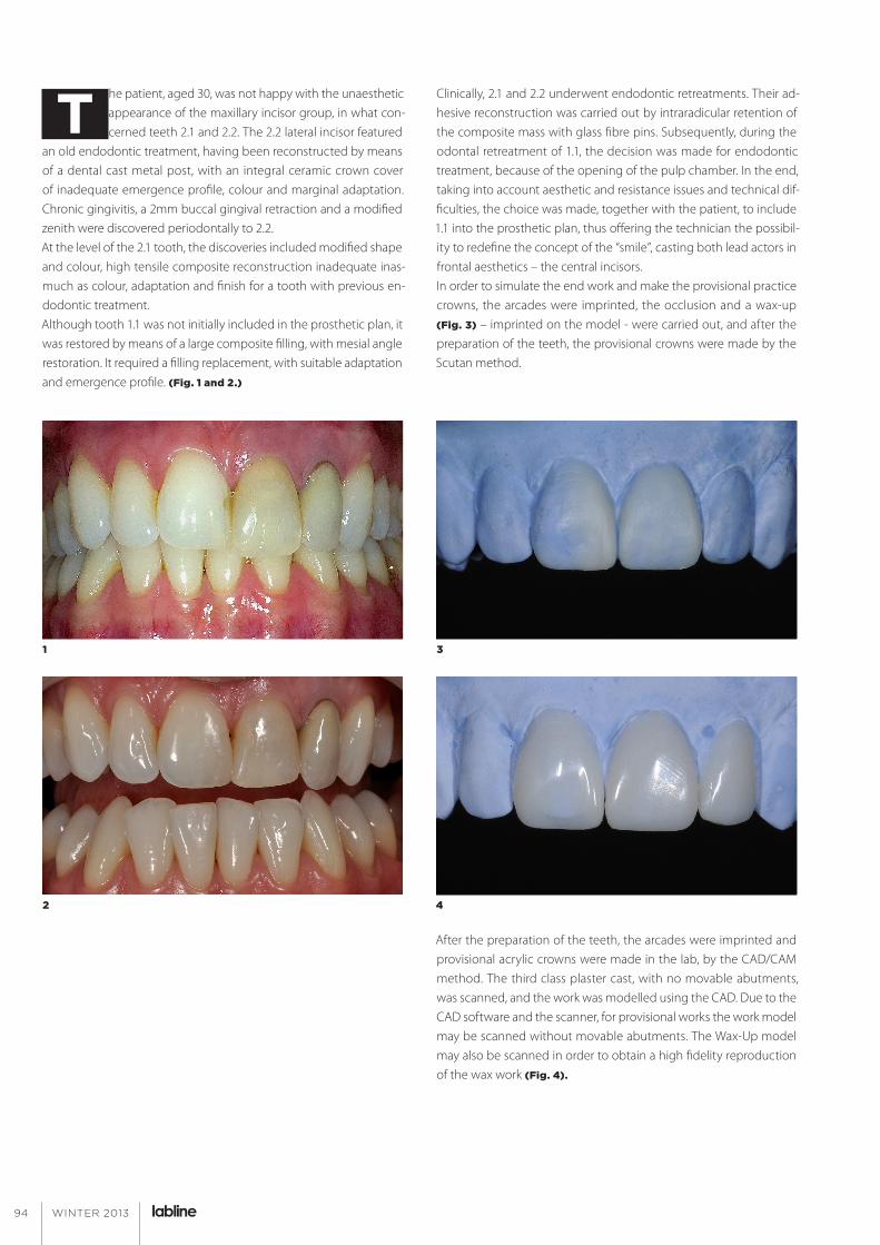
T he patient, aged 30, was not happy with the unaesthetic appearance of the maxillary incisor group, in what con-cerned teeth 2.1 and 2.2. The 2.2 lateral incisor featured
an old endodontic treatment, having been reconstructed by means of a dental cast metal post, with an integral ceramic crown cover of inadequate emergence profile, colour and marginal adaptation. Chronic gingivitis, a 2mm buccal gingival retraction and a modified zenith were discovered periodontally to 2.2.At the level of the 2.1 tooth, the discoveries included modified shape and colour, high tensile composite reconstruction inadequate inas-much as colour, adaptation and finish for a tooth with previous en-dodontic treatment. Although tooth 1.1 was not initially included in the prosthetic plan, it was restored by means of a large composite filling, with mesial angle restoration. It required a filling replacement, with suitable adaptation and emergence profile. (Fig. 1 and 2.)
Clinically, 2.1 and 2.2 underwent endodontic retreatments. Their ad-hesive reconstruction was carried out by intraradicular retention of the composite mass with glass fibre pins. Subsequently, during the odontal retreatment of 1.1, the decision was made for endodontic treatment, because of the opening of the pulp chamber. In the end, taking into account aesthetic and resistance issues and technical dif-ficulties, the choice was made, together with the patient, to include 1.1 into the prosthetic plan, thus offering the technician the possibil-ity to redefine the concept of the “smile”, casting both lead actors in frontal aesthetics – the central incisors. In order to simulate the end work and make the provisional practice crowns, the arcades were imprinted, the occlusion and a wax-up (Fig. 3) – imprinted on the model - were carried out, and after the preparation of the teeth, the provisional crowns were made by the Scutan method.
After the preparation of the teeth, the arcades were imprinted and provisional acrylic crowns were made in the lab, by the CAD/CAM method. The third class plaster cast, with no movable abutments, was scanned, and the work was modelled using the CAD. Due to the CAD software and the scanner, for provisional works the work model may be scanned without movable abutments. The Wax-Up model may also be scanned in order to obtain a high fidelity reproduction of the wax work (Fig. 4).
1 3
2 4
95WINTER 2013
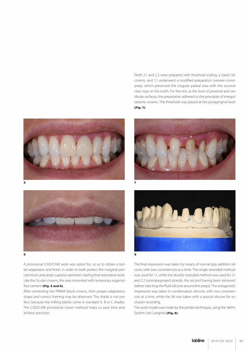
A provisional CAD/CAM work was opted for, so as to obtain a bet-ter adaptation and finish, in order to both protect the marginal peri-odontium and attain superior aesthetics during final restorative work.Like the Scutan crowns, this was cemented with temporary eugenol-free cement (Fig. 5 and 6).After cementing the PMMA block crowns, their proper adaptation, shape and correct framing may be observed. The shade is not per-fect, because the milling blanks come in standard A, B or C shades. The CAD/CAM provisional crown method helps us save time and achieve precision.
Teeth 2.1 and 2.2 were prepared with threshold scaling, a classic for crowns, and 1.1 underwent a modified preparation (veneer-crown prep), which preserved the cingular palatal area with the second-class stop on the tooth. For the rest, at the level of proximal and ves-tibular surfaces, the preparation adhered to the principles of integral ceramic crowns. The threshold was placed at the juxtagingival level (Fig. 7)
The final impression was taken by means of normal grip addition sili-cone, with two consistencies at a time. The single-stranded method was used for 1.1, while the double-stranded method was used for 2.1 and 2.2 (unimpregnated strands, the second having been removed before injecting the fluid silicone around the preps). The antagonists’ impression was taken in condensation silicone, with two consisten-cies at a time, while the IM was taken with a special silicone for oc-clusion recording.The work model was made by the pindex technique, using the JetPin System (Jan Langner) (Fig. 8).
5 7
6 8

96 WINTER 2013
In this case, the most important is the means of obtaining the inte-gral ceramic crowns. Because 12 and 11 were devital, of modified shade and with provisional work, as may be observed in the image, their thickness was minimal. For the ¾ crown on 11, the vestibular facet thickness was 0.2mm. The fact that the abutment was the shade of the healthy teeth helped us obtain the final shade easier. We opted for making the crowns by the method of sintered ceramics on refractory die. After being mounted in the articulator, the abut-ments were sectioned, prepared at prep level and duplicated using the Jan Langner duplication cuvette. This type of cuvette helped us obtain refractory die abutments 100% identical to those of the work model. (Fig. 9)
For ease of work and to make optimal crowns, a gingival mask was also produced, to replace the milled plaster in order to make the movable abutments. The technique of sintered ceramics on refrac-tory die abutments is one of the most difficult methods of obtain-ing integral ceramic crowns, after the platinum foil technique. At the same time, the aesthetics rendered by it is clearly superior to any pressed ceramics, but inferior to the platinum foil technique. For ceramics sintered on refractory die, we lack the possibility to test at “biscuit” stage, in order to see if the shade is right. One other as-pect that makes it difficult to tell the correct shade is that the refrac-tory die mount is white, not identical to the shades of the abutments. This is why an individual colour key is created to test the chosen shade combinations. (Fig. 10)
The chosen shades were nearly identical, so we could go on to sin-tering ceramics on the refractory die abutments. Before the first wash firing, we needed to carry out a heat treatment of the refrac-tory abutments, consisting of their drying and degassing in an oven, at 1100°C. After degassing, the refractory die pores were closed by wash firing, using a transparent ceramic mass. For teeth 21 and 22, the stratification was carried out with opaque ceramic masses in the beginning, in order to mask the colour of the abutments.
9 10

97WINTER 2013
The next firing was carried out with dentin masses, and for tooth 11, incisally, with opals. After sintering and after ob-taining the shape of the crowns, I manually polished the crowns using rubber wheels and Pumice powder. (Fig. 11)
To check the shape and texture of the teeth after the final processing, we used a Majesthetik-silver texturpowder to highlight their shape. (Fig. 12)
Certain of their shape and texture, we may now carry out the glaze firing. After the last sintering, the abutments are sectioned from the refractory die under the facet pack and the extra refractory die is sandblasted using glass pearls. Before sending them to the clinic, the adaptation of the crowns is double-checked on the control model. (Fig. 13)
Cementing was made by the adhesive technique for inte-gral ceramics described by Pascal Magne.
The crowns received from the lab, uncarved, were tested using a transparent glycerine gel in order not to influence their aesthetics and to stimulate cementing. The threshold adaptation was tested along with the points of contact, the static and dynamic occlusal contacts, the emergence profile and, last but not least, the aesthetic framing. After the patient accepted the restorations, the actual cementing began. (Fig. 14)
11
12
13
14

98 WINTER 2013
AT THE RESTORATIVE LEVEL1. The crowns were prepared by being placed in a silicone matrix
that made them easier to handle.2. They were carved in 9.5% hydrogen fluoride for 90 seconds,
which was removed under running water.3. The white precipitate was removed in 37% phosphoric acid
stirred with an adhesive applicator, then washed under running water and wiped with an acetone-soaked applicator.
4. After drying and checking the inner surfaces, they were silanised with two-component silane for 60 seconds, then dried with hot air by means of a hairdryer for 120 seconds.
5. The inner surfaces were styled with a fifth generation adhesive, compatible with the dual grip cements. They were then pro-tected from light.
AT THE LEVEL OF DENTAL PREPARATIONS1. The teeth were properly isolated by the dam system, through
the split dam technique, then the dental facets were carved in 37% phosphoric acid, 30 seconds on the enamel and 15 seconds on the dentin.
2. The facets were styled with adhesive without polymerisation. 3. The crowns were filled with translucent dual grip resin ce-
ment and then applied over the preparations. After a 2-3 sec-ond polymerization, the extra cement in its gel-state was removed marginally and interdentally. In order to avoid oxy-gen inhibition of the polymerization, the edges were distem-pered with glycerine. The facets were polymerized for 60 sec-onds each, then the edges were polished with silicone gums. (Fig. 15–18)
15–18
99WINTER 2013
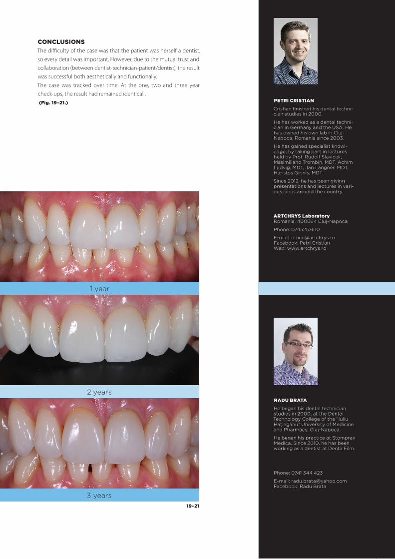
CONCLUSIONSThe difficulty of the case was that the patient was herself a dentist, so every detail was important. However, due to the mutual trust and collaboration (between dentist-technician-patient/dentist), the result was successful both aesthetically and functionally. The case was tracked over time. At the one, two and three year check-ups, the result had remained identical . (Fig. 19–21.) PETRI CRISTIAN
Cristian finished his dental techni-cian studies in 2000.
He has worked as a dental techni-cian in Germany and the USA. He has owned his own lab in Cluj-Napoca, Romania since 2003.
He has gained specialist knowl-edge, by taking part in lectures held by Prof. Rudolf Slavicek, Masimiliano Trombin, MDT, Achim Ludvig, MDT, Jan Langner, MDT, Haristos Girinis, MDT.
Since 2012, he has been giving presentations and lectures in vari-ous cities around the country.
ARTCHRYS Laboratory Romania, 400664 Cluj-Napoca
Phone: 0745257610
E-mail: [email protected] Facebook: Petri Cristian Web: www.artchrys.ro
RADU BRATA
He began his dental technician studies in 2000, at the Dental Technology College of the “Iuliu Haţieganu” University of Medicine and Pharmacy, Cluj-Napoca.
He began his practice at Stomprax Medica. Since 2010, he has been working as a dentist at Denta Film.
Phone: 0741 344 423
E-mail: [email protected] Facebook: Radu Brata
1 year
2 years
3 years19–21

PH
OT
O: D
R. M
EN
TE
S Á
RPÁ
D
9400 Sopron, Virágvölgyi u. 59.www.dentavantgart.huinfo@dentavantgart.huwww.lablinemagazine.com
www.facebook.com/ Labline Magazine
We wish all our Readersa Merry Christmas
and a Happy New Year!