Embed Size (px)
Citation preview

VOLUME 60, NUMBER 3 121
Journal of the Lepidopterists’ Society60(3), 2006, 121–137
FIVE NEW SPECIES OF PAUCIVENA DAVIS, 1975 (LEPIDOPTERA: TINEOIDEA:PSYCHIDAE) FROM CUBA
RAYNER NÚÑEZ AGUILADivisión de Colecciones Zoológicas y Sistemática, Instituto de Ecología y Sistemática, Carretera deVarona km 3.5,
Capdevila, Boyeros, C. de La Habana, Cuba. AP 8029. CP 10800, Cuba.E-mail: [email protected]
ABSTRACT. Five new species of Paucivena Davis (Lepidotera: Tineoidea: Psychidae), P. ferruginea, P. pinarensis, P. fusca, P. cubana and P.orientalis are described from Cuba and compared with relatives. The females of P. ferruginea and P. orientalis are described being the first fe-males known within genus; their characteristics confirm the intermediate position of the genus among the American psychids. Notes on naturalhistory of new species (e.g. hosts, habitat) are given as available. Keys for identification of known stages of all Paucivena species are provided.
Additional key words: Tineoidea, Psychidae, bagworm, natural history, West Indies.
The Neotropical region has the richest Lepidopteradiversity but one of the least studied psychid fauna withonly 61 known species (Heppner, 1998; Davis, 2000).This species number is extremely low compared withthat of other faunal regions. The Paleartic, for example,has more than 300 described species (Heppner, 1998).Members of this family were last reviewed by Davis(1975) who described two genera and five species fromthe West Indies. However, none of these taxa wasknown from Cuba. Recent collections from Cuba,chiefly on the three main mountain chains, haveresulted in the discovery of new records includingseveral new species. In this work, five new speciesbelonging to Paucivena Davis, 1975 are described. Keysfor identification of known stages of all species andinformation on natural history are also provided.
MATERIALS AND METHODS
Individuals of all species, except one whoserepresentatives were taken flying at day, were collectedas larvae and pupae in the field and reared in thelaboratory. Lab-reared larvae were provided with fieldcollected hosts until pupation. All type material isdeposited at Instituto de Ecología y Sistemática(CZACC).
Diagnostic morphological characters employedfollows Davis (1964, 1975) and Henderickx (1982). Setalmaps of larvae follow Hinton (1946) and Stehr (1987).Measurements were taken with an ocular micrometer ina Carl Zeiss Stemi 2000 stereoscopic microscope.Interocular index of head was calculated as a ratiobetween the vertical diameter of the compound eye andinterocular distance measured at a point across the fronsmidway between the base of antennal sockets and theanterior tentorial pits (Davis, 1975):
Interocular index= vertical eye diameter/ interoculardistance
Characters of the two previously known species,
Paucivena reticulata Davis, 1975 and Paucivenahispaniolae Davis, 1975, were taken from originaldescriptions and illustrations. Additionally, one P.reticulata specimen placed at CZACC, was examined.
Abbreviations: —×- mean, SD- standard deviation, CV-coefficient of variation.
RESULTS
Paucivena Davis, 1975
This genus is known only from the western part of theAntilles. Davis (1975) described P. hispaniolae fromDominican Republic, Hispaniola, and P. reticulata fromPuerto Rico and Jamaica. In the same work, this authormentioned the possible presence of Paucivena on Cuba.This was confirmed recently by Núñez (2004) based onunknown species from Topes de Collantes, in theCuban central mountains, which are described here.
Characters that best define Paucivena males are labialpalpi with a single segment not fused, origin of antennalrami at base of each antennal segment, two pairs oftibial spurs on mid and hindlegs, reduced wing venationand the abbreviated genitalia. Females may berecognized by the possession of compound eyes andfunctional legs, and the lack of antennae and wings.Larvae feed on several hosts including mosses andlichens growing on rocks and bark, and detritus.
Paucivena ferruginea Núñez, new species (Figs. 1, 6, 11, 16, 17, 22, 24, 26, 28–35, 41, 45–46, 52)
Diagnosis: Paucivena ferruginea male differs fromall other Paucivena by its brown coloration with slightferruginous iridescence. Other diagnostics charactersare the acute and heavily sclerotized sacculus and thebifid saccus of its genitalia.
Male (Figs. 1, 6, 11, 16, 17). Head: brown. Antennae with 23segments; lateral pectinations about 2–2.5 times length of supportingsegment. Vertical diameter of eye 0.8 the interocular distance. Thorax(Figs. 6, 11): anterior half dark brown, posterior half brown with slightferruginous iridescence; underside pale brown except inner surface of

122122 JOURNAL OF THE LEPIDOPTERISTS’ SOCIETY
FIGS. 1–5. Paucivena spp. adult males. 1 P. ferruginea, n. sp.; 2 P. pinarensis, n. sp.; 3 P. fusca, n. sp.; 4 P. cubana, n. sp.; 5 P. orientalis, n. sp. Scale= 3 mm.
coxa and femur of forelegs which are dark brown. Vestiture dense,scales hairlike. Wings brown with slight ferruginous iridescence; basaltwo thirds of costa on FW dark brown. Tibial spurs approximately 0.35the length of basal tarsal segments (Fig. 6). Scales at discal cellvariable in shape: oblanceolated, ovobated, with rounded or acuteapices, or hairlike. FW with 9 veins, all veins separated (Fig. 11);accessory cell present; CuP not reaching inner margin; one anal vein.HW with 7 veins, all veins separated except M2+3 and CuA1 which havea unique origin; cross vein between Sc and Rs absent; two anal veins.Wing expanse: 11 mm. Abdomen: brown with slight ferruginousiridescence; underside pale brown. Vestiture dense, scales hairlike.Genitalia (Figs. 16–17): tegumen broad, with a pair of sparsely setoseapical lobes. Valvae with pulvilli setose; apex of sacculus acute andheavily sclerotized, armed with three spines; cucullus apically roundedand sparsely setose. Saccus bifid, apices blunt; approximately 0.1 thelength of main body. Aedeagus simple, cylindrical, 0.7 times thelength of valvae.
Female (Figs. 22, 24, 26). Length: 7.5 mm. Vermiform.Stramineous, with two longitudinal bands of brown spots on dorsum.Head (Fig. 22): stramineous, eyes black. Slightly sclerotized. Shapenear ovoid (ventral view); eyes compound, well developed, subventral.Labial palpi 1-segmented, 100% fused; antennae absent. Thorax (Fig.24): patterned as above; body wall slightly sclerotized. Legs functional,armed with numerous tiny spines; tarsi 1-segmented with a pair ofclaws at distal end (Fig. 24); wings absent. Abdomen: color patterndisappearing at A2–A3; membranous and naked except for a ring ofdense brownish ochre hairlike scales around A7. External genitaliareduced (Fig. 26), largely membranous. Two pairs of apophysespresent; anterior pair elongated, free except bifid base fused withtegument; posterior pair straight and free.
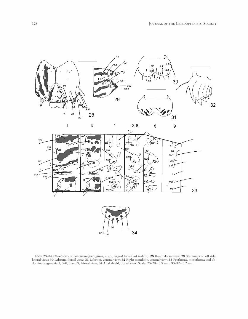
Larva (Figs. 28–34). Length of largest larva 11 mm, maximumwidth of head capsule 1.2 mm. Head and thorax whitish with dark
fuscous longitudinal bands continued on thorax forming a striatedpattern; spiracle on TI as large as spiracle on A8, both larger thanspiracles on A1–A7. Head (Figs. 28–32): patterned as above, lateralarea with five elongated bands; labrum ochre; an elongated band onadfrontal sclerite and frons extending from C1 to slightly beyond AF1;AF2 and P2 absent (Fig. 28). Six stemmata present; five arranged inan inverted semicircle, a sixth more distant and ventrad, immediatelyanterior to S3 (Fig. 29). Labrum (Figs. 30, 31) with LA3 isolated.Mandibles with four acute teeth and a fifth, blunt tooth (Fig. 32).Thorax (Fig. 33): patterned as above, three longitudinal bandsbetween body axis and lateral margin of shield, interrupted onmetathorax. TI with shield bearing D, SD, XD and L groups; XD-group and L2 in vertical line near anterior margin of shield, SD-groupslightly posterior; XD1 about equal in length to SD1, about 2 timeslonger than XD2 and D2; D1 dorsoposterior to XD1, about 1/4 itslength; SD2 above SD1, slightly posterior and about 1/4 its length; L-group trisetose, L1 about 3 times longer than L2 and L3,posteroventral to L2; L3 slightly longer than L2; spiracledorsoposterior to L-group, diagonal; SV-group in horizontal line onelongated pinnaculum, SV2 about 3/5 length of SV1; MV2 on samepinnaculum, anterior to SV2; V1 about equal in length to SV2,posteroventral to SV1. TII-TIII: D and SD groups in a vertical line onsame pinnaculum; D2 about 2–21/2 times longer than D1; SD1 about3 times longer than SD2; L2 separated from pinnaculum bearing L1and L3, about 1/2 length of L3; L1 3/5 length of L2, dorsoposterior toL3; SV group on same pinnaculum; SV1 about twice length of SV2; V1slightly shorter than SV1. Abdomen (Figs. 33, 34): integument darkbrown, pinnacula brownish ochre. A1: D-group on separatedpinnacula, D1 dorsoposterior to D2 and about 3 1/2 times longer; SD1above and slightly anterior to spiracle, slightly shorter than D2; SD2minute, anterodorsal to spiracle; L-group trisetose, on separatedpinnacula; L1 posterior to L2 and about twice its length; L3 below,

VOLUME 60, NUMBER 3 123
midway between L1 and L2, equal in length to L1; SV-group bisetoseand on same pinnaculum, SV2 anterodorsal to SV1 and about 1/3 itslength; V1 anteroventral to SV1 and about half its length (Fig. 33). A2(not shown) equal to A1 except SV- group trisetose, SV3 onpinnaculum bearing V1, below SV2 and about equal its length. A3–A6with four pairs of prolegs, crochets (22–24) uniordinal, uniserial,arranged in a lateral penellipse; setae as above except SV-group onpinnaculum containing proleg. A7 (not shown) as above except SV-group bisetose. A8 with setae as above except L-group arranged in amore or less vertical line, L1 on same pinnaculum bearing SD-groupand spiracle; SV-group unisetose. A9 with all setae arranged in a moreor less vertical line; SD1 and D2 on same pinnaculum, SD1 about 5times longer than D2 and about equal in length to D1. A10 (Fig. 34):anal plate with SD1 slightly longer than D1, about 2–2 1/2 timeslonger than D2; prolegs bearing 24 uniordinal crochets, uniserial,arranged in a lateral penellipse; anterior margin of shield irregular.
Larval case (Fig. 41). Dimensions: length of main body: m 12mm (—×=12, SD=0, CV=0, n=2), total length of projecting fragments:17–18 mm (—×=17.5, SD=0.71, CV=4%, n=2); f 13 mm, total lengthof projecting fragments: 22 mm; maximum diameter: m 2.6 mm (—×=2.6, SD=0, CV=0, n=2), f 3.3 mm. Almost cylindrical in its entirelength; soft. External cover formed by a basal layer of tiny vegetalfragments covered by large fragments of thin herbaceous stems,lengthwise arranged and parallel, various projecting backward fromcase.
Male pupa (Figs. 45–46). Length 5.8 mm. Uniform yellowishochre. Frontal ridge absent, frons rounded. Antennal scleritesextending slightly beyond apex of prothoracic legs (Fig. 45). Wing
sheaths extending midway along A3. Sclerites of metathoracic legsextending to anterior margin of A4. Cremaster reduced, consisting ina pair of small spines, ventrally curved, arising form a broad conicalbase; anal groove Y-shaped. Dorsum of A3–A7 with 2–3 irregular rowsof spines directed caudad on anterior margin, both end of rowsthickened (Fig. 46); A8 with spines grouped in an elliptical patch;areas surrounding rows covered by hundreds of tiny spines arrangedin 2–5 series or solitaire. Dorsum of A5–A7 with single posterior rowof slender spines oriented caudad. Tabulation of spines shown in Table1.
Female pupa (Figs. 50–51). Length 7.5 mm. Uniform yellowishochre. Head with eyes and labial palpi distinct (Fig. 50). Thorax withleg sclerites distinct; wings absent. Cremaster vestigial, reduced to acoarse and rough area around anal groove; anal groove Y-shaped.Dorsum of A6–A7 with 2–4 irregular rows of small spines directedcaudad on anterior margin, A8 with spines grouped in an ellipticalpatch. A4–A6 with a single posterior row of slender spines cephaladoriented (Fig. 51). Tabulation of spines shown in Table 2.
Types. Holotype m (with associated larval case and pupal exuvium),reared from larva (emerged 22 June 2003), CUBA: Sancti Spiritusprovince, Topes de Collantes, Pico Potrerillo, 973 m, 18 May 2003 (R.Núñez), slides RNA 014, 038, 046, 077, 078. Paratypes, f (withassociated larval case and pupal exuvium), reared from pupa (emerged20 May 2003), CUBA: Sancti Spiritus province, Topes de Collantes,Parque Codina, 800 m, (R. Núñez), slide RNA 042; 2 larvae (withassociated larval case), same data as holotype; 3 larval cases, same dataas holotype; 1 larval case, same data as holotype except 17 June 2004.
FIGS. 6–10. Paucivena spp. male legs. 6 P. ferruginea, n. sp.; 7 P. pinarensis, n. sp.; 8 P. fusca, n. sp.; 9 P. cubana, n. sp.; 10 P. ori-entalis, n. sp. Top- prothoracic leg, middle- mesothoracic leg, bottom-metathoracic leg. Scale= 2 mm.

124124 JOURNAL OF THE LEPIDOPTERISTS’ SOCIETY
Natural history observations. Larvae were foundfeeding on mosses, Orthostichidium guyanense (Mont.)V.F. Brotherus (Pterobryaceae) and anotherunidentified species, on bark of an unidentified bush.
Two adults were reared from larvae: a male emergedfrom the pupa after a month; a female emerged and wasobserved hanging from distal end of the case.
The species was found in two localities with verydifferent vegetation and climatic conditions. ParqueCodina is characterized by a secondary and very humidevergreen forest with the lower strata protected by adense canopy. A dry scrub, included in the mogotevegetational complex, grows on top of Pico Potrerillo, anenviroment very exposed to wind and solar radiation.
Distribution (Fig. 52). Known only from twolocalities at Topes de Collantes region, TrinidadMountains, central Cuba.
Etymology. The species name is derived from theslight ferruginous iridiscence of male wings.
Remarks. The female may be separated from that ofP. orientalis, n. sp., by its greater size (length 7.5 versus5 mm), its more elongated genitalia, the presence of
only two longitudinal spot bands on tegument (six in theother species) and its unswollen tibiae. Larvae may berecognized by the loss of AF2 on head and the isolationof LA3 on labrum. The larval case is also diagnostic forthis species within the genus. Davis (1964, 1975)reported similar cases from Haiti and Trinidad. Davisnoted the similarity of these cases with thoseconstructed by species of Epichnopteryx Hübner andPsyche Schrank, two Old World genera, consideringthem as a probable introduction from the Old World.Larval cases of P. ferruginea prove that this constructionpattern is not exclusive to Old World species.
Paucivena pinarensis Núñez, new species (Figs. 2, 7, 12, 18, 42, 47, 52)
Diagnosis: Paucivena pinarensis males possess acolor pattern similar to that of P. cubana, sp. n., P.orientalis, sp. n., and P. reticulata. However, thereticulated pattern is weaker in P. pinarensis due to itsmore obscure background color. Compared to otherPaucivena within this group, P. pinarensis males exhibitseveral diagnostic characters: absence of saccus in its
FIGS. 11–15. Paucivena spp. wing venation. 11 P. ferruginea, n. sp.; 12 P. pinarensis, n. sp.; 13 P. fusca, n. sp.; 14 P. cubana, n. sp.;15 P. orientalis, n. sp. Scale= 3 mm.

VOLUME 60, NUMBER 3 125
Rows Abdominal segments
I II III IV V VI VII VIII
Paucivena ferruginea
n=1 anterior 0 0 269 135 140 176 122 48
posterior 0 0 0 0 42 44 48 0
Paucivena pinarensis
n=1 anterior 0 0 57 63 64 60 49 49
posterior 0 0 1 7 38 38 14 0
Paucivena cubana
n=3anterior 0 0 0 52–70 47–58 42–55 29–45 22–24
posterior 0 0 31 35–44 37–46 36–42 32–36 0
Paucivena orientalis
n=3anterior 0 0 0 102–136 96–125 95–111 90–96 25–32
posterior 0 0 0 27–38 23–39 24–32 23–50 0
Rows Abdominal segments
I II III IV V VI VII VIII
Paucivena ferruginea
n=1anterior 0 0 0 0 0 61 90 21
posterior 0 0 0 7 58 53 0 0
Paucivena cubana
n=1anterior 0 0 0 3 14 20 21 0
posterior 0 0 6 56 78 71 0 0
Paucivena orientalis
n=3anterior 0 0 0 8–10 9–10 29–30 92–96 18–-22
posterior 0 0 0 16–17 23–27 21–22 0 0
TABLE 2. Rows and spines numbers per rows on dorsum of abdominal segments of Cuban Paucivena female pupae.
TABLE 1. Rows and spines numbers per rows on dorsum of abdominal segments of Cuban Paucivena male pupae.

126126 JOURNAL OF THE LEPIDOPTERISTS’ SOCIETY
FIGS. 16–21. Paucivena spp. male genitalia, ventral view. 16 P. ferruginea, n. sp., main body; 17 P. ferruginea, n. sp., aedeagus; 18P. pinarensis, n. sp.; 19 P. fusca, n. sp.; 20 P. cubana, n. sp.; 21 P. orientalis, n. sp. Scale= 0.5 mm.
FIGS. 22-23. Paucivena spp. female head, ventral view. 22 P. ferruginea, n. sp.; 23 P. orientalis, n. sp. Scale= 1 mm.

VOLUME 60, NUMBER 3 127
FIGS. 24–25. Paucivena spp. female legs. 24 P. ferruginea, n. sp., scale= 0.5mm; 25- P. orientalis, n. sp., scale= 0.25 mm. Top-prothoracic leg, middle- mesothoracic leg, bottom-metathoracic leg.
FIGS. 26–27. Paucivena spp. female genitalia, ventral view. 26 P. ferruginea, n. sp.; 27 P. orientalis, n. sp. Scale= 0.5 mm.
128128 JOURNAL OF THE LEPIDOPTERISTS’ SOCIETY
FIGS. 28–34. Chaetotaxy of Paucivena ferruginea, n. sp., largest larva (last instar?). 28 Head, dorsal view; 29 Stemmata of left side,lateral view; 30 Labrum, dorsal view; 31 Labrum, ventral view; 32 Right mandible, ventral view; 33 Prothorax, mesothorax and ab-dominal segments 1, 3–6, 8 and 9, lateral view; 34 Anal shield, dorsal view. Scale, 28–29= 0.5 mm; 30–32= 0.2 mm.

genitalia; the relative length of its tibial spurs, 0.3 versus0.15 (P. cubana, P. orientalis) and 0.5 (P. reticulata); andinterocular index 0.7 versus 1.1 (P. reticulata) and 1.5 (P.cubana).
Male (Fig. 2, 7, 12, 18).: Head: pale brown, labial palpi darkbrown. Antennal tips broken; lateral pectinations 3 about times lengthof supporting segment. Vertical diameter of eye 0.7 the interoculardistance. Thorax (Figs. 7, 12): dark brown; underside pale brown,inner surface of fore and midlegs dark brown, joints pale brown.Vestiture dense, scales hairlike. Tibial spurs approximately 0.3 thelength of basal tarsal segments (Fig. 7). FW with basal two thirds ofcosta dark brown; ground brown streaked with dark brown forming afaint reticulated pattern; fringe with various tones of brown. Scales atdiscal cell variable in shape: oblanceolated and ovobated, withrounded or acute apices, with scattered hairlike scales. Venation (Fig.12) as in P. ferruginea. HW uniform brown; fringe with various tonesof brown. Venation as in P. ferruginea except M2+3 and CuA1 whicharise separate from cell, M2+3 equidistant from CuA1 and M1, only oneanal vein present. Wing expanse: 9.2 mm. Abdomen: pale brown.Vestiture dense, scales hairlike. Genitalia (Fig. 18): tegumen broad,with a pair of sparsely setose apical lobes. Valvae with pulvilli setose;apex of sacculus armed with five strong spines; cucullus apicallyrounded and sparsely setose. Saccus absent. Aedeagus simple,cylindrical, 0.6 times length of valvae.
Female. Unknown. Larva. Unknown. Larval case (Fig. 42). Dimensions: length: m 9.8 mm; maximum
diameter: m 3.4 mm. Fusiform in outline; soft. Exterior heavilycovered with vegetal fragments, leaves and short stems, and mossesvarious shaped and oriented lengthwise. Cases were found hangingfrom silk filaments (3.5 mm in length) attached to rocks and treetrunks.
Male pupa (Fig. 47). Length 5.1 mm. Ochre, wing sheaths reddishbrown. Frontal ridge absent, frons rounded. Antennal scleritesextending slightly beyond apex of prothoracic legs. Wing sheathsextending to posterior margin of A3. Sclerites of metathoracic legsextending to posterior margin of A4. Cremaster consisting in a pair ofstrong spines ventrally curved; anal groove Y-shaped. Dorsum ofA3–A8 with an irregular row of spines on anterior margin; areassurrounding rows covered by hundreds of tiny, solitaire spines.Dorsum of A3–A7 with single posterior row of slender spines.Tabulation of spines shown in Table 1.
Female pupa. Unknown. Types. Holotype, m (with associated larval case and pupal
exuvium), reared from larvae (emerged 28 February 2004), CUBA:Pinar del Río province, Sierra del Rosario, Taco Taco River shore 1 kmnortheast from Jardín de Aspiro, 200 m, 28 November 2003 (R.Núñez), slides RNA 054, 079, 080. Paratypes: 4 larval cases, CUBA:Pinar del Río province, Sierra del Rosario, Jardín de Aspiro, 150 m,
VOLUME 60, NUMBER 3 129
FIGS. 35–40. Chaetotaxy of Paucivena cubana, n. sp., largest larva (last instar?). 35 Head, dorsal view; 36 Stemmata of left side,lateral view (scale= 0.5 mm); 37 Labrum, dorsal view; 38 Labrum, ventral view; 39 Right mandible, ventral view (scale= 0.2 mm);40 Prothorax and mesothorax, lateral view (abdomen damaged). Scale, 35–36= 0.5 mm; 37–39= 0.2 mm.
130130 JOURNAL OF THE LEPIDOPTERISTS’ SOCIETY
Key to the adult males of Paucivena
1. Wings dark brown with a ferruginous shine; genitalia with an acute and heavily sclerotized sacculusand a bifid saccus (Fig. 16) P. ferruginea
- Wings with a different color pattern; genitalia with sacculus not acute and weakly sclerotized, saccusnot bifid 2
2. Dorsum of wings and body entirely dark brown, almost black 3- Dorsum of wings and body with light color pattern 43. Body whitish grey ventrally; eyes of medium size (vertical diameter of eye 1.1 the interocular
distance); genitalia with the margins of apex of sacculus and the apical lobes of tegumen smooth P. hispaniolae
- Body dark brown ventrally; eyes small (vertical diameter of eye 0.8 the interocular distance);genitalia with the margins of apex of sacculus and the apical lobes of tegumen serrulated (Fig. 19)
P. fusca4. Tibial spurs much reduced, approximately 0.15 the length of basal tarsal segment 5- Tibial spurs less reduced, approximately 0.3 the length of basal tarsal segment or longer 65. Eyes very large (vertical diameter of eye 1.5 the interocular distance); wing expanse: 12 mm
P. cubana- Eyes very small (vertical diameter of eye 0.7 the interocular distance); wing expanse: 8.2-9.1 mm
P. orientalis6. Eyes of medium size (vertical diameter of eye 1.1 the interocular distance); tibial spurs large,
approximately 0.5 the length of basal tarsal segment; FW reticulated pattern distinct P. reticulata- Eyes very small (vetical diameter of eye 0.7 the interocular distance); tibial spurs reduced,
approximately 0.3 the length of basal tarsal segment; FW reticulated pattern weak, indistinct P. pinarensis
Key to the known larvae of Paucivena (excludes P. reticulata, P. pinarensis, P. fusca and P. orientalis,which are unknown)
1. Head and thorax whitish to light tan with irregular patches of dark fuscous; meso and metathoraxwith an extra seta (SD1a?) P. hispaniolae
- Head and thorax whitish with longitudinal dark fuscous bands arranged in a striated pattern; mesoand metathorax without an extra seta 2
2. Head with AF2 absent (Fig. 28); LA3 on labrum isolated from the rest (Fig. 30); abdominalintegument dark brown P. ferruginea
- Head with AF2 present (Fig. 35); LA3 on labrum not isolated (Fig. 37); abdominal integument dirtywhite P. cubana
28¬29 November 2003, (R. Núñez). Natural history observations. The single larva was
found on limestone rock near the Taco Taco River shoreand its food source can not be accurately identified. Inthe lab, the larva was fed with several crustose lichensand mosses collected on its substrate. Other larval caseswere found on rocks and tree trunks in an oldabandoned Botanical Garden (Jardín de Aspiro). Theunique adult emerged after a month; emergence tookplace between 0900 and 1130h.
At the Taco Taco River shore secondary remnants ofgallery forest are present whereas at Jardín de Aspiroseveral introduced and native trees grow forming grovesseparated by cleared areas occupied by campinginstallations.
Distribution (Fig. 52). Known only from two close
localities at Sierra del Rosario, Pinar del Río province. Etymology. The species name is derived from the
name of the Cuban province where the type locality,Pinar del Río, is located.
Remarks. The larval case is identical to that of P.cubana. The male pupa may be easily identified by theunique arrangement of spine rows on the dorsum ofabdominal segments (Table 1).
Paucivena fusca Núñez, new speciesPaucivena sp. n. 1: Núñez, 2004: 155
Figs. 3, 8, 13, 19, 52
Diagnosis. Males of P. fusca may be recognized bytheir uniform dark brown coloration. Within the genusonly P. hispaniolae exhibits a similar coloration but it has
VOLUME 60, NUMBER 3 131
Key to the known male pupae of Paucivena (excludes P. reticulata and P. fusca, which are unknown)
1. Anterior margin of A3 without spines 2- Anterior margin of A3 with at least one row of spines 32. Spines absent from posterior margin of A3; rows at anterior margin of A4-A7 with 90 or more spines
(Table 1); length 3.8-4.8 mm P. orientalis- Spines present on posterior margin of A3; rows at anterior margin of A4-A7 with 70 or fewer spines
(Table 1); length 5.0-5.4 mm P. cubana3. Rows on anterior margin of A3-A7 with more than 100 spines (Table 1); length 5.8 mm
P. ferruginea- Rows on anterior margin of A3-A7 with less than 100 spines (Table 1); length 5.0-5.1 mm 44. Spines present, although reduced, on posterior margin of A3-A4; cremaster consisting in a pair of
strong spines ventrally curved P. pinarensis- Spines completely absent from posterior margin of A3-A4; cremaster consisting in a pair of small
spines ventrally curved P. hispaniolae
Key to the female pupae of Paucivena (excludes P. reticulata, P. pinarensis and P. fusca, which areunknown)
1. Anterior rows of spines absent from dorsum of A3-A4 2- Anterior rows of spines present on dorsum of A3-A4 32. Posterior row of spines reduced but present on dorsum of A4; cremaster vestigial, reduced to a
coarse and rough area around anal groove; length 7.5 mm P. ferruginea- Posterior row of spines absent from dorsum of A4; cremaster relatively well developed, consisting in
pair of short acute spines; length 10-11 mm P. hispaniolae3. Anterior margin of A8 with a row of spines surrounded by hundreds of tiny spines; length 4.9-5.1
mm P. orientalis- Anterior margin of A8 covered only by hundreds of tiny spines; length 8.0 mm P. cubana
the underside of body whitish grey. Other usefulcharacters are FW shape (more rounded in the Cubanspecies), eye size (interocular index 0.8 in P. fusca and1.1 in P. hispaniolae) and the serrated margins at apex ofsacculus and apical lobes of tegumen in the genitalia ofP. fusca, both smooth in P. hispaniolae.
Male (Figs. 3, 8, 13, 19). Head: dark brown. Antennae with 21–22segments; lateral pectinations 2 times length of segment. Verticaldiameter of eye 0.8 the interocular distance. Thorax (Figs. 8, 13):uniform dark brown. Vestiture dense, scales hairlike. Tibial spursapproximately 0.25 the length of basal tarsal segments (Fig. 8). Scalesat discal cell of FW oblanceolated and ovobated with rounded oracute apices. Venation (Fig. 13) as in P. ferruginea. HW venation as inP. pinarensis, except the origin of M2+3 which is closer to CuA1 than toM1. Wing expanse: 10–11 mm (—×=10.2, SD=0.23, CV=4%, n=9).Abdomen: dark brown. Vestiture dense, scales hairlike. Genitalia (Fig.19): tegumen broad with apical cleft, lobes minutely serrated andsparsely setose. Valvae with pulvilli setose; apex of sacculus stronglyserrated; cucullus rounded, apex sparsely setose. Saccus reduced,approximately 0.2 the length of main body. Aedeagus simple,cylindrical, 0.6 times the length of valvae.
Female. Unknown. Inmature stages. Unknown. Larval case. Unknown. Types. Holotype, m CUBA: Sancti Spiritus province, Topes de
Collantes, Pico Potrerillo, 973 m, 6 May 2002 (R. Núñez), slides RNA011, 015, 039. Paratypes, 3 m, same data as holotype, slides RNA 010,022. 5 m, same data as holotype except 17 June 2004, slides RNA 026,029, 041.
Natural history observations. All individuals werefound flying at noon on the top of Pico Potrerillo except
a single specimen seen flying between rocky walls atpeak access. This species shares its habitat, dry scrub onthe top of Pico Potrerillo, with P. ferruginea and P.cubana.
Distribution (Fig. 52). Known only from PicoPotrerillo at Trinidad Mountains, central Cuba.
Etymology. The species name is derived from itsuniform dark brown color.
Paucivena cubana Núñez, new speciesPaucivena sp. n. 2: Núñez, 2004: 155Figs. 4, 9, 14, 20, 35–40, 43, 48, 52
Diagnosis. Males of P. cubana may be separatedfrom other Paucivena with reticulated wing pattern bythe following characters: 12 mm of wing expanse (thelargest within the genus), elongated legs with tiny tibialspurs (approximately 0.15 the length of basal tarsalsegment) and large eyes (interocular index 1.5, thelargest within the genus).
Male (Figs. 4, 9, 14, 20). Head: pale yellowish ochre. Antennaewith 18 segments; lateral pectinations 1.5–2 times the length ofsegment. Eyes large, vertical diameter of eye 1.5 the interoculardistance. Thorax (Fig. 9, 14): pale yellowish ochre with scattered darkbrown scales. Vestiture dense, scales hairlike. Tibial spursapproximately 0.15 the length of basal tarsal segments (Fig. 9). Wingsthinly scaled. FW (faded) yellowish ochre with scattered dark brown

132132 JOURNAL OF THE LEPIDOPTERISTS’ SOCIETY
FIGS. 41–44. Paucivena spp. larval cases. 41 P. ferruginea, n. sp.; 42 P. pinarensis, n. sp.; 43 P. cubana, n. sp.; 44 P. orientalis, n.sp. Scale= 3 mm.
scales; dark brown scales concentrated at basal half of anterior andposterior margins, forming a faint reticulated pattern on basal twothirds; fringe ochre. Scales at discal cell variable in shape:oblanceolated and ovobated, with rounded or acute apices, or hairlike.Venation as in P. ferruginea, except accessory cell which is wider,closing below origin of R4+5 (Fig. 14). HW pale yellowish ochre withscattered dark brown scales, paler than FW; fringe pale yellowishochre. Venation as in P. fusca. Wing expanse: 12 mm. Abdomen: paleyellowish ochre with scattered dark brown scales. Vestiture dense,scales hairlike. Genitalia (Fig. 20): tegumen broad, apex damaged.Valvae with pulvilli sparsely setose; apex of sacculus armed with threespines; cucullus rounded with apex sparsely setose. Saccus reduced,approximately 0.2 the length of main body. Aedeagus simple,cylindrical, 0.7 times the length of valvae.
Female. Unknown. Larva (Figs. 35–40). Length of longest larva 6.9 mm, maximum
width of head capsule 1.0 mm. Head and thorax whitish with darkfuscous longitudinal bands continued on thorax forming a striatedpattern. Head (Figs. 35–39): as in P. ferruginea except, AF2 present;adfrontal sclerite with elongated spot on upper third covering origin ofAF2; frons with spot covering origin of F1 and C2 (Fig. 35); AFacloser to AF2 than to AF1. Sixth stemma immediately anterior to S2and S3, midway between them (Fig. 36). Labrum (Figs. 37, 38) withsetae approximately mesad except, LA1 and M2 distinctly closer toborder. Mandibles with four acute teeth and a fifth, blunt tooth (Fig.39). Thorax (Fig. 40): as in P. ferruginea with the following exceptions.TI with XD1 about equal in length to XD2 and SD1, and about 1 1/2times longer than D2; MV2 separated from pinnaculum bearing SV-

VOLUME 60, NUMBER 3 133
FIGS. 45–46. Paucivena spp. male pupa. 45 P. ferruginea, n. sp., ventral view; 46 P. ferruginea, n. sp., dorsal view
group. TII-TIII: D2 about 3 times longer than D1; SD1 about 4 timeslonger than SD2. Abdomen damaged, integument dirty white.
Larval case (Fig. 43). Dimensions, length: m 10–11 mm (—×=10.5,SD=0.71, CV=7%, n=2), f 18 mm; maximum diameter: m 4.0 mm (—×=4.0, SD=0, CV=0, n=2), f 6.5 mm. Fusiform in outline, soft.Exterior densely covered by fragments of leaves, small herbaceousstems and mosses of different shape and lengthwise oriented. Caseswere found attached to rocky walls at Pico Potrerillo and banana trees,Musa paradisiaca L. (Musaceae), at Mogote Mi Retiro, hanging fromsilk threads (length: m 3.5 mm, f 5 mm).
Male pupa (Fig. 48). Length 5.0–5.4 mm (—×=5.2, SD=0.23,CV=4%, n=2). Uniform brownish ochre. Frontal ridge absent, fronsrounded. Antennal sclerites extending slightly beyond apex ofprothoracic legs. Wing sheaths extending to anterior margin of A4.Sclerites of metathoracic legs extending midway along A5 or itsposterior margin. Cremaster consisting in a pair of strong and veryclose spines, ventrally curved and abruptly tapered at apex; analgroove Y-shaped. Dorsum of A4–A8 with 1–2 irregular rows of spineson anterior margin; anterior margin A3 and areas surrounding spinerows on A4–A8 covered by hundreds of tiny, solitaire spines. Dorsumof A3–A7 with single posterior row of slender spines. Tabulation ofspines shown in Table 1.
Female pupa. Length 8 mm. Uniform ochre. Head with eyes andlabial palpi distinct. Thorax with leg sclerites distinct; wings absent.
Cremaster vestigial, reduced to a small pair of blunt, widely separatedspines; anal groove Y-shaped, with a pair of small rounded tubercleson either side. Dorsum of A4 with single anterior row of reduced,widely spaced spines; A5–A7 with 2–3 irregular rows of spines onanterior margin; anterior margin of A3, areas surrounding rows onA4–A8 and anterior margin of A8 covered by hundreds of tiny spines,solitaire or in 2–4 series. A3–A6 with single posterior row of slenderspines. Tabulation of spines shown in Table 2.
Types. Holotype, m (with associated larval case and pupalexuvium), reared from larva (emerged June 2002), CUBA: SanctiSpiritus province, Topes de Collantes, Caburní River depression, 500m, 30 April 2002 (R. Núñez), slides RNA 013, 016, 023, 040, 045.Paratypes, 1 larva, same data as holotype, slides RNA 065, 066, 070; 6larval cases (some with associated pupal exuvium), CUBA: SanctiSpiritus province, Topes de Collantes, mogote Mi Retiro northernbase, 800 m, 17 May 2003 (R. Núñez); 4 larval cases (some withassociated pupal exuvium), CUBA: Sancti Spiritus province, Topes deCollantes, southern side of rocky outcrop at Pico Potrerillo access, 850m, 18 May 2003 (R. Núñez), slides RNA 075, 076; 3 larval cases (onewith associated pupal exuvium), same data as preceding except 17June 2004; 2 larval cases, CUBA: Sancti Spiritus province, Topes deCollantes, Parque Codina, 800 m, 20 May 2003 (R. Núñez).
Natural history observations. Larvae fed onPlagiochila sp. (Plagiochilaceae), a hepatic growing on

134134 JOURNAL OF THE LEPIDOPTERISTS’ SOCIETY
FIGS. 47–49. Paucivena spp. male pupa. 47 P. pinarensis, n. sp., ventral view; 48 P. cubana, n. sp., ventral view; 49 P. orientalis,n. sp. Scale= 0.25 mm.
FIGS. 50–51. Paucivena ferruginea, n. sp., female pupa. 50 Ventral view; 51 Dorsal view. Scale= 0.25 mm.

VOLUME 60, NUMBER 3 135
rocks on the Caburní River shore. However, larvaeprobably use other hosts since cases were found ondifferent substrates at other localities. At the PicoPotrerillo access cases were found attached to rockywalls covered by crustose lichens, at mogote Mi Retirobase they were located on banana plants whereas atParque Codina they live on trunks of native trees. Thesingle adult emerged after a month as pupa.
Paucivena cubana inhabits localities with verydifferent vegetation and climatic conditions. At ParqueCodina it was found in the lower strata of secondaryevergreen forest, a very humid habitat, and at themogote Mi Retiro base in cultivated land, banana andcoffee (Coffea arabica L., Rubiaceae), close to themogote rocky wall. On the other hand, Caburní Rivershores are covered by gallery forest remnants, todaydominated by an introduced tree (Syzigium jambos L.,Myrtaceae), whereas at the Pico Potrerillo access caseswere attached to rocky walls surrounded by evergreenforest.
Distribution (Fig. 52). Known from four localities atTopes de Collantes, Trinidad Mountains, central Cuba.
Etymology. The species name is derived from thename of the Cuban island.
Remarks. The larval color pattern is identical to thatof P. ferruginea; however, differences in chaetotaxy andabdominal coloration are present. Male and femalepupae are easily distinguished by the arrangement ofabdominal dorsal spines (Tables 1, 2).
Paucivena orientalis Núñez, new speciesFigs. 5, 10, 15, 21, 23, 25, 27, 44, 49, 52
Diagnosis. Adult males of P. orientalis possess acolor pattern similar to that of P. pinarensis, P. cubana,and P. reticulata. P. orientalis may be separated from P.pinarensis by its smaller tibial spurs (0.15 versus 0.3 thelength of basal tarsal segment), the presence of a saccusin its genitalia and its more distinct FW reticulatedpattern. From P. reticulata, it may be distinguish by itssmaller eyes (interocular index 0.7 versus 1.1) and tibialspurs (0.15 versus 0.5 the length of basal tarsalsegment). From P. cubana it differs by its smaller size(8.2–9.1 versus 12 mm of wing expanse) and eye size(interocular index 0.7 versus 1.5).
Male (Figs. 5, 10, 15, 21). Head: pale greyish brown. Anten-nae with 16 segments; lateral pectinations 2–2.5 times the lengthof segment. Eyes small, vertical diameter of eye 0.7 the interoc-ular distance. Thorax (Figs. 10, 15): dorsum dark brown; under-side pale greyish brown, inner surface of legs dark brown. Vesti-ture dense, scales hairlike. Tibial spurs approximately 0.15 thelength of basal tarsal segments (Fig. 10). Wings thinly scaled.FW variously rounded; pale grayish brown streaked with darkbrown scales to form a reticulated pattern; fringe with varioustones of brown. Scales at discal cell mostly oblanceolated andovobated, with rounded, or rarely acute apices. Venation (Fig.15) as in P. ferruginea. HW uniform pale greyish brown; fringe
with various tones of brown; venation as in P. fusca. Wing ex-panse: 8.2–9.1 mm (—×=8.8, SD=0.49 CV=6%, n=3). Abdomen:dorsum dark brown at both ends, remainder pale greyish brown.Vestiture dense, scales hairlike. Genitalia (Fig. 21): tegumenbroad, with a pair of sparsely setose apical lobes. Valvae withpulvilli sparsely setose; apex of sacculus armed with three to fourspines; cucullus with apex rounded and sparsely setose. Saccusreduced, approximately 0.1 the length of main body. Aedeagussimple, cylindrical, 0.6 times the length of valvae.
Female (Figs. 23, 25, 27). Length 5.0 mm. Vermiform.Stramineous with six longitudinal bands of brown spots on dorsumand sides of body. Head (Fig. 23): stramineous, eyes black. Slightlysclerotized. Shape near ovoid (ventral view); eyes compound, welldeveloped, subventral. Labial palpi 1-segmented, almost 100% fused;antennae absent. Thorax (Fig. 25): patterned as above; body wallslightly sclerotized. Legs functional, armed with numerous tiny spines;tibiae swollen; tarsi 1-segmented with a pair of claws at distal end (Fig.25); wings absent. Abdomen: color pattern disappearing at A2–A3;membranous and naked except for a ring of dense brownish ochrehairlike scales around A7. External genitalia reduced (Fig. 27), largelymembranous. Two pairs of apophyses present; anterior pair elongated,free except, bifid base fused with tegument; posterior pair straight andfree.
Larval case (Fig. 44). Dimensions, length: m 6.5–7.1 mm (—×=6.7, SD=0.32, CV=5%, n=3), f 9–12 mm (—×=10.8, SD=1.10, CV=10%, n=5); maximum diameter: m 1.5–1.7 mm(—×=1.6, SD=0.12, CV=8%, n=3), f 1.8–2.7 mm (—×=2.2, SD=0.34,CV=15%, n=5). Fusiform in outline, soft. Exterior densely covered byelongated and divergent fragments of leaves and stems of bryophytesand herbaceous plants, occasionally hair fragments are added;material is arranged lengthwise.
Male pupa. Length 3.8–4.8 mm (—×=4.3, SD=0.71, CV=17%,n=2). Uniform brownish ochre. Frontal ridge absent, frons rounded.Antennal sclerites usually extending beyond apex of prothoracic legs(Fig. 49). Wing sheaths extending midway along A3. Sclerites ofmetathoracic legs usually extending to A4 posterior. Cremasterconsisting in a pair of strong and widely separated, ventrally curvedspines; anal groove Y-shaped. Dorsum of A4–A8 with 2–3 irregularrows of spines on anterior margin; anterior margin of A3 and areassurrounding rows at A4–A5 covered by hundreds of tiny spines,solitaire or in 2–5 series. Dorsum of A4–A7 with single posterior rowof slender spines. Tabulation of spines shown in Table 1.
Female pupa. Length 4.9–5.1 mm (—×=5.0, SD=0.1, CV=2%,n=3). Uniform ochre. Head with eyes and labial palpi distinct. Thoraxwith legs sclerites present; wings absent. Cremaster vestigial, reducedto a coarse and rough area around anal groove; anal groove Y-shaped.Dorsum of A4–A6 and A8 with single row of spines on anteriormargin, A7 with two rows; areas surrounding rows at A4–A6 andanterior margin of A8 covered by hundreds of tiny spines, solitaire orin 2–5 series. A4–A6 with single posterior row of slender spines.Tabulation of spines shown in Table 2.
Types. Holotype, m (with associated larval case and pupalexuvium), reared from pupa (emerged 28 April 2004), CUBA:Santiago de Cuba province, La Gran Piedra, Estación MeteorológicaLa Gran Piedra, 1100 m, 23 April 2004 (R. Núñez), slides RNA 024,031, 035, 036. Paratypes, 3 larval cases (some with associated pupalexuvium), CUBA: Granma province, La Bayamesa, abandoned coffeplantation at Nuevo Mundo stream shore, 1600 m, 21 April 2004 (R.Núñez); m (with associated larval case and pupal exuvium), rearedfrom pupa (emerged 2 May 2004), same data as holotype; m (withassociated larval case and pupal exuvium), reared from pupa (emerged9 May 2004), same data as holotype; f (with associated larval case andpupal exuvium), reared from pupa (emerged 24 May 2004), same dataas holotype, slides RNA 030, 034; 1 larva with its larval case, same dataas holotype; 9 larval cases (some with associated pupal exuvium), samedata as holotype.
Natural history observations. Larvae werecollected on substrates covered by several lichens andmosses and on external walls of edifications; thus, this

136136 JOURNAL OF THE LEPIDOPTERISTS’ SOCIETY
FIG. 52. Distribution of Cuban Paucivena spp. Scale bar in kilometers.
species probably also feeds on detritus. The adult rearedfrom a larva, a female, emerged after three weeks. Onecase was collected with an egg cluster and 53 larvaehatched 1 May 2004.
At Nuevo Mundo larval cases were found on trunks ofold isolated Mangifera indica L. (Anacardiaceae) trees,in an abandoned coffee plantation. At Gran Piedra allindividuals were located on edification walls.
Distribution. Known only from two widelyseparated localities at Sierra Maestra, southeasternCuba (Fig. 52).
Etymology. The species name is referred to itsdistribution range, restricted to the oriental Cubanregion.
Remarks. The larval case is diagnostic for thisspecies. Although constructed with small dry vegetalfragments like those of P. pinarensis and P. cubana,material is always cut in slender pieces and arrangedlengthwise but in a divergent way. Pupae of both sexesare easily distinguished by the arrangement ofabdominal dorsal spines (Tables 1, 2).
DISCUSSION
All new species described here fit the genus
description. However, interespecific variation in theinterocular index was noted with values between 0.7 and1.5. This is the only character that shows significantdeviation. Measurement given by Davis (1975) was 1.1.Deviation in Cuban representatives may be due todifferences in daily activity between species. Powell(1973) also used an “eye index” in his study on NewWorld Ethmia Hübner (Oecophoridae: Ethmiinae).Index values were between 0.9 and 1.2 in moths knownor presumed to be nocturnal whereas diurnal speciesexhibited ratios from 0.7 to 0.8. All adult males ofPaucivena species described here were lab-reared,except those of P. fusca that always were collected flyingduring the day. Data on daily activity of speciesdescribed by Davis (1975) are unavailable so this matterwill only be clarified with future work.
The female, described here for the first time, showscharacters that confirm Paucivena intermediate positionamong the American psychids proposed by Davis (1975)based on male characters. Female primitive features arethe presence of well developed compound eyes,functional legs and the behaviour of leaving the larvalcase and climbing on it, as occurs in some primitive OldWord forms (Davis, 1964). Specialization evidences are
the reduced genitalia, the complete loss of antennae andwings and the slightly sclerotized body wall.
In the larvae, the color pattern of the head and thoraxapparently lack diagnostic value, at least in Cubanspecies. In P. ferruginea, P. cubana, P. pinarensis and P.orientalis (the last two not described but observedduring rearing), the pattern is white to greyish whitewith dark fuscous longitudinal bands. However, theabdominal integument is differently colored in P.ferruginea (dark brown) and P. hispaniolae and P.cubana (dirty white). Differences in chaetotaxy may bealso used for species recognition.
Davis (1975) mentions that P. hispaniolae larvae feedon crustose lichens growing on the bark of anunidentified tree. Larvae of Cuban Paucivena feed on awide variety of hosts including mosses, hepatics, lichensand detritus. All these food preferences have beenobserved before in the Psychidae (Davis, 1964;Hättenschwiler, 1985; Davis & Robinson, 1998) andperhaps explain in part genus diversification togetherwith geographic isolation.
Paucivena appears to be well expanded on Cubacompared to other Antillean islands such as Hispaniola,Jamaica or Puerto Rico. However, this may due to lackof sampling on these islands. In Cuba, more collecteffort on Psychidae is also needed. The Nipe-Sagua-Baracoa Mountains, in the northeast part of the island,and other habitats like coastal forests, ultramafic scrub,and white sand savannahs are yet unexplored.
ACKNOWLEDGEMENTS
I wish to thank Adriana Lozada, Arturo Ávila, Carlos Sánchezand Ledis Regalado for their collaboration in the expeditionsand to executive and administrative personnel of FacultadAgropecuaria de Montaña del Escambray (FAME), at Topes de
Collantes, for support during surveys. Also, I wish to thank Ricardo Herrera and Rigel Fernández for the photographs andLuis Manuel Díaz for his help with the Corel Draw computerprogram. Luis F. De Armas, Emily V. Saarinen and AlejandroBarro read the manuscript suggesting valuable changes and helpwith literature.
LITERATURE CITED
DAVIS, D. R. 1964. Bagworm Moths of the Western Hemisphere. Bull.Unit. St.. Nat. Mus. 244:1–385.
——. 1975. A review of the West Indian moths of the family Psychi-dae with Descriptions of New Species. Smithson. Contr. Zool.188:1–66.
——. 2000. Brachygyna incae, a new genus and species of Psychidaefrom Perú with atypical larval biology (Lepidoptera: Tineoidea).Trop. Lepid. 10(2): 51–58.
DAVIS, D.R. & G.S. ROBINSON. 1998. The Tineoidea and Gracillari-oidea, pp 91–117. In N.P. Kristensen (ed.), Lepidoptera, Mothsand Butterflies. Handbuch der Zoologie IV (Arthropoda): Insects35. Walter de Gruyter and Co., Berlin and New York.
HÄTTENSCHWILER, P. 1985. Psychidae, pp 128–151. In J. Heath (ed.),The Moths and Butterflies of Great Britain and Ireland. Vol. 2Cossidae-Heliodinidae. Blackwell Scientific Ltd, Oxford, and TheCurwen Press, London.
HENDERICKS, H. 1982. Possible determination characters in psychidfemales. SHILAP Rev. Lepid. 10(39): 174.
HEPPNER, J.B. 1998. Classification of Lepidoptera. Part 1 Introduc-tion. Hol. Lepid. 5 (Suppl. 1): 1–148.
HINTON, H.E. 1946. On the homology and nomenclatura of the setae of lepidopterous larvae, with some notes on the phylogenyof the Lepidoptera. Trans. Roy. Entomol. Soc. Lond. 97: 1–37.
NÚÑEZ, R. 2004. Lepidoptera (Insecta) de Topes de Collantes, SanctiSpíritus, Cuba. Bol. S.E.A. 34: 151–159.
POWELL, J. A. 1973. A Systematic Monograph of New World EthmiidMoths (Lepidoptera: Gelechioidea). Smiths. Contrib. Zool.120:1–301.
STEHR, F.W. 1987. Order Lepidoptera, pp 288–331. In F.W. Stehr(ed.), Immature Insects. Kendall/Hunt Publishing Co., Dubuque.
Received for publication 3 May 2005; revised and accepted 30 May2006
VOLUME 60, NUMBER 3 137