Embed Size (px)
Citation preview
Volatile and Intravenous Anesthetics have Different Capacities in Depressing Spinal Neurons in vitro
Christian Grasshoff and Bernd AntkowiakExperimental Anesthesiology Section, Department of Anesthesiology and Intensive Care, Eberhard-Karls-University, Tübingen, Germany.
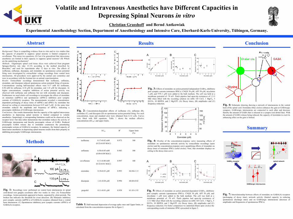
AbstractAbstract ResultsResults ConclusionConclusionBackground: There is compelling evidence from in vitro and in vivo studies that the capacity of propofol to suppress spinal neurons is limited compared to sevoflurane. This raises two questions: (I) Can it be generalized that intravenous anesthetics are limited in their capacity to suppress spinal neurons? (II) Which are the underlying mechanisms? Methods: Organotypic spinal cord tissue slices were achieved from pregnant Sprague-Dawley rats (day 13-15) according to the method described by Braschler,2 and used for experiments after 12 days in vitro. The effects ofisoflurane, enflurane, diazepam, and etomidate on spontaneous action potential firing were investigated by extracellular voltage recordings from ventral horn interneurons. All procedures were approved by the animal care committee and were in accordance with the German law on animal experimentation.Results: Extracellular recordings demonstrated that isoflurane, enflurane, etomidate, and diazepam reduced spontaneous action potential firing of neurons. Concentrations causing half-maximal effects were 0.17 mM for isoflurane, 0.50 mM for enflurane, 0.18 µM for etomidate, and 1.41 µM for diazepam. At higher concentrations, complete inhibition of action potential activity was observed with isoflurane and enflurane but not with etomidate and diazepam. Furthermore we used whole cell recordings to investigate the effects of etomidate on miniature (mIPSCs) and spontaneous (sIPSCs) inhibitory post synaptic currents of GABA(A) receptors. The measurements revealed a concentration-dependent prolonging of decay times of mIPSCs and sIPSCs by etomidate but showed no ceiling at concentrations between 0.25 and 5 µM. At the same time etomidate reduced the amplitude and frequency of sIPSCs, indicating a presynaptic inhibition of GABAergic interneurons. Conclusions: Our results demonstrate that the capacity of the applied intravenous anesthetics in depressing spinal neurons is limited compared to volatile anesthetics. Surprisingly a corresponding limitation could not be observed on the receptor level. Our data rather demonstrate that etomidate reduces the activity of GABAergic interneurons and thereby presynaptic release of GABA. Reduced presynaptic GABA release due to etomidate counteracts the potentiating postsynaptic actions at GABA(A) receptors. In summary, the limited capacity of intravenous anesthetics in depressing spinal neurons results from their property in inhibiting presynaptic GABAergic interneurons.
MethodsMethods
Fig. 2: Concentration-dependent effects of isoflurane (A), enflurane (B), etomidate (C), and diazepam (D) on spontaneous action potential firing. For each concentration, mean and standard error were obtained from 6-12 cells. Curves were fitted with Hill equations. Table 1 shows the median effective concentrations (EC50) and upper limits.
Fig. 1: Recordings were performed on ventral horn interneurons in spinal cord-dorsal root ganglia cocultures after two weeks in vitro. (A) Extracellular recordings. Action potentials appeared in bursts, separated by silent periods. The broken line indicates the threshold for event detection. (B) Miniature inhibitory post synaptic currents (mIPSCs) of GABA(A) receptors obtained from a ventral horn interneuron. (C) Spontaneous inhibitory post synaptic currents (sIPSCs) of GABA(A) receptors. Table 1: Half-maximal depression of average spike rates and Upper limits, as
calculated from the concentration-response fits in figure 2.
EC50 R2 Upper limit(%)
isoflurane 0.17±0.02 mM(0.52±0.05 MAC)
0.975 100
enflurane 0.50±0.05 mM(0.80±0.08 MAC)
0.983 100
sevoflurane 0.11±0.00 mM(0.32±0.01 MAC)
0.997 100
etomidate 0.18±0.01 µM 0.993 66.84±1.13
diazepam 1.41±0.04 µM 0.996 60.68±0.82
propofol 0.11±0.01 µM 0.958 61.43±1.93
Fig. 6: Schematic drawing showing a network of interneurons in the ventral horn of the spinal cord. Etomidate (blue circles) enhances the gain at GABAergic synapses. GABAergic interneurons are connected to each other and decrease thereby the amount of GABA that is released at a synapse (red dotted line). With the amount of GABA release being reduced, the capacity of etomidate to exert its enhancing effect on the gain is limited.
SummarySummary
Fig. 3: Effects of etomidate on action potential-independent GABAA inhibitory post synaptic currents (miniature IPSCs). CNQX 50 µM, AP5 50 µM, strychnine 1 µM, and TTX 1 µM were added to the bath medium. The cell was held at a membrane potential of -70 mV. Patch pipettes had open-tip resistances of 1.5-3 MΩ when filled with the recording solution (in mM) 145 CsCl, 1 MgCl2, 5 EGTA, 10 HEPES, and 2 MgATP. (A) Decay times, (B) amplitudes and (C) frequency reduction.
Fig. 4: Overlay of the concentration-response curve measuring effects of etomidate on spontaneous network activity by extracellular recordings (open circles) and the concentration-response curve quantifying effects of etomidate on decay times of miniature IPSCs (solid circles). Note, that etomidate displays no ceiling in the decay-time-curve.
0 1 2 3 4 50
50
100
150
t1/2
(ms)
Etomidate (µM)0 1 2 3 4 5
0
25
50
75
100
125
Am
plitu
de (p
A)
Etomidate (µM)0 1 2 3 4 5
-100
-50
0
50
100
Dep
ress
ion
of fi
ring
rate
(%)
Etomidate (µM)
A B C
0 1 2 3 4 50
25
50
75
100
125
50
75
100
125
150
Dep
ress
ion
of fi
ring
rate
(%)
Etomidate (µM)
t1/2
(ms)
t ½t ½
GABAA-receptor
GABAergic interneurons
spinal network
IPSC
A
B
C
A B C
0 1 2 3 4 5
-100
-50
0
50
100
Dep
ress
ion
of fi
ring
rate
(%)
Etomidate (µM)0 1 2 3 4 5
0
25
50
75
100
125
Am
plitu
de (p
A)
Etomidate (µM)0 1 2 3 4 5
0
50
100
150
t1/2
(ms)
Etomidate (µM)
1 mmmIPSC
sIPSC
Fig. 5: Effects of etomidate on action potential-dependent GABAA inhibitory post synaptic currents (spontaneous IPSCs). CNQX 50 µM, AP5 50 µM, and strychnine 1 µM were added to the bath medium. The cell was held at a membrane potential of -70 mV. Patch pipettes had open-tip resistances of 1.5-3 MΩ when filled with the recording solution (in mM) 145 CsCl, 1 MgCl2, 5 EGTA, 10 HEPES, and 2 MgATP. (A) Decay times, (B) amplitudes and (C) frequency reduction. For better comparison we underlaid (black open circles) the
Fig. 7: Interrelationship between effects of etomidate on GABA(A) receptors (prolonging of decay time), network activity (limited capacity in reducing spontaneous discharge rates) and on GABAergic interneurons (decrease of amplitudes and frequencies of spontaneous IPSCs).
corresponding results of miniature IPSCs presented in figure 3.


![Effects of the Abused Inhalant Toluene on the Mesolimbic ... · nitrite, (2) anesthetic gas nitrous oxide, and (3) volatile solvents, fuels, and anesthetics [1]. Volatile solvents](https://img.dokumen.tips/doc/110x75/5f066e857e708231d417f681/effects-of-the-abused-inhalant-toluene-on-the-mesolimbic-nitrite-2-anesthetic.jpg)
![Biochimica et Biophysica Acta - COnnecting REpositoriesby volatile anesthetics [20,21]. Reliable structures for individual sub-types of nAChRs, especially their TM domains, are also](https://img.dokumen.tips/doc/110x75/60f824a6246e9522bd1db7e7/biochimica-et-biophysica-acta-connecting-repositories-by-volatile-anesthetics.jpg)


