Embed Size (px)
Citation preview


2
XVI Congress of Euroepan Mycologists, N. Marmaras, Halkidiki, Greece September 18-23, 2011 Abstracts NAGREF-Forest Research Institute, Vassilika, Thessaloniki, Greece.
XVI CEM Organizing Committee Dr. Stephanos Diamandis (chairman, Greece) Dr. Charikleia (Haroula) Perlerou (Greece) Dr. David Minter (UK, ex officio, EMA President) Dr. Tetiana Andrianova (Ukraine, ex officio, EMA Secretary) Dr. Zapi Gonou (Greece, ex officio, EMA Treasurer) Dr. Eva Kapsanaki-Gotsi (University of Athens, Greece) Dr. Thomas Papachristou (Greece, ex officio, Director of the FRI) Dr. Nadia Psurtseva (Russia) Mr. Vasilis Christopoulos (Greece) Mr. George Tziros (Greece) Dr. Eleni Topalidou (Greece)
XVI CEM Scientific Advisory Committee Professor Dr. Reinhard Agerer (University of Munich, Germany) Dr. Vladimir Antonin (Moravian Museum, Brno, Czech Republic) Dr. Paul Cannon (CABI & Royal Botanic Gardens, Kew, UK) Dr. Anders Dahlberg (Swedish Species Information Centre, Uppsala, Sweden) Dr. Cvetomir Denchev (Institute of Biodiversity and Ecosystem Research , Bulgarian Academy of Sciences, Bulgaria) Dr. Leo van Griensven (Wageningen University & Research, Netherlands) Dr. Eva Kapsanaki-Gotsi (University of Athens, Greece) Professor Olga Marfenina (Moscow State University, Russia) Dr. Claudia Perini (University of Siena, Italy) Dr. Reinhold Poeder (University of Innsbruck, Austria)
Governing Committee of the European Mycological Association (2007-2011)
Dr. David Minter President, UK Dr. Stephanos Diamandis Vice-President, Greece Dr. Tetiana Andrianova Secretary, Ukraine Dr. Zacharoula Gonou-Zagou Treasurer, Greece Dr. Izabela Kalucka Membership Secretary, Poland Dr. Ivona Kautmanova Meetings Secretary, Slovakia Dr. Machiel Nordeloos Executive Editor, Netherlands Dr. Beatrice Senn-Irlett Conservation officer, Switzerland Only copy-editing and formatting of abstracts have been done, therefore the authors are fully responsible for the scientific content of their abstracts Abstract Book editors Dr. Stephanos Diamandis & Dr. Eleni Topalidou

3
Welcome preface Dear mycologists of Europe and the world,
On behalf of the Organizing Committee and the European Mycological Association (EMA), we have the honour to welcome you to Greece, to Halkidiki, and to the XVI Congress of European Mycologists.
Greece is internationally famous for its long history and beautiful nature, and Halkidiki especially so. There are sandy beaches and pine forests along the coast, with dense oak and beech forests inland, and mountain villages renowned for their mushroom gastronomy. All of these combine to make it a wonderful place for mycologists and for anyone interested in fungi. The excursion day will give you the opportunity to appreciate the wonderful mountain scenery, and we hope you will use your free time to enjoy the area around the Congress venue.
Although the climate is hot and dry, Greece hosts an amazing variety of habitats and a wealth of fungi. The Forest Research Institute along with the Department of Biology of the University of Athens and the Agricultural University of Athens has been studying Greek fungi for over 3 decades. In recent years, many new species have been recorded, we have re-introduced cultivation of truffles and an interest in truffle gastronomy, and we are encouraging the cultivation of edible mushrooms to fill a gap in the Greek market.
We have also seen an interest in fungi develop in several parts of the country. Six mycological societies have been established, and these organize forays and outdoor mushroom festivals as well as seminars in an effort to promote public interest in fungi. Holding this Congress in Greece will undoubtedly act as a stimulus for more activity of this sort, and we can hope for a growing awareness in Greece of the need for fungal conservation - a very promising trend which should, in the future, result in a greater security for fungal species and their habitats.
We thank you for participating in this Congress and, in particular, for choosing to promote your research results and ideas through this congress. By doing so, you have put in place all the prerequisites for a very exciting scientific event. The quality of research, the wide range of topics including both traditional and innovative themes, and the international co-operation so clearly visible in your research teams show that collectively you form an excellent cadre of scientists. We are particularly happy to see many young mycologists, who will tomorrow continue our efforts to study and promote mycology.
The Organizing Committee of this Congress has made a special effort to promote important topics which have not received adequate attention in past congresses. These include aeromycology, alien and invasive fungi, insect-fungus associations and conservation, particularly the application to fungi of IUCN criteria. To achieve our aim, we have invited as keynote speakers some of the real stars in world mycology, and we offer all of them warm thanks for accepting our invitation to attend.
We would like to acknowledge the help of all those involved in the organization of this congress. We warmly thank Members of the Organizing Committee who, although far away, contributed with ideas and advice, and special mention should be made of the Members of the Scientific Advisory

4
Committee who helped substantially with their long experience and knowledge.
The Congress is hosting 230 participants from 37 countries and every inhabited continent - not only Europe, but also Africa, Asia, Australia, North America and South America. It is very encouraging for European mycology that our Congress is being attended by so many mycologists outside Europe, and we believe this is a clear indication that this meeting is maintaining a high scientific standard and that this series of congresses, organized under the auspices of the European Mycological Association, is on the right track.
Our meeting also provides an opportunity to review the work of the European Mycological Association. Since the Association was established at the XIV Congress in Crimea in 2003, European mycology has seen many changes. Given its limited resources, the Association has only been able to contribute to some of these, its biggest success since the XV Congress in St Petersburg in 2007 being to play a leading role, through our conservation wing, the European Council for Conservation of Fungi, in founding the International Society for Fungal Conservation, the first society anywhere in the world with the explicit objective of protecting fungi.
There is still, however, a long way to go before international organizations, governments and the general public understand that fungi are a separate biological kingdom and very special organisms, that fungi are important in ecosystems, in the food chain, in medicine and in life, that fungi merit conservation just as much as birds, mammals, plants, reptiles and sea creatures. While we believe our association can take great pride in being a parent of this newly created Society, it is clear that the European Mycological Association faces many challenges and will need to adapt if it is to meet them successfully.
Our Association, and the new society it helped to create can, however, do nothing without members. Our location today makes it appropriate to recall a famous saying of the Ancient Greeks: «Η δύναµις εν τω πολλώ» (Strength lies in numbers) and, with that in mind, we urge you to join the International Society for Fungal Conservation and to encourage others to join it and the European Mycological Association. What we cannot achieve as individuals we can achieve when we are united.
We wish you all success in your scientific goals and a pleasant stay at the village of Neos Marmaras and the Porto Carras Resort. Dr. Stephanos Diamandis Dr. David Minter Chairman of the Organizing Committee President of the EMA

5
CONGRESS PROGRAMME
Sunday, 18th September
Time Meliton Veranda
Event
16.00-19.00 Registration open
20.30-23.00 Welcome Reception and Wine Tasting
Monday, 19th September
Congress Opening Ceremony
Plenary Session
Moderators: Dr. Stephanos Diamandis & Organizing Committee
08.00-09.00 Registration
09.00-09.30 Meliton Hall (at middle floor)
Congress Opening Ceremony
Speakers: Dr. S. Diamandis Mr. I. Tzitzios Mayor Dr. K. Mallidis NAGREF Dr. D. Minter president EMA & ISFC
09.30-10.00 Meliton Hall (at middle floor)
Fungal evolution: divergence and adaptation
Keynote speaker: Prof. John Taylor
10.00-10.30 Meliton Hall (at middle floor)
Fungal families: morphology, phylogeny and conflict resolution
Keynote speaker: Dr. Paul Cannon
10.30-11.00 Meliton Hall (at middle floor)
Discussion
11.00-11.20 Coffee break
11.30-13.00 Parallel Sessions in
3 Rooms

6
Thematic Area: Developmental Mycology
Moderator: Professor R. Poeder
11.30-11.45 CHLOE (Room I)
Fungal interactions of Hypholoma fasciculare.
E. Pereira, D. Baptista, P. Baptista, Teresa Lino-Neto
11.45-12.00 CHLOE (Room I)
Measurement of mycelium growth rate of homokaryotic mycelium obtained from single spore isolates of Hericium erinaceus in different culture media and their compatibility.
Ilgaz Akata, E. Kalmis, F. Kalyoncu, M. Atmaca
12.00-12.15 CHLOE (Room I)
Lipid metabolism in Aspergillus niger under heat shock.
Vera M. Tereshina, A.S. Memorskaya, E.R. Kotlova
12.15-13.00 CHLOE (Room I)
Discussion
13.00-14.00 Lunch break
14.00-15.00 Poster session
Thematic Area: Edible and medicinal fungi
Moderator: Professor Joao Baptista-Ferreira
15.00-15.15 CHLOE (Room I)
Saprotrophic and mycorrhizal wild edible mushrooms from Portuguese mycoflora as a source of nutrients and nutraceuticals.
C. Grangeia, S.A. Heleno, L. Barros, Anabela Martins, I.C.F.R. Ferreira
15.15-15.30 CHLOE (Room I)
Localization of the phenolic compounds on the surface of micelle cells of Lentinula edodes (Berk) Pegler cultivated without or with 20 ppm of na2seo3 added to the media.
J. Turło, A. Zobel, B. Gutkowska
15.30-15.45 CHLOE (Room I)
Perspectives to use of basidiomycetes in cancer treatmeht (an experimental investigation).
E. Kadukova, S. Sushko, T. Terpinskaya, A. Naumov, V. Truchonovets

7
15.45-16.00 CHLOE (Room I)
Reproductive isolation between closely related Pleurotus species is not reflected by morphological and physiological individuality.
A.V. Shnyreva, A.B. Sivolapova
16.00-16.30 CHLOE (Room I)
Discussion
16.30-16.50 Afternoon refreshments
Thematic Area: Systematics and evolution of fungi
Moderator: Dr. Cvetomir Denchev
11.30-11.45 THALLIA (Room II)
Phylogeny and intrafoliar genetic diversity of endophytic Colletotrichum from three tropical plant species.
C. Douanla-Meli, E. Langer
11.45-12.00 THALLIA (Room II)
What are the differences between Lactarius sensu novo and Lactifluus: the former milkcaps?
Jorinde Nuytinck, A. Verbeken
12.00-12.15 THALLIA (Room II)
Molecular phylogeny and phylogeography of Physarum notabile (Myxomycetes).
Mikhail Okun, A. M. Fiore-Donno, Yu.K. Novozhilov, M. Schnittler, I.V. Zemlianskaia, D.A. Erastova
12.15-12.30 THALLIA (Room II)
Fusarium divided: new generic concepts in the Nectriaceae.
Tom Gräfenhan, H.-J. Schroers, H.I. Nirenberg,
K.A. Seifert
12.30-12.45 THALLIA (Room II)
New species and new records of the genus Camarophyllopsis from Russia.
Alexander E. Kovalenko, E.F. Malysheva and O.V. Morozova
12.45-13.00 THALLIA (Room II)
Discussion
13.00-14.00 Lunch break

8
14.00-15.00 Poster session
Moderator: Dr. Paul Cannon
15.00-15.15 THALLIA (Room II)
Taxonomic study on Anthracoidea (Ustilaginomycetes) in Japan.
Teodor T. Denchev, C.M. Denchev
15.15-15.30 THALLIA (Room II)
Molecular characterization of true morels (Morchella) in Turkey.
Hatira Taşkın, S. Büyükalaca, S.A. Rehner, K. O’Donnell
15.30-15.45 THALLIA (Room II)
Lactifluus volemus (Russulaceae): a species rich complex revealed by molecular phylogenetics
Kobeke Van de Putte, J. Nuytinck and A.Verbeken
15.45-16.00 THALLIA (Room II)
Phylogeography of the Ganoderma australe (Basidiomycota) complex based on Brazilian specimens.
NC. Lima Júnior, T.B. Gibertoni, Elaine Malosso
16.00-16.15 THALLIA (Room II)
A three-chracter name for naming fungal genera.
Saeid Mahdavi Omran, S.M.B. Norozian, S.J. Mosavi
16.15-16.30 THALLIA (Room II)
Electronic publishing in the epoch of the semantic web: Mycokeys, the next generation journal in mycology.
Lyubomir Penev, T. Lumbsch
16.30-16.50 Afternoon refreshments
17.00-17.15 THALLIA (Room II)
The systematics of the Mortierellales revisited (Mortierellomycotina ex Zygomycetes)
Kerstin Voigt, P.M. Kirk, U. Münchberg, L. Wagner, K. Hoffmann, P. Rösch, J. Popp, C. Vágvölgyi, T. Papp

9
17.15-18.30 THALLIA (Room II)
Discussion
Thematic area: Conservation of Fungi
Moderator: Dr. Claudia Perini
11.30-11.45 ERATO
(Room III)
Fungal conservation - a political issue.
David W. Minter
11.45-12.00 ERATO
(Room III)
Climate change and fungal conservation.
Gregory M. Mueller
12.00-12.15 ERATO (Room III)
Repeated surveys yield insights on fruiting strategies, community assemblage and optimal survey methods of wood-inhabiting fungi. Panu Halme, J. Purhonen, V. Norros, S. Huhtinen, H. Kotiranta, J.S. Kotiaho
12.15-12.30 ERATO (Room III)
At which scale are fungi dispersal limited? quantifying the airborne dispersal of Phlebia centrifuga P. Karst Veera Norros, R. Penttilä, M.E. Niemi, O. Ovaskainen
12.30-12.45 ERATO
(Room III)
Molecular detection and diversity restoration of threatened wood-decaying Basidiomycetes. Dmitri S. Schigel, O. Ovaskainen, H. Ali-Kovero, V. Norros
12.45-13.00 ERATO (Room III)
Discussion
13.00-14.00 Lunch break
14.00-15.00 Poster session

10
Moderator: Dr. Vladimir Antonin
15.00-15.15 ERATO
(Room III)
A new window for natural reserves: the mycological view.
E. Ambrosio, M. Danielli, P. Leonardi, M. Landi, C. Saveri, E. Salerni, Claudia Perini
15.15-15.30 ERATO
(Room III)
Fungal conservation and the encyclopedia of life in Egypt.
A.M. Abdel-Azeem, G.S. Soliman
15.30-15.45 ERATO
(Room III)
Using local ecological knowledge for fungal conservation policy and decision making.
Elizabeth S. Barron, C. Sthultz, D. Hurley, A. Pringle
15.45-16.00 ERATO (Room III)
Conservation aspects of some rare species from genus Physarum (Myxomycetes) in Ukraine.
Irina O. Dudka, T.I. Kryvomaz, D.V. Leontyev
16.00-16.15 ERATO
(Room III)
Virtual herbarium of Brazilian plant and fungi as inducer of advances on taxonomy and mycological collections.
Leonor Maia, D. Canhos, A. Peixoto
16.15-16.30 ERATO
(Room III)
Effect of seed treatment with Brassicaceae on fungal disease incidence of wheat and tomato.
Nazira Aitkhozhina
16.30-16.50 Afternoon refreshments
17.00-17.15 ERATO
(Room III)
Fungal conservation in Portugal: a progress report of a 20-year productive campaign.
Joao L. Baptista-Ferreira
17.15-17.30 ERATO
(Room III)
Discussion
Workshop: Conservation of Ascomycetes
Moderator: Dr. David Minter
17.30-19.00 ERATO (Room III)
Introduction to the workshop

11
Tuesday, 20th
September
Plenary Session
08.30-09.00 Meliton Hall (at middle floor)
Outdoor Airspora: Patterns, Prevalence & Impacts
Keynote speaker:
Dr. Christine Rogers
Meliton Hall (at middle floor)
Recent advances in Indoor mycology
Keynote speaker:
Dr. Robert Samson
CANCELLED
09.00-10.00 Meliton Hall (at middle floor)
Discussion
10.00-10.20 Coffee break
10.30-13.00 Parallel Sessions in 3 Rooms
Thematic Area: Aeromycology
Moderator: Dr. E. Kapsanaki-Gotsi
10.30-10.45 CHLOE (Room I)
Αn assessment of airborne fungi in museum premises.
Eva Kapsanaki-Gotsi, A. Zervas, A. Patra and M. Koumbourou
10.45-11.00 CHLOE (Room I)
Αerobiological monitoring of fungi in a newly built haematology/oncology paediatric hospital.
A. Velegraki, K. Xerakia, A. Charissiadou, V. Konte, A. Milioni, S. Kritikou, Ch. Rhodaki, A. Stathi, A. Pangalis
11.00-11.15 CHLOE (Room I)
Effect of dust storms on concentration and content of fungi in the atmosphere of Haifa, Israel.
Isabella Grishkan, P. Schlesinger, Y. Mamane

12
11.15-11.30 CHLOE (Room I)
Diversity of airborne fungi in Athens and annual variation associated with meteorological factors.
Ioanna Pyrri, E. Kapsanaki-Gotsi
11.30-11.45 CHLOE (Room I)
Fugal aerobiology, spore morphology and genetics: a triple-fusion challenge for mid-term biosecurity.
M.E. Kambouris, A. Velegraki
11.45-12.00 CHLOE (Room I)
Arborne opportunistic microfungi in outdoor urban environments.
Olga E. Marfenina, N.V. Makarova, A.E. Ivanova, A.A. Danilogorskaja
12.00-12.15 CHLOE (Room I)
The level and species of moulds in indoor air of daycare centers in Korea.
Seong H. Kim, G.R. Ahn
12.15-12.30 CHLOE (Room I)
Identification of Lichtheimia, a causative agent of emerging Mucormycoses
W. Schrödl, T. Heydel, V.U. Schwartze, K. Hoffmann, G. Walther, A. Alastruey-Izquierdo, J.L. Rodriguez-Tudela, P. Olias, I.D. Jacobsen, G. Sybren de Hoog, Kerstin Voigt
12.30-13.00 CHLOE (Room I)
Discussion
13.00-14.00 Lunch break
14.00-15.00 Poster session
Symposium: Insect-fungus associations
Moderator: Dr. Dmitri Shigel
15.00-15.15 CHLOE (Room I)
Introduction.
Dmitri Shigel
13
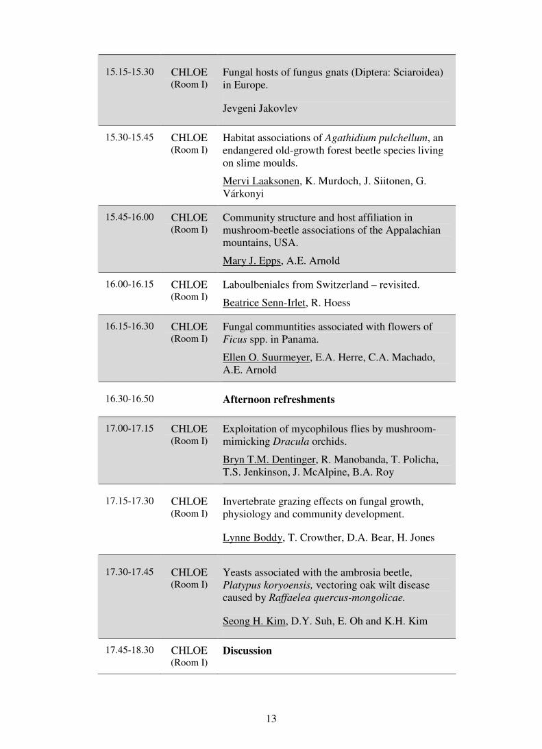
15.15-15.30 CHLOE (Room I)
Fungal hosts of fungus gnats (Diptera: Sciaroidea) in Europe.
Jevgeni Jakovlev
15.30-15.45 CHLOE (Room I)
Habitat associations of Agathidium pulchellum, an endangered old-growth forest beetle species living on slime moulds.
Mervi Laaksonen, K. Murdoch, J. Siitonen, G. Várkonyi
15.45-16.00 CHLOE (Room I)
Community structure and host affiliation in mushroom-beetle associations of the Appalachian mountains, USA.
Mary J. Epps, A.E. Arnold
16.00-16.15 CHLOE (Room I)
Laboulbeniales from Switzerland – revisited.
Beatrice Senn-Irlet, R. Hoess
16.15-16.30 CHLOE (Room I)
Fungal communtities associated with flowers of Ficus spp. in Panama.
Ellen O. Suurmeyer, E.A. Herre, C.A. Machado, A.E. Arnold
16.30-16.50 Afternoon refreshments
17.00-17.15 CHLOE (Room I)
Exploitation of mycophilous flies by mushroom-mimicking Dracula orchids.
Bryn T.M. Dentinger, R. Manobanda, T. Policha, T.S. Jenkinson, J. McAlpine, B.A. Roy
17.15-17.30 CHLOE (Room I)
Invertebrate grazing effects on fungal growth, physiology and community development.
Lynne Boddy, T. Crowther, D.A. Bear, H. Jones
17.30-17.45 CHLOE (Room I)
Yeasts associated with the ambrosia beetle, Platypus koryoensis, vectoring oak wilt disease caused by Raffaelea quercus-mongolicae.
Seong H. Kim, D.Y. Suh, E. Oh and K.H. Kim
17.45-18.30 CHLOE (Room I)
Discussion

14
Thematic Area: Fungi in ecosystems; effects of climate change
Moderator: Professor Lynne Boddy
10.30-10.45 THALLIA (Room II)
Conidial fungi in protected ecosystems: examination for conservation strategy.
Tetiana V. Andrianova
10.45-11.00 THALLIA (Room II)
Bioaccumulation of the artificial radionuclide 137 CS in Basidiomycota in Greece.
V. Kioupi, E. Florou, Z. Gonou-Zagou, P. Delivorias, E. Kapsanaki-Gotsi
11.00-11.15 THALLIA (Room II)
Heavy metal uptake of mushrooms from a former uranium mining site in eastern Thuringia.
Matthias Gube, E. Kothe
11.15-11.30 THALLIA (Room II)
Alpine vegetation rich in Salix shows various changes in its macromycetes over 25 years- a case study from Switzerland.
Beatrice Senn-Irlet
11.30-11.45 THALLIA (Room II)
Effects of soil warming on three soil and plant debris-borne fungal pathogens of oilseed rape.
Magdalena Siebold, A.von Tiedemann
11.45-12.00 THALLIA (Room II)
Characteristics of Paecilomyces lilacinus (Thom) Samson strains from various habitats.
Tatiana Belozerskaya, A. Egorova, N. Gessler, A. Ivanova
12.00-12.15 THALLIA (Room II)
Effects of climate change on saprotrophic and ectomycorrhizal fungi, revealed by long-term fruiting datasets.
Lynne Boddy, A. Gange, H. Kauserud
12.15-12.30 THALLIA (Room II)
Arbuscular mycorrhizal fungi in natural and revegetated dunes after mining activity.
D. Silva, Leonor Maia, C. Pereira, R. Souza, F. Oehl

15
12.30-12.45 THALLIA (Room II)
Conservation of ecosystem with fungal properties.
Yung-Hyun Ryu, Hyeokjun Yoon, Joo-Ri Woo, In-Jung Lee, Jae-Ho Shin, Yeon-Sik Choo, Jong-Guk Kim
12.45-13.00 THALLIA (Room II)
Discussion
13.00-14.00 Lunch break
14.00-15.00 Poster session
Thematic Area: Fungus-Plant interactions; mycorrhizal systems
Moderators: Professor R. Agerer
15.00-15.15 THALLIA (Room II)
Ectomycorrhizal fungal communities of native versus non-native trees. A common garden study of Pinus and Quercus species.
Iza Kałucka, L.K. Trocha, M. Stasińska, W. Nowak, M. Dabert, T. Leski, M. Rudawska, J. Oleksyn
15.15-15.30 THALLIA (Room II)
Abilities of mycorrhizal fungi in eliminating toxic substances.
Katrin Krause, I. Schlunk, T. Asiimwe, C. Henke, E. Kothe
15.30-15.45 THALLIA (Room II)
The ectomycorrhizal fungi in a forest chronosequence of European larch (Larix decidua).
Tomasz Leski, M. Rudawska
15.45-16.00 THALLIA (Room II)
Influence of mycorrhizal symbiosis in antioxidant potential of fungi and seedlings.
F.S. Reis, I.C.F.R. Ferreira, L. Barros, C. Santos-Buelga, Anabela Martins
16.00-16.15 THALLIA (Room II)
Can ectomycorrhizal fungi be cheaters?
Reinhard Agerer
16.15-16.30 THALLIA (Room II)
Study of dark septate endophytic fungi colonizing invasive and indigenous plants on semiarid sandy areas.
Daniel G. Knapp, A. Pintye, G.M. Kovács

16
16.30-16.50 Afternoon refreshments
17.00-17.15 THALLIA (Room II)
Unravelling an enigma: ecology of waxcaps (Hygrocybe: Agaricomycetes)
Patricia Silva-Flores, R. Agerer
17.15-18.30 THALLIA (Room II)
Discussion
Thematic Area: Fungal distribution and diversity
Moderators: Dr. Zapi Gonou-Zagou
10.30-10.45 ERATO
(Room III)
Diversity of soil microbial communities along climatic altitudinal gradients
Aurore Coince, M. Buée, B. Marçais
10.45-11.00 ERATO
(Room III)
Size matters not: some minute yet interesting ascomycetes from the mountainous region of Agrapha, Central Greece
Panos Delivorias, Z. Gonou-Zagou, E. Kapsanaki-Gotsi
11.00-11.15 ERATO
(Room III)
Contribution of metagenome pyrosequencing of soil fungi to nature conservation: a case study from sand dune communities in the Netherlands.
József Geml, M.E. Noordeloos
11.15-11.30 ERATO
(Room III)
Macrofungi of Abies cilicica and Abies borisii regis in Turkey and Central Balkans.
Hasan Hüseyin Doğan, M. Karadelev, K. Rusevska
11.30-11.45 ERATO (Room III)
Ecological features of Tricholoma anatolicum in Turkey.
Hasan Hüseyin Doğan, I. Akata
11.45-12.00 ERATO (Room III)
The impact of earthworms on microscopic fungi.
Alexander V. Kurakov, S.A. Kharin

17
12.00-12.15 ERATO (Room III)
Geoglossoid fungi in Slovakia.
V. Kučera, Pavel Lizoň
12.15-12.30 ERATO (Room III)
Molecular biogeography of arbuscular mycorrhizal fungi.
Maarja Öpik
12.30-13.00 ERATO (Room III)
Discussion
13.00-14.00 Lunch break
14.00-15.00 Poster session
Moderator: Dr. Stephanos Diamandis
15.00-15.15 ERATO (Room III)
Diversity of wood-inhabiting Basidiomycota in Leivaditis area (Thrace, Greece).
Athanasia Sergentani, Z. Gonou-Zagou, D.G. Hatzinikolaou, E. Kapsanaki-Gotsi
15.15-15.30 ERATO (Room III)
A reappraisal of existing knowledge on the diversity of the genus Lactarius Pers. in Greece.
Marina Triantafyllou, E. Polemis, D.M. Dimou, Z. Gonou-Zagou, P. Delivorias, G.I. Zervakis
15.30-15.45 ERATO (Room III)
Studies on Myxobiota of Canakkale (Turkey) and its environment.
Tülay Bican Süerdem, B. Dülger
15.45-16.00 ERATO (Room III)
Determining rarity of fungi.
Branislav Uzelac
16.00-16.15 ERATO
(Room III)
Morphology and ecology of Rhizophydium mammilatum – a parasitic chytrid fungus. Isolation and cultivation methods.
M.A. Mamkaeva

18
16.15-16.30 ERATO (Room III)
The distribution of some macromycetes in Europe (ECCF Mapping programme)
Andre Fraiture
16.30-16.50 Afternoon refreshments
17.00-17.15 ERATO (Room III)
The occurrence of aquatic fungi and fungus-like organisms in rivers in the northeastern part of Poland
Bozena Kiziewicz, A. Godlewska, E. Muszyńska, B. Mazalska
17.15-17.30 ERATO (Room III)
Biodiversity of “Cilento e vallo di Diano” national park: macrofungal communities in old-growth forests.
M. D’Aguanno, E. Ambrosio, P. Leonardi, Claudia Perini, E. Salerni
17.30-17.45 ERATO
(Room III)
Estimation of the fungal diversity in Bulgaria.
Cvetomir M. Denchev, T.T. Denchev
17.45-18.00 ERATO
(Room III)
Myxomycete assemblages from steppes of Tabernas and Monegros deserts (spain): biodiversity, ecology and biogeography.
E. García-Carvajal , C. Lado, Yu. K. Novozhilov
18.00-18.15 ERATO (Room III)
Macromycetes associated with Pinus peuce at the Pelister mountain (FYROM).
G. Kost, Mitko Karadelev
18.15-18.30 ERATO (Room III)
Myxomycetes from Antakya-Hatay Turkey.
H. Baba
18.30-18.50 Discussion

19
Tuesday, 20th
September
Meeting of the ECCF
19.00-20.30 ERATO (Room III)
Agenda will be announced by the ECCF secretariat
Wednesday, 21st September
Excursions to
1. Mount Holomon (broad leaved forests)
2. Parthenon (Mediterranean pines)
08.30 to approx. 18.30

20
Thursday, 22nd
September
Plenary Session
08.30-09.00 Meliton Hall (at middle
floor)
Alien and invasive fungi-what can we expect from a changing climate
Keynote speaker:
Prof. Jan Stenlid
09.00-09.30 Meliton Hall (at middle
floor)
Fungal conservation: insights from population biology and the impacts of past, present and future human land use
Keynote speaker:
Dr. Anders Dahlberg
09.30-10.00 Meliton Hall
(at middle floor)
Discussion
10.00-10.20 Coffee break
10.30-13.00 Parallel Sessions in 3 Rooms
Thematic Areas: Alien and invasive fungi Biological control
Moderator: Dr. Iza Kalucka
10.30-10.45 CHLOE (Room I)
Morphological and molecular characterization of Inonotus levis: a new fungal invasive pathogen for Europe.
Zapi Gonou-Zagou, V.N. Kouvelis, A. Krimitzas, D. Floudas, M.A. Typas
10.45-11.00 CHLOE (Room I)
Genetic diversity and spread of the chestnut blight fungus Cryphonectria parasitica in Switzerland.
Simone Prospero, D. Rigling

21
11.00-11.15 CHLOE (Room I)
National project of biological control of chestnut blight in Greece-area results.
S. Diamandis, C. Perlerou, G. T. Tziros, V. Christopoulos, E. T. Topalidou, D. Avtzis
11.15-11.30 CHLOE (Room I)
Use of Trichoderma harzianum in biological control of wheat root rot caused by Bipolaris sorokiniana.
A. Foroutan, S.A. Rezaei, E. Yasari, H. Barary, M. Aldaghi
11.30-11.45 CHLOE (Room I)
Morphology alteration of fungal cells at presence of fungicides used for books damaged by mould.
T.D. Velikova, E.S. Trepova, E.V. Lebedeva
11.45-12.00 CHLOE (Room I)
Selection and characterisation of an antagonistic yeast for biocontrol of the brown rot pathogen, Monilinia laxa.
Nattawut Rungjindamai, X-M. Xu, P. Jeffries
12.00-12.15 CHLOE (Room I)
Molecular ecology of a mycotrophic fungus Hypocrea/Trichoderma.
Irina S. Druzhinina
12.15-13.00 Discussion
Thematic Area: Plant pathogenic fungi
Moderator: Dr. J. Fatehi
10.30-10.45 THALLIA (Room II)
Asian populations of the wheat stripe rust pathogen as a potential source of new emergences, due to their high genotypic and phenotypic diversity.
Sajid Ali, J. Enjalbert, P.Gladieux, M. Leconte, A.Gautier, M.S. Hovmøler, C. de Vallavieille-Pope
10.45-11.00 THALLIA (Room II)
Population dynamics of Trichoderma in wheat rhizosphere in Mazandaran.
Abdolreza Foroutan, A. Foroutan, A.Yasari

22
11.00-11.15 THALLIA (Room II)
Presence of Phytophthora species in alluvium of Sava river.
Ivan Milenkovic, N. Keca, L. Letic, V. Nikolic
11.15-11.30 THALLIA (Room II)
Physiological response of Quercus spp. invaded by Phytophthora spp. plant pathogens.
Slavi Slavov, I. Tzvetkov, Zh. Yordanova, V. Kapchina-Toteva
11.30-11.45 THALLIA (Room II)
Microorganisms causing to rotting of grape roots infected by phylloxera in the Asgeran region.
H.M. Shikhlinski
11.45-12.00 THALLIA (Room II)
Phytopathologic estimation of cotton hybrid resistance to Verticillium dahliae Klebahn.
N.Kh. Mammadova
12.00-12.15 THALLIA (Room II)
Sample survey of Erysiphe alphitoides populations on oak trees.
Eleni T. Topalidou, M.W. Shaw
12.15-12.30 THALLIA (Room II)
Development of a highly specific diagnostic tool
for Verticillium species.
Mireille Dessimoz, J. Enkerli, V. Michel, F. Widmer
12.30-13.00 THALLIA (Room II)
Discussion
13.00-14.00 Lunch break
14.00-15.00 Poster session
Thematic Area: Fungal distribution and diversity
Moderator: Dr. Paul M. Kirk
10.30-10.45 ERATO
(Room III)
Distribution and diversity of the clavarioid fungi in the Eurasian Arctic.
Anton G. Shiryaev

23
10.45-11.00 ERATO (Room III)
Fungal biodiversity in a natural truffiére of Tuber magnatum.
E. Salerni, M. Iotti, P. Leonardi, A. Zambonelli and C. Perini
11.00-11.15 ERATO (Room III)
Diversity of macrofungi in islands of the Aegean archipelago.
Elias Polemis, D.M. Dimou, G.I. Zervakis
11.15-11.30 ERATO (Room III)
Distribution and ecology of the genus Battarrea in FYROM.
Katerina Rusevska, M.P. Martín, M. Karadelev
11.30-11.45 ERATO (Room III)
Characterization of yeast flora isolated from cheeses at Central Anatolia, Turkey.
T. Turgut Genç, İ.N. Çıldır, Tugba Çelik, N. Demir
11.45-12.00 ERATO (Room III)
Biodiversity of fungal endophytes in semi-evergreen vine thickets.
Rachel R. Graham, J.D.W. Dearnaley
12.00-12.15 ERATO (Room III)
Contribution to knowledge of the macromycetes fungi from Bolintin Deal forest – Giurgiu, Romania.
Mihai-Iulian Radu, Tatiana Eugenia Sesan
12.15-13.00 ERATO (Room III)
Discussion
13.00-14.00 Lunch break
14.00-15.00 Poster session

24
Symposium on Application of IUCN criteria
Moderator: Dr. Anders Dahleberg
15.00-16.00 CHLOE (Room I)
Introduction
16.00-16.30 Group work
16.30-16.50 Afternoon refreshments
17.00-19.00 Group work
20.30-24.00 “Under the pines”
Gala Dinner-Offered by the Organizing Committee
Friday, September 23
Plenary Session
08.30-09.00 Meliton Hall
(at middle floor)
A new Imaging Nanotechnology for Mycology
Keynote speaker:
Prof. Lodewyk Kock
09.00-09.30 Meliton Hall
(at middle floor)
MtDNA and rDNA: two different evolutionary lines combined for genetic differentiation, taxonomy and phylogeny in ascomycetes
Keynote speaker:
Prof. Milton A. Typas
09.30-10.00 Meliton Hall (at middle floor)
Discussion
10.00-10.20 Coffee break
10.30-13.00 Parallel Sessions in 3 Rooms

25
Thematic Area: Fungal genetics and genomics
Moderator: Professor Milton A. Typas
10.30-10.45 CHLOE (Room I)
Comparative genomic, phylogenetic, and functional investigation of the xenobiotic metabolizing arylamine n-acetyltransferase enzyme family among fungi.
Sotiria Boukouvala, E. Kontomina, E.P. Karagianni, B. Ormiston, T. Tsirka, A.E. Glenn
10.45-11.00 CHLOE (Room I)
Quantitative trait loci controlling vegetative growth rate of edible mushroom Pleurotus ostreatus.
Anastasia Sivolapova, J. Baars, A. Sonnenberg, A. Shnyreva, B. Lavrijssen, P. Hendricks
11.00-11.15 Comparative analysis of mitochondrial genome isolated from three Flammulina velutipes strains.
Hyeokjun Yoon, Young-Hyun You, Ju-Ri Woo, Young-Jin Park, Won-Sik Kong, Byoung-Moo Lee, Jong-Guk Kim
11.15-12.00 Discussion
Thematic Area: Fungal distribution and diversity
Moderator: Dr. A. Abdel-Azeem
11.30-11.45 CHLOE (Room I)
A new basidiomycete genus, Scotomyces from Turkey.
Halil Güngör, H. Alli, M. Işiloğlu
11.45-12.00 CHLOE (Room I)
A new and interesting record (Fenugreek Stalkball) from Turkey.
M. Işiloğlu, Hakan Alli, S. Helfer
12.00-12.15 CHLOE (Room I)
Yeast flora of different varieties of grapes used for wine making in Bozcaada (Canakkale, Turkey).
T. Turgut Genç, İlknur N. Çıldır

26
12.15-12.30 CHLOE (Room I)
Macrofungi of Liquidambar orientalis mill. forests in Muğla (Turkey).
M. Işıloğlu, H. Allı, H. Güngör, S. Candar
12.30-12.45 CHLOE (Room I)
Hypogeous fungi and perspectives for truffle cultivation in Greece.
Stephanos Diamandis, C. Perlerou, V. Christopoulos
12.45-13.00 CHLOE (Room I)
Discussion
13.00-14.00 Lunch break
Thematic Area: Fungal Biotechnology
Moderator: Professor John Taylor
10.30-10.45 THALLIA (Room II)
Isolation and cloning of manganese peroxidase (mnp) gene from the white button mushroom.
M. Farsi, J. Hasan-Janpour, H.R. Pourianfar
10.45-11.00 THALLIA (Room II)
Endochitinase gene expression in tomatoes after simultaneous treatment with arbuscular mycorrhizal fungi and Trichoderma harzianum.
M. Ene, M. Alexandru, Tatiana E. Şesan, M. Cutrubinis
11.00-11.15 THALLIA (Room II)
Cyclopiazonic acid and sclerotia producing ability in aflatoxigenic and non-aflatoxigenic Aspergillus flavus strains from peanuts field soils.
Masoomeh Shams-Ghahfarokhi, S. Amani, M. Banasaz, M. Razzaghi-Abyaneh
11.15-11.30 THALLIA (Room II)
Glycogen and trehalose accumulation in debaryomyces occidentalis at different carbon sources.
Tulay Turgut Genç, T. Çelik, İ. N. Çıldır
11.30-11.45 THALLIA (Room II)
Recent advances in conservation and study of macromycetes genetic resources in the LE-BIN culture collection.
Nadya Psurtseva, A. Kiyashko, N. Shakhova, M. Shevchenko, K.Barinova

27
11.45-12.30 THALLIA (Room II)
Discussion
13.00-14.00 Lunch break
15.00-16.30 Meliton Hall (at middle floor)
EMA General Assembly
Elections
16.30-17.00 Closing Ceremony

28
All abstracts are arranged in thematic areas following the congress scientific
programme
Oral lectures and poster presentations are arranged in two separate sections for easier follow up by the
participants
Oral lectures
pages 29 - 195
Poster presentation pages 196 - 312

29
Oral lectures
Thematic area: Systematics and evolution of fungi
Keynote lectures
FUNGAL FAMILIES: MORPHOLOGY, PHYLOGENY AND CONFLICT RESOLUTION
Paul F. Cannon
CABI and Royal Botanic Gardens, Kew, Richmond TW9 3AB, UK
Keywords: systematics and evolution of fungi, structure and function, parallel evolution, use of taxonomic ranks This paper will review historical use of the family as a taxonomic concept within the Fungi, focusing particularly on the recent increase in numbers of accepted families based primarily on molecular evidence. Is it correct to use sequences as the principal data supporting recognition of fungal families? And why, in many cases, do morphological features not reflect a more accurate and detailed phylogenetic signal? The background to these conundra needs to be analyzed in the context of systematics and nomenclature in general, as many of the issues are not confined to the family as a level in the taxonomic hierarchy. The adoption of the binomial nomenclatural system for plants and fungi by Linnaeus (1753) constituted a step change in biological communication in that organisms could be referred to by short-hand names rather than descriptions in their entirety, and their binomial nature provided a brief summary of their relationships as well as names. However, that meant that names changed as a result of new research, leading to miscommunication and instability. As family names are based on generic names, they too are subject to periodic change as taxonomic concepts evolve, and more significantly their circumscriptions change depending on the taxonomic opinion of the scientist revising the group. Phylogenetic methodologies offer some promise of taxonomic stability due to their more objective nature, but this stability must be considered as a long-term goal due to the challenges of sequence acquisition and data analysis. We are therefore in a period of significant and substantial change in our understanding of fungal evolution, which must necessarily lead to many changes in classification. The concept of the family as applied to plant and fungal systematics seems to originate with Ray (1682), but its use was sporadic and unstandardized for most of the succeeding two hundred years (Hawksworth and David,

30
1989). Linnaeus (1751) used the term “familia” but as a second-order hierarchical term; the plant kingdom was divided into seven “families”, the fungi, algae, mosses, ferns, grasses, palms and other plants. In common with many contemporary authors, Linnaeus (1753) gave the Fungi little more than postscript status, describing them in only 15 pages (of 1230) within a single “ordo” (one of 67), though 80 species of Lichen and seven of Tremella were classified in the Algae. A more significant milestone in the acceptance of the family as a taxonomic unit was reached by Adanson (1763) in his book Les Familles des Plantes. Here the “famille” Fungi (this time including Lichen) was divided into seven sections based on sporulating structures, though Tremella, Aspergillus and Botrytis were placed in the “famille” Byssi. Adanson was far ahead of his time. Much of his book was devoted to a primitive type of multi-access key, and his greatest contribution was to insist that classifications should be based on all available characters rather than on a small set of supposedly critical ones. Regrettably his work was unrecognized by most of his contemporaries, but it laid the foundations for all modern taxonomic systems. Persoon (1801) was among the first to recognize units that roughly correspond to the fungal families that we recognize today (e.g. Agaricoideae, Boletoideae) although he did not adopt the term. Fungal family names (either with the familiar –aceae ending or an equivalent) for groups of genera began to be adopted widely in the 1820s by mycologists including Fries, Dumortier, Chevallier, Link and Zenker, and the number published has increased steadily ever since (Fig. 1). Fungal families based on phylogenetic principles are now almost universally introduced using molecular sequence data.
Fig. 1. Numbers of fungal families published each decade since 1760; data from Index Fungorum. The 463 family names published by Marcel Locquin in the 1970s and 1980s (mostly either invalidly published or based on fossil taxa) have been excluded.

31
The first purported natural classification based on Adansonian criteria was published by de Bary (1869). This formed the foundation for classifications of the Fungi using morphological methods (with varying degrees of inclusion) throughout the twentieth century (Hawksworth and David, 1989) with important contributions made by Nannfeldt (1932), Luttrell (1951), Ainsworth et al. (1973) and Singer (1986) among others. The compilation of families of “bitunicate ascomycetes” by Eriksson (1980) was perhaps the most significant publication at the fungal family level in the twentieth century. This included a partial phylogenetic reconstruction of the Ascomycota in its entirety by clustering the accepted families in a series of 109 clades. The methods used were described by the author as “eclectic” and would certainly not pass muster today, and no attempt was made to cluster the clades into larger units. Nevertheless, the work was influential, leading to regularly updated systems published in Systema Ascomycetum and Myconet, on which were based in turn successive classifications in the Dictionary of the Fungi and in Species Fungorum. The 2005 Dictionary classification for the Ascomycota was presented in a fully illustrated and rather more approachable manner by Cannon and Kirk (2007). All of these took full advantage of the rapidly expanding literature on fungal phylogeny using molecular sequence data. The most recent major milestone was achieved by the major US-based fungal phylogenetics projects AFTOL (Hibbett et al. 2007) and Deep Hypha (Blackwell et al. 2006) with a full multigene phylogeny of the Fungi, though these focused on ordinal levels and above and did not directly address the issues of classification of fungal families. Families of fungi continue to be described on a routine basis (28 in 2008, 18 in 2009 and 16 in 2010), but there are few or no agreed criteria for their recognition, or indeed the assignment of rank to any fungal taxon except perhaps that of species. This is not an issue that is confined to the Fungi. Heywood (1977) stated that “there is no agreed principle for the limitation of [plant] families other than comparability of status in relation to allied families.” Today, the situation is perhaps even more fluid: Stevens (2008) observed that: “Taxa at the same rank are equivalent only by designation and have nothing necessarily in common other than their monophyly. Rank as used here [i.e. the system adopted by the Angiosperm Phylogeny project and website] has no meaning other than signifying a monophyletic group that includes other monophyletic groups with appropriately subordinate rank terminations.” Perhaps this is not an issue of great import; a large proportion of fungal taxa is now primarily defined in terms of monophyletic clades within a more extensive phylogeny, and linked where possible to shared derived morphological, cultural or ecological characteristics. A sizeable group of phylogenetists consider that traditional taxonomic categories obscure relationships rather than create order within them; see Potter & Freudenstein (2005) for a critique of their approach. We should at least take the trouble to ask questions as to the comparability of families within the Fungi, and by extension with other organism groups. Do we have too many families? Or not enough? Table 1 shows the number

32
of species currently accepted within a series of major organism groups (phyla and subphyla), along with the number of families to which they are assigned. It would also be reasonable to take into account the evolutionary age of the groups. Perhaps unsurprisingly, the groups most similar to ourselves appear to have been subdivided more than others. This will certainly be related to the amount of research on relationships that has been carried out, and perhaps too to the number of contributors - an extreme example of the phenomenon that taxonomists split their own groups and lump those of others. Also unsurprisingly, the Coleoptera and Lepidoptera have rather few families compared with the number of accepted species, though it is unclear whether this is due to differing patterns of evolutionary radiation or merely exhaustion on the part of their taxonomists. Workers on bryozoans seem to be particularly enthusiastic family describers and botanists reticent in this respect (although the latest angiosperm phylogeny contains substantially fewer families than the two previous published systems – a conscious decision to make the classification more approachable for students; Mark Chase, personal communication July 2011), but otherwise the rate of acceptance of fungal families seems largely in step with those of other groups. It must be emphasized that this is a particularly crude comparative measure, but it does indicate that we should not have immediate cause for concern. Table 1. Numbers of species and families of a range of major organism groups (taken largely from Heywood (1995) and the Species 2000 Catalogue of Life (www.sp2000.org/)).
Evolutionary age (My)
Species Families Species/Family
Arthropoda Arachnida 450 98000 625 156 Coleoptera 300 400000 137 2919 Lepidoptera 200 175000 125 1400 Orthoptera 300 25000 40 625 Bryozoa 500 5000 225 22 Chordata amphibians 350 6300 60 105 birds 150 10000 179 56 fish 500 32000 652 49 mammals 200 5700 146 39 reptiles 320 8200 63 130 Fungi 760-1060 100000 536 186 Mollusca 500 85000 492 172 Nematoda 600-1300 28000 171 163 Plantae 500 270000 445 606
How should we assign ranks to fungal taxa? Except at the species level, this question has scarcely been asked. Here, biological and phylogenetic concepts both have their proponents as reflecting evolutionary events more realistically than morphological features. Research into biological species concepts using experimental methods can tell us much about the behaviour of fungal species (e.g. Brasier 1993). Phylogenetic species concepts, especially using gene genealogical concordance methods (Koufopanou et al. 1997, Taylor et al. 2000), have been widely adopted to provide indirect

33
evidence of speciation. Hybridization and polyploidy in fungi have been inadequately studied (Schardl and Craven 2003) and may be very widespread, especially between taxa separable using phylogenetic species recognition that have retained their ability to mate. Recent research has shown that the “species” Verticillium longisporum is an allopolyploid that has originated at least three times from four different lineages and three different parental taxa (Inderbitzin et al. 2011). These complexities force us to adopt a pragmatic, perhaps even relaxed view of fungal species definition, as evidenced especially by the fungal barcoding initiatives that seek to define species using homology in single genes (Begerow et al. 2010). If a universal code for species definition eludes us, how are we to establish ranks for higher taxa? It has long been maintained that ranks differ in their innate reality; Linneaus (1751) stated that “The species and the genus are always the work of nature [i.e. specially created]; the variety mostly that of circumstance; the class and the order are the work of nature and art.” (trans. Stafleu 1971). The current view is that a taxon of any rank is a real entity if it is monophyletic, but monophyly cannot be an indication of rank. It would be wonderful if similar hiatuses in barcode sequence variation to those at the species boundary (that show promise for automated identification, at least in some fungal groups) could be found for higher taxa. This may be possible in some circumstances for generic delimitation, (see Fig. 2) but effective family delineation is unlikely, even using very conserved genes. Phylogenetic trees depict relationships between clades, but branch length (or tree length) bears little relationship to the taxonomic rank at which those clades are separated.
Fig. 2. Frequency diagram of ITS sequence divergence within the Colletotrichum gloeosporioides species aggregate and between it and other species of Colletotrichum, derived from BLASTn data. The left-hand solid line at the top of the diagram (~0-3%) denotes within-aggregate variation, the right-hand line (~8-12%) denotes homology with other species of Colletotrichum. Sequences with greater divergence are not derived from Colletotrichum.

34
We therefore have no objective method for determining rank (at least above species level). Groups of taxa can be demonstrated to be related in clear hierarchies using robust statistical methods, but their degree of relatedness is obscure. Nevertheless, almost all would agree that genera and families are real entities, and useful constructs to convey information. Therefore, we need to adopt pragmatic principles to determine the rank at which taxa are separated. The first of these is that the taxon should be monophyletic. The recognition of paraphyletic genera, families etc. is advocated by some for practical reasons of identification, but there is no reason why artificial schemes based on few characters should obscure genuine relationships. Rather, they can exist in parallel. Secondly, there needs to be a balance between maintenance of monophyletic groups that are also recognizable using traditional methods, with newly recognized taxa that are likely to contain less variation. It may be an ideal that a family of the Ascomycota should be broadly similar in terms of the variation of its constituent taxa to one of the Basidiomycota, but it is not worth significant disruption in order to achieve this. Thirdly, there needs to be consideration as to whether intermediate taxa should be adopted. Mycologists use fewer categories of the taxonomic hierarchy than students of almost all major organism groups. Rather than separating a traditional family into two or three units, it may be preferable to adopt one of the intermediate categories such as subfamily, tribe etc. Similarly, superfamily and suborder are quite acceptable as intermediate taxa between order and family. Phylogenetic classifications can potentially utilise an almost unlimited number of such categories. New fungal families are described for a variety of reasons. They may represent previously unknown lineages that have not previously been discovered using traditional methods (e.g. Letcher et al., 2008; Jones et al. 2011). Sometimes, existing taxa are found to occupy clades quite separate from those where they had previously been placed using morphological criteria, e.g. the Quambalariaceae (de Beer et al. 2006) or the Schizothyriaceae (Crous et al. 2007). Families may be found to be paraphyletic, as the same authors noted when they found that the Capnodiaceae clustered within the Mycosphaerellaceae as previously accepted. Traditionally circumscribed families may be found to be monophyletic, but contain a number of discrete monophyletic groups of genera that lead to a more restricted circumscription of the family. So why is the number of accepted families increasing? Molecular methods can identify monophyly at any level, so families might equally well be united rather than divided. There is probably a tendency to recognize new taxa rather than synonymize old ones. The vast majority of molecular phylogenetic research papers are incomplete, in that taxa not found to belong to well-defined monophyletic clades are left as unnamed branches, and there is a reluctance to recognize clades containing single taxa. Phylogenetic methods such as bootstrapping (Felsenstein 1985) give us hard evidence about the likelihood of a particular tree reflecting accurately gene change over time, but except in unusual cases (where data sets are really comprehensive) they can’t provide a completely accurate picture of evolution. Contrast this situation with morphological systems, though – their

35
subjective nature makes shoe-horning taxa easy into places where they don’t necessarily fit, and outliers are often not recognized at all. The overwhelming reason why we are increasingly finding morphological classifications at the family level (and at other ranks) to be inadequate is the tiny suite of characters that are used in their construction. This is compounded by the fact that phenetic characters are frequently subject to extreme selection pressures (which stimulate parallel evolution), and may be greatly influenced by the environment. In addition, there is insufficient attention paid to the energetic requirements for morphological structures, which also strongly influence character states. Others may represent trivial differences, even if they have been used in the past as convenient divisors to make sense of a complex world. Fungi exhibit rather few unique evolutionary events (game-changers) in terms of morphological structures. The most prominent of these must be the ascus and the basidium. In the case of the ascus, the so-called “basal” Ascomycota include groups with active and passive spore discharge, and it is not clear which is the ancestral state. The Taphrinomycetes include species with active spore discharge, including Taphrina and Neolecta (Landvik et al. 2003). On the other hand, Schizosaccharomyces has asci with passive discharge, and the functionality of Pneumocystis asci (if they exist at all) is obscure. The Saccharomycetes have passively discharging asci. Basal members of the Pezizomycotina (Spatafora et al. 2007) including the Orbiliomycetes and the Pezizales have active discharge, and their asci are simple in structure with rudimentary discharge mechanisms, little variation in apical wall structure and no subapical ring. We can be reasonably confident that the ancestral state is a simple sac with undifferentiated wall structures, but we cannot be sure of its discharge mechanism. At this point it is worth considering the energy economics of ascus production and discharge. An actively discharging ascus would appear to have an evolutionary advantage in that ascospores can be efficiently launched into the air to form new colonies. Passive dispersal also works effectively, but generally either requires water-splash or an animal vector. An actively discharging ascus needs relatively strongly constructed walls to allow hydrostatic pressure to build up for spore dispersal, and it needs to be elongated with an apical discharge mechanism in order that the spores are ejected in the right direction. Finally, it needs a well-constructed foundation. Actively discharging asci are therefore almost always cylindrical or near-cylindrical in shape. In contrast, passively discharging asci are exclusively thin-walled, and almost all globose or saccate in shape. Passively discharging asci may not be so efficient in terms of ascospore dispersal, but they require substantially less energy to contruct, not only because the walls can be thinner and no apical structures are required, but because their globose/saccate shape is more efficient in terms of surface area/volume ratio than a cylindrical ascus. There are therefore strong opposing selection pressures influencing ascus form. If we survey the families of the Ascomycota, we should not be surprised to see polarity in this feature. One
36
might surmise that active discharge appeared early on in evolution of the Ascomycota – perhaps another game-changer – but has been lost on a number of occasions as other pressures favour the simplicity of a passive ascus discharge mechanism. Moving on, actively discharging asci need a structure on or in which to develop (i.e. the ascoma), so that the ascus gun can be aimed into the air. Passively discharging asci do not need this, so species of the Ascomycota without fruit-body walls (i.e. yeasts) never have actively discharging asci. The ascoma wall has a further function, however, in that it protects the developing asci from predation, and from other external dangers such as UV radiation. The most efficient protection might therefore be expected to be a thick, strongly melanized ascoma wall that completely encloses the developing asci with only a small opening to allow spore release – and indeed the pyrenomycetous fruit-body type can be seen in a wide range of families of the Ascomycota. This type of fruit-body however has two important evolutionary disadvantages; it is energetically expensive to contruct, and ascospore discharge can only happen at a slow rate in good environmental conditions as asci extend in turn to the ostiole to allow spore ejection. The other main type of ascoma type – the disc- or cup-shaped structure – protects the developing asci less effectively (although their disposition in a compact palisade at least affords some protection), but ascus discharge can take place simultaneously so spore dispersal can be much more rapid. Some protection can be given to the asci, for example hairs or spines surrounding the hymenium or a well-developed epithecium, but many discomycete fungi have fruit-bodies that develop relatively quickly with thin-walled cells, and with ascomatal walls that are rarely strongly melanized. Those that are long-lived may protect their developing asci by inrolled ascomatal margins (in which case they are frequently leathery and melanized). Many lichenized discomycetous fungi have long-lived fruit-bodies, but usually have a well-developed and often melanized epithecial layer. Ascoma shape therefore also shows polarity of form due to opposing selection pressures, intermediates between the discomycetous and pyrenomycetous forms are rare, and it is not unusual to find cases where selection pressure has caused “flips” between the different types. A further twist to the tale can be encountered with cleistothecial fruit bodies. Here, asci are protected during development but actively discharged asci do not occur, as air-borne spore dispersal is impractical. Most cleistothecia are small and thin-walled – a thick wall would protect the asci well but needs to be broken down to allow spore release. Again, the energetics don’t work in this scenario – the only cleistothecial fungi with thick-walled fruit bodies are truffles, where the spores are dispersed by animals rather than air dispersal or water-splash. These are a few examples of the manner in which selection pressure reduces options in terms of the development of physical structures, and it is not surprising that molecular phylogenetic analysis demonstrates that the gross divisions that have been used in the past reflect little relationship signal. This does not mean that they lack utility – until all identification can be

37
done with hand-held molecular sequencers we will need to maintain systems that use basic morphological features in addition to phylogenies. We must accept these as parallel systems that serve different purposes. Many families of the fungi are real entities with a common ancestor. Molecular methods allow us to analyse their composition and relationships far more effectively than has been the case in the past, and importantly allow us to identify shared derived characters that reflect phylogeny in external appearance. These may not be the gross features that have been championed in the past, but it is frequently possible to correlate such features with phylogenetic position. The presentation will illustrate a number of cases where morphology and phylogeny converge. We can therefore have the best of both worlds. Literature Adanson, M. 1763: Familles des Plantes. 640 pp. Vincent, Paris. Ainsworth, G.C., Sparrow, F.K. & Sussman, A.S. 1973 (eds): The Fungi.
An Advanced Treatise vols 4A, 4B. Academic Press, New York etc. Bary, A. de 1869: Vergleichende Morphologie und Physiologie der Pilze,
Flechten und Myxomyceten. Engelmann, Leipzig. Beer, Z.W. de, Begerow, D., Bauer, R., Pegg, G.S., Crous, P.W.,
Wingfield, M.J. 2006: Phylogeny of the Quambalariaceae fam. nov., including important Eucalyptus pathogens in South Africa and Australia. Studies in Mycology, 55:289-298.
Begerow, D., Nilsson, H., Unterseher, M., Maier, W. 2010: Current state and perspectives of fungal DNA barcoding and rapid identification procedures. Applied Microbiology and Biotechnology, 87:99-108.
Blackwell, M., Hibbett, D.S., Taylor, J.W. & Spatafora, J.W. 2006: Research coordination networks: a phylogeny for kingdom Fungi (Deep Hypha). Mycologia, 98:829-837.
Brasier, C. M. 1993: The genetic system as a fungal taxonomic tool: gene flow, molecular variation and sibling species in the ‘Ophiostoma piceae–Ophiostoma ulmi’ complex and its taxonomic and ecological significance. In Ceratocystis and Ophiostoma: taxonomy, ecology and pathogenicity (M.J. Wingfield, K.A. Seifert & J.F. Webber, eds): 77–92. APS Press, St Paul, MN.
Cannon, P.F., Kirk, P.M. 2007: Fungal Families of the World. 456 pp. CAB International, Wallingford.
Crous, P.W., Braun, U. & Groenewald, J.Z. 2007: Mycosphaerella is polyphyletic. Studies in Mycology, 58: 1-32.
Eriksson, O.E. 1981: The families of bitunicate ascomycetes. Opera Botanica 60:1-220.
Felsenstein, J. 1985: Confidence limits on phylogenies: an approach using the bootstrap. Evolution, 39:783-791.
Hawksworth, D.L., David, J.C. 1989: Family Names. Index of Fungi Supplement. 75 pp. CAB International, Wallingford.
Heywood, V.H. 1977: Principles and concepts in the classification of higher taxa. Plant Systematics and Evolution, Supplement 1:1-12.
Heywood, V.H. 1995 (ed.): Global Biodiversity Assessment. 1140 pp. Cambridge University Press, Cambridge.

38
Hibbett, D.S. and 66 others 2007: A higher-level phylogenetic classification of the Fungi. Mycological Research, 111:509-547.
Landvik, S., Schumacher, T.K., Eriksson, O.E. & Moss, S.T. 2003: Morphology and ultrastructure of Neolecta species. Mycological Research, 107:1021-1031.
Letcher, P.M., Powell, M.J., Barr, D.J.S., Churchill, P.F., Wakefield, W.S., Picard, K.T. 2008: Rhizophlyctidales – a new order in Chytridiomycota. Mycological Research, 112: 1031-1048.
Inderbitzin, P., Davis, P.M., Bostock, R.M. & Subbarao, K.V. 2011: The ascomycete Verticllium longisporum is a hybrid and a plant pathogen with an expanded host range. PLoS ONE 6(3):e18260. doi:10.1371/journal.pone.0018260.
Jones, M.D.M., Forn, I., Gadelha, C., Egan, M.J., Bass, D., Massana, R. & Richards, T.A. 2011: Discovery of novel intermediate forms redefines the fungal tree of life. Nature,doi:10.1038/nature09984.
Koufopanou, V., Burt, A. & Taylor, J.W. 1997: Concordance of gene genealogies reveals reproductive isolation in the pathogenic fungus Coccidioides immitis. Proceedings of the National Academy of Sciences, USA 94:5478-5482.
Linneaus (Linné, C. von) 1751: Philosophia Botanica. Godofr. Hesewetter, Stockholm.
Linneaus (Linné, C. von) 1753: Species Plantarum. 2 vols. Laurentius Salvius, Stockholm.
Luttrell, E. 1951: Taxonomy of the pyrenomycetes. University of Missouri Studies, 24 (3):1-120.
Nannfeldt, J.A. 1932: Studien über die Morphologie und Systematik der nichtlichenisierten inoperculaten Discomyceten. Nova Acta Regiae Societatis Scientiarum Upsaliensis, IV (8):1-368.
Persoon, C.H. 1801: Synopsis Methodica Fungorum. Dietrich, Göttingen. Potter, D. & Freudenstein, J.V. 2005: Character-based phylogenetic
Linnean classification: taxa should be both ranked and monophyletic. Taxon, 54:1033-1035.
Ray, J. 1682: Methodus Plantarum Nova. Faitborne & Kersey, London. Schardl, C.L. & Craven, K.D. 2003: Interspecific hybridisation in plant- associated fungi and oomycetes: a review. Molecular Ecology, 12:2861- 2873.
Singer, R. 1986: The Agaricales in Modern Taxonomy. 981 pp. Koeltz, Königstein.
Stafleu, F. 1971: Linnaeus and the Linnaeans: The Spreading of their Ideas in Systematic Botany, 1735-1789. IAPT, Utrecht.
Stevens, P.F. 2001 (onwards): Angiosperm Phylogeny Website. Version 9, June 2008 [and more or less continuously updated since]. http://www.mobot.org/MOBOT/research/APweb/.
Taylor, J.W., Jacobson, D.J., Kroken, S., Kasuga, T., Geiser, D.M., Hibbett, D.S. & Fisher, M.C. 2000: Phylogenetic species recognition and species concepts in fungi. Fungal Genetics and

39
Thematic area: Developmental mycology
FUNGAL INTERACTIONS OF HYPHOLOMA FASCICULARE
E. Pereira 1, D. Baptista 2, P. Baptista 1, T. Lino-Neto 2
1 CIMO / School of Agriculture, Polytechnic Institute of Bragança, Campus de Santa Apolónia, Apartado 1172, 5301-854 Bragança, Portugal
2 BioFIG / Centre for Biodiversity Functional and Integrative Genomics, University of Minho, Campus de Gualtar, 4710-057 Braga, Portugal
E-mail: [email protected] Keywords: developmental mycology, Hypholoma fasciculare, fungal interactions The microorganisms living in the soil establish constant interactions among themselves and also with plant roots. This huge diversity of interactions contributes to the soil fertility and to plant development, nutrition and health. Hypholoma fasciculare is a common woodland basidiomycete in the chestnut orchards of Trás-os-Montes region (Portugal). Due to its high antagonistic activity, this saprotrophic fungus has already been described as a biological agent to control Armillaria root disease. In order to evaluate the consequences arising from the use of H. fasciculare as a biological control agent, the antagonistic spectrum of this fungus was assessed against different fungi present in chestnut orchards. Using an in vitro dual culture method, H. fasciculare exerts an antagonist action against distinct fungi, but also presents its growth affected by the interaction. A dense and compact H. fasciculare mycelium was observed in the interacting zone, which could function either as a defensive barrier or as invasive cords. During interaction, the detection of amylase, cellulase, laccase and lipase activities, all produced by H. fasciculare, suggests its involvement in the mechanism of interaction. Acknowledgments: This work has been supported by FCT (PTDC/AGR-AAM/099556/2008).
MEASUREMENT OF MYCELIUM GROWTH RATE OF HOMOKARYOTIK MYCELIUM OBTAINED FROM SINGLE
SPORE ISOLATES OF HERICIUM ERINACEUS IN DIFFERENT CULTURE MEDIA AND THEIR COMPATIBILITY
I. Akata 1, E. Kalmis 2, F. Kalyoncu 3, M. Atmaca 4
1Ankara Univ., Faculty of Science, Dept. of Biology, Ankara, Turkey
2Ege Univ., Faculty of Engineering, Dept. of Bioengineering, İzmir, Turkey 3Celal Bayar Univ., Faculty of Science & Arts,
Dept. of Biology, Manisa, Turkey 4Agroma Mushroom Production Center, Hacıeyüplü, Denizli, Turkey

40
Keywords: developmental mycology, Hericium erinaceus, mycelium, edible and medicinal fungi Abstract The vast majority of researches on macrofungi physiology have used heterokaryons obtained from fruit body tissues. Actually, there is evidence that heterokaryons and homokaryons exhibit differences in performance such as mycelium growth rate, colony morphology etc. Ten homokaryons were obtained from single spore germination in 2% malt extract agar medium. Extension rate of homokaryons were measured on 2% malt extract, potato dextrose, Hagem and minimal agar media. There were some significant differences in measured mycelium growth rates of commercial Hericium erinaceus (Bull.) Pers. After 27 days of incubation, mycelium growth of one homokaryon reached only 90 mm diameter. Nine heterokaryotic mycelia were created from pairing test on potato dextrose agar.
LIPIDS METABOLISM IN ASPERGILLUS NIGER UNDER HEAT SHOCK
V.M. Tereshina 1, A.S. Memorskaya 1, E.R Kotlova 2
1Winogradsky Institute of Microbiology Russian Academy of Sciences,
Moscow, Russian Federation 2Komarov Botanical Institute Russian Academy of Sciences, St. Petersburg,
Russian Federation E-mail: [email protected]
Keywords: developmental mycology, heat shock, phosphatidic acids The biochemical heat shock (HS) response fundamentally differs from response on heat influence in zone of tolerance and does not correspond to recently proposed hypothesis of Sinensky and Hazel, who postulated that the adaptation to heat influence goes by force of increasing level of bilayer phospholipids and saturation of their fatty acids. Our previous data confirm the proposed by us hypothesis of membrane protection under HS with the help of membrane-stabilizing compounds – trehalose, sphingolipids and sterol, that promote acquirement of tolerance to the lethal HS. Independently of the growth phase and duration of HS there were observed the increase of nonbilayer phosphatidic acids content in the membrane lipids. The goal of this work was to investigate the of lipid synthesis under HS conditions using labeling of cells with [2-14С] sodium acetate in pulse-chase manner and to find out the reason for phosphatidic acids shift. The membrane lipids were analysed by quantitative two-dimensional TLC. After 1 h of HS the submerged culture of the fungus have acquired the thermotolerance. The incorporation of label in all phospholipids was increased for 10-30%, but in DG and TAG the label was increased

41
remarkably (two fold). Removing of the labeled substrate and replacement of culture medium after 3 h HS led to the decreasing of labeled phosphatidylethanolamines (PE) and particularly phosphatidylcholines (PC) value on the background of increase of labeled phosphatidic acids (PA). These date give evidence, that the origin of PA is the PC and PE degradation by phospholipase D. PA, as PC and PE, was the main component of the membrane lipids under HS. We propose that PA performs the essential role in adaptation to HS. Perhaps, PA participates in formation of negative curvature of membranes and subsequent vesicle formation, endo- and exocytosis. Literature Kooijman, E.E.,Chupin, V., de Kruif, B., Burger, N.J. 2003: Modulation of
membrane cutvature by phosphatidic acid and lyso phosphatidic acid. Traffic, 4:162-174.
McMahon, H.T, Gallop, J.L. 2005: Membrane curvature and mechanisms of dynamic cell membrane remodeling. Nature, 438:590-596.
Thematic area: Edible and medicinal fungi
SAPROTROPHIC AND MYCORRHIZAL WILD EDIBLE MUSHROOMS FROM PORTUGUESE MYCOFLORA AS A
SOURCE OF NUTRIENTS AND NUTRACEUTICALS
C. Grangeia 1,2, S. A. Heleno1,2, L. Barros 1,2, A. Martins 2 and I.C.F.R. Ferreira 1,2
1 CIMO-ESA, Instituto Politécnico de Bragança, Campus de Santa
Apolónia, Apartado 1172, 5301-855 Bragança, Portugal 2 Escola Superior Agrária, Instituto Politécnico de Bragança, Campus de
Santa Apolónia, Apartado 1172, 5301-855 Bragança, Portugal
Keywords: edible and medicinal fungi, saprotrophic, mycorrhizal, nutrients, nutraceuticals
Consumption of wild growing mushrooms has been preferred to eating of cultivated fungi in many countries of central and Eastern Europe. Nevertheless, the knowledge of the nutritional value of wild growing mushrooms is limited. The present study reports the effects of trophism on mushrooms nutritional and nutraceutical potential. In vitro antioxidant properties of five saprotrophic (Calvatia utriformis, Clitopilus prunulus, Lycoperdon echinatum, Lyophyllum decastes, and Macrolepiota excoriata) and five mycorrhizal (Boletus erythropus, Boletus fragrans, Hygrophorus pustulatus, Russula cyanoxantha, and Russula olivacea) wild edible mushrooms were accessed and compared to individual compounds identified by chromatographic techniques. Mycorrhizal species

42
revealed higher sugar concentration (16–42 g/100 g dw) than the saprotrophic mushrooms (0.4–15 g/100 g). Furthermore, fructose was found only in mycorrhizal species (0.2–2 g/100 g). The saprotrophic L. decastes, and the mycorrhizal species B. erythropus and B. fragrans gave the highest antioxidant potential, mainly due to the contribution of polar antioxidants such as phenolics and sugars. The bioactive compounds found in wild mushrooms give scientific evidence to traditional edible and medicinal uses of these species. Literature Grangeia, C., Sandrina A. Heleno , Lillian Barros, Anabela Martins, Isabel
C.F.R. Ferreira 2011: Effects of trophism on nutritional and nutraceutical potential of wild edible mushrooms. Food Research International, 44:1029–1035.
Heleno, S. A., Barros, L., Sousa, M. J., Martins, A., & Ferreira, I. C. F. R. 2009: Study and characterization of selected nutrients in wild mushrooms from Portugal by gas chromatography and high performance liquid chromatography. Microchemical Journal, 93:195−199.
Heleno, S. A., Barros, L., Sousa, M. J., Martins, A., & Ferreira, I. C. F. R. 2010: Tocopherols composition of Portuguese wild mushrooms with antioxidant capacity. Food Chemistry, 119:1443−1450.
Acknowledgements: The authors are grateful to Fundação para a Ciência e a Tecnologia (FCT, Portugal) and COMPETE/QREN/EU (research project PTDC/AGRALI/ 110062/2009) for financial support. L. Barros and S.A. Heleno also thank to FCT, POPH-QREN and FSE for their grants (SFRH/BPD/4609/2008 and SFRH/BD/70304/2010, respectively).
LOCALIZATION OF THE PHENOLIC COMPOUNDS ON THE SURFACE OF MICELLE CELLS OF LENTINULA EDODES (BERK) PEGLER CULTIVATED WITHOUT OR WITH 20 PPM OF Na2SeO3
ADDED TO THE MEDIA.
J. Turło 1, A. Zobel 2, B. Gutkowska 1
1Medical University of Warsaw, Department of Drug Technology and Pharmaceutical Biotechnology, Faculty of Pharmacy,
Banacha1 Str., 02-097 Warszawa, Poland 2Spectroscopy Institute, Trent University,
Peterborough, ON, Canada K9J 6Y1
Keywords: edible and medicinal fungi, Lentinula edodes, mycelial culture, poliphenolics, fungal biotechnology Localization and quantitative and qualitative analysis of the phenolic compounds in mycelial cultures of Lentinula edodes cultivated in liquid medium was examined. We were interested in smaller phenolics because they were located both on the surface and inside plant cells under stress conditions and their quality and quantity were different (Zobel and Brown

43
1988, Zobel 1991). Our goal was to test, if similar phenomenon exists in mushroom. Materials and methods: The L. edodes strain used in this study was ATCC 48085. The mycelial culture was grown under the conditions described in our previous reports (Turlo et al. 2010). Cultivation medium was enriched or not enriched in sodium selenite in concentration of 20 ppm. Total quantity of the phenolics in the mycelial extracts, eluted from the surface of micelle cells and exuded to the medium was determined by spectrophotometric method. Speciation analysis of phenolics was performed by reversed phase liquid chromatography (RP HPLC). Results and discussion: Great difference in quantity and quality of phenolics in mycelium after treatment of the selenium was observed. In the isolates from the mycelium cultivated in not Se-enriched medium there were 5 identified peaks of phenolic acids. During the same time of growth with 20 ppm of selenium peaks were larger and five new not identified peaks were observed. Especially on the surface of micelle cells treated with Na2SeO3 there were detected many phenolics not present when medium was not Se-enriched. Great difference observed in quantity and quality of phenolics after treatment of the L. edodes mycelia cultures with the selenium was probably effect of stress conditions. Literature Zobel, A. and Brown S. 1988: Determination of Furanocoumarins on the
Leaf Surface of Ruta graveolens with an Improved Extraction Technique. Journal of Natural Products, 51:941-946.
Zobel, A.1991: Effect of the change from field to greenhouse environment on the linear furanocoumarin levels of Ruta chalepensis. Journal of
Chemical Ecology, 17: 21-27. Turło, J., Gutkowska, B. and Herold, F. 2010: Effect of selenium
enrichment on antioxidant activities and chemical composition of Lentinula edodes (Berk.)Pegl. mycelial extracts. Food and Chemical Toxicology, 48:1085–1091.
PERSPECTIVES TO USE OF BASIDIOMYCETES IN CANCER TREATMEHT (AN EXPERIMENTAL INVESTIGATION)
E. Kadukova , S. Sushko, T. Terpinskaya, A. Naumov and
V. Truchonovets
Institute of Radiobiology of NAS of Belarus, 246 007 Fedyninskaya 4, Gomel, Belarus
E-mail: [email protected]
Keywords: edible and medicinal fungi, lung adenoma, Ehrlich`s carcinoma, irradiation

44
In Belarus there is a special interest in cultivation of medicinal mushrooms as a source of new medicines with wide range of therapeutic effects. We have studied the antitumor activity of aqueous extracts of the following species: Flammulina velutipes (Curt.:Fr.) Sing (strain 229) and Auricularia auricular-judae (Bull.) J. Schröt, which were grown in the laboratory of food and medicinal resources of forests (Forest Institute of NAS of Belarus) and Phallus impudicus L.: Pers. (forests of Gomel region). Experiments were performed on Af mice (2-3 months of age, body weight of 19-22 g). The antitumor activity of aqueous extracts of mushrooms has been researched in the test definition out of spontaneous and chemical induced (mutagen-urethane) lung adenomas in mice. The impact of P. impudicus aqueous extract on the growth of tumors and the efficacy of cytostatic therapy (cyclophosphan) female mice with Ehrlich`s carcinoma were studied. Evaluation of radioprotective efficacy aqueous extract of P. impudicus were investigated by survival rate of mice after irradiation (7.0 Gr), as well as on its impact on bone marrow cells after an exposure dose of 5.5 Gr (Cs137). Determination of endogenously formed colonies in spleen conducted 9 days after the exposure. It was shown, that reception of extract P. impudicus raises survival rate and weakens expression of symptoms of radiation sickness after a unitary external irradiation of animals in a dose of 7.0 Gr. Therapy with extract of P. impudicus was shown to result in the increase in cytostatic therapy efficacy for mice with inoculated Ehrlich’s carcinoma. A 14-day use of water extract of F. velutipes and A. auricular-judae decreased the quantity of lung adenomas/mouse and mice with adenomas after introduction urethane. Thus, it is shown that extracts of tested mushrooms have antitumor and radio protective properties and can be a source to create prophylactic drugs for stabilization of cancer patients with radiation therapy.
REPRODUCTIVE ISOLATION BETWEEN CLOSELY RELATED PLEUROTUS SPECIES IS NOT REFLECTED BY
MORPHOLOGICAL AND PHYSIOLOGICAL INDIVIDUALITY
A.V. Shnyreva and A.B. Sivolapova
Department of Mycology and Algology, Faculty of Biology, Moscow State University, Moscow 119991, Russia
E-mail: [email protected]
Keywords: edible and medicinal fungi, sexual compatibility, reproductive isolation, species differentiation Several species of the oyster mushroom Pleurotus are cultivated and gathered in Central Russia. P. pulmonarius and P. ostreatus are the most popular cultivated species. These closely related species are very similar in morphology and substrate specificity. The purpose of this research was to

45
analyze cold (low temperature) sensitivity of the commercial and wild-collected oyster mushroom strains of P. pulmonarius, P. ostreatus, and P. sajor-caju and to determine whether the physiological individuality of the species is influenced by environmental factors in relation to the species identity and reproductive isolation. The cold temperature (cold shock at 10º
C) is the environmental factor which is required for successful fruit body initiation. Likewise, it is one of the crucial factors for mushroom cultivation since the crop cycle time is also affected by the temperature optimum. In the field study, P. pulmonarius was shown to be predominated mostly during the summer period in Central Russia when the average day-night temperatures fluctuate at an interval of 18-24º C. On the contrary, P. ostreatus was adapted to produce fruit bodies in early autumn under cooler temperatures (10-12º C). Decreasing temperature up to 10º C (cold shock) induced fruit body development for most P. ostreatus strains. Low temperature sensitivity is well known to be a trigger mechanism for P. ostreatus fruiting. In the laboratory study, cold sensitivity was tested for commercial strains and wild-collected isolates of different origin. Three species – P. ostreatus, P. pulmonarius, and P. sajor-caju – were shown to be reproductive isolated with particular concern to mating incompatibility between P. sajor-caju and P .pulmonarius species. Reproductive barriers (sexual incompatibility) were clearly demonstrated in crossing experiments against monokaryotic testers. As for fruiting, P. ostreatus strains were not affected by low temperature treatment, whereas the fructification of P. pulmonarius was shown to be stimulated only under constant temperature of 22-24º C. Thus, data obtained suggest that genetic divergence of the closely related Pleurotus species is more likely based on adaptation to variable temperature optimum in natural habitats, reproductive isolation being a step ahead of morphological and physiological differentiation between the species. The research was supported by RFBR grant.
Thematic Area: Systematics and evolution of fungi
PHYLOGENY AND INTRAFOLIAR GENETIC DIVERSITY OF ENDOPHYTIC COLLETOTRICHUM FROM THREE TROPICAL
PLANT SPECIES
C. Douanla-Meli and E. Langer
Universität Kassel, FB 10, Institut für Biologie, FG Ökologie, Heinrich-Plett-Straße 40, D-34132 Kassel, Germany
E-mail: [email protected] Keywords: systematics of fungi, appressorium, anthracnose, AFLP analysis
46
Although species of Colletotrichum Corda continue to draw attention as causal agents of Anthracnose diseases of fruit and forest plants and potential producers of bioactive metabolites, their taxonomy remains challenging with a lack of accurate species resolution. A better understanding of ecology, total diversity and systematics needs intense survey in unexplored tropical areas. Our study aimed at assessing the species diversity and coexistence, and intrafoliar genetic variability of Colletotrichum on Anthocleista nobilis G. Don., Diospyros crassiflora Hiern and Myrianthus arboreus P. Beauv. in the Mbalmayo Forest Reserve, Cameroon. We isolated about seventy Colletotrichum strains from mature healthy leaves and their study included AFLP analysis, rDNA ITS sequencing and morphocultural characterisation. Degree of leaf infection varied (50-95%) and was highest in M. arboreus. Total specific diversity of Colletotrichum was very low but the related morphological variability (conidia size and shape, appressorium, culture growth and pigmentation) within-host and at intraspecific level was higher. ITS-analysis including GenBank reference sequences clearly grouped most isolates to C. gloeosporioides (Penz.) Penz. & Sacc. in at least two clusters and C. incarnatum Zimm. in one cluster. Many other isolates from D. crassiflora and M. arboreus failed to group in any cluster, among which are some isolates showing the falcate spore type of C. graminicola (Ces.) G.W. Wilson but could not be assigned to this species. Leaves of D. crassiflora supported the highest diversity (at least three species) whereas only C. gloeosporioides colonised leaves of A. noblis. AFLP and ITS-analyses both claimed strong genetic diversity with few isolates having identical DNA fingerprints. Different isolates from same leaf of D. crassiflora referred to as C. gloeosporioides had different fingerprints and formed separate genetic lineages. These isolates may represent sibling species since they are morphologically very similar. Also, at least two species (C. gloeosporioides and C. incarnatum) coexisted on the same leaf of M. arboreus. Consistent with findings of many previous works using various molecular methods (e.g. Freeman et al. 1993), results obtained in this study have provided further insight into important genetic variability but unfortunately not reliable species delimitation of detected Colletotrichum. Literature Freeman, S., Pham, M., and Rodriguez, R.J. 1993: Molecular genotyping of
Colletotrichum species based on arbitrarily primed PCR, A+Trich DNA, and nuclear DNA analyses. Experimental Mycology, 17:309–322.
WHAT ARE THE DIFFERENCES BETWEEN LACTARIUS SENSU
NOVO AND LACTIFLUUS: THE FORMER MILKCAPS?
J. Nuytinck and A. Verbeken
Research group Mycology, Department of Biology, Ghent University, K.L. Ledeganckstraat 35, BE-9000 Ghent, Belgium
E-mail: [email protected]

47
Key words: systematics and evolution of fungi, Lactarius, Lactifluus, morphology, molecular phylogeny
Buyck et al. (2008) once and for all changed the generic landscape of the Russulaceae. Their phylogenetic results show that it is preferable to accept, besides Russula and Multifurca, at least two new genera of milkcaps. If our proposal to conserve Lactarius with a conserved type will be approved (Buyck et al. 2010, to be decided in July 2011), the resulting genera will be Lactarius (with type species L. torminosus) and Lactifluus (with type species L. piperatus). But what are the differences between Lactarius sensu novo and Lactifluus? While the genus Lactarius sensu novo has its main distribution in the Northern hemisphere, the genus Lactifluus occurs mainly in the Southern hemisphere with a few very different representatives in the Northern part. Genetically the two genera are very distinct: Lactarius is a large genus with a relatively low genetic diversity. We see many taxa where the morphological variation is high, but is not confirmed molecularly. While the genus Lactifluus is a smaller group, it shows very high genetic diversity and subgroups in very different and distant clades. Typical for this group are the species complexes where the molecular variation is much higher than the morphological variation. We discuss the morphological trends in these two genera with their different phylogeographic history and evolutionary rate. Thick-walled elements in the pileipellis and stipitipellis, as well as lamprocystidia, are general in Lactifluus but only rarely observed in Lactarius. A hymenophoral trama composed of sphaerocytes (as in the genus Russula) is common in Lactifluus but seldomly occurs in Lactarius. Pleurotoid milkcaps are so far only known in the genus Lactifluus. Angiocarpic milkcaps are only known in Lactarius and originated more than three times in this group, spread over three subgenera (Piperites, Russularia, Plinthogalus). Literature Buyck, B., Hofstetter, V., Eberhardt, U., Verbeken, A., Kauff, F. 2008:
Walking the thin line between Russula and Lactarius: the dilemma of Russula subsect. Ochricompactae. Fungal Diversity, 28:15-40.
Buyck, B., Hofstetter, V., Verbeken, A., Walleyn, R., 2010: Proposal 1919: to conserve Lactarius nom. cons. (Basidiomycota) with a conserved type. Mycotaxon, 111:504-508.

48
MOLECULAR PHYLOGENY AND PHYLOGEOGRAPHY OF PHYSARUM NOTABILE (MYXOMYCETES)
M. Okun 1, A. M. Fiore-Donno 2, Yu.K. Novozhilov 1, M. Schnittler 2,
I.V. Zemlianskaia 3, D.A. Erastova 1
1Komarov Botanical Institute RAS, Prof. Popov Street 2, 197376 Saint-Petersburg, Russia
E-mail: [email protected] 2Institute of Botany and Landscape Ecology, EMAU Greifswald,
Grimmer Str. 88, D-17487 Greifswald, Germany 3Volgograd Medical University, Department of Pharmacognosy, 400066,
Volgograd, Pavshikh Bortzov Square 1, Russia
Keywords: systematics and evolution of fungi, molecular taxonomy, intraspecific diversity, rDNA, slime molds, distribution patterns Myxomycetes, or plasmodial slime molds, are a monophyletic group of terrestrial fungi-like amoeboid organisms with a wide range of possible habitats. The taxonomy of the group is currently based on morphological characteristics of aerial spore-bearing fruit bodies (sporocarps). It seems likely that many of myxomycete morphospecies are simply morphologically distinct biotypes present in particular habitats. For instance, Physarum notabile T. Macbr., demonstrating a large morphological variability, is a common species on coarse woody debris in humid temperate and boreal deciduous and coniferous forests. However, during previous projects and expeditions in the arid areas of Eurasia (Russia, Spain, Kazakhstan, Mongolia), America (USA, Argentina) and Africa (Oman) we found numerous isolates with considerable variation of size, shape and color of sporocarps which do not agree perfectly with the original description of this species. We have chosen a part of the 18S SSU rDNA gene as the marker sequence; we also obtained sequences of the full SSU gene for several node samples presented in each clade in order to verify the topologies of the trees obtained. To further improve the resolution of our trees we compared the obtained sequences for not only different isolates of Ph. notabile but also for morphologically related but ecologically different species Ph. leucophaeum, Ph. compressum and Ph. nutans. Our results show that the analyzed samples form several clusters depending on the geographical location of the collection site. These “phylogeographical splits” support the model of moderate endemism, meaning that there are indeed certain limits for the distribution of myxomycete populations. The isolates from arid regions show morphological as well as genetical differences from those from boreal and temperate forests. They probably correspond to a widespread xerotolerant species consisting of a number of local sexual populations and numerous asexual clones that do not match the Ph. notabile sensu stricto.

49
The work was funded by the grant of the RFBR № 10-04-00536-а and the project the Federal goal-oriented state program “Scientific and scientific-teaching staffs of innovative Russia” (project P1300).
FUSARIUM DIVIDED: NEW GENERIC CONCEPTS IN THE NECTRIACEAE
T. Gräfenhan 1, H.J. Schroers 2, H.I. Nirenberg3 and K.A. Seifert 4
1CGC – GRL, Winnipeg, MB, Canada 2Agricultural Institute of Slovenia, Ljubljana, Slovenia
3Julius-Kühn-Institute, Berlin, Germany 4AAFC – ECORC, Ottawa, ON, Canada
E-mail: [email protected]
Keywords: systematics and evolution of fungi, ascomycete taxonomy, phylogeny, nomenclature A comprehensive phylogenetic reassessment of several ascomycete genera of the Nectriaceae (Hypocreales) was undertaken using fresh isolates and historical strains. As a result, the taxonomic concepts and nomenclatural details of many anamorph- and teleomorph-typified genera of the Nectriaceae, most notably Cosmospora and Fusarium, were revised extensively. Phylogenetic analyses showed that the present concept of Fusarium is not monophyletic and that the genus divides into two large groups, one basal in the family, the other terminal, separated by a large group of species classified in genera such as Atractium, Calonectria, Neonectria, and Volutella. The genus Fusarium is now restricted to members of the former Gibberella clade in the terminal group, which also includes Albonectria, Cyanonectria, Haematonectria, and Neocosmospora as well as the newly described genus Geejayessia. In the basal group, Cosmospora is restricted to fungicolous species with acremonium-like microconidial anamorphs. The genus Dialonectria was resurrected for species of the ‘Nectria’ episphaeria clade with macro- and microconidial synanamorphs. Seven species were classified in Fusicolla, formerly considered a synonym of Fusarium, including members of the F. aquaeductuum and F. mersimoides species complex, with several former varieties raised to species rank. Microcera is recognized as distinct from Fusarium, comprising mainly entomogeneous species. Originally a section of Nectria, Macroconia is raised to generic rank for species with minute perithecia and fusarium-like anamorphs. Members of the ‘Nectria’ purtonii complex are now classified in Stylonectria producing teleomorphs as well as micro- and macroconidial synanamorphs. In Atractium, which was removed from synonymy with Fusarium, three anamorphic species are recognized that are associated with waterlogged wood. The Volutella species sampled fell into three clades. Pseudonectria is accepted for a perithecial and sporodochial species that occurs on Buxus.

50
Volutella sensu stricto also includes perithecial and/or sporodochial species and was revised to include a synnematous species formerly included in Stilbella. The third volutella-like clade remained unnamed. In this taxonomic revision we followed a single name system that gives priority to the oldest generic names and species epithets, irrespective of whether they are originally based on anamorph or teleomorph structures. According to the International Code of Botanical Nomenclature, this approach sometimes violates article 59, but the rationale behind this will be discussed. Literature Gräfenhan, T., Schroers, H.-J., Nirenberg, H.I. and Seifert, K.A. 2011: An
overview of the taxonomy, phylogeny, and typification of nectriaceous fungi in Cosmospora, Acremonium, Fusarium, Stilbella, and Volutella Studies in Mycology, 68:79–113.
Schroers, H.-J., Gräfenhan, T., Nirenberg, H.I. and Seifert, K.A. 2011: A revision of Cyanonectria and Geejayessia gen. nov., and related species with Fusarium-like anamorphs. Studies in Mycology, 68:115–138.
NEW SPECIES AND NEW RECORDS OF THE GENUS CAMAROPHYLLOPSIS FROM RUSSIA
A.E. Kovalenko, E.F. Malysheva and O.V. Morozova
Komarov Botanical Institute, RAS,
2, Prof. Popov street, 197376 Saint Petersburg, Russia E-mail: [email protected]
Keywords: systematics and evolution of fungi, Camarophyllopsis, new species, phylogenetics, EF1-α Earlier, only two species of the genus Camarophyllopsis (C. foetens and C. schulzeri) were known for the territory of Russia (Kovalenko 1989, Svetasheva 2004, Kolmakov 2005, Marina 2006). As a result of long-term research, six species of the genus are known now in Russia, four of them are recorded for the first time for the territory, including one new species: C. schulzeri (Bres.) Herink, C. atropuncta (Pers.: Fr.) Arnolds, C. foetens (W. Phillips )Arnolds, C. micacea (Berk.et Broome) Arnolds, C. phaeophylla (Romagn.) Arnolds and C. albofloccipes Kovalenko, E.F. Malysheva et O.V. Morozova (the last one to be described as a new species in Kovalenko et al. 2012). In addition to detailed morphological analysis of the specimens collected, the evolutionary relationships between the taxa were examined using EF1-α (translation elongation factor) dataset. As outgroup were used Clavaria inaequalis and C. zollingeri. The position in the phylogenetic tree of three specimens collected in two regions of Russia clearly showed a separate taxon.

51
The distinctive features of the new species (C. albofloccipes) are: strong unpleasant odor, yellow stem covered with numerous small white scales, the presence of cheilocystidia and characteristic caulocystidia. The closest species is, probably, C. micacea. Literature Kolmakov, P.Yu. 2005: Agaricoid basidiomycetes of the Byelorussian-
Valdai Lakeland (within the Republic of Belarus and Pskov region of Russian Federation): Thesis PhD degree. Biol. Science. St Petersburg, 220 pp. (in Russian).
Kovalenko, A.E. 1989: Definitorium fungorum URSS. Ordo Hygrophorales. 176 pp. (in Russian).
Kovalenko, A.E., Malysheva, E.F., Morozova, O.V. 2012: The genus Camarophyllopsis in Russia: new records and new species C. albofloccipes. Mikologiya i fitopatologiya, 46 (1). (in Russian). (in press).
Marina, L.V. 2006: Agaricoid basidiomycetes of the Visim Nature Reserve (Middle Urals): Folia Cryptogamica Petropolitana. No 4. 124 pp. (in Russian).
Svetasheva/ T.Yu. 2004: Agaricoid basidiomycetes of Tula region: Thesis PhD degree. Biol. Science. (in Russian). 215 pp. (in Russian).
TAXONOMIC STUDY ON ANTHRACOIDEA (USTILAGINOMYCETES)
IN JAPAN
T.T. Denchev and C.M. Denchev
Institute of Biodiversity and Ecosystem Research, Bulgarian Academy of Sciences, 2 Gagarin St., 1113 Sofia, Bulgaria
Keywords: systematics and evolution of fungi, Anthracoideaceae, Japan, smut fungi The genus Anthracoidea is a natural group of smut fungi in the Anthracoideaceae, parasitising plants of Cyperaceae. In the present study, 16 species of Anthracoidea are reported from Japan, including two species new to science, A. blepharicarpae and A. dispalatae. Five, other species, Anthracoidea capillaris, A. humilis, A. irregularis, A. michelii, and A. sempervirentis, are new records for Japan. All species are provided with descriptions, illustrations in LM and SEM, information about their hosts and distribution in Japan, and taxonomic notes. The current taxonomic status of Anthracoidea obovoidea is also discussed.
52
MOLECULAR CHARACTERIZATION OF TRUE MORELS (MORCHELLA) IN TURKEY
H. Taşkın 1, S. Büyükalaca 1, S.A. Rehner 2 and K. O’Donnell 3
1 Unıversity of Çukurova, Faculty of Agriculture, Department of Horticulture, 01330 Adana, Turkey
E-mail: [email protected] 2 Systematic Mycology and Microbiology Laboratory, Agricultural Research
Service, United States Department of Agriculture, Beltsville, MD 20705, USA
3 Bacterial Foodborne Pathogens and Mycology Research Unit, National Center for Agricultural Utilization Research, Agricultural Research Service,
United States Department of Agriculture, Peoria, IL 61604, USA
Keywords: systematics and evolution of fungi, EF1-α, ITS rDNA, LSU rDNA, RPB1, RPB2
A collection of 247 true morels (Morchella spp.) was made from 10 different provinces of Turkey during the 2007-2008 growing season. This collection was analyzed for species diversity using phylogenetic analyses of partial RNA polymerase I (RPB1) and nuclear ribosomal large subunit (LSU) rDNA gene sequences. Based on the result of this initial screen, 62 collections were chosen to represent the full range of genetic diversity detected. To investigate species diversity, all 62 collections were analyzed using DNA sequences from the two additional nuclear genes: RNA polymerase II (RPB2) and translation elongation factor (EF1-α). Also nuclear ribosomal internal transcribed spacer (ITS) rDNA sequences were generated for 36 collections within a species-rich lineage within the M. elata clade designated the Elata Subclade.
Phylogenetic analyses of the individual and combined dataset indicated that the Elata Clade (black morels) and Esculenta Clade (yellow morels) in Turkey were represented by 13 and 2 species, respectively. Seven of the Elata Clade species and one within the Esculenta Clade are currently only known from Turkey. This research represents the first molecular systematic assessment of Morchella in Turkey. Detailed knowledge of species diversity is essential for developing conservation policies that insure annual commercial harvests of morels are sustainable and ecologically sound.
LACTIFLUUS VOLEMUS (RUSSULACEAE): A SPECIES RICH COMPLEX REVEALED BY MOLECULAR PHYLOGENETICS
K. Van de Putte, J. Nuytinck and A. Verbeken
Research group Mycology, Department of Biology, Ghent University,
K.L. Ledeganckstraat 35, BE-9000 Ghent, Belgium E-mail: [email protected]
Keywwords: systematics and evolution of fungi, Lactifluus volemus, morphological species, molecular phylogenetics, cryptic, pseudo-cryptic

53
Over the past few decades, molecular phylogenetic studies have perturbed fungal taxonomy. A recent molecular phylogeny of the agaricoid ectomycorrhizal Russulaceae identified four distinct genera instead of two (Buyck et al. 2008): the former Lactarius and Russula no longer exist as such, but are now split up into Lactarius1, Lactifluus1, Multifurca and Russula. Also at the species level, the inaccuracy of morphology-based taxonomy has become evident: many traditional morphological species have been shown to comprise cryptic and pseudo-cryptic species. Lactifluus volemus (Fr.: Fr.) Kuntze [syn.: Lactarius volemus (Fr.: Fr.) Fr.] is such a traditional morphological species. It has the reputation of being easily recognizable because of its dry, clay-buff to orange-brown or reddish-brown cap, fishy smell, greenish reaction with FeSO4, abundant white but brown staining latex, lamprocystidia, reticulate spore ornamentation, and pileipellis with lampropalisade structure. It has been described from Sweden by Fries in 1821, but the name has been applied for morphological look-a-likes in Guatemala, Mexico, eastern North America and Asia ever since. Is Fries’ L. volemus indeed such a widespread species? Or is it in fact a complex of different species? If L. volemus constitutes different species, are these cryptic or pseudo-cryptic, and which morphological features have a diagnostic value? Are the different species geographically restricted, or does intercontinental conspecificity exist?
Molecular phylogenetic analyses based on ITS, LSU, rpb1, rpb2 and atp6 sequence data confirmed that L. volemus indeed represents a species complex with both cryptic and pseudo-cryptic diversity. At least 18 species in northern Thailand, 5 species in Sikkim-Himalaya (India), 3 species in Europe and 3 species in North America have been identified. At present, 6 out of 18 Thai species, and 3 out of 5 Sikkimese species could be distinguished based on morphology and have been described as new species.
Literature Buyck, B., Hofstetter, V., Eberhardt, U., Verbeken, A., Kauff, F. 2008:
Walking the thin line between Russula and Lactarius: the dilemma of Russula subsect. Ochricompactae. Fungal Diversity, 28:15-40.
Buyck, B., Hofstetter, V., Verbeken, A., Walleyn, R. 2010: Proposal 1919: to conserve Lactarius nom. cons. (Basidiomycota) with a conserved type. Mycotaxon, 111:504-508
1 Genus names to be applied if the proposal submitted by Buyck et al. (2010) will be approved.
PHYLOGEOGRAPHY OF THE GANODERMA AUSTRALE (BASIDIOMYCOTA) COMPLEX BASED ON BRAZILIAN
SPECIMENS
N.C. Lima Júnior, T.B. Gibertoni and E. Malosso
Universidade Federal de Pernambuco / CCB / Dept. Micologia, 50.670-420, Recife-PE, Brasil
E-mail: [email protected]
Keywords: systematics and evolution of fungi, macrofungi, tropical woods

54
Phylogeography combines biogeography, population genetics and molecular phylogeny for studies of gene polymorphism in species populations. This approach has proved valuable for the development of hypothesis about evolution processes. Ganoderma australe (Fr.) Pat. (Ganodermataceae) is a widely distributed polypore fungus of ecological importance as organic matter decomposer in tropical forests. However, besides general approaches comparing neotropical specimens of G. australe with those from other continents, there is no phylogeographical study of this species in Brazil. The aim of this paper is to present a phylogeographical analysis of G. australe based on Brazilian specimens sampled at different localities. Eight specimens of G. australe from Brazil were used for DNA extraction, ITS-rDNA amplification and sequencing. Twenty four GenBank sequencies were found for South America, representing Argentina, Chile, Equador, French Guiana and Central America and Caribean, and used in the construction of a Maximum Parsimony phylogenetic tree. G. applanatum, G. australe, G. lobatum and G. tornatum were considered as belonging to G. australe complex. Strong correlation between ITS-rDNA and geographical distribution was found for the G. australe complex. Two groups were formed for the neotropical specimens and those from Argentina and Chile. Neotropical clade is further divided into two close subclades, indicating recent separation event for the neotropics. These as yet poorly studied events are generally regarded as the result of human activities as, differently from animals, fungi have spores involved in the dispersion process that can be transported to long distances. The separation between Brazilian taxa from the Amazon Forest and the Atlantic Forest is further evidence supporting that environmental variations influence geographical distribution of this species complex, giving a clue for speciation process. This is a preliminary study that can be used as basis for future studies with Ganoderma specimens from other Brazilian biomes. Acknowledgements: The authors thank FACEPE and CNPq for financial support.
A THREE-CHRACTER NAME FOR NAMING GENERA OF FUNGI
S. Mahdavi Omran, S.M.B. Norozian and S.J. Mosavi
Dept. of medical Parasitology & Mycology, Faculty of Medicine,
Babol University of Medical Sciences, Babol, Iran E-mail: [email protected]
Keywords: systematics and evolution of fungi, character, nomenclature Background: There are many species of fungi which have the same characters as initial of their genus name. As we usually use only the first

55
character of the genus name as abbreviation before the species name, it can cause confusion in the determination of the genus of fungi. This problem is magnified in web searching and also for names of new genera of fungi. Here, we propose a three-character system for the naming of fungi. Method: On the above basis, the three first characters of the genus name were selected as abbreviation of the genus name. In same genus name it can selected according to the famous name, important fungi or another characters. Discussion: The three-character name as genus name can be useful in decreasing some abnormality. There are many ways for resolving this problem and the present proposal is one of them. Using the new system of fungal naming may cause confusion in some cases, but acceptance of the new system by researchers and teachers will put creative ideas and thinking around it, and may end in a good, final opinion. Acceptance of the new system by international organizations (ICBN) as a code for botanical nomenclature is desirable. Conclusion: The three-character genus naming of fungi may solve the confusion and difficulties caused by the one-character system that is currently used. ELECTRONIC PUBLISHING IN THE EPOCH OF THE SEMANTIC
WEB: MYCOKEYS, THE NEXT GENERATION JOURNAL IN MYCOLOGY
L. Penev 1 and T. Lumbsch 2
1 Pensoft Publishers, Sofia, Bulgaria E-mail: [email protected]
2 The Field Museum Chicago, USA Keywords: systematics and evolution of fungi, electronic publishing, taxonomy, electronic registration, semantic Web There are four main challenges for the publishing practices in mycology in our time: (1) Allowance of electronic publication and mandatory registration of nomenclatural acts for fungi approved by the International Botanical Congress Melbourne in July 2011 (Miller et al. 2011); (2) Open access publishing of information and data as emerging best practice in academic communication; (3) Need of cross-linking and data exchange between the leading biodiversity platforms (GBIF, Encyclopedia of Life (EOL), MycoBank, Index Fungorum, Genbank, etc.); (4) Semantic markup of published texts to increase visibility, citations and re-use of the published information. MycoKeys (www.pensoft.net/journals/mycokeys) is a new, peer-reviewed, open-access journal in systematics and biology of fungi, launched to

56
respond to the aforementioned challenges. MycoKeys builds upon the tremendous success of its sister journals ZooKeys and PhytoKeys. The journal will pursue cutting-edge technologies in publication and dissemination while strictly following the requirements of the future International Code for Nomenclature of algae, fungi and plants. MycoKeys provides mandatory registration of new taxonomic names and other nomenclatural novelties in MycoBank and Index Fungorum and inclusion of record numbers in the original publication (protologue). All new species are supplied to EOL and Wiki on the day of publication. MycoKeys is more than a journal. It is a linked environment at the internal level (within an article and within the journal) and to external resources (GBIF, EOL, Biodiversity Heritage Library, Genbank, Barcode of Life, Morphbank, Wikipedia, Wikispecies, etc.). Geo-referenced localities can be mapped within taxon treatments or for the entire paper. The journal can be followed on Twitter, Facebook, Mendeley, and several other social networks. MycoKeys provides a workflow for data publication through GBIF, the Dryad Data Repository as well as other data repositories. MycoKeys is published in four different formats: (1) high-resolution, full-color print version; (2) PDF identical to the printed version; (3) HTML to provide links to external resources and semantic enhancements to published texts for interactive reading; (4) XML version compatible to PubMedCentral archiving, thus providing a machine-readable copy to facilitate future data mining. Neither restriction nor charges are imposed on the use of color illustrations. There are no restrictions on the manuscript size. All papers are open access and free to read, download, print, and distribute. Authors retain copyright on their materials. Literature Miller, J., Funk, V., Wagner, W., Barrie, F., Hoch, P., and Herendeen, P.
2011: Outcomes of the 2011 Botanical Nomenclature Section at the XVIII International Botanical Congress. PhytoKeys 5: 1-4. doi: 10.3897/phytokeys.5.1850
THE SYSTEMATICS OF THE MORTIERELLALES REVISITED (MORTIERELLOMYCOTINA ex ZYGOMYCETES)
K. Voigt 1,2, P.M. Kirk 3, U. Münchberg 4, L. Wagner 1, K. Hoffmann 1,
P. Rösch 4, J. Popp 4,5, C. Vágvölgyi 6 and T. Papp 6
1University of Jena, Institute of Microbiology, Department of Microbiology and Molecular Biology, Neugasse 25, 07743 Jena, Germany
2Leibniz-Institute for Natural Product Research und Infection Biology, Dept. Molecular and Applied Microbiology, Jena Microbial Resource
Collection, Beutenbergstrasse 11a, 07745 Jena, Germany 3CABI UK Centre, Bakeham Lane, Egham, Surrey TW20 9TY, U.K
4University of Jena, Institute of Physical Chemistry, Helmholtzweg 4, 07743 Jena, Germany

57
5Institute for Photonic Technology, IPHT, Albert-Einstein-Str. 9, 07745 Jena, Germany
6University of Szeged, Faculty of Science and Informatics, Department of Microbiology, 6726 Szeged, Közép fasor 52, Hungary
Email: [email protected] Keywords: systematics and evolution of fungi, Mortierella, phylogeny, classification, polyunsaturated fatty acids Members of the Mortierellales constitute a remarkable group of the basal lineages of terrestrial fungi, showing extremely high ecological and physiological diversity and world-wide distribution. Most species are lipid accumulating organisms (e.g. Mortierella alpina) having great biotechnological importance as industrial producers of polyunsaturated fatty acids, such as arachidonic acid or eicosapentaenic acid. Both the content of fatty acids and their rate of saturation are known to be dependent on the temperature during production and also vary due to utilization of different cultivation media.
The fatty acid content and composition of Mortierella species was elucidated by Raman spectroscopy. Fatty acid composition was also measured directly in the hyphal lipid enclosures in time lapse experiments. These data are mapped on a phylogenetic tree based on the nuclear ribosomal DNA cluster consisting of more than 4,000 aligned nucleotides of the small subunit (SSU), the internal transcribed spacer ITS1 – 5.8S – ITS2 and the large subunit (LSU) rDNA. The elements of the rDNA cluster were separately aligned with Clustal W and Mafft alignment procedures, re-concatenated and comparatively subjected to Maximum Parsimony, RAxML and Bayesian Inference analyses. The phylogenetic signal of each of the gene region of the nuclear ribosomal DNA cluster was individually tested in several clade stability partitions.
The data set comprises about 150 taxa, which serve as a reference dataset for reliable molecular identification of mortierellalean fungi down to the species level. The biochemical data are discussed with respect to the phylogeny and the systematics of Mortierella spp. Literature Hoffmann, K., Voigt K. and Kirk P.M. 2011: Mortierellomycotina subphyl.
nov., based on multi-gene genealogies. Mycotaxon, 115(1):353-363.

58
Thematic area: Conservation of Fungi
FUNGAL CONSERVATION - A POLITICAL ISSUE
D.W. Minter
International Society for Fungal Conservation, 4 Esk Terrace, Whitby, North Yorkshire, YO21 1PA, UK
E-mail: [email protected]
Keywords: conservation of fungi Fungi are neglected organisms: endangered, like animals and plants, and frequently overlooked by conservationists, an estimated 95% await discovery. The 1992 Rio Convention recognized the right of all species to life on this planet, but its wording talked of “animals, plants and micro-organisms”, categories which fail to accommodate fungi. As a result, for fungi, the convention failed to provide the means to enforce the very right it recognized. Fungi are rarely mentioned in conservation plans and, since Rio, numbers of mycologists able to identify fungi have declined sharply in many parts of the world, making a crisis in fungal biodiversity studies: fungi are truly the orphans of Rio [www.biodiversityislife.net/?q=node/382]. This presentation describes what mycologists are doing about fungal conservation in that unfavourable environment.
Most significantly, mycology has developed an infrastructure for fungal conservation. In Europe, the European Council for Conservation of Fungi was established in 1985; for Australasia, there is the Australasian Mycological Society, with an explicit conservation remit, established in 1995; in North America the Conservation Committee of the Mycological Society of America was set up in 2008); for Africa, the Fungal Conservation Group of the African Mycological Association, began in 2009); for Asia the Fungal Conservation Group of the Mycological Committee for Asia, also began in 2009). Only South America does not yet have a fungal conservation group at continent level, although at the VI Congress of the Latin American Mycological Association in 2008, a Fungal Conservation Committee Steering Group was appointed. In 2009, in another major step, the IUCN recognized fungi as different from animals and plants, and agreed to increase its Species Survival Commission fungal committees from 2 to 5. The first IUCN conference explicitly devoted to fungi took place in October 2009, resulting in a landmark agreement to move towards some sort of global federation of fungal conservation groups, and in August 2010, as a direct result, the International Society for Fungal Conservation was established [www.fungal-conservation.org].
An infrastructure on its own is not enough. It has to be used. Red listing has been one area of activity. Since 2005 mycologists have increasingly used IUCN categories and criteria to prepare red lists of fungi. Workshops in Africa, Europe, North America and South America have trained mycologists to evaluate fungi using these categories and criteria, and IUCN-compatible red lists have now been produced in several countries, with more in

59
preparation. Globally, the Sampled Red List Index of the Ascomycetes project of the IUCN Species Survival Commission Specialist Group for Cup Fungi, Truffles & their Allies [www.cybertruffle.org.uk/ascos/iucn.htm] has assessed 1500 randomly selected species as part of the IUCN’s wider aim to carry out sampled red list evaluations of all groups of organisms. Results are both predictable and shocking, with more than 95% of the sample (itself from the pool of 5% known fungi) being evaluated as “data deficient”.
It has also, however, become clear that fungal conservation is more than just science. There is also a political dimension. Science says “populations of this species are declining”. Politics says “something must be done about this problem”. Learned scientific societies are not well adapted to deal with this. The Mycological Society of America, for example, is forbidden by its own constitution from engaging in political lobbying [see “Fungi, Politics, Conservation and Tax”, Inoculum 62 (1):1-3, 2011]. A major task of the new International Society for Fungal Conservation will therefore be to develop this political element. The first step is to learn. Fungi and mycology are undervalued and neglected. In the political arena, how have other undervalued and neglected groups promoted their causes? It may sound strange, but the study of the feminism, or the civil rights movement, or of movements for national liberation in Latin America have much to teach us. Certain steps are already clear. Mycologists need to affirm the unique identity of fungi. Having fungi confused in the public mind with plants is disastrous. If fungi are neither animals nor plants, then phrases like “fauna and flora” cannot encompass them. Every time “fauna and flora” is used as shorthand for biodiversity, fungi are excluded. Inappropriate use of such phrases and, more importantly, the lack of thought which lies behind them need to be challenged. Mycology is not an obscure corner of botany, it’s a biological discipline of the same rank. Getting that recognized will be a long and hard road.
CLIMATE CHANGE AND FUNGAL CONSERVATION
G.M. Mueller
Chicago Botanic Garden, Glencoe, Illinois 60022 USA
Keywords: conservation of fungi, effects of climate change, extinction threat, ecology Global climate change is expected to affect patterns of precipitation, temperature and frequency and intensity of severe weather events. Such climatic changes would strongly influence species richness and composition of the flora, fauna, fungi and microbes in an ecosystem, and therefore overall ecosystem function. These changes would also increase stress on threatened and endangered species already impacted by anthropogenic factors, and potentially increase the extinction risk of species currently considered to have stable or even expending populations. The potential impact of climate change on fungal communities is large but under discussed. These impacts will be both direct (e.g., earlier springs coupled

60
with longer autumns will cause changes in fungal phenology, moisture and temperature changes will impact fungi at the edge of their tolerance ranges, changes in competitive interactions will influence community structure) and indirect (e.g., changes in species richness and composition of whole communities and changes in abundance and distribution of individual plant and animal species). All of these factors, plus more, will likely impact species ranges and population structure resulting in increased threats to species of fungi. Thus, factoring impacts on fungi due to climate change is critical in fungal conservation discussions, and it is important to increase efforts to document the conservation status of fungal species and include climate change factors in the assessment of threats. Published data and predictions on climate change impacts on plants and animals provide some insight on potential responses of climate change to fungi. However, much more information on fungal biology is needed to accurately predict and monitor climate change effects on fungi. For example, how plastic are fungi, especially threatened and endangered species, i.e., can they adapt, switch hosts or habitat? How will competition among fungal species be impacted given that there will be differential responses by individual species and populations to changes in temperature and moisture? Will threatened and endangered fungi do relatively worse than common species? We also need better and more extensive data on how fungal communities assemble as new communities that lack analogues will be formed through species migrations (direct, co-migration with hosts, and human assisted migration of plants and animals). And, importantly, we need to ensure that fungi are included in climate change impact discussions, not just in a negative light (e.g., increase in pathogens and allergens) but as important components of the ecosystem that are susceptible to irreversible, negative impacts due to climate change.
REPEATED SURVEYS YIELD INSIGHTS ON FRUITING STRATEGIES, COMMUNITY ASSEMBLAGE AND OPTIMAL
SURVEY METHODS OF WOOD-INHABITING FUNGI
P. Halme 1, J. Purhonen 1, V. Norros 2, S. Huhtinen 3, H. Kotiranta 4 and J.S. Kotiaho 1
1Centre of Excellence in Evolutionary Research, Department of Biological and Environmental Science,
P.O. Box 35, FI-40014 University of Jyväskylä, Finland 2Department of Biological and Environmental Sciences, P.O. Box 65, FI-00014 University of Helsinki, Finland
3Herbarium, Department of Biology, FI-20014, University of Turku, Finland
4Finnish Environment Institute, Biodiversity Unit, P.O. Box 140, FI-00251 Helsinki, Finland

61
Keywords: conservation of fungi, survey methodology, wood-inhabiting fungi Many aspects related to the fruiting of wood-inhabiting fungi are still poorly known, even though research focusing on their ecology has been increasing during the last decade. A repeated survey is a powerful tool to study different fruiting strategies and also their influence on the optimal survey methods of different groups of wood-inhabiting fungi. We conducted two sets of repeated surveys in a boreal forest in Central Finland. First, we conducted an intensive study of several groups of wood-inhabiting fungi by repeating the survey 36 times on the same 107 decaying trunks during six years. Second, we conducted a more intensive study of the whole visible mycota (excluding lichens) by repeating 12 surveys on 26 trunks during one season. Considering this study, we marked each fruit body group and measured their size and position in each survey to be able to follow their life spans and spatial distribution on the trunk surface throughout the season. Based on the first data we studied the effects of timing and number of surveys on the detected species assemblage. It was revealed that by conducting the survey in suboptimal season, the detected number of species may be less than half of the number detected in the optimal season. By repeating the survey for two to three times the noise in the data can be substantially reduced, but the optimal timing of all the surveys remains critical. Based on the second data we studied the different fruiting strategies in different groups of wood-inhabiting fungi. We showed that the fruit body life spans of many poorly known species groups, such as Discomycetes and corticioids, were surprisingly long and many of them stayed alive on the trunks for the whole season. The variation in the fruit body life spans, however, was high both within and between species groups. Based on this data, we argue that the fungal diversity on decaying logs is mostly based on corticioids, Discomycetes and agarics. They were the dominant groups considering both the number of species and number of occurrences.
AT WHICH SCALE ARE FUNGI DISPERSAL LIMITED?
QUANTIFYING THE AIRBORNE DISPERSAL OF PHLEBIA
CENTRIFUGA P. KARST
V. Norros 1, R. Penttilä 2, M.E. Niemi 3 and O. Ovaskainen 1
1Metapopulation Research Group, Department of Biosciences, P.O. Box 65, 00014 University of Helsinki, Finland
2 Finnish Forest Research Institute, P. O. Box 18, FI-01301 Vantaa, Finland
3 School of Forest Science, University of Eastern Finland, P.O. Box 111, FI-80101 Joensuu, Finland

62
Keywords: conservation of fungi, establishment limitation, forest fragmentation, wood decay fungi, atmospheric diffusion In fungal ecology, a central question with both conservational and economic consequences is whether or not the colonisation of new habitat patches by fungi is typically dispersal limited. The answer remains unclear despite study effort, partly due to the methodological difficulties of obtaining quantitative dispersal data over greater distances. Here, we used species-specific spore traps to measure airborne dispersal of the wood-decay fungus Phlebia centrifuga P. Karst. up to 1,000 m distance from a point source. Two simple dispersal models, an empirical power law model and a semi-mechanistic diffusion model were fitted to the data using the Bayesian approach. The diffusion model provided a better fit than the power law model, which underestimated deposition at 5-50 m and overestimated deposition at longer and shorter distances. Model fit improved by allowing overdispersion, suggesting that spores are not dispersed independently but wind can transport packages of spores to considerable distances. Using the fitted diffusion model and the available information on the establishment rates of wood decay fungi, we examine the spatial scale on which dispersal limitation of P. centrifuga is likely to occur. We conclude that although spore deposition rates can be high up to hundreds of meters under favourable conditions, the low probability of establishment makes it still conceivable that the landscape-level occurrence of P. centrifuga is dispersal limited. This conclusion is likely to hold generally for those species inhabiting fragmented landscapes for which the resource and habitat requirements are specialised.
MOLECULAR DETECTION AND DIVERSITY RESTORATION OF THREATENED WOOD-DECAYING BASIDIOMYCETES
D.S. Schigel, O. Ovaskainen, H. Ali-Kovero and V. Norros
Metapopulation Research Group
PO Box 65 (Viikinkaari 1) FI-00014 University of Helsinki, Finland
E-mail: [email protected] Keywords: conservation of fungi, species inventory, high-throughput sequencing, metagenomics, polypores Biodiversity studies and nature conservation actions depend on the species inventory data, and wood-decaying fungi serve as important indicators of valuable forest habitats. What are the benefits and limitations of molecular inventory in comparison with conventional survey of fruit bodies, followed by microscopic scrutiny? We combined 454FLX sequencing with a fruit body inventory aiming to detect the mycelium and fruit body life stages of Basidiomycetes in a spruce-dominated forest in Finland. Molecular species

63
identification was based on ITS1/2, the reference database, and the sequence similarity / identification probability pipeline. Two surveys of fruit bodies in seventy 100 m2 study plots uncovered 111 species of fungi, and in thirty-four of these plots 100 spruce logs were sampled for sawdust. In these logs, several hundred species of fungi were discovered, of which 198 were identified molecularly, but only 65 observed as fruit bodies. The sawdust samples were well representing the dominating fungal communities, but species varied markedly in the rate of fruiting and in the time delay from colonization until fruiting. The trade-offs of two inventory approaches include high laboratory costs vs. high expertise needed for in identification of fruit bodies. In the nature conservation context, species inventory results based on the fruit bodies and on mycelial DNA imply different interpretations. The molecular survey demonstrated a greater power in detecting species than fruit body inventories, with more species found in 100 logs than in two inventories of the 7000 m2 forest stand. However, fruiting of the species is a signal of the established presence and dispersal, and, in most cases, of at least two genets within a microhabitat. Molecular surveys disclose both the established, fruiting species, and species which may not reproduce. Many of the non-fruiting species, as well as the resource-use specialists, even though still detectable in the slowly-changing environment of dead wood, are, in fact, already beyond the threshold of local extinction. Furthermore, we are testing the species restoration technology of growing the regionally extinct species on wood plugs and placing them on the logs of varying decay stages, where the resident fungal community is sampled as mycelia and as fruit bodies. Monitoring the survival and fruiting of the inoculums is expected to deliver a deeper understanding of species interactions and success in the human-altered habitats.
A NEW WINDOW FOR NATURAL RESERVES: THE MYCOLOGICAL VIEW
E. Ambrosio, M. Danielli, P. Leonardi, M. Landi, C. Saveri, E. Salerni
and C. Perini
Department of Environmental Sciences, via Mattioli 4, 53100 Siena – Italy Ufficio Territoriale per la Biodiversità, via Cassia Nord 7, 53100 Siena
E-mail: [email protected]
Keywords: conservation of fungi, fungal distribution and diversity, deciduous oak woods Thanks to the attention given to all organisms by the Forest corps for the preservation of Biodiversity of Siena, a new aspect of the Natural heritage of two National Natural Reserves was studied: the mycological one. The Natural Reserves of the Sienese territory (Tuscany, Italy) have been established in the ‘80s and have among the aims the safeguard of animals,

64
plants and habitats of comunitary interest. In addition conservation of historical worth and traditional activities but also breeding of threatened races of domestic animals and horse-training are on the agenda. Localized in the hilly areas between the Merse and Cecina rivers in a geothermal zone, the Reserves of Cornocchia and Palazzo are far from the main streets, little anthropized and few urbanized. The areas are characterized by deciduous oak forests dominated by Quercus cerris where small puzzle of grasslands and temporary swamp are distributed. 30 plots of 10 x 10 meters per side have been selected in these deciduous woods following a random sampling design and have been investigated from a mycological point of view during the past 2 years. Differences among both, number of species and composition of fungal community have been observed in the same vegetation type in nearby areas similar for climate and geology. Cornocchia show a greater biodiversity and many species are associated to thermophilous Mediterranean woods (Amanita franchetii, A. ovoidea, Boletus subtomentosus, Lactarius atlanticus, Russula maculata), while various macrofungi of Palazzo are more linked to beech-forests of fresh mountain areas. These results and the role of ecological factors together with the use of surrogate species, quick and cheap to study, as a basic tool in conservation actions will be discussed. In fact, some groups of organisms seem to be correlated with the diversity of other ones. According to various authors vascular plants, better known and more or less easy to observe, can be potentially considered a good surrogate for different groups but seems not applicable for the fungal community. In this context the present contribution with the aim to bring fungi as active actors in programmes of conservation and management guidelines.
FUNGAL CONSERVATION AND THE ENCYCLOPEDIA OF LIFE IN EGYPT
A.M. Abdel-Azeem and G.S. Soliman
Botany Department, Faculty of Science,
University of Suez Canal 415, Egypt Educational Consultant and Chairlady of International Curricula
Association, 318 Zahraa Madinet Nasr, Cairo, Egypt E-mail: [email protected]
Keywords: fungal conservation, EOL, orphan of Rio, schools, status, threats A comprehensive review of Egypt’s fungi, the first for many years, has recently been completed (Abdel-Azeem 2010). All possible information sources were screened, including publications, reference collections and databases. The resulting checklist contains more than 2200 species of fungi, belonging in about 750 genera, plus 57 myxomycete species, all recorded

65
from Egypt. 105 of these organisms have been described from Egypt as new species, one belonging to the Chytridiomycota, 47 to the teleomorphic Ascomycota, 55 to anamorphic fungi, and one to the Basidiomycota. When species richness of different systematic and ecological groups of fungi in Egypt is compared with that of the same groups worldwide, it is clear that knowledge of Egyptian fungi is fragmentary, especially for the Agaricales, Glomeromycota, yeasts, and lichen-forming, nematode-trapping, entomopathogenic, marine, fresh-water and coprophilous fungi. For some groups, like desert fungi and thermophilic fungi, it is already clear that Egypt is a biodiversity hot-spot. Other groups, such as the Trichomycetes and black yeasts, have never been studied in Egypt, and coverage of groups known to occur in the country still remains very incomplete. By identifying these deficiencies, the new review provides an opportunity to stimulate the next generation of mycologists to extend inventorial and ecological investigations of fungi in Egypt. It is therefore a priority to make available on-line the information on which the checklist is based, and to make it available in the Arabic language. With that objective in mind, as a first step, Arabic language versions have been prepared for Cybernome [www.cybertruffle.org.uk/cybernome/ara] and Robigalia [www.cybertruffle.org.uk/robigalia/ara], the main fungal databases of the Cybertruffle website. These are now fully functional and can be accessed alongside the other languages available for working with those databases [Chinese, English, French, Georgian, German, Polish, Portuguese, Russian, Spanish and Ukrainian]. The next step will be to edit the records of Egyptian fungi from the checklist to a form suitable for the Robigalia database. Along with a significant number of records from other Arab countries, including Morocco and Sudan, they will then be incorporated in Robigalia and made available on-line. At the same time, efforts will be made to enlarge the content of Arabic mycological literature available on Cyberliber [www.cybertruffle.org.uk/cyberliber]. Thanks to the generous support of a Rubenstein fellowship, work is also currently in progress to contribute descriptions of 400 Egyptian ascomycetes to the Encyclopedia of Life. The resulting species-level web pages with their descriptions and, where possible, illustrations and conservation status evaluations will complement the observational and specimen level Cybertruffle on-line databases. All of this represents an exciting infrastructural development for Egyptian mycology, and makes it possible for the first time to start to think about fungal conservation in Egypt. Fungi have been described as “the orphans of Rio” (Minter, 2010), a reflection of how ineffective the Convention on Biological Diversity (Rio de Janeiro, 1992) has been in protecting them. The challenges involved in addressing fungal conservation in Egypt are therefore, predictably, daunting. Up to now, in Egypt, the issue of fungal conservation has been almost totally overlooked. Some threats can already clearly be identified: climate change, human population growth, the vulnerability of Egypt’s one great

66
river to pollution, and the fragility of desert ecosystems (the low levels of plant coverage encourage the misleading idea that deserts house no biodiversity). Other threats doubtless exist. In most cases, however, the scarcity of information about fungal populations makes conservation status evaluations beyond “data deficient” difficult or impossible. Public awareness of fungi also remains very low, and a lot of education is necessary. There is a need to integrate fungal diversity and conservation into the science curricula and extra-curricular activities of schools and colleges, an issue vital for REAL educational reform in Egypt. A recent survey of 400 students at 20 schools, however, showed that more than 85% of the students misunderstood the nature of fungi, and 0% correctly answered the question, "how many protected areas for nature are there in Egypt". About two-thirds of them were unclear about the relationship between protecting the environment and protecting biodiversity, and less than 5% had ever visited a protected area in Egypt (Abdel-Azeem & Soliman 2011). Taking the same survey to a sample of 40 journalists produced largely similar results, At present, therefore, the main objective for fungal conservation in Egypt is to raise awareness of the issue among the public in general and students’ in particular. To achieve that, a group of Egyptian scientists have established an NGO called the International Foundation for Environment Protection and Sustainability to work on protecting the environment and biodiversity. International support will, however, be needed if progress is to be made. More specifically, there is the need for an Arab Mycological Association to promote all aspects of mycology, including fungal conservation, in the Arab world. Literature Abdel-Azeem, A. M. 2010: The history, fungal biodiversity, conservation,
and future perspectives for mycology in Egypt. IMA Fungus, 1(2):123-142.
Abdel-Azeem, A. M. and Solima, G. S. 2011: Biodiversity and conservation of fungi in Egypt, A survey of school students and multimedia reporters (Unpublished data).
Minter, D.W. 2010: Conservation of fungi: the orphans of the Rio de Janeiro Convention. In: The First International Conference on Basic and applied Mycology, 9–11 March 2010, Assiut, Egypt, pp. 22–23.

67
USING LOCAL ECOLOGICAL KNOWLEDGE FOR FUNGAL CONSERVATION POLICY AND DECISION MAKING
E.S. Barron 1, C. Sthultz 1, D. Hurley 2 and A. Pringle 1
1Organismic & Evolutionary Biology, Harvard University, 16 Divinity Ave., Biolabs Rm. 3100, Cambridge, MA 02138
2Smithsburg, MD 21783 Keywords: conservation of fungi, fungi in human culture, edible and medicinal fungi, morels, nontimber forest products People collect wild fungi worldwide for food, medicine, income or crafts, in many different social contexts. These collectors include amateurs in mycological societies, commercial harvesters, recreational harvesters and subsistence users. Mushroom harvesters’ knowledge about fungi, sometimes called local ecological knowledge (LEK), often includes details on species’ ecology, phenology, and micro-scale habitat requirements. Because of its development in situ over long periods of time, LEK can be a source of historical and baseline data for a variety of research questions and hypotheses encompassing nomenclature, ecology, and conservation, and is increasingly being used in complement with scientific knowledge. However, to effectively use these data, scientific and folk taxonomies must be reconciled. Therefore, in the current study, we correlate ethnomycological knowledge about samples of Morchella from the mid-Atlantic United States with molecular data on morel taxonomy. To do this, we collected fresh Morchella specimens for identification through both amateur and scientific methods. Amateur identification was obtained in the field by a fourth generation local expert with 51 years of field experience. Molecular identification was obtained through PCR amplification and sequencing of fungal ITS and LSU regions of DNA. Results from each method of identification were matched against a recently published morel phylogeny. Our findings indicate that in the mid-Atlantic region, identification of species with LEK is consistent with identification from molecular techniques, making the use of ethnomyological data on Morchella phenology, habitat, vegetative associations, and responses to disturbance appropriate for management and conservation decisions. Building on this case study and other related research, we provide a model for the “matching” of species concepts generated by amateurs and scientists that will facilitate the use of LEK for management and conservation action, without requiring the time and expense of molecular work. ¹By subsistence users we mean those people who have documented, long-standing cultural practices of collecting wild mushrooms, such as the Karuk people of Oregon and northern California who collect matsutakes and local communities in Maryland who collect morels.

68
CONSERVATION ASPECTS OF SOME RARE SPECIES FROM THE GENUS PHYSARUM (MYXOMYCETES) IN UKRAINE
I.O. Dudka ¹, T.I. Kryvomaz 2, D.V. Leontyev 3
¹M.G. Kholodny Institute of Botany of National Academy of Sciences of
Ukraine, Tereshchenkivska str., 2, Kyiv-1, 01601, Ukraine, E-mail: [email protected]
2NGO “Ukrainian Ecological Society”, Olegivs’ka, 39, Kyiv, 04071,
Ukraine, E-mail: [email protected] 3Kharkiv State Zooveterinary Academy, Akademichna, 1, Mala Danylivka,
Kharkiv 62341, Ukraine, E-mail: [email protected]
Keywords: conservation of fungi, fungal distribution and diversity, myxomycetes, Physarum
Many fungi and fungi-like organisms, in particular protozoan fungal analogues from Myxomycetes, are threatened and declining globally. Most existing fungal conservation projects, however, deal only with macrofungi. This study evaluates Physarum, the biggest genus of Myxomycetes, from a conservation viewpoint. Out of forty Physarum species recorded in Ukraine, only three – P. album (Bull.) Chevall., P. cinereum (Batsch.) Pers. and P. viride (Bull.) Pers. – can confidently be evaluated as “Least Concern”, being widely distributed and showing no sign of population change. Six species – P. bivalve Pers., P. leucopus Link, P. compressum Alb. et Schwein., P. contextum (Pers.) Pers., P. globuliferum (Bull.) Pers. and P. psittacinum Ditmar – are also likely to be of “Least Concern”, as they are fairly common in Ukraine. Seven species – P. flavicomum Berk., P. leucophaeum Fr., P. citrinum Schumach., P. decipiens M.A. Curtis, P. gyrosum Rostaf., P. mutabile (Rostaf.) G. Lister and P. pulcherripes Peck – are possibly of “Least Concern”, having been recorded from several Ukrainian regions. The remaining 24 species (60% of the genus) have been found once or just a few times from a small number of Ukrainian regions. Of these, P. albescens Ellis ex T. Macbr. P. alpestre Mitchel, S.W. Chapm. et M.L. Farr and P. vernum Sommerf. belong to the special ecological group of nivicolous myxomycetes, and can be considered as “Vulnerable” because of the effects of climate change. Physarum lakhanpalii Nann.-Bremek. et Y. Yamam., usually tropical, has been found in the mediterranean climate of Crimea. Some species from this big group – P. bitectum G. Lister, P. didermoides (Pers.) Rostaf., P. pusillum (Berk. et M.A. Curtis) G. Lister and P. virescens Ditmar – are rare not only in Ukraine, but have also been included in the “Red list of Leningrads’ka oblast” (Russia). There are, furthermore, several Physarum species which are known only from localities threatened by urban development (P. licheniforme (Schwein.) Lado - near Lviv; P. digitatum G. Lister & Farquhorson - near Kyiv). Myxomycete conservation may be effectively realized through protected areas. It is therefore necessary to study these organisms in the biosphere,

69
nature reserves and nature parks with the main objective to find new locations for rare species and to monitor their populations.
VIRTUAL HERBARIUM OF BRAZILIAN PLANT AND FUNGI AS INDUCER OF ADVANCES ON TAXONOMY AND
MYCOLOGICAL COLLECTIONS
L. Maia ¹, D. Canhos², A. Peixoto ³
¹Universidade Federal de Pernambuco, 50670-940, Recife, PE, Brasil,
²Centro de Referência em Informação Ambiental, 13084-791, Campinas, SP, Brasil, ³Instituto de Pesquisas Jardim Botânico,
22460-038, Rio de Janeiro, RJ, Brasil E-mail: [email protected]
Keywords: conservation of fungi, diversity, taxonomy The “Virtual Herbarium of the Brazilian Plant and Fungi” is one of the National Institutes of Sciences and Technology created to stimulate and support research nets in the country. The herbarium mission is to provide a free and open data infrastructure for the scientific community, for policy and decision makers, and for society. Its goals should be achieved by integrating data about plants and fungi collected in the country and also deposited abroad. To fulfill its mission the Virtual herbarium aims to: Expand the knowledge about the diversity of flora and mycota in Brazil; improve the quality of the herbaria collection; encourage the formulation of public policies aimed at ensuring the sustainability of collections, the training of taxonomists, and support for biodiversity studies; encourage free and open access to data and information in an useful and friendly format; offer data and information to support that environmental sustainability is just as important as social and economic development for public policies. Today the Institute is formed by more than 50 herbaria from all Brazilian regions and its activities are focused on research, training, and knowledge transfer to society (http://inct.florabrasil.net). The research activities involve (a) taxonomic and diversity studies; (b) the integration of Mycology/Botany/Information Technology to offer tools that make online search easier; (c) the use of data on species-occurrence in the formulation of public policies regarding diversity and conservation of fungi and plants. During these two years of functioning the Institute promoted: (a) 23 courses on taxonomy and herbarium management, benefiting > 400 students and technicians; (b) visits of 32 specialists who revised/identified 23,924 exsicatae in 43 herbaria of the country. Only last year, > 200 million entries attended the search criteria of the users and > 46 million records were accessed, which means 26 times the total number of records in the Virtual Herbarium net in December 2010. This shows that the Institute is bringing together research groups and information about Brazilian mycological and

70
botanical collections, promoting a wide propagation and improvement of scientific knowledge about diversity of plants and fungi. Acknowledgements: To all participants and collaborators of the Institute, and the financial support of CNPq (Conselho nacional de Desenvolvimento Científico e Tecnológico) and FACEPE (Fundação de Apoio à Pesquisa de Pernambuco).
EFFECT OF SEED TREATMENT WITH BRASSICACEAE ON FUNGAL DISEASE INCIDENCE OF WHEAT AND TOMATO
N. Aitkhozhina
Institute of Microbiology and Virology
103 Bogenbai batyr str., Almaty, Kazakhstan E-mail: [email protected]
Keywords: biological control, plant pathogenic fungi, Brassicaceae While effect of Brassicaceae incorporation into soil has been widely studied, tuber and seed powder of Brassicaceae plants has not been investigated with regard to their potential for fungal suppression during the vegetation season. The objective of this study was to evaluate the potential of mustard dryed seed powder water extract (MSPE) and horse-radish tuber tissue atmosphere to promote wheat and tomato plant health through the suppression of Alternaria alternata, Bipolaris sorokiniana, Fusarium solani, Botrytis cinerea and other pathogenic fungi. Three concentrations (0,25, 0,5, and 1%) of mustard (Brassica nigra) seed powder water extract were tested. 100 seeds each of tomato (Lycopersicum esculentum) and wheat (Triticum durum) soaked in extracts for 5 h followed by their plantation in plots. The same was done with seeds exposed by atmosphere for 2 hours of horse-radish (Armoracea lapathifolia) pasta prepared from crashed tuber tissue. Microbiological control for the presence of fungi and bacteria in the plant tissue material was made every other week during the growing season until and after harvest time. Seed treatment with 1% MSPE resulted in reduced disease incidence by 75% compared to water control. The highest healthy plants status (95%) recorded where seeds were exposed to HTA. In addition, exposure to HTA during 5-7 h was effective for growth stimulation and against soil-borne fungi such as Rhizoctinia spp. Microbiological control during plant onthogenesis has shown no fungal colonies recovered from plant tissue. Total number of bacteria in treated plants increased starting first month of vegetation season. Control plant samples exhibited no bacteria and presence of fungal pathogens belonging to five genera recorded until and after harvest. These results suggest that Brassicaceae tuber and seed application could be an alternative to chemical fungicides. The efficiency of black radish water extracts and combinations of water extracts and exposure to

71
tuber volatile components to Sclerotinia sclerotiorum and apple scab under field conditions currently being evaluated.
FUNGAL CONSERVATION IN PORTUGAL: A PROGRESS REPORT OF A 20-YEAR PRODUCTIVE CAMPAIGN
J.L. Baptista-Ferreira
Centro de Micologia da Universidade de Lisboa,
Rua da Escola Politécnica 58, 1250-102 Lisboa, Portugal
Keywords: conservation of fungi, Portugal Some twenty years ago at Centro de Micologia (University of Lisbon Mycology Research Centre) we were aware that our knowledge on the distribution and ecology of fungi in Portugal was insufficient to address any programme on the conservation of fungi, due to the lack of available organized data. Therefore, along with several inventories made on selected areas, we started the compilation of all existing data from herbaria records and bibliographic references leading to the production of a preliminary checklist essential for any further work on the conservation of fungi, such as the elaboration of a meaningful red list. At the same time several initiatives were launched aiming at making people aware of the importance of fungi in ecosystems and of the factors that threaten fungal species conservation. A first report about the situation in Portugal was presented in 1997 (Baptista-Ferreira 1997) at the meeting of the ECCF at Vipiteno, which not only showed concerns about threatening situations but also, and above all, pointed out the necessary measures to be taken to transmit information that could make people aware of the importance of fungi and of their role in ecosystems. At that time an emphasis was put on calling governmental authorities’ attention for the need of a conservation policy on fungi, and this goal was achieved. Since then, in governmental directives and legislation about biodiversity, fungi started to be mentioned as independent entities along with plants and animals, and many actions took place in order to contribute for the knowledge of good practices (Baptista-Ferreira 2003) to protect fungi and their diversity. Meanwhile, several projects continue to provide mycota inventories contributing to the knowledge of the country’s mycota, thus gathering important information indispensable for fungal conservation appraisal. Another important step as regards fungi and their protection, is the recently promulgation of the Forestry Code (2009) published by a decree from the Ministry of Agriculture that regulates forestry practices, the activity of mushroom harvesting and introduces the obligation of a licence for the professional mushroom pickers that is issued after a training course.

72
So far, and making an assessment over this 20-year period, this “campaign” has already achieved many important goals. The actual situation on the conservation of fungi in Portugal will be presented. Literature Baptista-Ferreira, J. L. 1997: What’s going on about conservation of fungi
in Portugal. IV Meeting of the European Council for the Conservation of Fungi, Vipiteno, Italy.
Baptista-Ferreira, J. L. 2003: Can we reconcile commercialisation and conservation of wild edible mushrooms? XIV Congress of European Mycologists, Yalta, Crimea, Ukraine.

73
Thematic Area: Aeromycology OUTDOOR AIRSPORA: PATTERNS, PREVALENCE, & IMPACTS
Christine A. Rogers
Environmental Health Science, School of Public Health & Health Sciences,
University of Massachusetts Amherst, USA E-mail: [email protected]
Keywords: aeromycology, airspora, spores, health impact, climate change Introduction Most fungi propagate via airborne dispersal of their spores, produced either meiotically (sexual spores such as ascospores or basidiospores) or mitotically (asexual spores such as conidia or sporangiospores), therefore, the airspora (the composite of all viable and non-viable fungal spores that are airborne at a particular place and time) is an integration and sensitive indicator of the presence, abundance and activity of diverse fungal communities within the environment. Fluctuations in the airspora are the consequence of both short term and longer term fluctuations in meteorological conditions that influence the dynamic mosaic of fungi occurring within the ecosystem, and the production, dispersal and transport of their airborne spores. Inventories of fungal spores in the air provide a valuable complement to terrestrial studies. First, the airspora is a heterogeneous mixture that comes from fungi at the soil–air interface, micro-fruiting bodies on litter, leaves, twigs, stems, and buds, both from understory plants and forest canopy, and macro-fruiting bodies on the forest floor and trees. Hence, the profile of spores in the air comes from all of the fungi releasing spores within the source area, representing an integration of active fungi at a particular time in the diverse ecosystem habitats. Second, sampling for airborne spores is non-intrusive and non-destructive to the environment or source organisms. Third, an air sample contains fungal spores in high proportion compared to terrestrial substrates, therefore soil and other substances do not obscure fungal spore morphology, and DNA from other groups of organisms (e.g. plants, bacteria) are less obstructive to analysis of fungal DNA. Fourth, samples can be collected continuously throughout all seasons and over multiple years, and are useful in assessing the changes in fungal activity over time and in response to meteorological factors. Hence, continuous analysis of the airspora will detect spores from fungi regardless of their temporal activity patterns, including those active during under-studied times of the year (e.g. winter, early spring), ephemeral species with short-lived fruiting bodies lasting only a few hours, and fungi that may produce fruiting bodies only once every few years. Finally, the profiles of airborne spores are representative of wider geographic areas than those from terrestrial plots, although source area size varies with atmospheric conditions. We know from numerous spore dispersal studies that the majority (typically 80 to

74
90%) of passively dispersed fungal spores released from a source within a meter of the ground will deposit within ~20 to 50 m downwind and airborne concentrations will then approach background ((Ingold 1971) pp182-5; (Zhang et al. 2005)). Hence, an air sample will be influenced largely by local sources, but may include a small but significant proportion of particles from sources considerable distances away. Long-distance transport events of all types of particles (e.g. pollen, pollution) are known to occur (Rogers and Levetin 1998, Gregory 1978, Summers 1987) influenced by large scale atmospheric motion systems embedded in global scale circulations. Since the presence and abundance of fungal spores has a wide array of impacts, from plant pathology to human allergy and asthma, a greater understanding of the influences on the airspora is needed. Consequently, it is also important to investigate the influences that climate change will undoubtedly have on the airspora. Profiles Although fungi are critical to ecosystem function and stability, and form complex interactions with other organisms (as decomposers, pathogens, or symbionts), fungal biodiversity remains one of the most poorly characterized aspects of ecosystems and only about 5% of the total number of species worldwide have been named and characterized (Hawksworth 1991, Kirk et al. 2001). Fungal biodiversity inventories continually identify new fungal species even though they typically only focus on the macrofungi. These studies are incompletely representative, largely due to logistical difficulties, may be highly site specific, are often based on similarities of habitat or substratum, and occur over limited temporal scales. In contrast, long-term sampling of the airspora can eliminate many of these limitations. Typically, surveys of microbes in the environment have relied upon culture onto artificial media, followed by morphological or molecular characterization of the microbes in pure culture. This approach is inadequate, as a majority of microbes are not culturable. Many fungi, including common components of the airspora [e.g., Uredinales (rust fungi), ectomycorrhizal Hymenomycetes (including mushrooms)], are obligate biotrophs or grow poorly in culture. Recent analyses of outdoor airspora have relied less on culturing methods, but instead use either direct microscopic observation of spores or DNA analysis of air samples. Microscopic counts of individual morphotypes are converted into atmospheric concentration and expressed as the number of spores per cubic meter of air. Counts can be made over short time periods of particular interest; for example, to determine diurnal patterns of spore abundance at hourly or two hourly intervals.
A very small number of categories (<60) are used in routine aeromycological analyses although there are several hundred spore types that are possible to identify by spore morphology alone from the many thousands of species that are likely to be present in the air (e.g. Table 1).

75
Spores can be identified to various levels of resolution based on size, pigmentation, shape, septation, and surface ornamentation. Spore morphology for some types is too indistinctive to provide any level of differentiation. As Table 1 shows, the majority of spores we identify are at the broad taxonomic group (i.e. hyaline basidiospores – 64% of the total). On the other hand, for a few types (8 regularly encountered) we are entirely confident in the microscopic identification at the species level, have confirmed it with monograph descriptions and/or field collection (including Parasphaeosphaeria michotii Westend, Phaeosphaeria annulata Shoemaker & C. E. Babc. 1989, Fomes fomentarius (L.), Pleuroceras tenella (Ellis & Everh.), Nigrospora sphaerica (Sacc.), Pithomyces chartarum (Berk. & M.A. Curtis), Polythrincium trifolii Kunze, Epicoccum nigrum Link), and have excluded other spore types with which the morphology may be confused. At other levels of morphological differentiation, e.g. genus, where about 20% of the spores are identifiable to this level, the underlying genetic variation will vary from a few species (Ganoderma) to several hundred (Leptospheria) depending on the type. Automated image analysis systems are still in the developmental stages, so, while tedious, visually quantifying airspora with the microscope is currently the only practical method. Hence to avoid errors due to variability in visual identification skills, adequate training is necessary. Table 1. Over 60 identifiable spore categories; data from two seasons of air sampling at Worcester, Mass, USA; presented as a percentage of the total spores counted. Ascospores % Basidiospores % Mitospores % Apiospora/ Apiosporina 0.0080 Agaricaceae 0.5628 Alternaria 0.1614 Chaetomium 0.0006 Agrocybe-like 0.0872 Arthrinium 0.0002 Diatrypaceae 0.7234 Conocybe-like 0.0318 Botrytis 0.0001 Lasiospheria 0.0025 Coprinus-like 1.1792 Cercospora 0.0194
Leptosphaeria-like 0.3900
Panaeolus/
Psathyrella 0.0343 Cladosporium 11.744 Ophiobolus 0.0004 Entoloma 0.0896 Curvularia 0.0073
Paraphaeosphaeria michotii 0.0104 Laccaria 0.1090
Bipolaris/ Drechslera-like 0.0250
Phaeosphaeria annulata 0.0158 Boletaceae 0.5594
Epicoccum nigrum 0.0887
Pleospora-like 0.0239 Cortinariaceae 0.0440 Oidium 0.0068 Pleuroceras tenella 0.8697 Inocybe-type 0.0506 Fusarium 0.0018
Sporormiella 0.0008 Ganoderma applanatum 4.0161
Nigrospora sphaerica 0.0045
Venturia-like 0.0008 Calvatia/ 0.1668 Penicillium / 1.5529

76
Lycoperdon Aspergillus-like
Xylariaceae 0.1178
Fomes fomentarius 0.0807 Periconia 0.0199
2-celled colored 0.3696 Russula/ Lactarius 0.3984 Pestalotia-like 0.0081
2-celled colorless 1.2745 Scleroderma 0.0310 Pithomyces chartarum 0.0325
Other ascospores 2.3586 Tomentella-like 0.2653 Polythrincium trifolii 0.0097
Hyaline basidiospores 63.969 Scopulariopsis 0
Colored basidiospores 3.3428 Spegazzinia 0.0019
Oomycete spores Allantoid basidiospores 0.2466 Stachybotrys 0
Peronospora 0.0618 Stemphylium 0.0026
Teliospores - Smuts Tetraploa 0
Myxomycete spore 0.4572 Tilletia-like 0.0096 Torula 0.0175
Urocystis 0.0013 Other mitospores 0.4610
Ustilago-type 0.0035 Unknown 2.5065 other smuts 1.3331
Urediniospores – Rusts 0.0304
In order to expand the repertoire of types that we can visually identify, members of the Pan American Aerobiology Association periodically organize a special activity called “Spore Camp”. Air samples are taken at the site and scanned for the most abundant and distinctive spore types for which we do not currently have a precise identification. Then the surroundings are scoured for the source of the spores with hopes that the fungal fruiting structure will allow a precise identification. There have been several successes through these activities such as Fomes fomentarius and Pleroceras tenella. Recent advances in molecular diagnostics now allow us to assay directly the diversity of fungi, by direct extraction of environmental DNA followed by PCR amplification, cloning, and sequencing of diagnostic molecular markers. These techniques have been successfully used to characterize soil and other terrestrial fungal communities (Vandenkoornhuyse et al. 2002, Schadt et al. 2003, O'Brien et al. 2005), efficiently identifying large numbers of new species and revolutionizing the tasks of surveying fungal biodiversity. However, these techniques have only recently been applied to airspora recoveries. Genomic techniques have the advantage of being highly specific, but the disadvantage of being less accurate quantitatively

77
(Hospodsky, Yamamoto and Peccia 2010). Used jointly, phylotyping is valuable in either confirming or refuting morphological analysis and is most helpful in differentiating taxonomic groupings within our broad categories from microscopic identification. Yearly totals of airborne spores, from either method, should be useful as indicators of fungal abundance within the environment. However, the amount and rate of spore production for individual taxa, and the quantitative relationships between airborne spore concentrations and fungal biomass for any individual taxon has not yet been worked out. This is a research area in great need of attention. To the detriment of human health and productivity, the same circumstances that benefit fungi in natural settings also promote fungal growth in man-made settings. Under normal circumstances, the profile of fungal spores in indoor air should be qualitatively similar to, and quantitatively lower than, that found outdoors in the surrounding area if there is no opportunity for fungi to grow in the building and ventilation systems are providing adequate filtration. In northeastern North America, the outdoor air is dominated by hyaline basidiospores, Cladosporium, and Ganoderma, whereas in damp or wet indoor environments the air is typically dominated by Penicillium and Aspergillus spores and other moisture loving saprotrophic fungi (e.g., Chaetomium, Stachybotrys) that are often relatively rare in outdoor air.
Patterns of Prevalence While highly variable both temporally and spatially, the fluctuations in the airspora also reflect the responses of fungi to environmental conditions driven by meteorological factors over various scales. The large-scale synchrony of temperature and moisture associated with the progression of the seasons is the primary control of the phenological development of organisms including fungi, their hosts, and consequently the airspora profile (Gage, Isard and Colunga-GG. 1999). The yearly flush of fungal spores is highly seasonal with very low amounts in snow-covered areas in winter and the highest amounts in late summer. The spring spore profile is dominated by ascospores, and the summer and fall period by mitosporic ascomycetes and basidiospores. Collection of daily air samples allows determination of the seasonality of individual taxa. For example, Pleuroceras tenella, an ascomycete that grows on the petioles of maple leaves, has a season of about 100 days beginning early May and extending through August, with a peak period in early June. Of course, tropical climates will show a less pronounced seasonality of airspora. Within-season weather fluctuations on the order of weeks (e.g. droughts, rainy seasons, cold) also influence the ability of particular fungi to thrive and sporulate creating corresponding fluctuations in the airspora. Meteorological factors in local environments also vary on time scales that range from minutes to days, and influence the production, release, entrainment, and deposition of the airspora. This causes great day-to-day variability in spore profiles and concentrations. Similarly, diurnal fluctuations in atmospheric conditions create warm, dry, windy conditions

78
during the day and cool, moist surfaces at night, carrying clouds of familiar mitospores, such as Cladosporium, Alternaria, Epicoccum, during the day and copious amounts of hyaline basidiospores at night. Finer temporal fluctuations also occur; the explosive increase in Diatrypaceae spores within the first moments of rainfall is one example that is easily noticed on air sample slides. At the widest scale, climatic factors govern the distribution of major biomes and hence the fungi within them. Highly detailed, long-term airborne fungal spore records are relatively rare; hence, comparisons of geographic differences in spore abundance profiles are few. However, it is clear that the genus Cladosporium has a global distribution and is the dominant genus of the airspora worldwide. For many very distinctive spores an illusion is created that these spore types are also highly abundant simply because they are so recognizable. Hence, it has been erroneously stated that Alternaria is one of the most abundant spore types outdoors whereas there are few places where this would be so. Similarly, because Stachybotrys is so common in wet indoor environments it is presumed that it is also abundant in outdoor air; however it would never be in the top ten taxa at any location. Evidence of geographic differences in the airspora is obviously restricted to the more distinctive spore types such as Beltrania which is moderately abundant in southern California but absent in northern regions of North America. Impacts As plant and human pathogens, and primary decomposers of dead organic matter, the impact of fungi is great and is directly related to the transport of spores in air. Occasionally a small number of non-indigenous fungi appear in the airspora profile (e.g., soybean rust, (Stokstad 2004) due to large-scale weather events (e.g., hurricanes, mid-latitude cyclones)). Aeromycology has been employed to develop forecast systems for predicting the movement of important plant pathogens e.g. Phakopsora pachyrhizi (soybean rust) (Isard, Russo and Ariatti 2007), and Peronospora tabacina (tobacco blue mold)(Main et al. 2001). A number of fungal taxa are considered human pathogens and are capable of causing lung infections through inhalation of airborne spores (Table 2). These fungi cause a significant amount of morbidity and mortality yearly. Aeromycology can be used to learn the pathways of exposure to these organisms. Table 2. Fungal infectious diseases, their source organism, and the environmental source of airborne spores.
Disease Source
Acute allergic alveolitis (from various fungal and actinomycete spores)
Fungal or actinomycete spores from decomposing organic matter (composts, grain stores, hay, etc.)
Aspergillosis Fungal spores inhaled from decomposing

79
(Aspergillus fumigatus, A. flavus, A. niger)
organic matter
Pulmonary Blastomycosis (Blastomyces dermatitidis)
Spores of the fungus which is found in moist soil with high acidity and organic content near waterways
Histoplasmosis (Histoplasma capsulatum)
Spores of the fungus, in old, weathered bat or bird droppings
Coccidioidomycosis (Coccidioides immitis)
Spores in air-blown soil dust in desert regions (Central, South and North America)
Cryptococcosis (Cryptococcus gattii)
Soilborne basidiomycete yeast originally isolated to the tropics now found in Vancouver Island, Canada
It is also well known that airborne fungal spores elicit allergic and asthmatic responses in sensitized populations. Once thought only to be important in indoor environments, recent studies have elucidated a relationship between outdoor fungal aerosols and asthma (Pongracic et al. 2010, Delfino et al. 1996, Dales et al. 2004). Interestingly, basidiospores seem to consistently have the strongest association (Atkinson et al. 2006, Dales et al. 2004, Delfino et al. 1996). Unfortunately, few skin test materials exist for establishing the extent of sensitivity in the population or for identifying those who might be at risk due to exposure. Predicted levels of atmospheric CO2 change will not directly impact fungal processes in the environment. However, other climate change parameters that result from elevated CO2, such as increased temperatures or changed precipitation regimes, may have pronounced effects on fungal abundance and/or activity. Because of their intimate relations with plants (as pathogens, saprobes, and mutualists), there may also be large indirect effects of CO2 on fungi as a result of enhanced plant growth under elevated CO2. Therefore, plausible arguments can be made for the likelihood of increased fungal biomass which would: (1) result from increased photosynthate being channeled below ground, (2) be needed to facilitate nutrient uptake to support CO2 driven increases in plant biomass, and (3) be needed to degrade the increased plant biomass that will likely result under climate change scenarios. Little information exists on the response of saprobic fungi to climate change but activity patterns can be inferred from studies on soil respiration. Soil respiration appears to be positively linked to soil moisture content and temperature (Borken et al. 2003), which may be higher under elevated CO2 scenarios. Increased amounts of plant biomass generated as a result of increased CO2 will provide added resources for saprobic microbes and some studies show increases in fungal biomass as a result (Lipson, Wilson and Oechel 2005). However, critical studies are needed to establish whether increased organic matter and fungal activity will translate into greater fungal biomass across a range of ecosystems. Another important

80
question is whether these expected changes in fungal abundance and activity will translate into greater quantities of released spores. Where studied, increased fungal abundance has led to increased sporulation (Chakraborty and Datta 2003, Klironomos et al. 1997, Wolf et al. 2010) and lower antigenicity, but much more research is needed. In addition, only a very few long term spore monitoring sites exist in order to make these evaluations. At one site, (Corden, Millington and Mullins 2003) increasing numbers of Alternaria spores over 26 years of record were found, although this is partly attributable to increases in cereal production. Because of increased outdoor temperatures due to climate change, our reliance on air conditioning for controlling temperature in the indoor environment will increase. Frequently, improper installation and management of air-conditioning systems, poorly designed building envelope, or mismanagement of building ventilation, leads to inappropriate moisture conditions in buildings which can lead to fungal growth. In addition, changes in precipitation regimes are anticipated, with heavier downpours creating local flooding as was experienced in the northeastern United States in May 2006 and 2009. Increased flooding in coastal areas is also projected with increases in sea level. All of these scenarios indicate a higher likelihood of wet interior surfaces that are prone to fungal growth and subsequent human exposure to released spores. This will increase problems for those with allergies and asthma as several studies show that home dampness (presumably also related to mold growth) is a significant predictor of respiratory symptoms (Dales et al. 1991, Bornehag et al. 2001, Institute of Medicine of the National Academies 2004). Any change in the severity of storms, flooding, and subsequent changes in indoor molds may exacerbate the indoor fungal problem. Conclusion Sampling and analyzing the airspora provides a unique perspective on the profile, abundance, and activity of fungi in the environment. This approach is well suited to characterizing the timing and intensity of plant and human exposure, but could be used more broadly for biodiversity studies, estimating specific fungal biomass in an environment, and characterizing time activity patterns. Literature Atkinson, R. W., D. P. Strachan, H. R. Anderson, S. Hajat & J. Emberlin
(2006) Temporal associations between daily counts of fungal spores and asthma exacerbations. Occup Environ Med, 63:580-590.
Borken, W., E. A. Davidson, K. Savage, J. Gaudinski & S. E. Trumbore (2003) Drying and wetting effects on carbon dioxide release from organic horizons. Soil Science Society of America Journal, 67:1888-1896.
Bornehag, C.-G., G. Blomquist, F. Gyntelberg, B. Jarvholm, P. Malmberg, L. Nordvall, A. Nielsen, G. Pershagen & J. Sundell (2001) Dampness in buildings and health. Indoor Air, 11:72-86.

81
Chakraborty, S. & S. Datta (2003) How will plant pathogens adapt to host plant resistance at elevated CO2 under a changing climate? New Phytol, 159:733-742.
Corden, J. M., W. M. Millington & J. Mullins (2003) Long-term trends and regional variation in the aeroallergen Alternaria in Cardiff and Derby UK: Are differences in climate and cereal production having an effect? Aerobiologia, 19:191-199.
Dales, R. E., S. Cakmak, S. Judek, T. Dann, F. Coates, J. R. Brook & R. T. Burnett (2004) Influence of outdoor aeroallergens on hospitalization for asthma in Canada. J Allergy Clin Immunol, 113:303-6.
Dales, R. E., H. Zwanenburg, R. Burnett & C. A. Franklin (1991) Respiratory health effects of home dampness and molds among Canadian children. Am J Epidemiol, 134:196-203.
Delfino, R. J., B. D. Coate, R. S. Zeiger, J. M. Seltzer, D. H. Street & P. Koutrakis (1996) Daily asthma severity in relation to personal ozone exposure and outdoor fungal spores. Am J Respir Crit Care Med: 154,633-41.
Gage, S. H., S. A. Isard & M. Colunga-GG. (1999) Ecological scaling of aerobiological dispersal processes. Agricultural and Forest Meteorology, 1999:249-261.
Gregory, P. H. (1978) Distribution of airborne pollen and spores and their long distance transport. Pure Appl. Geophys., 116: 309-315.
Hawksworth, D. L. (1991) The fungal dimension of biodiversity: magnitude, significance, and conservation. Mycological Research, 95:641-655.
Hospodsky, D., N. Yamamoto & J. Peccia (2010) Accuracy, Precision, and Method Detection Limits of Quantitative PCR for Airborne Bacteria and Fungi. Applied and Environmental Microbiology, 76:7004-7012.
Ingold, C. T. 1971. Fungal spores: their liberation and dispersal. Oxford: Clarendon Press.
Institute of Medicine of the National Academies. 2004. Damp Indoor Spaces. Washington, DC: The National Academies Press.
Isard, S. A., J. M. Russo & A. Ariatti (2007) The Integrated Aerobiology Modeling System applied to the spread of soybean rust into the Ohio River valley during September 2006. Aerobiologia 23:271-282.
Kirk, P. M., P. F. Cannon, J. C. David & J. A. Stalpers. 2001. Ainsworth and Bisby's Dictionary of the Fungi. Egham, UK: CAb International.
Klironomos, J. N., M. C. Rillig, M. F. Allen, D. R. Zak, K. S. Pregitzer & M. E. Kubiske (1997) Increased levels of airborne fungal spores in response to Populus tremuloides grown under elevated atmospheric CO2. Canadian Journal of Botany-Revue Canadienne De Botanique, 75: 1670-1673.
Lipson, D. A., R. F. Wilson & W. C. Oechel (2005) Effects of elevated atmospheric CO2 on soil microbial biomass, activity, and diversity in a chaparral ecosystem. Applied and Environmental Microbiology, 71:8573-8580.
Main, C. E., T. Keever, G. J. Holmes & J. M. Davis (2001) Forecasting long-range transport of downy mildew spores and plant disease epidemics. APSnet.

82
O'Brien, H. E., J. L. Parrent, J. A. Jackson, J.-M. Moncalvo & R. Vilgalys (2005) Fungal community analysis by large-scale sequencing of environmental samples. Appl. Environ. Microbiol., 71:5544-5550.
Pongracic, J. A., G. T. O'Connor, M. L. Muilenberg, B. Vaughn, D. R. Gold, M. Kattan, W. J. Morgan, R. S. Gruchalla, E. Smartt & H. E. Mitchell (2010) Differential effects of outdoor versus indoor fungal spores on asthma morbidity in inner-city children. J Allergy Clin Immunol, 125:593-9.
Rogers, C. A. & E. Levetin (1998) Evidence of long-distance transport of mountain cedar pollen into Tulsa, Oklahoma. Int. J. Biometeor., 42:65-72.
Schadt, C. W., A. P. Martin, D. A. Lipson & S. K. Schmidt (2003) Seasonal dynamics of previously unknown fungal lineages in tundra soils. Science, 301:1359-1361.
Stokstad, E. (2004) Plant pathologists gear up for battle with dread fungus. Science, 306:1672-1673.
Summers, P. W. 1987. Empirical source-receptor relationships in Eastern Canada determined from monitoring data and airmass trajectory climatologies. Toronto: Atmospheric Environment Service.
Vandenkoornhuyse, P., S. L. Baldauf, C. Leyval, J. Straczek & J. P. W. Young (2002) Evolution - Extensive fungal diversity in plant roots. Science, 295:2051-2051.
Wolf, J., N. R. O'Neill, C. A. Rogers, M. L. Muilenberg & L. H. Ziska (2010) Elevated Atmospheric Carbon Dioxide Concentrations Amplify Alternaria alternata Sporulation and Total Antigen Production. Environmental Health Perspectives, 118:1223-1228.
Zhang, W., K. M. Parker, Y. Luo, S. Wan, L. L. Wallace & S. Hu (2005) Soil microbial responses to experimental warming and clipping in a tallgrass prairie. Global Change Biology, 11:266-277.
AN ASSESSMENT OF AIRBORNE FUNGI IN MUSEUM PREMISES
E. Kapsanaki-Gotsi, A. Zervas, A. Patra and M. Koumbourou
National and Kapodistrian University of Athens Faculty of Biology, Department of Ecology & Systematics
Panepistimioupoli, GR-157 84 Athens, Greece E-mail: [email protected]
Keywords: aeromycology, indoor air quality, museums The knowledge on the composition and abundance of the fungal bioaerosol is of great importance for museums and archives, due to the degradation activity of fungi on precious objects, in addition to some adverse health effects posed for humans. Nevertheless, the aerobiological research in museums is sporadic and limited with regard to its extent and scope, although it may contribute to the preventive conservation of cultural heritage.

83
A continuous monitoring in the National Gallery in Athens was undertaken, in order to study the airborne fungi and their fluctuations during the years 2008-2010. Several sites were sampled in exhibition halls, storage rooms and restoration and conservation laboratories. An outdoor site was also sampled for comparison. A portable Burkard sampler for agar plates was used, with Malt Agar as the nutrient medium. The Petri dishes were incubated at room temperature for at least 2 weeks. The colonies were studied and counted, and the fungal bioaerosol was assessed quantitatively regarding the total fungi and the prevalent genera. A total of 32 genera of fungi have been recovered and identified, in addition to yeasts, Sphaeropsidales and Non Sporulating Fungi which were recorded as groups. The genus Penicillium predominated in concentration and frequency indoors, versus the genus Cladosporium outdoors. Also, the genera Cladosporium, Aspergillus and Alternaria were common in the indoor environment, whereas Aureobasidium, Emericella, Trichoderma, Botrytis, Geotrichum, Paecilomyces, Eurotium, Fusarium, Verticillium and Rhizopus were less commonly found. The annual mean concentration of the total fungi, for each one of the three years, was much lower in the indoor air than that in the ambient air and varied remarkably according to the sampling site. Several factors have been evaluated for their impact on the proliferation of fungal propagules in the museum premises and should be considered for the improvement of the indoor air quality and for a more effective conservation planning and management.
AEROBIOLOGICAL MONITORING OF FUNGI IN A NEWLY BUILT HAEMATOLOGY/ONCOLOGY PAEDIATRIC HOSPITAL
A. Velegraki 1, K. Xerakia 2, A. Charissiadou 2, V. Konte2 , A. Milioni 1,
S. Kritikou 1, Ch. Rhodaki 1, A. Stathi 2, and A. Pangalis 2
1Mycology Laboratory (K.A 70/4/5905), Department of Microbiology, Medical School, National and Kapodistrian University of Athens, Greece 2Microbiology Laboratory, “Aghia Sophia” Children’s Hospital', Athens,
Greece
Keywords: aerobiology, hospital air quality, monitoring, polyphasic identification The complex hospital environment warrants special alertness to assure indoor air quality (IAQ) to protect patients and healthcare employees from nosocomial and occupational diseases. The aim of this survey was to generate baseline data for indoor viable airborne fungal species in a newly built paediatric oncology/haematology hospital before the transfer of patients. The study assessed the hospital IAQ during 48 h operation of the ventilation system, to evaluate its efficacy in delimiting the number of airborne fungi and was compared against the outdoor fungal concentrations at each indoor sampling occasion.

84
Air samples were collected onto malt extract agar plates from chemotherapy, bone marrow transplant, oncology and haematology clinics comprising patient wards, medication preparation rooms, septic surgery rooms, play rooms, kitchens, medical and nursing staff rooms, medical and nursing equipment store rooms, patient record and archive rooms and hospital corridors, using a Burkard continuous recording air sampler. The plates were incubated at 25º C for at least 7 days after sampling and were examined once when the colonies were young and again after approximately one week of incubation. During the first examination, fungi were counted to determine fungal load concentrations (CFU/m3 air) and identified to genus level by microscopy. Further physiological and molecular tests were used for polyphasic identification of fungi to species level.
Total indoor fungal CFU concentrations were approximately 16% of outdoor concentrations. Abundant indoor fungal genera included Aspergillus, Penicillium, Bipolaris, Cladosporium, and Trichoderma, all of which followed a uniform distribution pattern throughout the new building. The Aspergillus/Penicillium-CFU count was common outdoors and exceeded indoor levels (P=0.003).
Human exposure to airborne fungi may result in a variety of adverse health effects, including infectious diseases in hospitalized patients, allergic and irritant responses, respiratory problems, and hypersensitivity reactions in hospitalized and community-based individuals. This study provides primary data that can encourage large scale aerobiological surveys, which can contribute in the formulation of guidelines on acceptable countable fungal taxa that can be comparatively used in clinical evaluations of fungal exposure–related disease.
EFFECT OF DUST STORMS ON CONCENTRATION AND CONTENT OF FUNGI IN THE ATMOSPHERE OF HAIFA, ISRAEL
I. Grishkan 1, P. Schlesinger 2 and Y. Mamane 2
1Institute of Evolution, University of Haifa, Haifa 31905, Israel E-mail: [email protected]
2Environmental, Water and Agricultural Engineering, Technion, Haifa 32000, Israel
Keywords: aeromycology, airborne fungi, dust storms, particulate matter Dust storms heavily affect the East Mediterranean mostly during the spring season. The physical and chemical nature of the dust storms, their origin (mainly the Saharan desert), and the meteorological conditions leading to the generation of storms are fully documented, but knowledge about their biological content barely exists. The main goal of our study addresses the qualitative and quantitative aspects of dust-associated fungal communities sampled during dust events in the years 2004-2005 in Haifa, Israel, and their comparison with the communities sampled in the adjacent clear days. The effect of particulate matter concentrations and elemental composition of the

85
atmospheric particles on fungal communities was also estimated. Airborne fungi were collected with the Six Stage Andersen Viable Impactor. Their taxonomic identification was based mainly on the morphological characteristics; one repeatedly isolated type of non-sporulated colonies was identified employing the polymerase chain reaction (PCR). During six dust events and the adjacent clear days, 98 species were collected - 79 and 32 species in dusty and clear days, respectively. The dust-associated fungal communities were significantly richer than the communities of clear days (Wilcoxon signed-rank test, p=0.03). Remarkable increases in concentration of airborne fungi during the dust events compared to the adjacent clear days have been also revealed. The following species were most frequently and abundantly isolated: Alternaria alternata, Aspergillus niger, Cladosporium cladosporioides, Penicillium glabrum, P. chrysogenum, Phlebia sp. (the basidiomycete species identified by PCR), Sporotrichum aureum (>70% of samplings), A. versicolor, C. sphaerospermum, P. aurantiogriseum, P. griseoroseum, P. purpurogenum and Pleospora tarda (>40% of samplings). The Canonical correspondence analysis revealed that the dominant environmental factors which influenced the distribution of these species were concentration of fine atmospheric particles followed by concentration of geological elements and coarse particles. As a whole, many of fungal species collected during dust events are known as potential pathogens and allergens that produce huge amounts of small spores (2.1-3.3 µm) able to penetrate easily the human respiratory system and cause severe public health problems.
DIVERSITY OF AIRBORNE FUNGI IN ATHENS AND ANNUAL VARIATION ASSOCIATED WITH
METEOROLOGICAL FACTORS
I. Pyrri and E. Kapsanaki-Gotsi
National and Kapodistrian University of Athens, Faculty of Biology, Department of Ecology & Systematics, Panepistimioupoli,
GR-157 84 Athens, Greece Keywords: aeromycology, fungal bioaerosol, culturable, seasonal fluctuations The spores of fungi suspended in the atmosphere constitute a significant part of the bioaerosol. The diversity and the concentration of the fungal spores are influenced by several environmental factors including meteorological conditions. The aim of this investigation was to study the diversity of the airborne mycobiota as well as their seasonal distribution in relation to meteorological parameters. Fungal spores were collected by a portable air sampler for agar plates (Burkard Manufacturing Company, Ltd). Malt Agar 2% and Potato Dextrose Agar were used as nutrient media. The samples were collected
86
from the ambient air at the top of a 30m building in the Athens city center, from 8 to 8:30 a.m., three times a week, for a period of four years. The majority of the airborne mycobiota recovered were anamorphic fungi (75%), followed by Non Sporulating Fungi (13.8%), yeasts (8.5%), Ascomycota (1.3%), Basidiomycota (1.2%) and Zygomycota (0.3%). A total of 320 strains, including 148 species in 54 genera, were isolated in pure culture. Ninety three species are reported for the first time from Greece. A relatively high diversity was recognized in the prevalent genera Cladosporium (12 species), Penicillium (29 species), Aspergillus (30 species) and Alternaria (4 species). It is noteworthy that twelve of the species are recorded for the first time worldwide as airborne. The airborne fungi presented seasonal periodicity both qualitatively and quantitatively. The diversity and the concentration of the total fungi as well as of the genera Cladosporium, Aspergillus and Alternaria were increased during the warm months of the year while those of the genus Penicillium were significantly decreased at the same period. The annual fluctuations in the spore concentration of the prevalent genera as well as of the total fungi, were correlated with various meteorological parameters. Statistically significant positive or inverse correlations were found with temperature, relative humidity, solar radiation and precipitation.
FUNGAL AEROBIOLOGY, SPORE MORPHOLOGY AND GENETICS: A TRIPLE-FUSION CHALLENGE FOR MID-TERM
BIOSECURITY
M. E. Kambouris and A. Velegraki
Dept of Medical laboratories, TEI of Athens & Dept of Microbiology, Medical School, National and Kapodistrian University of Athens
Keywords: aeromycology, biosecurity, spore aerodynamics, genetic manipulation Fungi comprise the most flyable microorganisms. The passive flight of large numbers of mitospores ensures propagation and survival of the species; thus adaptation is proven/shown in both spore aerodynamics and attachment mechanisms (Kambouris & Velegraki 2001). Moreover, many fungi have a surplus DNA context which allows for easier manipulation and tampering, especially in the event of use of eukaryotic genes and translation systems. These facts taken together suggest a high prominence of the sporogonic airborne fungi in biosecurity/ biodefence priority lists (Casedeval & Pirosfki 2006). Compared to B. anthracis, the prominent bioweapon (Inglesby et al. 2002), Aspergillus fumigatus spores travel much further without costly fabrication techniques to improve flight characteristics of its conidia. Due to the low virulence and mortality of the fungal pathogens, they are not included in such lists but sporadically. This is so, mainly because they are compiled by medical-oriented and trained personnel (Casedeval & Pirosfki

87
2004). Though, the disruptive effect of fungal-based biosecurity threats is important and genetic manipulations are to further destructive potential as well to unheard-of levels. In such cases, even the most modern monitoring approaches, based on database compilation and imaging procedure of collected data may prove inadequate. Effects-based, proteomic approaches may well be a possible solution for perpetrator-induced incidents (Grivas et al. 2008), whereas polyphasic taxonomy may revolutionize spontaneously originated incidents and medical diagnosis and taxonomy. Nonetheless, in biosecurity such complex approaches might prove more of a hindrance than an asset. Literature Casadevall A. & Pirofski L. 2004: The weapon potential of a microbe. Trends Microbiol., 12:259-63. Casadevall, A. and Pirofski, L. 2006: The weapon potential of human
Pathogenic fungi. Med Mycol., 44:689-96. Grivas, K., Velegraki, A. and. Kambouris, M. E. 2008: Mid-Term
Deployability and geointegration concerns in biodefense sampling and detection hardware design and procedures. Defensor Pacis, 22:111-6.
Inglesby, TV, O’Toole, T et al. 2002: Anthrax as a biological weapon. JAMA 287, 17:2236-51
Kambouris, M.E. and Velegraki, A. 2001: Aspergillus fumigatus, A. flavus and A. niger: aerodynamic, immunological and metabolic virulence
determinants. Archives of Hellenic Medicine, 18(1):20-34.
AIRBORNE OPPORTUNISTIC MICROFUNGI IN OUTDOOR URBAN ENVIRONMENTS
O.E. Marfenina, N.V. Makarova, A.E. Ivanova, A.A. Danilogorskaja
Soil Biology Department, Soil Science Faculty,
Moscow State University, Moscow, 119992, Russia E-mail: [email protected]
Keywords: aeromycology, air, microfungi, opportunistic, outdoor, urban environments Accumulation of microfungi potentially dangerous for man in urban environments can be considered as a phenomenon of biological pollution. Our previous studies carried out in the European part of Russia have demonstrated that there is a clear-cut tendency of growth of the potentially pathogenic microfungi group in urban soils. The goal of the present research was to investigate the profiles of airborne cultivated microfungi (with the emphasis on opportunistic species) in outdoor urban environments in mega polis (the city of Moscow). The investigation was carried out in 2007-2011 in city districts of varying built up age (of 6 and 40 years) at sites of traffic-caused pollution along highways and at underground stations. The air samples were collected on
88
Czapek agar with PU-1B sampler (250 l) in the underground air (0,2 m) and at human respiration level (1.5 m above the ground). The fungal biomass and biomass structure in the air were evaluated using luminescent microscopy with Calcofluor white. The identification of unsporulated strains was performed using molecular analysis of ITS regions of rDNA. The abundance of cultivated microfungi in the air was greater in summer and in fall. Atmospheric precipitation reduced the number of airborne microfungi, especially those with large spores, manifold. The highest number of microfungi (up to 1500 CFU/ m3) was found in summer in districts recently builtup with rather scarce vegetation. The estimation by the direct account of fungal diaspores has established that the air dust content of microfungi is considerably higher - up to 400 thousand diasporas/m3. The microfungal composition in the air just above soil does not correlate with that in soil. That implies that soil is not the main source of airborne microfungi. In winter the air content of cultivated microfungi is low (maximum up to 100 CFU/ m3) and is qualitatively different from that noted in summer and fall. There is a clear-cut season-related pattern in occurrence of different airborne opportunistic microfungi in cities. In late summer and in fall the dominating species in urban air are dark colored microfungi known as highly allergenic species (mainly Cladosporium spp., Alternaria spp.). In late fall there is a growing proportion of airborne Aspergillus, including A. fumigatus, A. flavus, etc., while in winter, of Penicillium. Microfungal assemblages in the snow and ice cover in the urban areas include more opportunistic species than in soils of natural forest parks. The air content of microfungi in cities is frequently most elevated near highways. By contrast, the abundance of airborne fungal diaspores at underground stations and inside trains is comparatively low, while the level of Aspergillus opportunistic microfungi may be higher that at surrounding outdoor plots. The work was supported by grant 11-04-00857-а RFBR.
THE LEVEL AND SPECIES OF MOULDS IN INDOOR AIR OF DAYCARE CENTERS IN KOREA
S.H. Kim and G.R. Ahn
Department of Microbiology, Dankook University Cheonan, Chungnam 330-714, Republic of Korea
E-mail: [email protected]
Keywords: aeromycology, indoor air quality, moulds, daycare centers Indoor air quality (IAQ) is very important because it has an impact on human health. Mould has been known as allergens, pathogens, irritant, and toxigenic agents. Thus, its presence in indoor air could affect the IAQ. Currently, information is rarely available on moulds in indoor air of public facilities in Korea. In this study, we investigated mould species present in

89
indoor and outdoor air of 13 daycare centers located at Seoul Metropolitan City, Incheon Metropolitan City, Kyunggido, and Chungcheongnamdo in Korea. A single stage Anderson air sampler was used for air sampling. Moulds grown on the sampled media were counted and identified at the level of species. Mould diversity was higher in fall than other seasons. A total of 39 species belonging to 18 genera and 12 families were indentified. The major genera of seasonally distributed indoor moulds were Alternaria, Cladosporium, and Penicillium in spring, Alternaria, Aspergillus, and Cladosporium in summer, Cladosporium, Alternaria, and Aspergillus in fall, and Cladosporium in winter. The level of mould concentration (cfu/m3) in the air was 170 in spring, 289 in summer, 331 in fall, and 115 in winter, respectively. In general, mould concentration was higher in the daycare centers built before year 2000 than those built after year 2000. The number of children also affected on mould concentration in the air of daycare centers. This is first report on mould survey in daycare centers in Korea. IDENTIFICATION OF LICHTHEIMIA, A CAUSATIVE AGENT OF
EMERGING MUCORMYCOSES
W. Schrödl 1, T. Heydel 1, V.U. Schwartze 2,3 K. Hoffmann 2, G. Walther 2,4, A. Alastruey-Izquierdo 5, J.L. Rodriguez-Tudela 5,
P. Olias 6, I.D. Jacobsen 3, G. Sybren de Hoog 4 and K. Voigt 2,3
1Universität Leipzig, Veterinärmedizinische Fakultät, Institut für Bakteriologie und Mykologie, Abt. Mykologie, An den Tierkliniken 29,
04103 Leipzig, Germany 2University of Jena, School of Biology and Pharmacy, Institute of
Microbiology, Department of Microbiology and Molecular Biology, Neugasse 25, 07743 Jena, Germany
3Leibniz-Institute for Natural Product Research und Infection Biology – Hans-Knöll-Institute, Beutenbergstrasse 11a, 07745 Jena, Germany
4CBS-KNAW Fungal Biodiversity Centre, Uppsalalaan 8, 3584CT Utrecht, The Netherlands
5Instituto de Salud Carlos III National Centre of Microbiology, Ctra. Majadahonda-Pozuelo Km 2, 28220 Majadahonda (Madrid), Spain
6Freie Universität Berlin, Department of Veterinary Pathology, Robert-von- Ostertag-Str. 15, 14163 Berlin, Germany
Keywords: medical and veterinary mycology, diagnostics, zygomycosis The genus Lichtheimia encompass ubiquitously distributed saprotrophic soil- or dead plant material-inhabiting fungi of the order Mucorales (subphyl.: Mucoromycotina, formerly classified into the polyphyletic class Zygomycetes). Lichtheimia spp. inhabit different growth temperature optima ranging from 33° C to 42° C. While L. sphaerocystis and L.

90
hyalospora exhibit lower temperature optima, L. corymbifera, L. ramosa and L. ornata represent species with higher growth temperature optima and are of clinical relevance as opportunistic human pathogens. Identification of human pathogenic Lichtheimia species was achieved by Matrix-Assisted Laser Desorption/Ionization -Time Of Flight (MALDI-TOF) mass spectrometry. Single and combined genealogies based on distance, maximum parsimony, maximum likelihood and Bayesian analyses of aligned nucleotide sequences of the nuclear-encoded genes for actin (act) and for the 5.8S ribosomal RNA flanked by the internal transcribed spacer (ITS) regions 1 and 2 of a total of 60 Absidia s.l. species were reconstructed. The phylogenetic reconstructions suggest a trichotomy of the Absidia genus consisting of a mesophilic, a fast-growing thermotolerant and a slowly growing mycoparasitic Absidia group comprising the Cunninghamellaceae, Lichtheimiaceae and Lentamycetaceae fam. nov. respectively (Hoffmann et al. 2007, 2009, Alastruey-Izquierdo et al. 2011, Hoffmann and Voigt 2009). Based on a nine-gene genealogy using genes encoding the nuclear genes of actin, β-tubulin, translation elongation factor EF-1α, the largest subunit of the RNA polymerase II (RPB1), the second largest subunit of the RNA polymerase II (RPB2), the ITS, the D1/D2 region of the large subunit (nucLSU) ribosomal RNA as well as the mitochondrial cytochrome c oxidase subunit I (COI) and the mitochondrial ribosomal small subunit (mtSSU) rDNA on fifty-one isolates of Lichtheimia confirmed the coherence of the Lichtheimiaceae (Hoffmann et al. 2009) which comprises the five species Lichtheimia corymbifera, L. ramosa, L. hyalospora comb. nov and L. ornata comb nov. and L. sphaerocystis sp. nov. (Alastruey-Izquierdo et al. 2011). The results and synoptic keys are outlined in more detail in references [5] and [6]. Literature Hoffmann, K., Discher, S. and Voigt, K. 2007: Revision of the genus
Absidia (Mucorales, Zygomycetes) based on physiological, phylogenetic and morphological characters: Thermotolerant Absidia spp. form a coherent group, Mycocladiaceae fam. nov. Mycol. Res., 111:1169-1183.
Hoffmann, K., Walther, G. and Voigt, K. 2009: Mycocladus vs. Lichtheimia,a correction (Lichtheimiaceae fam. nov., Mucorales, Mucoromycotina). Mycol. Res., 113:277-278.
Alastruey-Izquierdo et al. 2011: Species recognition and clinical relevance of the zygomycetous genus Lichtheimia (syn. Mycocladus, Absidia pp.). J. Clin. Microbiol., 48 (6):2154-2170.
Hoffmann, K. and Voigt, K. 2009: Absidia parricida plays a predominant role in biotrophic fusion parasitism among mucoralean fungi (Zygomycetes): reclassification as Lentamyces parricida gen. nov., comb. nov. Plant Biol., 11:537-554.

91
Thematic Area: Insect-fungus associations
FUNGAL HOSTS OF FUNGUS GNATS (DIPTERA: SCIAROIDEA) IN EUROPE
J. Jakovlev
Finnish Forest Research Institute, P.O. Box 18,
FIN-01301, Vantaa, Finland E-mail: [email protected]
Keywords: insect-fungus associations, Diptera, host-fungal preferences Fungi are an important food source for larvae of various flies and gnats (Insecta: Diptera). Fungus associations are now known for about one thousand species of European Diptera. Fungus gnats (Diptera: Bolitophilidae, Ditomyiidae, Diadocidiidae, Keroplatidae, and Mycetophilidae) with more than 1100 species that occur in Europe comprise the largest group. Fungal hosts are discovered for a total of 417 species that comprises 38% of the European fungus gnat fauna (Jakovlev 2011). A list of recorded fungal hosts covers a total of ca 650 species of macrofungi including a wide range of systematic and ecological groups. There are also numerous rearing records from larvae collected under bark, in decaying wood, soil and litter without indication of fungal host species. The patterns of fungus gnat’s host selection on the level of different fungal taxa and ecological groups are discussed.
Fungus gnat groups and numbers of insect species reared from different fungal hosts Fungal groups and number of
fungal host species Bol Dia Dit Ker Sci Myc Total
- Agaricales, epigeic, ca 240 17 - - 0 11 91 119 - Agaricales, lignicolous, ca 60 15 - - 1 14 74 104 - Boletales, ca 70 10 - - 3 20 64 97 - Russulaceae, ca 90 3 - - 1 14 65 83 - Hydnums, ramarioid and clavarioid fungi, ca 20
- - 1 1 17 15 34
- Lycoperdales, ca 5 - - - 0 2 7 9 - Polypores, soft, ca 30 9 - 6 3 15 25 72 - Polypores, hard,ca 60 1 1 5 12 50 40 121 - Corticioid fungi, ca 30 - 1 1 5 24 37 68 - Jelly fungi, ca 10 - - - 2 8 15 25 - Ascomycota, Pezizales, ca 20 - - - 1 11 22 34 - Ascomycota,lignicolous,ca 10 - - 1 1 5 7 14 - Myxomycota, ca 5 - - - 0 0 6 4 No named fungal host species:
- reared from decaying wood 8 6 5 39 161 141 362 - reared from soil and litter 1 3 0 12 34 14 64

92
Fungus gnat groups: Bol – Bolitophilidae, Dia – Diadocidiidae, Dit –Ditomyiidae, Ker – Keroplatidae, Sci – Sciophilinae s.l., Myc – Mycetophilinae Literature Jakovlev, J. 2011: Fungus gnats (Diptera: Sciaroidea) associated with dead
wood and wood growing fungi: new rearing data from Finland and Russian Karelia and general analysis of known larval microhabitats in Europe. Entomologica Fennica 22 (1) (submitted).
HABITAT ASSOCIATIONS OF AGATHIDIUM PULCHELLUM, AN ENDANGERED OLD-GROWTH FOREST BEETLE SPECIES
LIVING ON SLIME MOULDS
M. Laaksonen, K. Murdoch, J. Siitonen and G. Várkonyi
Finnish Environment Institute, Friendship Park Research Centre, Lentiirantie 342 B, FI-88900 Kuhmo, Finland
E-mail: [email protected] Keywords: insect-fungus associations, Myxomycetes, Coleoptera The beetle genus Agathidium is the largest insect group documented that principally feeds on slime moulds. Listed in the EU's Habitats Directive, Agathidium pulchellum Wankowicz is one of the rarest and most poorly known Agathidium species in Europe. We studied the biology and habitat associations of Agathidium species occurring on slime moulds in old-growth and mature managed spruce forests in eastern Finland (Laaksonen et al. 2010). We searched for a predefined set of 33 species of slime moulds. Both adults and larvae of A. pulchellum were found exclusively on Trichia decipiens. The host was associated with mid-decayed aspen and spruce logs, and its incidence grew with both increasing log diameter and stand-level log density. We observed that even if its host was present, the beetle was absent from sites with less than 80 aspen and spruce logs per hectare. All sites with A. pulchellum were natural forests of high conservation value. Our results show that it is possible to systematically survey the occurrence of A. pulchellum in its potential habitats, which may facilitate monitoring the conservation status of the species in the future. Literature Laaksonen, M. Murdoch, K., Siitonen, J. and Várkonyi, G. 2010: Habitat
associations of Agathidium pulchellum, an endangered old-growth forest beetle species living on slime moulds. Journal of Insect Conservation, 14:89–98.

93
COMMUNITY STRUCTURE AND HOST AFFILIATION IN MUSHROOM-BEETLE ASSOCIATIONS OF THE APPALACHIAN
MOUNTAINS, USA
M.J. Epps 1 and A.E. Arnold 2
1Department of Ecology and Evolutionary Biology, The University of Arizona, Tucson, Arizona, USA 85721
2School of Plant Sciences, The University of Arizona, Tucson, Arizona, USA 85721
Keywords: insect-fungus associations, Basidiomycota, Coleoptera, diversity, mycophagy, network analysis, specialization Although arthropods play important roles in the life history of many macrofungi (Lilleskov and Bruns 2005, Guevara et al. 2000) little is known about the factors shaping their community composition on fungal sporocarps. Here, we use ecological network analysis and other methods to evaluate diversity, specificity, and variation in the community structure of beetles as a function of macrofungal abundance, fruiting longevity, water content, developmental stage, and insect trophic status (predator or fungivore). Adult beetles were collected from sporocarps in repeated censuses of four hardwood forest plots in the central Appalachian Mountains (Virginia, USA) from June-August, 2009-2011. To date, 547 beetle-hosting sporophores representing ca. 65 species (19 families), and >24,500 individual beetles representing ca. 119 species (18 families) have been collected and identified. Analyses reveal that beetle communities are strongly dominated by Staphylinidae (Latreille), with highest abundance and diversity in the subtribe Gyrophaenina Kraatz (Staphylinidae: Aleocharinae). Community composition differs strongly with host developmental stage, showing notable species turnover through sporocarp expansion and decay. Ecological network structure varies in characteristics such as connectance as a function of sporocarp age and water content, as well as beetle trophic status. Patterns of host affinity at the study site are discussed for both beetle and fungal taxa; for instance, wide variation in both specificity and host preference was found among common species of Gyrophaenina. This work not only represents one of the largest collections of whole-community beetle-mushroom association data to date, but offers new insight into the structure and dynamics of a common and ecologically important fungal-insect interaction. Literature Guevara R, Rayner ADM, Reynolds SE. 2000a. Effects of fungivory by two
specialist ciid beetles (Octatemnus glabriculus and Cis boleti) on the reproductive fitness of their host fungus, Coriolus versicolor. New Phytol., 145:137–144.
94
Lilleskov EA, Bruns TD. 2005. Spore dispersal of a resupinate ectomycorrhizal fungus, Tomentella sublilacina, via soil food webs. Mycologia, 97:762–769.
LABOULBENIALES FROM SWITZERLAND – REVISITED
B. Senn-Irlet 1 and R. Hoess 2
1WSL, Swiss Federal Research Institute, Birmensdorf, Switzerland E-mail: [email protected]
2Normannenstrasse 35, 3018 Bern, Switzerland E-mail: [email protected]
Keywords: insect-fungus associations, Laboulbeniales, Coleoptera, Diptera, Switzerland Laboulbeniales on Coleoptera (Carabidae, Haliplidae, Dytiscidae, Gyrinidae, Hydrophilidae, Staphylinidae) and Diptera (Fanniidae, Opomyzidae, Ephidridae, Drosophilidae) from three assemblages (personal collection Hoess, Museum Basel, Museum Geneva) collected over a period of about 100 years have been checked for parasitic fungi and compared to published Swiss data by Baumgartner (1923-1951). A total of 35 Laboulbeniales species are new records for Switzerland. Carabidae showed an infection rate of 20%, i.e. 140 carabide species carried thalli of 35 fungal species belonging to four genera (mainly Laboulbenia), thus enlarging the knowledge of infected beetles in Switzerland considerably. In addition, influences of habitat and altitude of sampling sites on infestation were analyzed. Highest infection rates are observed in lowland wetlands, lowest in forests and alpine habitats. A comparison between presence and absence of fungi before and after 1950 did not result in significant changes. However, with the probable loss of one host species, the specific fungal parasite will also have been lost to the Swiss biota. Dytiscidae were infected by Chitonomyces, from which 5 species have been identified. In this group male beetles were found to be infected twice as often as females with the thalli fixed at very specific sites, on the elytron and the male left hind tarsus respectively. The highest record, C. italicus, is from 1100 m. Staphylinidae were infected by a variety of morphologically very different fungi with thalli usually dispersed over the entire body, resulting in 16 species from 12 genera observed. The highest record, Rhachomyces philonthinus, is from 1780 m. Diptera were rarely infected, those with fungi displayed 4 Stigmatomyces and 1 Fanniomyces species, each with numerous thalli dispersed over all parts of the host.

95
The other groups resulted in less infections and low species richness (Gyrinidae with 1 species, Haliplidae with 1 species, Hydrophilidae with 3 species). As the studied museum collections are biased towards collections from the first half of the 20th century, for many Laboulbeniales more recent data are not available.
FUNGAL COMMUNTITIES ASSOCIATED WITH FLOWERS OF FICUS SPP. IN PANAMA
E. O. Suurmeyer 1, E. A. Herre 2, C. A. Machado 3, A.E. Arnold 4
1Ecology and Evolutionary Department, The University of Arizona, Tucson, AZ 85721 USA
2Smithsonian Tropical Research Institute, Balboa, Ancon Republic of Panama 3Department of Biology, The University of Maryland,
College Park, MD 20742 USA 4 School of Plant Sciences, The University of Arizona,
Tucson, AZ 85721 USA Keywords: insect-fungus associations, diversity, Ficus, fig wasps, mutualism, phylogenetic analysis, yeasts Fruit and seed production by fig trees (Ficus spp.) depends on pollination by minute wasps. In turn, the wasps depend on figs for reproduction, ovipositing in some flowers in order to produce offspring. This fig-wasp interaction represents perhaps the most intimate and obligate pollination mutualism known (Machado et al. 2001, Herre et al. 2008). Fungi have been recorded previously from figs, but the potential for fungal communities to affect the fig-wasp association has never been assessed rigorously. We combined culture-based and culture-free approaches to examine the diversity and composition of fungal communities associated with developing fig flowers and fruits in six species of Ficus in a lowland, moist tropical forest in Panama. Analyses of species-rich fungal communities dominated by Saccharomycotina revealed similar fungal assemblages associated with the flowers of all fig species prior to pollination. Pre-pollination communities differed significantly in fungal species- and clade-level composition from communities in flowers after pollination. After pollination, community composition differed significantly among figs as a function of the phylogenetic position of pollinating wasps, suggesting transport of different fungal communities by different wasp lineages. Together our results suggest the potential for these previously unstudied fungi to affect the ecologically important fig-wasp pollination mutualism. Literature Herre, E.A., Jander, K.C., and Machado, C.A. 2008: Evolutionary ecology
of figs and their associates: ongoing progress and outstanding puzzles. Annual Review of Ecology and Systematics, 39:439-458.

96
Machado, C.A., Jousselin, E., Kjellberg, F. W., Compton, S. G., Herre, E. A. 2001: Phylogenetic relationships, historical biogeography and character evolution of fig-pollinating wasps. Proceedings of the Royal Society, B 268:685-94.
EXPLOITATION OF MYCOPHILOUS FLIES BY MUSHROOM-MIMICKING DRACULA ORCHIDS
B.T. M. Dentinger 1, R. Manobanda 2, T. Policha 3, T.S. Jenkinson 4,
J. McAlpine 3, and B.A. Roy 3
1Mycology Section, HLAA, Royal Botanic Gardens, Kew, Richmond, Surrey TW9 3AB, United Kingdom
2Instituto de Ciencias Ambientales y Ecológicas, Universidad de Los Andes, Núcleo La Hechicera, Mérida, 5101, Venezuela
3Center for Ecology and Evolutionary Biology, University of Oregon, Eugene, OR 97403, USA
4Department of Biology, San Francisco State University, San Francisco, CA 94132, USA
Keywords: insect-fungus associations, mushroom mimicry, mycophily Epiphytic orchids in the genus Dracula are found primarily in wet, undisturbed cloud forests in Central and South America. Their flowers are modified to look and smell like small, fleshy mushrooms, and they are pollinated through attraction of fungus-seeking flies in the genus Zygothrica. At present, Zygothrica are not known to use fleshy fungi as brood sites, but rather males utilize mushroom caps as arenas for the display of mating behaviors, while the females oviposit in nearby flowers. In order to determine if the pollinating species of Zygothrica utilize mushrooms and/or Dracula flowers as brood sties, we initiated a rearing study from naturally co-occurring fungi and flowers at Los Cedros Reserve, which encompasses a pristine cloud forest on the west slope of the Andes in northwestern Ecuador. Over a two-month period, a total of 1153 insects were reared to adulthood from 18 different species of fungi and 24 different flowers, including 4 species of Dracula. Diversity, abundance, and host associations of Zygothrica and other mycophilous flies and their significance to the ecology and evolution of mushroom-mimicking Dracula orchids will be discussed.

97
INVERTEBRATE GRAZING EFFECTS ON FUNGAL GROWTH, PHYSIOLOGY AND COMMUNITY DEVELOPMENT
L. Boddy, T. Crowther, D.A. Bear and H. Jones
Cardiff School of Biosciences, Biomedical Building, Museum Avenue, Cardiff CF10 3AX, UK
E-mail: [email protected] Keywords: insect-fungus associations, saprotrophic cord-forming basidiomycetes, soil microcosms
Fungal mycelia are nutrient rich, and form a major food source for a huge diversity of mycophagous soil fauna, including arthropods and nematodes. Grazing dramatically affects mycelial growth patterns and foraging activity. High intensity grazing often reduces fungal growth and activity, while low intensity grazing can have a stimulatory effect. Grazing intensity is directly related to invertebrate abundance, and effects on mycelia vary between species and functional groups, depending on grazing intensity and location of grazing. Fungal physiological responses to grazing include changes to hydrolytic enzyme production and respiration rate, and these directly influence nutrient mineralisation and the flux of CO2 between terrestrial and atmospheric pools. Preferential grazing can also exert selective pressures on saprotrophic communities, driving shifts in fungal succession and community composition. Predicted changes to soil invertebrate abundance, diversity and species composition, brought about by changes in land use, pollution or climatic warming, are therefore likely to drive changes in the belowground nutrient cycling and carbon storage.
Recent results on effects of invertebrate (Collembola, Isopoda, Myriapoda, Oribatida and Nematoda) grazing on growth, physiology and outcome of interspecific interactions of the saprotrophic cord-forming basidiomycetes Hypholoma fasciculare, Phanerochaete velutina and Resinicium bicolor in soil microcosms will be presented.
YEASTS ASSOCIATED WITH THE AMBROSIA BEETLE, PLATYPUS KORYOENSIS, VECTORING OAK WILT DISEASE
CAUSED BY RAFFAELEA QUERCUS-MONGOLICAE
S.H. Kim 1, D.Y. Suh 1, E. Oh 2 and K.H. Kim 2
1Department of Microbiology, Dankook University Cheonan, Chungnam 330-714, Republic of Korea
E-mail: [email protected] 2Korea Forest Research Institute, Seoul 130-171, Republic of Korea
Keywords: insect-fungus associations, yeast, Platypus koryoensis, oak wilt disease

98
Recently, oak wilting caused by symbiotic interactions between the ambrosia beetle Platypus koryoensis and the beetle vectored fungus Raffaelea quercus-mongolicae, has been threatening forest health in Korea. To find out other possible factors involved in the oak wilting mechanism, we investigated yeast species that might have symbiotic relationships with P. koryoensis. A total of 195 yeast isolates were obtained from the ambrosia beetle that were sampled in the beetle’s galleries formed inside wood of wilted oak trees grown in three different geographic locations (Cheonan, Goyang and Paju) in Korea. Based on the results of 28S rDNA sequence analysis and taxonomic character examination, nine yeast species were identified. Among the identified species, Candida guilliermondii and Candida kashinagacola were shown as dominant species. In the extracellular enzyme test using chromogenic media containing each of nine kinds of polymeric substrate tested, these two yeast species showed avicelase, CM-cellulase and xylanase activity that are known for roles in wood degradation. While the oak wilt pathogen R. quercus-mongolicae did not show avicelase, CM-cellulase, and xylanase activity. When the nine yeast species were co-cultured with R. quercus-mongolicae on YEME plate, no antagonistic relationship was observed between them. Our results suggest that the isolated yeast species may have symbiotic relationship with R. quercus-mongolicae. This is first report of yeast species associated with P. koryoensis.
Thematic Area: Fungi in ecosystems; effects of climate change
CONIDIAL FUNGI IN PROTECTED ECOSYSTEMS: EXAMINATION FOR CONSERVATION STRATEGY
T.V. Andrianova
Mycology Department, M.G. Kholodny Institute of Botany NAS Ukraine,
Tereshchenkivs'ka str., 2, Kiev, 01601, Ukraine E-mail: [email protected]
Keywords: fungi in ecosystems, conservation of fungi, anamorphic fungi Conidial fungi are an inseparable quota of biodiversity, being components of sustainable conservation and susceptible indicators of threats. Plant-associated conidial fungi conservation and estimation of their diversity are closely related to protection of plant communities’ diversity, fluctuations of the fungal richness as affected by annual life cycles and climate/environmental changes.

99
It is supposed that assemblage of organisms in nature protected areas can represent primary biodiversity and, consequently, set of fungi from natural habitats. Study of the plant pathogenic and saprobic conidial fungi in nature reserves and national nature parks revealed shortages for their diversity assess and conservation on a model territory that was Ukraine. By applying the Hawksworth’s estimation rate for European countries, the number of expected plant-associated conidial fungi is not less than 5,000-6,000 species for this territory. Meanwhile, collected data on conidial fungi for 17 nature protected areas of Ukraine comprises 648 species of 34 hyphomycetous and 84 coelomycetous genera based on field studies, dry reference collections revision and early publications. Thirty-seven anamorphic fungi were recorded for the first time in long-term study, e.g. Apiocarpella anisomera, Marssonina sennenis, M. stellariae, Monochaetia saccardoana, Passalora comari, Septoria geranii, S. tabacina etc. Fungal diversity is conditioned by the edges of some boreal natural habitats there; besides, light-spore conidial fungi related to Mycosphaerella which are abundant in steppe grasslands and coloured-spore ones are typical for scrubs and devastated lands on the borders of protected areas. Observation problems, data availability, lack of long-term monitoring in most of the areas preserved for conservation are discussed for conidial fungi. It is demonstrated that vast territories are not reliable way to conserve diversity of this group of organisms, and larger areas do not have more extensive representation of conidial fungi. Edge effects, as biological phenomena associated with artificial boundaries of habitat fragments, are more powerful for the diversity of these fungi. Edge effects evidently alter structure, dynamics and enrich species composition of conidial fungi in preserved plots. Small reserves scattered across the natural zone can protect and sustain locally endemic or rare species that would, in other cases, become extinct. Conceptual frameworks for conservation of important plant-associated conidial fungi have to develop toward foundation of small preserved territories that would include most of the existed native and some important for these fungi modified habitats and comprise a net of protected territories.
BIOACCUMULATION OF THE ARTIFICIAL RADIONUCLIDE 137Cs IN BASIDIOMYCOTA IN GREECE
V. Kioupi 1, E.Florou 2, Z. Gonou-Zagou 1, P. Delivorias 1 and
E. Kapsanaki-Gotsi 1
1National and Kapodistrian University of Athens, Faculty of Biology, Department of Ecology & Systematics, Panepistimioupoli,
GR-15784 Athens, Greece, 2NCSR “Demokritos”, Institute of Nuclear Technology & Radiation
Protection, Environmental Radioactivity Laboratory, GR-15310 Aghia Paraskevi, Athens, Greece
E-mail: [email protected]
100
Keywords: fungi in ecosystems, Basidiomycota, 137Cs activity concentration, concentration factor, bioindicators After the Chernobyl accident, a considerable number of studies in various countries have suggested that fungi, among other organisms, have the ability to accumulate 137Cs, a major radionuclide released and dispersed after the accident, and one of great radiological significance. Limited data are available from Greece. In the present study, a bulk of 32 fungal specimens representing 30 species of Basidiomycota, were collected, along with the associated soil samples, from five forest areas mainly of central Greece. The fruitbodies and the soil samples were properly treated and their 137Cs activity concentrations were measured in an HpGe γ-spectrometry system. The 137Cs concentrations ranged from ≤ 0.1 (system detection limit) to 87.2 Bq/Kg fresh weight, while the Concentration Factors for 137Cs (C.Fs) from 0.0005 to 0.2780. The highest values of 137Cs were detected in Ramaria formosa, Hydnellum concrescens and Russula delica, whereas the lowest in Agaricus xanthodermus, Armillaria mellea, Clitocybe sp., Lepista flaccida and Lepista nuda. The data analysis strongly suggests that the bioaccumulation of 137Cs is more species-specific than dependent on the soil concentrations. Among the species examined, Ramaria formosa and Hydnellum concrescens could be considered as bioindicators for the radiological assessment of 137Cs deposition.
HEAVY METAL UPTAKE OF MUSHROOMS FROM A FORMER URANIUM MINING SITE IN EASTERN THURINGIA
M. Gube and E. Kothe
Friedrich-Schiller-University, Institute for Microbiology, Microbial
Phytopathology, Neugasse 25, D-07743 Jena, Germany E-mail: [email protected]
Keywords: fungi in ecosystems, metal accumulation, bioremediation The uptake of heavy metals by fungi has great impact on public health and the environment. Consumption of edible mushrooms collected on contaminated sites may pose a health threat. On the other hand, knowledge on uptake capacities and mechanisms can potentially facilitate ecotoxicological risk assessment or bioremediation applications. The former uranium mining district in Eastern Thuringia and Saxony is well suited to study the impact of heavy metals. Mining of the sulfidic ore was undertaken between 1949 and 1990, with an outcome of more than 220,000 tons of uranium. Today, the remnants of mining include various heaps in different stages of restoration. Element contents of fruiting bodies collected at the northwestern part of the former Lichtenberg pit near Ronneburg/ Thuringia were analysed by ICP-

101
MS to evaluate their individual uptake capacities in relation to bioavailable and total soil content. Accumulation of certain heavy metals varies greatly among species. In some cases, this reflects phylogenetic relationships, as shown in the considerable cadmium accumulation capacity of Agaricaceae and Lycoperdaceae. Certain species, such as Pisolithus tinctorius (Mont.) E. Fisch. and Thelephora terrestris Ehrh., are hyperaccumulators of multiple elements, often widely surpassing total soil content. Element uptake is in many cases two to three magnitudes higher than soil element contents considered as bioavailable, suggesting increased availability of mineral bound elements for fungi. Bioremediation by semi-controlled cultivation is suggested as affordable and yet effective way to remove toxic elements from soil.
ALPINE VEGETATION RICH IN SALIX SHOWS VARIOUS CHANGES IN ITS MACROMYCETES OVER 25 YEARS- A CASE
STUDY FROM SWITZERLAND
B. Senn-Irlet
WSL Swiss Federal Research Institute, Birmensdorf Switzerland, E-mail: [email protected]
Keywords: fungi in ecosystems, alpine ecology, macromycetes, mycorrhizae, Salix, succession Fruiting patterns of macromycetes in four different alpine habitats indicate different responses in species composition and abundance to plant succession and climate change within a period of 20 years. Climate change scenarios predict strong and rapid changes in alpine ecosystems as alpine shrub- and grasslands are shaped by extreme climatic conditions such as a long-lasting snow cover and a short vegetation period. Estimates indicate earlier melt out (6-26 days earlier), earlier onset of plant growth and higher productivity in the Swiss Alps, influencing carbon and nutrient cycling. If sporocarp productivity of saprobic and mutualistic macromycetes is seen as a proxy of general activity in nutrient cycling, long term observations may give hints to recent and ongoing changes. Five plots in Oberaar/Switzerland, have been revisited in period 2 (2003 and 2008) where species richness, abundance and sporocarp numbers of macromycetes have been studied in period 1 (1981-1985). Already the vegetation composition has changed to various extents, with graminoids becoming more dominant in snowbed and pioneer vegetation. In macromycetes, total species richness between the periods (1 vs 2) increased whereas the total sporocarp production between the periods decreased. Succession processes in moraine pioneer vegetation within 20 years changed the macromycete composition, less fungi are observed, most prominent among mycorrhizal species, probably caused by a denser graminoid vegetation. In more stable alpine vegetation such as snow-bed,

102
mires and willow thickets, species and sporocarp numbers increased, mainly from saprobic ubiquitous species.
EFFECTS OF SOIL WARMING ON THREE SOIL AND PLANT DEBRIS-BORNE FUNGAL PATHOGENS OF OILSEED RAPE
M. Siebold and A. von Tiedemann
University of Goettingen, Department of Crop Science,
Division of Plant Pathology and Crop Protection, Grisebachstr. 6, D-37077 Goettingen, Germany
E-mail: [email protected]
Keywords: fungi in ecosystems, effects of climate change, Sclerotinia sclerotiorum, Leptosphaeria maculans, Verticillium longisporum, oilseed rape, agriculture
The rise of mean air and hence soil temperature due to global warming is likely to have effects on both crop and fungal pathogen development. Within the research framework KLIFF (Climate Change Research in Lower Saxony, Germany), potential effects of higher air and soil temperatures on the life cycle of economically important oilseed rape pathogens are investigated both theoretically (Siebold and Tiedemann 2011) and experimentally. A soil warming experiment, covering 12 mini-plots of winter oilseed rape (Brassica napus L.) in a randomised block design, was established in order to study effects of rising soil temperatures on the life cycle of the three economically important, soil- and debris-borne oilseed rape pathogens under field conditions. The experimental setup consists of electric heating cables, sensors for measuring soil temperature in different depths, sensors for monitoring changes in soil water content due to soil warming, a weather station to obtain above-ground meteorological parameters, several dataloggers, and a computer controlled soil temperature regulation system with high accuracy. Treatments are repeated four times and include (i) unheated control (the actual soil temperature continuously measured), (ii) soil warming continuously 1.6° C above control plots and (iii) soil warming continuously 3.2° C above control plots. Investigations include (1) the infection of winter oilseed rape by Verticillium longisporum comb. nov. Karapapa, (2) ascospore release of Leptosphaeria maculans (Desm.) Ces. et de Not. (Anamorph: Phoma lingam (Tode ex Schw.) Desm.) in autumn, and (3) apothecia production of Sclerotinia sclerotiorum (Lib.) de Bary in spring. First year experiences and results of this soil warming experiment will be presented, including warming effects on plant growth, microclimate and fungal pathogen development. To our knowledge, this experiment is the first one where soil warming effects on fungal pathogens of agricultural crops are investigated under field conditions.

103
Literature Siebold, M. and Tiedemann, A.V. 2011: Potential effects of global warming
on oilseed rape pathogens in Northern Germany. Fungal Ecology, in press.
CHARACTERISTICS OF PAECILOMYCES LILACINUS (THOM) SAMSON STRAINS FROM VARIOUS HABITATS
T. Belozerskaya 1, A. Egorova 1, N. Gessler 1 and A. Ivanova2
1 A.N. Bach Insitute of Biochemistry, RAS, Moscow, Russia 2 Soil Science Department, Moscow State University, Moscow, Russia
E-mail: [email protected] Keywords: fungi in ecosystems, Paecilomyces lilacinus, resistance to various extreme habitats Paecilomyces lilacinus is found in many soil types. It frequently occurs in ecotopes exposed to various anthropogenic influences (urbanization, pollution, and radioactive contamination). P. lilacinus is a potentially pathogenic fungus because of its tolerance to antifungal agents. It shows ability to anaerobic growth. Its wide distribution through Chernobyl Nuclear Power Plant (ChNPP) alienation zone points to high resistance to ionizing radiation. The resistance mechanisms of this fungus to extreme environments are poorly understood. The aim of the work was a comparative study of antioxidant resistance systems of P.lilacinus strains from ChNPP zone with various radioactivity (from 1.2x10²-5.9x10⁵ Bq/kg), and strains from various habitats with background radiation.
Comparison of growth rates of P. lilacinus strains from habitats with background radiation and the strains from ChNPP revealed an increase in growth rates of about 1.5 times under low glucose concentrations (0,1-0.5%) of ChAES strains, and strains from increased Cu2+ in soil. Variations in protein carbonyls as indicators of oxidative stress have been elucidated under Н2О2 treatment. All the Chernobyl strains from various radioactivity zones showed resistance to oxidative stress whereas the strains from “background” zones differently responded to peroxide stress. Protein carbonyls increased mostly in the strain from undisturbed zonal soil upon Н2О2 addition. Strains from habitats with increased Cu2+ levels in soil and from peat bog soil were resistant to peroxide stress similarly to strains from ChNPP. Change in activity of enzyme protective mechanisms SOD and catalase upon H2O2 treatment varied between P. lilacinus strains irrespective to their habitat. It was apparently caused by individual peculiarities of the strains tested.
Electron spin resonance spectroscopy (ESR) revealed melanins in P. lilacinus for the first time. High numbers of stable free radicals were found in Chernobyl strains from different habitats (3.41-7.93х1016 spins/g), widely

104
varying between the strains tested. Only 0.2-2.2х1016 spins/g were detected in the mycelium of the strains from background radiation habitats. Thus all Chernobyl P. lilacinus strains displayed high resistance to peroxide stress and high melanin level in mycelium. Activity of antioxidant enzymes SOD and catalase varied widely between the strains tested. The activity levels did not depend on radioactive contamination of the strain’s habitat. Data revealed point to significance of melanin pigments for P. lilacinus resistance to ionizing radiation.
EFFECTS OF CLIMATE CHANGE ON SAPROTROPHIC AND ECTOMYCORRHIZAL FUNGI, REVEALED BY LONG-TERM
FRUITING DATASETS
L. Boddy 1, A. Gange 2 and H. Kauserud 3
1Cardiff School of Biosciences, Biomedical Building, Museum Avenue, Cardiff CF10 3AX, UK
E-mail: [email protected] 2School of Biological Sciences, Royal Holloway University of London,
Egham, Surrey, TW20 0EX, UK 3Microbial Evolution Research Group, Department of Biology,
University of Oslo, P.O. Box 1066 Blindern, NO-0316 Oslo, Norway Keywords: fungi in ecosystems, effects of climate change, phenology, basidiomycetes Climate change is dramatically affecting fungal behaviour, and clues to these effects can be obtained from long-term datasets. Analysis of a data set of fruiting events in macrofungi over the last 56 years in a small region of southern England showed that since 1978, on average, the first fruiting date of over 300 species has become earlier, while the last fruiting date has become later, thus extending the autumn fruiting season. However, effects vary between species. For saprotrophic basidiomycetes that typically appear in early autumn, the average fruiting date has become later, but for those that fruit in late autumn, average fruiting date has become earlier. This reflects the fact that these two groups respond differently to summer temperature and rainfall. Some wood decay species appear recently to have switched hosts, reflecting changes in climate. Fruiting of basidiomycetes ectomycorrhizal with deciduous trees, but not with coniferous trees, now begins later, indicating physiological differences between these ecosystems in terms of response to climate change. Many species now fruit in spring as well as autumn, indicating increased mycelial activity in winter and spring. There is a trend towards progressively earlier fruiting during spring, correlated with higher winter temperatures. There is also significant correlation between weather conditions in one year and timing of spring fruiting the following year. This indicates that not only is fungal activity being affected by climate change,

105
but that fruiting is influenced by mycelial activity many months previously. Overall, long-term datasets reveal dramatic changes in the activity of decomposer and mycorrhizal communities that are likely to alter the structure and functioning of forest ecosystems.
ARBUSCULAR MYCORRHIZAL FUNGI IN NATURAL AND REVEGETATED DUNES AFTER MINING ACTIVITY
D. Silva, L. Maia, C. Pereira, R. Souza and F. Oehl
Universidade Federal de Pernambuco /CCB / Depto. de Micologia
50670-420 Recife, Pernambuco, Brasil E-mail: [email protected]
Keywords: fungi in ecosystems, coastal environment, Glomeromycota, species richness Arbuscular mycorrhizal fungi (AMF) contribute to the maintenance of the functionality and sustainability of the terrestrial ecosystems. In coastal dunes environmental conditions with sandy soils and low nutrient content are adverse, and the AMF, associated with most plants are important for vegetation establishment. However, mining activities involving vegetation and soil removal strongly threaten the occurrence of AMF affecting plant succession. Management of soil microbiota together with introduction of native plants may help ecosystem restoration considering that AMF propagules are not destroyed. The aim of this study was to determine the richness of AMF species in dunes subject to mining activity. Soil samples were collected at Mataraca, PB - Northeast Brazil, in five areas of restinga (a typical formation that occurs in the Brazilian coast and varies from low forest to herbaceous physiognomies): restinga forest; restinga herbaceous; three dunes revegetated 22, 10, and 2 years ago. Trap cultures were made to help identification of the fungi. Glomerospores were extracted from each sample by wet sieving and sucrose centrifugation, counted, and identified. Thirty one species of AMF from nine genera and eight families of AMF (Glomeromycota) were found. The dune which revegetated 10 years ago was the richest in AMF species, in comparison with the other ones. Glomus Tul. & C. Tul. and Acaulospora Gerd. & Trappe were the genera most commonly identified. As observed in a previous study (Souza et al. 2011), glomoid and acaulosporoid species predominated. AMF species producing large spores and high amount of mycelium are important in dune environments, favoring soil aggregation and physical structure of the ecosystem (Córdoba et al. 2001). Gigaspora margarita W.N. Becker & I.R. Hall was the only species occurring in all areas, indicating that supports this type of environmental impact. Ten years of dune revegetation allowed increase of AMF species richness and all studied areas maintained a fungal

106
community potentially favorable for conservation and/or recovery of the mining explored dunes. Literature Córdoba, A.S., Mendonça, M.M., Stürmer, S.L. and Ryglewicz, P.T. 2001:
Diversity of arbuscular mycorrhizal fungi along a sand dune stabilization gradient: a case study at Praia de Joaquina, Ilha de Santa Catarina, South Brazil. Mycoscience, 42:379-387.
Souza, R.G., Silva, D.K.A., Mello, C.M.A., Goto, B.T., Barbosa, F.S.B.,Sampaio, E.V.S.B., Maia, L.C. 2011: Arbuscular Mycorrhizal
Fungi in revegetated mined dunes. Land Degradation & Development (Published online DOI: 10.1002/ldr.1113).
CONSERVATION OF ECOSYSTEM WITH FUNGAL PROPERTIES
Y.H. Ryu, H. Yoon, J.-R. Woo, I.J. Lee 2, J.H. Shin 3, Y.S. Choo 4
and J.G. Kim 1
1School of Life Sciences and Biotechnology, 2Department of Agronomy,
3Department of Agricultural Chemistry, 4Department of Biology,
Kyungpook National University, 702-701, Daegu, South Korea Keywords: fungi in ecosystems, gibberellins, plant growth promotion Microorganisms play a beneficial role for humans in many ways. They are important sources of antibiotics and medicines and are also crucial for food, bio-energy, and the environment. In this study, about 1,500 fungal strains were isolated from the root systems of various plants grown in sand-dunes in Korea and their plant growth promoting properties were studied. Finally, three strains were selected for their plant growth promoting characteristics. One of these strains was identified as Penicillium citrinum KACC43900. Analysis of the culture filtrate of P. citrinum showed the presence of the physiologically active gibberellins, GA1, GA3, GA4 and GA7 (1.95 ng/ml, 3.83 ng/ml, 6.03 ng/ml and 2.35 ng/ml, respectively) along with other physiologically inactive Gas, such as GA5, GA9, GA12, GA15, GA19, GA20 and, GA24. The plant growth promotion and gibberellin producing capacity of P. citrinum was much higher than that of the wild type Gibberella fujikuroi, which was used as control during this study. When sand dune plants were treated with the culture filtrate of P. citrinum KACC43900, their growth was enhanced and the rate of photosynthesis was increased. Also, proteomic analysis showed that the expression level of gibberellins 3-oxidase, cytochrome P450 family protein and ent-kaurene synthase was increased. Therefore, P. citrinum KACC43900 can be used for growth promotion of plants in declining ecosystems.

107
Thematic Area: Fungus-plant interactions; mycorrhizal systems
ECTOMYCORRHIZAL FUNGAL COMMUNITIES OF NATIVE VERSUS NON-NATIVE TREES. A COMMON GARDEN STUDY OF
PINUS AND QUERCUS SPECIES
I. Kałucka 1, L.K. Trocha 2, M. Stasińska 3, W. Nowak 4, M. Dabert 4, T. Leski 2, M. Rudawska 2 and J. Oleksyn 2
1 University of Łódź, Department of Algology and Mycology, Banacha 12/16, 90-237 Łódź, Poland
2 Polish Academy of Sciences, Institute of Dendrology, Parkowa 5, 62-035 Kórnik, Poland
3 University of Szczecin, Department of Botany and Nature Protection, Felczaka 3c, 71-412, Szczecin, Poland
4 Adam Mickiewicz University, Laboratory of Molecular Biology Techniques, Umultowska 89, 61-614 Poznań, Poland
Keywords: fungus-plant interactions, ectomycorrhiza, alien tree species, related tree species Nowadays, many tree species grow outside their natural ranges as a result of intentional or unintentional human activity. Weather they are planted, naturalized or invade novel places, they have to rely on symbiotic fungi and succeed only if they find indigenous mycorrhizal partners or are co-introduced with non-native fungi. However, it is not clear how much non-native tree species differ in mycorrhizal communities from that of related native tree species and to what extent they share the same fungal symbionts. Alternatively, the similarity may be more determined by host plant phylogeny than by weather the plant is native or non-native. Ectomycorrhizal fungal (EMF) communities associated with two tree genera widely used in forestry were examined in a 35-year-old common garden in Poland, native and non-native Quercus robur and Q. rubra and native and non-native Pinus sylvestris and P. nigra (Trocha et al. 2011). As a result of molecular and morphological analyses of ectomycorrhizal root tips and sporocarps collected in the monoculture tree plots, a total of 69 EMF species were found. All of them were native and commonly associated with a holarctic range of distribution. Native Q. robur had ca. 120% higher EMF species richness than the non-native Q. rubra, while native P. sylvestris had ca. 25% lower EMF species richness than non-native P. nigra. Across genera, there was no evidence that native species have higher diversity than introduced species. However, higher similarity in EMF communities was found between the two European Pinus species than between the European and American Quercus species. Alien trees seem to adapt to novel places by forming symbiotic associations mainly with cosmopolitan EMF species.

108
Literature Trocha, L.K., Kałucka, I., Stasińska, M., Nowak, W., Dabert, M., Leski, T.,
Rudawska, M and Oleksyn, J. 2011. Ectomycorrhizal fungal communities of native and non-native Pinus and Quercus species in a common garden of 35-year-old trees. Mycorrhiza, in press.
ABILITIES OF MYCORRHIZAL FUNGI IN ELIMINATING TOXIC SUBSTANCES
K. Krause, I. Schlunk, T. Asiimwe, C. Henke and E. Kothe
Friedrich-Schiller-University, Institute of Microbiology, Microbial
Phytopathology, Neugasse 25, D-07743 Jena, Germany E-mail: [email protected]
Keywords: fungus-plant interactions, mycorrhizal systems, aldehyde dehydrogenase, MATE transporter, spruce Various toxic compounds, e.g. xenobiotics, heavy metals and aldehydes, are found in ectomycorrhizal habitats. Potentially involved elements of detoxification are the modification of compounds by enzymes, extracellular chelation by excreted ligands, cell wall binding, reduced influx across the plasma membrane, enhanced efflux, intracellular chelation by metallothioneins or glutathione, transport into subcellular compartments like the vacuole, protection against toxic metal-induced oxidative stress by thioredoxins and superoxide dismutases and filter function of the mycelial mantle. To study how the fungus can survive these conditions without damages, genes (and corresponding proteins) involved in detoxification mechanisms were investigated in the basidiomycete Tricholoma vaccinum. The genes ald1 and mte1, upregulated in ectomycorrhiza with spruce were investigated in more detail. The specific fungal aldehyde dehydrogenase Ald1 catalyzes the conversion of different aldehydes to the corresponding carboxylic acids. By using competitive and real-time RT-PCR, ald1 was shown to be induced in response to alcohol- and aldehyde-related stress. Ald1 overexpressing mutants of T. vaccinum showed increased ethanol stress tolerance in comparison to wildtype. Mte1 of the multidrug and toxic compound extrusion (MATE) family exports different compounds. By heterologous expression in Saccharomyces cerevisiae, different metals, xenobiotics like DNA-intercalating dyes and fungicides were identified as substrates for this specific transporter.

109
THE ECTOMYCORRHIZAL FUNGI IN A FOREST CHRONOSEQUENCE OF EUROPEAN LARCH (LARIX DECIDUA)
T. Leski and M. Rudawska
Institute of Dendrology Polish Academy of Sciences
Parkowa 5, 62-035 Kórnik, Poland E-mail: [email protected]
Keywords: fungus-plant interactions, ectomycorrhizal communities The aim of the study was to characterize the belowground ectomycorrhizal (ECM) fungal communities of Larix decidua Mill. along a chronosequence (10-150 years) of monoculture or mixed larch forests from two mountain regions of Poland (six age classes in Świętokrzyskie Mts and five age classes in Opawskie Mts respectively). ECM community was characterized by combination techniques of morphological (morphotyping) and molecular methods (sequencing of fungal ITS r DNA region). In total 27 ECM fungal taxa were identified (22 taxa in Świętokrzyskie Mts and 17 taxa in Opawskie Mts). 16 taxa were shared among both sites. Along the chronosequence the number of encountered taxa ranged from 7 to 16 in the Świętokrzyskie Mts and from 4 to 9 in the Opawskie Mts. The total ECM fungal taxa richness on comparable forest age classes was always higher in Świętokrzyskie Mts than in Opawskie Mts. The most common and abundant larch symbionts in our studies were fungi belonging to Thelephoraceae (Tomentella, Pseudotomentella and Theplephora) and Russulaceae family (Russula and Lactarius). There was no clear pattern of abundance of EMC fungal taxa across sites and age classes. In Świętokrzyskie Mts, however, Tomentella sublilacina, Cenococcum geophilum and Russula ochroleuca have been found in all five age classes, often with high abundance. In Opawskie Mts the most frequent (present in 5 age classes) and abundant ECM species were Suillus grevillei and R. ochroleuca. Regression analysis (carried out using pooled data from both sites) revealed that tree age and overstory tree composition (number of tree species at individual stand) have a significant effect on ECM fungal taxa richness (R2=0.21, p=0.023 and R2=0.61, p=0.32, respectively) There was also a significant relationship between larch tree age and the abundance of the mycorrhizas from the Russulaceae family (R2=0.53, p=0.038). From the RDA (redundancy analysis) it can be inferred that some ECM fungal species (e.g. Lactarius camphoratus) were positively associated with K, Mg, Ca and P content in the soil, and negatively with C/N ratio. Our results indicated that in a forest chronosequence of European larch forests, stand age, overstory tree composition and soil nutrients are collectively responsible for controlling ECM fungal community structure.

110
INFLUENCE OF MYCORRHIZAL SYMBIOSIS IN ANTIOXIDANT POTENTIAL OF FUNGI AND SEEDLINGS
F.S. Reis1, 2, I.C.F.R. Ferreira1, 2, L. Barros1, 3, C. Santos-Buelga 3
and A. Martins 2
1CIMO-ESA, Instituto Politécnico de Bragança, Campus de Santa Apolónia, Apartado 1172, 5301-855 Bragança, Portugal
2Escola Superior Agrária, Instituto Politécnico de Bragança, Campus de Santa Apolónia, Apartado 1172, 5301-854 Bragança, Portugal
3Grupo de Investigación en Polifenoles (GIP-USAL), Facultad de Farmacia, Universidad de Salamanca, Campus Miguel de Unamuno, 37007
Salamanca, Spain Keywords: mycorrhizal systems, antioxidant potential, fungus-plant interactions Ectomycorrhizal (ECM) symbiosis plays a major role in nutrient cycling and the functioning of forest ecosystems. Trees with well developed ectomycorrhizas are more resistant to environmental stresses such as drought and to biotic stresses such as root pathogens (Smith and Reid 2008). The establishment of ECM symbiosis is triggered by signals produced by both partners. These signals lead to morphological changes and a complex development of specific structures in both the plant root and the fungus Martin et al. 2001). In the present work, the development of the ECM fungi, Paxillus involutus and Pisolithus arhizus, in presence and absence of the symbiont – Pinus pinaster- was evaluated, as well as their antioxidant properties and phenolic compounds composition in response to the symbiotic association. Phenolic compounds were analyzed by reversed phase HPLC-DAD and the antioxidant properties were evaluated by three in vitro assays: 2,2-diphenyl-1-picrylhydrazyl (DPPH) radical scavenging activity, reducing power and lipid peroxidation inhibition through β-carotene bleaching inhibition (Reis et al. 2011). ECM fungi grew less in the presence of P. pinaster, with P. arhizus being less affected in growth and thus being more adapted to this association. Protocatechuic acid was found only in P. involutus, while P. pinaster roots, both in association and isolated, proved to have other phenolic acids, such as p-hydroxybenzoic and p-coumaric acids. The symbiosis between P. involutus and P. pinaster had no major effects on the symbionts. Otherwise, the association between P. arhizus and P. pinaster seems to generally decrease the antioxidant effects of both symbionts, despite the increase in p-coumaric and cinnamic acids in the ECM fungi, accounting for the hypothesised reduced oxidative stress of the mycorrhizal association induction for both partners. Literature Smith, S.E., Read, D.J. 2008: Mycorrhizal symbiosis. Academic Press, San

111
Diego, CA, USA. Martin, F., Duplessis, S., Ditengou, F., Lagrange, H., Voiblet, C., Lapeyrie,
F. 2001: New Phytol., 151:145–154. Reis FS, Ferreira ICFR, Barros L, Santos-Buelga S, Martins, A. 2011:
Chemoecology DOI 10.1007/s00049-011-0079-1. Acknowledgments: The authors are grateful to the Foundation for Science and Technology (Portugal) and COMPETE/QREN/UE for financial support through the research project PTDC/AGR-ALI/110062/2009 and L. Barros grant (SFRH/BPD/4609/2008). The GIP-USAL is financially supported by the Spanish Ministerio de Ciencia e Innovación through the Consolider-Ingenio 2010 Programme (FUN-C-FOOD, CSD2007-00063), and Junta de Castilla y León (Grupo de Investigación de Excelencia, GR133).
CAN ECTOMYCORRHIZAL FUNGI BE CHEATERS?
R. Agerer
Ludwig-Maximilians-University München Department Biology I, Division of Organismic Biology: Mycology
80638 München, Germany E-mail: [email protected]
Keywords: mycorrhizal systems, root meristem, ectomycorrhizae, intracellular hyphae, Hartig net, cheater Fungi are commonly regarded as ectomycorrhizal when they are able to form ectomycorrhizae (ECM) with roots, irrespective of whether they can cause in addition arbutoid, ericoid, orchidoid or jungermannioid classes of mycorrhizae, indicating a decisive influence of the plant upon the anatomical relation between root cells and hyphae. Whereas the latter three classes present exclusively intracellular hyphae and lack a hyphal mantle, ECM develop apart from a hyphal mantle also intercellular hyphae, the Hartig net. Arbutoid mycorrhizae discern in addition to intracellular hyphae a Hartig net and a hyphal mantle. The latter mycorrhizal classes, are–as indicated by their designation-mostly restricted to special plant relationships whereas ECM plants are widely distributed over angiosperm and gymnosperm families. For all classes there seems to be no possibility to grow beyond the endodermis and to colonize the root meristem. Although it is known that the Hartig net of ECM can differently deeply protrude into the root cortex, all ECM that form any kind of Hartig net are regarded as typical ECM. As also the mantle can differ in thickness from very wide and compact to a mantle consisting of almost only a single hyphal layer, a high variability of root fungus relations is evident. Extreme issues are examples where hyphae form only a mantle and no Hartig net (Agerer & Beenken 1998). The relation between hyphae and roots is even more diversified, when ECM fungi use foreign ECM as their nutritive basis (Agerer 1990, Beenken 2004), or grow into the meristem and hamper cell

112
divisions (di Marino et al. 2008), or digest the meristem (Agerer and Waller 1993), or even digest extended portions of the roots (Agerer 2011).
Literature Agerer, R. 1990: Studies on ectomycorrhizae XXIV. Ectomycorrhizae of
Chroogomphus helveticus and C. rutilus (Gomphidiaceae, Basidiomycetes) and their relationship to those of Suillus and Rhizopogon. Nova Hedwigia, 50(1-2): 1-63.
Agerer, R. 2011: Asexual reproduction of Hygrophorus olivaceoalbus by intracellular microsclerotia in root cells of Picea abies – A winner of ozone stress? Mycol Progress (accepted).
Agerer, R. and Beenken, L. 1998: Geastrum fimbriatum Fr. + Fagus sylvatica L. Descr Ectomyc, 3:13-18.
Agerer, R. and Waller, K. 1993: Mycorrhizae of Entoloma saepium: parasitism or symbiosis? Mycorrhiza, 3(4):145-154.
Beenken, L. 2004: Die Gattung Russula. Untersuchungen zu ihrer Systematik anhand von Ektomykorrhizen. Diss Uni München. di Marino, E., Montecchio, L. and Agerer, R. 2008: Hygrophorus penarius
L. + Fagus sylvatica L. Descr Ectomyc, 11/12:77-82.
STUDY OF DARK SEPTATE ENDOPHYTIC FUNGI COLONIZING INVASIVE AND INDIGENOUS PLANTS ON SEMIARID SANDY
AREAS
D.G. Knapp, A. Pintye and G.M. Kovács
Eötvös Loránd University, Institute of Biology, Department of Plant Anatomy, Pázmány Péter sétány 1/C, Budapest 1117, Hungary
E-mail: [email protected]
Keywords: fungus-plant interactions, endophytic fungi, dark septate endophytes, DSE, invasive plants, semiarid areas Most vascular plants form mutualistic symbiosis with various non-pathogenic fungi. Besides mycorrhizas, the root colonization by endophytic fungi is also important type of plant-fungus interactions. Previous studies revealed the frequent colonization of the plants of semiarid sandy areas of the Great Hungarian Plain by ‘dark septate endophytes’ (DSE). The aims of our study were (i) to gain data on compositional diversity of DSE fungi, and (ii) to compare the DSE fungi of invasive and indigenous plants of semiarid sandy areas of the Great Hungarian Plain. Root-samples were collected from three sandy areas of the region. Endophytic fungi were isolated from the roots of three invasive (Ailanthus altissima, Ambrosia artemisiifolia and Asclepias syriaca) and eight indigenous species (Ephedra dystachia, Festuca vaginata, Fumana procumbens, Helianthemum ovatum, Juniperus communis, Medicago minima, Populus alba and Stipa borysthenica). ITS region of nrDNA of strains with different morphology isolated from each plant was sequenced and compared with sequences deposited in public databases. In vitro tests

113
with strains representing the main clades with Allium porrum plants were established to test whether the fungi were endophytic and formed microsclerotia. Partial LSU region (LR0R-LR5) of nrDNA was also sequenced of representative strains of the different DSE clades. Approximately 300 strains were isolated from nearly 200 samples collected from the three sites. ITS region of 241 isolates were sequenced. Based on the in vitro tests, nearly 60% of the strains were DSE belonging to Eurotiales, Helotiales, Hypocreales, Pleosporales and Xylariales of the Ascomycota. Neither seasonality nor area specificity of the DSE strains were detected. The same DSE lineages colonized the native and the invasive plants which show that these DSE fungi are generalists. The study was supported by the Hungarian Research Fund (OTKA K72776).
UNRAVELLING AN ENIGMA: ECOLOGY OF WAXCAPS (HYGROCYBE: AGARICOMYCETES)
P. Silva-Flores and R. Agerer
Department Biology I, Organismic Biology: Mycology
Menzinger Str. 67, D-80638 München, Germany E-mail: [email protected]
Keywords: fungus-plant interaction, fungi in ecosystems, waxcaps, Hygrocybe, biotrophy Waxcaps are the most distinctive and visible components of grassland fungi in Europe, however, many aspects of their ecology are poorly understood. There is still an ongoing debate regarding what exact role waxcaps play in the soil. They have been considered as ectomycorrhizal, however, no direct evidence of ectomycorrhizal structures has been reported. Waxcaps have also been considered as saprotrophic fungi, however no axenic cultures have been obtained to date, what should be expected doing so from saprotrophs. Indirect evidence to solve the enigma of the trophic status of waxcaps has been obtained with the use of stable isotopes. They have pointed out waxcaps to be in a biotrophic relation with an unknown partner (Seitzman et al. 2011). In order to help solving the enigma I will investigate the new idea, which suggest waxcaps as biotrophic fungi. The aim of this research is to find first hints of waxcaps in relation with organisms that inhabit grasslands, too. This is the first research in the area in which possible partners of waxcaps will be directly investigated. In order to do so I am currently studying the possible relation of waxcaps with the flora of grasslands habitats. The approaches that are being used for this study are anatomical, molecular and experimental.

114
It is important to mention that results are not available yet for this research as this project will be performed in the context of a Master Thesis from March to August of 2011. Results will be available completely at the beginning of September this year. Finally, this research is not only important to help solving a current debate with the use of new approaches, it is also important for conservational issues. Hygrocybe is a genus seriously endangered in Europe and Australia. Literature Seitzman, B.H., Ouimette, A., Mixon, R.L., Hobbie, E.A., Hibbett, D.S.
2011: Conservation of biotrophy in Hygrophoraceae inferred from combined stable isotope and phylogenetic analyses. Mycologia, 103:80-290. DOI: 10.3852/10-195.
Thematic Area: Fungal distribution and diversity
DIVERSITY OF SOIL MICROBIAL COMMUNITIES
ALONG CLIMATIC ALTITUDINAL GRADIENTS
A. Coince, M. Buée and B. Marçais
UMR1136 INRA Nancy Université, «Interactions Arbres/Micro-organismes», Centre de INRA de Nancy,
IFR110, 54280 Champenoux, France Keywords: fungal distribution and diversity, climate change A relationship between energy gradient and community richness has been documented in community ecology for numerous groups of organisms (Hawkins et al. 2003). Interestingly, human pathogen diversity follows this common ecological pattern (Guernier et al. 2004). By contrast, fungal communities do not appear to follow this common diversity pattern (Amend et al. 2010, Tedersoo and Nara 2010). However, little data exist on the impact of these energy gradients on fungal communities and especially on those associated to plants. In the context of climate warming, more information is needed on this topic and we thus set up an experiment to document whether Oomycetes and fungal communities, in particular Phytophthora spp. and ectomycorrhizal species, are structured by climatic factors. The study was performed in beech (Fagus sylvatica) forests along altitudinal gradients, which are also gradients of mean annual temperature and precipitation. Several gradients were studied, as independent spatial repetitions across France and Italy (Vosges, Alps, Pyrenees, and Apennine mountains). In all, 14 and 11 altitudes were chosen for respective fungal and oomycete community studies. Three plots of five trees were chosen per

115
altitude. Three soil cores per tree were sampled in 2010. Primer pair ITS1F-ITS2 was used to amplify fungal communities with DNA extracted from fine roots. A nested amplification, with ITS4/ITS6, then ITS6/ITS7 primer pairs, was done to target oomycetes from soil. High-throughput sequencing was used to get Internal Transcribed Spacer (ITS) regions. Data description, diversity indices with preliminary analyses will be presented to highlight a possible pattern of microbial distribution along altitudinal gradients. Literature Amend A.S., Seifert K.A., Samson R., Bruns T.D. 2010: Indoor fungal
composition is geographically patterned and more diverse in temperate zones that in the tropics. PNAS, 107 (31):13748-13753.
Guernier V., Hochberg M-E. and Guégan J-F. 2004: Ecology drives the worldwide distribution of human infectious diseases. Plos Biology, 2:740-
746. Hawkins B.A., Field R., Cornell H.V., Currie D.J., Guégan J-F., Kaufman
D.M., Kerr J.T., Mittelbach G.G., Oberdorff T., O’Brien E.M., Porter E.E., Turner J.R.G. 2003: Energy, water, and broad-scale geographic patterns of species richness. Ecology, 84(12):3105–3117.
Tedersoo L., Nara K. 2010: General latitudinal gradient of biodiversity is reversed in ectomycorrhizal fungi. New Phytologist, 185:351–354. *Collaboration with UMR BioGeCo (INRA, Bordeaux), CEMAGREF Grenoble and LaMFoP (UNITUS, Viterbo).
SIZE MATTERS NOT: SOME MINUTE YET INTERESTING ASCOMYCETES FROM THE MOUNTAINOUS REGION OF
AGRAPHA, CENTRAL GREECE
P. Delivorias, Z. Gonou-Zagou and E. Kapsanaki-Gotsi
University of Athens, Faculty of Biology, Department of Ecology and Systematics, Panepistimioupoli, GR–157 84, Athens, Greece
Keywords: fungal distribution and diversity, ascomycetes The mountainous region of Agrapha is situated at the southernmost part of the Pindos mountain range, in Central Greece, covering a large part of the prefectures of Karditsa and Evrytania. Its overall area extends to approximately 2600 km2. A significant part of this region consists of mountainous woodland mostly covered by fir, oak and beech forests. The hitherto unknown macromycetes of this area have been extensively studied over the past decade as part of a taxonomic and biogeographic study on its mycobiota. In these studies, fungi of miniature proportions often provide the most interesting finds, as they are easily overlooked in various forays and are frequently considered rare or uncommon, even though this may be far from the truth.

116
In this work, some interesting and relatively little-known helotiaceous ascomycetes collected from the Agrapha region are presented. The identified taxa belong to the genera Calycina, Ciboria, Hymenoscyphus, Lachnellula, Lachnum, Perrotia, Phaeohelotium, and Rutstroemia. Most species were found either for the first or for the second time in Greece, albeit some do not seem to be particularly rare. All collected specimens are deposited at the Mycological Herbarium of the University of Athens.
CONTRIBUTION OF METAGENOME PYROSEQUENCING OF SOIL FUNGI TO NATURE CONSERVATION: A CASE STUDY FROM SAND DUNE COMMUNITIES IN THE NETHERLANDS
J. Geml and M.E. Nordeloos
National Herbarium of the Netherlands, Netherlands Centre for Biodiversity
Naturalis, 2300RA Leiden, Netherlands E-mail: [email protected]
Keywords: fungal distribution and diversity, sequencing, conservation, fungal communities There are serious concerns among researchers and the public alike related to the future of biodiversity. Although current conservation efforts focus on vascular plants and vertebrates, these groups only make up a small portion of biodiversity. Fungi are still poorly understood and appreciated compared to plants and animals. Nonetheless, significant advances have been made in the taxonomy, distribution and conservation status of macrofungi in the last two decades. Collecting such data has been particularly successful in the Netherlands, where a nation-wide network of amateurs, paraprofessionals, and professionals have been mapping mushrooms since 1980, coordinated by the Werkgroep Paddenstoelenkartering Nederland (WPN). An official national Red List of fungi was published in 1996, then revised in 2008. Traditionally, our knowledge of fungal diversity and distribution has been based almost entirely on collection of sporocarps. In recent years, however, DNA-based studies of soil fungal communities have provided valuable insights into the biodiversity and ecology of fungi. Building upon the data accumulated by the WPN, we have begun a series of 454 sequencing projects to supplement long-term sporocarp records with DNA-based species identification from soil samples for mapping purposes. The first of our fungal diversity assessments has focused on Salix repens sand dune communities along the North Sea coast, because these areas are highly important for nature conservation, water resource management, and recreational purposes. In the more than 600 000 ITS sequences generated from 10 sampling sites, we detected 1222 fungal 97% sequence similarity OTUs. Of these, 42% belonged to the phylum Ascomycota, 27% to Basidiomycota, 7% to Glomeromycota, 2% to Zygomycota, 1,5% to Chytridiomycota, while 20,5% remained unidentified. Besides the providing the first kingdom-wide diversity assessment for this coastal ecosystem, we

117
detected numerous red listed species in our samples, often from previously unknown locations. In addition, we found several species that had never been reported from the Netherlands. This project provides examples for the potential contribution of high-throughput soil sequencing studies to fungal mapping and nature conservation.
MACROFUNGI OF ABIES CILICICA AND ABIES BORISII REGIS IN TURKEY AND CENTRAL BALKANS
H.H. Doğan 1, M. Karadelev 2 and K. Rusevska 2
1Selçuk University, Faculty of Science, Department of Biology, Campus, 42031 Konya, Turkey
2Institute of Biology, Faculty of Natural Science and Mathematics, Ss. Cyril and Methodius University, Arhimedova 5, 1000 Skopje, FYROM
E-mail: [email protected]
Keywords: fungal distribution and diversity, fir forests, Turkey, Central Balkan The biodiversity of macromycetes in Abies associations (Abies cilicica (Ant. & Kotschy) Carr. subsp. cilicica, Abies cilicica subsp. isaurica Coode & Cullen and Abies borisii-regis Mattf.) in Turkey and FYRo Macedonia were investigated. The results from both countries were compared for distributions of species. For that purpose, mycological materials were collected from different localities in Turkey (101 localities) and Macedonia (6 localities) in the years of 2006 and 2010. As a result of field and laboratory studies in Turkey, 380 taxa belonging to two division, 70 families and 162 genera were determined. Of them, 13 families, 16 genera and 27 taxa belonged to Ascomycota and 57 families, 146 genera and 353 taxa to Basidiomycota. As a result of field and laboratory studies in Macedonia, 323 taxa belonging to two division were determined. Of them, 16 families and 28 taxa belonged to Ascomycota and 62 families and 295 taxa to Basidiomycota. The biggest family determined in Turkey is Tricholomataceae with 57 taxa. This family is followed by Agaricaceae with 29 taxa, Mycenaceae with 26 taxa, Russulaceae and Strophariaceae with 18 taxa for each, Gomphaceae with 13 taxa, Hygrophoraceae with 12 taxa, Marasmiaceae with 11 taxa, Polyporaceae and Tubulicrinaceae with 10 taxa for each, Cortinariaceae, Inocybaceae and Stereaceae with 9 taxa for each, Atheliaceae, Bolbitiaceae and Morchellaceae with 7 taxa for each and other families have less than 7 taxa. 43 taxa were determined as new records for Turkey. Of them, 8 taxa were found for genus level and other 35 taxa for species level. New genera are Boidinia, Ceraceomyces, Craterocolla, Ditiola, Fibulomyces, Leptosporomyces, Resinicium and Tulasnella. In comparison of the species between Turkey and FYRo Macedonia, 105

118
species are the same for both countries and similarity percentage is 15%. Literature Doğan H.H, Karadelev M and İsiloglu M. 2010: Macrofungal distribution in
Abies cilicica forests in Turkey. IMC 9 The Biology of Fungi, Edinburgh, 01-06 August.
ECOLOGICAL FEATURES OF TRICHOLOMA ANATOLICUM IN TURKEY
H.H. Doğan 1 and I. Akata 2
1Selçuk University, Faculty of Science, Department of Biology, Campus, 42031 Konya, Turkey
2Ankara University, Faculty of Science, Department of Biology, 06100, Ankara Turkey
E-mail: [email protected]
Keywords: fungal distribution and diversity, ectomycorrhizal fungi, matsutake group, Mediterranean region, Tricholoma anatolicum, Turkey Tricholoma anatolicum Doğan & Intini was first published as a new species in 2003 and it has been known as “Katran Mantarı” in Turkey. It has a big importance for trading and is also exported to Japan. Nevertheless, there is not extensive information about its ecological status. To reveal its ecological status features, we studied eight different places in Turkey in the years of 2005 and 2009. According to our results, this species makes an ectomycorrhizal association with Cedrus libani A.Rich. The distribution area of the species is Taurus Mountain between 1400 and 1700 m elevation in the Mediterranean region. The morphological features of the species are closer to Tricholoma magnivelare (Peck) Redhead than the other members of matsutake group. Its characteristic features are white to cream coloured fruiting bodies, a special odour “like Tar”, different aroma and cyanophilic spores. In general, it grows on well drained and infertile sandy soil in C. libani forests which are more than 25 years old. The growing season is from October to November. It grows in Mediterranean climate type. Literature Intini, M., Doğan, H.H. and Riva, A. 2003: Tricholoma anatolicum Spec.
Nov.: A New Member of The Matsutake Group. Micol. e Veget. Medit., 18(2):135-142.

119
THE IMPACT OF EARTHWORMS ON MICROSCOPIC FUNGI
A.V. Kurakov and S.A. Kharin
Lomonosov Moscow State University, International Biotechnological Center and Soil Biology Department,Moscow, 119992, Russia
E-mail: [email protected]
Keywords: fungal diversity and distribution, earthworms Fungi and earthworms are major components of soil biota and the interrelation between them undoubtedly play vital role in the functioning of terrestrial ecosystems. The changes of composition and activity of the fungal community under soil passage through the digestive tract of earthworms Aporrectodea caliginosa and clarification of their mechanisms were investigated. The comparative study was done on the samples of soddy-podzolic soils, the digestive tract and cast of A. caliginosa. Fungal composition was investigated by plate method. Species were identified according to cultural-morphological characteristics and by the molecular-genetic method. The physiological state of the fungi was assessed by analysis of dynamics of colonies formation on medium, metabolic activity- by Biolog approach with antibiotics for the separation of the fungal from the total microbial activity, biomass - by luminescent microscopy. Germination and vitality of spores and mycelial fragments of species were estimated in the midgut liquid of A. caliginosa. The fungal growth, N transformation and water stability of cast aggregates were studied in the incubation experiments with cycloheximide. Fungal biomass is lower in the gut and the fresh casts of A. caliginosa than in the soil. It increases several times after few days of incubation of the casts. During further incubation (for 2 weeks), the structure of microbial biomass (pools and ratio of fungi and bacteria) becomes close to that of the soil. Fungal populations have a longer lag-phase period and lower indexes of probability of propagation in the gut than in the soil. Sharp activation of fungi occurs after short-term incubation of the cast. Decline of metabolic activity of fungi in the digestive tract and increase in the cast was established. Fungal diversity decreases in the gut and the cast compared to the soil. Species which were found in the digestive tract and developing in fresh cast were identified. Germination of spores of the majority of fungi was inhibited after incubation in digestive liquid of earthworms and increased only single species. Decrease of spore germination and death of mycelial fragments of some fungi was detected under short-term (1-2 min) impact of the digestive liquid. Activities of immobilization, ammonification, nitrification and denitrification in the fresh casts were higher than in the soil, N2-fixation - lower. Fungal immobilization of N in the casts is a significant mechanism of the decline N losses from soil in the form of nitrous oxide. The inhibition of

120
fungal growth by cyclohemide decrease of water stability of cast aggregates on 15-20%.
GEOGLOSSOID FUNGI IN SLOVAKIA
V. Kučera and P. Lizoň
Institute of Botany, Slovak Academy of Sciences, Dúbravská cesta 9, SK - 845 23 Bratislava, Slovakia
E-mail: [email protected] Keywords: fungal distribution and diversity, conservation of fungi Geoglossoid fungi belong to rare and threatened species in most countries. Targeted field research of forest and non-forest biotopes in the last decade resulted in a distinct increase of our knowledge on their distribution (7 new species for Slovakia) and ecology in Slovakia. The research proved also that typical habitats for these fungi are not peat-bog-like wetlands (as frequently stated in publications) but grasslands – extensively maintained meadows and pastures and their bushy margins. Sixteen species were identified in Slovakia until now: Trichoglossum hirsutum (24 localities), T. walteri (2 localities), T. variabile (2 localities), T. octopartitum (2 localities), Geoglossum umbratile (7 localities), G. glutinosum (4 localities), G. glabrum (4 localities), G. fallax (6 localities), G. montanum (1 locality), G. cookeianum (2 localities), Thuemenidium atropurpureum (1 locality), Microglossum viride (7 localities), M. olivaceum (4 localities), M. fuscorubens (1 locality), M. rufescens (1 locality), M. nudipes (1 locality). Annual fructification in monitored collecting sites was not accidental but regular, influenced especially by local rainfall. Geoglossoid fungi are threatened mainly by decay of traditional farming of pastures and meadows and drainage of countryside so their protection should be based on the conservation of their habitats.
MOLECULAR BIOGEOGRAPHY OF ARBUSCULAR MYCORRHIZAL FUNGI
M. Öpik
Department of Botany, University of Tartu 40 Lai Str., 51005 Tartu, Estonia
E-mail: [email protected]
Keywords: fungal distribution and diversity, arbuscular mycorrhiza, biogeography, database There is an increasing number of studies about Glomeromycota (arbuscular mycorrhizal fungi) molecular diversity from various ecosystems. Glomeromycota sequence database MaarjAM

121
(http://www.maarjam.botany.ut.ee) summarizes publicly available Glomeromycota occurrence data based on DNA sequences and now allows description of global distribution and richness patterns of these fungi. DNA sequences from collated studies were subjected to phylogenetic analysis in order to establish synonymics between phylogroups established in individual studies and create molecular taxa (virtual taxa, VT) with the same taxon delimitation principles. The database is regularly updated and currently contains data from over 100 publications and nearly 300 VT. Existing data in the MaarjAM database indicate the presence of some ubiquitous AM fungal VT and the majority of specific ones in geographic regions and in climatic zones. The opposite is true for host-related patterns: a large proportion of Glomeromycota VT have been recorded colonising many hosts. The data also allows direct investigation of AMF diversity in specific ecosystems such as forests or habitats under human impact. Data in the MaarjAM database also make it very clear that most of the DNA-based Glomeromycota community surveys have been conducted in Europe and North America, whilst other regions are poorly studied if at all. Furthermore, there is limited data from major biomes. In order to improve both geographic and biome coverage of global Glomeromycota occurrence data, we sampled locations in South America, Africa, Australia and Asia. These newly obtained DNA sequence data will be presented and the current gaps in knowledge of Glomeromycota distribution and diversity discussed. The majority of DNA-based Glomeromycota taxa (VT) in the MaarjAM database do not presently include a related known species. At the same time, a small proportion of morphologically described Glomeromycota species have been sequenced. Whilst this proportion is due to increase, the detection rate of new molecular taxa is higher than the description rate of new morphospecies. Implications of using DNA-based taxa in Glomeromycota diversity studies will be discussed.
DIVERSITY OF WOOD-INHABITING BASIDIOMYCOTA IN LEIVADITIS AREA (THRACE, GREECE)
A. Sergentani 1, Z. Gonou-Zagou 1, D.G. Hatzinikolaou 2 and
E. Kapsanaki-Gotsi 1
1Department of Ecology & Systematics, Faculty of Biology, University of Athens, 157 84 Athens, Greece
2Microbiology Group, Department of Botany, Faculty of Biology, University of Athens, 157 84 Athens, Greece
E-mail: [email protected] Keywords: fungal distribution and diversity, wood-decaying fungi, Thrace Leivaditis is a mountainous village in Thrace, Northern Greece, located in the Rhodope mountain range, at an altitude of 1200 m. This region is of great ecological importance with several types of forest ecosystems

122
representing the southernmost distribution limit for some Central-European tree species. In the area of Leivaditis, which has pure and mixed beech forests, the diversity of Basidiomycota is almost unknown. The wood-inhabiting Basidiomycota include numerous species and most of them are wood-decaying fungi causing white- and brown-rots due to the action of extracellular enzymes that degrade cellulose, hemicellulose and lignin. Basidiocarps were collected from various woody substrates in the forests around Leivaditis and were indentified. Fungal strains from selected specimens have been isolated in pure culture and screened for their lignocellulolytic ability on solid media. All specimens and strains are deposited at the Mycological Herbarium and Culture Collection of Fungi of the University of Athens (ATHUM). Sixty six specimens have been assigned to at least 35 species representing 32 genera including Bjerkandera, Cerrena, Coprinopsis, Fomes, Ganoderma, Hypholoma, Ischnoderma, Kuehneromyces, Lycoperdon, Merulius, Oudemansiella, Phellinus, Pholiota, Piptoporus, Pleurotus, Plicaturopsis, Polyporus, Psathyrella, Schizophyllum, Stereum, Trametes, Tricholomopsis, Volvariella. A total of 25 strains, belonging to 15 genera, were isolated in pure culture and most of them exhibited lignocellulolytic activity. According to this study, the diversity of wood-inhabiting Basidiomycota in Leivaditis may be considered relatively high. Most of the species are reported for the first time in this area and a few of them represent new records for Greece.
A REAPPRAISAL OF EXISTING KNOWLEDGE ON THE DIVERSITY OF THE GENUS LACTARIUS PERS. IN GREECE
M. Triantafyllou 1, E. Polemis 1, D.M. Dimou 1,2, Z. Gonou-Zagou 3,
P. Delivorias 3and G.I. Zervakis 1
1Agricultural University of Athens, Department of Agricultural Biotechnology, Laboratory of General and Agricultural Microbiology,
Iera Odos 75, 11855 Athens, Greece E- mail: [email protected]
2Korytsas 10, 15343 Agia Paraskevi, Greece 3University of Athens, Faculty of Biology, Department of Ecology and
Systematics, 15784 Athens, Greece Keywords: fungal distribution and diversity, Lactarius, Greece, Russulaceae Lactarius is an ectomycorrhizal genus of the family Russulaceae with a cosmopolitan distribution, and basidiomata characterized by the production of a milky latex when injured. This study aims at the re-evaluation of all available specimens identified as Lactarius species in Greece.

123
Well over one-hundred herbarium (incl. some fresh) samples originally assigned to 34 species have been examined until now. In addition, voucher material from several Lactarius species was obtained from abroad and was included in this study as well. As a result, the presence of 32 Lactarius species was established, and the existence of L. lacunarum and L. glaucescens is reported for the first time in Greece, whereas the presence of L. scrobiculatus, L. leonis, L. ilicis, L. circellatus, L. musteus and L. torminosus cannot be confirmed from the specimens examined. Despite the fact that a few additional Lactarius species have also been reported to exist in Greece, no pertinent material is available for confirming initial identifications. As regards the new records from Greece: L. lacunarum is characterized by the reddish to red-orange pileus, the white latex changing to pale yellow on paper, the incomplete reticulation -with several closed meshes- of the spores and the oedotrichodermal pileipellis; L. glaucescens was identified after re-determination of mixed herbarium material consisting of basidiomata from both L. piperatus and L. glaucescens collected from the same sites; the latter is distinguished by the thicker suprapellis, by the presence of longer, strongly emergent cheilomacrocystidia and a sterile lamellar edge. It is noteworthy that the presence of the species L. scrobiculatus and L. leonis cannot be confirmed in Greece since all specimens originally determined as such represent instead the species L. intermedius. L. intermedius is characterized by the relatively large, sub-reticulate spores and its association with Abies, while L. scrobiculatus or L. leonis are associated with Picea and present a different pattern of spore ornamentation. Finally, the relationships between L. atlanticus, L. serifluus and L. subumbonatus, three of the microscopically similar species of section Olentes, is discussed based on both morphological and molecular data.
STUDIES ON MYXOBIOTA OF CANAKKALE (TURKEY) AND ITS ENVIRONMENT
T.B. Süerdem and B. Dülger
Canakkale Onsekiz Mart University, Faculty of Science and Arts,
Biology Department, 17020 Canakkale, Turkey E-mail: [email protected]
Keywords: fungal distribution and diversity, Canakkale, Myxobiota, moist chamber technique Studies on Turkish Myxomycetes in Turkey are very recent and myxobiota has not been investigated comprehensively yet. Up to now, the number of species determined in the studies conducted on myxomycete taxonomy is 222 (Sesli and Denchev 2009) and this figure indicates a very small ratio considering the over 1000 myxomycete species mentioned all over the

124
world. Therefore, there is a great need for an increase in the frequency of taxonomical and floristical studies in our country. This study intends to determine Myxobiota of Canakkale and its environment on organic materials such as bark, cut wood blocks, rubbish, decaying leaves, tree branches and plant materials which were collected in the excursions conducted between March 2006 and September 2009. The moist chamber technique was applied by ensuring incubation conditions suitable for the material collected. Myxomycete fructifications, which had completed development in the natural environment, were attached to appropriate boxes in the field, the location and field details were recorded and they were transported to the laboratory for preparing diagnosis preparations. As a result of the study, 38 taxa in total belonging to 10 families and 17 genera were determined including 23 taxa from natural environment and 15 taxa from the moist chamber technique. Two of these taxa have been recorded for the first time for Turkey. These new records are: Comatricha suksdorfii Ellis & Everh. and Diderma effusum (Schwein.) Morgan. Up to now, the total number of the myxomycetes in Canakkale province has reached 61 species belonging to 12 families and 21 genera with the previous studies.
Literature Seski, E., and Denchev, C.M. 2009: Checklist of the myxomycetes, larger
ascomycetes and larger basidiomycetes in Turkey. Mycotaxon 106:65- 68.
Acknowledgements: The first author would like to thank Çanakkale Onsekiz Mart University, the Scientific Research Projects Commission, for supporting this PhD thesis as a project (Project No: BAP-2006/29).
DETERMINING RARITY OF FUNGI
B. Uzelac
Petra Konjovica 12e, 11000 Beograd, Serbia E-mail: [email protected]
Keywords: fungal distribution and diversity, ecology During our field work on the book “Fungi of Serbia and the Western Balkans” we have witnessed changes in the frequency of fruiting and distribution of fungal species. Each year we are surprised by the number of rare fungal species which suddenly appear common throughout Serbia, and which, until then, were considered very rare. Furthermore, for certain species these patterns of ‘outbursts’ appear to be in cycles of once in five to ten years. Our observations challenge the mechanisms to determine the rarity of fungi.

125
We will discuss species which possess these fruiting and distribution patterns and raise questions as to how fungal species are qualified as ‘rare’. We will suggest that the rarity of fungal species cannot be understood without a global understanding of their distributions.
MORPHOLOGY AND ECOLOGY OF RHIZOPHYDIUM
MAMMILATUM – A PARASITIC CHYTRID FUNGUS. ISOLATION AND CULTIVATION METHODS
M.A. Mamkaeva
Saint-Petersburg State University, Laboratory of Microbiology
199504, Oranienbaumskoye sh.2, Saint-Petersburg, Russia E-mail: [email protected]
Keywords: fungal distribution and diversity, chytrids, parasitic fungi, isolation, cultivation
Chytrids are widespread microorganisms in nature. A lot of species inside the phyla Chytridiomycota are parasites of freshwater algae. The laboratory of microbiology (Saint-Petersburg State University) has a collection of microorganisms, including parasitic forms of chytrids (CALU). One of the most frequently observed parasites of filamentous algae in the North-West of the Russian Federation is Rhizophydium mammilatum (Braun) Fisher. Methods for the detection, isolation and cultivation of this parasitic fungus have been developed. The isolation and further cultivation was carried out by using the freshwater alga Tribonema gayanum Pasch. Strain CALU-20. Four strains of Rhizophydium mammilatum have been isolated. Morphology and physiology of these strains were investigated. All stages of the life cycle were described in culture and the dynamics of the parasite in culture was examined. The ultrastructure of sporangium, zoospore and rhizoid was obtained. It was shown that some of the characteristics such as papilla size, the mechanism of zoospore release from the sporangium, host-specificity, and dynamics of development may vary in different strains of one species. The ecological studies have shown that Rhizophydium mammilatum is a widespread parasite of algae. The quantity of this parasite was examined in a few natural ponds of the North -West of Russian Federation. Obviously, the species Rhizophidium mammilatum plays an important role in the biocenosis and may influence the strength of the host algae in natural waters.

126
THE DISTRIBUTION OF SOME MACROMYCETES IN EUROPE (ECCF MAPPING PROGRAMME)
A. Fraiture
National botanic garden of Belgium,
Domain of Bouchout, B-1860 Meise, Belgium E-mail: [email protected]
Keywords: fungal distribution, Europe, distribution maps, macromycetes During the last years, a mapping program has been developed by the European Council for the Conservation of Fungi (ECCF), to examine the distribution of 51 species on the European continent. The species belong to different groups of Ascomycetes (Discomycetes, Pyrenomycetes) and Basidiomycetes (Agaricales, Boletales, Aphyllophorales, Gasteromycetes). Most of these taxa are threatened in some way in Europe. A total of 39 countries (i.e. all the countries of the continent, except Bosnia, Albania, Moldavia and Belarus) participated in the study. In each country, a national coordinator collected, with the help of local mycologists, all the available information about the distribution of each of the 51 species, put all that information together in a homogenous way and sent it to the ECCF (A. Fraiture). In addition, the information concerning the ecology and status of the different species in the country has also been collected and summarized by the national coordinator and then sent to the ECCF (P. Otto). In total, we can say that the results of the study have been produced thanks to the efforts of more than 200 mycologists. After a long process to collect and homogenize the data, produce the provisional maps and check them, ask for corrections and additions, try to obtain the cooperation of still missing countries, produce new maps and check them again…, the final maps are now finished and will be published this year. The distributions appear to be very different from one species to the other: e.g. northern versus southern species, continental versus coastal species, … Similarities can, however, be observed among the distribution patterns of different species. On the other hand, the maps can also help to understand the ecology of the different taxa and to follow the evolution of their populations. European distribution maps have already been published for some taxa (Lange 1974). Ten species are common between both studies. The comparison between ancient and recent maps shows that the distribution area of the species often seems to have increased in 30 years, but it is difficult to know whether it is due to a better exploration of the territory or to an expansion of the species e.g. as a consequence of global warming. Literature Lange, L. 1974: The distribution of Macromycetes in Europe. Dansk Botanisk Arkiv, 30(1):1-105.

127
THE OCCURRENCE OF AQUATIC FUNGI AND FUNGUS-LIKE ORGANISMS IN RIVERS IN THE NORTHEASTERN PART OF
POLAND
B. Kiziewicz, A. Godlewska, E. Muszyńska and B. Mazalska
Medical University, Department of General Biology, Mickiewicza 2C, 15-222 Białystok, Poland
E-mail: [email protected]
Keywords: fungal distribution and diversity, aquatic fungi, fungus-like organisms, hydrochemistry, rivers, Poland
Aquatic fungi take part in decomposition of organic matter commonly encountered in inland waters. Studies of fungi and fungus – like organisms in the northeastern part of Poland have mainly referred to the running waters, including rivers in the vicinity of Białystok in the northeastern part of Poland. The main aim of the present study was to investigate the biodiversity of the fungi and fungus-like organisms in a few rivers such as Biala, Horodnianka, Suprasl and Narew.
In total, twenty five aquatic fungi including 18 fungus-like organisms from kingdom Chromista/Protista, class Oomycetes and 7 fungi from kingdom Fungi, class Ascomycetes (2), Chytridiomycetes (3) and Zygomycetes (2) were noted in water from rivers. The most common species were fungus-like organisms like Achlya americana, A. polyandra, Aphanomyces laevis, Dictyuchus monosporus, Pythium debaryanum, Saprolegnia ferax and S. parasitica. Several aquatic fungus and fungus-like organisms found in rivers were either new to Polish fungal biota Achlya ambisexualis, Aphanomyces frigidophilus, Saprolegnia papillosa and Lagenidium giganteum or rare Achlya crenulata and A. flagellate. The presence of Leptomitus lacteus in water indicates that research rivers are heavily polluted with sewage of various origins, including waste. Leptomitus lacteus is usually reported as a minor component of sewage fungus. Presence sewage fungus in the water of research rivers offers the possibility of using them as indicators of water quality. The content of nitrogen forms of research waters demonstrated second and third class degree of phosphate of cleanliness. Literature Fuller, M. S. and Jaworski, A. 1987: Zoosporic Fungi in Teaching and
Research. Southeastern Publishing Corporation, Athens, GA.
BIODIVERSITY OF “CILENTO E VALLO DI DIANO” NATIONAL PARK: MACROFUNGAL COMMUNITIES IN OLD-GROWTH
FORESTS
M. D’Aguanno, E. Ambrosio, P. Leonardi, C. Perini and E. Salerni
Dipartimento di Scienze Ambientali "G. Sarfatti" Università degli Studi di Siena
Via P.A. Mattioli, 4 – 53100, Siena, Italy
E-mail: [email protected]

128
Keywords: fungal distribution and diversity, mycocenology, old-growth forests The results of a mycological study carried out over a three-year period (2008-2010) in old-growth forests in southern Italy are reported. This work is a part of the wider research project: Monitoring of the old growth forests in “Cilento e Vallo di Diano” National Park, that involves the presence of various Italian Universities. Monitoring areas are characterized by different value of old-growth and the aim of the project is to monitor the biodiversity and to predispose the principal indications for bioecological and structural riarrangments. In particular the bases of this study are the qualitative and quantitative analyses (floristic investigations and carpophore count) of macrofungi present in four vegetation types of different history dominated by Fagus sylvatica, Castanea sativa, Quercus cerris or Q. ilex. According to the research project, this study increases the mycological knowledge. 437 fungal species were found during the sampling, some of them characteristic of the real old-growth forests. In this context, is interesting to note that 6 of these species are reported as indicators of old-growth by some authors (Boletus badius, Fomes fomentarius, Lycoperdon pyriforme, Mycena arcangeliana, M. renati, Xylaria polymorpha). The correlation between species richness and structural indicators of old-growth and some activities to maintain the biodiversity and the conservation of fungal species in their natural habitat will also be discussed.
ESTIMATION OF THE FUNGAL DIVERSITY IN BULGARIA
C.M. Denchev and T.T. Denchev
Institute of Biodiversity and Ecosystem Research Bulgarian Academy of Sciences, 2 Gagarin St., 1113 Sofia, Bulgaria
Keywords: fungal distribution and diversity, Bulgaria Contemporary knowledge of fungal diversity in Bulgaria is based on 110 years of research. The current status of the inventory of fungi in Bulgaria is discussed. Information about the total number of recorded fungal species of oomycetes, hyphochytrids, chytrids, zygomycetes, ascomycetes (including lichen-forming species), basidiomycetes proper, smut fungi, rust fungi, and anamorphic fungi is given. The most important task in front of the Bulgarian mycologists is organization and publication of the monograph series Fungi of Bulgaria. To date, seven volumes have been published, dealing with the Erysiphales, Peronosporales, pycnidial anamorphic fungi, smut fungi, Helotiales, cercosporoid hyphomycetous fungi, and Diaporthales. Monographs about fungi of the Melanconiales, Physciaceae, and Boletales are also in preparation. The importance of this monograph series in the fungal inventory in Bulgaria is discussed.

129
The financial support from the Bulgarian National Science Fund (grant no. DO 02-181/2008) is gratefully acknowledged.
MYXOMYCETE ASSEMBLAGES FROM STEPPES OF TABERNAS AND MONEGROS DESERTS (SPAIN): BIODIVERSITY, ECOLOGY
AND BIOGEOGRAPHY
E. García-Carvajal 1, C. Lado 1 and Yu. K. Novozhilov 2
1Royal Botanic Garden, CSIC, Plaza de Murillo, 2. 28014 –Madrid, Spain E-mail: [email protected]
2Komarov Botanical Institute RAS, Prof. Popov Street 2, 197376 Saint-Petersburg, Russia
Keywords: fungal distribution and diversity, slime molds, arid areas
Myxomycetes are a group of amoeboid fungi-like protists that occupy a great variety of habitats. Although recent studies have provided information about diversity and ecology in different world biomes, the myxomycete biota of arid areas is still poor studied. Monegros desert (Valley of Ebro River) and Tabernas desert (Southeastern of Spain) are two arid areas of Spain where scrub-steppe on salty and sandy soils occur. Many Saharo-Shindian and Irano-Thuranian elements within the endemic flora are found in these regions. Nowadays, little information is available on myxomycete diversity, ecology and distribution from scrub-steppe. The research is focused on the assessment of the myxomycete species richness, community structure, species distribution in this region and on the analysis of the influence and how they are influenced by microenviromental conditions. Another aim of our work was to estimate the levels of similarity between myxomycete assemblages of the studied regions and those found in other arid regions of Eurasia. We carried out a survey in May 2009 to collect suitable substrates (bark, litter and dung) to bring myxomycetes by applying the moist chamber culture technique. Due to the specific arid microclimate of the studied regions, all specimens were obtained by this method. The ordination multivariate analysis and the Shannon index were used to compare the different components of myxomycete communities. The Chao-Jaccard similarity coefficient was calculated to estimate the level of similarity between myxomycete biotas. As preliminary results, 33 taxa were identified from 143 records originated from 98 moist chamber cultures in the studied region. Only 6 species had a frequency of occurrence higher than 1.5%. Physarum notabile cf. and Didymium anellus, as the most common species, were recorded 16 times but about a half of all species was classified as occasional in this area. In the studied regions the main bark-inhabiting species were Echinostelium colliculosum and Didymium anellus. The main litter-inhabiting species was Perichaena vermicularis; the main coprophilous species was Didymium

130
difforme. A recently species found in other arid regions of Eurasia, Perichaena polygonospora, which is connected with specific microhabitats in saline scrub-communities was occurred in Spain arid areas. There seem to be different preferences trends in species richness and substrate specificity: bark had the richest but least specific, dung the poorest but most specific myxomycete assemblage. A cluster analysis indicates similarity with others local myxomycete biotas of Eurasian arid regions, supporting the moderate endemicity model postulated for various other groups of microbes and protists.
MACROMYCETES ASSOCIATED WITH PINUS PEUCE AT THE PELISTER MOUNTAIN (FYROM)
G. Kost 1 and M. Karadelev 2
1University of Marburg Germany, FB 17 Systematic Botany & Mycology, 35032 Marburg, Germany
E-mail: [email protected] 2Mycological Laboratory, Institute of Biology, Faculty of Natural Science
and Mathematics, Ss. Cyril and Methodius University 1000 Skopje, FYROM
Keywords: fungal distribution and diversity, macrofungi, ecology, Pinus peuce In the Southern Part of the Former Yugoslav Republic of Macedonia near the borders of Greece and Albania the Pelister Mt. is situated with a max. altitude of around 2600 m. Because of the geographic and edaphic situation this forest of this Mountain was not removed and destroyed during the ice ages. At the Northern side of the Mountain grows a unique forest type composed by two main tree species, the endemic Pinus peuce and the taxonomic interesting Abies borisii regis.
Between 1999 and 2010 we collected more than 2000 specimens in the Pelister Mountain area, 618 macrofungi. 112 species of them are new for the Former Yugoslav Republic of Macedonia. Dry specimen of all species have been preserved and collected in the mycological collection at the Biology Institute within the Faculty of Natural Science and Mathematics in Skopje. The collection is available for observation and it facilitates further identification of critical species. Also, during the field research, photo documentation of registered species was made.
As well within saprophytic fungi as the mycorrhizal fungi this area has a high biodiversity of the fungal flora, which are adapted to these two tree species. The mycorrhizal fungi, which are associated to Pinus peuce divided in three groups, are presented on the species level.
• Mycorrhizal species strictly associated with 5 needle pine trees • Mycorrhizal species associated with Pinaceae • Unspecific mycorrhizal species

131
Also the saprophytic and parasitic fungal species can be distinguished in a similar way. Some of the new detected species had to be inserted into the Red List of the FYR of Macedonia. Literature Tortic, M. 1987: Main Character of the Mycoflora in Forests of Pinus
peuce Griseb. Acta Bot.Croat., 46:145-151. Karadelev, M. 1998: Lignicolous Basidiomycetes on Molika Pine (Pinus
peuce Grieseb.) - Relict and Endemic Pine on Central Balkans. Forest Research Institute - Bulgarian Academy of Sciences, pp. 266-269.
Karadelev, M., Kost, G. and Rexer, K. 2007: New Macromycetes Species (Ascomycetes and Basidiomycetes) for Mycobiota of the Republic of Macedonia. Maced. Acad. Sci. Arts, Skopje, pp. 311-327.
MYXOMYCETES FROM ANTAKYA-HATAY TURKEY
H. Baba
Mustafa Kemal University, Faculty of Science and Arts, Biology Department, Alahan-31000 Antakya- Hatay – Turkey
E-mail: [email protected]
Keywords: fungal distribution and diversity, Myxomycetes, Antakya (Hatay), Turkey
In this taxonomic study, Myxomycetes of Antakya (Hatay) were collected during the period of 2010-2011. As a result of field and laboratory studies we report forty-four species in Protosteliomycetes and Myxomycetes.
The area is situated at Akdeniz phytogeographical region and C6 square according to Davis in the years of grid system. The study area is naturally covered with Pinus brutia Ten., scrub vegetation and furigana formation. The specimens on natural substrata, bark and debris material, bark of living trees as well as decaying bark, wood, leaves and litter were collected. Natural mature fructifications were gently and directly collected from the substratum and placed in cardboard herbarium boxes. In addition, the fructifications of Myxomycetes were as obtained from the moist chamber culture in the laboratory.
Microscopic and macroscopic features of the samples were determined in the laboratory. The Myxomycete specimen was identified with the aid of the literature listed in the literature. The samples were prepared as fungarium material and stored in the laboratory of Department of Biology, Faculty of Science and Arts Mustafa Kemal University in Hatay.
Literature Sesli, E. and Denchev, C.M. 2010: Checklists of the Myxomycetes, Larger
Ascomycetes and Larger Basidiomycetes in Turkey. Mycotaxon, 106:65-68.
Stephenson, S.L. and Stempen, H. 2000: A Handbook of Slime Moulds. Portland, Oregon: Timber Press, Inc. pp 183.

132
Thematic Area: Alien and invasive fungi
Keynote lectures
ALIEN AND INVASIVE FUNGI- WHAT CAN WE EXPECT FROM A CHANGING CLIMATE?
Jan Stenlid
Department of Forest Mycology and Pathology,
Swedish University of Agricultural Sciences, Box 7026, 75007 Uppsala, Sweden
E-mail: [email protected]
Keywords: alien and invasive fungi, plant pathogenic fungi As integrated parts of their ecosystems, fungi are influenced by their biotic and abiotic environment. Local adaptations are the result of these influences and, importantly, include coevolutionary interactions with trees, other fungi, insects etc. Climate change will inevitably have a profound effect on the distribution ranges, the fruiting periods and the ecosystem impact of fungi. One commonly used definition of alien invasive species is those that occur as the result of an introduction, intentionally or accidentally by humans, and that reproduce in considerable numbers away from their parental range. Alien species may arrive and enter a new region through three broad mechanisms: importation of a commodity, arrival of a transport vector, and/or natural spread from a neighbouring region where the species is itself alien. Already Darwin put forward that the kinds of new interactions gained by an introduced population will depend on its relatedness to native populations. There is increasing evidence that these altered interactions jointly influence the success of introduced populations. Although invasive species can cause the extinction of native ones, good quality data are scarce. However, invaders can alter ecological interactions that have arisen over evolutionary time scales, and thus may have wide reaching impact on ecosystems and evolutionary trajectories. Introductions have clearly increased in frequency due to escalating trade and international travel. For fungi, probably pathogenic and symbiotic fungi will have strongest effects on the abundance of individual species, community diversity and ecosystem functioning while invasive decomposers probably have little impact, because of limited specificity and great functional redundancy. Understanding, predicting and counteracting consequences of enhanced global homogenization of natural communities through introducing exotic plants, animals and fungi will require future studies on how pathogenic, symbiotic and decomposer fungi interact, how they are influenced by higher trophic level organisms and how their combined effects are influencing the composition and functioning of ecosystems.

133
With the exception of pathogenic fungi, which cause emergent infectious diseases, the impact of fungal invasions is often difficult to quantify owing to limited baseline data on fungal communities. The most obvious impacts of fungal invasions are epidemics caused by exotic pathogenic fungi. New diseases in forest ecosystems have been reported in an increasing rate over the last century. Some reasons for this include the increased disturbance by humans to forest ecosystems, changed climatic conditions and intensified international trade. Introduced species do not share a common co-evolution with their host populations thus new diseases impose complex or new patterns of interaction with host trees that may not carry the appropriate resistance to the disease. The society has not been able to respond in a constructive way to stop these new and invasive challenges. Part of the problem is the lack of a common understanding of the nature of the threat, and part is the lack of proper tools to approach these challenges. Climatic conditions such as temperature and precipitation can strongly influence the activity of forest pathogens and the severity of disease. The anticipated future changes in climate may affect the distribution of current forest pathogens by altering the balance between host, pathogen and environment. Changing climatic conditions may also increase the introductions of new diseases by removing abiotic constraints that have previously limited the geographical distribution of pathogenic fungi. Milder winters, higher nocturnal temperatures and higher overall temperatures will enable increased winter survival of plant pathogens, accelerated vector and pathogen life cycles, and increased sporulation and infectiousness of foliar fungi. An increase in the number of invasive pathogens can be predicted because climate change will enable plants and pathogens to survive outside their historic ranges. The effects of climate on pathogens are not only direct but also indirect via changed physiology or geographic range of hosts. Most mycorrhizal plants form associations with arbuscular mycorrhizal fungi which, because of their low specificity, do not seem to play a major role in facilitating or hindering plant invasions. The lack of symbionts has, however, been a major barrier for establishment of many ectomycorrhizal plants, notably for Pinus spp. in parts of the southern hemisphere. Some of the world's worst invasive alien plant species only invaded after the introduction of symbionts. Mutualisms in the new environment sometimes re-unite the same species that form partnerships in the native range of the plant. Very often, however, different species are involved, emphasizing the diffuse nature of many (most) mutualisms. Mutualisms in new habitats usually duplicate functions or strategies that exist in the natural range of the plant. The season for fungal fruiting may change substantially in response to changed climatic conditions. Expanded fruiting periods as well as earlier fruiting has been reported and these changes may very well be associated with new niches opening for changed geographical ranges of fungal species. In conclusion, there are still many unresolved issues and contradictory observations associated with how perceived climate change will influence the invasiveness of fungi. This should inspire mycologists to new

134
investigations and experimental work. Answers to such studies will help us to understand ecosystem dynamics and to counteract challenges to vital processes for human society such as food production.
FUNGAL CONSERVATION: INSIGHTS FROM POPULATION BIOLOGY AND THE IMPACTS OF PAST, PRESENT AND
FUTURE HUMAN LAND USE
Anders Dahlberg
Swedish Species Information Centre, Department of Forest Mycology and Pathology,
Swedish University of Agricultural Sciences, P.O. Box Box 7007/ 7026, 750 07 Uppsala, Sweden
E-mail: [email protected]
Keywords: conservation of fungi
This lecture will elaborate on five themes; (1) why bother about conservation and the role of fungi in it, (2) means to increase consideration of fungi in conservation, (3) how to use the Red List, (4) the significance of the historic human impacts on fungal diversity, and (5) procedures for effective fungal conservation. Why bother about conservation and the role of fungi in it Biodiversity loss is one of the world’s most pressing issues and there is growing global concern about the status of the biological resources on which so much of human life depends. Many species are declining to critical population levels, habitats are being degraded, fragmented and destroyed, and ecosystems are changing due to land transformation, pollution, invasive species and other contemporary human impacts. The main drivers of this loss are converting natural and semi-natural areas to intense forestry and farming and urban development, introducing invasive alien species, polluting or over-exploiting natural resources and species. At the same time, there is also a growing awareness of the importance of biodiversity, the potential to manage land for desired biodiversity and conservation actions are also increasingly taking place. Means to increase consideration of fungi in conservation Until recently, fungi were hardly considered in conservation largely due to lacking, or perhaps rather communicated, knowledge of fungal status and means to promote different aspects of fungal diversity. Setting conservation priorities is a political decision using the information or inferred information that is available. Such priorities will be set and actions taken whether or not appropriate and adequate information is available. For obvious reasons such information has been much more available for organisms such us birds, mammals, fishes and vascular plants. Advances in mycological knowledge, taxonomy, distribution, ecology and threats now definitely enable fungi to be included within national,

135
continental and global conservation agendas. In order to make conservation actions effective, to prioritize and to make appropriate management decision, it is necessary to have knowledge of the status, trends and autecology of species as well as of their habitats. Commonly, the status and trend analysis of fungal species has to be interpreted on the basis of their habitats. Since the first efforts in the 1980s, substantial knowledge and experiences has accumulated to evaluate status and threats to fungal species. Guidelines for fungal red lists have been produced. Even though much remains to be accomplished, the conservation status has been evaluated for more than 15 000 macrofungi in at least 36 national fungal red-lists. Red-listed species are starting to be taken into account in conservation activities. These lists indicate that about 10% of the European larger fungi are threatened, e.g. have declining populations or are extremely rare, mainly due to changing land use. How to use the Red List Red Lists document the conservation status of individual species and therefore, provide invaluable data for conservation decisions. The aim of red-listing is to evaluate the risk of extinction for a species in any group of organisms by describing the threat status in a comparable, revisable, transparent and objective system using an internationally accepted set of criteria. The core of the evaluation is estimating the potential change of a species’ population size over time. By employing common criteria evaluations, the approach used by IUCN, permits the status of diverse organisms, and also from different countries, to be compiled, compared and analyzed together or in a similar manner. This enables political conservation decisions to consider all types of species including also relatively poorly known ones such as many fungi or insects. Effective conservation of biodiversity relies on accessibility to Red List documentation and harmonized conservation evaluations from different groups. It is important to be aware of that the listing is not a list of how threatened species should be prioritized in conservation, it is a political decision. Furthermore, red-listing evaluations have by definition limitations in time and space. Typically, changes in population size in fungi are evaluated in a 10-50 years time span, depending on fungal life-form and generation turn-over.
The significance of the historic human impacts on fungal diversity Human land use throughout Holocene, and not only during the last decades or the last century, has significantly influenced the conditions for biological diversity and changed biological legacies. During thousands of years land has been managed for hunting, farming, cattle herding, and to get tree-based products such as fire wood, charcoal, tar, pulp and timber. Also introduction of plants and pathogens, and more recently, pollution and urban development have had strong effects on biodiversity. The presence of species and ecosystems is since long to a larger degree influenced by humans than by natural processes. Human land use in Europe, with the possible exceptions of certain alpine and arctic environments and the small

136
remaining areas of virgin boreal forests, has significantly affected more than 90% of the land in Europe. Consequently, historic human land use has also affected the conditions for fungal species. Largely indirectly by influencing plant and animal occurrences and abundances, hence changing habitats and in many cases resulting in ecosystem shifts over large areas. Obviously, some fungal species been – or are - favoured, some not favoured and others barely affected. The influence on specific species has likely shifted over time due to changing biological legacies and climatic conditions. A lesson from this reasoning when appreciating the human influence on today’s fungal diversity is that (1) the large potential to influence tomorrows fungal diversity by today’s land management and (2) that the presence of certain aspects of appreciated, sometimes threatened, fungal diversity is due to human activities. Some habitats and areas that are considered to host “high fungal diversity values”, such as semi-natural grassland and forests that for thousands of years have been grazed and browsed by cattle, are sometimes a result of man’s long land use. Recognition of how contemporary distribution, abundance and habitat occurrences of fungal species relates to evolutionary and historic factors will add explanatory power to our understanding of the presence and fate of fungal species. It will also, help to identify effective conservation measures and reduce missteps in anticipating or managing for future conditions. Procedures for effective fungal conservation The basic requirements for evaluating the status of fungal species are that taxonomy is resolved and knowledge of autecology, distribution and frequency. However, nature is not static but dynamic, it is changing due to natural processes and human activities. Conditions in a specific location change, appropriate sites for species vary in time and space why species have to be mobile in order to live on. Obviously, this requires considerable understanding of what determines the presence of species, how they complete their life-cycles and how land should be managed to ensure their continued existence. At what distances does fungal species effectively disperse at different settings? At what conditions and at what frequencies may establishment occur? What generation times do different fungi have? What ecological requirements does specific species have? May certain Basidiomycetes survive extended periods as homokaryotic mycelia? How can existing locations for specific fungi be managed to extend their suitability as appropriate habitat? The cryptic lifestyle of many fungi makes these questions particularly challenging. Fungal conservation needs and research initiatives are clearly synergistic activities. Red-listing efforts identify research needs and will help to advance the knowledge in fungal population ecology.

137
MORPHOLOGICAL AND MOLECULAR CHARACTERIZATION OF INONOTUS LEVIS: A NEW FUNGAL INVASIVE PATHOGEN
FOR EUROPE
Z. Gonou-Zagou 1, V.N. Kouvelis 2, A. Krimitzas2, D. Floudas 1 and M.A. Typas 2
1Department of Ecology & Systematics, Faculty of Biology, University of Athens, GR-15784 Athens, Greece,
2Department of Genetics & Biotechnology, Faculty of Biology, University of Athens, GR-15784 Athens, Greece
E-mail: [email protected]
Keywords: alien and invasive fungi, basidiomycetes, tree pathogen, ITS1-5.8S-ITS2 region, mitochondrial small ribosomal RNA subunit (rns) The basidiomycete Inonotus levis is newly recorded in Europe occurring as an invading pathogen in planted species of poplar trees in urban, suburban and rural regions in Greece. Invasive fungal species and emergent fungal diseases are serious threats for European native forest ecosystems, as well as for non-native trees that are important in forestry or urban settings. I. levis is a particularly interesting and rare species of one of the largest genera of the family Hymenochaetaceae, belonging to the Inocutis group, which is characterized by the absence of setae and the formation of mycelial core. The species was first described from Turkmenistan on Salix and has been quite recently reported from a few more Asian countries. In Greece, I. levis was first detected 6 years ago in many sites of Athens, attacking Populus nigra or P. x canadensis and gradually causing their decline and finally their death. Since that time, the fungus has spread from Peloponnese to western Macedonia. Numerous basidiocarps have been sampled from different areas of Greece and many pure cultures have been obtained from them. The macroscopic and microscopic characteristics of the basidiocarps and their isolates have been extensively described. Species identification was verified from comparative studies with type material from Turkmenistan, and specimens from China, Uzbekistan, Iran and Egypt. In addition, the nucleotide sequences of the nuclear rRNA gene complex ITS1-5.8S-ITS2 region and the mitochondrial small ribosomal RNA subunit –rns– gene were obtained and analysed for species variability. Morphological and molecular results were combined for the first time in the examination of the taxonomic and phylogenetic status of the Greek I. levis leading to useful conclusions.

138
GENETIC DIVERSITY AND SPREAD OF THE CHESTNUT BLIGHT FUNGUS CRYPHONECTRIA PARASITICA IN
SWITZERLAND
S. Prospero and D. Rigling
WSL Swiss Federal Research Institute, CH-8903 Birmensdorf, Switzerland E-mail: [email protected]
Keywords: alien and invasive fungi, introduction, clonality, recombination, admixture Cryphonectria parasitica is the best known example of an invasive forest pathogen in Europe. In southern Switzerland, chestnut blight was first reported in 1948, whereas north of the Alps it did not appear until the 1980s. Between 1995 and 2008, we sampled 640 C. parasitica isolates from nine populations south of the Alps and nine norths of the Alps. Twelve historical isolates, collected between 1950 and 1972 in the south, were obtained from our collection. All 652 isolates were screened at ten microsatellite loci to test for the existence of divergent genetic pools and to infer possible origins of haplotypes. A total of 52 haplotypes were identified. STRUCTURE analysis indicated that 43 haplotypes (including all historical haplotypes) belonged to a main cluster, six haplotypes to a different cluster, and three haplotypes had an intermediate allele pattern. All newly founded populations in northern Switzerland were initiated by one or just a few haplotypes from the main cluster, which probably came directly from the populations south of the Alps. Subsequently, genetic diversity increased through mutations, sexual reproduction, and/or new migrations. In the absence of admixture, the increase in genetic diversity was relatively low. Indeed, encounters of haplotypes from different genetic pools considerably increased genetic diversity, which could negatively affect the biological control of chestnut blight by hypovirulence.
Thematic Area: Biological control
NATIONAL PROJECT OF BIOLOGICAL CONTROL OF CHESTNUT BLIGHT IN GREECE-AREA RESULTS
S. Diamandis, C. Perlerou, G. Tziros, V. Christopoulos,
E. Topalidou and D. Avtzis,
NAGREF – Forest Research Institute, GR-570 06 Vassilika, Greece E-mail: [email protected]
Keywords: biological control, chestnut blight, vc types, hypovirulence

139
Chestnut blight has been one of the main causes of the reduction in chestnut production in Greece, which fell from 18,000 tons in the 1960s to the present level of 12,000 tons. The disease has spread throughout the country since its first recording in 1963. After almost 20 years of research, biological control against the disease has been applied on a national scale. Hypovirulent (hv) strains of Cryphonectria parasitica have been introduced by artificial inoculation into 17 prefectures where chestnut orchards and forests are important for the local economy. Inoculations were made in the period 2007-2009 when 1,000 lit. of compatible hv inoculumum (in the form of paste) was used. The inoculation material was produced and accordingly delivered after four vc types of the pathogen had been identified and mapped (EU-1, EU-2, EU-10, EU-12). For this evaluation, two areas were selected: Mt. Pangaeon, N. Greece – Coppice forest Prefecture of Agia, Central Greece - Orchards In Mt. Pangaeon, two round plots of 1,000 m² each were randomly selected. Bark samples of 5 mm in diameter were removed from the lowest canker from each diseased tree using a special cork borer. Samples were taken from the upper and lower ends of each canker and also from the middle. The samples were wrapped in absorbent paper and inserted into vials. They were kept in a cooler until they were plated out on PDA. Plates were incubated at 24º C for 10 days until the yielded colonies were evaluated as virulent (v) or hypovirulent (hv). In the Prefecture of Agia, seven round plots of 1,000 m² were randomly selected. Bark samples were taken and treated as in the previous area. Only two years after the end of artificial inoculations, out of 158 cankers sampled in Mount Pangaeon 59.4 – 64.5% yielded hv strains. Similarly, in the Prefecture of Agia and out of 158 cankers, 43.8 – 91.6% of the isolates were hv. More importantly, in Mount Pangaeon, 60.3 – 63.8% of the NON inoculated cankers which were sampled yielded hv strains, while in the Prefecture of Agia, the corresponding figures were 46.1 - 100%. Introduced hv by artificial inoculation was successful in both of the treated areas. Hv strains not only settled in orchards and coppice stands but also began spreading naturally to non inoculated cankers. It is concluded that natural appearance of hv and introduced hv by artificial inoculations can succumb the disease. When artificial inoculations are implemented in a way that all involved elements, such as mapping of vc types, quality of produced hv inoculum, trained personnel for field work, continuous monitoring and education of chestnut producers are thoroughly considered, then the results should be expected to be satisfactory.

140
USE OF TRICHODERMA HARZIANUM IN BIOLOGICAL CONTROL OF WHEAT ROOT ROT CAUSED BY BIPOLARIS
SOROKINIANA
A. Foroutan 1, S. A. Rezaei 2, E. Yasari 3, H. Barary1 and M. Aldaghi 1
1Department of Plant Protection, Agricultural and Natural Resources Research Center of Mazandaran, Sari-Iran,
2Department of Natural Resources, Agricultural and Natural Resources Research Center of Mazandaran, Sari-Iran,
3Payame Noor University, Sari-Iran E-mail: [email protected]
Keywords: biological control, wheat diseases Common root rot of wheat caused by Bipolaris sorokiniana, is an important and prevalent disease of wheat in Mazandaran province. In this study, Trichoderma harzianum strains were isolated from the wheat samples that collected from different fields of Mazandaran including Kohkheil, Pahnab and Larim (Jouibar), Serajmohaleh,Tirtash, Yanehsar (Galogah), Rostamkola, Hoseinabad, Zirvan (Behshahar), Baiekola, Estakhrposht, Nozarabad (Neka), Dashtnaz, Farahabad, Makran (Sari), Gharakeil, Arateh, Chmazcoti (Ghaemshahar), during 2008 and 2009 cropping season. The antagonistic effect of Trichoderma strains was evaluated on the basis of their inhibitory zone of fungal growth. Eleven out of 150 isolates that showed the highest inhibitory effect on B. sorokiniana in vitro were selected and tested in greenhouse experiments, using as seed dressing and soil drenching methods. The results showed that 5 out of 11 isolates were more effective in reducing the disease severity, and increasing the dry weight of shoots and roots either in seed dressing or soil drenching methods, in the presence of the pathogen. Seed coating was more effective than soil drenching method on disease severity and dry weight of shoots and roots in the present investigation.
MORPHOLOGY ALTERATION OF FUNGAL CELLS AT PRESENCE OF FUNGICIDES USED FOR BOOKS
DAMAGED BY MOULD
T.D. Velikova, E.S.Trepova, E.V.Lebedeva
FCCD – National Library of Russia, 18, Sadovaya, 191 069, St. Petersburg, Russia
E-mail: [email protected]
Keywords: biological control, micromycete, morphology, fungicide
Viability of fungi in paper of documents damaged by mould, especially after treatment of books with fungiсides is defined with standard microbiological

141
methods. Morphology differences of unviable fungi cells after treatment with biocides from morphology of living cells are not always appreciable with microscope. Distinctions of biocides (under biostatic and biocidic concentrations) action on fungal viability are uncertain. Morphology alteration of spores and mycelium of Alternaria alternata (Fr.) Keissl, Aspergillus niger Tiegh. and A.versicolor (Vuill.) Tirab. under the influence of fungicides under biostatic and biocidic concentrations is studied. Spores and mycelium were treated with 5 various biocides: PHMG (poly-hexa-methylen-guanidin), Lichenicida (N,N-bimethyl-N-phenil-N-fluorobichlormethyl-tiosulphamid) and three derivatives of isothiazol. Change of morphology was defined with a light microscope with multiplication х40 and х90, and also with a JSM35C scanning electronic microscope with multiplication х3000. Significant increase of adsorption ability of spores was observed when concentrations of biocides were biostatic: a very long chain formation, spore agglutinations on the surface of hyphae and on crystals of almost insoluble fungicides. The spore size of Alternaria alternata under action of all investigated biocides decreased to 7-10 micron. The largest change in spore size was observed after treatment with fungicide Lichenicida, and the least – with polymeric fungicide PHMG. The most numerous and various morphological changes of Aspergillus niger were observed under the action of all fungicides. The most significant destruction of mycelial hyphae and decondensation of a cellular wall of Aspergillus niger were revealed as a result of PHMG action. However, some fungicides did not cause visible damage of a surface of mycelium cellular wall. Various kinds of damages and modifications of cellular wall such as deformation, swellings and chlamydospores of all investigated micromycetes were revealed under concentration exceeding the biocidic level. Significant changes of spore surface and forms under action of biocides were revealed only with electronic microscopy. Thus, both spores and mycelium underwent morphological changes under action of fungicides, that became apparent in occurrence of chlamydospores, integrity violation and deformation of mycelium, change of surface and size of spores.
SELECTION AND CHARACTERISATION OF AN ANTAGONISTIC YEAST FOR BIOCONTROL OF THE BROWN ROT PATHOGEN,
MONILINIA LAXA
N. Rungjindamai 1, X-M. Xu 1 and P. Jeffries 2
1School of Biosciences, University of Kent, Canterbury, Kent, CT2 7NJ, UK 2East Malling Research, New Road, East Malling, Kent, ME19 6BJ, UK
E-mail: [email protected] Keywords: biological control, brown rot disease, Monilinia laxa

142
Resident microbes were isolated from plant materials collected from orchards in Kent. Over 260 strains of microbes were screened, and 57 isolates were shown to inhibit the growth of Monilinia laxa using a dual culture technique. The ability of these isolates to tolerate and grow at low temperatures was initiated. Yeast isolate Y80 showed the highest inhibition in the primary screen and was chosen as the primary candidate for a growth rate. The isolate was grown in malt extract broth (MEB) at six temperatures (0, 5, 10, 15, 20 and 25o C) and daily viable counts were made over 8 days. The survival rate was also studied using a similar approach in the absence of nutrients. The growth study showed that the log phase was longer at lower temperatures (0, 5, 10 and 15° C) than at 20 and 25o C. Stationary phase was reached within 3 days at all temperatures except 0° C. Nevertheless, slow growth still occurred at 0° C. The survival study showed that viable cell counts dramatically decreased at high temperatures (20 and 25o C), but viable counts increased slowly at low temperatures (0, 5 and 10o C) for the first three days and then decreased slightly. In conclusion, temperature affects the growth and survival of this potential biocontrol agent, but it tolerates and grows across the wide range of temperature that it would experience within field conditions.
MOLECULAR ECOLOGY OF A MYCOTROPHIC FUNGUS HYPOCREA/TRICHODERMA
I.S. Druzhinina
Research Area Gene Technology and Applied Biochemistry, Institute of
Chemical Engineering, Vienna University of Technology, Getreidemarkt 9-1665, A-1060 Vienna, Austria
Keywords: fungal biotechnology, biological control Fungi of a ubiquitous genus Trichoderma (teleomorph Hypocrea, Ascomycota, Dikarya) are among the most frequently found filamentous fungi. They have been isolated from an innumerable diversity of natural and artificial substrata that demonstrates their high opportunistic potential and adaptability to various ecological conditions. Trichoderma species are economically important producers of industrial enzymes, opportunistic pathogens of immunocompromised humans and causative agents of mushroom diseases. On a genus-wide scale, Hypocrea/Trichoderma parasitism on other fungi (hyperparasitism or mycoparasitism) is used in patented biofungicides for the biological control of plant diseases. This property is frequently combined with the superior ability of the fungus to stimulate plant growth and resistance to pathogens. The exploitation of the latter properties of Trichoderma in biotechnology and agriculture made the genus well studied and brought it in focus of numerous -omic studies including the three complete genome sequences recently released for public access.

143
Here, I will present the first insides in molecular ecology of the genus Trichoderma achieved by the application of modern tools of molecular phylogeny, metagenomic and cultivation-based diversity surveys in various ecosystems and ecological niches. The extensive in vitro assessments of mycoparasitic abilities show that the affinity to mycotrophy (or mycoparasitism in a broad sense) is the innate property of the genus which gave rise to the outstanding environmental opportunism of these fungi.
Thematic Area: Plant pathogenic fungi
ASIAN POPULATIONS OF THE WHEAT STRIPE RUST PATHOGEN AS A POTENTIAL SOURCE OF NEW EMERGENCES,
DUE TO THEIR HIGH GENOTYPIC AND PHENOTYPIC DIVERSITY
S. Ali 1, J. Enjalbert 2, P. Gladieux 3, M. Leconte 1, A. Gautier 1, M.S. Hovmøler 4 and C. de Vallavieille-Pope 1
1UMR1290 BIOGER-CPP, INRA-AgroParisTech, 78850 Thiverval-Grignon, France
2UMR 320 Génétique Végétale, INRA, Ferme du Moulon, 91190 Gif sur Yvette, France
3Laboratoire Ecologie Systématique Evolution, UMR8079, Univ. Paris-Sud, CNRS, AgroParisTech, Orsay, France
4 University of Aarhus, Forsøgsvej 1, DK-4200 Slagelse, Denmark E-mail: [email protected] E-mail: [email protected]
Keywords: plant pathogenic fungi, alien and invasive fungi, Puccinia striiformis f.sp. tritici, microsatellites, telial production
Understanding the origin, migration routes, and possible changes in population biology of fungal pathogens is essential for durable disease management. Here, we used microsatellite genotyping to infer the invasion history of the wheat pathogen Puccinia striiformis f.sp. tritici (PST), based on a set of 385 worldwide isolates. The pathogen has been reported clonal with cryptic molecular variation in Europe, USA and Australia, but recent works highlighted the existence of a high diversity in Asian PST populations (Bahri et al. 2011, Mboup et al. 2009). In the last decade, a high temperature adapted strain, first reported in USA, has spread globally, with no known origin of emergence.
Our analyses, based on Bayesian and multivariate methods, clustered worldwide PST populations into six distinct groups, corresponding to their geographical origin. Asiatic populations (China, Nepal and Pakistan) were found recombinant and diverse, while Australian-European, African, and North American populations were clonal. Molecular data support a Mediterranean-Middle-Eastern origin of the recently spread aggressive strain. The distribution of genetic variability was consistent with an East-West genotypic diversity gradient, with PST populations from Middle-East and Central Asia of intermediate diversity but lacking the recombination

144
signature. To assess the role of sex in this diversity cline, we used telial production as a proxy for sex-ability; telia being the sex-specific obligatory structures for sexual cycle. The geographic cline in telial production was found significantly correlated with observed gradient of genotypic diversity/recombination in a subset of 56 representative isolates, defending the scenario of a loss of sex ability in clonal PST populations. Temperature adaptation tests for the same 56 isolates demonstrated the existence of high variability in temperature adaptation in Middle-East and Asian populations. We conclude that Asian populations having recombination/high diversity, high sex ability and high variability in temperature adaptation could serve as potential source of new emergences. Literature Bahri, B., Shah, S. J. A., Hussain, S., Leconte, M., Enjalbert, J., and De
Vallavieille-Pope, C. 2011: Genetic diversity of wheat yellow rust population in Pakistan and its relationship with host resistance. Plant Pathology, Doi: 10.1111/j.1365-3059.2010.02420.x.
Mboup, M., Leconte, M., Gautier, A., Wan, A. M., Chen, W., de Vallavieille-Pope, C., and Enjalbert, J. 2009: Evidence of genetic recombination in wheat yellow rust populations of a Chinese oversummering area. Fungal Genetics and Biology, 46 (4):299-307.
POPULATION DYNAMICS OF TRICHODERMA IN WHEAT RHIZOSPHERE IN MAZANDARAN
A. Foroutan1, A. Foroutan2 and A. Yasari3
1Department of Plant Protection of Agricultural and Natural Resources Risearch Center of Mazandaran,Iran
2Department of Plant Biotechnology, Faculty of Agricultural Sciences, Mazandaran University, Sari-Iran
3Department of Plant Biotechnology, Faculty of Agricultural Sciences and Natural Resources, Science and Research Branch of Islamic Azad
University, Tehran-Iran E-mail: [email protected]
Keywords: plant pathogenic fungi, biological control, Trichoderma Determination of antagonist population densities in different regions and strengthening the compatible agents is important in effectiveness of the biological control. For this purpose, Trichoderma harzianum, T. virence and T. atroviride strains were isolated from the wheat samples that collected from different fields of Mazandaran including Kohkheil, Pahnab and Larim (Jouibar), Serajmohaleh,Tirtash, Yanehsar (Galogah), Rostamkola, Hoseinabad, Zirvan (Behshahar), Baiekola, Estakhrposht, Nozarabad (Neka), Dashtnaz, Farahabad, Makran (Sari), Gharakeil, Arateh, Chmazcoti (Ghaemshahar), during 2008 and 2009 cropping season, and their population densities were evaluated. For isolation of Trichoderma, one gram of fresh root segments

145
that collected from the above mentioned areas was shaken in 100 ml of sterile distilled water for 15 minutes, after vigorous shaking of excised roots to remove all, but slightly adhering soils. Then after suspension of 102-105 serial dilutions were plated out on Mc-Fadden and sutton selective medium. The plates were incubated at 26±2o C. The number of Trichoderma colonies was enumerated after 48 hours. The results indicated that the Trichoderma populations varied from 1×10³ 1×10⁸ conidia per gram soil. Baiekola (Neka), showed to have the highest number of Trichoderma, amongst the studied areas, and T. harzianum was the most dominant fungi compared with T. virence and T. atroviride.
PRESENCE OF PHYTOPHTHORA SPECIES IN ALLUVIUM
OF SAVA RIVER
I. Milenkovic, N. Keca, L. Letic and V. Nikolic
Faculty of Forestry, Belgrade University, Kneza Viseslava 1, 11030 Belgrade, Serbia
E-mail: [email protected] Keywwords: plant pathogenic fungi, Phytophthora, oak stands, water regime, soil characteristics This paper points out the occurrence of Phytophthora species in hydrophilic forests, Fraxino angustifoliae - Quercetum roboris, in the alluvium of Sava river, in the management unit “Grabovacko-Vitojevacko ostrvo”. The area is under the impact of flooded water from Sava river or under impact of underground water in the no flooded period (Nikic et al. 2010). This characteristic habitat creates favourable conditions for the development of pathogens from the genus Phytophthora. In the soils, tested for the presence of Phytophthora species, it was found that P. quercina (Jung, T.) and P. cambivora (Peter.)Buisman are very aggressive species and are responsible for the deterioration of offspring oak seedlings (Quercus robur) (Jung et al. 1999a). Tests performed with various Phytophthora species, on a large number of broadleaved hosts showed that P. quercina is a specific species of the genus Phytophthora and is related to the species of the genus Quercus (Jung et al. 1999b, 2000.). The research was done at the beginning of the 2011 growing season in five locations. At the same time, observations were carried out on the fluctuation of groundwater levels. The aim of this research is to determine the presence of these pathogens, and their prevalence in different depths, going from the surface to the groundwater level. Preliminary results showed that all five localities were positive for the presence of Phytophthora species. The results of these studies will contribute to better understand the impact of flood or groundwater for the presence of Phytophthora species in this area.

146
Literature Jung, T.; Cooke, D. E. L.; Blaschke, H.; Duncan, J.M.; Oßwald, W. 1999:
Phytophthora quercina sp. nov., causing root rot of European oaks. Mycol. Res,. 103:785–798.
Jung, T., Blaschke, H., Osswald, W. 2000: Involvement of Phytophthora species in Central European oak decline and the effect of site factors on
the disease. Plant Pathol., 49:706–718. Nikić Z., Letić Lj., Nikolić V., Filipović V. 2010: Procedure for
underground water calculation regime of Pedunculata oak habitat in Plain Srem. Bulletin of the Faculty of Forestry, 101:125-138.
PHYSIOLOGICAL RESPONSE OF QUERCUS SPP. INVADED BY PHYTOPHTHORA SPP. PLANT PATHOGENS
S. Slavov 1, I. Tzvetkov 2, Zh. Yordanova 3 and V. Kapchina-Toteva 3
1AgroBioInstitute, Agricultural Academy, 8 Dragan Tsankov Blvd., 1164 Sofia, Bulgaria
2Forest Research Institute, Bulgarian Academy of Sciences, 132 Kliment Ohridsk Blvd., Sofia, Bulgaria
3Faculty of Biology, Sofia University “St. Kliment Ohridski”, 8 Dragan Tsankov Blvd., 1164 Sofia, Bulgaria
Keywords: plant pathogenic fungi, oak, Phytophthora spp., plant physiology, response Invading plant pathogens of the genus Phytophthora cause escalating economical losses in many forest plant species. Among them the oak (Quercus spp.) is one of the most threatened with damages caused by Phytophthora spp. The fungicide treatment efficiency of large forest areas has a questionable advantage. Learning the mechanism of the pathogen host interactions is a base for new control strategies development. The response mechanism of oak host plants in early stage of infection is giving important information about the ability of the plant to trigger its own resistant mechanisms in order to protect itself from the pathogen. One of the earliest responses of the plant to infection is reactive oxygen species (ROS) bursted and malondialdehide (MDA) formation. The aim of the current work was to investigate the role of the ROS and MDA in the early stage of the oak/Phytophthora spp. infection process, as a base for better understanding their role in the initial phase of this particular plant/microbial interactions. Two species of oak (Q. cerris L. and Q. rubra L.) were root infected with two different species of oomycetes fungi P. cinnamomi and P. quercina. Mycelium mats of both pathogens grown on PDA for a week were placed on the bottom of the flasks and covered by sterile distilled water. The 2-yr-old oak plants were taken out of the soil and the washed roots were positioned into the flasks. The plants were kept under greenhouse conditions. Accumulation of reactive oxygen species (ROS) and

147
the levels of (MDA) were detected in leaves 24, 72, 96 hrs after inoculation and compared with those measured in non treated control plants. Under the both pathogen infections the accumulation of ROS was observed in both plant species. However MDA levels were higher in case of Q. rubra/P. cinnamomi 48 hrs after the contact of Q. rubra with the fungus. In case of Q. cerris/P. cinnamomi higher levels of MDA were observed as soon as 24 hrs after inoculation. It is concluded that formation and accumulation of ROS is a required episode in the early response of the oak to the infection of Phytophthora spp. and may trigger the subsequent enhanced resistance responses.
MICROORGANISMS CAUSING ROTTING OF GRAPE ROOTS
INFECTED BY PHYLLOXERA IN THE ASGERAN REGION
H.M. Shikhlinski
Genetic Resources Institute of the Azerbaijan National Academy of Sciences, Azadlig avenue 155, Baku, AZ 1106, Azerbaijan
E-mail: [email protected]
Keywords: plant pathogenic fungi, phylloxera, pathogenic bacteria, saprotrophic fungi
Root samples from the infected by phylloxera grape varieties Tebrizi, Xindogni and Bayanshirae were collected from farms of the Asgeran region. The samples were analyzed and species composition of the microorganisms causing secondary rotting were determined. Roots of the Tebrizi grape variety were infected by at 100%. The pathogenes detected were Cylindrocarpon spp. 22%, Gliocladium spp. 11%, whereas 23% were Fusarium spp. At the same time 10% of the pathogens detected were bacteria of the genus Pseudomonas while 20% belonged to the genus Bacillus. Also, among the microorganisms found in this grape variety, 5% were saprophytic fungi of the genus Penicillium, 4% of the genus Mucor, 3% of the genus Molissia and 2% were saprophytic fungi of the genus Rhacodiella. The spreading rates of Fusarium spp. (23%) and of Penicillium spp. (5%) were the highest. Roots of the Xindogni grape variety were infected by pathogens also at 100%. These were Gliocladium spp. 14%, Cylindrocarpon spp. 15% and Fusarium spp. 32%. At the same time 25% of bacteria were from the genus Bacillus. Furthermore, 3,5% were saprophytic Penicillium spp., 2.5% were Mucor spp., 2% were Molissia spp., 3% were Rhacodiella spp. and 3% were fungi of the genus Absidia. The spreading rates of Fusarium spp. (32%) and of Penicillium spp. (3.5%) were the highest. The spreading rate of Bacillus bacteria (25%) was also the highest.

148
Roots of the Bayanshirae grape variety were infected by microorganisms at 86%. These were Gliocladium spp. 14%, Cylindrocarpon spp. 16% and Fusarium spp. 10%. At the same time Pseudomonas bacteria were determined to be 22% and Bacillus bacteria were 23%. Also saprophytic fungi Absidia spp. reached 4%. The spreading rates of Gliocladium spp. (16%) and the saprophytic Absidia spp. (1%) were again the highest. Bacillus bacteria had the highest spreading value (23%) in comparison with other genera.
PHYTOPATHOLOGIC ESTIMATION OF COTTON HYBRID
RESISTANCE TO VERTICILLIUM DAHLIAE KLEBAHN
N.Kh. Mammadova
Genetic Resources Institute of the Azerbaijan National Academy of Sciences, Azadlyg avenue, 155, Baku, AZ 1106 Azerbaijan
E-mail: [email protected] Keywords: plant pathogenic fungi, cotton, Gossypium hirsutum, G. barbadense, interspecific hybrids, diseases, wilt Because cotton is a valuable crop significant attention is being paid to its cultivation in Azerbaijan. Creation of highly productive varieties of cotton which are resistant against diseases and their introduction in agriculture is a very important issue. Research is conducted on immunity and also on the selection of cotton against the most harmful diseases. One of the most dangerous diseases of cotton is wilt. The disease is caused by the fungus Verticillium dahliae Klebahn. We studied the resistance of interspecific hybrids of Gossypium hirsutum L. and G. barbadense L. to wilt. The phytopathologic estimation of cotton hybrid resistance was carried out on an artificial – infectious background by the Vaytenoks method on a five-ball scale. Characteristic symptoms of the disease are the appearance of yellowish round and angular spots on the leaves. The estimation of the resistance of cotton hybrids has shown different levels of sensitivity which allows to select the most resistant one against wilt. Our results showed that the hybrids of G. barbadense L.x G. hirsutum L. cotton species turned out more resistant to wilt. The percentage of sensitive and highly sensitive hybrids of G. hirsutum L. x G. barbadense L. was almost twice as high compared to the hybrids of G. barbadense L. x G. hirsutum L. The figures estimated were 15.7% - 12.7% and 9.8% - 7.3% respectively. The percentage of immune hybrids of this cotton species equalled 29.4% and 45.4% respectively. The most resistant ones were Pima-S-4 x 18819; S-2607 x kk-1543; Antep x 159-F x S-5497; Acala-1517 BR x Antep.

149
According to the above results it is concluded that the cotton hybrids G. hirsutum L. x G. barbadense L. are more resistant to wilt than the hybrids G. barbadense L. x G. hirsutum L. These hybrids can be used in selection as donors of resistance against this disease.
SAMPLE SURVEY OF ERYSIPHE ALPHITOIDES POPULATIONS
ON OAK TREES
E.T. Topalidou1,2 and M.W. Shaw 1
1School of Biological Sciences, The University of Reading, Whiteknights, Reading RG6 6AS, UK
2 National Agricultural Research Foundation (NAGREF), Forest Research Institute, GR-57006, Vassilika, Thessaloniki, Greece
E-mail: [email protected] Keywords: plant pathogenic fungi, population dynamics, powdery mildew Powdery mildew of oak (Quercus robur), caused by Erysiphe alphitoides, is a serious introduced pathogen of Q. robur in Northern Europe. Since it was first reported in Europe it has spread through almost the whole continent. It is the most common fungal disease of oaks in many settings and has been reported to be responsible for the failure of Q. robur to regenerate under its own canopy. The aim of this study was to measure the size, variation and variability of E. alphitoides population between and within trees of different ages and relate these to spatial and temporal patterns of colonization. Stratified random surveys were conducted over 2.5 years on 11 parkland oak trees in two locations separated by about 400 m within the campus of University of Reading, UK. The trees were chosen in three different height-classes: 1-3 m, 3-9 m and 9-12 m, so as to study differences in pathogen population dynamics and disease severity among juvenile, semi-mature and mature trees. Disease assessments were made twice per year, in early summer and in autumn. On each tree 50 leaves (25 branches were chosen at random around the perimeter of the lower part of the canopy) were visually assessed by recording the percentage of the leaf area which was covered by E. alphitoides mycelium on a scale 0-100%. During the last year of the survey, mildew population was also assessed microscopically by using sellotape strips of mildew infected leaves. Variance in average severity due to differences between years was important; variance due to the individual location of each sampled tree within a single location was negligible; most of the variance in severity was attributed to differences between leaves within the canopy of a single tree. Height-class also affected mildew severity. Although young oaks are normally considered more vulnerable to the disease than older ones because they have a high proportion of new shoots, semi-mature trees (3-9 m) were the most severely diseased trees on both leaf surfaces at all sampling times. Mature trees (9-12 m) also carried heavy infection loads. Mildew population

150
increased between summer and autumn. Trees of all height-classes were more severely diseased in autumn (as expected since the disease had more time to multiply) although the level of the disease varied among the autumns of the successive years of the study. However, the summer mildew severity largely determined the autumn severity and there was a suggestion that trees with more mildew in autumn had less disease in the following summer. The patterns of disease incidence and severity observed were consistent with control by the availability of both inoculum from the previous year or elsewhere susceptible young leaves on the trees, in turn determined by the time of flushing and the rate of maturation.
DEVELOPMENT OF A HIGHLY SPECIFIC DIAGNOSTIC TOOL FOR VERTICILLIUM SPECIES
M. Dessimoz 1,2, J. Enkerli 1, V. Michel 2 and F. Widmer1
1 Molecular Ecology, Agroscope Reckenholz-Tänikon, 8046 Zürich, Switzerland
2 Berries and Medicinal Plants, Agroscope Changins-Wädenswil, 1964 Conthey, Switzerland
E-mail: [email protected]
Keywords: plant pathogenic fungi, Verticillium wilt, microsclerotia, tricorpus, nubilum, mass spectrometry The genus Verticillium comprises five major and minor soil-borne plant pathogenic species which are responsible for billions of dollars of crop loss each year. V. dahliae and V. albo-atrum attack strawberries and can cause more than 50% loss in infested fields (Wilhelm and Koch 1956, Pegg and Brady 2002). Reliable assessment of pre-plant infestation of the soil would reduce such losses. However, neither cultivation-dependent nor PCR-based diagnostics have so far allowed for reliable identification of these two species. Indeed, distinction among the closely related V. dahliae, V. longisporum and V. albo-atrum has not been achieved with genes classically used as phylogenetic markers such as the internal transcribed spacers (ITS), the DNA-dependent RNA polymerase (RDP) or the small-subunit ribosomal DNA (SSU rDNA) (Morton et al. 1995, Fahleson et al. 2004, Pantou et al. 2005).
Therefore, a new locus as target for a new diagnostic tool for Verticillium species was needed. We aimed to investigate the Verticillium phylogeny based on a genomic and a proteomic approach to determine its congruence with the current phylogeny, and to select a target locus that allows for specific PCR primers applicable to strawberry production and other agricultural systems. The complete ribosomal intergenic spacer (IGS) was sequenced from a collection of 44 Verticillium strains and in parallel protein profiles were determined with MALDI-TOF (Matrix-Assisted Laser Desorption Ionization – Time Of Flight). Phylogeny based on either data set was consistent and species-specific clusters were defined for the design of

151
specific PCR primers which target the IGS of V. dahliae, V. longisporum, V. albo-atrum and V. tricorpus. The primers showed species-specificity on the strain collection and quantitative PCR allowed for quantification of Verticillium species in soil. The developed diagnostic tool will allow for the identification and quantification of Verticillium species in various agricultural systems.
Literature Fahleson, J. Hu, Q. and Dixelius, C. 2004: Phylogenetic analysis of
Verticillium species based on nuclear and mitochondrial sequences. Archives of Microbiology, 181:435-442.
Morton, A., Tabrett, A.M., Carder, J.H. and Barbara, D.J. 1995: Sub-repeat sequences in the ribosomal-RNA intergenic regions of Verticillium albo-atrum and V. dahliae. Mycological Research, 99:257-266.
Pantou, M.P., Strunnikova, O.K., Shakhnazarova, V.Y., Vishnevskaya, N.A., Papalouka, V.G. and Typas, M.A. 2005: Molecular and immunochemical phylogeny of Verticillium species. Mycological Research, 109:889-902.
Pegg, G. and Brady, B. 2002: Verticillium wilts. CABI Publishing. Wilhelm, S. and Koch, E. 1956: Verticillium wilt controlled. California Agriculture, 10:3-14.
Thematic Area: Fungal distribution and diversity
DISTRIBUTION AND DIVERSITY OF THE CLAVARIOID FUNGI IN THE EURASIAN ARCTIC
A.G. Shiryaev
Plant and Animal Ecology Institute, Russian Academy of Science, 620144 RUSSIA, Ekaterinburg, 8 March str., 202,
E-mail: [email protected] Keywords: fungal distribution and diversity, Arctic, clavarioids, fungi in ecosystems, mycogeography The mycobiota of clavarioid fungi of the Eurasian Arctic (in the borders of CAVM Team, 2003) include 41 species in 12 genera. It is a depauperate variant of the boreal mycobiota, since the species richness is only 20% of that found in the boreal zone. In longitudinal scale, species diversity is significantly decreasing with increasing continentality of the climate. Thirty three species (10 genera) were collected in the western regions (from Fennoscandia till Polar Ural) situated under the warm influence of Gulfstream (Shiryaev 2010), and only two genera (Clavicorona, Ramaria) were found here. In the most continental, dry and cold areas (Siberia) 27 species (8 genera) were collected. East of the Kolyma river, species richness slowly grows towards Beringia (30 species, 10 genera) and again only two

152
genera (Mucronella, Pistillaria) were found here. All in all, systematical, morphological, geographical and ecological analyses of clavarioid fungi could be the first step in classifying the mycogeographical zones in the Arctic. The western sector (European) includes three regions – a) Fennoscandia & Murmansk, b) East-European-Russian and c) Polar Ural. The Siberian sector contains also three regions: a) Yamal, Gydan and western Taimyr, b) eastern Taimyr and western, central Arctic Yakutia, c) eastern Yakutia (areas between the estuaries of Lena and Kolyma rivers). The easternmost Eurasian sector (Beringian) includes Chukotka and Alaska in North America. Literature CAVM Team, 2003: Circumpolar Arctic vegetation map. Scale 1:7.500.000.
Conservation of Arctic flora and fauna (CAFF) Map N 1. U.S. Fish & Wildlife Service, Anchorage, Alaska.
Shiryaev, A. G. 2010: A spatial structure of Arctic complexes of clavarioid fungi. Bull. Ecol. Forestry Landsc., 11:39–49.
FUNGAL BIODIVERSITY IN A NATURAL TRUFFIÉRE OF TUBER
MAGNATUM
E. Salerni 1, M. Iotti 2, P. Leonardi 1, A. Zambonelli2 and C. Perini 1
1Dipartimento di Scienze Ambientali “G. Sarfatti”, via P.A. Mattioli 4, 53100 Siena, Italy
2Dipartimento di Protezione e Valorizzazione Agroalimentare, via Fanin 46, 40127 Bologna, Italy
E-mail: [email protected]
Keywords: fungal distribution and diversity, mycorrhizal systems, mycocoenology, direct PCR, truffle The data presented in this work make part of an inter-regional project (MAGNATUM) funded by 4 regions Abruzzo, Molise, Tuscany and Emilia – Romagna. The aim of this project is to obtain scientific information useful for the safeguard of natural T. magnatum truffiéres integrating traditional methods of study and molecular tools of analyses. Particularly here, the results regarding fungal biodiversity above and below ground present in a natural truffle area localized in central Tuscany (Italy) are compared. The first approach follows the classic mycocoenological method counting and identifying sporomata of all macromycetes observed in 9 plots of 500 m2 (Arnolds 1981), while the second is addressed to identify molecularly the ectomycorrhizal (ECM) morphotypes isolated from soil cores through direct PCR techniques as described by Iotti and Zambonelli (2006). This in fact gives a more precise description of the fungal diversity existing underground (Dahlberg 2001). During the research period 2007-2010 among the fungal simbionts identified above-ground the genus Inocybe results with 17 species the mostly represented followed by Cortinarius with only 6 and Amanita with 4 species. The fungal community

153
below-ground shows a high percentage of ECM morphotypes belonging to Tomentella and Sebacina followed by Inocybe. This last genus present with 8 different OTU confirms a high biodiversity as observed with the sporocarp survey. To underline an interesting pattern above/below ground given by Russula sp. In fact Russula basiomata were commonly found inside the studied truffiére and Russula mycorrhizas were also identified in some soil samples. On a preliminary basis, the unique Russula OTU can be assigned to Russula fragilis, the sporomata of which are also the most abundant and frequent in the studied areas. Further studies would be addressed on the possible role of this species and the other identified ECM fungi on the development of Tuber magnatum. Literature Arnolds, E. 1981: Ecology and coenology of macrofungi in grasslands and
moist heathlands in Drenthe, the Netherlands. Part 1. Introduction and Synecology. Bibl. Mycol., 83:1-410.
Iotti, M. and Zambonelli, A. 2006: A quick and precise technique for identifying ectomycorrhizas by PCR. Mycol. Res., 110:60-65. Dahlberg, A 2001: Community ecology of ectomycorrhizal fungi: an advancing interdisciplinary field. New Phytol., 150:555–562.
DIVERSITY OF MACROFUNGI IN ISLANDS OF THE AEGEAN ARCHIPELAGO
E. Polemis 1, D.M. Dimou 1,2 and G.I. Zervakis 1
1 Agricultural University of Athens, Lab. of General & Agricultural Microbiology, Iera Odos 75, 11855 Athens, Greece
E-mail: [email protected] E-mail: [email protected] 2 Korytsas 10, 15343 Agia Paraskevi, Greece
E-mail: [email protected]
Keywords: fungal distribution and biodiversity, macromycetes, east Mediterranean, Aegean islands Although the Aegean islands are considered to be one of the most interesting Mediterranean areas from the floristic point of view, the study of macrofungi was until recently largely neglected. It was not until the late 90s that a long-term inventory of mushrooms was initiated in the island of Andros, to be later extended to Naxos and Amorgos. This work yielded more than 450 taxa of basidiomycetes, of which the one-third constitutes new records for Greece. In addition, four new species for science were described: Gymnopus amygdalisporus, G. dysosmus, Entoloma alnicola and E. leuconitens (Polemis and Noordeloos 2007, Noordeloos and Polemis 2008). Several other taxa were recorded, which were previously known from the west Mediterranean only: e.g. the ectomycorrhizal fungi Cortinarius caligatus, Russula ilicis, R. prinophila and Xerocomus ichnusanus growing in association with Quercus ilex and/or Q. coccifera, or

154
rare and insufficiently known species such as Entoloma griseopruinatum, E. griseorugulosum, Mycena bertaultiana, M. marocana, Phellinus erectus and P. rosmarini. Moreover, several corticioid basidiomycetes have been recorded for the first time on phrygana with an east Mediterranean distribution (e.g. Hyphodontia juniperi on Centaurea spinosa and Salvia fruticosa, Henningsomyces candidus on Genista acanthoclada and Phlomis fruticosa, Peniophora lycii on Ballota acetabulosa, Anthyllis hermaniae and Euphorbia dendroides, Perenniporia meridionalis on S. fruticosa, and Radulomyces confluens on Sarcopoterium spinosum). Further work in east Aegean includeds a long-term investigation of Agios Efstratios island (producing several first national records, e.g. Conocybe pubescens, Inocybe pseudoasterospora, Phanerochaete arizonica and Hyphoderma obtusiforme), as well as examination of collections from Lesvos island (the largest in this region; first record: Endoptychum arizonicum), Oinousses island (Amanita ponderosa), and from the volcanic island of Nisyros (representative first records: Lepiota subgracilis, Radulomyces notabilis, Skeletocutis percandida and Tulostoma giovanellae). Hence, in the frame of this study, ca. 530 macromycete species have been recorded from the Aegean islands. On-going research includes additional islands of Kyklades, as well as Ikaria in east Aegean, while inventories of macromycetes with particular ecological preferences together with monitoring of selected biotopes is currently under way in Andros.
Literature Polemis, E., Noordeloos, M.E. 2007. Mycotaxon, 102:171-178. Noordeloos, M.E., Polemis, E. 2008. Mycotaxon, 105:301-312.
DISTRIBUTION AND ECOLOGY OF THE GENUS BATTARREA IN THE FYR OF MACEDONIA
K. Rusevska 1, M.P. Martín 2 and M. Karadelev 1
1Faculty of Natural Science and Mathematics Ss. Cyril and Methodius University, Arhimedova 5, 1000 Skopje, FYROM
E-mail: [email protected] 2Real Jardín Botánico, CSIC, Plaza de Murillo 2, 28014 Madrid, Spain
E-mail: [email protected]
Keywords: fungal distribution and diversity, Battarrea, FYROM Distribution and ecology of the genus Battarrea Pers. in the FYR of Macedonia are presented. Two taxa, Battarrea phalloides (Dickson) Pers. and B. stevenii (Lib.) Fr. have been cited in the country. B. phalloides published from river Radika valley (Lindtner 1932), but it is not found again and from population on Golem Grad Island (Prespa Lake), where it grows in Greek juniper forest (Karadelev 2000). There is only one data for B. stevenii, vicinity of Dojran Lake (1988), and its habitat now is under strong anthropogenic pressure. B. phalloides is a part of the National Preliminary Red List of the Macromycetes in the category of species existing in

155
endangered or rare habitats (Karadelev 2000). To clearly delimitate the taxa, the ITS nrDNA sequences obtained from 9 basidiomata under B. phalloides and B. stevenii from the study area, were compared with homologous sequences published in GenBank. The sequences group in two of the three clades obtained in Martín & Johannesson (2000). However, the B. phalloides sequences did not cluster together, supporting the idea that both taxa belong to the same species. Molecular analyses of type collections of both taxa are important to clarify belonging to one or two species. Literature: Lindtner, V. 1931-32: Battarrea phalloides (Dicks.) Pers aus Südserbien.
Extrait du Bulletin de l’Institut et du Jardin Botaniques de l’Université de Beograd, Tome II, No. 1-2:104-105.
Karadelev, M. 2000: Preliminary Red List of Macrofungi in the Republic of Macedonia. ECCF Newsletter, 10:7-11.
Martín, M. P. and Johannesson, H. 2000: Battarrea phalloides and B. stevenii, insight into a longstanding taxonomic puzzle. Mycotaxon, 76:67-75 (A). IF (2004):0.450.
CHARACTERIZATION OF YEAST FLORA ISOLATED FROM CHEESES AT CENTRAL ANATOLIA, TURKEY
T. Turgut Genç, İ. N. Çıldır, T. Çelik and N. Demir
COMU- Science Institute, 17100 Çanakkale, Turkey
E-mail: [email protected]
Keywords: fungal distribution and diversity, yeast flora, cheese, glucose repression, Candida kefyr Yeast microflora on the surface and interior part of different cheeses were determined. The cheese samples were collected at six dairies in central Anatolia, Turkey. The collected cheese samples were grouped in three types, white cheese, cedar cheese and goat cheese. For identification API ID 32C (BioMérieux S.A., Marcy-L’Étoile, France) system was used following the instructions given by the suppliers (Anonymous 1993). Yeast populations of the cheeses ranged from 13x103 cfu/gr to 22x105 cfu/gr. Seventy eight yeast species isolated and identified from these cheeses. All isolated yeast species belonging to Candida genus were identified as Candida famata, C. kefyr, C. pelliculosa, C. crusei, C. lusitaniae, C. guillermondii and C. parapsilopsis. C. kefyr and C. parapsilopsis were spreading in white cheeses whereas Candida pelliculosa was common in goat and cedar cheeses. Alkalising power, urea hydrolysis, 50% dextrose growth, growth in different temperatures and fermentation-assimilation tests were examined for all isolated Candida species. In these tests some yeast species showed different growth patterns and enzymatic activities with in the members of same species. C. intermedia, C. kefyr and C. famata yeast species had beta galactosidase activity and all isolated and

156
identified C. kefyr yeast species showed higher beta galactosidase activity which was not affected from glucose repression. Literature Anonymous, 1993: Analytical Profile Index ID 32C system. BioMérieux,
Marcy-l’Étoile, France.
BIODIVERSITY OF FUNGAL ENDOPHYTES IN SEMI-EVERGREEN VINE THICKETS
R.R. Graham and J.D.W. Dearnaley
Australian Centre for Sustainable Catchments, University of Southern Queensland, Baker Street, Toowoomba, Queensland, Australia 4350
Keywords: fungal distribution and diversity Endophytes form a large but relatively understudied group of fungi. Rainforests, with their high moisture environments and host plant richness are thought to harbour the greatest diversity of endophytic fungi. Semi evergreen vine thickets (SEVT), a type of dry rainforest, are a nationally endangered ecosystem occurring sporadically along the east coast of Australia. Very little is known about the fungal diversity within this ecosystem (McDonald 2010). In this study, six leaves were sampled from each of 21 plants in SEVT in the south-east Queensland region. Samples were collected from high in the canopy, mid-canopy and from lower tree regions. Within 10 hours of collection, leaves were surface sterilised and eight tissue samples were hole-punched per leaf onto PDA plates containing antibiotics. Plates were checked daily and fungi observed growing from the leaf fragment were subcultured. Isolated fungi were identified using molecular and morphological methods. Molecular methods involved sequencing of the ITS region using primers ITS1F and ITS4 and then phylogenetic analysis using Mega5. A total of 239 different fungal species were isolated from 21 plants found in the SEVT, averaging 10 endophytes per plant (std=5.4). Common species were Nigrospora spp., Preussia spp., Cladosporium spp., Xylaria spp., Epicoccum spp., Pestalotiopsis spp., and Phomopsis spp. Many of these are cosmopolitan endophytes, with the exception of the Preussia spp. which are more commonly known as dung dwelling species. When a number of Preussia isolates were tested for bioactivity, 87% of isolates showed some level of activity against gram positive bacteria. These findings highlight the importance of preserving endangered vegetation types such as Australian SEVT. Literature McDonald, W. 2010: National recovery plan for the “Semi-evergreen vine

157
thickets of the Brigalow Belt (North and South) and Nandewar Bioregions” ecological community. Report to Department of the Environment, Water, Heritage and the Arts, Canberra.
CONTRIBUTION TO KNOWLEDGE OF THE MACROMYCETES FUNGI FROM BOLINTIN DEAL FOREST – GIURGIU, ROMANIA
M. Iulian Radu and T. Eugenia Sesan
Faculty of Biology, University of Bucharest,
1-3 Portocalelor St., district 5, Bucharest, Romania E-mail: [email protected]
E-mail: [email protected]
Keywords: fungal distribution and diversity, macromycetes, taxonomy, Romania The paper contains the results of a study concerning the macromycetes within the Bolintin Deal forest near the town of Bucharest. The research objective was to inventory the species for this area. There were identified 39 species of macromycetes fungi. All the identified species represent a new contribution due to the lack of mycological research in the given area. The study is part of a larger research regarding the diversity and distribution of macromycetes in the areas near Bucharest, Romania. The mycological data and material was gathered during many trips to the specified area in different seasons of the years 2009 and 2010. The material collected was brought to the laboratory. The examinations included macroscopic as well as microscopic aspects. The macroscopic consisted in the analysis of the color (cap, gills, spore-print, stalk), consistency, morphology, taste, odor, presence and characteristics of the latex etc. The microscopic features are referring to the morphology of the spores and other structures (cystidia, cap cuticle etc.). The observations made were noted and used in the process of identification the species.
A NEW BASIDIOMYCETE GENUS, SCOTOMYCES
FROM TURKEY
H. Güngör, H. Alli and M. Işiloğlu
Muğla University, Faculty of Science, Biology Department, 48170, Muğla, Turkey
E-mail: [email protected]
Keywords: fungal distribution and diversity, Scotomyces, new genus record, Turkey The genus Scotomyces Jülich, is represented by one taxa in the world. Scotomyces is saprophytic under trunks and branches lying on the ground

158
and rotten plant debris. After an investigation in the laboratory and fungarium, Scotomyces subviolaceus (Peck) Jülich was identified (Ellis and Ellis 1990, Breintenbach and Kränzlin 1986). According to our records (Solak et al. 2007, Sesli and Denchev 2009), the genus was recorded for the first time in Turkey at genus level and added to the macrofungi flora of Turkey. Literature Breitenbach, J. and Kränzlin, F. 1986: Fungi of Switzerland Volume 2
Nongilled Fungi. Verlag Mykologia, Switzerland. Ellis, M.B. and Ellis, J.P. 1990: Fungi without Gills (Hymenomycetes and
Gasteromycetes). Chapman and Hill: London. Sesli, E. and Denchev, C.M. 2009: Checklist of the Myxomycetes, Larger
Ascomycetes, and Larger Basidiomycetes in Turkey. Mycotaxon, 106: 65-68.
Solak, M.H. Işiloğlu, M. Kalmiş, E. and Alli, H. 2007: Macrofungi of Turkey Checklist, Üniversiteliler ofset: İzmir.
A NEW AND INTERESTING RECORD (FENUGREEK STALKBALL) FROM TURKEY
M. Işıloğlu 1, H. Allı 1 and S. Helfer 2
1Muğla University, Science Faculty, Biology Department, Kötekli, Muğla 2Royal Botanic Garden, Edinburgh, Scotland, UK
E-mail: [email protected]
Keywords: fungal distribution and diversity, Phleogenaceae, new record, Turkey Phleogenaceae, in the sub-phylum Pucciniomycotina of the Basidiomycota was first recorded as a new family for Turkey and added to the Macrofungi Flora of Turkey (Solak et al. 2007; Sesli and Denchev 2010). It is gasteroid, stipitate and capitate, the fertile head globose, smooth, white or pale brown, not viscid, the stipe cylindrical. Basidiospores are ovoid to reniform, brown, thick-walled and smooth. As a result of morphologic and microscopic studies, it was identified as Phleogena faginea (Fr.) Link (Ellis and Ellis 1990, Bessette and Bessette 1997, Sterry and Hughes 2009, Cannon and Kirk 2007). Kırklareli, Demirköy, İğneada, Sislioba village (41º 53' N / 27º 59' E) 5 m, on dead standing birch trunk, 21.10.2009, Işıloğlu 9132. Literature Bessette, A., Bessette, A.R., Fischer, D.W. 1997: Mushrooms of
Northeastern North America, Syracuse University Press. Cannon, P.F and Kirk, P.M. 2007: Fungal Families of the World. CAB
International, Oxfordshire, UK, Kyodo Press. Ellis, M.B. and Ellis, J.P. 1990: Fungi Without Gills (Hymenomycetes and
Gasteromycetes., Chapman and Hill, London.
159
Sesli, E. and Denchev, C.M. 2010: Checklists of the myxomycetes, larger ascomycetes, and larger basidiomycetes in Turkey. Mycotaxon, 106:65-67 + on-line version, 1-133.
Solak, M.H., Işıloğlu, M., Kalmış, E., Allı H. 2007: Macrofungi of Turkey, Checklist. Volume 1, İzmir, Üniversiteliler Ofset. Sterry, P. Hughes, B. 2009: Collins Complete Guide to British Mushrooms & Toadstools. Harper Collins Publishers Ltd.
YEAST FLORA OF DIFFERENT VARIETIES OF GRAPES USED FOR WINE MAKING IN BOZCAADA (ÇANAKKALE, TURKEY)
T. Turgut Genç and İ.N. Çildir
COMU- Science Institute, 17100 Çanakkale, Turkey.
E-mail: [email protected] Keywords: fungal distribution and diversity, yeast flora, grape, Bozcaada Grape is a highly preferred habitat for yeasts. The natural yeast flora on the grape berries affect the wine production. Even though, many researchers have conducted research on the identification of yeast species on grape, the yeast flora of grapes is surprisingly poorly documented. In our research, we are going to isolate and describe non-Saccharomyces yeast flora on grape berries collected from Bozcaada, Turkey. For identification the API ID 32C (BioMérieux S.A., Marcy-L’Étoile, France) system was used following the instructions given by the suppliers (Anonymous 1993). The grape varieties, Karasakız, Efes Karası, Çavuş, Atasarısı, Alfanoz, Şensu, Kınalı Yapıncak, Cardinal, Karalahna, Pembe Gemre, Kokulu Kabak, and Cabernet are widely used for wine making in Bozcaada. 435 yeast species were isolated from these grape varieties and 219 yeast species were identified. Fifteen different yeast species belonging to four genera were identified: Cryptococcus laurentii, C. neoformans, C. albidus, C. humicola, Candida. famata, C. holmii, C. zeylanoides, C. sake, C. magnoliae, C. pulcherrima, C. membranifaciens, Kloeckera apis, K. apiculata, Rhodotorula mucilaginosa and R. glutinis. Yeast species belonging to genera Rhodotorula and Cryptococcus were the predominant yeast species in these grape varieties. Literature Anonymous, 1993: Analytical Profile Index ID 32C system. BioMérieux,
Marcy-l’Étoile, France.

160
MACROFUNGI OF LİQUİDAMBAR ORİENTALİS MİLL. FORESTS IN MUĞLA (TURKEY)
M. Işiloğlu, H. Alli, H. Güngör and S. Candar
Muğla University, Faculty of Science, Biology Department,
48170, Muğla, Turkey E-mail: [email protected]
Keywords: fungal distribution and biodiversity, macrofungi, Liquidambar orientalis, new records, Turkey
Macrofungi specimens were collected from Liquidambar orientalis forest in Muğla (Turkey) in 2010 and 2011. As a result of the field work and laboratory studies, 30 species were identified. According to our records (Solak et al. 2007, Sesli and Denchev 2009) Scutellinia umbrarum (Fr.) Lambotte and Mollisia cinerea (Batsch) P. Karst. were recorded for the first time in Turkey and added to the Macrofungi flora of Turkey. Locations: Köyceğiz (Muğla), Fethiye (Muğla), Marmaris (Muğla). Literature Breitenbach, J. and Kränzlin, F. 1984-2000: Fungi of Switzerland Vols. 1-5
Ascomycetes. Verlag Mykologia, Luzerne. Ellis, M.B. and Ellis, J.P. 1990: Fungi without Gills (Hymenomycetes and
Gasteromycetes). Chapman and Hill, London. Jordan, M. 2004: The Encyclopedia of Fungi of Britain and Europe, Frances
Lincoln, London. Knudsen, H. And Vesterholt, J. 2008: Funga Nordica, Nordsvamp,
Copenhagen. Sesli, E. and Denchev, C.M. 2009: Checklist of the Myxomycetes, Larger
Ascomycetes, and Larger Basidiomycetes in Turkey. Mycotaxon, 106:65-68.
Solak, M.H. Işıloğlu, M. Kalmış, E. and Allı, H. 2007: Macrofungi of Turkey Checklist, Üniversiteliler ofset, Izmir.
HYPOGEOUS FUNGI AND PERSPECTIVES FOR TRUFFLE CULTIVATION IN GREECE
S. Diamandis, C. Perlerou and V. Christopoulos
NAGREF–Forest Research Institute, 570 06 Vassilika, Thessaloniki, Greece E-mail: [email protected]
Keywords: fungal distribution and diversity, hypogeous fungi, Tuber
Twenty eight species of mycorrhizal fungi which produce hypogeous carpophores have been recorded up to date in Greece (Diamandis and Perlerou 2008). Among them Tuber melanosporum, T. aestivum, T. uncinatum, T. brumale and T. borchii are known worldwide for their gastronomically precious truffles. All the above mentioned species with the

161
exception of T. melanosporum are ubiquitous in different types of forests. Before wild truffle picking starts in Greece threatening both fungi and their habitats, a project on setting out truffle plantations started in Greece six years ago. Historic documentation about truffle picking in antiquity, information on high quality of Greek truffle and recent picking of significant quantities of autochthonous truffles in Greece lead to the conclusion that perspectives for truffle cultivation are high. Given that truffle plantations need well draining, sloppy land and poor, alkaline soil, truffle cultivation could exploit a significant area of mountainous and hilly fields. If two more features are added, one that truffle Table 1. Hypogeous fungi recorded in Greece.
Species name Host trees Balsamia vulgaris Choiromyces meandriformis Elaphomyces muricatus Fr. Pinus halepensis, P. brutia,
Carpinus orientalis Gautieria graveolens Vittad. var. graveolens
Quercus frainetto
Gautieria morchellaeformis Vittad. Pinus nigra, Quercus ilex Genea fragrans Genea vessucosa Hymenogaster bulliardii Vittad. Q. frainetto, Q. ilex Hymenogaster citrinus Hymenogaster lycoperdinus Hysterangium calcareum Hesse Corylus avellana, Fagus
sylvatica Melanogaster variegatus (Berk.)Zeller & Dodge
Q. ilex, Cistus incanus
Melanogaster vittadinii Soehner et Knapp
Q. frainetto
Rhizopogon obtextus Rhizopogon roseolus Rhizopogon vulgaris Terfezia terfezioides Asparagus sp., Malus domestica Tuber aestivum Vittad. P. brutia, Q. coccifera,
Q. pubescens, C. avellana, Cistus spp.
Tuber borchii Vittad. P. brutia, P. pinea, Q. coccifera Tuber brumale Vittad. Q. frainetto Tuber brumale f. moschatum (Ferry)Montecchi & Lazzari
Q. pubescens
Tuber excavatum Vittad. Q. pubescens, Q. coccifera, C. orientalis
Tuber macrosporum Tuber magnatum Tuber melanosporum Vittad. Q. pubescens, Q. ilex, C.

162
orientalis Tuber mesentericum Vittad. Q.pubescens, C. avellana Tuber panniferum Tul.& C. Tul. Q. ilex Tuber panniferum Tul.& C. Tul. Q. pubescens, Q. frainetto Tuber rufum (Pico ex Fr.) var. rufum Q. pubescens, Q. coccifera, C.
orientalis
cultivation is by nature organic and second that it may generate a satisfactory income, then truffle cultivation may be an attractive alternative for Greek farmers. Literature Diamandis, S. and Perlerou, C. 1997: Vol. Abstracts, XV CEM,
St. Petersburg, p. 56. Diamandis, S. and Perlerou, C. 2008: Recent records of hypogeous fungi in
Greece. Acta Mycologica, 43(2):139-142. Konstantinides, G. 2009: Mushrooms. A photographic guide. Grevena,
559 pp. (in Greek).
163
Thematic Area: Fungal Biotechnology A NEW IMAGING NANOTECHNOLOGY FOR MYCOLOGY
J.L.F. Kock 1, C.W. Swart 1, C.H. Pohl 1, D.M. Ncango 1, H.C. Swart 2,
E. Coetsee 2 and P.W.J. van Wyk 3
1Department of Microbial, Biochemical and Food Biotechnology, 2Department of Physics,
3Centre for Microscopy, University of the Free State, Bloemfontein, South Africa
E-mail: [email protected]
Keywords: fungal biotechnology, element analysis, nano-etching, imaging, SAM, SEM Abstract 3-D architecture and element composition of yeast sexual cells treated with novel anti-mitochondrial antifungal drugs were successfully examined using a PHI 700 Nanoprobe equipped with Scanning Auger Microscopy (SAM), Scanning Electron Microscopy (SEM) and nano-etching facilities. With this nanotechnology, the influence of the antifungal fluconazole on ascus development as well as element composition was successfully analysed in the yeast Nadsonia fulvescens. It was found that this azole inhibited ascus maturation by targeting ascospore development, yielding an empty, spherical structure enveloped by a cell wall of different element composition. The empty structure was verified by a decrease in carbon (C) intensity as etching proceeded into the fluconazole treated ascus. A significant difference in C/O ratio patterns, while etching proceeded into the asci, was also measured when comparing the control and fluconazole treated cells. Pulsing of element intensities was also observed and is ascribed to changes in chemical structures and concentrations as etching proceeds through asci inclusions such as organelles. Strikingly, the distribution of fluconazole could be followed throughout the cell by tracking fluorine (F), which is part of the fluconazole structure. Fungal dispersal mechanisms can now be visualized and studied in detail, while the effects of different antifungal agents are exposed. Even the metabolic fate of these and other compounds can be monitored throughout the cell via element analysis. For more in-depth knowledge regarding this nanotechnology and application to fungi, the reader is referred to our e-conference at: http://vimeo.com/21056636 Introduction Research shows that sexual reproductive stages in yeast (asci) and fruiting structures (e.g. sporangia) of other fungi and fungi-like organisms are characterised by increased levels of mitochondrial activity (Figure 1). This may be ascribed to the fact that more energy is needed for the development of such fruiting structures compared to normal vegetative cells (Kock et al. 2011a).

164
We found that these asci and other fruiting structures are selectively inhibited by anti-mitochondrial drugs such as antimycin A, rotenone, acetylsalicylic acid and many more while the vegetative growth i.e. yeast cells and hyphae remained unaffected - a phenomenon that is probably highly conserved amongst fungi (Kock et al. 2011a,b,c). As a result of these findings, anti-mitochondrial bio-assays, where yeast sexual stages are used as indicators to screen for new anti-mitochondrial drugs or drugs that may pose mitochondrial liabilities have been developed (Kock et al. 2009, 2011a). In this study we describe a new imaging nanotechnology for mycology (i.e. Nano Scanning Auger Microscopy also abbreviated as NanoSAM) that can be used to gain more insight into the effects of such anti-mitochondrials on fungi. Fig. 1. A collage showing selectively fluorescing asci, ascospores and fruiting structures of various fungi and fungi-like organisms. Fluorescence indicates increased mitochondrial activity as determined by β-oxidation and transmembrane potential activity probes and analysed by Confocal Laser Scanning Microscopy (Kock et al. 2007, 2011a). (a) Ascospore of Eremothecium ashbyi; (b), Asci of Galactomyces reessii; (c,q,t), Asci of Dipodascopsis; (d,i,f,l,p,u), Asci of Ascoidea; (e), Zoosporangia of Phytophthora; (g,j), Asci of Pachysolen; (h), Asci of Saccharomyces cerevisiae; (k), Ascospores of Dipodascopsis; (m), Ascospores of Eremothecium gossypii; (n,o,r), Sporangia and sporangiospores of Mucor; (s), Asci of Eremothecium ashbyi. Taken from Kock et al. (2011c). Nano Scanning Auger Microscopy (NanoSAM) In depth element analysis as well as 3-D imaging of fungal cells during sequential nano-etching using NanoSAM is now a reality (Kock et al. 2011a,b). This nanotechnology may be useful in antifungal, metabolism, bioprocess and ultrastructural research.

165
How does NanoSAM work? The integral parts of NanoSAM consist of Scanning Electron Microscopy (SEM), Auger Electron Spectroscopy (AES) and Scanning Auger Microscopy (SAM), which are combined with an etching device using argon (Swart et al. 2010). SEM works on the following principle: (i) an electron gun bombards the sample in vacuum with electrons, known as an electron beam, (ii) the electrons collide with the sample that is covered with gold to make it more electron conductive and (iii) electrons are then scattered from the sample and detected by a Secondary Electron Detector (SED) that converts the signal into an image that we observe on a computer screen. AES may be explained by the Auger Effect. This involves the following: (i) an incident beam causes an electron in the inner shell of the atom to become excited, (ii) this electron is then ejected from the inner shell leaving an empty space, (iii) the resultant vacancy is soon filled by an electron from one of the outer shells, (iv) this electron releases energy in the process of relaxation, (v) the energy is transmitted to an electron in the outer shell and this electron is then ejected from the atom. These are called Auger electrons. Each element has a specific Auger profile that is used to identify the element. The last integral part of this nanotechnology, SAM, works on the same principle as AES, Fig. 2. A Nano Scanning Auger Microscope photo showing (a) the electron gun at the top as well as the different detectors similar to that found in Scanning Electron Microscopy, the ion gun (b) that uses argon to etch the samples, the viewport (c) where samples can be viewed in the working chamber and the introductory chamber (d), where the samples are placed before entering the working chamber. however, instead of determining the elements in one small target area, the electron beam or nanoprobe scans across the whole sample surface. Different colours can then be assigned to different elements to give a selectively coloured element map. In addition, an argon etching gun “slices”
166
the sample in nanometer thin sections (Figure 2). This allows targeted etching of samples, along with simultaneous element analysis and SEM imaging. In the study this nanotechnology was applied for the first time to biological material. Application of NanoSAM. Cells of the yeast Nadsonia fulvescens were treated with the anti-mitochondrial antifungal, fluconazole (Swart et al. 2010; Kock et al. 2011a), which served as model for applying this nanotechnology. In short, cells were spread over an YM agar plate to form a homogenous lawn. An E-test strip containing a concentration gradient of fluconazole was then overlaid on the plate and incubated at 25º C until a white zone, with no mature asci, and a brown zone, with mature asci, could be observed (Figure 3). Cells from different zones were then viewed with a light microscope to determine the morphology and effect of fluconazole without any sample preparation steps, such as dehydration that may lead to artifacts. Cells from the two different zones were also scraped off and subjected to SEM sample preparation. This includes fixation, followed by critical point drying, mounting on stubs, sputter coating with gold and then viewing of the samples with normal SEM. Here the challenge was to completely dehydrate the samples for safe use in this nanotechnology with minimum artifact formation. Next, the samples prepared for SEM, were subjected to NanoSAM (SEM, AES, SAM and etching) analysis and images obtained (Figure 4). Results and Discussion A characteristic of the yeast Nadsonia fulvescens is that the sexual stage, or ascospores, produces a brown colour on the plate (Figure 3a) due to melanin production (Kock et al. 2011a). If no mature ascospores are formed, the growth will remain white. Therefore, after incubation three zones, i.e. a transparent zone (blue), where little or no growth could be observed; a white zone, where only asexual growth occurred and a brown zone, where asexual and sexual growth occurred, can be observed on the plate. A. Using light microscopy, the effect of fluconazole on the ascospore development of this yeast could be observed. In the brown zone a large, mature ascospore with spiny protuberances inside the ascus was visible (Figure 3b). In the white zone, however, a smaller, smooth immature ascospore that seems to be a hollow ring-like structure, was present (Figure 3c). These samples were further evaluated with this nanotechnology to determine the 3-D architecture of the cells obtained from the white and brown zones respectively. Figure 4(a) indicates asci obtained from the brown zone. The wrinkled surface appearance is due to the shrinking of the ascus wall around the spiny ascospore protuberances. As etching proceeds, we observed the wrinkled protuberances in Figure 4(b). Even further etching, to 1030nm into the ascus, discloses a solid ascospore structure (Figure 4c) with surrounding protuberances, as expected. In the white zone, a smooth walled ascus is observed in Figure 4(j). As expected, etching exposed a sphere (Figure 4k). Figure 4(l) shows the disintegration of this spherical structure to disclose, as expected from the corresponding light

167
micrograph (Figure 3c), a hollow structure. This indicates the effect that fluconazole has on ascospore development in this yeast. Cells from the white and brown zones were also subjected to element analysis. Various targets were chosen as indicated by 1 to 4 in Figure 5(a) and 1 and 2 in Figure 5(c). Figure 5(b) depicts the element analysis for target 3 in Figure 5(a). Here we observe various elements including carbon (C), oxygen (O), gold (Au) and osmium (Os). Figure 5(b) indicates a high C/O ratio, which could be due to the high C/O ratio of melanin deposits that give the mature ascospores its brown colour. Figure 5(d) indicates the Fig. 3. The effect of fluconazole on growth and asci (As) development of Nadsonia fulvescens. (a), Antifungal bio-assay based on the dilution plate method showing inhibition of cell growth (blue zone) at high concentrations of fluconazole, inhibition of mainly ascus development (white zone) at lower concentrations and eventually no inhibition of sexual stage development (brown zone) at even lower concentrations. (b), Light micrograph of mature ascus with ascospore surrounded by spiky protuberances (isolated from brown zone in a). (c), Light micrograph of ring-like structure with less dense area inside positioned within an ascus (isolated from white zone in a). Reproduced with permission from Swart et al. (2010). element analysis of target 2 in Figure 5(c). F (fluorine) is indicative of fluconazole used in the treatment of the cells. It was possible to follow the dispersal of this element throughout the cell with this nanotechnology. Here the C/O ratio is lower, probably due to the absence of melanin and also the presence of low C/O intensity ratio compounds such as chitosan. Further element analysis should be performed to determine the exact composition and reasons for the variation in element ratios. The presence of gold (Au) and osmium (Os) can be ascribed to the sample preparation techniques used. After every etching an element analysis was performed showing that the intensities of the various elements vary as etching continues. This pulsing effect can be ascribed to the different organelles and other inclusions in

168
different areas of the cell (Kock et al. 2011a,b). A drastic decrease in C-intensity occurred towards the centre of the ascus that was treated with fluconazole (Figure 5d). This is ascribed to the less dense area inside the ascus (Figure 4l).
Fig. 4. Scanning Electron Microscopy micrographs at different stages of sequential targeted etching into asci (As) of Nadsonia fulvescens developed in the presence of various concentrations of the antifungal fluconazole. (a-f), Sequential etching through spiky ascospores (Asp) within shrunken asci (As) from the brown zone. (g-i), Colour maps of the various elements present in the sample. These maps correspond to the etching micrographs (d-f). Blue – carbon (C), Green – gold (Au), Red – oxygen (O). (j-l), Scanning Electron Microscopy micrographs at different stages of sequential targeted etching into asci (As) of Nadsonia fulvescens developed in the presence of higher concentrations of fluconazole, indicating a less dense area (Lda) beneath the ascus wall. “Asp” = malformed ascospore; Asw = ascus wall; Csp = crunched spiky protuberances; Sp = spiky protuberance. The question which now arises concerns the possibility of applying SAM to yeasts in order to observe cell inclusions in different colours. So far, colour SAM maps have been constructed of the surface of an ascospore and also after a single etching procedure (Figure 4g-i). Here we can see gold (Au) in green, before etching starts (Figure 4g). As the gold and cell wall are etched away, the carbon (C) can be seen in blue as well as some oxygen (O) in red

169
(Figure 4h,i). Further studies should be conducted to obtain a SAM colour map after etching has proceeded into the ascospore. Here one would probably be able to observe the different elements of the spiny protuberances as well as the cell inclusions inside the ascospore. Fig. 5. Element analysis through asci (As) during sequential etching of the yeast Nadsonia fulvescens. (a), Scanning Electron Microscopy (SEM) micrograph of two asci from brown zone, each attached to a parental cell (Control). The crossed circles show targets for element analysis. (b), A graph depicting element intensity over sputter time of the Control sample in (a) (Target 3). (c), An SEM micrograph of a smooth walled fluconazole treated ascus attached to a parental cell as found in the white zone. Crossed circles indicate the targets for element analysis. (d), A graph showing element intensity over sputter time of a fluconazole treated ascus taken from the white zone (Target 2). Taken with permission from Swart et al. (2010). Conclusions This nanotechnology was found to be applicable as research tool to biological material, yet it is still in its infancy and its full potential should now be evaluated. Also, the possibility of visualizing the 3-D structure of cell inclusions should be assessed using SEM and argon etching as well as SEM, argon etching and SAM in combination with the use of element ratio comparisons and tagged probes that target cell inclusions. The possibility of visualizing cell metabolism should be investigated using SEM, argon
170
etching and SAM in conjunction with tagged probes that target enzymes and enzyme location. A drawback to this technique is that there are only a few such modern apparatus available worldwide and it is expensive. Acknowledgements: The authors thank The National Research Foundation (NRF) and The Claude Leon Foundation, South Africa, for financial support. Literature Kock, J.L.F., Sebolai, O.M., Pohl, C.H., van Wyk, P.W.J. and Lodolo, E.J.
2007: Oxylipin studies expose aspirin as antifungal. FEMS Yeast Research, 7:1207-1217.
Kock, J.L.F., Swart, C.W., Ncango, D.M., Kock (Jr), J.L.F., Munnik, I.A., Maartens, M.M.J., Pohl, C.H. and van Wyk, P.W.J. 2009: Development of a yeast bio-assay to screen anti-mitochondrial drugs. Current Drug Development Technologies, 6(3):186-191.
Kock, J.L.F., Swart, C.W. and Pohl, C.H. 2011a: The anti-mitochondrial antifungal assay for the discovery and development of new drugs. Expert Opinion on Drug Discovery, 6(6):671-681.
Kock, J.L.F. and Swart, C.W. 2011b: A New Nanotechnology for Translational Medicine.
http://obiocon.blogspot.com/2011/03/new-nanotechnology-for-translational.html Kock, J.L.F., Swart, C.W., Ncango, C.H. and Pohl, C.H. 2011c: Yeast “contraceptives” also novel drugs. http://obiocon.blogspot.com/2011/05/yeast-contraceptives-also-novel-drugs.html Swart, C.W., Swart, H.C., Coetsee, E., Pohl, C.H., van Wyk, P.W.J. and
Kock, J.L.F. 2010: 3-D architecture and elemental composition of fluconazole treated yeast asci. Scientific Research and Essays, 5(22): 3411-3417.
MtDNA AND rDNA: TWO DIFFERENT EVOLUTIONARY LINES COMBINED FOR GENETIC DIFFERENTIATION, TAXONOMY
AND PHYLOGENY IN ASCOMYCETES
M.A. Typas, M.P. Pantou and V.N. Kouvelis
Department of Genetics & Biotechnology, Faculty of Biology, University of Athens, Panepistimiopolis, Kouponia 15701, Athens, Greece
E-mail: [email protected] Keywords: Ascomycetes; mitochondrial genomes; nuclear ribosomal rRNA gene-complex; molecular phylogenetics; genetic fingerprinting; evolutionary relationships
Abstract Molecular methods and in particular analyses of gene sequences have provided in recent years a wealth of data in the study of fungi for genetic,
171
phylogenetic, evolutionary and taxonomic purposes. Undoubtedly, the nuclear rRNA gene complex is the most popular and extensively used region in all fungi, including Ascomycota. The ITS1-5.8S-ITS2, the small (SSU or 18S rRNA) and large (LSU or 28S rRNA) ribosomal subunit genes, introns –and their secondary structures– inserted after conserved sites of SSU and LSU, even IGS sequences, have played a pivotal role in such studies and have resolved ambiguities on taxonomy, identification, differentiation and phylogenies in many cases. As multi-gene approaches provide more accurate information in taxonomy and phylogeny of fungi, the use of nuclear genes with conserved-functions has been used to several other, with most prominent those coding for: β-tubulin (benA), RNA polymerase subunit genes (rpb1 and 2), elongation factor (elf) etc. In addition, analysis of mitochondrial (mt) genes and, moreover, mt genomes demonstrated that they are capable to offer similar –if not better– results for fungal intra- and inter- species discrimination and phylogeny. MtDNA changes faster than the nuclear genome but always co-evolves with it. Mt genes studied alone or in combination with rDNA and/or conserved nuclear genes were a powerful tool for species identification and mt intergenic regions proved ideal for typing groups and strains within a species of interest. Finally, whole mt genomes and more particularly the concatenated sequences of the 14 protein coding mt genes –either as nucleotide or amino acid data matrix– were shown to resolve phylogenetic relations with excellent bootstrap support.
Introduction Fungi are a diverse group of eukaryotic organisms, comprising a kingdom that proliferates on all known habitats. They have the ability to modify their environment and become indispensable as they decay organic matter, recycle nutrients and have fundamental role in nutrient cycling and exchange. Several species within this kingdom parasitize on other organisms like animals and plants with noxious results, but they are also capable of producing valuable secondary metabolites like antibiotics, ethanol and fine chemicals. It is currently accepted that the 1.5 million different species estimated on our planet (Hawksworth 2001) is a rather conservative estimation, even though only 100 thousand different taxa have roughly been identified (Crous et al. 2006). Hence, the development of tools which identify the different fungi, classify them and thus, reveal their phylogenetic and evolutionary relationships is, beyond any doubt, extremely important. For years, the taxonomy and identification of fungi was mainly based on their morphological and biochemical characteristics. However, it was soon realized that morphological characteristics have only limited potential to distinguish fungal species and their enzyme synthesis can vary significantly during fungal growth (Bruns et al. 1992, Berbee and Taylor 2001). The development of molecular techniques during the last two decades, and more importantly the boost of the sequencing technology, led to the notion that morphological and molecular data must be implemented in combination for the study of fungi (Taylor et al. 2004, Hawksworth 2006). Thus, fungal classification is nowadays, based on cumulative data from molecular

172
analyses with an emphasis on genes and regions that can distinguish the different species with excellent discriminatory results. The knowledge that certain genes/regions, like the nuclear RNA ribosomal gene complex, are not only highly conserved but also present in the genome of all organisms in multiple copies made them the most promising candidates for PCR-amplification. Thus, the easy amplification of parts of this region with conserved primers that could be applied to all fungi influenced the work of all mycologists and became the starting point of molecular fungal classification and evolution (White et al. 1990). Since then numerous studies were based on this nuclear domain (e.g. Bruns et al. 1992), while several other introduced new genes of nuclear or mitochondrial origins (e.g. Keeling et al. 2000, Kretzer et al. 1999) as powerful alternatives for fungal identification and discrimination. Soon after the introduction of molecular genetics and the sequencing of specific genes as phylogenetic and taxonomic markers, a major collaborative initiative with more than 120 participants from different international scientific groups was initiated in 2003. This effort is called “Assembling the Fungal tree of life (AFTOL)” (Lutzoni et al. 2004, Spatafora 2005, Celio et al. 2006, McLaughlin et al. 2009) and may be considered as the offspring of a previous effort called Deep Hypha (Blackwell et al. 2006). The classification of fungi is based on the analyses of nine genes in about 1,500 species, selected in a careful way to cover all fungal groups (McLaughlin et al. 2009). In this talk, examples from studies performed in our laboratory will be given using genes from two different evolutionary lineages, the nuclear and the mitochondrial. Their impact on genetic differentiation, taxonomy and phylogenesis in the subphylum of ascomycetes will be presented and their combined power in solving phylogenetic problems will be discussed. Particular emphasis will be placed on the use of protein coding mt gene sequences as a single unit, alone or in combination with nuclear genes, to resolve phylogenetic relations of fungi with excellent bootstrap support. Most commonly used molecular tools for phylogenetic analyses of fungal
species Every molecular approach aiming to detect species differentiation or to clarify the phylogenetics of members of a family, genus, species or even isolates, is principally based on the search for polymorphisms in the genetic material of these organisms. The most common molecular approaches in studies of phylogenesis and consequently classification, evolution and taxonomy of fungi are: (a) Random Amplification of Polymorphic DNA (RAPDs), (b) Restriction Fragment Length Polymorphisms (RFLPs) and Amplified Fragment Length Polymorphisms (AFLPs), (c) microsatellites and telomeric fingerprinting, and (d) direct sequencing and analysis of particular genome regions and genes, alone or in combination with other genes (multi-genic approach). The type of random differences (mutations) all over the genome and later on specific regions of the genetic material that provided clear advantages in handling, namely multiple copies of a gene (nuclear rRNA gene complex, repetitive regions like microsatellites,

173
transposable elements, direct or inverted repeats, etc, and the mtDNA) were easily detected with the introduction of Polymerase Chain Reactions (PCR), restriction and hybridisation analyses but most importantly with the direct sequencing of certain genes or regions. Taxonomy, phylogeny and genetic fingerprinting studies of ascomycetes
based on nuclear genes/regions Today, it is generally accepted that the best results for inferring phylogenetic and evolutionary relationships are obtained from the full exploitation of DNA sequences (as nucleotide or amino acid datasets). The most popular genetic region used in the phylogeny of fungi is the nuclear rRNA gene complex (Fig. 1), which can be divided into three different domains with variable importance in fungal genetic studies. In detail:
Fig. 1. Schematic representation of the fungal nuclear rRNA gene complex. Dotted triangles represent introns and numbers underneath triangles indicate the exact insertional position in the respective gene sequence of Escherichia coli. (a) Internal Transcribed Spacer 1 and 2 (ITS1 and ITS2) region, including the 5.8S rRNA gene (i.e., ITS1-5.8S-ITS2 or ITS) The ITS domain is the region used as the basis for every phylogenetic analysis of all ascomycetes and lately even for fungi of the other subphyla. In the case of entomopathogenic and phytopathogenic fungi, almost every research group that has worked with these fungi has used this region with different conserved primers which easily provide the amplified region of ~550bp that presents a remarkable variability (e.g., Driver et al., 2000; Zare and Gams, 2001; Rehner and Buckley, 2005). After several detailed studies in different genera, like the phytopathogenic genus Verticillium (Pramateftaki et al. 2000) or the entomopathogenic genera Metarhizium (Pantou et al. 2003) and Lecanicillium (Kouvelis et al. 2008), it proved an excellent tool for discriminating fungi at the level of species. (b) the 18S (SSU) and 28S (LSU) genes and the introns harboured within These genes, coding for the small and large rRNA subunits, respectively, are the second most commonly used regions in fungal molecular studies (an example of phylogenetic analysis based on 18S is shown in Fig. 2). Since these genes often harbour group-I introns they provide additional information for phylogenetic analyses. The introns located within these genes belong to group-I according to their secondary structures, and interestingly enough, they are inserted at exactly the same point of the 18S (516, 943, 989 and 1199 of the Eschericia coli SSU or 563, 1168, 1214 and 1430 of the Saccharomyces cerevisiae SSU, respectively) or 28S (1921, 2066, 2449 and 2563 of E. coli or 2263, 2407, 2814 and 2928 of S. cerevisiae LSU, respectively) sequence in all fungi, always flanked by

174
highly conserved target sequences. More particularly, in population studies with the entomopathogenic fungi Metarhizium anisopliae and Beauveria bassiana, and the phytopathogenic Verticillium dahliae it was shown that for the LSU, Ec positions 1921 and 2449 harboured group-IC1 introns and 2066 and 2563 group-IE introns, whereas for the SSU, Ec positions of 516, 989 and 1199 always contained group-IE introns and Ec 943, group-IC1 introns (Pramateftaki et al. 2000, Mavridou et al. 2000, Pantou et al. 2003, Wang et al. 2003, examples in Fig. 2 – SSU- and Fig. 3 –LSU-). Since this seems to be true for most of the 18S and 28S rRNA gene introns examined in many other fungi, valuable information can be extracted not only from the gene sequences but also from the introns for well supported phylogenetic and taxonomic analyses of several ascomycetes (Nikoh and Fukatsu, 2001; Pantou et al., 2003; Wang et al. 2003, Yokoyama et al. 2006, Garrido-Jurado et al. 2011).
Fig. 2. Parsimony analysis of 888 nucleotides of the SSU rDNA identified 36 MP trees requiring 111 steps, as presented by Pantou et al., 2003 – Fungal Genet. Biol. 38:159-174. Bootstrap percentages over 50% from 500 replicates are shown above each supported branch. CI, consistency index; RI, retention index; RC, rescaled consistency index. (c) the intergenic spacer region (IGS) Although avoided by many due to its large size (often larger than 3,000 nt), the many direct and inverted repeats it usually contains (with the consequence to cause the formation of secondary structures and interfere with the yield of PCR products), and the difficulties to design “universal

175
primers”, it may become -in cases- a very informative domain, especially when intra-species differentiation and/or genetic fingerprinting of an isolate within a fungal species is desired (Pantou et al. 2003, Hughes et al. 2004, example in Fig. 4). Even though, the importance and the value of the rDNA gene-complex based phylogeny is firmly established and usually resolves many taxonomic problems, there are still several examples where rRNA gene sequences, like morphology traits, failed to provide solid evidence for differentiation of close kinship fungal species (Peterson 2000, Berbee and Taylor 2001, Pantou et al. 2005). Thus, alternative nuclear genes with high variability and different rates of evolution have been examined and applied successfully in several cases. For example, rpb1, the gene of RNA polymerase large
(Fig. 3 continued)

176
Fig. 3. Phylogenetic analyses of (a) the SSU rRNA group-I introns, as presented by Pantou et al., 2003 – Fungal Genet. Biol. 38: 159-174, and (b) the LSU rRNA group-I introns, as presented by Wang et al., 2003 – Mycol. Res. 107: 1189-1200. Relationships are inferred from parsimony analysis of introns inserted at indicated preferred sites of the nuclear (a) SSU and (b) LSU rDNA. Bootstrap percentages over 50% from 500 replicates are shown below each supported branch. The accession numbers of the sequences used are shown. CI, consistency index; RI, retention index; RC, rescaled consistency index.
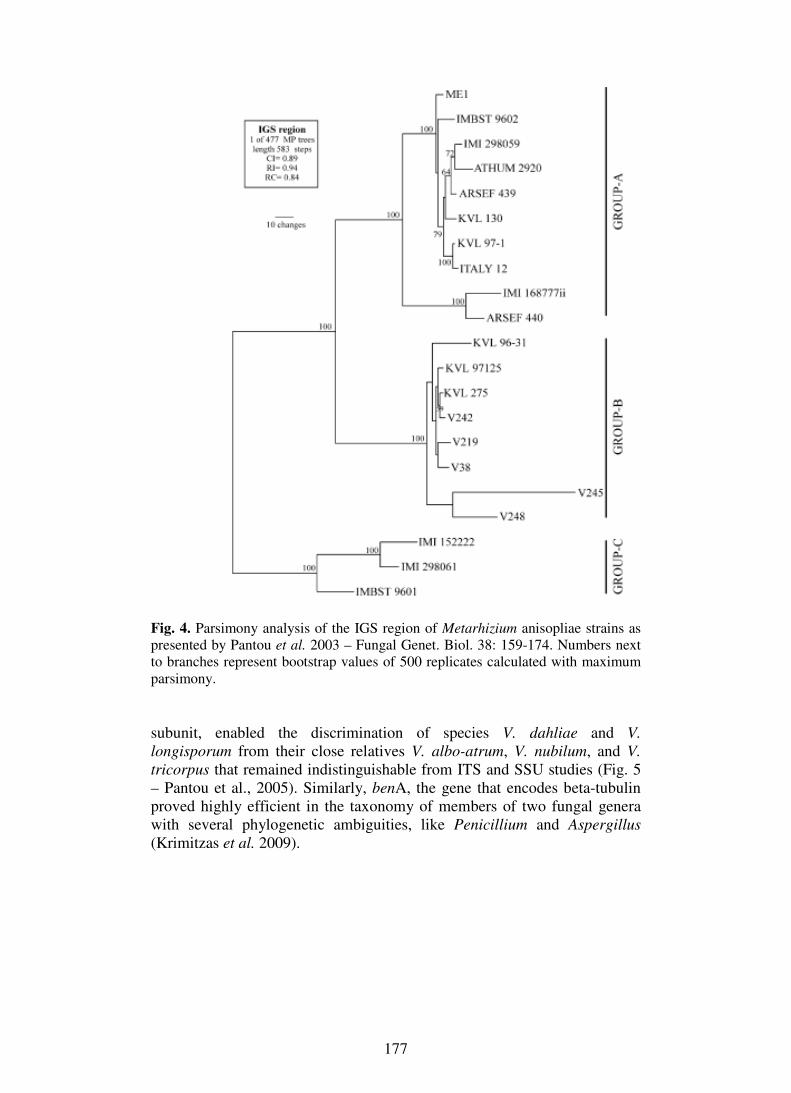
177
Fig. 4. Parsimony analysis of the IGS region of Metarhizium anisopliae strains as presented by Pantou et al. 2003 – Fungal Genet. Biol. 38: 159-174. Numbers next to branches represent bootstrap values of 500 replicates calculated with maximum parsimony. subunit, enabled the discrimination of species V. dahliae and V. longisporum from their close relatives V. albo-atrum, V. nubilum, and V. tricorpus that remained indistinguishable from ITS and SSU studies (Fig. 5 – Pantou et al., 2005). Similarly, benA, the gene that encodes beta-tubulin proved highly efficient in the taxonomy of members of two fungal genera with several phylogenetic ambiguities, like Penicillium and Aspergillus (Krimitzas et al. 2009).

178
Fig. 5. Parsimony analysis of the rpb1 gene, as presented in article of Pantou et al. 2005 – Mycol. Res. 109: 889-902. Numbers next to branches represent bootstrap values of 500 replicates calculated with maximum parsimony and maximum likelihood (in brackets). Mitochondrial genomes and their synteny as an indicator of fungal
evolution Mitochondrial (mt) genomes represent an alternative route of evolution within the organisms, since their rate of change is faster in comparison with their nuclear counterparts (Burger et al. 2003, Kouvelis et al. 2004). Mt genomes contain 14 genes encoding for proteins which participate in cellular respiration and production of ATP, two rDNA genes for the large and small rRNA subunits, and a variable number of trn coding tRNAs (Kouvelis et al. 2004).

179
In a relatively recent study in which the 14 protein coding genes were used as a single unit -either as nucleotide or amino acid datasets- to examine the phylogenetic relationships of all Pezizomycotina fungi with known mt genomes, the result was a single tree (irrespective to method implemented, i.e., Maximum Parsimony, Neighbour Joining and Bayesian Inference) with excellent support (bootstrap and posterior probabilities values of 100% – Fig. 6; Pantou et al. 2008). Phylogenetic relationships within the Class of Sordariomycetes, where the majority of fungi with known mt genomes belong, were well defined based on these mt datasets. For less-studied classes like the Leotiomycetes and Dothideomycetes, their exact phylogenetic placement is still rather ambiguous. Phylogenies based on RPB2 protein sequences found Leotiomycetes monophyletic and clustered them with Sordariomycetes, placing Dothideomycetes at a more basal position (Liu and Hall 2004). In other studies, Leotiomycetes were presented as polyphyletic and therefore, divided into three distinct clades, based on the concatenation of 2 and 3 genes (Lutzoni et al. 2004, Reeb et al. 2004). According to the mt protein dataset, Dothideomycetes and Leotiomycetes form a well-supported sister group to Sordariomycetes (BP: 73% and PP: 100%). A result which is in accordance with previous studies on Pezizomycotina based on nuclear and mt sequence data (Lumbsch et al. 2002, Lumbsch et al. 2005). On the other hand, nucleotide analysis of the 1st and 2nd codon positions showed that Leotiomycetes cluster with Sordariomycetes while Dothideomycetes are placed at a more basal position (Fig. 3b) with strong support (BP: 80% and PP: 100%). A topology further supported by a recent study of a multi-gene nuclear analysis (Spatafora et al. 2006). In a similar phylogenetic analysis within the subphylum of Saccharomycotina (Pramateftaki et al. 2006), mt protein dataset helped in the correct placement of a wine yeast Hanseniaspora uvarum as a sister clade to the rest of the known yeast mt genomes of the ‘Saccharomyces complex’ and along with Candida metapsilosis, Candida orthopsilosis, Candida parapsilosis and Candida albicans and received excellent clade credibility and bootstrap support (Pramateftaki et al. 2006). However, a more recent effort of phylogenesis within the same subphylum for another wine species, like Candida zemplinina revealed the need for additional data from deep-diverging members before divergent lineages of hemiascomycetes can receive a solid phylogenetic classification (Pramateftaki et al. 2008). When first applied, comparisons of mt genome organization and gene synteny of the protein-coding genes for fungi with known complete mt genomes revealed differences associated with the order in which the fungal taxa belonged and common elements that were maintained throughout evolution (Kouvelis et al. 2004). Later, the addition of trn genes in synteny analyses upgraded the information on Sordariomycetes mt genome evolution (Ghikas et al. 2006) and subsequently for the entire subphylum of Pezzizomycotina (Pantou et al. 2008). Especially when the number of complete mt genomes was substantially increased, it became evident that there are four conserved for all Sordariomycetes syntenic units [namely, rnl-

180
trn(11-12)-nad2-nad3; nad4L-nad5-cob-cox1; nad1-nad4-atp8-atp6; and rns-trn(1-5)-cox3-trn(1-5)-nad6-trn(2-5)] which, with few exceptions, are also conserved in all Pezzizomycotina and that trn genes may act as transposable elements that help the occurrence of rearrangements on the entire mt genome (Fig. 7 – Pantou et al. 2008).
Fig. 6. Phylogenetic trees constructed from (a) concatenated protein data and (b) combined 1st and 2nd codon positions of 14 mt genes, as produced by BI (and in accordance to the ML tree). Clade credibility using MrBayes (upper numbers) and parsimonial bootstrap support (lower numbers) calculated from 1000 replicates is shown only when below 100%. Boxes indicate the taxonomic status of each species: A = Ascomycota, B = Basidiomycota, C = Chytridiomycota, Z = Zygomycota, P = Pezizomycotina, S = Saccharomycetes, Sc = Schizosaccharomycetes, Ho = Homobasidiomycetes, Het = Heterobasidiomycetes, So = Sordariomycetes, E = Eurotiomycetes, D = Dothideomycetes, L = Leotiomycetes, H = Hypocreales, He = Helotiales, Sor = Sordariales, Ph = Phyllachorales, Pl = Pleosporales, Eu = Eurotiales, O = Onygenales, Sa = Saccharomycetales, Sch = Schizosaccharomycetales, Ag = Agaricales, Bl = Blastocladiales, T = Tremalles, M = Monoblepharidales, Mo = Mortierellales, Mu = Mucorales. Taxonomic units of the lineage of Fusarium oxysporum are shaded, as presented by Pantou et al. 2008, Gene 419: 7–15.

181
Fig. 7. Gene order comparisons of complete mt genomes from Pezizomycotina. Representatives from three orders of Sordariomycetes are shown: Fusarium oxysporum (Fo), Giberella zeae (Gz), Hypocrea jecorina (Hj), Metarhizium anisopliae (Ma) and Lecanicillium muscarium (Lm) of the order Hypocreales; Verticillium dahliae (Vd) of the order Phyllachorales; and Neurospora crassa (Nc) of the order Sordariales. Eurotiomycetes from the order of Eurotiales: Aspergillus nidulans (An) and Penicillium marneffei (Pm) and the order of Onygenales: Coccidoides immitis (Ci) and Epidermophyton floccosum (Ef), as well as Leotiomycetes from the order of Helotiales: Botryotinia fuckeliana (Bf) and Sclerotinia sclerotiorum (Ss) are also presented. Dothideomycetes are represented with Phaeosphaeria nodorum (Pn) from the order of Pleosporales. Additional representatives of Schizosaccharomycetes: Schizosaccharomyces pombe (Sp) and Saccharomycetes: Yarrowia lipolytica (Yl) and Saccharomyces cerevisiae (Sc) are also included. The detected four units of synteny are either shaded or underlined. [Pantou et al. 2008, Gene 419: 7–15]

182
Mitochondrial genes for genetic, taxonomic and phylogenetic studies of
fungi Mt genes present the advantages and disadvantages of the genetic loci which are essential for the cell throughout the evolution of life. In other words, they contain regions which are highly conserved and therefore, suitable for designing primers, but also domains with high variability and in cases, species- or group-specific, like the gene domains encoding the conserved inter-membrane and variable intra-membrane domains of the NADH subunits (nad1-6 and nad4L genes), which appear highly suitable to explore inter-species differences (Kouvelis et al. 2008). A number of studies in which different mt genes were used to establish intra- and inter-species variation clearly show that the significance of a gene may differ from fungus to fungus. For example, an effort to molecularly identify the different species of the newly introduced Lecanicillium genus -replaced the previously known diverse entomopathogenic species Verticillium lecanii (Zare and Gams 2001)- showed that rns and nad1, alone or in combination, led to the clear differentiation and classification of 28 of the 30 uncharacterised strains (Kouvelis et al. 2008). On the other hand, in the same study, nad3 presented a level of variability within the different Lecanicillium species which was informative but not well substantiated (low bootstrap support). In a similar study within the phytopathogenic species of the Verticillium genus (order: Phyllachorales) four mt genes presented variable levels of information: nad1 was the most informative and discriminated the majority of the species examined. It was followed by rns and cox3 and lastly by nad3, the less variable gene, due to its small size (Pantou et al. 2005). Finally, the introns usually located within the mt genes can be another source of variability and further discrimination of fungal species and, in cases, of isolates within a species (Pantou et al. 2005, Kouvelis et al. 2008, Ghikas et al. 2010). Even though, the insertion sites of the introns within the genomes seem to be conserved for all fungal species and the introns themselves present identical elements, like secondary structure of group-I and ORFs encoding for putative GIY-YIG or LAGLI-DADG endonucleases, their presence/absence and variability in their non-conserved domains is yet another source for discrimination and consequently identification of isolates that carry them (Ghikas et al. 2010). Intergenic mt regions are the most useful for phylogenetic studies The availability of complete mt genomes presented the possibility of utilizing the most variable regions within the mt genomes, i.e., the intergenic regions. As the sizes of the mt genomes are highly variable, ranging from 17-100 Kb, due to the presence of different numbers of introns, but most importantly to their highly different intergenic domains, the selection of the latter as sequences for species identification at intra- and inter- specific level proved until now, the best choice for studying any fungal species (e.g. Ghikas et al. 2006, Pantou et al. 2006, Kouvelis et al. 2008b, Ghikas et al. 2010). The benefits of exploiting these regions are easily understood if we consider the main conclusions of the above mentioned studies: a) the intergenic nad1-nad4 region allowed the
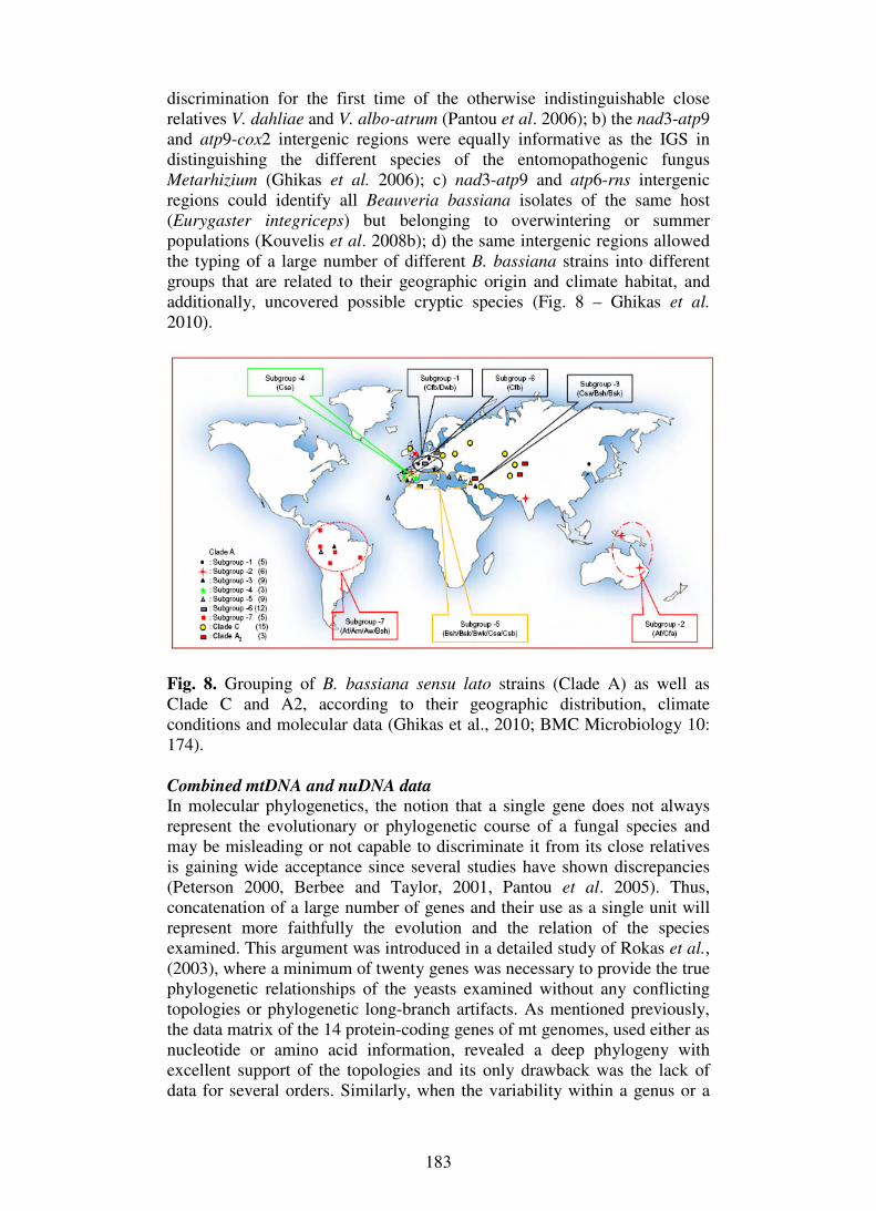
183
discrimination for the first time of the otherwise indistinguishable close relatives V. dahliae and V. albo-atrum (Pantou et al. 2006); b) the nad3-atp9 and atp9-cox2 intergenic regions were equally informative as the IGS in distinguishing the different species of the entomopathogenic fungus Metarhizium (Ghikas et al. 2006); c) nad3-atp9 and atp6-rns intergenic regions could identify all Beauveria bassiana isolates of the same host (Eurygaster integriceps) but belonging to overwintering or summer populations (Kouvelis et al. 2008b); d) the same intergenic regions allowed the typing of a large number of different B. bassiana strains into different groups that are related to their geographic origin and climate habitat, and additionally, uncovered possible cryptic species (Fig. 8 – Ghikas et al. 2010).
Fig. 8. Grouping of B. bassiana sensu lato strains (Clade Α) as well as Clade C and A2, according to their geographic distribution, climate conditions and molecular data (Ghikas et al., 2010; BMC Microbiology 10: 174). Combined mtDNA and nuDNA data In molecular phylogenetics, the notion that a single gene does not always represent the evolutionary or phylogenetic course of a fungal species and may be misleading or not capable to discriminate it from its close relatives is gaining wide acceptance since several studies have shown discrepancies (Peterson 2000, Berbee and Taylor, 2001, Pantou et al. 2005). Thus, concatenation of a large number of genes and their use as a single unit will represent more faithfully the evolution and the relation of the species examined. This argument was introduced in a detailed study of Rokas et al., (2003), where a minimum of twenty genes was necessary to provide the true phylogenetic relationships of the yeasts examined without any conflicting topologies or phylogenetic long-branch artifacts. As mentioned previously, the data matrix of the 14 protein-coding genes of mt genomes, used either as nucleotide or amino acid information, revealed a deep phylogeny with excellent support of the topologies and its only drawback was the lack of data for several orders. Similarly, when the variability within a genus or a

184
species was examined, the combined use of concatenation nuclear and mitochondrial genes produced the most solid results with the best discriminatory data (Kouvelis et al. 2008; Krimitzas et al. 2009; Ghikas et al. 2010). Therefore, at a time that sequencing cost is daily reduced and sequence data from large numbers of genes from an organism and entire fungal genomes become increasingly frequently available to the scientific community, the combined study of the nuclear and the mitochondrial genomes becomes all the more likely in the near future. Undoubtedly combining data from the prospect view of two different evolutionary lineages, is expected to reveal the evolutionary progress of the entire organism and further to resolve any ambiguities on taxonomy and phylogenies. Even at a smaller scale, today that the data from many genes is not yet feasible for the average laboratory, suitable gene markers from both nuclear and mt genomes can be chosen and used in taxonomy, phylogenesis and fingerprinting studies. However, it is important to realise that each gene contributes differently in such studies and the right choice must be made depending on the question addressed. Literature Berbee, M.L., Taylor, J.W. (2001). Fungal molecular evolution: gene trees
and geologic time. In “The Mycota. Vol. VII. Part B, Systematics and Evolution” (McLaughlin D.J., McLaughlin E.G. and Lemke P.A., eds), Springer-Verlag, Heidelberg pp. 229–245.
Blackwell, M., Hibbett, D.S., Taylor, J.W., Spatafora, J.W. (2006). Research Coordination Networks: a phylogeny for kingdom Fungi (Deep Hypha). Mycologia 98: 829–837.
Burger, G., Gray, M.W., Lang, B.F. (2003). Mitochondrial genomes: anything goes. Trends in Genetics 19: 709–716.
Bruns, T.D., Vilgalys, R., Barns, S.M., Gonzalez, D., Hibbett, D.S., Lane, D.J., Simon, L., Stickel. S., Szaro, T.M., Weisburg, W.G., Sogin, M.L. (1992). Evolutionary relationships within the Fungi: analyses of nuclear small subunit rRNA sequences. Molecular Phylogenetics and Evolution 1: 231–241.
Celio, G.J., Padamsee, M., Dentinger, B.T., Bauer, R., McLaughlin, D.J. (2006). Assembling the Fungal Tree of Life: constructing the structural and biochemical database. Mycologia 98: 850–859.
Crous, P.W., Rong, I.H., Wood, A., Lee, S., Glen, H., Botha, W., Slippers, B., de Beer, W.Z., Wingfield, M.J., Hawksworth, D.L. (2006). How many species of fungi are there at the tip of Africa? Studies in Mycology 55: 13–33.
Driver, F., Milner, R.J., Trueman, J.W.H. (2000). A taxonomic revision of Metarhizium based on phylogenetic analysis of rDNA sequence data. Mycological Research 104: 134–150.
Garrido-Jurado, I., Marquez, M., Ortiz-Urquiza, A., Santiago-Alvarez, C., Iturriaga, E., Quesada-Moraga, E., Monte, E., Hermosa, R. (2011). Genetic analyses place most Spanish isolates of Beauveria bassiana in a molecular group with word-wide distribution. BMC Microbiology 11: 84.

185
Ghikas, D.V., Kouvelis, V.N., Typas, M.A. (2006). The complete mitochondrial genome of the entomopathogenic fungus Metarhizium anisopliae var. anisopliae: gene order and trn gene clusters reveal a common evolutionary course for all Sordariomycetes, while intergenic regions show variation. Archives of Microbioliology 185: 393–401.
Ghikas, D.V., Kouvelis, V.N., Typas, M.A. (2010). Phylogenetic and biogeographic implications inferred by mitochondrial intergenic region analyses and ITS1-5.8S-ITS2 of the entomopathogenic fungi Beauveria bassiana and B. brongniartii. BMC Microbiology 10: 174
Hawksworth, D.L. (2001). The magnitude of fungal diversity: the 1.5 million species estimate revisited. Mycological Research 105: 1422–1432.
Hawksworth, D.L. (2006). Pandora's mycological box: molecular sequences vs. morphology in understanding fungal relationships and biodiversity. Revista Iberoamericana de Micología 23: 127–133.
Hegedus, D.D., Pfeifer, T.A., Mulyk, D.S., Khachatourians, G.G. (1998). Characterization and structure of the mitochondrial small rRNA gene of the entomopathogenic fungus Beauveria bassiana. Genome 41: 471–476.
Hughes, W.O.H., Thomsen, L., Eilenberg, J., Boomsma, J.J. (2004). Diversity of entomopathogenic fungi near leaf-cutting ant nests in a neotropical forest, with particular reference to Metarhizium anisopliae var. anisopliae. Journal of Invertebrate Pathology 85: 46–53.
Keeling, P.J., Luker, M.A., Palmer, J.D. (2000). Evidence from beta-tubulin phylogeny that microsporidia evolved from within the fungi. Molecular Biology and Evolution 17: 23–31.
Kouvelis, V.N., Ghikas, D.V., Typas, M.A. (2004). The analysis of the complete mitochondrial genome of Lecanicillium muscarium (synonym Verticillium lecanii) suggests a minimum common gene organization in mtDNAs of Sordariomycetes: phylogenetic implications. Fungal Genetics and Biology 41: 930–940.
Kouvelis, V.N., Sialakouma A., Typas, M.A. (2008). Mitochondrial gene sequences alone or combined with ITS region sequences provide firm molecular criteria for the classification of Lecanicillium species. Mycological Research 112: 829–844.
Kouvelis, V.N., Ghikas, D.V., Edgington, S., Typas, M.A., Moore, D. (2008b). Molecular characterisation of isolates of Beauveria bassiana obtained from overwintering and summer populations of Sunn Pest (Eurygaster integriceps). Letters in Applied Microbiology 46: 414–420.
Kretzer, A.M., Bruns, T.D. (1999). Use of atp6 in fungal phylogenetics: an example from the boletales. Molecular Phylogenetics and Evolution 13: 483–492.
Krimitzas, A., Kouvelis, V.N., Typas, M.A. (2009). Phylogenetic discrimination of Aspergillus and Penicillium species with nuclear and mitochondrial genes. Proceedings of 2nd Conference of “Mikroviokosmos”, pp. 32–33.
Liu, Y.J., Hall, B.D. (2004). Body plan evolution of ascomycetes, as inferred from an RNA polymerase II phylogeny. Proceedings in National Academies of Science U.S.A. 101: 4507–4512.

186
Lumbsch, H.T., Wirtz, N., Lindemuth, R., Schmitt, I. (2002). Higher level phylogenetic relationships of euascomycetes (Pezizomycotina) inferred from a combined analysis of nuclear and mitochondrial sequence data. Mycological Progress 1: 57–70.
Lumbsch, H.T., Schmitt, I., Lindemuth, R., Miller, A., Mangold, A., Fernandez, F., Huhndorf, S. (2005). Performance of four ribosomal DNA regions to infer higher level phylogenetic relationships of inoperculate euascomycetes (Leotiomyceta). Molecular Phylogenetics and Evolution 34: 512–524.
Lutzoni, F., Kauff, F., Cox, C.J., McLaughlin, D., Celio, G., Dentinger, B., Padamsee, M., Hibbett, D.S., James, T.Y., Baloch, E., Grube, M., Reeb, V., Hofstetter, V., Schoch, C., Arnold, A.E., Miadlikowska, J., Spatafora, J., Johnson, D., Hambleton, S., Crockett, M., Shoemaker, R., Sung, G.-H., Lücking, R., Lumbsch, T., O’Donnell, K., Binder, M., Diederich, P., Ertz, D., Gueidan, C., Hansen, K., Harris, R.C., Hosaka, K., Lim, Y.-W., Matheny, B., Nishida, H., Pfister, D., Rogers, J., Rossman, A., Schmitt, I., Sipman, H., Stone, J., Sugiyama, J., Yahr, R., Vilgalys, R. (2004). Where are we in assembling the fungal tree of life, classifying the fungi, and understanding the evolution of their subcellular traits? American Journal of Botany 91: 1446–1480.
Mavridou, A., Cannone, J., Typas, M.A. (2000). Identification of group-I introns at three different positions within the 28S rDNA gene of the entomopathogenic fungus Metarhizium anisopliae var. anisopliae. Fungal Genetics and Biology 31: 79–90.
McLaughlin, D.J., Hibbett, D.S., Lutzoni, F., Spatafora, J.W., Vilgalys, R. (2009). The search for the fungal tree of life. Trends in Microbiology 17: 488–497.
Nikoh, N., Fukatsu, T. (2001). Evolutionary dynamics of multiple group I introns in nuclear ribosomal RNA genes of entomoparasitic fungi of the genus Cordyceps. Molecular Biology and Evolution 18: 1631–1642.
Pantou, M.P., Mavridou, A., Typas, M.A. (2003). IGS sequence variation, group-I introns and the complete nuclear ribosomal DNA of the entomopathogenic fungus Metarhizium: excellent tools for isolate detection and phylogenetic analysis. Fungal Genetics and Biology 38: 159–174.
Pantou, M.P., Strunnikova, O.K., Shakhnazarova, V.Y., Vishnevskaya, N.A., Papalouka, V.G., Typas, M.A. (2005). Molecular and immunochemical phylogeny of Verticillium species. Mycological Research 109: 889–902.
Pantou, M.P., Kouvelis, V.N., Typas, M.A. (2006). The complete mitochondrial genome of the vascular wilt fungus Verticillium dahliae: a novel gene order for Verticillium and a diagnostic tool for species identification. Current Genetics 50: 125–136
Pantou, M.P., Kouvelis, V.N., Typas, M.A. (2008). The complete mitochondrial genome of Fusarium oxysporum: insights into fungal mitochondrial evolution. Gene 419: 7–15.
Peterson, S.W. (2000). Phylogenetic relationships in Aspergillus based on rDNA sequence analysis. In: “Integration of modern taxonomic methods for Penicillium and Aspergillus classification” (Samson R.A.

187
and Pitt J.I., eds), Harwood Academic Publishers, Amsterdam, pp. 323–355.
Pramateftaki, P.V., Antoniou, P.P., Typas, M.A. (2000). The complete DNA sequence of the nuclear ribosomal RNA gene complex of Verticillium dahliae: intraspecific heterogeneity within the intergenic spacer region. Fungal Genetics and Biology 29: 135–143.
Pramateftaki, P.V., Kouvelis, V.N., Lanaridis, P., Typas, M.A. (2006). The mitochondrial genome of the wine yeast Hanseniaspora uvarum: a unique genome organization among yeast/fungal counterparts. FEMS Yeast Research 6: 77–90.
Pramateftaki, P.V., Kouvelis, V.N., Lanaridis, P., Typas, M.A. (2008). The complete mitochondrial genome sequence of the wine yeast Candida stellata: intra-species distribution of a novel group-IIB1 intron with eubacterial affiliations. FEMS Yeast Research 8: 311–317.
Reeb, V., Lutzoni, F., Roux, C. (2004). Contribution of RPB2 to multilocus phylogenetic studies of the euascomycetes (Pezizomycotina, Fungi) with special emphasis on the lichen-forming Acarosporaceae and evolution of polyspory. Molecular Phylogenetics and Evolution 32: 1036–1060.
Rehner, S.A., Buckley, E.P. (2005). A Beauveria phylogeny inferred from nuclear ITS and EF1-α sequences: evidence for cryptic diversification and links to Cordyceps teleomorphs. Mycologia 97: 84–98.
Rokas, A., Williams, B.L., King, N., Carroll, S.B. (2003). Genome-scale approaches to resolving incongruence in molecular phylogenies. Nature 425: 798–804.
Spatafora, J.W. (2005). Assembling The Fungal Tree of Life (AFTOL). Mycological Research 109: 755–756.
Spatafora, J.W., Sung, G.H., Johnson, D., Hesse, C., O'Rourke, B., Serdani, M., Spotts, R., Lutzoni, F., Hofstetter, V., Miadlikowska, J., Reeb, V., Gueidan, C., Fraker, E., Lumbsch, T., Lücking, R., Schmitt, I., Hosaka, K., Aptroot, A., Roux, C., Miller, A.N., Geiser, D.M., Hafellner, J., Hestmark, G., Arnold, A.E., Büdel, B., Rauhut, A., Hewitt, D., Untereiner, W.A., Cole, M.S., Scheidegger, C., Schultz, M., Sipman, H., Schoch, C.L. (2006). A five-gene phylogeny of Pezizomycotina. Mycologia 98: 1018–1028.
Taylor, J.W., Spatafora, J., O’Donnell, K., Lutzoni, F., James, T., Hibbett, D.S., Geiser, D., Bruns, T.D., Blackwell, M. (2004). The fungi. In: “Assembling the Tree of Life” (Cracraft J. and Donoghue M.J., eds), Oxford University Press, Oxford, pp. 171–196.
Wang, C., Li, Z. Typas, M.A., Butt, T.M., (2003). Nuclear large subunit rDNA group I intron distribution in a population of Beauveria bassiana strains: phylogenetic implications. Mycological Research 107: 1189–1200.
White, T.J., Bruns, T.D., Lee, S., Taylor, J.W. (1990). Amplification and direct sequencing of fungal ribosomal RNA genes for phylogenetics. In: “PCR Protocols: a Guide to Methods and Applications” (Innis M.A., Gelfand D.H., Sninsky J., White T.J. eds), Academic Press, San Diego, pp. 315–322.

188
Yokoyama, E., Arakawa, M., Yamagishi, K., Hara, H. (2006). Phylogenetic and structural analyses of the mating-type loci in Clavicipitaceae. FEMS Microbiology Letters 264: 182-191.
Zare, R., Gams, W. (2001). A revision of Verticillium section Prostrata. IV. The genera Lecanicillium and Simplicillium gen. nov. Nova Hedwigia 73: 1 –50.
Thematic area: Fungal genetics and genomics
COMPARATIVE GENOMIC, PHYLOGENETIC, AND FUNCTIONAL INVESTIGATION OF THE XENOBIOTIC
METABOLIZING ARYLAMINE N-ACETYLTRANSFERASE ENZYME FAMILY AMONG FUNGI
S. Boukouvala 1, E. Kontomina 1, E.P. Karagianni 1,
B. Ormiston 2, T. Tsirka 1 and A.E. Glenn 2
1Department of Molecular Biology and Genetics, Democritus University of Thrace, Building 10, University Campus, Alexandroupolis 68100, Greece
2Toxicology and Mycotoxin Research Unit, Russell Research Center, Agricultural Research Service, United States Department of Agriculture,
950 College Station Road, Athens, GA 30605, USA
Keywords: fungal genetics and genomics, plant pathogenic fungi xenobiotic metabolism Arylamine N-acetyltransferases (NATs) are xenobiotic metabolizing enzymes well-characterized in several bacteria and higher eukaryotes. The role of NATs in fungal biology has only recently been investigated (Glenn and Bacon, 2009; Glenn et al., 2010). The NAT1 gene of Gibberella moniliformis was the first NAT cloned and characterized from fungi and is essential for the metabolism of antimicrobial compounds (benzoxazolinones) produced by cereals (maize, wheat, rye) and some wild grasses. We report a phylogenetic analysis employing an exhaustive annotated dataset of homologous NAT amino acid sequences recovered through inspection of 146 fungal genomes. We proceeded to amplification, exon-intron characterization and cloning of 16 NAT loci (3 of which are pseudogenes), predicted in Gibberella moniliformis, Gibberella zeae, Fusarium oxysporum, Aspergillus flavus and Aspergillus nidulans. Expression of recombinant NATs from these loci has enabled enzymatic profiling of their co-factor and substrate selectivity, as well as protein purification for future structure-function investigations. Real-time PCR quantification of the corresponding NAT transcripts is also in progress, supporting an effect of xenobiotics (including benzoxazolinone) to endogenous expression of certain NAT genes.

189
Our findings suggest an association between fungal NAT metabolic capacity and affinity for hosts that produce benzoxazolinone defense compounds. Future studies will investigate more closely this possible association and its implications on agricultural management practices. Literature Glenn, A.E. and Bacon, C.W. 2009: FDB2 encodes a member of the
arylamine N-acetyltransferase family and is necessary for biotransformation of benzoxazolinones by Fusarium verticillioides. J. Appl. Microbiology, 107:657-671.
Glenn, A.E., Karagianni, E.P., Ulndreaj, A. and Boukouvala, S. 2010: Comparative genomic and phylogenetic investigation of the xenobiotic metabolizing arylamine N-acetyltransferase enzyme family. FEBS Lett., 584:3158-3164.
QUANTITATIVE TRAIT LOCI CONTROLLING VEGETATIVE GROWTH RATE OF EDIBLE MUSHROOM PLEUROTUS
OSTREATUS
A. Sivolapova 1, J. Baars 2, A. Sonnenberg 2, A. Shnyreva 1, B. Lavrijssen2 and P. Hendricks2
1Moscow State University, Faculty of Biology, Mycology & Algology Department, 119991, Moscow, Russian Federation
E-mail: [email protected] 2Wageningen University, PRI, Plant Science Group,
6700 AA Wageningen, Netherlands
Keywords: fungal genetics and genomics, oyster mushroom, QTL’s, growth rate Pleurotus ostreatus is commercially important edible mushroom commonly known as oyster mushroom. Mycelium growth rate has applied interest, as faster growing dikaryons are able to colonize the substrate faster than competitors responsible for reductions in the industrial yield of this fungus. In current research we tryed to find QTL's responsible for dikaryotic substrate colonization rate, that is possibly of interest for process of waste degradation as well as for mushroom cultivation. The growth rate segregation was analysed on the wheat straw as the culture substrate. Our analysis showed 12 genomic regions (quantitative trait loci, QTL’s) controlling vegetative growth rate of Pleurotus ostreatus which were mapped on the genetic linkage map of this fungus. This linkage map can be useful in the design of breeding programs based on genomic data.

190
COMPARATIVE ANALYSIS OF MITOCHONDRIAL GENOME ISOLATED FROM THREE FLAMMULINA VELUTIPES STRAINS
H. Yoon 1, Y.H. You 1, J.R. Woo 1, Y.J. Park 2, W.S. Kong 3, B.M. Lee 4 and J.G. Kim 1
1Department of Life Sciences and Biotechnology, Kyungpook National University, South Korea
2Department of Applied Biochemistry, Konkuk University, South Korea 3National Institute of Horticultural & Herbal Science, RDA, South Korea
4National Academy of Agricultural Science, RDA, South Korea
Keywords: fungal genetics and genomics, Flammulina velutipes
Flammulina velutipes, also known as “winter mushroom”, is a very popular edible mushroom in East Asia and Europe. F. velutipes belongs to the class Agaricomycetes, the order Agaricales and the family Physalacriaceae, and this mushroom is the white-rot fungus which has the capacity to degrade both lignin and cellulose. In this study, two monokaryotic strains 4019-18 and 4019-20 and one dikaryotic strain 4019-18x20 were selected from the Rural Development Administration in Korea, and their mitochondrial genomes were analysed for the first time.
The total sequence length of mtDNA from both 4019-18 and 18x20 strains is 88,513 bp, and that of 4019-20 is 88,508 bp, 5 bp shorter than other two strains, due to single-nucleotide deletions. The annotation of mtDNA of F. velutipes strains was performed using blast algorithms with Genbank sequence database (NCBI).
As a result, it includes the 14 genes encoding proteins which are related with 6 NADH dehydrogenases, one apocytochrome b, 3 cytochrome c oxidases, 3 ATP synthases and one ribosomal protein. It contains the 28 structural genes involved with ribosomal RNA and transfer RNA. Comparative analysis was performed with mtDNA sequences of other basidiomycetes, and diverse results were generated about gene order, genome size, GC contents and phylogenetic relationships.
Thematic area: Fungal Biotechnology
ISOLATION AND CLONING OF MANGANESE PEROXIDASE (mnp) GENE FROM THE WHITE BUTTON MUSHROOM
M. Farsi 1, J. Hasan-Janpour 1, H.R. Pourianfar 2
1Faculty of Agriculture, Ferdowsi university of Mashhad 2Department of Industrial Fungi Biotechnology, (ACECR)
University campus, Mashhad, Iran E-mail: [email protected]
Keywords: fungal biotechnology, Agaricus bisporus, cDNA of manganese peroxidase, molecular cloning

191
In addition to their nutritional values, mushrooms are currently considered as useful tools of agriculture wastes remediation. In the button mushroom (Agaricus bisporus), there are several enzymes which catalyze lignin compounds of the compost during the mycelia growth and the fructification phases. Manganese Peroxidase (MnP), as one of the most important lignin-degrading enzymes, plays a key role in degradation of lignin compounds in the button mushroom (Lankinen et al. 2005, Nagai et al. 2007). In order to achieving a high yield of MnP in the A. bisporus, the gene encoding MnP was isolated, characterized and cloned. The total RNA was extracted from the mycelium growing on the liquid compost extract medium, followed by construction of its cDNA by reverse transcriptase. The PCR products were then inserted into the pTZ57R/T cloning vector, and transferred into E. coli (the DH5α strain). Finally, the plasmid was extracted from the transgenic bacteria, followed by enzymatic digestion and nucleotide sequencing. The BLAST analysis revealed two different nucleotides (657 and 850) between the cloned fragment (generated in this research) and the mnp1 (available in the gene bank of NCBI). The difference in the nucleotide positions of 657 and 850 subsequently changed Isoleucine to Valine and Serin to Alanine, respectively. Literature Lankinen, P., Hilden, K., Aro, N., Salkinoja-salonen, M. and Hatakka, A.
2005. Mangnese peroxidase of Agaricus bisporus: grain bran-promoted production and gene characterization. Applied Microbiology and Biotechnology, 66:401-407.
Nagai, M., Sakamoto, Y. and Nakade, K. 2007. Isolation and characterization of the gene encoding a manganese peroxidase from Lentinula edodes. Mycoscience, 48:125-130.
ENDOCHITINASE GENE EXPRESSION IN TOMATOES AFTER SIMULTANEOUS TREATMENT WITH ARBUSCULAR MYCORRHIZAL FUNGI AND TRICHODERMA HARZIANUM
M. Ene, M. Alexandru, T.E. Şesan and M. Cutrubinis
University of Bucharest, Faculty of Biology,
Department of Botany & Microbiology, Romania
Keywords: fungal biotechnology, fungus-plant interactions, endochitinase, tomatoes, Q-Real-Time PCR, Glomus intraradices, Trichoderma harzianum Diseases resistance in plants can be improved by treatment with various fungi, most of mechanisms involved leading to the expression modulation of pathogenesis-related proteins (PR). One of the most important, often affected classes of PR proteins is chitinases. Fungi in the Trichoderma genus are often used as biocontrol agents, generally yielding an increase in chitinase synthesis. The effect of their combination with Arbuscular Mycorrhizal Fungi (AMF) is often controversial, depending on the host

192
plant and species of fungus. In this paper we have analysed the expression of a gene encoding for one endochitinase in tomatoes (Lycopersicum esculentum), after infection with Glomus intraradices and Trichoderma harzianum, applied separate or combined. After 35 days from the germination and interaction with the fungi, the expression decreased 6.6 times in treatments that included mycorrhizal fungus (with or without Trichoderma), comparing to the untreated control, whereas in treatment with T. harzianum only, the expression decreased 55.5 times. For this analysis, Q-Real-Time PCR reactions were normalized simultaneously against EF1 alfa and Ribosomal Protein L2 genes, combination that we have found to be the most stable from a total of four commonly used house-keeping genes, by using the GeNorm calculation algorithm. Microscopical observation, after classical cotton-blue staining, did not show any opposition or root-space competition between the two fungi. The conclusion is that the endochitinase could be involved in the penetration permission and that mycorrhizal symbiosis controls physiological effect of infection with other fungi, including the beneficial mould Trichoderma harzianum.
CYCLOPIAZONIC ACID AND SCLEROTIA PRODUCING ABILITY IN AFLATOXIGENIC AND NON-AFLATOXIGENIC
ASPERGILLUS FLAVUS STRAINS FROM PEANUTS FIELD SOILS
M. Shams-Ghahfarokhi 1, S. Amani 1, M. Banasaz 2 and M. Razzaghi-Abyaneh 2
1Department of Mycology, Faculty of Medical Sciences, Tarbiat Modares University, Tehran 14115-331, Iran
2Department of Mycology, Pasteur Institute of Iran, Tehran 13164, Iran
Keywords: fungal biotechnology, Aspergillus flavus, cyclopiazonic acid, aflatoxins, sclerotia, chemotype pattern, HPLC Cyclopiazonic acid (CPA) is a toxic endole tetramic acid which mainly produces by some species of the genera Aspergillus and Penicillium. In the present study, the CPA and sclerotia producing ability was investigated in a total of 53 aflatoxigenic and non-aflatoxigenic Aspergillus flavus strains isolated from peanuts field soils. The isolates were cultured on czapek dox agar plates and sclerotia production was recorded after 14 days incubation at 28° C. For CPA assessment, the isolates were cultured on yeast extract-sucrose broth for 4 days at 28° C and CPA producing ability was evaluated by observing purple spots on thin layer chromatography (TLC) plates. Quantitation of CPA as well as aflatoxin B1 (AFB1) was carried out by high performance liquid chromatography (HPLC).

193
Based on the results obtained, 45 of the 53 A. flavus isolates (84.9%) were able to produce either CPA or AFB1, while 8 of the isolates (15.1%) were non-toxigenic. The amounts of CPA and AFB1 produced by the isolates were reported in the range of 18.22 to 403.85 µg/g and 53.27 to 7446.28 µg/g fungal dry weights, respectively. Chemotype classification of A. flavus isolates based on the ability for producing mycotoxins and sclerotia showed that 43.40% were producers of CPA, AFB1 and sclerotia (group I), 13.21% of CPA and AFB1 (group II), 9.43% of AFB1 and sclerotia (group IV), 15.09% of AFB1 (group VI), 3.77% of CPA and sclerotia (group VII) and 15.09% were non-toxigenic with no sclerotia (group VIII). None of the A. flavus isolates were able to produce only CPA (group III) or sclerotia (group V). The presence of CPA-producing A. flavus strains in peanuts field soils reported in the present study may be quite important due to the natural occurrence and proven toxicity of CPA in food, feed and agricultural commodities.
GLYCOGEN AND TREHALOSE ACCUMULATION IN DEBARYOMYCES OCCIDENTALIS AT DIFFERENT CARBON
SOURCES
T.T. Genç, T. Çelik and İ.N. Çildır
COMU- Faculty of Arts and Science, 17100 Çanakkale, Turkey E-mail: [email protected]
Keywords: fungal biotechnology, trehalose, glycogen, Debaryomyces occidentalis Saccharomyces cerevisiae accumulates glycogen and trehalose when growth conditions become unfavorable (Parrou et al. 1997). Trehalose and glycogen contents in Debaryomyces occidentalis yeast species were examined in a time course manner in three different carbon sources: glucose, galactose and glycerol. The S. cerevisiae strain was used as control for comparing the patterns of reserve carbohydrates accumulation. Trehalose, accumulated during the overnight growth, was more slowly degraded in glucose grown D. occidentalis yeast cells than in S. cerevisiae. When D. occidentalis yeast cells reached a high cell density (nearly 60 hours later), yeast cells started to accumulate trehalose again in the stationary phase. But in S. cerevisiae, trehalose accumulation started 36 hours later, at the beginning of stationary phase. There was no glycogen accumulation during these phases in either yeast species. When the carbon source became galactose, D. occidentalis degraded overnight-accumulated-trehalose very rapidly and started to accumulate trehalose again (24 hours later). Then it degraded half of the logarithmic trehalose (5.29 mg/g) within the stationary phase (60-72 hour later). Low levels of glycogen accumulation (4.11mg/g) were detected 24 hours later and it decreased to 1.07 mg/g after 72 hours. But in S. cerevisiae the accumulated glycogen level was high (5.39 mg/g) and degraded slowly. Trehalose accumulation

194
started after 48 hours, at the beginning of the stationary phase and reached a high concentration (12.19 mg/g) 72 hours later. In D. occidentalis very low level glycogen and trehalose accumulation was detected during the 72 hours when it was grown in glycerol. However, in S. cerevisiae glycogen and trehalose accumulation reached a maximum level (10.33 mg/g and 12.4 mg/g, respectively) after 24 hours, then degraded to half of that 48 hours later. Results indicated that glycogen and trehalose accumulation patterns are completely different in D. occidentalis from S. cerevisiae. Literature Parrou, J., Teste, M., and Fransois, J., 1997: Effects of various types
of stress on the metabolism of reserve carbohydrates is Saccharomyces cerevisiae: Genetic evidence for a stress-induced recycling of glycogen and trehalose. Microbiology,143:1891-1900. RECENT ADVANCES IN CONSERVATION AND STUDY OF
MACROMYCETES GENETIC RESOURCES IN THE LE-BIN CULTURE COLLECTION
N. Psurtseva, A. Kiyashko, N. Shakhova, M. Shevchenko
and K. Barinova
Komarov Botanical Institute RAS, Prof. Popov Str., 2, St. Petersburg 197376, Russia
E-mail: [email protected]
Keywords: fungal biotechnology, macromycetes, culture collection, oxidases, biological activity Fungal biotechnology has become an integral part of our life. Culture Collections play a significant role in conservation and maintenance of macromycetes genetic resources that can be successfully used for human interests. LE-BIN is the largest Russian basidiomycete culture collection, holding over 1600 strains of about 600 species of agaricoid, aphyllophoroid, and gasteroid fungi. Most of the strains are original isolates from field works in various regions of Russia. The main functions of the LE-BIN are conservation ex situ of taxonomical and ecological diversity of macromycetes in Russia with emphases on rare and endangered, ectomycorrhizal and useful for biotechnology and medicine species; study and development of the biological resources, and provision of high quality biological material for research and industry. The aim of preservation is to maintain purity, viability and genomic integrity, avoid selection of variants, and lessen the prospects of strain deterioration. Collection strains are maintained as sub-cultures in tubes, on beer-ale agar slants at 4–6º C and in screw-cap vials under distilled water at room temperature. The process of cryopreservation by deep-freeze method has recently been started. Verification of cultures is conducted by a cumulative approach using cultural, biochemical, and molecular methods.

195
Primary researches carried out in the collection include study of strains from various macromycete taxa for enzymatic and biological activity. Screening for novel laccase producers demonstrated that species from Steccherinaceae family could be a matter of research interest in this regard. Culture characteristics, evaluation of biosynthetic potential and submerged cultivation under various conditions for laccase regulation were carried out to show a great laccase potential of Antrodiella faginea, Junghuhnia nitida, Steccherinum murashkinskyi and S. ochraceum. Study of basidiomycetes for antifungal activity revealed some species (e.g. Agrocybe species, Lampteromyces japonicus and Marasmius scorodonius) with sufficient activity against micromycetes-biodestructors. Some of the newly isolated strains of xylotrophic fungi collected in sparsely populated and unexplored areas of the Altai Republic were studied for antiviral activity. Strains of Daedaleopsis confragosa, Datronia mollis, Ischnoderma benzoinum and Ganoderma valesiacum were the most promising against flue virus type A.

196
Poster presentations
PROGRAMME
Monday, 19th September
Thematic area Title-Authors Stand No
Developmental mycology
Trehalose effect on spore viability of Blakeslea trispora.
Olga A. Vereshchagina, A.S. Memorskaya, V.M. Tereshina
1
Investigation of morphological and anatomical features of Helvella crispa fr. spores and mycelium.
Perihan Güler, F. Kutluer2, A. Türkoğlu, İ. Kunduz, H. Biçer
2
Regulation of chitinase (chi1 and chi2) expression in Coprinellus congregatus.
H. Kim, Hyoung T. Choi 3
Systematics and evolution of fungi
A morphological and molecular study of Seiridium associated with stem-canker disease on Cupressaceae in Greece.
Athanasios Angelopoulos, V.N. Kouvelis, Μ.Α. Τypas, E. Kapsanaki-Gotsi
4
Gymnopus inusitatus var. cystidiatus, a new European bisporic taxon (Basidiomycota, Omphalotaceae).
V. Antonín, P. Finy, M. Tomšovský
5
DNA isolation of macrofungi.
M. Bozkurt, Mehmet A. Karaselek, S. Aktaş, T. Uysal
6
Comparative analyses of alignment and phylogeny reconstruction strategies reveal unexploited phylogenetic signal in the ITS sequences at multiple taxonomic ranges.
Lazlo G. Nagy, S. Kocsubé, G.M. Kovács, Z. Csanádi, T. Petkovits, C. Vágvölgyi, T. Papp
7

197
Type studies of some Cystoderma species.
Irja Saar 8
A contribution towards the clarification on the species concept of Tricholoma equestre.
Sofia Gomes, J.L.Baptista-Ferreira 9
A five-gene phylogeny of the Mortierellales.
Tamas Petkovits, L.G. Nagy, I. Nyilasi, S.A. Kovács, K. Voigt, C. Vágvölgyi, T. Papp
10
Morphological and physiological variation among species of the genus Circinella.
J. Budziszewska, B. Szmaus, M. Wilk,
Marta Wrzosek
11
A morphological analysis of strains of the genus Monascus isolated from olives.
Eva Kapsanaki-Gotsi, K. Efthymiou 12
Determination of phylogenetic relationship in multinucleate and binucleate Rhizoctonia with 18 s rDNA sequence analysis.
Eda Uğurtay, İ. Özkoç
13
Lactifluus piperatus and company (Russulaceae).
Eske De Crop, J. Nuytinck, A. Verbeken 14
Delimitation of almost forgotten species Spongipellis litschaueri and its taxonomic position within the genus.
Michal Tomšovský
15
Russula sect. Maculantinae in Europe – taxa accepted consistently versus dubious ones.
S. Adamčík, Sonia Jančovičová 16
Using ITS2 secondary structure for species identification in Lecanicillium W. Gams & Zare
J. Małagocka, Julia Budziszewska, M. Wrzosek 17
Conservation of fungi
The Red Book of plants of the republic of Armenia (plants and fungi).
Siranush G. Nanagulyan and L.V. Margaryan 18

198
Aphyllophoroid fungi in forest ecosystems in Vodlozero National Park.
Petr Zavodovskiy 19
Succession of macromycetes in disused gravel-sand pits: a comparison between restoration and spontaneous succession.
Lucie Zíbarová
20
Contribution of the Komarov Botanical Institute basidiomycetes culture collection to the fungal conservation in Russia.
A.A. Kiyashko, Nadya V. Psurtseva
21
Conservation of fungi in the Management Plans of Protected Areas of Greece.
Charikleia. Perlerou, V. Christopoulos, S. Diamandis
22
Fungi in ecosystems
Organic acid production by Penicillium citrinum upon the influence of copper and zinc.
Katerina V. Barinova, S.M. Schiparev 23
Climatic inversion and distribution of macrofungi – case study from the Bohemian Switzerland National Park (Czech Republic).
Jan Holec, Jan Wild
24
Edible and medicinal fungi
Influence of Agaricus bisporus, Cantharellus cibarius, Lentinula edodes crude water extracts on Drosophila melanogaster locomotor activity
Elina Svilpe, K. Serstnova, N. Matjuskova
25
Antioxidant properties of the Pleurotus ostreatus mycelium obtained in the presence of corn extract.
Emanuel Vamanu, M. Ene, I. Avram, D. Pelinescu 26
Fungus – plant interactions
The ectomycorrhizal fungi in decling pedunculate oak (Quercus robur) stands.
Marcin Pietras, R. Jaszczak, M. Miotke, T. Leski, M. Rudawska
27
On the true size and shape of cortical cells in Norway spruce mycorrhizae.
Bernhard Stögmann, R. Pöder 28

199
The use of ectomycorrhizal fungi to restore root growth during in vitro rooting and minimize losses during the acclimation of stone pine (Pinus pinea L.).
C. Ragonezi, K. Klimasewska, L.S. Dias, A.T. Caldeira, M.R. Martíns, C. Santos-Silva, R. Louro, M.O. Miralto, E. Ganhão, Amely Zavattieri
29
The role of fungal aldehyde-dehydrogenase
in ectomycorrhizal symbiosis.
Catarina Henke, K. Krause, T. Asiimwe, E. Kothe
30
A surface hydrophobin in ectomycorrhiza interaction.
Dominik Senftleben, K. Krause, E. Kothe 31
Mycorrhizal status and presence of pathogens in roots of Norway spruce growing in Radziejowa massif in the Polish Carpathians.
Dorota Hilszczańska, A.Żółciak, W. Grodzki
32
Mycorrhiza-like structures during in vitro culture of stone pine (Pinus pinea L.). A matter of stress?
A. Zavattieri, Maria O. Miralto, C. Ragonezi, K. Klimaszewska, L.S. Dias, I. Pereira
33
Selection of Amanita caesarea (Scop.: Fr.) Pers. strains for mycelial inoculant production.
A. Daza, M. Camacho, M.J. Grande, L. Romero de la Osa, Carmen Santamaría
34
Ectomycorrhizae of adventive and indigenous plants in a semiarid grassland.
Diána Seress, L.G. Nagy, A.F. Lukács, J.B. Németh, G.M. Kovács
35
Inoculation of containerized Pinus nigra seedlings with “native” ectomycorrhizal fungi in Montenegro.
Jelena Lazarević 36

200
Tuesday, 20th September
Thematic area Title - Authors Stand No
Aeromycology Could exposition to fungal diversity protect against the development of allergic asthma in early childhood? – a cross-sectional study using pcr-sscp (Gabriela)
Tobias Janke, M.J. Ege, E. von Mutius, C. Fahn, M. Mayer, J. Bauer
1
Concentrations of indoor and outdoor fungi and pollen in child day care centers in Canakkale, Turkey.
T.B. Süerdem, Candan Şahin, T.T. Genç, H. Akyalçın
2
Fungal contamination of dental unit water in routine use and effect on quality of indoor air.
Duygu Kadaifçiler, A. Çotuk 3
Research of fılamentous fungı ın water and bıofılm samples ın coolıng towers.
D. Kadaifçiler 4
Paper deterioration by micromycetes isolated from library storage air.
T.D. Velikova, E.A. Popikhina, A.G. Goryaeva, N.J. Mamaeva
5
Potentially pathogenic mycotic fungi species composition of the ear-nose-throat organs.
Yeva Kh. Hovhannisyan, J.H. Abrahamyan, S.G. Nanagulyan, I.N. Eloyan
6
Insect-fungus associations
Fungal diversity among different annual generations of Prays oleae.
I. Oliveira, J.A. Pereira, A. Bento, P. Baptista, Teresa Lino-Neto
7
Fungal biota associated with pine shoot beetle Tomicus piniperda in Finland.
Ximena Silva, E. Terhonen, R. Kasanen, K. Heliövaara, R. Jalkanen, F. Asiegbu
8

201
Rove beetles (Staphylinidae) associated with mushrooms (Agaricales)
Olga Kochetova, V. Semenov, E.Voronina 9
Evolution of the genus Geosmithia, symbionts of bark beetles, its physiology and role of genome size.
Teresa Veselska, J. Svoboda, M. Kolarik 10
Fungal distribution and diversity
Abundance and diversity of macrofungi in differently-aged Pinus pinaster forests.
Paula Baptista, E. Pereira 11
Spatial distribution of fungal and oomycete communities in a beech forest using high-throughput sequencing.
Aurore Coince, B. Marçais, M. Buée
12
Macrofungal diversity of Araban district (Gaziantep – Turkey)
Abdullah Kaya, K. Demirel, Y. Uzun 13
Molecular ecology of Trichoderma in Nile river basin and surrounding ecosystems in Egypt.
Maryam A. Marzouk, M.I.A. Ali, M.M. Yasser, A.S. Mousa, C.P. Kubicek, I.S. Druzhinina
14
Aliving fossil? Palaeoecological study of Desmidiospora-like fungus from poor fen in north-eastern Poland.
Mateusz Wilk, M. Mętrak, J. Budziszewska, M. Suska-Malawska, E. Bulska, M. Wrzosek
15
Fungi of Serbia and the western Balkans.
G, Milošević, Damjan Krstajić, B. Uzelac 16
Influence of Hypholoma fasciculare abundance on macrofungi diversity in chestnut orchards.
E. Pereira, T. Lino-Neto, Paula Baptista 17
Macrofungi of Akşehir (Konya, Turkey) district.
Sinan Aktaş, S. Alkan, C. Öztürk, M. Ekmekciler 18
Macrofungi of Kefe (Denizli, Turkey) plateau.
G. Kaşık, C. Öztürk, S. Aktaş, Sinan Alkan 19

202
Macrofungi of Mustafa Kemal University Tayfur Sökmen campus (Hatay- Turkey) and near environment.
H. Baba, Sinan Alkan, G. Kaşık
20
A new record of Diderma (Myxomycetes) from Turkey.
T.B. Süerdem, Ersin Karabacak, B. Dülger 21
Macrofungi of Anamur (Mersin, Turkey) Province
Celaleddin Öztürk, S. Aktaş, S. Alkan, A. Türkmenoğlu
22
Flagelloscypha faginea, an unusual basidiomycete new for Greece.
Panos Delivorias, Z. Gonou-Zagou, E. Kapsanaki-Gotsi
23
Four new records for Stemonitales from Hadim (Konya-Turkey).
Gonul Eroğlu, G. Kaşık 24
Some Myxomycetes from Civril (Denizli-Turkey) Province.
G. Eroğlu, C. Öztürk, G. Kaşık, S. Aktaş 25
Towards a better knowledge of the phoenicoid fungi in Greece.
Z. Gonou-Zagou and Z. Atsa 26
Contribution to the unexplored diversity of mycophilic fungi in Greece
Z. Gonou-Zagou, M. Vranakis 27
Hypogeous fungi diversity and their habitats in FYROM.
T. Grebenc, Iskra Kajevska, K. Rusevska, M. Karadelev
28
Determination of genetic diversity of isolates obtained from the diseased common bean (Phaseolus vulgaris L.) plants in the Black Sea coastal region of Turkey.
M. Çebi, İbrahim Özkoç
29
203
Macrofungi in the phytocoenoses of the Populetum albae br.-bl. 1931 association in Krajkowo Reserve (Western Poland).
Mateusz Stefaniak
30
Microscopic structures of Kombucha mushroom that was cultivated in various sugar media.
Perihan Güler, F. Kutluer 31
Acidiella bohemica (Teratosphaeriaceae) prov gen. et sp. nom. from acidic saline soils.
Martina Hujslová, A. Kubátová, M. Kostovčík M. Kolařík
32
Some data on distribution and ecology of the lignicolous basidiomycetes in the Orel region, Russia (forest-steppe zone).
S.V. Volobuev
33
Ecology and distribution of Armillaria spp.
in FYROM.
Kiril Sotirovski, I. Kajevska, M. Risteski, M. Karadelev
34
Morels of Turkey.
H. Baş Sermenli, M. Işıloğlu 35
Filamentous fungi in aviation fuel tanks and storages.
Anastasia A. Vasilyeva, L.N. Hekunova, A.V. Polyakova
36
Medical and veterinary mycology
Inhibitory effects of Foeniculum vulgare and Platycladus orientalis on the growth of Aspergillus parasiticus and aflatoxin production.
Mehdi Razzaghi-Abyaneh, S. Alinezhad, A. Kamalzadeh, M. Shams-Ghahfarokhi
37
Teaching Mycology
Promotion of the educational subjects on mycology in arid zones through international cooperation as strategy for the Euro-Mediterranean area: experiences in a preparatory action,
J. Sánchez, M.E. Bonilla, Eduardo Gallego
38

204
Thursday, 22nd September
Thematic area Title-Authors Stand No
Biological control
Biological control of Pythium aphanidermatum and Fusarium oxysporum f. sp. melonis with aqueous extracts of a Euro-Mediterranean dryland common weed (Zygophyllum fabago l.)
J. Sánchez, E.D. Dana, J. García de Lomas, E. Gallego
1
Screenıng of antıfungal effects of Pseudoclitocybe cyathiformis Bull. (Singer).
Basri Hakan Hakyemez, P. Güler, F. Kutluer, T. Erol, E.Eren, İ. Kunduz, H.Biçer
2
Antifungal effects of Cantharellus lutescens Fr. against the plant pathogen Fusarıum culmorum and Fusarıum monılıforme.
Fatih Kutluer, P. Güler, B. H. Hakyemez, T. Erol, E. Eren, İ. Kunduz,
3
Antifungal effects of Cantharellus lutescens Fr. against the plant pathogen Fusarıum solani and Fusarıum equıseti Fatih Kutluer, P. Güler, B.H. Hakyemez, T. Erol, E. Eren, İ. Kunduz and H. Biçer
4
Antifungal properties of Allium species.
Oana Roşca-Casian, M. Pârvu, L. Vlase, G. Groza
5
Assessment of resistance in tomato varieties under greenhouse conditions against Fusarium wilt, and biological control of the disease.
A. Babay-ahari, R. Manafi, M. Arzanloo, M. Valizadeh
6
Plant pathogenic fungi
Occurrence, distribution and characteristics of Sclerotinia sclerotiorum in the Canakkale Province of Turkey.
Figen Mert-Türk
7

205
The g protein β subunit gene is essential for virulence and physiology of the wilt causing fungus Verticillium dahliae.
A. Tzima, E.J. Paplomatas, D.I. Tsitsigiannis, S. Kang
8
Characterization of fungi involved in brown needle cast disease of Pinus sylvestris.
I. Moročko-Bičevska, Jamshid Fatehi, I. Baumanis, I. Veinberga
9
Molecular characterization of Polish and Scottish isolates OF Plasmodiophora brassicae (Clubroot).
K. Pieczul, E.Jajor, A. Perek, M. Jedryczka, M. Korbas
10
Evaluation of Trichoderma isolates for biological control of barley Fusarium root rot.
Abdolreza Foroutan, A. Yasari, A. Foroutan 11
Pathogenic fungi associated with cankers and dieback symptoms of fruit trees in Latvia.
I. Moročko-Bičevska, J. Fatehi 12
Infection of winter oilseed rape cultivars by Typhula spp. and its effect on seed yielding in Poland.
E. Jajor, M. Korbas, J. Horoszkiewicz-Janka, K. Pieczul
13
Induction of resistance to powdery mildew in wheat.
A. Foroutan, S. Raiatpanah, A. Dalili, R. Valiollahpour, M. Nooralizadeh
14
Identification of DON and NIV producing chemotypes of Fusarium culmorum and Fusarium graminearum in Poland.
J. Horoszkiewicz-Janka, K. Pieczul, E. Jajor, M. Korbas
15
Gnomonia fragariae agressiveness and susceptibility of strawberry cultivars.
O. Sokolova, I. Moročko-Bičevska 16

206
Fungal Biotechnology
Enhancement of fungal secondary metabolites in five Mycogone strains under liquid fermentation conditions.
A. Liakouri, Z. Gonou-Zagou, A.L. Skaltsounis, N. Fokialakis
17
Perspectives on the cultivation of oyster mushroom in Armenia
S.G. Nanagulyan, A.A. Amiryan, A.A. Babayan 18
Evaluation of genetic distances in homokaryotic populations of white button mushroom.
B. Jalalzadeh Moghaddam Shahri,
K. Malekzadeh, M. Farsi
19
Effect of sodium nitrate, saccharose and zinc sulfate concentration on pigment production by Monascus purpureus.
M. Saberi Nadjafi, K. Malekzadeh,
B. Jalalzadeh Moghaddam Shahri
20
Biotechnological potential of Steccherinaceae fungi.
M. Shevchenko, N. Shakhova, N. Psurtseva 21
Fungal laccase production and their application for the degradation of aromatic compounds.
I.V. Pavlidis ,G. Drivas, A. Dados, A.A. Taha,
Ch. Papastephanou, P. Katapodis, H. Stamatis
22
Selenium absorption in Lentinula edodes (Berk.)Pegl. liquid mycelium culture depending on zinc.
J. Turło, B. Błażewicz , A. Gutkowska, R. Kocjan
23
Natural durability of Norway spruce clones to degradation by brown-rot fungus Coniophora puteana??
I. Irbe, A. Jansons, U. Grinfelds, A. Verovkins
24
Genus-specific sequestration of zinc in fruit-bodies of ectomycorrhizal Russula spp. and Hebeloma spp.
Jan Sácký, A. Briksí, T. Pikalová, J. Borovička,
P. Kotrba
25

207
Assessing the ligninolytic potential of Basidiomycota from Greek ecosystems.
S. Kouloumpis, Z. Gonou-Zagou, D. G. Hatzinikolaou, E. Kapsanaki-Gotsi
26
Bioactivity of detonation nanodiamonds towards dark- and light-colored species of micromycetes.
V. Terekhova, S. Patsaeva, M. Gladkova, D. Khundzhua, V. Vavilova
27
Genus-wide screening for class II hydrophobins in Hypocrea/Trichoderma using phylogenetic and metagenomic approaches.
M. Komon-Zelazowska, Alexey Kopchinskiy, C.P. Kubicek, I.S. Druzhinina
28
Phenolic and flavonoid contents of Lactarius scrobiculatus extract.
R. Ozturk, E. Maltas, H.H. Doğan 29
Fungal genetics and genomics
Two genes encode the dynein heavy chain in the Agaricomycete Schizophyllum commune.
Melanie Brunsch, E. Kothe 30
Phylogenetic relationships among Armillaria mellea based on ITS1 region of rDNA sequence data.
S.A.R. Dalili , S.G. Nanagulyan, S.V. Alavi 31
Hunting mutants in phytopathogenic fungi: the case of the wilt fungus Verticillium dahliae.
I.A. Papaioannou, M.A. Typas 32
Characterization of laccases in Schizophyllum commune.
Soumya Madhavan, K.Krause, E. Kothe 33
Expression of non-mating g-protein coupled receptors brl2, brl3 and brl4 in Schizophyllum commune.
Daniela Freihorst, E. Kothe
34
RAS proteins as signal transduction elements in Schizophyllum commune.
E.M. Jung, N. Knabe, E. Kothe 35

208
Polyploidy of the endophytic ascomycete Epicoccum nigrum.
K. Kasekamp, T. Varvas, B. Kullman 36
Pathogenic and genetic diversity among Iranian isolates of Macrophomina phaseolina.
S. Rayatpanah, S.G. Nanagulyan, S.V. Alavi, M. Razavi
37

209
Thematic area: Developmental Mycology
TREHALOSE EFFECT ON SPORE VIABILITY OF BLAKESLEA TRISPORA
O.A. Vereshchagina, A.S. Memorskaya and V.M. Tereshina
Winogradsky Institute of Microbiology, Russian Academy of Sciences,
Moscow, Russian Federation E-mail: [email protected]
Keywords: developmental mycology, carotenoids, Blakeslea trispora, spores, viability, trehalose, lipids, carbohydrates The mycelial fungus Blakeslea trispora is industrially used for the production of two carotenoids, β-carotene and lycopene. Combined culture of (+) and (-) strains is applied in fermentation, with (-) strain playing the main role in carotenogenesis. Spores are used as inoculum, but those of the (-) strain lose their germination ability within a month, which poses a problem for industrial production. Disaccharide trehalose is known to accumulate in spores where it functions as a protectant of membranes and macromolecules. In earlier papers the spores of (-) strain were shown to have a low level of trehalose (2%). Therefore we propose that the viability of spores may be affected by trehalose addition into the medium (T-spores). In the control variant, sucrose was added instead of trehalose in order to equalize the concentrations of carbon source (S-spores). While cultivating in a medium with trehalose, the quantity of spores increased 1,5-2 fold in comparison with the control. Besides, T-spores remained viable more than 66 days, whereas S-spores completely lost their germination ability in 30 days. In both variants the increase in total soluble carbohydrates (and trehalose in particular, 4-6%) was observed, but there were substantial differences in carbohydrates and lipid composition. Thus, in fresh T-spores only trehalose and glucose were found, while in S-spores arabitol and glycerol were also detected. After a 30-day period of being kept at room temperature, trehalose was depleted and arabitol was found in T-spores and in S-spores while glycerol level was raised. The main distinctive feature of T-spores membrane lipids appeared to be a high content of sphingolipids (40%) and low levels of phosphatidylethanolamine (PE) and cardiolipin. While T- and S-spore sterol level was falling, phospatidylcholine and PE levels were rising in both variants. Altogether, the changes of chemical composition indicate that trehalose introduction in a cultivating medium results not only in a higher trehalose level in spores, but in a decrease in their metabolic activity. As a result, deeper dormancy of spores may be attained, which makes it possible to preserve the spores viable.

210
INVESTIGATION OF MORPHOLOGICAL AND ANATOMICAL FEATURES OF HELVELLA CRISPA FR. SPORES AND
MYCELIUM
P. Güler 1, F. Kutluer 2, A. Türkoğlu 3, İ. Kunduz 1 and H. Biçer 1
1 Kırıkkale University Faculty of Science and Literature, Department of Biology, Yahsihan-Kırıkkale, Turkey
2 Kırıkkale University Kırıkkale Vocational High School, Mushroom Programme, Yahsihan-Kırıkkale, Turkey
3Nevsehir University Faculty of Science and Literature, Department of Biology, Nevsehir, Turkey
E-mail: [email protected] Keywords: developmental mycology, Helvella crispa, morphological characteristics, anatomical characteristics In this study, cultural properties as well as morphological and anatomical features of Helvella crispa Fr. spores and mycelium were examined. Helvella crispa grows in broad-leaved or mixed forests from summer through autumn (Buckzacki 1989). In Turkey H. crispa was recorded in pine forests (Yabanlı 2003). Mushroom specimens were brought to the laboratory, tissue fragments were taken and were cultured on potato dextrose agar (PDA). The plates were then incubated in the dark for 7 days at 28° C. During the incubation period, the radial growth of the colony was measured on a daily basis. During the development, the mycelium developed parallel to the agar medium surface. There were no air hyphae. In the present study, spores and mycelium of H. crispa were also investigated with the help of both light microscopy and scanning electron microscopy (SEM) in the Kırıkkale University Scanning Electron Microscopy Laboratory. Literature Buckzacki, S. 1989: Fungi of Britain and Europe. Glasgow: William Collins Sons & Co. Ltd. Yabanlı, M. 2003: Taxonomic Researh on Ula (Muğla) District macrofungi.
REGULATION OF CHITINASE (CHI1 AND CHI2) EPRESSION IN COPRINELLUS CONGREGATUS
Hyewon Kim and Hyoung T. Choi
Molecular Microbiology Lab, Department of Biochemistry, Kangwon National University, Chunchon 200-701, S. Korea
E-mail: [email protected]

211
Keywords: fungal genetics and genomics, chitinases, Coprinellus congregatus, mushroom physiology All fungi have chitinase activities for their vegetative growths; chitin polymer must be cut to insert chitin monomer for the expansion of cell wall during active tip growth. Many fungi also show autolysis of sexual/asexual fruit bodies for their successful spore/conidia dissemination. Coprinellus congregatus, an inky cap, can be grown and induced to form mushrooms very easily in lab conditions. As the mushroom is getting mature, the pileus is rapidly autolyzed almost at the same time. When the autolyzed tissue of the mushroom was observed under the microscope, there was no fungal cell, which meant the fungal cell wall was also degraded during the mushroom autolysis. We would like to isolate a chitinase which is involved in the degradation of the cell wall in the mushroom tissue. We have isolated two chitinase cDNAs (chi1 and chi2) from this fungus and determined their expression patterns by quantitative RT-PCR and real time PCR during the fungal life cycle. These chitinases can be used in the regulation of other fungal growth. Literature Leem, H. and Choi, H.T. 2009: Enhanced expression of chitinase during the
autolysis of mushrooms in Coprinellus congregatus. J. Microbiology, 47:225-228.
Thematic area: Systematics and evolution of fungi
A MORPHOLOGICAL AND MOLECULAR STUDY OF SEIRIDIUM
ASSOCIATED WITH STEM-CANKER DISEASE ON CUPRESSACEAE IN GREECE
A. Angelopoulos1, V.N. Kouvelis 2, Μ.Α. Τypas 2, E. Kapsanaki-Gotsi
1Department of Ecology & Systematics, Faculty of Biology, University of Athens, Panepistimiopolis, Τ.Κ 157 84, Athens, Greece
2Department of Genetics & Biotechnology, Faculty of Biology, University of Athens, Panepistimiopolis, Τ.Κ 157 84, Athens, Greece
E-mail: [email protected]
Keywords: systematics and evolution of fungi, Seiridium, ITS1-5.8S EF1α, rns The stem canker is a serious disease of several members of Cupressaceae in many parts of the world. In Greece, the stem canker disease, mainly caused by Seiridium cardinale, is considered as one of the major threats for the

212
genera Cupressus and Juniperus. In addition, the species Seiridium cupressi, has been reported as agent of the stem canker disease on Cupressus sempervirens. A disease survey in various areas of Greece revealed the presence of the disease in many regions. Specimens were collected from cankered trees of Cupressus and Juniperus. A total of 180 fungal strains were isolated, and all isolates were deposited in the ATHUM Culture Collection of Fungi in the University of Athens. Most of the strains belong to the species S. cardinale except one strain of the species S. cupressi. The aim of the present study is the comparison of Seiridium strains obtained from various geographic areas of Greece and diverse hosts, using molecular and morphological data. Eighteen strains of S. cardinale originating from several regions of Greece and one strain of S. cupressi from the island of Kos, were selected for further analysis. The strains were compared for the cultural characteristics, such as the colony texture, colour, density of aerial hyphae, growth rate etc., for the microscopic characteristics of the conidiogenous cells and conidia, for the sporulation potential and for the production of spermatia. Molecular analysis of the strains was based on three genetic loci: the nuclear ribosomal ITS1-5.8S-ITS2 region, the translation elongation factor 1-alpha (EF1-α) gene and the mitochondrial ribosomal small rRNA subunit (rns) gene. Analysis of the sequence data were based on three different methods, the Neighbour-Joining (NJ), the Maximum Parsimony (MP) and the Bayesian Inference (BI). The phylogenetic trees produced after the molecular analyses have not revealed any remarkable variation within the genus Seiridium.
GYMNOPUS INUSITATUS VAR. CYSTIDIATUS, A NEW EUROPEAN BISPORIC TAXON (BASIDIOMYCOTA,
OMPHALOTACEAE)
V. Antonín1, P. Finy 2 and M. Tomšovský 3
1Moravian Museum, Dept. of Botany, Zelný trh 6, CZ – 659 37 Brno, Czech Republic
E-mail: [email protected] 2Zsombolyai u. 56, H – 8000 Székesfehérvár, Hungary
E-mail: [email protected] 3Faculty of Forestry and Wood Technology, Mendel University in Brno,
Zemědělská 3, CZ – 613 00 Brno, Czech Republic E-mail: [email protected]
Keywords: systematics and evolution of fungi, Basidiomycota, Gymnopus, taxonomy, DNA, new species A new species, Gymnopus inusitatus var. cystidiatus, was found by the second author on Salix or grass remnants in sandy grassland in Hungary.

213
Macroscopically, it is similar to Mycetinis scorodonius. Microscopically it is characterized by the rather large basidiospores ((10–)10.5–13×4.5–6.0 µm), bisporic basidia, clavate or subutriform, often capitate cheilocystidia, a pileipellis composed of hyphae with greenish brown incrustation in KOH forming a cutis to a poorly developed dryophila-structure. It belongs to sect. Levipedes (Fr.) Halling. Its taxonomic position was also confirmed by the studies of DNA sequences.
In comparison with other European species with bisporic basidia, Gymnopus bisporus has a more brightly coloured pileus, cream to yellow brownish lamellae, smaller basidiospores (9.0–11(–12)×4.5–5.5(–6.0) µm), and the absence of true cheilocystidia, G. inusitatus especially differs by a reddish brown to violaceous brown pileus becoming cream to ochraceous-brown on drying, slightly narrower basidiospores (10–14×4.0–5.0 µm), and the absence of true cheilocystidia. Species from the G. dryophilus complex have a well-developed pileipellis dryophila-structure, tetrasporic basidia, and smaller basidiospores (Antonín and Noordeloos 2010). The macroscopically similar North-American G. earleae has a larger, up to 35 mm broad pileus, buff or pale orangish yellow, then orangish buff lamellae, a more robust stipe (10–46(–90)×2–5 mm), smaller basidiospores (5.6–7×2.8–3.5 µm), mostly tetrasporic basidia, and inconspicuous cheilocystidia (Halling 1983), G. bicolor has similar cheilocystidia and a poorly developed dryophila-structure, but it has crowded lamellae, a larger, 22–28 mm broad pileus, a more robust stipe (30–42×2–3 mm), tetrasporic basidia, and smaller basidiospores (5.2–8×2.4–3.6 µm) (Wilson et al. 2004).
Literature Antonín, V. and Noordeloos M.E. 2010: A monograph of marasmioid and collybioid fungi in Europe. IHW-Verlag, Eching, 480 pp. Halling, R.E. 1983: The genus Collybia (Agaricales) in the Northeastern
United Sates and adjacent Canada. Mycological Memoirs, 8:1–147.
DNA ISOLATION OF MACROFUNGI
M. Bozkurt, M.A. Karaselek, S. Aktaş and T. Uysal
Selçuk University, Science Faculty, Biology Department, 42075, Campus/KONYA, Turkey
E-mail: [email protected]
Keywords: systematics, DNA isolation, macrofungi, Tricholoma, Lepista Amazing developments in molecular biology have positively affected whole fields of biology in recent years. These developments have also showed their effects on fungal systematics and have provided fast and reliable identifications. Previously, molecular studies, morphological and biochemical techniques have been extensively used in research. but it was hard to obtain results, especially with the morphological examinations, and lots of time and experience were required for these studies. The results of these studies have also been subjective, sometimes showing inconsistencies among the researchers. For these reasons, molecular techniques have been

214
increasingly used along with conventional techniques in fungal systematics. Although there are many molecular studies about macrofungi, material and methodology for DNA isolation of macrofungi is unclear. DNA isolation is the basic and most important step for all molecular studies. So, when DNA isolation is performed from macrofungi, high quality and pure DNA should be obtained from the best part of the fungus by employing a suitable methodology. This is especially important for restriction analyses and PCR-based approaches. Thus, we used different parts of selected fungal species by using suitable and effective DNA isolation methods. For this aim, fruit body, stipe and lamellae parts belonging to Tricholoma and Lepista species were used for DNA isolations. The obtained results are discussed in this paper.
COMPARATIVE ANALYSES OF ALIGNMENT AND PHYLOGENY RECONSTRUCTION STRATEGIES REVEAL UNEXPLOITED
PHYLOGENETIC SIGNAL IN THE ITS SEQUENCES AT MULTIPLE TAXONOMIC RANGES
L.G. Nagy 1, S. Kocsubé 1, G.M. Kovács 2, Z. Csanádi 1,
T. Petkovits 1, C. Vágvölgyi 1 and T. Papp1
1Department of Microbiology, University of Szeged, Faculty of Sciences and Informatics, Közép fasor 52., H-6726 Szeged, Hungary
2Institute of Biology, Department of Plant Anatomy, Eötvös Loránd University, Pázmány Péter sétány 1⁄C, H-1117 Budapest, Hungary
Keywords: systematics and evolution of fungi, internal transcribed spacer, alignment, phylogenetic signal, taxonomic range
The nuclear ribosomal ITS region constitutes a multicopy gene region used widely to infer phylogenies at the species level across a broad range of organisms. However, it has been criticized for alignment problems and consequently, the loss of reliable phylogenetic signal above the species level. In this study, we reanalyzed 112 previously published ITS alignments covering a wide spectrum of taxonomic ranges (species - class level) and groups (Asco- and Basidiomycetes, "Zygomycota", etc.). We show that both the quantity and the quality of phylogenetic signal in the ITS can be augmented (i) by using probabilistic alignment algorithms that model nucleotide substitutions and insertion/deletion events by Markov models and (ii) by incorporating indels in the phylogenetic reconstruction. Because nucleotide substitutions are more frequent than indel events, the latter retains the signature of early evolutionary events better, which can be exploited in the phylogenetic analyses. By using both nucleotide and indel data in the phylogeny reconstructions, we show that the resolvability of trees can be extended towards earlier divergences. Mean Bayesian posterior probabilities and consensus tree resolution increase ca. 12% when indels are incorporated in the analyses, respectively. We argue that "ambiguously

215
aligned regions" are a result of the failure of the alignment algorithm to capture patterns of length mutations properly during multiple alignment and show that the exclusion of these regions always results in a loss of information and the overall performance of the remaining alignment sites is below that of the uncurated alignments. Finally, we make suggestions about how to align and handle fungal ITS sequences to exploit phylogenetic signal as properly as possible.
TYPE STUDIES OF SOME CYSTODERMA SPECIES
I. Saar
University of Tartu, 51005 Tartu, Estonia E-mail: [email protected]
Keywords: systematics, Cystoderma, type study Type specimens of four species from the genus Cystoderma (Agaricales, Basidiomycota) were studied. These were C. simulatum P.D. Orton (1960) collected from England, C. clastotrichum (G. Stev.) E. Horak (1971) from New Zealand, C. neoamianthinum Hongo (1974) from Japan, and C. freirei Justo & M.L. Castro (2003) from Spain. Studies revealed that all specimens belong to one single species. Thus, Cystoderma simulatum, as the earliest name, should be used and other three – C. clastotrichum, C. freirei and C. neoamianthinum – are considered as synonyms. The species C. simulatum is characterized by rather small basidiomes, pileus 1–3.5 cm, ochraceous yellow; lamellae adnate, white to pale yellowish; stipe 2–7×0.2–0.7 cm, concolorous with pileus, stuffed to hollow, having ring-zone. Basidiospores broadly ellipsoid to ellipsoid, rarely subglobose, 3.5–5×2.5–3.5 µm, amyloid; arthrospores present in the context under pileipellis; growing on rotten wood. Distribution is known from Europe (Denmark, England, France, Germany, Spain), Asia-Temperate (Japan, Turkey) and Australasia (New Zealand). Literature Hongo, T. 1974: Notes on Japanese larger fungi 21. Journal of Japanese
Botany, 49(10):294-304. Horak, E. 1971: A contribution towards the revision of the Agaricales
(Fungi) from New Zealand. N. Zealand Journal of Botany, 9(3):403-462. Justo, A., Castro, M. L. 2003: Cystoderma freirei, a new species from
Galicia (Spain). Cryptogamie Mycologie, 24(4):309-316. Orton, P.D. 1960: New check list of British agarics and boleti. Part III.
Notes on genera and species in the list. Transactions of the British Mycological Society, 43(2):159-439.

216
A CONTRIBUTION TOWARDS THE CLARIFICATION ON THE SPECIES CONCEPT OF TRICHOLOMA EQUESTRE
S. Gomes 1 and J.L.Baptista-Ferreira 2
1Departamento de Biologia Vegetal, Faculdade de Ciências da Universidade de Lisboa, Portugal
E-mail: [email protected]
2Centro de Micologia da Universidade de Lisboa, Portugal
Keywords: systematics and evolution of fungi, Tricholoma equestre, molecular analysis
In the past few years the yellow tricholoma - Tricholoma equestre (L.) P. Kumm. – has caused delayed rhabdomyolysis in humans after repeated consumption of large quantities of this edible wild mushroom. As a result, in many European countries legislation has been passed in order to prohibit its collection, commercialization and consumption. However, in some directives, the preventive and transitory character of the application was referred to, as long as no further evidence that could ensure it safe for consumption would be produced. According to several authors, Tricholoma equestre, T. flavovirens and T. auratum are synonymous while others consider that there are enough differences to treat them as more than just one species. Despite recent substantial contributions on the Tricholoma systematics (Bon 1991, Christensen and Heilmann-Clausen 2008, Nooderloos and Christensen 1999, Riva 2003), the taxonomical issues concerning this species remain unclear, and it is generally agreed that studies are still needed to search for a better understanding of the variability of taxa, their ecology and distribution. For this purpose, it is essential to investigate the existence (or not) of distinct species and to provide tools for their correct identification. Since these yellow tricholomas show high levels of phenotypic plasticity, the morphological data is not sufficient. In the present work, specimens collected in distinct habitats (pinewoods and mixed forests from littoral to inland locations) were studied. Along with the morphological characterization, molecular analysis was also performed in order to find a reliable molecular identification method. A preliminary phylogenetic study was done based on the internal transcribed spacer (ITS), which is a gene with high significance in systematic, and the discussion of the results will be presented. Literature Riva, A. 2003: Fungi Europaei, vol. 3: Tricholoma. Ed. Candusso, Italy.
Bon, M. 1991: Flore Mycologique d’Europe vol. 2: Les Tricholomes et Ressemblants. Ed. Association d'Ecologie et de Mycologie, France. Noordeloos, E. and Christensen, M. 1999: Flora Agaricina Neerlandica, vol. 4 (B3) Genus Tricholoma. A.A. Balkema, Netherlands. Christensen and Heilmann-Clausen 2008: In: Funga Nordica, Knudsen and Vesterholt eds., Denmark.

217
A FIVE-GENE PHYLOGENY OF THE MORTIERELLALES
T. Petkovits 1, L.G. Nagy 1, I. Nyilasi 1, S. A. Kovács 1, K. Voigt 2, C. Vágvölgyi 1 and T. Papp 1
1Department of Microbiology, University of Szeged, Faculty of Sciences and Informatics, Közép fasor 52, H-6726 Szeged, Hungary
2University of Jena, School of Biology and Pharmacy, Institute of Microbiology, Department of Microbiology and Molecular Biology, Jena
Microbial Resource Collection, Neugasse 25, 07743 Jena, Germany E-mail: [email protected]
Keywords: systematics and evolution of fungi, basal fungi, Mortierella, moloecular phylogeny, paraphyly, rDNA, protein-coding Among the basal fungi, Mortierellales is one of the largest groups of “Zygomycota”. Most of the species can be isolated from soil where - like many other organisms - they play a key role in decomposition. Some of them are also known as opportunistic animal pathogens (such as Mortierella wolfii B.S. Mehrotra & Baijal 1963) or as producers of polyunsaturated fatty acids (e.g. Mortierella alpina Peyronel 1913) widely used in the biotechnological industry. At the same time the phylogenetic relationships of these fungi have been poorly understood. Previously, we have demonstrated the paraphyly of the genus Mortierella which included the genera Dissophora, Gamsiella and Lobosporangium. However, using ribosomal genes only, deep branchings could not been resolved. Here, we address the early divergences in the Mortierellales by using a combined data set of two protein coding (tef and RPB1) and 3 ribosomal sequences (the nrSSU and nrLSU genes and the complete ITS region). We employed partitioned Bayesian and Maximum Likelihood approaches and paid special attention to dealing with the rate of heterogeneity between the different loci and partitions. The phylogeny inferred in this study demonstrates the improved power of combined protein coding and ribosomal data sets to resolve early nodes of the Mortierellales. The phylogenetic position of several taxa and the taxonomic, as well as evolutionary, implications of the new phylogeny are discussed in detail.
MORPHOLOGICAL AND PHYSIOLOGICAL VARIATION AMONG SPECIES OF THE GENUS CIRCINELLA
J. Budziszewska 1, B. Szmaus 1, M. Wilk 2 and M. Wrzosek 1
1Department of Plant Systematics and Geography, Faculty of Biology, University of Warsaw, Al. Ujazdowskie 4, 00-478 Warsaw, Poland
2Department of Plant Ecology and Environmental Protection, Faculty of Biology, University of Warsaw, Al. Ujazdowskie 4, 00-478 Warsaw, Poland

218
Keywords: systematics and evolution of fungi, Mucoromycotina, Mucorales, molecular taxonomy
The genus Circinella was erected by van Tieghem and Le Monnier in 1873 and it comprises currently nine widespread species (Arambarri and Cabello 1996). They are characterized by the presence of sporangiophores bearing circinate branches terminated by globose sporangia with persistent sporangial wall (Hesseltine and Fennell 1955). The polyphyly of the genus Circinella has already been supposed. The aim of this study was to analyze morphological and physiological variation among species of the genus Circinella. For this purpose genealogies based on distance, MP, ML and Bayesian analyses of aligned nucleotide sequences of ITS, 18S and 28S rDNA (ca. 2500 bp overall) were reconstructed. Careful morphological and physiological observations were conducted on different media. The phylogenetic reconstructions confirm the polyphyly of the genus. Two species, C. rigida and C. simplex, that do not form zonate growth pattern, are not monophyletic with type strain of the genus. They also differ in their thermotolerance. Significant morphological and physiological variability of strains was observed depending on type of media that had been used. The study was supported by Polish Ministry of Science and Higher Education grant NN_303548839. Literature Arambarri, A.M., Cabello, M.N. 1996: Circinella lacrymispora sp. nov. a
new mucoral isolated from Argentine soils. Mycotaxon, 57:145-149. Hesseltine, C.W., Fennell, D.I. 1955: The genus Circinella. Mycologia,
47(2):193-212. Van Tieghem, P., Le Monnier, G. 1873: Recherches sur les Mucorinées.
Annales des Sciences Naturelles, Botanique, Séries V, 17:261-399.
A MORPHOLOGICAL ANALYSIS OF STRAINS OF THE GENUS MONASCUS ISOLATED FROM OLIVES
E. Kapsanaki-Gotsi and K. Efthymiou
National and Kapodistrian University of Athens, Faculty of Biology, Department of Ecology & Systematics,
Panepistimioupoli, GR-157 84 Athens, Greece E-mail: [email protected]
Keywords: systematics and evolution of fungi, Monascaceae, Eurotiales, olives
The Monascus-fermented rice is a food product traditionally used in countries of SE Asia as a food colorant, preservative and flavoring agent. It has been used in Chinese folk medicine for centuries and is considered as a health promoting and therapeutic agent. Nowadays, it is used as a dietary

219
supplement because of various active ingredients, including the anti-cholesterol compounds monacolins. However, its usefulness is questioned because of the toxic effects of citrinin, which may potentially be produced, depending on the fungal strain and the fermentation conditions.
The genus Monascus has been proposed by van Tiegham (1884) and several species have been described from cereals, agricultural products and soil. A heat resistant strain of Monascus ruber has been isolated from the Conservolea variety of green olives (Panagou et al. 2002).
Recently, a strain of Monascus ruber was found to cause spoilage in a commercially available variety of black olives which had been thermally processed. This finding stimulated our further investigation on the possible occurrence of Monascus in Greek table olives. A great number of olive samples, including homemade and marketed olives of several varieties, were inspected to identify signs of fungal contamination. Selected olive samples were inoculated in several nutrient media and incubated for a long time. Several strains of Monascus, including the Basipetospora anamorph, have been isolated and their cultural and microscopic characteristics have been studied in detail. The isolated strains are considered to represent the holomorph of Monascus ruber. Nevertheless, a remarkable variability in morphological characteristics of the ascomata and ascospores was recorded. A molecular analysis of these isolates in comparison to authentic material may confirm any inter- or intraspecific variability and resolve their taxonomic affinities.
Literature Panagou, E.Z., Katsaboxakis, C.Z., Nychas, G.-J.E. 2002: Heat resistance
of Monascus ruber ascospores isolated from thermally processed green olives of the Conservolea variety. Int. J. Food Microbiol., 76:11–18.
DETERMINATION OF PHYLOGENETIC RELATIONSHIP IN MULTINUCLEATE AND BINUCLEATE RHIZOCTONIA WITH
18 S rDNA SEQUENCE ANALYSIS
E. Uğurtay and İ. Özkoç
Ondokuz Mayıs University Faculty of Art and Sciences Department of Biology Atakum Samsun, Turkey
E-mail: [email protected]
Keywords: systematics and evolution of fungi, phylogeny, Rhizoctonia, anastomosis grouping, and 18 S rDNA Fourty isolates of multinucleate and binucleate Rhizoctonia were used to determine phylogenetic relationship in different anastomosis groups (AGs) and intraspesific groups (ISGs). Genomic DNA was extracted using a Qiagen DNeasy Plant Mini Kit and stored at -20° C. The oligonucleotide primers NS1, NS3, NS4 and NS8 (White et al. 1990) were used for amplification and sequencing of the small subunit rDNA region.

220
Selected Rhizoctonia isolates were examined in sequence variations in the 18 S rDNA (small subunit) region. Amplified products were sequenced by Macrogen Inc.(Korea). For each data sets, sequences were aligned using the Clustal X and adjusted by visual examination. The phylogenetic analysis was performed using the maximum parsimony and neighbour joining algorithm in PAUP v. 4.0b10 (Swofford 2002). In this group, the phylogenetic relationships determined with ITS sequences earlier, were firstly assessed with the full 18 S rDNA sequence data. Literature Swofford, D.L. 2002: Phylogenetic Analysis Using Parsimony. Version 4. Sunderland, MA, Sinauer Associates. White, T.J., Bruns, T., Lee, S., Taylor, J. 1990: Amplification and direct
sequencing of fungal ribosomal RNA genes for phylogenetics Academic Press, San Diego, 315-322.
ITSLACTIFLUUS PIPERATUS AND COMPANY (RUSSULACEAE)
E. De Crop, J. Nuytinck and A. Verbeken
Ghent University, Department of Biology, Research group Mycology, K.L. Ledeganckstraat 35, 9000 Ghent, Belgium
E-mail: [email protected] Keywords: systematics and evolution of fungi, Lactifluus piperatus, molecular phylogenetics, cryptic species Recent research demonstrated that the traditional classification of the agaricoid Russulaceae into the two genera Lactarius and Russula is not supported (Buyck et al. 2008). Lactarius is paraphyletic and is split in two genera: Lactarius1 and Lactifluus1. Based on morphological and molecular characteristics, Lactifluus seems the most variable out of these two genera, with a lot of cryptic diversity. In Europe, one well known representative of the genus Lactifluus is Lactifluus piperatus (L.: Fr.) Kuntze (syn.: Lactarius piperatus (L.: Fr.) Pers.). There exists a lot of ambiguity concerning the relationships between L. piperatus and ITS relatives. Nowadays, two species are generally accepted in this group, namely L. piperatus and L. glaucescens, the latter being synonymised with L. pergamenus. However, there is some inconsistent morphological variation in some characteristics (e.g. colour change of the latex, chemical reactions of the latex with KOH and of latex and context with formaldehyde, micromorphological characters), which has also led to the description of doubtful and not generally accepted species such as L. pergamenus and L. spurius. Our hypothesis is that L. piperatus belongs to a species complex or at least has some close relatives that can be distinguished molecularly rather than morpholgically, just as was previously shown for other members of the genus Lactifluus: L. gerardii (Stubbe et al. 2010) and L. volemus (Van de Putte et al. 2010). Look-a-likes are found in

221
other continents too (North America and Asia), but in a first phase of this study, molecular phylogenetics based on ITS, LSU, rpb2 and atp6 are combined with morphological characteristics to unravel the relationships between the European species of this group. 1Genus names to be applied if the proposal submitted by Buyck et al. (2010) will be approved. Literature Buyck, B., Hofstetter, V., Eberhardt, U., Verbeken, A. and Kauff, F. 2008:
Walking the thin line between Russula and Lactarius: the dilemma of Russula subsect. Ochricompactae. Fungal Diversity, 28:15-40.
Buyck, B., Hofstetter, V., Verbeken, A. and Walleyn, R. 2010: Proposal 1919: to conserve Lactarius nom. cons. (Basidiomycota) with a conserved type. Mycotaxon, 111:504-508
Stubbe, D., Nuytinck, J. and Verbeken, A. 2010: Critical assessment of the Lactarius gerardii species complex (Russulales). Fungal Biology, 114(2-3):271–283.
DELIMITATION OF ALMOST FORGOTTEN SPECIES
SPONGIPELLIS LITSCHAUERI AND ITS TAXONOMIC POSITION WITHIN THE GENUS
M. Tomšovský
Faculty of Forestry and Wood Technology, Mendel University in Brno, Zemědělská 3, CZ – 613 00 Brno, Czech Republic
Email: [email protected]
Keywords: systematics and evolution of fungi, Spongipellis, Polyporales, ribosomal DNA, basidiospores, ring of species Spongipellis litschaueri, described in 1931 from Austria (Lohwag 1931), is currently synonymised with S. delectans in current literature. Morphological and molecular approaches were applied to revise the taxonomic position of this species (Tomšovský, in press). The basidiospore dimensions and DNA sequences confirmed differences between S. delectans and S. litschaueri, thus, the later should be kept as a distinct species. However, the relationship between S. litschaueri and its North American kin S. unicolor is complex. Although most of the examined specimens of S. unicolor differ significantly from S. litschaueri in morphological and molecular characteristics, one American isolate showed markedly high DNA sequence similarity to the European species. Moreover, specimens of S. unicolor from the, more or less, Western USA have larger basidiospores than the others and the respective values are similar to S. litschaueri. Thereby, S. unicolor may be split into two taxa where one is closely related to S. litschaueri. Additionaly, DNA sequences of large subunit of nuclear ribosomal RNA gene were analysed to elucidate the position of Spongipellis within Polyporales.

222
The results revealed unexpected polyphyly when the generic type S. spumeus fell in separate lineage than S. delectans, S. litschaueri, S. pachyodon, and S. unicolor. In addition, Tyromyces fissilis occasionally kept in Spongipellis is not related to any species of the genus nor to Tyromyces chioneus, the generic type of Tyromyces. Literature Lohwag, H. 1931: Mykologische Studien VI. Spongipellis litschaueri
(Polyporus Schulzeri Fr. sensu Bresadola). Archiv für Protistenkunde, 75:297-314.
Tomšovský, M.: Delimitation of almost forgotten species Spongipellis litschaueri and its taxonomic position within the genus. Mycological Progress, in press.
RUSSULA SECT. MACULANTINAE IN EUROPE – TAXA ACCEPTED CONSISTENTLY VERSUS DUBIOUS ONES
S. Adamčík 1 and S. Jančovičová 2
1Institute of Botany SAS, Dúbravská cesta 14, SK-845 23 Bratislava, Slovakia
E-mail: [email protected] 2 Comenius University in Bratislava, Faculty of Natural Sciences, Department of Botany, Révová 39, SK-811 02 Bratislava, Slovakia
E-mail: [email protected] Keywords: systematics and evolution of fungi, Russula Delimitation and classification of well known and generally accepted taxa of the Russula sect. Maculantinae Konrad et Joss. (Russulaceae, Basidiomycota, fungi) occurring in Europe is presented. The group, accepted here in the narrow sense, includes only taxa with red cap, acrid taste, yellow spore print, uninstructed pileocystidia and absence of diverticulate hyphae in pileipellis. Illustrations of microscopical structure and photos of basidiomata are supported with the key that has been prepared based on our observations. Insufficiently known and dubious taxa are also listed and discussed. Occurrence and ecological characteristics of the species in Slovakia are presented. The Slovak Grant Agency VEGA (grant no. 2/0028/11) supported this study.

223
USING ITS2 SECONDARY STRUCTURE FOR SPECIES IDENTIFICATION IN LECANICILLIUM W. GAMS & ZARE
J. Małagocka 1, 2, J. Budziszewska 1 and M. Wrzosek 1
1Department of Plant Systematics and Geography, Faculty of Biology, University of Warsaw, Al. Ujazdowskie 4, 00-478 Warsaw, Poland
2Department of Agriculture and Ecology, Faculty of Life Sciences, University of Copenhagen, Thorvaldsensvej 40,
DK 1871 Frederiksberg C., Denmark
Keywords: systematics and evolution of fungi, Hypocreales, ITS2 secondary structure, molecular taxonomy Lecanicillium is an anamorphic genus of the Ascomycota, with arthropod-pathogenic and fungicolous species, that were previously included in Verticillium sect. Prostrata. Simple morphology renders quick identification of the species difficult, with frequent uncertainties remaining to be resolved by multi-locus molecular analysis (Kouvelis et al. 2008). This is undesired for common work with these economically significant fungi. It has been shown that secondary structure of the internal transcribed spacer (ITS) provides useful taxonomical information, and ITS2 can be used as speciation marker (Coleman 2009) sufficient to identify most fungal species (Landis and Gargas 2007). This study aimed to develop a fast and reliable method of species identification in Lecanicillium. ITS sequences available in GenBank and obtained during this study from test-strains of ambiguous taxonomic status where used for ITS2 annotation and structure modelling as described in Shultz and Wolf 2009. As a result, 7 oligonucleotide signature sequences have been indicated, for Lecanicillium antillanum, L. aranearum, L. flavidum, L. fungicola, L. fusisporum, L. tenuipes and L. wallacei. However, remaining Lecanicillium species could not be differentiated by this method, including former Verticillium lecanii (Zimm.) Viégas complex. Literature Coleman, A.W. 2009: Is there a molecular key to the level of “biological
species” in eukaryotes? A DNA guide. Molecular Phylogenetics and Evolution, 50:197-203.
Kouvelis, V.N., Sialakouma, A., Typas, M.A. 2008: Mitochondrial gene sequences alone or combined with ITS region sequences provide firm molecular criteria for the classification of Lecanicillium species. Mycological Research, 112:829-844.
Landis, F.C., Gargas, A. 2007: Using ITS2 secondary structure to create species-specific oligonucleotide probes for fungi. Mycologia, 99(5):681-692.
Schultz, J., Wolf, M. 2009: ITS2 sequence-structure analysis in phylogenetics: A how-to manual for molecular systematics. MolecularPhylogenetics and Evolution, 52(2):520-523

224
Thematic area: Conservation of fungi
THE RED BOOK OF PLANTS OF THE REPUBLIC OF ARMENIA (PLANTS AND FUNGI)
S.G. Nanagulyan and L.V. Margaryan
Yerevan State University, A.Manoogian str. 1, Yerevan, 0025, Armenia
E-mail: [email protected]
Keywords: conservation of fungi, Red Book, plants and fungi Creation of the new Red Book of Armenia has been implemented during 2007-2009 on the basis of existing data and new field observations made by the specialists of the Institute of Botany of NAS RA and Yerevan State University. The Book was approved by the government of Armenia in January 2010 (Decree N 72-N, 29.01.2010). It is worth mentioning that macroscopic fungi are included in the new Red Book for the first time in Armenia. A first, preliminary Red List of macrofungi in Armenia was published in 2005 (Nanagulyan 2005). The list included 35 species of macromycetes. Taking into consideration that the conservation of fungi differs substantially from that of plants, the description of 40 species (ascomycetes and basidiomycetes) to be included in the new published Red Book have been represented by 6 internationally accepted categories. The evaluation of the status of all the species and their classification was done according to the internationally accepted standards - by applying the categories and criteria of the International Union for Conservation of Nature and Natural Resources (IUCN, 2001, version 3.1). Of the species evaluated, 15 species were classified as Endangered (EN), 12 as Vulnerable (VU), 6 as Critically Endangered (CR), 4 as Data Deficient (DD), 2 as Near Threatened (NT), and 1 as Extinct (EX). The alphabetical order of genera is accepted in the new publication. Changes of modern classification have been adopted. Latin, Armenian, Russian and English names for fungi are given for each species. The assessment of the species and the short explanation of the assessment are given, as well as the brief description of the species, its distribution, biological, ecological and phytocoenological peculiarities, limiting factors, conservation actions, and a dot distribution maps. The publication of the new Red Book is an important official document and a guidebook for the effective conservation of the exceptional fungal biota of Armenia. Literature Nanagulyan S.G. 2005: Endangered macrofungi and a Red Book in
Armenia. ECCF Newsletter, 14:2. Tamanyan, K., Fayvush, G., Nanagulyan, S., Danielyan T. 2010: The Red

225
Book of Plant and Fungi of the Republic of Armenia. Yerevan, 598 pp.
APHYLLOPHOROID FUNGI IN FOREST ECOSYSTEMS IN VODLOZERO NATIONAL PARK
P. Zavodovskiy
Petrozavodsk state University, Russia, Republik of Karelia,
Petrozavodsk, Lenin street, 33, 185910 E-mail: [email protected]
Keywords: conservation of fungi, fungal distribution and diversity, aphyllophoroid fungi, forest ecosystems In forest ecosystems in the Republic of Karelia, and particularly in the Vodlozero National Park which is the largest park in Europe, 488 fungal species have been recorded (Kotkova and Krutov 2009). The Vodlozersky Park was projected and developed in full conformity with the Seville strategy accepted in 1995. In 2001 this was the first among Russian parks to enter into the worldnet biospheric reserve by UNESCO. The Vodlozero National Park is located in the southeastern part of the Republic of Karelia in the Pudozhsky area near the border with the Arkhangelsk region and has an area of 468,193 hectares. The park includes the Vodlozero lake and the river of Ileksy (Hohlova, et al. 2000). Research carried out in the period 2002-2010 in the forest ecosystems of the Vodlozero National Park revealed 210 species of aphyllophoroid fungi from 97 genera, 35 families and 12 orders (Zavodovskiy 2010). In the Red Book of the Republic of Karelia (2007) the following species of aphyllophoroid fungi, found in the territory of Vodlozero National Park, are included: Antrodia crassa 2 (EN), Antrodia mellita 3 (VU), Antrodia primaeva 3 (VU), Antrodia pulvinascens 3 (VU), Antrodiella citronella 3 (VU), Dichomitus squalens 3 (NT), Ganoderma lucidum 3 (VU), Gloeophyllum protractum 3 (NT), Oligoporus hibernicus 3 (NT),Parmastomyces transmutans 3 (VU), Radulodon erikssonii 3 (VU), Rigidiporus crocatus 3 (VU), Skeletocutis lenis 3 (VU), Tyromyces fissilis 3 (VU). One species found in the park territory, Polyporus pseudobetulinus, was included in the Red book of East Fennoscandia (1998). Literature Hohlova T. J., Antipin V. K, Tokarev P. N. 2000: Especially protected
natural territories of Karelia. Petrozavodsk, 312 pp. Kotkova V. M., Krutov V. I. 2009: About distribution and the security status
of species aphyllophoroid fungi included in the Red book of Republic Karelia. Trudy KarNtS of the Russian Academy of Sciences, № 1 Petrozavodsk, 43-50.
Red book of Republic Karelia, 2007. E.V.Ivanter, O.L.Kuznetsov. Petrozavodsk 368 pp.
Red Data Book of East Fennoskandia 1998: H. Kotiranta, P. Uotila, S.

226
Sulkava et al. - Helsinki, 351 pp. Zavodovsky P. 2010: Aphyllophoroid fungi in wood ecosystems of Vodlozero National Park / the Dissertation on competition of a scientific degree of Cand.Biol.Sci. - Moscow, 315 pp.
SUCCESSION OF MACROMYCETES IN DISUSED GRAVEL-SAND PITS: A COMPARISON BETWEEN RESTORATION AND
SPONTANEOUS SUCCESSION
L. Zíbarová
University of South Bohemia, Faculty of Science, Department of Botany Branisovska 31, 370 05, Ceske Budejovice, Czech Republic
E-mail: [email protected] Keywords: conservation of fungi, restoration ecology, succession, sand pits It is well known, that presence of some formerly human-disturbed sites could increase habitat diversity in a landscape, providing new opportunities for rare or declining species to establish. In spite of this fact, the current practice of sand pits restoration mostly involves planting of uniform stands of Scots pine (Pinus sylvestris L.), which could hardly sustain such a function. Succession of vegetation on disused sand pits has been well described both in the world and the Czech Republic. This is, however, not the case if the succession of macrofungi in these habitats is considered. A succession of macrofungal fruitbodies was observed during three consecutive fruiting seasons (2008-2010) on five sand pits in Třeboň Basin, Southern Bohemia, Czech Republic using the space-for-time substitution approach. To do so, eighteen 400 m2 permanent plots of three different age classes (<10 yrs., 10-20 yrs., 20-30 yrs.), either reclaimed (R) by planting Scots pine or left for a spontaneous succession to occur (SS), were established and then sampled bi-monthly for fruitbodies during each growing season. Three replicates of each management vs. age class were available. The results of our study show that, at the youngest stage, species composition of the plots of both management regimes is fairly close, being inhabited mostly by typical ubiquitous early stage fungi. However, at both of the older age classes, a divergence between the 'SS' and 'R' plots increases. Notably, the 'R' plots quickly become dominated by a rather uniform macrofungal community of widespread saprotrophic and, to lesser extent, mycorrhizal species. On the other hand, the 'SS' plots undergo a different and slower course of succession, resulting in a rather diverse community, particularly of ectomycorrhizal fungi, and including some rare or even locally endangered species (e. g. Tricholoma focale (Fr.) Ricken). Therefore, we can conclude that the type of reclamation practice for the disused gravel-sand pits has had a significant impact on the composition of

227
the macrofungal community and, at least from a conservation perspective, this impact is far from being desirable. We hope that the presented results will provide some additional arguments to change the current reclamation policy regarding the habitats studied.
CONTRIBUTION OF THE KOMAROV BOTANICAL INSTITUTE BASIDIOMYCETES CULTURE COLLECTION TO THE FUNGAL
CONSERVATION IN RUSSIA
A.A. Kiyashko and N.V. Psurtseva
Komarov Botanical Institute, Prof. Popov Str. 2, Saint Petersburg, Russia 197022
E-mail: [email protected]
Keywords: conservation of fungi, macrofungi, culture collection Basidiomycetes Culture Collection (LE-BIN) was established in the Komarov Botanical Institute over 50 years ago. Originally maintaining strains were used for screening of biological activity and biochemical researches. Since then the number of preserved strains and species has been increasing gradually. At present the LE-BIN holds over 10 % of Russian macromycetes species diversity and plays an important role in the ex situ fungal conservation. Contribution of the LE-BIN to fungal conservation in Russia can be considered in two ways. The first one is maintaining of rare and endangered species. The Collection maintains 272 strains of 100 species included in official Red Books of various Russian administrative units. This is about 16 % of all macrofungal species protected in Russia. It should be noted that some of protected and preserved ex situ species are either rare only locally or present by scattered populations, or have local losses and some extinction only at the edge of their areal (e.g. Bjerkandera adusta, Mutinus caninus, Phallus impudicus, Pholiota flammans, Polyporus squamosus, etc.). In certain cases the inclusion of such species into the regional Red Books may be caused by lack of sufficient study of local biodiversity. These species are usually represented in the LE-BIN by several strains from different parts of their native areas. The most important is to preserve ex situ species which are rare and endangered overall and protected both in Russia and in European countries. Some of these species (e.g. Hericium alpestre, H. erinaceus, Laricifomes officinalis, etc.) maintained in the Collection are represented by 1-5 strains from one to several geographical regions. Increasing of ex situ representation of such species is one of the Collection priorities. The second aspect of the LE-BIN fungal conservation initiative is preserving cultures of macromycetes from protected areas. Thus, 685 strains (about 35% of total amount of the LE-BIN holdings) more than 357 macrofungal species originated from 22 nature reserves and national parks. These strains represent a native component of etalon ecosystems. Among them are strains considered as rare in Russia but not included in the official Red Books (Marasmius hudsonii, Mycena crocata) or obtained from type specimens (Resinomycena viscido-

228
cruenta). Representation of macromycetes in the LE-BIN from various protected areas in Russia are as follows: European part – 254 strains (37.1% of the strains from protected areas), Western Caucasus – 212 str. (30.9%), Western Siberia –105 str. (15.3%), Russian Far East – 80 str. (11.7%), Volga river basin – 47 str. (6,9%) and Ural region – 30 str. (4.4%). This work is supported by RFBR grant 11-04-01704.
CONSERVATION OF FUNGI IN THE MANAGEMENT PLANS
OF PROTECTED AREAS IN GREECE
C. Perlerou, V. Christopoulos and S. Diamandis
NAGREF – Forest Research Institute, 570 06 Vassilika, Thessaloniki, Greece
E-mail: [email protected]
Keywords: conservation of fungi, management plans, Zeus olympius After a long effort, consideration of fungi as a separate group of organisms in Management Plans for Protected Areas in Greece is now a reality. This can be regarded as an achievement because fungi are not mentioned as “protected objects” in the Habitats Directive 92/34/EEC of the Community Environmental legislation. As there is no Red Book of threatened fungi in Greece yet, the inclusion of fungi in Management Plans is an important step forward. According to the instructions for the elaboration of Management Plans, the following points have to be fulfilled and information presented:
1. A list of saprotrophic, wood decay and mycorrhizal species as well as their relative population densities in each studied area.
2. A list of the threatened fungal species. 3. The biological cycle of the individual species with emphasis on their
presence in the area. 4. A description of the habitats of the threatened species and the threats
and the stress factors which affect the fungal populations or their habitats.
5. A list of the existing types of habitats. 6. The state of isolation of the fungal population distinguished in 3
levels: A. isolated or almost isolated, B. non-isolated population which occurs at the limits of its distribution, and C. widespread population.
7. The state of conservation on national and international scales according to the IUCN criteria (CR, EN, VU, NT, DD).
8. The definition of threats according to the Directive 92/43/EEC. 9. The origin of information considered: e.g. based on bibliographic
references, own observations etc.

229
The above standards are being applied for the first time in the Management Plans of one of the major National Parks in Greece, Mt. Olympus. In 1987, the ascomycete Zeus olympius Minter & Diamandis was recorded on Pinus leucodermis for the first time. As there has not been a legal conservation frame, no conservation measures have been taken. The new administrative approach now offers the opportunity to propose measures for the protection of such endemic species. It also indirectly introduces monitoring with a surveillance which will be made every 10 years when the Management Plans have to be re-elaborated. Most importantly, however, is the fact that the administration recognizes fungi for the first time as important elements of ecosystems which need to be managed. Literature Minter, D.W., Lowen, R., Diamandis, S. 1987: Zeus olympius gen. et
sp. nov. and Nectria ganymede sp. nov. from Mt. Olympus, Greece. Transactions British Mycological Society, 88(1):55-61.
Thematic area: Fungi in ecosystems
ORGANIC ACID PRODUCTION BY PENICILLIUM CITRINUM UPON THE INFLUENCE OF COPPER AND ZINC
K.V. Barinova and S.M. Schiparev
Saint-Petersburg State University
199034 Universitetskay emb. 7/9, Saint-Petersburg, Russia E-mail: [email protected]
Keywords: fungi in ecosystems, organic acids, oxalate, copper, zinc Copper and zinc in a low concentration are essential elements for fungal growth and metabolism, but toxic metal ions in a high concentration can disrupt some cell structures and the metabolism processes. Fungi have an active defense mechanism to minimize this damage effect. Organic acids, and especially oxalic acid secreted by fungi, are powerful chelating agents that precipitate many metal ions, including copper and zinc. The aim of this work was to examine the influence of Cu and Zn on the organic acid production by Penicillium citrinum during growth in two different media. The fungus was cultivated in Czapek-Dox medium containing, per litre: NaNO3 – 3,0 g, КН2РО4 – 1,0 g, МgSO4 x 7 Н2О – 0,5 g, КСl – 0,5 g, FeSO4 x 7 Н2О – 0,015 g, sucrose – 30,0 g and Rollen medium containing, per litre: NН4NO3-3,0 g, KH2PO4-1,0 g, MgSO4 x 7 Н2О -1,0 g, FeSO4 x 7 Н2О -0,015 g, sucrose-50,0 g. Cu and Zn were added in the concentration 25 and 500 µM. For the control Cu- and Zn-free media was used. Organic acid analyse was carried out by chromate-mass-spectrometry on the Agilent

230
MSD 5975 instrument on the 7th, 17th and 30th days and on the Rollen medium also on the 3rd day. Oxalate, phosphate, fumarate, succinate, malate, gluconate and malonate were detected in the extracellular metabolites of P. citrinum. In the Czapek-Dox medium, both Zn concentrations have provided excretion of oxalate. Cu has also stimulated this acid formation, but mainly in a low concentration. The maximum oxalate concentration in this medium was detected on the 17th day, and afterwards its quantity decreased. Succinate, fumarate and malate were detected only on the 30th day in a low concentration but the metal influence on their quantities in the medium was not studied. In the Rollen medium, the quantities of oxalate, succinate, malate and fumarate have decreased upon the influence of metals. The maximums of succinate, malate and fumarate have been detected on the 17th day, while the largest quantity of oxalate was detected on the 30th day. In both media the gluconate production has increased upon Cu in low and high concentration and in the Rollen medium also upon high Zn concentration. This work is supported by Russian Foundation of Fundamental Research: № 09-05-01062 а; № 10-04-01181 а.
CLIMATIC INVERSION AND DISTRIBUTION OF MACROFUNGI – CASE STUDY FROM THE BOHEMIAN SWITZERLAND
NATIONAL PARK (CZECH REPUBLIC)
J. Holec and J. Wild
National Museum, Mycological Department, Václavské nám. 68, CZ-115 79 Praha 1, Czech Republic
E-mail: [email protected] Institute of Botany of the ASCR, v. v. i., CZ-252 43 Průhonice, Czech Republic
E-mail: [email protected]
Keywords: fungi in ecosystems, climatic inversion, microclimate, sandstone gorges, montane fungi Climatic inversion is an interesting ecological phenomenon enabling the occurrence of montane organisms at a low altitude. It is caused by the accumulation of cold and humid air in deep valleys, river canyons and gorges. Bohemian Switzerland National Park (northern Bohemia, Czech Republic) is an area of sandstone rocks where the phenomenon is well developed at altidudes of 180-270 m. Submontane and montane fungi Phellinus nigrolimitatus, Pholiota subochracea and Postia undosa live there together with some species preferring cold and wet conditions (Cyphellostereum laeve, Lichenomphalia umbellifera, Pleurocybella porrigens).

231
In 2010, the microclimate at 10 localities of P. nigrolimitatus was studied using sensors (dataloggers) measuring temperature and soil humidity at intervals of 30 minutes. The preliminary results show that bottoms of deep river canyons and sandstone gorges function as "buffers“ conserving a stable, cold and humid microclimate. On the other hand, the slopes and crests of the canyons and gorges exhibit microclimatic extremes such as severe frosts in winter and high temperatures connected with dryness in summer. Consequently, the species mentioned above are not found there. Generally, climatic inversion is a phenomenon markedly influencing the distribution of fungi in the landscape and enabling the,so called, extrazonal occurrence of some species.
Thematic area: Edible and medicinal fungi
INFLUENCE OF AGARICUS BISPORUS, CANTHARELLUS
CIBARIUS, LENTINULA EDODES CRUDE WATER EXTRACTS ON DROSOPHILA MELANOGASTER LOCOMOTOR ACTIVITY
E. Svilpe, K. Serstnova and N. Matjuskova
University of Latvia, Faculty of Biology, Department of Microbiology and
Biotechnology, Kronvalda blv 4, Riga, LV-1586, Latvia E-mail: [email protected]
Keywords: edible and medicinal fungi, hot water extract, fruit fly, climbing activity Macroscopic basidiomycetes are well known as a source of nutrients and biologically active substances. Basidiomycetes have been extensively studied due to their immunomodulating, antimicrobial, antioxidative, antigenotoxic a.o. effects. Based on these observations, it could be suggested that these fungi might decelerate the aging process and functional senescence associated with age-related pathophysiological changes. The fruit fly Drosophila melanogaster is one of the model organisms used for studying various fields of biology. In fruit flies, similar to other animals, age-related changes result in a decline of locomotor activity, incl. negative geotaxis or climbing activity. In this research, we evaluate the influence of Agaricus bisporus, Cantharellus cibarius, and Lentinula edodes extracts on fruit fly climbing activity, in terms of the percentage of flies that climb to a prescribed height on the container wall during a test. Extracts were obtained from fresh mushroom fruiting bodies by hot water extraction and the amount of crude polysaccharides was determined by ethanol precipitation. Lyophilized extracts were added to adult fly diets in respect to their crude polysaccharide content - 0.015% and 0.030% crude polysaccharides per volume of baker’s yeast suspension respectively.

232
Climbing activity decreased with age and reduction was more remarkable in male than in female flies. Sex dimorphism was observed also within response to mushroom extracts. A statistically significant positive effect on climbing activity was noted only in males. Increase of climbing activity, in comparison to control, was more expressed in C. cibarius and A. bisporus fed males than in males exposed to L. edodes. Acknowledgements: This work has been supported by the European Social Fund within the project “Support for Doctoral Studies at University of Latvia”.
ANTIOXIDANT PROPERTIES OF THE PLEUROTUS OSTREATUS MYCELIUM OBTAINED IN THE PRESENCE OF CORN EXTRACT
E. Vamanu, M. Ene, I. Avram, D. Pelinescu
University of Agronomic Sciences and Veterinary Medicine,
Faculty of Biotechnology, Bd. Mărăşti no. 59, district 1, Bucharest, Romania
Keywords: edible and medicinal fungi, batch, DPPH, antimicrobial activity The substances isolated from the Pleurotus ostreatus mycelium determine a different approach of its biological functions, beyond the ordinary consumption. In this study, the extract from the mycelium of four Pleurotus ostreatus strains (M2191, PSI101109, PBS281009, PQMZ91109) cultivated in the presence of corn extract was analyzed in order to determine in vitro the antioxidant and antimicrobial activity. The mycelium was obtained by submerged batch cultivation in a New Brunswick bioreactor, for five days. The culture medium had the following composition: 0.05% ammonium sulfate, 0.2% monopotassic phosphate, 0.5% calcium sulfate, 0.05% magnesium sulfate, 0.01% disodium phosphate, 0.2% corn extract (dry substance 40%) in solution of 5% corn flour extract. The mycelia were recovered from the liquid medium by filtration, washed with distilled water and submitted to freeze-drying in a Alpha 1-2 LD freeze-dryer, in the absence of a cryoprotector agent. The freeze-dried mycelia were submitted to ethanol extraction, for 24 hours at 150 rpm, 200C. The perfectly clear solution was freeze-dried with the same Alpha 1-2 LD freeze-dryer and it was tested at 1, 2.5, 5, 10, 15, 20, 25 mg/ml concentrations. The ethanolic extracts were investigated for total phenols, flavonoids and β-carotene contents and free radicals scavenging activity. The antioxidant activity was calculated by the reducing power assay, the scavenging effect on DPPH radicals. The results showed that the ethanolic extracts from the four Pleurotus ostreatus strains had a significant antioxidant activity and low EC50 values, namely an average of 0.2 mg/ml. Consequently, the ethanolic extract and the EC50 values were correlated to a high phenolic content (in average

233
43.04±0.2 mg/g gallic acid equivalent) and to the presence of β-carotene (1.06±0.003 µg/mg). The antimicrobial capacity was screened against Gram positive and Gram negative bacteria, and fungi. The extracts cultivated in the presence of corn extract selectively inhibited the growth of Gram positive and negative bacteria, Staphylococcus aureus ATCC 6588 and Pseudomonas aeruginosa ATCC 15442 being the most susceptible one. Fungi (Candida strains) were the most resistant to the extracts.
Thematic area: Fungus – plant ineractions
THE ECTOMYCORRHIZAL FUNGI IN DECLING PEDUNCULATE OAK (QUERCUS ROBUR) STANDS
M. Pietras 1, R. Jaszczak 2, M. Miotke 2, T. Leski 1 and M. Rudawska 1
1Institute of Dendrology Polish Academy of Sciences Parkowa 5, 62-035 Kórnik, Poland E-mail: [email protected]
2University of Life Sciences, Faculty of Forestry Wojska Polskiego 28, 60-625 Poznań, Poland
Keywords: fungus-plant interactions, ectomycorrhizal communities, crown defoliation Pedunculate oak (Quercus robur L.) is critically dependent on ectomycorrhizal (ECM) fungi for optimal growth and development. Large scale oak decline, observed from the 1980s, has become a worldwide problem and seems to be associated with a decrease in biomass and vitality of fine roots; some changes in the assemblage of ECM fungi has also been shown. The aim of the presented research was to describe the ECM species richness and composition of declining oaks and to determine the relationship between the functional diversity of ECM fungi and crown defoliation level. We investigated the ECM community composition of mature pendunculate oak trees in three declining stands of Krotoszyńskie Forests which are considered as the largest oak forests in Poland. In total, 45 root samples from oak trees with 25-99% level of crown defoliation as well as dead trees were sampled. Fruiting bodies from all study sites were collected and identified to complement the picture of the ECM fungal species composition. Molecular approach based on PCR (polymerase chain reaction) and sequencing of the ITS (internal transcribed spacer) of fungal rDNA were performed to identify ectomycorrhizas and fruiting bodies. Forty two ECM fungal taxa associated with declining oaks have been recorded belowground. Among them Cenococcum geophilum and Lactarius quietus were the most frequent and abundant taxa. Also, Tomentelloid fungi were widespread in analysed stands. We identified several taxa that rarely

234
occur in Poland (e.g. Tuber maculatum, T. puberulum, Russula graveolens, Tomentella punicea, T. lapida, Tomentelopsis submollis). During the aboveground survey, fruiting bodies of 10 more ECM fungal species were collected. At one of the tested sites, fruiting bodies of non-mycorrhizal and non-native to Europe species Clathrus archeri have been found. The lowest number of living root tips and ECM fungal species richness were noted for oaks with high defoliation level. We presume that crown defoliation level and, as a consequence, limited carbon flux from crown to roots may affect the belowground ECM community composition. Fungal species distributions among trees with different crown defoliation levels should be clarified to draw conclusions about the functional diversity and ecology of ECM communities.
ON THE TRUE SIZE AND SHAPE OF CORTICAL CELLS IN NORWAY SPRUCE MYCORRHIZAE
B. Stögmann and R. Pöder
Institute of Microbiology, University of Innsbruck, Austria
E-mail: [email protected] Keywords: mycorrhizal systems, structure analysis, cortical cells-Hartig net interface, 3-D reconstruction The characteristic structure of an ectomycorrhiza (ECM), besides its fungal mantle and extramatrical mycelium, is the Hartig net (HN), a complex, highly branched structure formed by the mycobiont within the host rootlets that envelopes the tannin and cortical cells without penetrating them. The cortical cell layer (CCL) represents the essential functional compartment within an ECM root tip, in which the symbiotic nutrient exchange takes place directly. Precise data on the actual shape, size, surface structure, and arrangement of the cortical cells (CCs) has been missing, so far. Obser-vations were limited by analyses of two-dimensional images (sections) unavoidably causing stereometrical problems, and thus leading to inadequate assumptions on the ECM architecture. Therefore, we used a computer-aided 3-D reconstruction based on serial sections to evaluate the stereometrical details of the CCL. A vital, fully developed ECM of Norway spruce [Tomentella terrestris (Berk. & Broome) M.J. Larsen] was fixed, resin-embedded, and cut into semi-thin (1 µm) serial sections. Within the CCL, an intact spatial region containing a group of 28 adjacent CCs was fully 3-D reconstructed. Individual cells as well as their in situ arrangement were visualised (incl. rotatable animation) and their volumes and surface areas measured/ computed using Synapse ReconstructTM software. The CC’s actual size and shape vary greatly: their average volume is 8.43×103µm3 (± 55 %), their shape being elongated in parallel to the length axis of the root tip, but highly irregular in outline. The cell surfaces are

235
strongly grooved by forces of the tightly appressed HN hyphae resulting in an enlarged fungus-plant interface area, which is 18% higher when compared to smooth, spheroid model cells of the same volume. The total interface area (the HN-CCs contact area) extrapolated to 1 mm3 of the CCL – it contains 9.24×10⁴ cells – is about 250 mm2. Compared with our earlier models, this means that in one Hectare of a healthy, mature, montane Norway spruce stand, the ECM cortical layers provide a total interface area for nutrient exchange of approximately 60 ha in the upper 5 cm of soil, an amount that doubles if the ECMs of deeper soil layers (down to 40 cm) are also taken into account. Considering that about 10% of the Earth’s surface (51×106 km2) is covered by forests, proper baseline data on fungal below-ground processes, e.g., the activity of ECMs, should be fundamentals for the design of reliable models on the global carbon cycle or other important environmental issues. THE USE OF ECTOMYCORRHIZAL FUNGI TO RESTORE ROOT GROWTH DURING IN VITRO ROOTING AND MINIMIZE LOSSES DURING THE ACCLIMATION OF STONE PINE (PINUS PINEA L.)
C. Ragonezi, K. Klimasewska, L.S. Dias, A.T. Caldeira, M.R. Martíns,
C. Santos-Silva, R. Louro, M.O. Miralto, E. Ganhão and A. Zavattieri
Laboratory of Plant Breeding and Biotechnology ICAAM Institute,
University of Évora, Ap. 94; 7002-554 Évora, Portugal E-mail: [email protected]
Keywords: fungus-plant interactions, in vitro mycorrhization, Pisolithus arhizus, rooting, acclimation The ICAAM Institute aims to study Mediterranean forest ecosystems in all aspects. Our Plant Breeding and Biotechnology Laboratory of ICAAM has always been involved in biotechnology of Mediterranean woody species, and has developed various in vitro techniques for vegetative propagation of Quercus and Pinus (Ragonezi et al. 2010, Zavattieri et al. 2009). The power of clonal propagation for the improvement of these woody species is indisputable. However, despite the fact that a lot of improvement in micropropagation has been made, we have always faced problems in the rooting phase (lack or reduced root growth), acclimation (water stress, loss of plants) and transfer to the field (low adaptability, low plant establishment). In this context, a few years ago we found an adequate “natural solution” for the in vitro mycorrhization. Different ECM fungi from pure and mixed stands of Pinus pinea were tested for their ability to enhance root formation and root sustainability, acclimation performance and survival of plants. Results of growth and physiological parameters during the in vitro and ex vitro acclimation phases of microplants inoculated with Phisolithus arhizus
236
growing in different substrates and different conditions during ex vitro development and colonization will be presented. Literature Ragonezi, C., Castro, M.R., Klimaszewska, K., Lima, M., Zavattieri, M.A.
2010: Influence of light quality and intensity on adventitious root formation in microshoots of Pinus pinea L. Acta Hort., (ISHS) 865:287- 291.
Zavattieri, A., Lima, M., Sobral, V., Oliveira, P., Costa, A. 2009: Effects of carbon source carbon concentration and culture conditions on in vitro rooting of Pinus pinea L. microshoots. Acta Hort., (ISHS) 812:173-180.
THE ROLE OF FUNGAL ALDEHYDE-DEHYDROGENASE IN ECTOMYCORRHIZAL SYMBIOSIS
C. Henke, K. Krause, T. Asiimwe and E. Kothe
Friedrich Schiller University of Jena, Institute of Microbiology,
Microbial Phytopathology, Neugasse 25, D-07743 Jena, Germany E-mail: [email protected]
Keywords: fungus-plant interactions, ectomycorrhiza, phytohormones, auxin, IAA-biosynthesis, differential gene expression, ethanol stress Ectomycorrhizal symbiosis plays an important role in ecosystem functioning, particularly known to improve plant growth, nutrient supply and plant protection against pathogens. The molecular level of the association of the basidiomycete fungus Tricholoma vaccinum and the specific host spruce (Picea abies) is, so far, only slightly understood and we intended to investigate the molecular mechanisms of interaction. Differential display analysis revealed a fungal aldehyde dehydrogenase encoding gene ald1 from the basidiomycete T. vaccinum specifically expressed in ectomycorrhiza during interaction with the compatible host. Ald1 has a function in the detoxification of alcohols and aldehydes occurring in mycorrhizal biotopes and is involved in phytohormone production. Competitive and real-time RT-PCR analyses showed that ald1 is upregulated in the presence of different alcohols and aldehydes. Agrobacterium tumefaciens mediated transformation was used to produce Ald1 overexpressing transformants of T. vaccinum and resulted in an increase in ethanol stress tolerance of the fungus. It is intended to confirm the functional analysis in a knock-down transformation system. Aldehyde dehydrogenase seems to be involved in the biosynthesis of the phytohormone indole-3-acetic acid in T. vaccinum. By using GC-MS we intend to elucidate the possible pathway.

237
A SURFACE HYDROPHOBIN IN ECTOMYCORRHIZA INTERACTION
D. Senftleben, K. Krause and E. Kothe
Friedrich-Schiller-University, Institute for Microbiology, Microbial
Phytopathology, Neugasse 25, D-07743 Jena, Germany, E-mail: [email protected]
Keywords: fungus-plant interactions, mycorrhizal systems, hydrophobin Hydrophobins are small secreted proteins with low sequence homology. However, all proteins contain eight cysteines, which form disulfide bridges. They are divided into two classes, depending on their solubility, and have a broad range of functions, e.g. formation of aerial structures in processes of growth and development of filamentous fungi,. Mutual symbiosis, as with ectomycorrhiza, is based on differential gene expression. This was shown for hydrophobin tthyd1 which is upregulated in the Hartig’net in the interaction of Tricholoma terreum with pine. We investigate hydrophobins in Tricholoma vaccinum, a widely spread basidiomycete (Agaricales –Tricholomataceae) – forming ectomycorrhiza with spruce. The aim is to analyze in which stage of the life cycle, respectively, symbiotic interaction hydrophobins are produced, what kind of role they play with respect to function in the symbiotic tissue and if they are regulated in relation to heavy metal response. Apart from the hypothesis of the heavy metal response, the regulation of hydrophobins will be analyzed through repression of an RGS and Gα protein. Literature Mankel, A. et al. 2002: Identification of a hydrophobin gene that is developmentally regulated in the ectomycorrhizal fungus Tricholoma terreum App. and Environ. Microbiol. 68:1408-1413.
MYCORRHIZAL STATUS AND PRESENCE OF PATHOGENS IN ROOTS OF NORWAY SPRUCE GROWING IN RADZIEJOWA
MASSIF IN POLISH CARPATHIANS
D. Hilszczańska, A.Żółciak and W. Grodzki
Forest Research Institute, 05-090 Raszyn, Sękocin Stary, Poland E-mail: [email protected]
Keywords: fungus-plant interactions, mycorrhizal systems, root pathogens, bark beetles, Norway spruce
Decline of Norway spruce stands, observed within the last few decades, was the main reason for investigating the structure of mycorrhizal and

238
pathogenic fungi in one of mountainous regions in Poland. The investigations were conducted in pure and mixed stands at the altitudes: 500, 700, 900 and 1100 m a.s.l., in Radziejowa Massif (Beskid Sądecki, Carpathians). Specimens of the fungi were collected in the experimental plots (No. 11-14 – 500 m a.s.l., No. 21-24 – 700 m a.s.l., No. 31-34 – 900 m a.s.l., No. 41-41 - 1100 m a.s.l.). At the last altitude no mycorrhizal samples were taken. Species richness differed between the altitudes: 24 species were found at 500 m a.s.l., 22 – at 700 m a.s.l. and 19 – at 900 m a.s.l. The most frequent mycorrhizae were created by Tomentelloid, Thelephora, Inocybe, Cortinarius, Cenoccocum and Paxillus fungi. Heterobasidion sp. was found at 500, 700 and 900 m a.s.l., while Armillaria sp. was present at all altitudes. The lack of root pathogens on one of the plots (No. 31 at 900 m a.s.l.) corresponds with the high number of mycorrhiza at this altitude. The lack of pathogens at the same altitude, on plots No. 33 and 34, can be an effect of the species composition of the stands, mainly a high percentage (regarding the DBH area) of tree species other than spruce,such as larch (over 30% in plot No. 33) and beech (over 50% in plot No. 34). Armillaria gap at 1100 m a.s.l. (plot No. 42) is characterized by a high abundance of bark beetles infesting standing trees, as well as those collected from sentinel bolts (Polygraphus poligraphus, Hylastes sp. and Ips amitinus). The above preliminary findings need to be confirmed by further investigation.
MYCORRHIZA-LIKE STRUCTURES DURING IN VITRO CULTURE OF STONE PINE (PINUS PINEA L.). A MATTER OF
STRESS?
A. Zavattieri, M.O. Miralto, C. Ragonezi, K. Klimaszewska, L.S. Dias and I. Pereira
Laboratory of Plant Breeding and Biotechnology,
ICAAM Institute,University of Évora, Ap. 94; 7002-554 Évora, Portugal
E-mail: [email protected]
Keywords: fungus-plant interactions, mycorrhizal systems, genotype, micropropagation, Pinus pinea, osmotic potential Pinus pinea is one of the most important species grown in the Iberian Peninsula and it is Portugal’s largest edible seeds producer. The induction and improvement of in vitro rhizogenesis of microshoots of Pinus pinea L. was developed in our laboratory using in vitro co-culture system of pine micro-shoots with ECM fungi. Unexpectedly, extensive dichotomous and coralloid branching of lateral roots occurred during in vitro rooting at the expression phase in our control plants. On the other hand, non inoculated plants that remained in the culture medium for longer than a month, in

239
increasingly dry medium, developed more numerous mycorrhizal-like structures. This would suggest a correlation between osmotic and/or nutritional stress and the abundance of these mimicing structures. Results of changes in the osmotic potential of the culture medium (water content) and their influence on the number of dichotomous branching as well as the genotype dependence on the production of such structures will be presented. Analysis of dichotomous and coralloid roots (derived from in vitro co-cultures) with and without fungus inoculation, were analyzed during the acclimation phase through histological observation. The cryostat sections revealed anatomical differences, both internal and external. The dichotomous branching of short lateral roots and the formation of coralloid organs are diagnostic of ectomycorrhizas in many pine species, but the micorrhyzae-like structures found in the control plants show a striking similarity to those of ectomycorrhizas. This phenomenon has been observed previously in other pine species and might be indicative of the long co-evolution of these two kingdoms for millions of years. Therefore, it is possible that in the past mycorrhiza-like structures might have been erroneously assumed as plant-fungi associations. SELECTION OF AMANITA CAESAREA (Scop.: Fr.) PERS. STRAINS
FOR MYCELIAL INOCULANT PRODUCTION
A. Daza, M. Camacho, M.J. Grande, L. Romero de la Osa and C. Santamaría
IFAPA Centro “Las Torres-Tomejil”, 41200 Alcalá del Río, Sevilla, Spain
E-mail: [email protected] Keywords: fungus-plant interactions, mycorrhizal systems, Amanita caesarea, mycorrhizosphere, inoculant production In the southwest of Spain, Castanea sativa Mill. is a host of Amanita caesarea but only a small percentage of roots formed mycorrhizas on inoculated seedlings in forest nursery conditions. In order to select the most effective A. caesarea strain, a total of seventeen isolates were obtained from sporocarps collected in three different chestnut groves located in the Natural Park of Sierra de Aracena y Picos de Aroche (Huelva) and checked to determine its growth in synthetic media. Since the establishment of ectomycorrhizal symbiosis can be improved by mycorrhizosphere bacteria some of the fungal isolates were firstly selected for compatibility with mycorrhizosphere phosphate solubilizing and siderophore-producing bacteria able to increase lateral root formation in chestnut seedlings. Two of those A. caesarea isolates were selected on the basis of their growth rate for inoculant production. Fungal inocula remained viable after twelve months. Dual inoculation with A. caesarea CT19 in combination with several mycorrhizosphere bacteria was efficient in

240
colonizing and promoting the growth of chestnut seedlings in forest nursery conditions.
ECTOMYCORRHIZAE OF ADVENTIVE AND INDIGENOUS PLANTS IN A SEMIARID GRASSLAND
D. Seress 1, L.G. Nagy 2, A.F. Lukács 1, J.B. Németh 1, G.M. Kovács 1
1 Eötvös Loránd University, Institute of Biology, Department of Plant Anatomy, 1117 Budapest, Pázmány Péter sétány 1/C, Hungary
E-mail: [email protected] 2University of Szeged, Faculty of Science and Informatics,
Department of Microbiology, 6726 Szeged, Közép Fasor 52, Hungary
Keywords: mycorrhizal systems, semiarid grassland, Suillus, Rhizopogon, Pinus, Fumana, Salix, Populus The mycorrhizal interactions of different fungi and vascular plants play an important role in terrestrial ecosystems. They can help plants surviving drought stress, so these interactions could have a great importance in semiarid areas. The aim of the present work was the study and comparison of ectomycorrhizal fungi (ECMF) of adventive Pinus sylvestris and native Fumana procumbens, Populus alba and Salix rosmariniifolia on a semiarid sandy grassland of the Great Hungarian Plain (Fülöpháza, Kiskunság National Park). Ectomycorrhizae (ECM) have been collected from soil and root samples since 2008. Sporocarps of different ECMF collected in the area in the last ten years were also involved in the analyses. ECM morphotypes were separated and characterized following well established methods. The mantle-structure, the hyphal and rhizomorphal characteristics were studied. In molecular taxonomic analyses, the ITS region of the nrDNA was amplified and sequenced from both ECM and fruitbody samples. The results gained from the study of more than 400 ECM samples and 170 fruitbodies will be presented and compared according to tree species. ECMF specialist (Suillus sp., Rhizopogon spp.) partners of the alien pines were detected in the area even on pine trees invading the grassland. The study was supported by the Hungarian Research Fund (OTKA K72776).

241
INOCULATION OF CONTAINERIZED PINUS NIGRA SEEDLINGS WITH “NATIVE” ECTOMYCORRHIZAL FUNGI
IN MONTENEGRO
J. Lazarević
University of Montenegro, Biotechical faculty, Forestry Department E-mail: [email protected]
Keywords: mycorrhizal systems, seedling mycorrhization, Pinus nigra, Pisolithus arhizus, Suillus granulatus, Montenegro Containerized Pinus nigra Arn. seedlings are commonly used in afforestation of the most unfavorable, generally dry, hot and soil-poor sites in both Serbia and Montenegro (Šijačić-Nikolić et al. 2010). Fungi originated from the territory of Montenegro were not examined before as the material for seedling mycorrhization, and controlled mycorrhizal inoculation is not a common practice in Montenegrin and Serbian nurseries. In order to improve the quality of seedlings which could be used in afforestation, we inoculated the seedlings of P. nigra from autochthonous seed stand (loc. Šaranske forests, Serbia) with spore suspension (106, 107, 108) and mycelial (1:16, 1:8 and 1:4) inoculum of Pisolithus arhizus (Scop.)Raucschert (Podgorica, MNE) and Suillus granulatus (L.)Rousele (Kučke mountains, MNE) in open field conditions in Podgorica. The effect of the treatments was evaluated after 11 and 22 months. All treatments proved effective. Better ectomycorrhizal development was achieved with spore inoculations, which could be recommended as a promising method for nursery inoculation. Ten additional fungal species from the territory of Montenegro were investigated for inoculation of containerized P. nigra seedlings in open field condition, with good results. Chosen fungi, same as S. granulatus, originated from relict and endemic forests of Pinus heldreichii Christ, so it was assumed that they represent the source of inoculum well adapted to hard environmental conditions or are listed on preliminary red list of macromycetes of Montenegro (Perić and Perić 2004). Literature Perić, B. and Perić, O. 2004: The provisory red list of endangered
macromycetes of Montenegro. Mycologia Montenegrina, VII:7-33. Šijačić-Nikolić, M. Vilotić, D., Milovanović, J., Veselinović, M., Stanković, D. 2010: Aplication of superabsorbent polymers in production of Scotch
pine (Pinus slvestris L.) and Austrian pine (Pinus nigra Arn.) seedlings. Fresenius Enviromental Bulletin, 19/6:1180-1185.
242
Thematic area: Aeromycology
COULD EXPOSITION TO FUNGAL DIVERSITY PROTECT AGAINST THE DEVELOPMENT OF ALLERGIC ASTHMA IN
EARLY CHILDHOOD? – A CROSS-SECTIONAL STUDY USING PCR-SSCP (GABRIELA)
T. Janke 1, M.J. Ege 2, E. von Mutius 2, C. Fahn1, M. Mayer 1
and J. Bauer 1
1Institute of Animal Hygiene, TU Munich, Weihenstephaner Berg 3, 85354 Freising, Germany
2Dr. von Hauner Children’s Hospital, University of Munich, Lindwurmstraße 4, 80337 München, Germany
E-mail: [email protected]
Keywords: aeromycology, medical mycology, PCR-SSCP, allergic asthma Farm-reared children statistically showed less manifestations of childhood asthma and atopy which is assumed to be due to the exposition to high numbers and great diversity of microorganisms (bacteria and fungi) which may effect the maturation of the immune system. So far, in the GABRIELA study (Multidisciplinary Study to Identify the Genetic and Environmental Causes of Asthma in the European Community [GABRIELA]), culturing of samples of settled dust from children’s rooms brought two fungal genera into focus showing strong negative correlation to asthma prevalence: Eurotium sp. [OR=0.37; p=0.0029] and Penicillium sp. [OR=0.56; p=0.0442]. However, as only a small fraction of the fungi present in dust and soil could be cultured, our aim was to establish and optimize the culture-independent PCR-SSCP analysis for investigation of fungal exposition on farm children utilizing mattress dust samples of children’s beds. With the Power SoilTM DNA isolation kit (MoBio, USA) and the primer pair ITS1/ITS4, high average SSCP profile similarities of the system could be demonstrated resulting in 97.77% [SD=1.32] for intragel, 88.13% [SD=7.24] for intergel and 94.53% [SD=4.32] for interday variation. Moreover, it could be shown that the systems detection limit is on the level of 200 CFU/g mattress dust, which is the known upper limit of air-borne fungal material in house dust environment. The cross-sectional study of 847 GABRIELA children’s mattress dusts of the three stratas exposed farmers, exposed non-farmers and non-exposed non-farmers, indicated great differences in fungal exposition of different children. The band profile similarities varied from 8.63 to 94.65%. Following biostatistical analysis, deep sequencing and asthma mouse models were intended. Literature Ege, M.J., Mayer, M., Normand, A.-C., Genuneit, J., Cookson W.O.C.M.,
Braun-Fahrländer, C., Heederik, D., Piarroux, R., von Mutius, E. 2011:

243
for the GABRIELA Transregio 22 Study Group 2011. New England Journal of Medicine, 364:701-709.
Janke, T., Ege, M.J., von Mutius, E., Fahn, C., Mayer, M., Bauer, J. Validation and improvement of PCR-SSCP analysis for fungal detection in environmental dust. Journal of Microbiological Methods.
Fig. 1: Flow-chart PCR-SSCP
Fig. 2: PCR-SSCP band profiles (GABRIELA); 17 mattress dust samples Lanes 1 / 11 / 20: Species standard (from above: Penicillium chrysogenum, Aureobasidium pullulans, Aspergillus versicolor, Acremonium strictum, Eurotium amstelodami, Cladosporium herbarum, Alternaria alternata, Wallemia sebi); Lanes 2 – 10 and 12 – 19: Mattress dust samples of GABRIELA children from three stratas: exposed farmers, non-exposed farmers and non-exposed non-farmers (control) � blind study

244
CONCENTRATIONS OF INDOOR AND OUTDOOR FUNGI AND POLLEN IN CHILD DAY CARE CENTERS IN ÇANAKKALE,
TURKEY
T.B. Süerdem 1, C. Şahin 2, T.T. Genç 1 and H. Akyalçın 1
1 Çanakkale Onsekiz Mart University, Faculty of Science and Arts, Biology Department, 17020 Canakkale, Turkey
2 Çanakkale Onsekiz Mart University, Institute for Natural and Applied Sciences, Division of Biology, 17020 Canakkale, Turkey
E-mail: [email protected]
Keywords: aeromycology, child day care centers, microfungi, yeast, pollen, Canakkale With the aim of monitoring airborne fungi, samples were collected from indoor and outdoor air between December 2010-and April 2011 in 5 child day-care centers (CDCC) that are controlled by the Ministry of Education. Also, for monitoring indoor pollen concentration, three glycerin-gelatine slides were placed in three different classrooms for each school. Fungal samples were collected twice a month and pollen slides were replaced weekly. Microfungi and yeast samples were collected by using the “petri plate gravitational method” exposing plates containing Rose Bengal Chloramphenicol Agar (RBCA) to the air for 10-15 min. Additionally, the “woodhouse method” was used to detect the concentration of pollen in indoor air. A total of 625 microfungal and 23 yeast colonies were counted on 350 petri plates. No pollen was recorded from December 2010 to February 2011 because of the winter season. However, it was recorded from March to April 2011. Our data showed that the concentration of indoor fungi was higher than outdoor fungi. This result comes from the differences in temperature and humidity of outdoor and indoor air, and the number of children and their activities in CDCCs.
Literature Aydogdu, H. ve Asan, A. 2008. Airborne fungi in child day care centers in Edirne City, Turkey. Aydogdu, H.., Asan, A., Otkun, M.T. 2010: Indoor and outdoor airborne bacteria in child day-care centers in Edirne City (Turkey), seasonal distribution and influence of meteorological factors.

245
FUNGAL CONTAMINATION OF DENTAL UNIT WATER IN ROUTINE USE AND EFFECT ON QUALITY OF INDOOR AIR
D. Kadaifçiler and A. Çotuk
Istanbul University, Faculty of Science, Department of Biology Vezneciler,
34134 Istanbul, Turkey E-mail: [email protected]
Keywords: aeromycology, medical mycology, microfungi, public health Dental unit waterlines (DUWLs) provide an ideal environment for microbial growth. By the direct contact of contaminated water with oral wounds and inhaling the aerosols which come out from dental units (DUs), health problems could be caused for both patients and dentists with suppressed immune systems (Szymanska 2005). This study aims to (i) determine both the level of microfungal contamination of 20 DUs and the quality of indoor air, (ii) survey the effect on the quality of indoor air when there is the existance of microfungi (MF) in dental unit water (iii) identify microfungi. Water samples were studied using a filtration method. Air samples were collected by active sampling. Sabouraud dextrose agar with streptomycine was used for isolation. MF were detected in 7 out of 20 (%35) DUs. All dental offices have a low amount of air contamination. There is no correlation between the counts of fungi in water and air samples. The most common MF genera that existed in water samples were Aspergillus P. Micheli ex Link and Cladosporium Link, and the most common MF for the indoor air were Alternaria Nees, Penicillum Link, Aspergillus P. Micheli ex Link and Cladosporium Link. This study indicates the significance of periodic control of fungal contamination, both in water at DUs and in the indoor air. By reason of the negative effects of fungi on public health, further research especially associated with fungal load in DUWLs and indoor environments is required. Literature: Szymanska, J. 2005: Evaluation of mycological contamination of dental unit waterlines. Annals of Agricultural and Environmental Medicine,
153-155.
RESEARCH OF FILAMENTOUS FUNGI IN WATER AND BIOFILM SAMPLES IN COOLING TOWERS
D. Kadaifçiler
Istanbul University, Faculty of Science, Department of Biology Vezneciler,
34134 Istanbul, Turkey E-mail: [email protected]

246
Keywords: aeromycology, bioaerosol, public health Cooling towers are known as harbors for many microorganisms like other man-made water systems. Hence, they have supplied ideal conditions for the formation of biofilm. Inhaling bioaerosols which spread from cooling towers can cause health problems for people with suppressed immune systems (Doğruöz et al. 2009). To improve our knowledge, we aim to investigate the filamentous fungi in cooling tower water and biofilm samples for the first time in Istanbul, Turkey.
Water and biofilm samples were collected from 5 different cooling towers. Filamentous fungi were investigated using the cultural method which allows isolation and preservation of the environmental strains. The most common filamentous fungal genera that existed in water samples were Aspergillus P. Micheli ex Link, Penicillium Link and Cladosporium Link. The filamentous fungi that existed in biofilm samples were Alternaria Nees, Penicillum Link, Aspergillus P. Micheli ex Link and Cladosporium Link. Members of these fungal genera could be infection agents of allergic rhinits, asthma and aspergillosis. These results reveal the importance of fungal load in a cooling tower system. Owing to the unfavourable effects of opportunistic fungal pathogens on the human population, further studies associated with fungal accumulation and the effectiveness of disinfectants, both for bacteria and fungi in man-made water systems, are required.
Literature Doğruöz , N., Göksay, D., Ilhan-Sungur, E., Cotuk, A. 2009: Pioneer
colonizer microorganisms in biofilm formation on galvanized steel in a simulated recirculating cooling-water system. Journal of Basic Microbiology, 49:1-5.
PAPER DETERIORATION BY MICROMYCETES ISOLATED FROM LIBRARY STORAGE AIR
T.D. Velikova, E.A. Popikhina, A.G. Goryaeva, N.Ju. Mamaeva
FDCC – National Library of Russia,
18, Sadovaya, 191 069, St. Petersburg, Russia E-mail: [email protected]
Keywords: aeromycology, aerobiologia, fungal distribution and diversity, CFU, fungi growth, paper deterioration According to the Russian Standards the quantity of microorganisms in air is the characteristic of a hygienic condition of library storages, archives and museums. This parameter is evaluated monthly in the National Library of Russia. The ability of fungi isolated from book-depositories air to paper deterioration was investigated. Fungal activity on paper was assessed by the

247
mass loss of paper and by quantity of the carbon dioxide produced during fungal growth. The average quantity of micromycetes in the total quantity of the microorganisms containing in air, was about 67-77%. The micromycete quantity in book-depositories air during a year did not exceed 90 CFU/м3, except for autumn months: from September till November fungi concentration measured up to 200-670 CFU/m3. The air temperature in book-depositories in which air probes was inserted, was always above 20º С. The relative humidity of the air during a cold season was 22–31%, from May till October relative humidity of air raised from 40 to 60% that was accompanied by increase of fungi quantity in air. In the course of one year, 28 species of fungi which belong to 7 genera, Alternaria, Aspergillus, Cladosporium, Mucor, Paecilomyces, Penicillium, Ulocladium were identified in the air of book-depositories. Micromycetes of the following genera prevailed: Penicillium (37-45%), Aspergillus (10-20%) and Cladosporium (9-21%). In September and October the part of micromycetes of genera Penicillium was the greatest, 85-95%. The large part of the representatives of the genus Cladosporium was observed in summer, the increase of their quantity up to 35-55% was found from June till September. All fungi isolated from air were capable of consuming cellulose of paper. The most active species were Penicillium lanoso-coeruleum, Aspergillus ustus and Acremonium roseum. The mass loss of paper came up to 9-10%, concentration of carbon dioxide to 30-32% after the growth of these fungi in paper after 24 day. By the extent of affection of the substrate the following fungi turned out to be of less activity: Botrytis cinerea, Stachybotrys chartarum, Aspergillus subolivaceus, A. versicolor, A. niger, Penicillium aurantioogriseum, P. camemberti. Mass loss of paper during the growth of these fungi for the same time did not exceed 5-6%. Under the same conditions Chrysosporium pannorum, Arthrobotrys cladodes, Penicillium decumbens, Botrytis pilulifera, destroyed less than 4% of the initial paper mass.
POTENTIALLY PATHOGENIC MYCOTIC FUNGAL SPECIES COMPOSITION OF THE EAR-NOSE-THROAT ORGANS
Y.Kh. Hovhannisyan, J.H. Abrahamyan, S.G. Nanagulyan
and I.N. Eloyan
Yerevan State University, A.Manoogian str. 1, Yerevan, 0025, Armenia E-mail: [email protected]
Keywords: aeromycology, medical mycology, fungi in human culture In the last few years in Armenia there has been a dramatically increasing number of potentially pathogenic fungi because of the increase in construction works, air pollution and growth of the number of vehicles and the mass felling of trees. These fungi by penetrating into different buildings

248
lead to the human mycotic diseases. A number of issues concerning the prophylaxis, diagnosis and therapy of the mycosis have not been well investigated. For the solution of these problems it is necessary to identify and investigate the fungal species composition, their ecology as well as to estimate the extent of their pathogenicity. The objective of the current study was to explore the above mentioned problems. In Armenia and for the first time the species composition of the air-polluting micromycetes in houses, museums and hospitals was described. Dangerous myco-destructors which colonize walls, wallpapers and ceilings were identified. A large percentage of these fungi cause mycotic diseases of the ear-nose-throat (ENT). During the study 91 species of micromycetes which belong to 30 genera, 8 families, 6 orders, 3 classes (Zygomycetes, Coelomycetes, Hyphomycetes) were detected. Representatives of the Mycelia sterilia group were also isolated. During this research, 406 patients were examined. In 70% of the cases mycotic infection of the ENT organs were detected. From the mycotic infected ENT organs, representative species of the following genera Aspergillus, Penicillium, Alternaria, Monilia, Mucor, Rhinocladium, Rhizopus, Paecilomyces, Scopulariopsis, Verticillium were isolated. These pathogens were widespread in the investigated buildings. Out of 39 isolated species, 35 were detected for the first time in Armenia. Most of these fungi lead to the otomycosis infections. The conditions which favour the infections and the clinical pattern of the infected patients were described. For the investigated micromycetes a conspectus was produced which includes all the details of taxonomic composition and the ways the pathogens can spread.
Thematic area: Insect – fungus associations
FUNGAL DIVERSITY AMONG DIFFERENT ANNUAL GENERATIONS OF PRAYS OLEAE
I. Oliveira 1, J.A. Pereira 1, A. Bento 1, P. Baptista 1, T. Lino-Neto 2
1 CIMO / School of Agriculture, Polytechnic Institute of Bragança, Campus de Santa Apolónia, Apartado 1172, 5301-854 Bragança, Portugal
2 BioFIG / Centre for Biodiversity Functional and Integrative Genomics, University of Minho, Campus de Gualtar, 4710-057 Braga, Portugal
E-mail: [email protected] Keywords: insect-fungus association, olive tree, Prays oleae Olives and olive oil production are important agricultural activities in Portugal. In the region of Trás-os-Montes, olive orchards are strongly affected by Prays oleae Bern., which is responsible for high losses in olive production, as much as 40% of the expected yield. This lepidopteran

249
presents three generations per year (phyllophagous, antophagous and carpophagous) that damage the olive tree in different organs (leaves, flowers and fruits, respectively). In an attempt to identify fungi that might cause the death of olive moths, larvae and pupae of the three annual generations were collected and surveyed for natural fungal infection. After their isolation, the fungal agents were molecularly identified by sequencing the amplified internal transcribed spacer (ITS) region of rDNA. In the present work, the diversity of fungal species associated to P. oleae, in several olive orchards located in Trás-os-Montes region, is discussed. The diversity and abundance of fungal species differed among all three generations. Higher diversity was found in the carpophagous generation, followed by the antophagous and phyllophagous generations. The use of already adapted fungal species to control one of the major pests of olive groves will increase the success of a future biocontrol strategy. In this context, the identification of fungi associated to P. oleae from olive orchards provided a pool of potential biocontrol agents. In this work, Beauveria bassiana proved to be the most promising fungus to be used as a biocontrol agent against the olive moth, being strongly associated to the phyllophagous generation. Other fungal species presenting entomopathogenic, antagonistic and phytopathogenic characteristics were also found. Acknowledgments: This work has been supported by FCT (PTDC/AGR-AAM/102600/2008).
FUNGAL BIOTA ASSOCIATED WITH PINE SHOOT BEETLE TOMICUS PINIPERDA IN FINLAND
X. Silva 1, E. Terhonen 1, R. Kasanen 1, K. Heliövaara 1, R. Jalkanen 2
and F. Asiegbu 1
1University of Helsinki, Department of Forest Sciences, P.O. Box 27, FI-00014 Helsinki, Finland,
2Finnish Forest Research Institute, Rovaniemi Research Unit, Eteläranta 55, FI- 96300 Rovaniemi, Finland
E-mail: [email protected] Keywords: insect-fungus associations, Tomicus piniperda, insect, fungus, Finland, DNA analysis, Ophiostoma Fungi associated with the Scots pine bark beetle Tomicus piniperda were studied. T. piniperda were collected from Pinus sylvestris in Northern (Rovaniemi) and Southern (Hyytiälä) Finland in June 2010. Both endo- and epi- entomophagus fungi were isolated. The fungi were identified using a combination of morphological features and molecular data. The results reveal a great diversity of fungal species associated with T. piniperda, with a total of 3073 isolates representing 23 species isolated. The

250
most frequently isolated fungi in the bark beetles from Northern Finland were Beauvaria bassiana, Kuraishia sp. and Penicillium sp., whereas P. brevicompactum and Mortierella sp. were mostly observed in the south. Ophiostoma canum and O. minus were also observed. The number of isolates per insect in the north was 2.83 epi- and 2.38 for endo entomophagus fungi. In the south, the number of isolates per insect was 4.1 for epi- and 3.5 for endo entomophagus fungi. A preliminary statistical analysis indicated that there was, however, no significant differences in fungal populations associated with the beetles in Southern and Northern Finland. The highest richness and diversity of the fungal species was observed in the south. However, the overall fungal diversity index analysis revealed that the mycobiota was undersampled.
ROVE BEETLES (STAPHYLINIDAE) ASSOCIATED WITH MUSHROOMS (AGARICALES)
O. Kochetova, V. Semenov and E.Voronina
Lomonosov Moscow State University, Faculty of Biology,
1-12 Leninskie Gory, 119991 Moscow, Russia E-mail: [email protected]
Keywords: insect-fungus associations, mushrooms, rove beetles, Agaricales, Staphylinidae
There are some works about fungus-insect interactions and particularly about polyporus fungi and insects associated with them. The information about mushroom-insect interactions, however, is limited. We decided to investigate this problem by studying the mushroom and insect community and determining the factors which influence the insect diversity. It has been shown that genera of fungi are not important for most of insects. We hypothesized that differences in fruit bodies at different decaying stages may exert a direct influence on the composition of insect communities. To test the above hypothesis we conducted a 3-year field research and analyzed the species composition of fungus-insect communities, especially rove beetles (Staphylinidae). Rove beetles are one of the most common group of insects which occur in mushrooms. We collected more then 800 fruit bodies of different species of mushrooms in Russia (Moscow region and Murmansk region near White Sea) from different biotopes and more then 10000 invertebrates. We collected more then 4000 individuals of the Staphylinidae family. We investigated the influence to the insect communities and examined many other factors such as type of hymenium, size of pileus, exposure, presence of larvae in fruit bodies and others.

251
EVOLUTION OF THE GENUS GEOSMITHIA, SYMBIONTS OF BARK BEETLES, ITS PHYSIOLOGY AND ROLE OF GENOME
SIZE
T. Veselska 1, 2, J. Svoboda 1 and M. Kolarik 1, 2
1Institute of Microbiology of the ASCR, v.v.i, Prague, Czech Republic
2Department of Botany, Faculty of Science, Charles University, Prague, Czech Republic
Keywords: insect-fungus association, fungi, phloeophagous beetles, ambrosia beetles The genus Geosmithia (Ascomycota: Hypocreales) belongs to the group of fungi which live in association with bark beetles under a broad range of ecological strategies. Most of the Geosmithia spp. are symbionts of phloeophagous beetles from conifers or broad leaved trees worldwide. At least two species live in obligatory symbiosis with ambrosia beetles (nutritionally depend on fungi). Their shift in ecology led to morphological changes (e.g. enlargement of the conidia size) commonly found in other species of ambrosia fungi. As the correlation between cell size and genome size is well reported for many eukaryotic species from wide taxonomic range, we assume that the change of genome size could play an important role in the evolution of large spored ambrosia species. Our goals are to describe the physiological and genetic features of Geosmithia spp. with respect to their ecological strategy and morphology. We characterized profile of utilization of C, N, P, S sources and essential compounds using a high throughput BIOLOG microarrays. For genome size estimation we chose flow cytometry in combination with confocal microscopy. Our first results indicate strong convergent evolution of the physiological features, where taxonomically unrelated species with the same ecology have also similar physiological profiles. The pathogenic fungus G. morbida living on walnut utilized only limited number of substrates, a feature unknown in other species from broad leaved trees. This confirms its role of highly specialized parasite. The genome size estimation of sister species Geosmithia sp. 8 (living with phloeophagous bark beetles) and G. microcorthyli (with ambrosia beetles) indicates that enlargement of conidia size of ambrosia fungus was accompanied by the genome size extension.

252
Thematic area: Fungal distribution and diversity
ABUNDANCE AND DIVERSITY OF MACROFUNGI IN DIFFERENTLY-AGED PINUS PINASTER FORESTS
P. Baptista and E. Pereira
CIMO / School of Agriculture, Polytechnic Institute of Bragança,
Campus de Santa Apolónia, Apartado 1172, 5301-854 Bragança, Portugal E-mail: [email protected]
Keywords: fungal distribution and diversity, Pinus pinaster, fungal succession, marketed-species Wild mushrooms are an economicly important non-wood forest resource. Besides being used as food for human consumption, mushrooms have also been used for medicine, agriculture, industry and bioremediation. From all the edible and marketed forest mushrooms, the most profitable are ectomycorrhizal (EM) fungi. Although the increasingly importance of EM mushrooms as a non-wood forest resource, knowledge about their ecology and factors that affect their productivity are scarce. The present study aims to examine the influence of Pinus pinaster forest age on the diversity and abundance of EM fungi, especially of the marketed species. Three neighbouring Pinus pinaster stands with different ages, young (10 years old), mid (30 years old) and old (50 years old) were selected in the northeast of Portugal. During autumn of 2007 and 2008, sporocarps were recorded in three transects (approximately 50 m long x 2 m wide) per stand. Sporocarps were counted and identified by genera or species following standard procedures for taxonomic identification. The 4352 sporocarps collected represent 22 species and 13 genera. Edible species represented 68% of total species, but only 3% are currently commercialized in local markets. Stand age was observed to be a factor that influences species composition and abundance. Young and mid age forests (10–30 years old) have the highest diversity and abundance. It was also noticed that Macrolepiota, Laccaria, Peziza and Gomphidius are the most abundant genera in young stand (10 years old); Suillus and Tricholoma species occurred in 10 and 30 years old stands; and Russula species occurred only in mid and old stands (30 and 50 years old). Lactarius deliciosus and Rhizopogon roseolus appeared in all stands. The obtained results may provide useful information to forest managers.

253
SPATIAL DISTRIBUTION OF FUNGAL AND OOMYCETE COMMUNITIES IN A BEECH FOREST
USING HIGH-THROUGHPUT SEQUENCING
A. Coince, B. Marçais and M. Buée
UMR1136 INRA Nancy Université, «Interactions Arbres/Micro-organismes», Centre de INRA de Nancy, IFR110, 54280 Champenoux,
France Keywords: fungal distribution and diversity, oomycetes, beech forest, pyrosequencing Fungi and Oomycetes are an extremely diverse eukaryotic component of soil microbial communities. High-throughput sequencing provides insight into the structure and richness complexity of these communities. Indeed, with the high number of sequences available, one can get deep information about richness of each sample, and the number of analyzed samples can also be increased. We aim at finding the best compromise between the number of samples and the number of sequences to have a full description of the spatial distribution of fungi and Oomycetes species at the stand scale (30m x 70m). The field study was conducted from a moder type humus soil in a temperate beech forest (Vosges, France). Twenty adjoining trees (Fagus sylvatica) were selected. For each tree, samples were taken in the organic (0-10 cm depth) and organo-mineral (10 to 20 cm depth) horizons.Total DNA was extracted from fine roots and from soil for each horizon. Primers ITS1F and ITS2 were used to focus on fungal communities in the different compartments (80 samples). A nested amplification, with ITS6/ITS4, then ITS6/ITS7 primer pairs, was done to target oomycetes from soil (40 samples). High-throughput sequencing generated 362 660 sequences, out of which 82.4 % were kept for analyses, thus leading to 2 400 sequences per samples on average. Approximately 45% of the operational taxonomic units (OTU) were singletons and were not further considered. The data strongly suggest that the communities are structured by the soil horizons. The spatial distribution, and its relation to soil characteristics, will be discussed for the different ecological groups (saprotrophic/mutualistic fungi and oomycetes).
MACROFUNGAL DIVERSITY OF ARABAN DISTRICT (GAZİANTEP – TURKEY)
A. Kaya 1, K. Demirel 2 and Y. Uzun 2
1Karamanoğlu Mehmetbey University, Science Faculty, Department of Biology, 70100, Karaman, Turkey
E-mail: [email protected] 2Yüzüncü Yıl University, Science Faculty, Department of Biology,
65080, Van, Turkey E-mail: [email protected] E-mail: [email protected]

254
Keywords: fungal distribution and diversity, mycota, Turkey This study presents the results of a preliminary project of an ongoing research on the macrofungal diversity of Gaziantep province within the Southeastern Anatolian region of Turkey. Starting from 2009, about 280 macrofungi samples were collected within the boundaries of Araban district and 45 taxa belonging to 40 genera and 20 families were identified. The taxa included 4 Ascomycota (2 Helvellaceae, 1 Morchellaceae, and 1 Pyronemataceae) and 41 Basidiomycota (6 Agaricaceae, 6 Tricholomataceae, 5 Psathyrellaceae, 4 Marasmiaceae, 4 Strophariaceae, 3 Bolbitiaceae, 2 Inocybaceae, 2 Pluteaceae, 1 Entolomataceae, 1 Meruliaceae, 1 Mycenaceae, 1 Pleurotaceae, 1 Polyporaceae, 1 Rhizopogonaceae, 1 Schizophyllaceae, 1 Stereaceae and 1 Suillaceae).
MOLECULAR ECOLOGY OF TRICHODERMA IN NILE RIVER BASIN AND SURROUNDING ECOSYSTEMS IN EGYPT
M.A. Marzouk 1, 3, M.I.A. Ali 2, M.M. Yasser 3, A.S. Mousa 3,
C.P. Kubicek1 and I.S. Druzhinina 1
1Research Area of Gene Technology and Applied Biochemistry, Institute of Chemical Engineering, Vienna University of Technology, Vienna, Austria
2Botany Department, Faculty of Science, Cairo University, Egypt 3Botany Department, Faculty of Science, Beni-Suef University,
Beni-Suef, Egypt Keywords: fungal distribution and diversity, Hypocrea, Trichoderma Sustainable development of modern Egypt demands environmentally friendly and efficient technologies for agriculture. It puts the genus of highly mycoparasitic filamentous fungi – Hypocrea/Trichoderma in special focus: these microorganisms can simultaneously protect crops from plant pathogens such as molds and nematodes and stimulate plant growth. To assess the diversity, distribution and environmental adaptations of Hypocrea/Trichoderma in Nile basin and surrounding ecosystems, the following strategy has been chosen. First, we took 190 soil samples from fertile cultivated lands and from relatively nutrient poor, hostile habitats such as saline lands and deserts and characterized soil properties. Then, we isolated 135 strains of Hypocrea/Trichoderma having 1.5% and 3.7% from the desert and saline habitats respectively. Purified single conidia cultures were identified to species level by multiloci DNA barcoding (www.isth.info) using the internal transcribed spacers 1 and 2 (ITS1 and 2) of the rRNA gene cluster, the long intron of the translation elongation factor 1-α (tef1) gene and other phylogenetic markers. As the diversity of Hypocrea/Trichoderma revealed by the cultivation-dependent method was surprisingly low (Simpson’s diversity index for all ecosystems 0.56), we then verified this result by the

255
metagenomic approach. To this end we collected fresh soil samples from five representative locations of four main ecosystems ( Desert, old cultivated, reclaimed, and saline land) and constructed Hypocrea/Trichoderma-specific DNA clone libraries of ITS1 and 2 for each ecosystem. The frequent species in Nile basin were T. harzianum sensu lato (several haplotypes), T. asperlloids, T. brevicompactum and T. ghanense, while saline and desert soils were inhabited exclusively by species from Section Longibrachiatun such as, T. longibrachiatum, H. schweinitzii/T. citrinoviride. The two isolates of the putative new taxa have been detected in fertile cultivated soil.
A LIVING FOSSIL? PALAEOECOLOGICAL STUDY OF DESMIDIOSPORA-LIKE FUNGUS FROM POOR FEN IN NORTH-
EASTERN POLAND
M. Wilk 1,3, M. Mętrak 1, J. Budziszewska 2, M. Suska-Malawska 1, E. Bulska 3 and M. Wrzosek 2
1Department of Plant Ecology and Environmental Protection, Faculty of Biology, University of Warsaw, Ujazdowskie 4, 00-478 Warsaw, Poland
2Department of Systematics and Plant Geography, Faculty of Biology, University of Warsaw, Ujazdowskie 4, 00-478 Warsaw, Poland
3Laboratory of Basic Aspects of Analytical Chemistry, Faculty of Chemistry, University of Warsaw, Pasteura 1, 02-093 Warsaw, Poland
Keywords: fungal distribution and diversity, Desmidiospora-like sporomorphs, palaeoecology, fen During a mycological investigation of Sphagnum moss on poor fen in Poland, an unculturable fungus was found, with structures resembling conidia of Desmidiospora myrmecophila Thaxt. (Thaxter 1891). This genus includes three species, but only the type species, D. myrmecophila Thaxt., is a living one. The other two, D. willoughbyi (W.H. Bradley) D.L.E. Glass, D.D. Br. & Elsik and D. marginiconvoluta Kalgutkar are known only from fossil specimens of Eocene age (Glass et al., 1986, Kalgutkar 1997). Interestingly, the specimen found in Poland bears the closer resemblance to D. willoughbyi. The aim of the study was to check for the presence of the newly found fungus in the peat profile taken at the place of discovery and, by doing this, to reconstruct the history of occurrence of this fungus on the fen. The whole peat core was divided into layers based on the morphological characteristics of peat. Each layer was subsequently characterized by identifying plant macro-remains and checked for the presence of Desmidiospora-like sporomorphs.

256
As a result, a distribution of this fungus within the peat profile and its possible associations with main types of vegetation during the history of the fen was obtained. Moreover, a disscussion on the nature of Desmidiospora-like sporomorphs, as well as on the validity of including extinct taxa in the modern genus Desmidiospora is provided. Literature Glass, L.E., Brown, D.D., Elsik, W.C. 1986: Fungal spores from the upper Eocene Manning Formation, Jackson group, east and south-central
Texas, USA. Pollen & Spores, 28(3-4):403-420. Kalgutkar, R.M. 1997: Fossil fungi from the lower Tertiary Iceberg Bay
Formation, Eukeka Sound Group, Axel Heiberg Island, Northwest Territories, Canada. Review of Palaeobotany and Palynology, 97(1-2): 197-226.
Thaxter, R. 1891: On certain new or peculiar North American Hyphomycetes. II. Helicocephalum, Gonatorrhodiella, Desmidiospora nov. genera and Everhartia lignatilis n. sp. Botanical Gazette 16:201-205.
FUNGI OF SERBIA AND THE WESTERN BALKANS
G. Milošević, D. Krstajić and B. Uzelac
Asocijacija Gljivara, Crvenih Hrastova 9,11000 Beograd, Serbia E-mail: [email protected]
Keywords: fungal distribution and diversity, fungi of Serbia, biodiversity In 2003, we began to systematically explore and describe the fungal diversity of Serbia and the Western Balkans. In 2009 we published a book entitled “Fungi of Serbia and Western Balkans”. All of the major macromycetes found in the region are described. Over 1500 species, varieties and forms are included and over 1200 of these are illustrated with a photograph. The definition and classification of fungi is based on recent AFTOL data. Over 20 species of hypogeous fungi are represented in the book, as well as many rare species, e.g. Agaricus decoratus, Rickenella mellea, and some possibly introduced to Serbia, e.g. Amanita velosa. Several species discovered in the last few years are also included, e.g. Xerocomus bubalinus, Xerocomus persicolor. Furthermore, we critically review our work and discuss our ongoing research of fungi of the Western Balkans. Literature Hibbett, D.S et al. 2007: “A higher level phylogenetic classification of the
Fungi”. Mycological Research, 111:509-547 Uzelac, B. 2009: “Gljive Srbije i Zapadnog Balkana” [Fungi of Serbia And Western Balkans]. BGV Logik, Serbia.

257
INFLUENCE OF HYPHOLOMA FASCICULARE ABUNDANCE ON MACROFUNGI DIVERSITY IN CHESTNUT ORCHARDS
E. Pereira 1, T. Lino-Neto 2 and P. Baptista 1
1CIMO / School of Agriculture, Polytechnic Institute of Bragança, Campus de Santa Apolónia, Apartado 1172, 5301-854 Bragança, Portugal
2BioFIG / Centre for Biodiversity Functional and Integrative Genomics, University of Minho, Campus de Gualtar, 4710-057 Braga, Portugal
E-mail: [email protected]
Keywords: fungal distribution and diversity, chestnut orchard, Hypholoma fasciculare In the northeast of Portugal, the macrofungal community associated with the chestnut tree (Castanea sativa Mill.) is rich and diversified. Among fungal species, the cord-forming basidiomycete Hypholoma fasciculare is common in this habitat. Previous results revealed that this saprotrophic species displays antagonistic activity against several soil-borne fungi frequently present in chestnut orchards. This study intends to evaluate the influence of H. fasciculare abundance on the macrofungal diversity in chestnut orchards. Three chestnut orchards with different H. fasciculare abundance (high, intermediate and low) were selected in the Bragança region (northeast of Portugal). Five quadrate plots (100 m2 each) were selected in each chestnut orchard, and all the sporocarps were collected weekly (autumn and spring) during 2009 and 2010. Sporocarps were counted and identified by genera or species following standard procedures for taxonomic identification. A total of 77 macrofungal species, belonging to 27 genera, were identified. H. fasciculare abundance was observed to be a factor that influences species composition. The number of species decreased as the abundance of H. fasciculare increased. The orchard with the highest abundance of H. fasciculare presented the lowest macrofungal diversity (8 species, belonging to 6 genera), followed by the orchards with intermediated (36 species, belonging to 18 genera) and low (52 species, belonging to 20 genera) abundance of H. fasciculare. The obtained results may contribute to wiser understanding of the impact of H. fasciculare on chestnut orchards and help to define better management practices for increasing chestnut orchards sustainability. Acknowledgments: This work has been supported by FCT (PTDC/AGR-AAM/099556/2008)

258
MACROFUNGI OF AKŞEHİR (KONYA, TURKEY) DISTRICT
S. Aktaş, S. Alkan, C. Öztürk and M. Ekmekciler
Selçuk University, Science Faculty, Biology Department, 42075, Campus/KONYA, Turkey
E-mail: [email protected]
Keywords: fungal distribution and diversity, new records, Akşehir, Konya, Turkey Ninety three macrofungi taxa have been identified from different localities of Akşehir (Konya) between the years 2005-2008. As a result of the field and laboratory studies, it was determined that 16 taxa belong to 7 families from Ascomycota and 77 taxa belong to 19 families from Basidiomycota. According to present literature on the macrofungi, 7 taxa are new records for the Turkish macrofungi. Mollisia cinerea (Batsch) P. Karst., Nectria episphaeria (Tode) Fr., Rosellinia mammiformis (Pers.) Ces. & De Not., Inocybe dunensis P.D. Orton, Inocybe geraniodora J. Favre, Inocybe maculipes J. Favre and Psathyrella friesii Kits van Wav.
MACROFUNGI OF KEFE (DENİZLİ, TURKEY) PLATEAU
G. Kaşık, C. Öztürk, S. Aktaş and S. Alkan
Selçuk University, Science Faculty, Biology Department, 42075, Campus/Konya, Turkey
E-mail: [email protected]
Keywords: fungal distribution and diversity, macrofungi, Kefe plateau, Denizli, Turkey In this study, 180 specimens of macrofungi were collected from different localities in Kefe (Denizli) plateau, particularly in spring and autumn months between 2008 and 2009. According to field and laboratory studies, 103 taxa belong to 2 divisions while 33 families were identified. These taxa were characterized as edible, poisonous and non-edible. Fourteen of the reported taxa belong to Ascomycota, and 89 species belong to Basidiomycota.

259
MACROFUNGI OF MUSTAFA KEMAL UNIVERSITY TAYFUR SÖKMEN CAMPUS (HATAY- TURKEY) AND NEAR
ENVIRONMENT
H. Baba, S. Alkan and G. Kaşık
Selçuk University, Science Faculty, Biology Department 42075, Campus/Konya, T urkey
E-mail: [email protected]
Keywords: fungal distribution and diversity, Tayfur Sökmen campus (Hatay), macrofungi diversity, Turkey In this taxonomic study, macrofungi of Tayfur Sökmen Campus (Hatay) were collected during the period of 2010 – 2011. As a result of field and laboratory studies we have reported 60 taxa, including Ascomycota and Basidiomycota. The distribution, habitat, and collection numbers of the identified species are given.
A NEW RECORD OF DIDERMA (MYXOMYCETES) FROM TURKEY
T.B. Süerdem, E. Karabacak and B. Dülger
Çanakkale Onsekiz Mart University, Faculty of Science and Arts, Biology
Department, 17020 Canakkale, Turkey E-mail: [email protected]
Keywords: fungal distribution and diversity, Diderma effusum, new record, Turkish myxobiota The exact evolutionary affinities of the myxomycetes are still debated, but these organisms constitute a well-defined and homogenous group (class Myxomycetes) of approximately 875 species (Lado, 2001) in the world. But this number is only 219 for Turkey (Sesli and Denchev, 2009). Because of this, there is a great need for study about the taxonomy of myxomycetes in order to increase this number.
In November 2006, during routine field trips to different localities, many samples of Myxomycetes were collected from Gökçeada (older name in Turkish: Imroz; Greek: Ίµβρος - Imbros), which is the largest island of Turkey, part of Çanakkale province. According to the checklists by Ergul & Dulger (2000), Sesli & Denchev (2005, 2009), Dulger (2007), Diderma effusum (Schw.) Morgan (Didymiaceae) was recorded for the first time in Turkey. The new record is described and photographed, and a distribution map is presented. The myxomycetes fructification has been obtained by using the moist chamber technique in the laboratory. This taxon was identified with the aid of the literature (Martin and Alexopoulos 1969 and Nannenga-Bremekamp 1991). Microscopic photographs were taken with the

260
Leica DM 2500 Trinoculer Microscope and Leica DFC 280 Model Camera in the pallinology laboratory. Macroscopic photographs were taken with a Nikon E8400 Model Camera. This specimen is stored in the first author’s personal collection. Literature Dulger, B. 2007: Checklist of the Myxomycetes in Turkey. Mycologica
Balcanica, 4: 151-155. Ergul, C.C. & Dulger, B. 2000: Myxomycetes of Turkey. Karstenia, 40: 39
41. Lado C 2001: Nomenmyx. A nomenclatural taxabase of myxomycetes.
Cuad Trab Flora Micol Ibér, 16:1–221 Martin, G.W. & Alexopoulos, C.J. 1969: The Myxomycetes. University of
Iowa Press, Iowa City, 560 pp. Nannenga-Bremekamp, N.E. 1991: A Guide to Temperate Myxomycetes.
Biopress Limited, Bristol., 460 pp. Sesli, E. and Denchev, C.M. 2005: Checklists of the Myxomycetes and
Macromycetes in Turkey. Mycologia Balcanica, 2: 119-160. Sesli, E. & Denchev, C.M. 2009: Checklist of the myxomycetes, larger
ascomycetes, and larger basidiomycetes in Turkey. Mycotaxon, 106:65- 68.
MACROFUNGI OF ANAMUR (MERSİN, TURKEY) PROVINCE
C. Öztürk, S. Aktaş, S. Alkan, A. Türkmenoğlu
Selçuk University, Science Faculty, Biology Department, 42075, Campus/KONYA, Turkey E-mail: [email protected]
Keywords: fungal distribution and diversity, macrofungi, taxonomy, Anamur, Mersin, Turkey. In this study, 190 specimens of macrofungi have been collected from different localities in the Anamur (Mersin) province between 2008 and 2009, particularly during the months of autumn and spring. As a result of the field and laboratory studies, 105 taxa were identified. Twenty-one of the reported taxa belong to the division of Ascomycota, and 84 species are belonging to the division of Basidiomycota. FLAGELLOSCYPHA FAGINEA, AN UNUSUAL BASIDIOMYCETE
NEW FOR GREECE
P. Delivorias, Z. Gonou-Zagou and E. Kapsanaki-Gotsi
National and Kapodistrian University of Athens, Faculty of Biology, Department of Ecology & Systematics
Panepistimioupoli, GR-157 84 Athens, Greece

261
Keywords: fungal distribution and diversity, cyphelloid fungi Flagelloscypha is an unusual genus of minute, whitish, cyphelloid basidiomycetes that resemble small discomycetes. About 25 species are known to exist worldwide. Flagelloscypha is currently placed in the family Niaceae, where it forms a monophyletic group with the closely related cyphelloid genus Lachnella, from which it is distinguished on account of the morphology of the surface hairs and the size of the basidia. An uncommon representative of this unusual genus, F. faginea, was recently collected on fallen leaves of Platanus orientalis from central Greece, and is hereby presented. This is the first report of the genus Flagelloscypha from Greece. F. faginea is closely related to the type species of the genus, F. minutissima, but is distinct on account of both its ecology and its micromorphology. The scope of this work is to contribute to the knowledge on the morphology, ecology and distribution of F. faginea, a seemingly rare species that is apparently inadequately known in Europe. The collected specimens are deposited at the Mycological Herbarium of the University of Athens.
FOUR NEW RECORDS FOR STEMONITALES FROM HADİM (KONYA-TURKEY)
G. Eroğlu, G. Kaşık
Selcuk University, Science Faculty, Biology Department,
Konya, Turkey E-mail: [email protected]
Keywords: fungal distribution and diversity, Myxomycetes, new record, Hadim, Konya, Turkey.
This study has been made on the specimens which were obtained from the province of Hadim (Konya) between the years 2007-2009. The samples were acquired from bark of trees, from leaves and materials of decayed trees. These materials were examined by using the “Moist Chamber Culture” in an effort to produce myxomycete sporophores. As a result of field and laboratory studies, 4 taxa have been recorded for the first time in Turkey, and they were added to the Turkish myxobiota. These new records were; Amaurochaete comata G. Lister & Brândza, Comatricha alta Preuss, Comatricha pulchelloides Nann.-Bremek. and Symphytocarpus confluensis (Cooke & Ellis) Ing & Nann.-Bremek. Literature Nannenga-Bremekamp, N.E. 1991: A Guide to temperate myxomycetes,
Biopress Limited,17 Wimbledon Road, Bristol, BS6 7 YA, England. Neubert, H., Nowotny, W., Baumann, K. and Marx, H. 2000: Die
myxomyceten (Band III), Karlheinz Baumann Verlag Gomaringen.

262
Sesli, E. and Denchev, C.M. 2008: Checklists of the myxomycetes, larger ascomycetes, and larger basidiomycetes in Turkey. Mycotaxon, 106:65– 67.
Online version 2011:1-136. http://www.mycotaxon.com/resources/checklists/sesli-v106-checklist.pdf)
SOME MYXOMYCETES FROM ÇİVRİL (DENİZLİ-
TURKEY) PROVINCE
G. Eroğlu, C. Öztürk, G. Kaşık and S. Aktaş
Selcuk Univercity, Science Faculty, Biology Department, Konya, Turkey
E-mail: [email protected]
Keywords: fungal distribution and diversity, Myxomycetes, new record, Çivril, Denizli, Turkey
This study has been made on the specimens which were obtained from the province of Çivril (Denizli) between the years 2006-2008. The samples were acquired from bark of trees, leaves and materials from decayed trees. These materials were examined by using the “Moist Chamber Culture” in an effort to produce myxomycete sporophores. In addition, myxomycete specimens from natural habitats were obtained. As a result of field and laboratory studies, seven taxa belonging to five genera were identified. One taxon was recorded for the first time in Turkey, and it was added to the Turkish myxobiota. This new record was: Physarum famintzinii Rostaf. Literature Neubert, H., Nowotny, W. and Baumann, K. 1993: Die myxomyceten (Band
I), Karlheinz Baumann Verlag Gomaringen. Neubert, H., Nowotny, W. and Baumann, K. 1995: Die myxomyceten (Band
II), Karlheinz Baumann Verlag Gomaringen. Neubert, H., Nowotny, W., Baumann, K. and Marx, H. 2000: Die
myxomyceten (Band III), Karlheinz Baumann Verlag Gomaringen. Sesli, E. and Denchev, C.M. 2008: Checklists of the myxomycetes, larger
ascomycetes, and larger basidiomycetes in Turkey. Mycotaxon, 106:65– 67 + onlineversion[2011]:1-136
(http://www.mycotaxon.com/resources/checklists/sesli-v106-checklist.pdf)

263
TOWARDS A BETTER KNOWLEDGE OF THE PHOENICOID FUNGI IN GREECE
Z. Gonou-Zagou and Z. Atsa
Department of Ecology & Systematics, Faculty of Biology, University of Athens, GR-15784 Athens, Greece
E-mail: [email protected] Keywords: fungal distribution and diversity, post fire, discomycetes, agarics Phoenicoid fungi are a specialized ecological group that produce their fruit-bodies on burnt substrates such as soil and woody or herbaceous remnants. They comprise mostly ascomycetes, as well as relatively few basidiomycetes. After a fire, the growth of phoenicoid fungi and the production of their fruit-bodies are stimulated as a result of the multiple chemical, physical and biological effects of fire on the soil. Such post-fire modifications include a release of nutrients and an increase in their availability, the elimination of competitors, the induction of spore germination and an increase of pH. In the time following a fire, the transformation of the soil leads to a sequence of habitat changes that affect the diversity of the fungal populations. Some phoenicoid fungi are found exclusively in burnt sites, demanding fire for their fruiting or at least showing a strong preference for burnt areas, while others may be merely favored by burning or simply are fire-tolerant. This work aims towards a better knowledge of the phoenicoid fungi of Greece and the recording of their diversity. Eighty-eight specimens were collected from various burnt sites of natural and cultivated forests, as well as open places and sandy ground, within a period ranging from a few months to 3 years after the fire. The specimens were taxonomically studied and assigned to 8 species of ascomycetes and 20 species of basidiomycetes. An apparent species succession was observed, with ascomycetes having a high frequency during the first year after fire, and basidiomycetes during the third. Species of phoenicoid fungi that are newly recorded for Greece are Peziza moseri, Peziza subviolacea, Plicaria leiocarpa, Tephrocybe atrata and Xerula mediterranea, while other species found in this study that have been previously reported from burnt areas are Anthracobia macrocystis, Melastiza chateri, Myxomphalia maura, Pholiota highlandensis, Plicaria endocarpoides and Psathyrella pennata. It should be noted that previous reports of Peziza violacea, which is considered a dubious name, could possibly correspond to P. moseri. A number of basidiomycetes such as Clathrus ruber, Coniophora olivacea, Coniophora puteana, Geastrum campestre, Hohenbuehelia petaloides, Laetiporus sulphureus, Psathyrella marcescibilis, Trichaptum abietinum, Tubaria furfuracea, and Xerula melanotricha, are reported for the first time from burnt sites.

264
CONTRIBUTION TO THE UNEXPLORED DIVERSITY OF MYCOPHILIC FUNGI IN GREECE
Z. Gonou-Zagou and M. Vranakis
Department of Ecology & Systematics, Faculty of Biology,
University of Athens, GR-15784 Athens, Greece E-mail: [email protected]
Keywords: fungal distribution and diversity, mycophilic, fungicolous fungi The term mycophilic or fungicolous fungi refers to species of fungi that consistently form lasting relationships with other fungal species. The nature of the trophic interaction may vary from parasitism to commensalism or saprotrophism and often cannot be accurately defined. Species of mycophilic fungi usually belong to ascomycetes or anamorphic fungi and less frequently to zygomycetes or basidiomycetes. They occur mainly on sporocarps of fungi, but may also grow on various other fungal substrates. Although mycophilic fungi are a widespread ecological group of significant taxonomic interest, their diversity is inadequately studied. Furthermore, due to their ability to produce various metabolites with diverse properties, mycophilic fungi are of great pharmaceutical and industrial significance with many potential applications. The goal of this work is to present a preliminary review on the biodiversity of mycophilic fungi in Greece, the knowledge of which was practically non-existent. The fungi collected in this study were found growing mainly on basidiocarps of various species of basidiomycetes and several taxa have been isolated in pure culture. After a taxonomic study of the samples collected from the field as well as of the isolated strains, the specimens were assigned to species of anamorhic or ascomycetous genera such as Mycogone, Cladobotryum, Sepedonium, Cosmospora, Calcarisporium, Cladosporium, Epicoccum, Hypomyces and Nectria, to the zygomycetous genera Mucor, Mortierella, Syzygites and Spinellus, and rarely to basidiomycetes. The identification at the species level proved to be a demanding task for many of the Greek specimens, revealing the need for a further taxonomic survey. The main species identified were Sepedonium microspermum, S. ampullosporum, Cladobotryum varium, C. verticillatum, C. dendroides, Mycogone calospora, Calcarisporium arbuscula, Mortierella hyalina, Syzygites megalocarpus and Spinellus fusiger.

265
HYPOGEOUS FUNGI DIVERSITY AND THEIR HABITATS IN FYROM
T. Grebenc 1, I. Kajevska 2, K. Rusevska 2 and M. Karadelev 2
1 Slovenian Forestry Institute, Vecna pot 2, 1000 Ljubljana, Slovenia 2 Institute of Biology, Faculty of Natural Science and Mathematics, Ss. Cyril and Methodius University, Arhimedova 5, 1000 FYROM
E-mail: [email protected]
Keywords: fungal distribution and diversity, truffles, ecology Hypogeous fungi are widespread yet their distribution and ecology remained understudied in FYRo Macedonia. We have reviewed the herbarium records and publications available and performed more than ten expeditions from 2009-2011, during autumn, winter and spring in order to estimate their presence, diversity, distribution and ecological requirements in the country. All collections were identified based on morphology and molecular criteria (Marjanović et al. 2010) and deposited in the herbarium MCF (Macedonian Collection of Fungi) and in the Herbarium and Mycotheca SFI (UNI LJU). Over fifty localities were surveyed with a potential vegetation and ecology. Fourteen species were recorded as listed in Chavdarova et al. (2011). In addition, we have (re)collected 12 species or forms of truffles s. str.: Tuber aestivum, T. borchii, T. brumale, T. foetidum, T. puberulum, T. excavatum, T. rufum f. nitidum, T. r. f. ferrugineum, T. r. f. lucidum. Carpinus and Quercus species were the most common plant partners on all sites. Regardless of the rock base (limestone in the west,, silicates in central and eastern parts of the country) the diversity of hypogeous fungi is relatively high. The key problems reducing their presence in some areas are intensive pasture grazing, even within forests, and frequent fires in nature. The presence of commercial truffles indicates suitable areas for their cultivation which remains an open opportunity. Literature Chavdarova, S., Kajevska, I., Rusevska, K., Grebenc, T., Karadelev, M.
2011: Distribution and ecology of hypogeous fungi (excluding Tuber) in the Republic of Macedonia. Biol. Macedonica, 62:7-14.
Marjanović, Z., Grebenc, T., Marković, M., Glišić, A., Milenković, M. 2010: Ecological specificities and molecular diversity of truffles (genus Tuber) originating from mid-west of the Balkan Peninsula. Sydowia, 62(1):67–87.

266
DETERMINATION OF GENETIC DIVERSITY OF ISOLATES OBTAINED FROM THE DISEASED COMMON BEAN
(PHASEOLUS VULGARIS L.) PLANTS IN THE BLACK SEA COASTAL REGION OF TURKEY
M. Çebi and İ. Özkoç
Ondokuz Mayıs University Faculty of Science and Arts,
Department of Biology, Atakum Samsun, Turkey E-mail: [email protected]
Keywords: fungal distribution and diversity, plant pathogenic fungi, Rhizoctonia solani (Thanatephorus cucumeris), Phaseolus vulgaris, rDNA-ITS phylogeny
In this study, 114 Rhizoctonia solani Kühn [teleomorph: Thanatephorus cucumeris (Frank) Donk] isolates were obtained from diseased common bean (Phaseolus vulgaris L.) plants in the Black Sea Coastal region of Turkey. Genomic DNA of the isolates was extracted following the procedure of Pascual et al. (2000) and stored at -20° C. The oligonucleotide primers ITS1, and ITS4 (White et al. 1990) were used for the amplification and sequencing of the DNA region encoding ITS1-5.8S-ITS2. Sequences of these isolates were aligned with other known R. solani sequences from the NCBI GenBank and distance and parsimony analysis (Swofford 2002) were used to obtain phylogenetic trees. Rhizoctonia AG-4 isolates were seperated into three main clusters corresponding to AG-4 HG-I, AG-4 HG-II and AG-4-HG-III. Based on phylogenetic analysis, we identified genetic variations in AG-4 subgroups. This study contains first reports for AG-4-HG-I and –HG-II reached with molecular methods from P. vulgaris in Turkey. It has also been found that the Giresun isolates are closely related to Italian isolates. Literature Pascual, C.B., Toda, T., Raymondo, A.D., Hyakumachi, M. 2000:
Characterization by conventional techniques and PCR of Rhizoctonia solani isolates causing banded leaf sheath blight in maize. Plant Pathology, 49:108-118.
Swofford, D.L. 2002: Phylogenetic Analysis Using Parsimony. Version 4. Sunderland, MA, Sinauer Associates.
White, T.J., Bruns, T., Lee, S., Taylor, J. 1990: Amplification and direct sequencing of fungal ribosomal RNA genes for phylogenetics. Academic Press, San Diego, 315-322.

267
MACROFUNGI IN THE PHYTOCOENOSES OF THE POPULETUM
ALBAE BR.-BL. 1931 ASSOCIATION IN KRAJKOWO RESERVE (WESTERN POLAND)
M. Stefaniak
Department of Plant Ecology and Environmental Protection,
Adam Mickiewicz University, Umultowska 89, 61-614 Poznań, Poland
E-mail: [email protected]
Keywords: fungal distribution and diversity, mycocoenology, floodplain forests The Populetum albae association is strictly confined to valleys of large and medium sized rivers, where it occupies regularly flooded habitats on alluvial sediments, between willow forests (Salicetum albae Issler 1926) and ash-elm forests (Querco – Ulmetum minoris Issler 1924) associations. In Poland, phytocoenoses of this association occurs in lowlands, up to the northern part of the country, where it reaches its northern boundary of distribution. For many years phytocoenoses of this association were exposed to different forms of human activities, such as changes in land use and water management, which led to loss of the area occupied by phytocoenoses of this association. At present the Populetum albae association in Poland is rare and endangered. So far there have been no studies on macrofungi of the Populetum albae association in Poland. The aim of the research is to obtain data on mycobiota in this association with reference to its vegetation.
The investigations were conducted in the Krajkowo reserve (Western Poland) in the Warta river valley, which is a typical large, lowland river, in the period of 2008-2011. Five permanent observation plots in phytocoenoses of the Populetum albae association, located on the lowest floodplain terrace, were established, covering the area of 200-400 m2 each. Observations were conducted in the growing season, twice a month on each observation plot. On average, 29 observations were made. The number of sporocarps and the exact type of substrate were determined every time.
During the observations period, over 160 species of macrofungi were found on all the observation plots, belonging mainly to Basidiomycota. Saprotrophic species occurring on dead wood remnants, as well as on litter were predominating, constituting over 93% of the mycobiota. Ectomycorrhizal fungi made up (5%) and parasitic (2%) species were rare. Most of the species recorded in investigated plots are connected with deciduous forests and shrubs, having a wide synecological range. On the other hand, some of the species found seems to prefer phytocoenoses of the Populetum albae association, e.g. saprotrophic species as Neolentinus schaefferi (Weinm.) Redhead & Ginns and Marasmius minutus Peck and ectomycorrhizal ones, as Naucoria salicis P.D. Orton or Inocybe salicis Kühner.

268
MICROSCOPIC STRUCTURES OF KOMBUCHA MUSHROOM THAT WAS CULTIVATED IN VARIOUS SUGAR MEDIA
P. Güler 1 and F. Kutluer 2
1 Kırıkkale University Faculty of Science and Literature, Department of Biology, Yahsihan-Kırıkkale, Turkey
2 Kırıkkale University Kırıkkale Vocational High School, Mushroom Programme, Yahsihan-Kırıkkale, Turkey
E-mail: [email protected]
Keywords: fungal distribution and diversity, Kombu, Kombucha The Kombucha mushroom is known by various names such as Combuchu, Fungus japonicus, Japanese Kombu Fungo, Cembuyaoriantalis, Tschambucho, Manchurian mushroom tea, Kwassan, Teakwass and Divina tsche (Dufresne and Farnworth 2000). The Kombucha mushroom is usually fermented black tea and therefore known as tea mushroom. In this study, microscopic structures of Kombucha were investigated with the aid of both a light microscope and a scanning electron microscope. The Kombucha mushroom was cultured in broth media containing various sugars such as glucose, fructose, maltose, lactose, sucrose and dextrose. For this work the culture media were prepared by using glucose, lactose, sucrose, maltose, dextrose and fructose separately. For each experiment, 30 g of sugar and one black tea bag were added to 500 ml of boiling water and left to cool to room temperature (25° C). The Kombucha mushroom was then added. This Kombucha culture medium was covered with a cloth to prevent contamination by dust or insects and then covered with a carton to ensure complete darkness. Anatomical studies of the Kombucha culture were conducted by the use of a scanning electron microscope (SEM) and a light microscope. In the anatomical observations, Kombucha fungus was observed to occur of acetic acid bacteria and yeasts (Teoh et al. 2004). Literature Dufresne, C. and Farnworth, E. 2000: Tea, Kombucha, and health: a review.
Food Research International, 33:409-421. Teoh, A.L., Heard, G. and Cox, J. 2004: Yeast ecology of Kombucha
Fermentation. International Journal of Food Microbiology, 95:119-126.

269
ACIDIELLA BOHEMICA (TERATOSPHAERIACEAE) PROV GEN. ET SP. NOM. FROM ACIDIC SALINE SOILS
M. Hujslová 1, 3, A. Kubátová 1, M. Kostovčík 2 M. Kolařík 1, 2
1 Department of Botany, Faculty of Science, Charles University in Prague,
Benátská 2, 128 01, Praha 2, Czech Rebuplic 2 Institute of Microbiology ASCR, Laboratory of Genetics, Physiology and Bioengineering of Fungi, Videnska 1083, 142 20 Prague 4, Czech Republic
3 Research Institute of Crop Production, Drnovská 507, 161 06 Prague 6, Czech Republic
Keywords: fungal distribution and diversity, microscopic fungi, acidotolerant Soils with high acidity (pH 0-3) and salinity harbor mycobiota highly different from less acidic soils and are dominated by few fungal species. During an investigation of mycobiota in such soil type in the Czech Republic a group of melanized fungal strains was obtained. Based on phenotype and ribosomal DNA (ITS region, SSU, LSU) sequences, the isolates were accommodated in two phylogenetic lineages within the family Teratosphaeriaceae (Capnodiales, Dothideomycetes). The first group of strains is here described as a new genus and species Acidiella bohemica gen. nov. et sp. nov. The latter one had identical rDNA sequences to the recently described taxon Acidomyces acidophilus. The most closely related species were other extremotolerant fungi isolated from rocks and lichens collected in Antarctica as well as leaf-spotting species or opportunistic human pathogens. The growth response of selected strains of the new fungus Acidiella bohemica to different pH values was determined.
SOME DATA ON DISTRIBUTION AND ECOLOGY OF THE LIGNICOLOUS BASIDIOMYCETES IN THE OREL REGION,
RUSSIA (FOREST-STEPPE ZONE)
S.V. Volobuev
V.L. Komarov Botanical Institute of the Russian Academy of Sciences, 197376 Prof. Popova str., 2, St. Petersburg, Russia,
E-mail: [email protected]
Keywords: fungal distribution and diversity, wood-decaying fungi, substrate preference, forest-steppe zone, the Orel region, Russia Wood-inhabiting fungi are an essential part of natural cenoses, they enable wood decomposition, which is a decisive process in nutrient recycling and soil formation of forest ecosystems. Until now the territories of many Russian regions have been poorly studied in relation to species richness and substrate preferences of lignicolous basidiomycetes. The Orel region (centre of the Middle Russian Elevation) is a typical area of forest-steppe

270
vegetation. Furthermore, most of the territory is represented by agricultural lands, whereas forests occupy only 9% of the whole area. The study of wood-inhabiting fungi in the Orel region was started by the author in 2008. At present more than 250 basidiomycetes species are known for this territory (Volobuev 2009, Kotkova et al. 2011). Our research, carried out both in a reserve and in agricultural landscapes, showed that the most frequent genera of lignicolous basidiomycetes are Bjerkandera, Byssomerulius, Daedaleopsis, Fomes, Fomitopsis, Hymenochaete, Irpex, Oxyporus, Peniophora, Phellinus, Piptoporus, Polyporus, Schizopora, Steccherinum, Stereum, and Trametes. This leads us to the conclusion that having mainly di- or trimitic hyphal systems, they can develop fruiting bodies in anthropogenically disturbed ecosystems as well as in conditions of water deficiency and short-time drought. As a consequence, the regular occurrence of Antrodiella fragrans is ordinary. Some species, such as Fomitopsis rosea, Phellinidium ferrugineofuscum and Pycnoporellus fulgens, occur only in the Orlovskoe Polesye National Park, where southern types of boreal ecosystems are common along with deciduous temperate forests. The amount of fungal species which have been found on Populus tremula and Betula pendula was larger than that on other tree genera (Quercus, Tilia, Pinus, etc.). The occurrences of fungal species listed above are likely to be due to the fact that essential zonal types of vegetation in the Orel region are deciduous forests and meadow steppes. The work was supported by the RFBR (grant 09-04-01064a). Literature Kotkova, V.M., Bondartseva, M.A. and Volobuev, S.V. 2011: The
aphyllophoraceous fungi of the national park “Orlovskoe Poles’e” (Orel region) [in Russian]. Mikologiya i Fitopatologiya, 45(1):35-48.
Volobuev, S.V. 2009: The study of aphyllophoroid fungi in the Orel region [in Russian]. The study of fungi in biogeocenoses: collected materials of 5th International conference, 45-48.
ECOLOGY AND DISTRIBUTION OF ARMILLARIA SPP. IN FYROM
K. Sotirovski 1, I. Kajevska 2, M. Risteski 1 and
M. Karadelev 2
1Faculty of Forestry, Ss. Cyril and Methodius University, A. Makedonski b.b. 1000 Skopje, FYROM
E-mail: [email protected] 2Institute of Biology, Faculty of Natural Science and Mathematics, Ss. Cyril
and Methodius University, Arhimedova 5, 1000 Skopje, FYROM E-mail: [email protected]
Keywords: fungal distribution and diversity, Armillaria spp., fungi in ecosystems

271
Through detailed investigation of samples collected in the last 20 years, four species of Armillaria (Fr.) Staude have been determined as present on the territory of FYRO Macedonia. The most widely distributed in respect of cover area and ecology preferences is A. mellea (Vahl) P.Kumm, found on all major mountains, ranging in altitudes from 200m to 1600m a.s.l. Of the 110 collected and determined samples of Armillaria spp., 92 belong to A. mellea, while the second most widespread was A. tabescens (Scop.) Emel, with 15 collected samples. There have been only 2 findings of A. ostoye (Romagn.) Herink, both in very different ecological conditions. One was collected at a Quercettum frainetto-cerris, while the other one at Piceetum excelsae - subalpinum scardicum plant association. Only a single sample has been collected of A. cepistipes Velen. from a plant association Quercetum frainetto-cerris. Further intensive investigation are planned for the following years, in which more focus will be given to the ecology of Armillaria spp., as well as to their association with processes of forest dieback and decline. Literature Karadelev, M., Kost, G. & K. Rexer, 2007: New Macromycetes Species
(Ascomycetes and Basidiomycetes) for Mycobiota of the Republic of Macedonia. Maced. Acad. Sci. Arts, Skopje, 311-327.
Karadelev, M., Rusevska, К & L. Taukcieva, 2009: Diversity and Ecology of Macromycetes on Ograzden Mountain, Republic of Macedonia. Biol. Macedonica, 61:29 - 45.
MORELS OF TURKEY
H. Baş Sermenli and M. Işıloğlu
Muğla University Science Faculty, Biology Department 48170 Kötekli, Muğla, Turkey
E-mail: hayba2000 @gmail.com
Keywords: fungal distribution and diversity, morel, rural economy, edible fungi, Turkey
The genus Morchella belongs to the Morchellaceae family of the Pezizales order of the Ascomycetes class.Morels are known as the “Kuzu Göbeği” in Turkey. They usually emerge in pine forests during the spring season under fairly suitable climatic conditions.They arouse the interest of many people because of their good taste. Another factor creating interest is the expanding demand from European countries. The exportation potential of Morchella species makes them a labor resource in some regions of Turkey during the spring season. They are harvested intensively in the Aegean and Mediterranean regions of Turkey.

272
Fourty one taxa of morels have been recently recorded in Turkey (Solak et al. 2007; Isiloglu et al. 2010). In this study, all morel taxa of Turkey are presented in a list including their localities and distribution.
Literature Isıloğlu, M., Allı, H., Spooner, B.M.., Solak, M.H. 2010: Morchella anatolica
(Ascomycota), a new species from southwestern Anatolia, Turkey. Mycologia, 102 (2):455-458.
Solak, M. H., Işıloğlu, M., Kalmış, E., Allı, H. 2007: Macrofungi of Turkey. Üniversiteliler Ofset, İzmir.
FILAMENTOUS FUNGI IN AVIATION FUEL TANKS AND STORAGES
A.A. Vasilyeva 1,2, L.N. Hekunova1, A.V. Polyakova 2
1Lomonosov Moscow State University, Faculty of Biology, Department of Mycology and Algology, Moscow, Russia
2All-Russian Scientific Research Institute of Aviation Materials, Moscow, Russia
E-mail: [email protected]
Keywords: fungal distribution and diversity, Hormoconis resinae, Monascus floridanus, fungi in aviation fuel, biodeterioration.
It is known that many species of bacteria and fungi can present in aviation fuel tanks and storage, using fuel hydrocarbons for their growth. Under such conditions microorganisms cause serious difficulties in providing efficient and smooth functioning in fuel preservation and exploitation. Filamentous fungi are the most dangerous microorganisms existing in fuel. They not only produce some enzymes and acids, which lead to biodeterioration of metal cells, their biomass can block up filters and other aviation techniques that it can lead to damages.
We studied contaminated aviation fuel samples from different tanks and storages. We used Czapek's medium and malt agar, 32 filamentous fungi species and 11 sterile mycelia strains were isolated. Many species of filamentous fungi can be present in aviation fuel tanks and storages distributing by air, but not all of these species are able to use fuel hydrocarbons for their metabolic processes. We carried out artificial fuel contamination by isolated fungi to reveal fungal species, which cause biodeterioration of fuel systems. To this effect we added spore suspension of each species separately into test tubes with sterile aviation fuel and mineral-water medium. Appearance of growth was observed during one month. Artificial fuel contamination by the isolated fungi showed that they can be divided into three groups: actively growing fungi, fungi partly adapted to fuel and random fungi that cannot grow in fuel. The first group includes two species Hormoconis resinae (Lindau) Arx & G. A. De Vries and Monascus floridanus P. Cannon & Barnard. These two species

273
prevailed among the isolated fungi in terms of numbers and grew more actively than the other fungi after artificial fuel contamination. H. resinae was present in all samples from tanks and in one sample from storage; M. floridanus was present in one sample from tank. We showed the capability of M. floridanus to grow in fuel for the first time. In addition, this strain M. floridanus has several characteristics, which were not described in the literature. There are the second anamorph like Phialophora, presence of prominent longitudinal stripe at the ascospore surface and the capable of growth in fuel. It is accepted earlier that Hormoconis resinae is the most active fungus growing in fuel. But we have studied that new strain Monascus floridanus could assimilate fuel hydrocarbons equally with H. resinae.
Thematic area: Medical and veterinary mycology
INHIBITORY EFFECTS OF FOENICULUM VULGARE AND PLATYCLADUS ORIENTALIS ON THE GROWTH OF
ASPERGILLUS PARASITICUS AND AFLATOXIN PRODUCTION
M. Razzaghi-Abyaneh 1, S. Alinezhad 2, A. Kamalzadeh 2 and M. Shams-Ghahfarokhi 3
1Department of Mycology, Pasteur Institute of Iran, Tehran 13164, Iran 2Institute of Scientific-Applied Higher Education of Jahad-E-Agriculture,
Tehran 1457896681, Iran 3Department of Mycology, Faculty of Medical Sciences, Tarbiat Modares
University, Tehran 14115-331, Iran
Keywords: medical and veterinary mycology, Aspergillus parasiticus, aflatoxin, Platycladus orientalis, Foeniculum vulgare In a search for novel antifungals from natural sources, two medicinal plants i.e. Foeniculum vulgare and Platycladus orientalis were identified as bioactive plants against a plant pathogenic fungus named Aspergillus parasiticus using a microbioassay technique. Aflatoxins as carcinogenic secondary metabolites were measured in culture broth by high performance liquid chromatography. The essential oils of studied plants were analyzed by gas chromatography/mass spectrometry (GC/MS). Based on the results obtained, the EOs of flowers and roots of Foeniculum vulgare significantly inhibited both fungal growth (~70.0%) and AFs B1 and G1 (~99.0%) production. The ethyl acetate extract of Platycladus orientalis leaves suppressed AFB1 (~90.0%) but not fungal growth and AFG1 production. Antifungal activities of bioactive plants introduced in the present study would be an important contribution to explain the use of these plants as effective antimicrobial candidates to protect foods and feeds from toxigenic fungus growth and subsequent AF contamination.

274
Thematic area: Teaching mycology
PROMOTION OF THE EDUCATIONAL SUBJETCS ON MYCOLOGY IN ARID ZONES THROUGH INTERNATIONAL
COOPERATION AS STRATEGY FOR THE EURO-MEDITERRANEAN AREA: EXPERIENCES IN A PREPARATORY
ACTION
J. Sánchez, M.E. Bonilla and E. Gallego
Department of Plant Biology and Ecology, University of Almeria E-04120 Almeria, Spain
E-mail: [email protected] E-mail: [email protected] Keywords: teaching mycology, arid zones, dry areas, Maghreb, Euro-Mediterranean, plant pathology This paper demonstrates the experiences (from Almería, Spain), of those who have been involved in the task of cooperating in the further development of education (research and teaching) on Mycology (and Plant Pathology) of arid zones in the southern Mediterranean. The authors are active in the province of Almeria, which belongs to the arid areas of Spain, and also includes the desert of Tabernas. Thus, previous experience in research and teaching, physical presence, b-learning and e-learning, and dissemination workshops and other activities in Almeria province, is used for the successful development of this international cooperative action. To this end, they are participating in a multiyear project under the inter-university cooperation program (PCI) of 2010, the Spanish Agency for Development Cooperation (AECID), the Ministry of Foreign Affairs and Cooperation (MAEC), where the C/030908/10 preparatory action entitled "Collaborative Research and Teaching about Mycology and Plant Pathology in Dry Areas" is being developed. This paper describes the factors affecting the preparatory phase of this collaboration between a European university (University of Almeria, Spain) and one of the Maghreb (University of Mouaskar, Algeria). In particular, it describes previous contacts, linguistic matters, created expectations, initial problems, current situation studies, planned fortifications, etc. of interest for other donors in educational and scientific subjects.

275
Thematic area: Biological control
BIOLOGICAL CONTROL OF PYTHIUM APHANIDERMATUM AND FUSARIUM OXYSPORUM F. SP. MELONIS WITH AQUEOUS
EXTRACTS OF A EURO-MEDITERRANEAN DRYLAND COMMON WEED (ZYGOPHYLLUM FABAGO L.)
J. Sánchez 1, E.D. Dana 1, J. García de Lomas 2 and E. Gallego 1
1Department of Plant Biology and Ecology, University of Almeria E-04120 Almeria, Spain
E-mail: [email protected] E-mail: [email protected] 2Department of Biology, University of Cadiz
E-11510 Puerto Real, Spain Keywords: biological control, Fusarium oxysporum, inhibitory effect, Pythium aphanidermatum, Zygophyllum fabago In this study, biological control under laboratory conditions of two phytopathogenic fungal species (Pythium aphanidermatum and Fusarium oxysporum f. sp. melonis) with aqueous extracts from the dryland common weed Zygophyllum fabago L. (Syrian bean-caper) were studied. The plant extracts (10% w/v) were prepared by using deionized water and fresh tissues. Dilutions (2, 4, 6, and 8% w/v) were obtained to test their effect on the mycelial growth of the phytopathogenic species. Additionally, the recovery of the fungi after their exposure to the Z. fabago extract was analyzed. The plant extracts inhibited the growth of P. aphanidermatum and F. oxysporum (the maximum mean inhibition that was recorded with the 10% w/v extracts was 85.3% and 42.9%, respectively). A second series of experiments demonstrated the existence of residual effects in both species. The amount of residual inhibition by the 10% w/v extracts was 53.8% in P. aphanidermatum and 28.6% in F. oxysporum. A dose-response was clearly observed in P. aphanidermatum, while an increase in extract concentration was not associated with a significantly greater reduction in the growth of F. oxysporum. These findings give insights into the potential of Z. fabago as a growth inhibitor of P. aphanidermatum and F. oxysporum, thus suggesting an interesting potential role for this Euro-Mediterranean dryland weed as a source of natural fungicides. Literature: Dana, E.D., García de Lomas, J. and Sánchez, J. 2010: Effects of the
aqueous extracts of Zygophyllum fabago on the growth of Fusarium oxysporum f. sp. melonis and Pythium aphanidermatum. Weed Biology and Management, 10:170-175.

276
SCREENING OF ANTIFUNGAL EFFECTS OF PSEUDOCLITOCYBE CYATHIFORMIS BULL. (SINGER)
B.H. Hakyemez 1, P. Güler 2, F. Kutluer 3,
T. Erol 1, E. Eren 4, İ. Kunduz 2 and H. Biçer 2
1Kırıkkale University Kırıkkale Vocational High School, Organic Agriculture Programme, Yahsihan-Kırıkkale, Turkey
2Kırıkkale University Faculty of Science and Literature, Department of Biology, Yahsihan-Kırıkkale, Turkey
3Kırıkkale University Kırıkkale Vocational High School, Mushroom Programme, Yahsihan-Kırıkkale, Turkey 4 Ege University Bergama Vocational High School,
Mushroom Programme, İzmir Turkey E-mail: [email protected]
Keywords: biological control, Pseudoclitocybe cyathiformis, Fusarium culmorum, Fusarium moniliforme, antifungal activity
In this study, the antifungal properties of Pseudoclitocybe cyathiformis extracts with the help of acetone and chloroform against Fusarium species (F. culmorum and F. moniliforme) were investigated. P. cyathiformis fruit bodies were dried in aseptic conditions and put through extractions for 12 hours in solvents. They were transferred to an evaporator at 40° C and finally, the dried material was stored at 4° C. (Jonathan and Fasidi 2003). Antifungal activities were measured by the Disc Diffusion method (Stoke and Ridgway 1980). According to this method the inoculum containing F. culmorum and F. moniliforme was spread on agar medium. As a result, antagonistic effects of P. cyathiformis were found against both Fusarium spp. The clear zone of inhibition was observed around the mushroom extracts. For control, water and only acetone and chloroform saturated discs were used. The results were compared with those of commercial antibiotics (amoxycillin and erythromycin). Literature Jonathan, S.G. and Fasidi, I.O. 2003: Antibacterial activities of Nigerian
edible macro fungi- Lycoperdon puslilum Batsch and Lycoperdon giganteum Pers. African J. Biomed. Research, 6:85-90.
Stoke, J.E. and Ridgway, G.L. 1980: Clinical Bacteriology, Edward, Arnold Ltd. London.

277
ANTIFUNGAL EFFECTS OF CANTHARELLUS LUTESCENS Fr. AGAINST THE PLANT PATHOGEN FUSARIUM SOLANI AND
FUSARIUM EQUISETI
F. Kutluer 1, P. Güler 2, B.H. Hakyemez 3, T. Erol 3, E. Eren 4, İ. Kunduz 2 and H. Biçer 2
1Kırıkkale University Kırıkkale Vocational High School, Mushroom Programme, Yahsihan-Kırıkkale, Turkey
2Kırıkkale University Faculty of Science and Literature, Department of Biology, Yahsihan-Kırıkkale, Turkey
3Kırıkkale University Kırıkkale Vocational High School, Organic Agriculture Programme, Yahsihan-Kırıkkale, Turkey
4Ege University Bergama Vocational High School, Mushroom Programme, İzmir- Turkey E-mail: [email protected]
Keywords: biological control, .Cantharellus lutescens, Fusarium solani, Fusarium equiseti, antifungal activity.
In this study, the plant pathogens Fusarium solani and Fusarium equiseti were chosen as material which causes paleness sickness and root corrosion in many plants. Carpophores of Cantharellus lutescens were dried in aseptic conditions and cut into small pieces. Dried mushrooms were pulverized in a blender and 50g batches of the powdered sample were individually soaked in 300ml of 95% acetone and chloroform in an Erlenmayer flask until complete exhaustion. The flasks were then covered with aluminum foil and allowed to stand for 7 days for extraction. These extracts were filtered through Whatman filter paper No.1 and were evaporated in vacuum and dried using a rotary evaporator at 40° C. The extracts were collected and dried (Jonathan and Fasidi 2003). Antifungal activities were measured by the Disc Diffusion method (Stoke and Ridgway 1980). As a result, antagonistic effects of Cantharellus lutescens were found against both Fusarium spp. A clear zone of inhibition was observed around the mushroom extracts. For control, water and only acetone and chloroform saturated discs were used. The results were compaired with commercial antibiotics (amoxycillin and erythromycin). Literature Jonathan, S.G. and Fasidi, I.O. 2003: Antibacterial activities of Nigerian
edible macrofungi- Lycoperdon pusillum Batsch and Lycoperdon giganteum Pers. African J. Biomed. Research, 6:85-90.
Stoke, J.E. and Ridgway, G.L. 1980: Clinical Bacteriology, Edward, Arnold Ltd. London.

278
ANTIFUNGAL EFFECTS OF CANTHARELLUS LUTESCENS Fr. AGAINST THE PLANT PATHOGEN FUSARIUM CULMORUM AND
FUSARIUM MONILIFORME
F. Kutluer 1, P. Güler 2, B.H. Hakyemez 3, T. Erol 3, E. Eren 4, İ. Kunduz 2, H. Biçer 2
1 Kırıkkale University Kırıkkale Vocational High School, Mushroom Programme, Yahsihan-Kırıkkale, Turkey
2 Kırıkkale University Faculty of Science and Literature, Department of Biology, Yahsihan-Kırıkkale, Turkey
3 Kırıkkale University Kırıkkale Vocational High School, Organic Agriculture Programme, Yahsihan-Kırıkkale, Turkey
4 Ege University Bergama Vocational High School, Mushroom Programme, İzmir- Turkey E-mail: [email protected]
Keywords: biological control, Cantharellus lutescens, Fusarium solani, Fusarium equiseti, antifungal activity.
In this study, the plant pathogens Fusarium solani and Fusarium equiseti
were chosen as material which causes chlorosis and root corrosion in many plants. Cantharellus lutescens was dried in aseptic conditions and cut into bits. Dried mushroom was pulverized in a blender and 50 g batches of the powdered sample were put into Erlenmayer flasks and soaked in 300ml of 95% acetone and chloroform until complete exhaustion. The flasks were covered with aluminum foil and allowed to stand for 7 days for extraction. These extracts were filtered through Whatman filter paper no.1 and were evaporated in vacuum and dried using a rotary evaporator at 40° C. The extracts were collected and dried (Jonathan and Fasidi 2003). Antifungal activities were measured by Disc Diffusion method (Stoke and Ridgway 1980). Antagonistic effects of Cantharellus lutescens was found against both Fusarium spp. The clear zone of inhibition was observed around the mushroom extracts. For control, water and only acetone and chloroform saturated discs were used. The results were compared with commercial antibiotics (amoxycillin and erythromycin). Literature Jonathan, S.G. and Fasidi, I.O. 2003: Antibacterial activities of Nigerian
edible macro fungi- Lycoperdon pusilum (Bat.Ex) and Lycoperdon giganteus (Pers.) African J. Biomed. Research, 6:85-90.
Stoke, J.E. and Ridgway, G.L. 1980: Clinical Bacteriology, Edward, Arnold Ltd. London.

279
ANTIFUNGAL PROPERTIES OF ALLIUM SPECIES
O. Roşca-Casian 1, M. Pârvu 2, L. Vlase 3 and G. Groza 4
1Babeş-Bolyai University, A. Borza Botanical Garden, Cluj-Napoca, Romania
2Babeş-Bolyai University, Faculty of Biology and Geology, Cluj-Napoca, Romania
3Iuliu Haţieganu University of Medicine and Pharmacy, Faculty of Pharmacy, Cluj-Napoca, Romania
4University of Agricultural Sciences and Veterinary Medicine, Cluj-Napoca, Romania
Keywords: biological control, allicine, Allium extract, HPLC, MIC, phytopathogenic fungi Plant extracts from four Allium species (A. fistulosum L., A. obliquum L., A. senescens L. ssp. montanum (F.W.Schmidt) Holub, A. ursinum L.) were analyzed regarding their allicin content using a newly developed liquid chromatography coupled with mass spectrometry detection (LC/MS) and were tested against in vitro germination and growth of Aspergillus niger Tiegh., Botrytis cinerea Pers., B. paeoniae Oudem., Fusarium oxysporum f.sp. tulipae W.C. Snyder and H.N. Hansen, Penicillium expansum Link, P. gladioli Machacek, and Sclerotinia sclerotiorum (Lib.) de Bary phytopathogenic fungi, on Czapek-agar nutritive medium. The antifungal activity of Allium plant extracts was expressed as minimum inhibitory concentration (MIC) and was compared to Fluconazole antimycotic drug and allicin standard, by agar-dilution assay (Bhandari et al. 2000). The antifungal properties of Allium plants are well known, but little research has been done in testing their action against plant pathogenic fungi. Also, there is little or no information about Allium obliquum which is an endemic plant and so experiments were performed for the first time on this species. A. fistulosum, A. obliquum, A. senescens ssp. montanum plant extracts were obtained from fresh plants (leaves, stems and flowers) and A. ursinum from fresh leaves and flowers, respectively, by modified Squibb’s repercolation method (Ionescu-Stoian and Savopol 1977), with 70% EtOH. The inhibitory action of plant extracts was dependent on Allium extracts, phytopathogenic fungi and allicin content. Literature Bhandari, D.K., Nath, G., Ray, A.B., Tewari, P.V. 2000: Antimicrobial
activity of crude extracts from Berberis asiatica stem bark. Pharmaceut. Biol., 38:254-257.
Ionescu-Stoian, P., Savopol, E. 1977: Extracte farmaceutice vegetale (Pharmaceutical plant extracts). Ed. Medicală, Bucureşti, 90-92.

280
Acknowledgements: This work was supported by CNCSIS –UEFISCSU, project number PNII – IDEI code 2272/2008.
ASSESSMENT OF RESISTANCE IN TOMATO VARIETIES UNDER GREENHOUSE CONDITIONS AGAINST FUSARIUM WILT, AND
BIOLOGICAL CONTROL OF THE DISEASE
A. Babay-ahari 1, R. Manafi 1, M. Arzanloo 1 and M. Valizadeh 2
University of Tabriz, 29 Bahman, BLVD, 5166614766 Tabriz Iran E-mail: [email protected]
Keywords: biological control, Fusarium wilt, tomato, Trichoderma harzianum, Pseudomonas flurescence Tomato crown and root rot caused by Fusarium oxysporum f. sp. radicis-lycopersici and Fusarium oxysporum f. sp. lycopersici, is one of the most important diseases of this crop. The disease control is mainly achieved by the use of resistant cultivars and crop rotation. Biological control is considered as a potential alternative strategy for disease management. In the present study, resistance of common tomato cultivars namely Ergon, Daehnfild, Clause grown in green houses in East Aazrabayjan province were evaluated. For this purpose, three Fusarium isolates including two reference isolates (F. oxysporum f. sp. radicis-lycopersici and F. oxysporum f. sp. lycopersici) and a highly virulent local isolate of F. oxysporum were used. Resistance of cultivars was assessed based on different factors such as disease percentage, wet and dry weight of foliage, wet and dry weight of roots and the height of each plant (Kamal et al. 2009, Amini 2009). Based on our results, Daehnfild showed highest degree of susceptibility and F. oxysporum f. sp. radicis-lycopersici showed the highest degree of pathogenicity. Biological control of disease was evaluated by using the most virulent isolate of Fusarium and the most susceptible tomato cultivar. Two isolates of Trichoderma harzianum (Strain T22 isolated from TRIANUM-P and the second was obtained from Iranian Institute of Plant Protection)and single strains of Pseudomonas fluorescens (CHAO) were evaluated for antagonistic potential on this disease in laboratory and greenhouse conditions by assessing weight of wet and dry foliage, root and height of each plant (Kamal et al. 2009, Amini 2009). Our results based on laboratory experiments showed that T.harzianum recovered from commercial biological control product (TRIANUM-P), together with P. fluorescens (CHAO) showed the highest degree of control in compare with control. But the greenhouse experiments revealed that the second isolate of T. harzianum showed the highest degree of control in compare with control.

281
Literature Kamal, A. M., Elyousr, A. and Mohamed, H. M. 2009: Biological control of
Fusarium wilt in tomato by plant growth-promoting yeast and Rhizobacteria plant. Pathology Journal, 25(2):199-204.
Amini, K. 2009: Physiological race of Fusarium oxysporum f. sp. lycopersici in Kurdistan Provice of Iran and reaction of some tomato cultivars to race 1 of pathogen. Plant pathology, 8:68-73.
Thematic area: Plant pathogenic fungi
OCCURRENCE, DISTRIBUTION AND CHARACTERISTICS OF SCLEROTINIA SCLEROTIORUM IN THE ÇANAKKALE
PROVINCE OF TURKEY
F. Mert-Türk
Çanakkale Onsekiz Mart University, Agricultural faculty, Plant Protection Dept. 17100, Canakkale, Turkey
E-mail: [email protected] Keywords: plant pathogenic fungi, Sclerotinia sclerotiorum Sclerotinia sclerotiorum is among the most destructive of plant pathogens. Plants susceptible to this pathogen encompass 64 families, 225 genera, and 361 species (Purdy 1979). Although it is most common in temperate regions, it has a broad ecological distribution. S. sclerotiorum is also a problematic pathogen in a range of agricultural crops in the Çanakkale Province. Lettuce grown in greenhouses is the most commonly affected among other hosts, such as oilseed rape, cauliflower, broccoli and cabbage. DNA analysis by microsattelite markers proved that there is a great variation among the population collected from infected lettuce even in the samples collected from the same greenhouse (Mert-Türk 2011). Similar results were obtained from other populations collected from oilseed rape, cauliflower, broccoli and cabbage (Mert-Türk et al. 2007). When the isolates were compared morphologically, they also exhibited distinct characteristics in regard to color and mycelial growth on the medium. The growth rate of the isolates also differed in the medium when fungicides were used. Some showed higher susceptibility to the fungicides, while the mycelial growth of the others was comparable to the control plates. The fungicide inhibition assays and molecular as well as morphological markers show that there is a great variation within the populations of the fungus; therefore, special attention must be taken when planning control measures. This abstract presents the results obtained from our S. sclerotiorum research in the area. Literature Mert-Türk, F., Ipek, M., Mermer, D. and Nicholson P.2007: Microsatellite

282
and morphological markers reveal genetic variation within a population of Sclerotinia sclerotiorum from oilseed rape in Çanakkale province of Turkey. J Phytopathol., 155:182-187.
Mert-Türk, F. 2010: Polymorphisms Exists in the Lettuce Population of Sclerotinia sclerotiorum in Greenhouses. Conference on “Impact of plant pathogens on food quality of agricultural crops and wine (Patholux)”. November 22-23. Remich, Luxembourg. Abstract book, 26.
Purdy, L.H. 1979: Sclerotinia sclerotiorum - history, diseases and symptomatology, host range, geographic distribution and impact. Phytopathology, 69:875-880.
THE G PROTEIN β SUBUNIT GENE IS ESSENTIAL FOR VIRULENCE AND PHYSIOLOGY OF THE WILT CAUSING
FUNGUS VERTICILLIUM DAHLIAE
A. Tzima 1, E.J. Paplomatas 1, D.I. Tsitsigiannis 1 and S. Kang 2
1Laboratory of Plant Pathology, Agricultural University of Athens, 75 Iera Odos, 11855 Athens, Greece
2Department of Plant Pathology, The Pennsylvania State University, University Park, PA 16802, USA
Keywords: plant pathogenic fungi, signaling, cAMP pathway G proteins transduce external signals to intracellular targets, regulating cellular and developmental processes, and also affect virulence in several plant pathogenic fungi. To gain insight into the role of the G protein signaling pathway in virulence and development of the soilborne, wilt causing fungus Verticillium dahliae, the G protein β subunit (VGB) was disrupted in the tomato race 1 strain of V. dahliae through gene replacement. VGB mutants showed drastic reduction in virulence, as they caused almost no visible symptoms. Nevertheless, during microscopic observation of the early infection behavior, a dsRed labeled VGB mutant germinated faster and showed increased hyphal elongation compared to a gfp-labeled wild type strain. Moreover, disruption of the VGB gene caused induction of microsclerotia production, increase in germination and decrease in ethylene production compared to the wild type strain. In addition, VGB mutants presented a vertical rather than a radial growth pattern on agar media. Introduction of an additional copy of the PKA catalytic subunit gene, VdPKAC1 in 70∆Gb mutant (resulting in 70∆GbCPK5) restored the radial wild type growth, germination and conidiation. Mutants were unable to produce sclerotia, but were able to cause typical disease symptoms on tomato plants. Phenotypical changes observed in 70∆Gb15 and 70∆GbCPK5 correlated with transcriptional changes in several genes involved in signaling and development of V. dahliae.

283
The findings of the present work suggest interaction between VGB and VdPKAC1 in regulating virulence, physiology and development in V. dahliae.
CHARACTERIZATION OF FUNGI INVOLVED IN BROWN NEEDLE CAST DISEASE OF PINUS SYLVESTRIS
I. Moročko-Bičevska 1, J. Fatehi 1, I. Baumanis 2
and I. Veinberga 2
1Latvia State Institute of Fruit-Growing, Graudu str. 1, Dobele LV-3701, Latvia
E-mail: [email protected] 2Latvian State Forest Research Institute "Silava”,
Rigas str. 111, Salaspils, LV2169, Latvia Keywords: plant pathogenic fungi, Lophodermium seditiosum, Lophodermium conigenum, pathogenicity Brown needle-cast, caused by an ascomyceteous fungus Lophodermium seditiosum, is an economically important disease with a worldwide distribution. The pathogen is aggressive in different species of pine trees and the infection can cause death of young needles within a year (Stenström and Ihmark 2005). The other Lophodermium species associated with brown needle cast disease are endophytes, or considered as weak pathogens, which cause only premature browning and loss of older needles on pine, spruce and fir (Minter and Millar 1980). In 2009, research was initiated in order to clarify the occurrence of brown needle cast disease in Latvia and to characterize the pathogens involved. Surveys and samplings were performed from May to June in natural and plantation stands of age 1-14 years in 44 locations in Latvia. Sampling has also been made from diseased trees in Sweden, Lithuania and Belorussia. The stands and trees were evaluated for the presence of needle-cast disease and the severity of the disease was scored for trees from which samples were taken. In total, 350 needle samples with brown needle cast symptoms were collected and the isolation of fungi associated with the infected tissues were performed in laboratory. Fungal isolates were identified by morphological and molecular means. The disease was detected in all stands surveyed while the disease severity varied among the locations and within a location. A large collection of fungi has been obtained from diseased needle samples. Two species of Lophodermium, L. seditiosum and L. conigenum were predominantly isolated from diseased needle samples. In more than 50% of the samples, L. seditiosum was isolated in combination with L. conigenum. However, no isolate of L. pinastri was found. We have also investigated the development of a reliable artificial inoculation method of pine seedlings by L. seditiosum

284
and research is in progress to characterize the variation of the isolates and the role of L. conigenum in the disease. Literature Minter, D.W. and Millar, C.S. 1980: Ecology and biology of three
Lophodermium species on secondary needles of Pinus sylvestris. European Journal of Forest Pathology, 10:169-181.
Stenström, E. and Ihmark, K. 2005: Identification of Lophodermium seditiosum and L. pinastri in Swedish forest nurseries using species-specific PCR primers from the ribosomal ITS region. Forest Pathology, 35:163-172.
PATHOGENIC FUNGI ASSOCIATED WITH CANKERS AND DIEBACK SYMPTOMS OF FRUIT TREES IN LATVIA
I. Moročko-Bičevska and J. Fatehi
Latvia State Institute of Fruit-Growing Graudu str. 1, Dobele LV-3701, Latvia
E-mail: [email protected] Keywords: plant pathogenic fungi, Malus, Pyrus, Prunus, Phomopsis, Cytospora, Dichomera Several canker, dieback and wood rot diseases are widespread on fruit trees and are caused by variety of pathogenic fungi. Among these, diseases caused by pathogenic species belonging to Diaporthe, Valsa and Botryosphaeria are considered as the most damaging to their hosts (Nakatani and Fujita 1997, Sakuma 1997, Wang et al. 2011). The research was initiated when severe canker and dieback symptoms and tree death were observed in fruit tree orchards in Latvia.
In order to identify the causes of the observed tree diseases, orchard surveys and samplings were performed from May to September. In total, more than 150 apple, pear, plum and cherry orchards were surveyed and about 1000 woody samples from branches and trunks with diverse symptoms of cankers and dieback were collected. Fungal isolations and identifications were carried out in the laboratory. More than 2000 fungal isolates were obtained in pure cultures and preserved in our fungal collection. So far, a number of isolates belonging to the known pathogenic genera causing tree cankers and dieback, such as Phomopsis, Cytospora, Cryptosporiopsis and Dichomera, have been identified. The research is in progress to evaluate the role of these fungi in canker and dieback diseases in the Latvian orchards. Literature Nakatani F. and Fujita K. 1997: Diaporthe canker. In: Jones A.L. and
Aldwinckle H.S. (ed.) Compendium of apple and pear diseases. APS Press, St. Paul Minesota, 38.
Sakuma T. 1997: Valsa canker. In: Jones A.L. and Aldwinckle H.S. (ed.)

285
Compendium of apple and pear diseases. APS Press, St. Paul Minesota, 39-40.
Wang H., Wei J., Huang L., Kang Z. 2011: Re-evaluation of pathogens causing Valsa canker on apple in China. Mycologia, 103:317-324.
MOLECULAR CHARACTERIZATION OF POLISH AND SCOTTISH ISOLATES OF PLASMODIOPHORA BRASSICAE
(CLUBROOT)
K. Pieczul, E.Jajor, A. Perek, M. Jedryczka and M. Korbas
Institute of Plant Protection, National Research Institute ul. W.Wegorka 20, 60-318 Poznan, Poland
E-mail: [email protected] Keywords: plant pathogenic fungi, Plasmodiophora brassicae, oilseed rape Clubroot, caused by the protozoa Plasmodiophora brassicae, is a serious disease of the brassicae family plants of the temperate climate zone. For the last few years an increase in its prevalence on winter rape fields in Poland has been observed. A long survival time of the pathogen spores in soil and the possibility of the infection of weeds from the brassicae family–a reservoir of pathogen inoculum- hinder effective limitation of the disease. It has been shown that P. brassicae isolates exhibit various pathogenicity and considerable genetic variability. The aim of the present study was to assess the genetic diversity of 70 isolates of P. brassicae from Poland and from southern Scotland. DNA isolation was performed from single root galls with the use of the DNeasy Plant Mini Kit. For the sequence analysis, 900 bp DNA fragments coding conservative ribosomal subunits and variable ITS regions were amplified by PCR (Faggian et al. 1998). The results were prepared by MEGA software. The analysis of results presented as a dendrogram enabled us to distinguish groups of isolates characterized by a greater degree of similarity. The division into separate genetic groups was not related to the place of origin of the isolates and year of their isolation. Literature Fagian, R., Bulman, S.R., Lawrie A.C. and Porter, I.J. 1998: Specific
polymerase chain reaction for the detection of Plasmodiophora brassicae in soil and water. Phytopathology, 89:392-397.
286
EVALUATION OF TRICHODERMA ISOLATES FOR BIOLOGICAL CONTROL OF BARLEY FUSARIUM ROOT ROT
A. Foroutan 1, A. Yasari 2 and A. Foroutan 3
1Department of Plant Protection of Agricultural and Natural Resources, Risearch Center of Mazandaran, Iran E-mail: foroutan@[email protected]
2Department of Plant Biotechnology, Faculty of Agricultural Sciences and Natural Resources, Science and Research Branch of Islamic Azad
University, Tehran, Iran
3Department of Plant Biotechnology, Faculty of Agricultural Sciences, Mazandaran University, Sari, Iran.
Keywords: plant pathogenic fungi, biological control, Trichoderma, Fusarium, barley root rot Barley root rots are caused by several species of fungi, including, Gaeumannomyces graminis var. tritici, Cochliobolus sativus, Fusarium graminearum, F. culmorum, Rhizoctonia solani and Pythium spp. Among them, F. graminearum is the most common pathogen of barley root rots in Mazandaran province of Iran. In this study, Trichoderma strains were isolated from wheat samples that were collected from different fields of Mazandaran including Kohkheil, Pahnab and Larim (Jouibar), Serajmohaleh,Tirtash, Yanehsar (Galogah), Rostamkola, Hoseinabad, Zirvan (Behshahar), Baiekola, Estakhrposht, Nozarabad (Neka), Dashtnaz, Farahabad, Makran (Sari), Gharakeil, Arateh, Chmazcoti (Ghaemshahar), during the 2008 and 2009 crop season. Dual culture and volatile metabolite methods were used to select the superior isolates of Trichoderma. In order to determine the effect of Trichoderma isolates for disease control, 5 selected antagonists including strains of T 22, T 35, T 43, T 122 (T. harzianu), T 63, T 85 (T. virens) were evaluated in green house and field trials against F. graminearum. In treatments containing Trichoderma and Fusarium isolates, the disease severity was lower than those containing only Fusarium isolate both in green house and in field trials. The lowest disease severity was 3% in T 43 in green house and 4.12% in field studies, compared with control (56% and 61.23% respectively).
PATHOGENIC FUNGI ASSOCIATED WITH CANKERS AND
DIEBACK SYMPTOMS OF FRUIT TREES IN LATVIA
I. Moročko-Bičevska and J. Fatehi
Latvia State Institute of Fruit-Growing Graudu str. 1, Dobele LV-3701, Latvia
E-mail: [email protected]

287
Keywords: plant pathogenic fungi, Malus, Pyrus, Prunus, Phomopsis, Cytospora, Dichomera Several canker, dieback and wood rot diseases are widespread on fruit trees and are caused by variety of pathogenic fungi. Among these, diseases caused by pathogenic species belonging to Diaporthe, Valsa and Botryosphaeria are considered as the most damaging to their hosts (Nakatani and Fujita 1997, Sakuma 1997, Wang et al. 2011). The research was initiated when severe canker and dieback symptoms and tree death were observed in fruit tree orchards in Latvia.
In order to identify the causes of the observed tree diseases, orchard surveys and samplings were performed from May to September. In total, more than 150 apple, pear, plum and cherry orchards were surveyed and about 1000 woody samples from branches and trunks with diverse symptoms of cankers and dieback were collected. Fungal isolations and identifications were carried out in the laboratory. More than 2000 fungal isolates were obtained in pure cultures and preserved in our fungal collection. So far, a number of isolates belonging to the known pathogenic genera causing tree cankers and dieback, such as Phomopsis, Cytospora, Cryptosporiopsis and Dichomera, have been identified. The research is in progress to evaluate the role of these fungi in canker and dieback diseases in the Latvian orchards. Literature Nakatani F. and Fujita K. 1997: Diaporthe canker. In: Jones A.L. and
Aldwinckle H.S. (ed.) Compendium of apple and pear diseases. APS Press, St. Paul Minesota, 38.
Sakuma T. 1997: Valsa canker. In: Jones A.L. and Aldwinckle H.S. (ed.) Compendium of apple and pear diseases. APS Press, St. Paul Minesota, 39-40.
Wang H., Wei J., Huang L., Kang Z. 2011: Re-evaluation of pathogens causing Valsa canker on apple in China. Mycologia, 103:317-324.
INFECTION OF WINTER OILSEED RAPE CULTIVARS BY TYPHULA SPP. AND ITS EFFECT ON SEED YIELDING IN
POLAND
E. Jajor, M. Korbas, J. Horoszkiewicz-Janka and K. Pieczul
Institute of Plant Protection, National Research Institute ul. W.Wegorka 20, 60-318 Poznan, Poland
E-mail: [email protected]
Keywords: plant pathogenic fungi, Typhula, oilseed rape Oilseed rape may be infected by many species of pathogenic fungi causing considerable losses in seed yield. In Poland, among others, the following fungal species: L. biglobosa, Sclerotinia sclerociorum, Alternaria spp., Plasmodiophora brassicae and Botryotinia fuckeliana are considered as disease casual agents of economic importance. In 2009 and 2010 plants
288
infected by the fungi of Typhula spp. were observed on some oilseed rape plantations. Both, occurrence and harmfulness of these pathogens depend on overwintering conditions and especially on the duration of snow covering. The fungal species T. incarnate, T. ishikariensis and Typhula spp. show differentiated pathogenicity to oilseed rape (Bruehl and Cunfer 1975, Dynowska 1984). According to other authors, Typhula gyrans (Batsch) Fr. (1821) (Paul 1988) or T. brassicae (Bergius) Vang (1945), are considered as the main disease casual agents. The aim of the study was to determine the effect of disease casual agents on the yield of oilseed rape cultivars. Winter oilseed rape cultivars showing a differentiated level of disease symptom intensity were used as material for this study. The studies were conducted on experimental fields in 2010. A level of infection of winter oilseed rape caused by the fungi Typhula spp. was assessed using a 9-point scale. After harvest, the seed yield per ha was determined. These studies revealed that the disease occurrence and harmfulness were strictly related to the duration of snow covering. The disease may present a potential threat locally to winter oilseed rape crops in conditions favouring its development. In 2009, a great differentiation of infection in oilseed rape cultivars was noted in contrast to a lower differentiation of cultivar infection in the second year of research. The results also allowed us to determine initially a significant effect on susceptibility of winter oilseed rape cultivars to the disease in seed yield. Literature Macdonald, J.A. 1934: The life history and cultural characteristics of
Typhula gyrans (Batsch) Fries. Ann. Appl. Biol., 21 (4):590–613. Paul, V.P. 1988: Krankheiten und Schädlinge des Rapses. Verlag Th. Mann, Gelsenkirchen–Bauer, 121 pp.
INDUCTION OF RESISTANCE TO POWDERY MILDEW IN WHEAT
A. Foroutan, S. Raiatpanah, A. Dalili, R. Valiollahpour,
M. Nooralizadeh
Department of Plant Protection, Agricultural and Natural Resources Research Center of Mazandaran, Iran E-mail: foroutan@[email protected]
Keywords: plant pathogenic fungi, foliar diseases of wheat Powdery mildew caused by Blumeria graminis f.sp. tritici, is a common and serious foliar disease of wheat in Mazandaran province of Iran. Site effects of chemical pesticides (i.e. increasing resistance of pathogen fungi to fungicides and environmental hazardous), caused to focus to other

289
alternative control managements such as induction of plant defense related genes, in recent years. In this study effects of chemical compounds including; Acibenzolar-S-methyl (Bion), Salicylic acid (SA) and yeast extract (YE) as inducers, were evaluated in field conditions. To this purpose an experimental design was carried out at the Gharakheil Agricultura Research Station of Mazandaran, Iran in 2008-9 cropping year, under artificial inoculation. The experiment was conducted as randomized complete block in 3 replicate. Seeds of Tajan cultivar wheat were planted in 4 rows (6m long with 30 cm row space). Wheat plants at the 4 leaf stage were treated with different Concentrations of Bion (15-30 g/h), SA (0.5-1 mM) and YE (0.5-1% W/V). Tebocunazol fungicide (0.5 l/h) and distilled water were used as control. After 24 hours, the treated plants were inoculated with fungi spore suspensions at a concentration of 1×10⁵ conidia ml-1. All plots were kept weeds free by hand weeding in several times as required. All plots were kept weeds free by hand weeding in several times as required. The results indicated that extension of fungal infection in all mentioned treatments had significant reduction in comparison with distilled water control, and also among different treatments, Tebocunazol (0.5 l/h) and Bion (30 g/h) were more effective than others.
IDENTIFICATION OF DON AND NIV PRODUCING CHEMOTYPES OF FUSARIUM CULMORUM AND FUSARIUM
GRAMINEARUM IN POLAND
J. Horoszkiewicz-Janka, K. Pieczul, E. Jajor, and M. Korbas
Institute of Plant Protection, National Research Institute ul. W.Wegorka 20, 60-318 Poznan, Poland E-mail: [email protected]
Keywords: plant pathogenic fungi, Fusarium, DON and NIV chemotypes Fusarium species cause a number of plant diseases and contamination of many important plant food products. Fusarium species, including F. culmorum, F. graminearum, F. avenaceum and F. poae are the most important pathogens of cereals in Poland. The pathogens can produce phytotoxic metabolites – mycotoxins, which are harmful for people and animals. The most common fusarium mycotoxins are deoxynivalenol (DON) and nivalenol (NIV) from a trichothecene B group (Chandler et al. 2003, Jennings et al. 2004).
The aim of the study was to establish the chemotypes of 100 isolates of F. culmorum and F. graminearum cereals collected from Poland. DNA isolations were performed with the use of the DNeasy Plant Mini Kit from fresh mycelium growing on PDA medium. Primers specific for the

290
chemotypes were used for PCR. The PCR products were separated in 2% agarose gel and visualized with ethidium bromide, using UV light.
Based on the PCR results DON (3Ac-DON or 15Ac-DON) and NIV chemotypes were successfully identified. The most of the tested isolates from Poland were classified as DON producing strains.
Literature
Chandler, E.A., Simpson, D.R., Thomsett, M.A., Nicholson, P. 2003: Development of PCR assays to Tri 7 and Tri 13 trichothecene biosynthetic genes, and characterisation of chemotypes of Fusarium graminearum, Fusarium culmorum and Fusarium cerealis. Physiological and Molecular Plant Pathology, 62:355-367.
Jennings, P., Coates, M.E., Turner, J.A., Chandler, E.A., Nicholson, P. 2004: Determination of deoxynivalenol and nivalenol chemotypes of Fusarium culmorum from England and Wales by PCR assay. Plant Pathology, 53:182-190.
GNOMONIA FRAGARIAE AGRESSIVENESS AND SUSCEPTIBILITY OF STRAWBERRY CULTIVARS
O. Sokolova and I. Moročko-Bičevska
Latvia State Institute of Fruit-Growing, Graudu str. 1,
Dobele LV-3701, Latvia E-mail: [email protected]
Keywords: plant pathogenic fungi, virulence, pathogenicity, Fragaria x ananassa Gnomonia fragariae Kleb. is a pathogenic fungus which was recently shown to be the cause of root rot and petiole blight on strawberry (Morocko et al. 2006). It is widespread and causes severe disease on perennial strawberry in Latvia. The fungus has been also reported on cultivated strawberry in Sweden, Switzerland, Germany, United Kingdom and Lithuania (Morocko et al. 2006, Bolay 1972). In order to evaluate aggressiveness of the pathogen and susceptibility of strawberry cultivars two field trials and detached leaf assays were performed.
The disease severity was evaluated in a cultivar collection field including 41 different strawberry genotypes and in an organic field trial including 14 different commercial strawberry cultivars for two years in a location with long history and high severity of the disease. The infection was evaluated visually and the disease severity was scored as 1 to 5. In the detached-leaf assay performed in the laboratory, 57 strawberry genotypes were inoculated with young mycelial plugs of 7 Gnomonia fragariae isolates originated in Latvia and Sweden. Development of necrosis was monitored daily and total necrotic area was measured after 10 days. Statistical analysis of the data was

291
done by SPSS software and mean values compared by Duncan’s multiple range test (P=0.05).
Significant differences among the cultivars in susceptibility to the pathogen were detected in field trials and in detached leaf assays. The incidence of perithecia on petiole bases varied greatly depending on cultivars in both fields. However, no correlation between incidence of perithecia and disease severity was detected. The tested isolates of G. fragariae showed substantial variations in their aggressiveness in detached leaf assays and specific interactions were observed in combinations of isolates and cultivars. These results indicate presence of possible races among G. fragariae isolates and they also suggest that there are resistant genetic material available for strawberry breeding.
Literature Bolay, A. 1972: Contribution a la connaissance de Gnomonia Comari
Karsten (syn. G. fructicola [Arnaud] Fall). Etude taxonomique, phytopathologique et recherches sur sa croissance in vitro. Berichte der Schweizerischen Botanischen Gesellschaft: Bulletin de la Société botanique Suisse, 81:398-482.
Morocko, I., Fatehi, J. and Gerhardson, B. 2006: Gnomonia fragariae, a cause of strawberry root rot and petiole blight. European Journal Plant Pathology, 114:235-244.
Thematic area: Fungal biotechnology
ENHANCEMENT OF FUNGAL SECONDARY METABOLITES IN FIVE MYCOGONE STRAINS UNDER LIQUID FERMENTATION
CONDITIONS
A. Liakouri 1, Z. Gonou-Zagou 1, A.L. Skaltsounis 2 and N. Fokialakis 2
1Department of Ecology & Systematics, Faculty of Biology, University of Athens, Panepistimioupolis, GR-15784, Athens, Greece
2Department of Pharmacognosy and Natural Products Chemistry, Faculty of Pharmacy, University of Athens, Panepistimioupolis, GR-15771, Athens,
Greece Keywords: fungal biotechnology, Mycogone, secondary metabolites, XAD7, metabolic profiling Fungi are known for their ability to produce bioactive small molecules otherwise known as secondary metabolites. Production of fungal secondary metabolites is a special process that occurs only under specific environmental conditions or at a specific stage in their life cycle. Over the last decade, several research teams have focused on the regulation of fungal metabolism by changing cultivation parameters or using molecular tools in order to enhance the production of small molecules.

292
In a continuation of our studies for the discovery of novel metabolites from filamentous fungi, we have investigated the effect of a synthetic adsorbent resin XAD7, in the production of secondary metabolites in five strains of the mycophilic fungus Mycogone in liquid fermentations. The strains derived from Greek samples and deposited at the ATHUM Culture Collection of Fungi have been cultivated in the presence and absence of the amberlite polymeric resin XAD7. After seven days, the produced biomass, the resin and the extracellular media, were separated and each one was then extracted by an organic solvent. The metabolic profiling of the three extracts of each strain was compared using HPTLC and HPLC methods. From the above experiments, it was concluded that the presence of the adsorbent resin enhanced, in all strains, both the over expression of specific secondary metabolites as well as the production of novel secondary metabolites, that in conditions of absence of the resin were completely lucking from the extracts. Βikaverin, the naphthoquinone derivative, was one of the secondary metabolites that was enhanced specifically in two strains and is known to possess antibiotic and antitumoral activities. Due to the fact that only specific compounds are adsorbed in the resin (thus fatty acids are absent from the extract) the isolation procedure of the secondary metabolites is significantly faster. In addition, while in the absence of the resin there was little quantitative and qualitative difference in the metabolites in favor of the extracellular media in comparison to the biomass, this effect was multiplied in the presence of the resin during the cultivations. Overall, this technique of using adsorbent resins that enhance the production of secondary metabolites during liquid fermentation looks promising and needs further investigation for the production, isolation and exploitation of novel fungal bioactive secondary metabolites.
PERSPECTIVES ON THE CULTIVATION OF OYSTER MUSHROOM IN ARMENIA
S.G. Nanagulyan, A.A. Amiryan, A.A. Babayan
Department of Botany, Faculty of Biology, Yerevan State University,
A. Manoogian str. 1, 0025, Yerevan, Armenia E-mail: [email protected]
Keywords: fungal biotechnology, mushroom cultivation, Pleurotus ostreatus, comparative study In Armenia there are different species of the genus Pleurotus which are adapted to local conditions. It is possible that their harvest in local conditions can be higher than the harvest of selected strains. Cultivation of edible mushrooms will contribute to solving some of the agricultural problems of the countries which have little arable land.

293
The aim of our research was a comparative study between local and selected strains of the oyster mushroom Pleurotus ostreatus. The strains were isolated from fruit bodies of mushrooms growing in different regions of Armenia: PO-8 - Garni; PO-21 - Tsitsernakaberd, Yerevan; PO-27 – Tsakhkadzor. The type strain was isolated from the industrially used strain HK-35 of "Sylvan" company as a control. The spawn was grown on wort-agar nutrient medium. All strains, wild and type strain, were cultivated on sterile grain substrate in polypropylene bags. P. orteatus was cultivated on industrial medium, which was wheat straw in 15-16 kg bags. Ready-made bags were hung in an incubation chamber, where a temperature of 24° C and 80% relative humidity were maintained, and then the mature bags were moved to the production chambers. In the incubation chambers, PO-8, PO-21 and HK-35 strains matured normally but the PO-27 strain, did not develop on the straw and the whole consignment had to be destroyed. In the production chambers conditions were maintained with a temperature of 24° C, fresh air exchange, 85-95% relative humidity, and 12-hour lighting. Full coverage of the substrate by the mycelium was achieved in 15-17 days. The average figures of the harvest obtained from the bags were HK-35 (control) - 3.3 kg, PO-21 - 4,45 kg, and PO-8 - 3,2 kg (per bag). The results of the experiment showed that the local strain PO-21 is a valuable food product and can be used in large-scale industrial production of P. ostreatus, because it not only yields the higher amount, but also with some indicators exceeds in yield the HK-35 strain of “Sylvan” company.
EVALUATION OF GENETIC DISTANCES IN HOMOKARYOTIC POPULATIONS OF WHITE BUTTON MUSHROOM
B.J. Moghaddam Shahri, K. Malekzadeh and M. Farsi
Department of Industrial Fungi Biotechnology,(ACECR)-Eghbal
building, University campus, Mashhad, Iran E-mail: [email protected]
Keywords: fungal biotechnology, Basidiomycetes, cross breeding, heterokaryons, homokaryons
Lack of heterogenesity in commercial populations of the common button mushroom Agaricus bisporus, hinders the analysis of breeding systems in this valuable species. The aim of this study was to produce a heterogenous segregating population of A. bisporus strains which can be used in breeding programs of this mushroom. For this purpose, slow growing isolates were derived from 10 commercial strains of A.bisporus bv. bisporus. Out of 377 slow growing isolates, 22 proved to be homokaryotic. Homokaryon isolates assessed for their similarity using nine RAPD primers in NTSYS software.
294
Similarity analysis revealed high polymorphism among these isolates. Based on the similarity matrix, isolates with the least similarity were put into crosses. Out of 92 crosses, seven proved to be compatible and resulted in stable heterokaryons after fruiting tests. The seven hybrids were introduced as the segregating maternal population to be used for future breeding programs of this specious.
Literature Kerrigan, R. W., Royse, J. C., Baller, L. M., Kohli, Y., and Horgen. P. A
1993: Meiotic behavior and linkage relationships in the secondarily homothallic fungus Agaricus bisporus. Genetics, 133:225-236.
Moore, A.J., Challen, M.P., Warner, P.J., Elliott, T.J. 2001: RAPD discrimination of Agaricus bisporus mushroom cultivars. Applied Microbiology and Biotechnology, 55:742–749.
EFFECT OF SODIUM NITRATE, SACCHAROSE AND ZINC SULFATE CONCENTRATION ON PIGMENT PRODUCTION BY
MONASCUS PURPUREUS
M. Saberi Nadjafi, K. Malekzadeh
and B.J. Moghaddam Shahri
Department of Industrial Fungi Biotechnology,
(ACECR)-Eghbal building, University campus, Mashhad, Iran E-mail: [email protected]
Keywords: fungal biotechnology, natural pigment, fungal metabolites Pigments of Monascus sp. comprise a group of fungal metabolites which are traditionally used in food and drug industries in some countries. In recent years, numerous studies have been conducted on Monascus growth media to optimize various conditions of pigment production in different species of this genus (Erdogrul and Azirak 2004, Lin et al 2008). In this research, biomass and pigment production of Monascus purpureus strain DSMZ 1603 was studied using three levels of sodium nitrate (1.5, 3 and 4/5 g/l) and five levels of saccharose (75, 100, 125, 150 and 175 g/l) in a factorial design and five levels of zinc sulfate (0, 5, 10, 15, 20 mg/l) in a completely randomized design. Treatments were cultured in a broth medium at 25o C and 150 rpm rotating shaker. The results showed pigment production was enhanced significantly by increasing the saccharose level whereas the effect of sodium nitrate was not significant on the pigment production. The highest rate of pigment production was induced in 175 g/l saccharose with 3 g/l sodium nitrate. The highest biomass produced in the medium contained 175 g/l saccharose with 4.5 g/l sodium nitrate.

295
The results showed pigment production decreased significantly by increasing the levels of zinc sulfate while the highest rate of pigment production was induced in the medium without zinc sulfate. Literature Erdogrul, O. and Azirak, S. 2004: Review of the studies on the red yeast rice (Monascus purpureus). Turkish Electronic Journal of
Biotechnology, 2:37-49. Lin, Y.L., Wang, L.T., Lee, M.H. 2008: Biologically active components and
nutraceuticals in the Monascus- fermented rice: a review. Applied Microbiology and Biotechnology, 77:965-973.
BIOTECHNOLOGICAL POTENTIAL OF STECCHERINACEAE FUNGI
M. Shevchenko 1, N. Shakhova 2 and N. Psurtseva 2
1St Petersburg State University, Universitetskaya nab., 7-9, St Petersburg 199034, Russia
2Komarov Botanical Institute RAS, Prof. Popov Str., 2, St Petersburg 197376, Russia
E-mail: [email protected]
Keywords: fungal biotechnology, Steccherinaceae, culture characters, oxidases Fungi from the Steccherinaceae family play an important role in forest ecosystems participating in wood decaying processes. They belong to the white rot fungi and are able to utilize lignin. However, this family is poorly investigated, and ecological, morphological, physiological and biochemical peculiarities of its representatives are worth studying. The Steccherinaceae fungi can be easily cultivated and maintained in pure culture. This feature gives us the possibility of using them in biotechnology. Strains of the Steccherinaceae species are kept in various culture collections all over the world as well as in the LE-BIN culture collection in St Petersburg. It was found that most of the Steccherinaceae fungi possessed high oxidative activity. Searching for strains from this family for new perspective laccase producers is a matter of interest. Culture characteristics and evaluation of biotechnological potential of strains from Steccherinaceae were the aim of the present work. New data on growth, morphological features, fruiting ability and enzymatic activity of species from Steccherinaceae were obtained. Some species were studied and fruited in culture for the first time. Hydrolytic activity was low for all studied strains of all genera, except Mycorrhaphium strains, that showed appreciable amylase activity. High extracellular laccase activity was detected in Antrodiella, Junghuhnia, Mycorrhaphium and Steccherinum species. Selected strains (Antrodiella faginea 1998, Junghuhnia nitida 2013 and Steccherinum murashkinskyi 1963) were studied for oxidoreductases

296
using submerged cultivation under various conditions. Strains were cultivated on rotational shakers in glucose-peptone liquid medium with CuSO4 as laccase inductor at 25°, 30° and 35° C for 19-24 days. Laccase activity (Lac) was determined by syringaldazine, lignin peroxidase activity (LiP) - by ABTS in the presence of H2O2. It was shown that cultivation temperature within the studied ambit had a great impact on enzyme production. Induction of the enzymes by increasing the cultivation temperature was 1.5-6.0 times for Lac and 1.2-2.7 times for LiP. The optimal temperature for the substrate transformation was 30° C for all strains. The highest oxidative potential was shown for the Steccherinum murashkinskyi strain (951.0 U ml-1for Lac and 1.59 µM ml-1 min-1 for LiP). This strain demonstrated a relatively high level of Lac at all studied temperatures (157.7 U ml-1 and 287.5 U ml-1 at 25° and 35° C, respectively). As a conclusion, species of Steccherinaceae with certain cultural characters have a great biotechnological potential as producers of extracellular oxidases. This work is supported by RFBR grant 11-04-01704.
FUNGAL LACCASE PRODUCTION AND THEIR APPLICATION FOR THE DEGRADATION OF AROMATIC COMPOUNDS
I.V. Pavlidis 1 ,G. Drivas 1, A. Dados 2, A.A. Taha 1,
Ch. Papastephanou 2, P. Katapodis 1 and H. Stamatis 1
1Department of Biological Applications & Technologies, University of Ioannina, 45110 Ioannina, Greece
2CP Foodlab Ltd, 2082 Nicosia, Cyprus E-mail: [email protected]
Keywords: fungal biotechnology, laccase, biodegradation, decolorization Laccases (p-diphenol: dioxygen oxidoreductases, EC 1.10.3.2) are multi-copper enzymes which catalyze the oxidation of a variety of organic and inorganic substrates coupled with the reduction of molecular oxygen to water with the one-electron reaction mechanism. The best known laccases are of fungal origin, especially those belonging to the class of white rot fungi. They are able to delignify wood pulp, decolorize and degrade toxic environmental pollutants and synthetic dyes which are carcinogenic and hazardous to the environment (Couto and Herrera 2006). In the present study we demonstrate the production, partial purification and biochemical characterization of laccases from Botrytis cinerea and Trametes hirsuta. These laccases, along with commercially available fungal laccases, were used for the degradation of aromatic compounds such as synthetic dyes and polycyclic aromatic hydrocarbons. Various parameters (e.g. mediator, pH, temperature, initial aromatic concentration, enzyme amount, and incubation time) which affect the biodegradation efficiency of laccases were

297
studied. Our study demonstrated that the degradation abilities of laccases were based on their origin, the type of mediator, the aromatic molecular structure of the pollutant, and the experimental conditions. The results of this study suggest the possible application of laccases for the bioremediation of soils polluted with polycyclic aromatic hydrocarbons. The work was supported by the Research Promotion Foundation's (Cyprus) Framework Programme for Research, Technological Development and Innovation 2008
Literature Couto S.R. and Herrera J.L.T. 2006: Industrial and biotechnological
Applications of laccases: A review. Biotechnol. Advances, 24:500–513.
SELENIUM ABSORPTION IN LENTINULA EDODES (BERK.)
PEGL. LIQUID MYCELIUM CULTURE DEPENDING ON ZINC
J. Turło 1, B. Błażewicz 1 , A. Gutkowska 2, R. Kocjan 2
1Department of Drug Technology and Pharmaceutical Biotechnology, Medical University of Warsaw, 1 Banacha St, 02-097 Warsaw, Poland
2Department of Analytical Chemistry, Medical University of Lublin, 4A Chodzki St, 20-093 Lublin, Poland
Keywords: fungal biotechnology, Lentinula edodes, selenium, zinc There are several types of dietary supplements of potential immunomodulating activity containing both selenium and zinc – two micronutrients necessary for functioning of the immune system. An interesting test is to find out if cultivation of L. edodes mycelium in a medium enriched in selenium and zinc would affect mycelium enrichment in both elements. This could be used as a substrate for the preparation of new immunostimulating food supplements. In our previous studies, we demonstrated that submerged cultivated mycelium of L. edodes accumulated selenium from the cultivation medium very effectively (Turlo et al. 2008). Selenium was well bioavailable from the mycelial preparations in in vitro and in vivo tests (Turlo et al. 2011). The goal of this study was to analyze selenium absorption itself in liquid media at different concentrations and to compare the obtained results to selenium uptake at the same concentration in the presence of zinc. The same experiment was performed using zinc. Different molar ratios of zinc and selenite ions were tested. Obtained results showed that accumulation of selenium in the presence of zinc, and zinc in the presence of selenium, significantly dropped in comparison to that when only one of them was supplemented in the medium. During interactions in a liquid environment between Zn2+ and SeO3
2-complex compounds could be formed with lower affiliation to the

298
mycelial cell. Depending on the molar proportions of the two ions, it could be charged positively or negatively, or be neutral. It seems to have importance in the case of the L. edodes mycelia, and the strongest proof comes from the results obtained when the concentration of Zn2+ and SeO3
2- was 1:1, and the complex had no charge.
Literature: Turło, J., Gutkowska, B., Herold, F., Klimaszewska, M., Suchocki, P. 2008:
Optimization of the selenium-enriched mycelium of Lentinula edodes (Berk.) Pegler as a food supplement. Food Biotechnology, 24 (2):180- 196.
Turło, J., Gutkowska, B., Herold, F., Dorociak, A., Gajzlerska, W., Dawidowski, M., Zobel, A. 2011: Biological availability and preliminary selenium speciation in selenium-enriched mycelium of Lentinula edodes (Berk.).Food Biotechnology, 25 (1):16-29.
NATURAL DURABILITY OF NORWAY SPRUCE CLONES TO DEGRADATION BY BROWN-ROT FUNGUS CONIOPHORA
PUTEANA??
I. Irbe 1, A. Jansons 2, U. Grinfelds 1, A. Verovkins 1
1Latvian State Institute of Wood Chemistry, 27 Dzerbenes Str., LV 1006, Riga, Latvia
E-mail: [email protected] 2Latvian State Forest Research Institute "Silava", 111 Rigas Str.,
Salaspils, LV 2169, Latvia E-mail: [email protected]
Keywords: fungal biotechnology, wood biodegradation, brown-rot fungi In this study, a new approach was applied to determine genetic variation of particular spruce clones regarding their resistance to biodegradation. Ten Norway spruce (Picea abies (L.) Karst.) clones: 26, 31, A10, A15, A7, B10, B15, B6, V7, V9, represented by 3 ramets on average, were selected across Latvia. The natural durability of clones was determined according to the European standard EN 350. The wood samples were exposed to the wood decay basidiomycete Coniophora puteana (Schum.: Fr.) Karst. (BAM Ebw.15). The effect of wood density on the rate of wood biodegradation was determined according to the DIN 50014-20/65-1 and DIN 52185:1979-09. The acetone-soluble extractives were analyzed according to the TAPPI T280 pm-99. The mass loss value x of all spruce clones was > 0.90 that corresponds to the durability class 5 (not durable). Three clones showed the mass loss value x ≤ 0.90 that relates to the durability class 4 (slightly durable). The average wood density of spruce clones ranged from 361 to 502 kg/m3, while the wood extractives were in the range from 1.1% to 1.75%.

299
The wood density and content of extractives were not sufficient to restrict the fungal degradation of spruce clones. No clear relation between the wood decay vs. origin of clones, wood density, or content of extractives was found. To improve the resistance of the spruce clones against biodegradation and to extend their service life as a construction material, additional wood protection measures should be applied. Acknowledgement: This study was funded by the European social fund’s project no 2009/0200/1dp/1.1.1.2.0/09/apia/viaa/146. We thank Mrs. Marite Skute for the performance of extractive analyses. GENUS-SPECIFIC SEQUESTRATION OF ZINC IN FRUIT-BODIES OF ECTOMYCORRHIZAL RUSSULA SPP. AND HEBELOMA SPP.
J. Sácký 1, A. Briksí 1, T. Pikalová 1, J. Borovička 2 and
P. Kotrba 1
1Department of Biochemistry and Microbiology, Institute of Chemical Technology, Technická 5, CZ-16628, Prague 6, Czech Republic
E-mail: [email protected] 2Nuclear Physics Institute, v.v.i., Academy of Sciences of the Czech
Republic, Rez 130, CZ-25068 Rez near Prague, Czech Republic
Keywords: fungal biotechnology, metal ligand, metallothionein, glutathione, metal tolerance Mycorrhizal fungi, including ectomycorrhizal (EM) species, play an important role in protection of associated plants against toxic effects of heavy metals. The delineation of the molecular basis of metallotolerance in metal-accumulating fungi may allow rating the host-protection capacity of particular fungi, with certain significance for bioremediation purposes. In fruit-bodies of EM fungi, including Hebeloma spp. and Russula spp., grown in pristine environments, Zn levels typically range from 50 to 150 mg kg−1. In contrast, R. atropurpurea, R. ochraleuca, R. pumila and R. viscida, which form a single clade together, are efficient Zn accumulators with the common fruit-body Zn concentrations of 300 to 1100 mg kg−1. Gel permeation chromatography was employed to investigate the intracellular speciation of accumulated metals. The fluorimetric assays were used to explore the metal complexes and the 4-fluoro-7-sulphobenzofurazane-labeled cysteine-containing ligands were characterized by RP-HPLC and SDS-PAGE. We show that in Zn accumulators of Russula spp., 80% of intracellular Zn is sequestered by 6-kDa metallothioneins (MTs), while 20% is complexed by glutathione (GSH). Independent of the phylogenetic relationship, we also detected Zn distributed between Zn-MT and Zn-GSH complexes in poor Zn accumulators of Russula spp., signifying that the sequestration traits are the same as in accumulating species. In contrast, analyzed fruit-bodies of

300
phylogenetically distant Hebeloma spp. showed virtually all intracellular Zn sequestered in a complex with GSH. Although we indentified a 3-kDa MT from in fruit-body of H. mesophaemum collected from an Ag-polluted site, this MT was detected only as Ag-MT and Zn remained complexed exclusively by GSH. The inspection of subcellular localization of metal complexes is under way in our laboratory and the results will be presented and discussed. It should be noted that most eukaryotic cells localize metal-MT complexes in the cytoplasm, while metal-GSH are compartmentalized. Our data show that Russula spp. and Hebeloma spp. employ distinct strategies for intracellular sequestration of excess Zn in their sporocarps, indicating that the tolerance against excess Zn would be dominated by different mechanisms also in the vegetative mycelia. This work was supported from P504/11/0484, 1M6837805002 and Specific University Research (MSMT No. 21/2011)
ASSESSING THE LIGNINOLYTIC POTENTIAL OF BASIDIOMYCOTA FROM GREEK ECOSYSTEMS
S. Kouloumpis 1, Z. Gonou-Zagou 1, D. G. Hatzinikolaou 2
and E. Kapsanaki-Gotsi1
1National and Kapodistrian University of Athens, Faculty of Biology, Department of Ecology & Systematics, Panepistimioupoli,
GR-15784 Athens, Greece 2National and Kapodistrian University of Athens, Faculty of Biology, Department of Botany, Panepistimioupoli, GR-15784 Athens, Greece
E-mail: [email protected]
Keywords: fungal biotechnology, Basidiomycota, ligninolytic activity Basidiomycota can efficiently degrade lignin; a heterogeneous branched aromatic polymer that represents 25-30% of wood biomass. Lignin degradation is achieved through the synergistic action of several extracellular enzymes, the most important of which are laccases (Lacs), lignin peroxidases (LiPs) and manganese peroxidases (MnPs). The aim of the present study was to examine the expression and activity levels of Lac, MnP and LiP in 12 strains of Βasidiomycota isolated from Greek natural ecosystems and deposited in the ATHUM Culture Collection of Fungi at the University of Athens. These strains had already been evaluated and selected for their general ligninolytic activity in Poly R-478 agar plates. Solid cultures of the strains were additionally grown in Petri dishes using various media supplemented with chromogenic substrates specific for the different ligninases (ABTS, RBBR, Phenol Red, Azure B plus various metals (MnCl2, MnSO4). The plates were examined for the formation of color zones in the medium under

301
and around the fungal colony, indicating the production of the corresponding enzyme. The time course of color intensity was recorded as indicative for the enzyme activity. In most of the strains, the color reaction indicated a remarkable Lac and MnP activity whereas the Lip activity was detected only for two of the strains. Furthermore, all the strains were grown in liquid cultures in minimal salt medium using 2% w/v wheat bran as sole carbon and energy source, and the culture supernatants were examined for the specific enzymes using appropriate assays. In full accordance with the solid state cultures, high amounts of Lac and MnP were also recorded for most of the strains, whereas LiP activity was detected in the supernatants of only two strains. The results of our study led to the identification of certain basiodiomycota strains with a complete ligninolytic system that will be further evaluated for biotechnological applications. BIOACTIVITY OF DETONATION NANODIAMONDS TOWARDS DARK- AND LIGHT-COLORED SPECIES OF MICROMYCETES
V. Terekhova, S. Patsaeva, M. Gladkova, D. Khundzhua, V. Vavilova
Institute of Ecology and Evolution RAS, Moscow State University Leninskie Gory, 1-12, Moscow, 119991 Russia,
E-mail: [email protected] Keywords: fungal biotechnology, nanomaterials, ecotoxicology, mycelia biomass, absorption spectra First studied in detail beginning in the 1960s in Russia, detonation nanodiamonds (NDs) have now gained world-wide attention (Schrand et al. 2009). NDs are already widely used in various industries. At the same time, their bioactivity and possible toxicity to biotic components of the environment are still unknown. Previously, the effects of NDs on green algae and the growth of higher plants have been studied (Gladkova and Terekhova 2010, Karateeva et al. 2010).
The goal of this work is to investigate the bioactivity of detonation NDs towards different colored species by measuring growth of mycelia and spectral characteristics of micromycete cultural fluids.
We have examined the effect of Ural NDs differing in the size of free particles - 15 and 100 nm NDs (manufactured in Snezhinsk, Russia) to growth of fungal biomass of 3 species - Alternaria alternata (Fr.) Keissl, Cladosporium cladosporioides (Fresen.) G.A. de Vries and Fusarium oxysporum Schltdl. after incubation in liquid Chapek medium (at 0.05 mg/ml). Fluorescence emission spectra of filtered culture fluid were measured by luminescence spectrometer Solar CM 2203 under excitation at 270, 310 and 355 nm. Fluorescence quantum yield QY was estimated using quinine sulphate dissolved in water as a reference.

302
Our experimental investigation demonstrated that: 1) light-colored fungi are more sensitive to NDs than dark-colored species; 2) NDs of 15 nm size are more effective on the growth of fungal biomass
stimulation than NDs of 100 nm; 3) NDs reduce the fluorescence quantum yield of fungal metabolites in culture fluid of all species, which is associated with an increase in size of the macromolecules in the medium.
Literature Gladkova, M. and Terekhova, V. 2010: Bioassay of detonation
nanodiamonds using the higher plants and detoxication effect of humate at joint application. Proceeding of the 3th Nanotechnology International Forum, Nov.1-3, Moscow, Russia.
Karateeva А., Terekhova V., Matorin D., Gubarevich T., Zaitova T., Kalachev A. 2010: Toxicity of Nanodiamonds to Chlorella vulgaris. SETAC Europe, 364 pp.
Schrand, A., Ciftan Hens, S. and Shenderova, O. 2009: Nanodiamond Particles: Properties and Perspectives for Bioapplications. Critical Reviews in Solid State and Materials Sciences, 34:18–74.
Acknowledgements: The research was supported by the Russian Education and Science Ministry (GK 02.740.11.06993).
GENUS-WIDE SCREENING FOR CLASS II HYDROPHOBINS IN HYPOCREA/TRICHODERMA USING PHYLOGENETIC AND
METAGENOMIC APPROACHES
M. Komon-Zelazowska 1, A. Kopchinskiy 1, C.P. Kubicek 1, 2 and I.S. Druzhinina 1,2
1 ACIB GmbH c/o Area Gene Technology and Applied Biochemistry, Institute of Chemical Engineering, Vienna University of Technology,
1060 Vienna, Austria 2 Area Gene Technology and Applied Biochemistry, Institute of Chemical
Engineering, Vienna University of Technology, 1060 Vienna, Austria
Keywords: fungal biotechnology, fungal genetics and genomics, hydrophobins Hydrophobins are proteins unique to filamentous fungi and characterized by a conserved sequence of eight cysteine residues. Their main function is to provide hydrophobicity to surfaces of hyphae and spores when in contact with air or during attachment to hydrophobic surfaces of other organism in various biotrophic and saprotrophic interactions. For example, hydrophobins have been described to guide cutinases along the surface of polyesters (i.e. cutin). In spite of the frequent occurrence of hydrophobins in filamentous fungi, the genus Trichoderma (teleomorph Hypocrea, Ascomycota, Dikarya) contains the highest numbers of hydrophobin genes in its genome known to date, although only 3 (of more than 160) species have been investigated. Considering that many biotechnologically relevant

303
applications, e.g. polymer modification, may have specific requirements, we aim to explore the infrageneric diversity of Hypocrea/Trichoderma hydrophobins as a source of novel proteins with unique properties. We first developed a database dedicated to Hypocrea/Trichoderma hydrophobins and performed a phylogenetic analysis of the protein sequences for the class II hydrophobins. Results showed that the HFB4 clade has the smallest infracladal variability of amino acid sequences and also contains representatives of phylogenetically distinct taxa. Then we designed a set of HFB4-specific degenerate primers and screened different taxa for the presence of these hydrophobines. As the number of individual strains of Hypocrea/Trichoderma available in our collection is close to 3000 and the number of distinct phylogenetic species in the genus exceeds one hundred fifty, a novel metagenomic phylogeny oriented approach has been developed. The resulted sequence library of HFB4 reveals a considerable primary polymorphism of these proteins, functional consequences of which are being currently investigated.
PHENOLIC AND FLAVONOID CONTENTS OF LACTARIUS
SCROBICULATUS EXTRACT
R. Ozturk 1, E. Maltas 2 and H.H. Doğan 1
1Selcuk University, Faculty of Science, Department of Biology, 42031 Konya, Turkey
2Selcuk University, Faculty of Science, Department of Chemistry, 42031 Konya, Turkey
E-mail: [email protected]
Keywords: fungal biotechnology, Lactarius scrobiculatus, flavonoid, antioxidant activity Lactarius scrobiculatus is a basidiomycete fungus which belongs to the genus Lactarius. Taxonomy places this species into subgenus Piperites, section Zonarii, subsection Scrobiculati (List of Lactarius species). Its distinctive large fruiting bodies are locally common in forests throughout Europe and North America. The white milk of L. scrobiculatus turns promptly yellow when exposed to air. The cap is whitish to olive buff (sometimes becoming yellowish at maturity) and it features fibrils that often darken to brown (Kuo 2011). In this study, antioxidant properties of L. scrobiculatus grown in Turkey were studied. Firstly, the milky liquid was extracted from carpophores with methanol. The antioxidant activity of the extract was evaluated by different antioxidant activity methods such as DPPH scavenging method and β-carotene-linoleic acid assay (Liu et al. 2007). In addition to antioxidant activitiy measurements, the total phenolic and flavonoid contents of the extract were determined by High performance liquid chromatography (Maltas et al. 2011). Results showed that the methanolic extract of L.

304
scrobiculatus exhibited strong antioxidant activity related to the high phenolic and flavonoid contents. Literature http://en.wikipedia.org/wiki/List_of_Lactarius_species. http://en.wikipedia.org/wiki/Lactarius_scrobiculatus. Kuo, M. 2011: March. Lactarius scrobiculatus var. canadensis. Retrieved
from the MushroomExpert.Com. Web site: http://www.mushroomexpert.com/lactarius scrobiculatus canadensis.html. Liu, X., Dong, M., Chen, X., Jiang, M., Lv, X. and Yan, B.G. 2007:
Antioxidant activity and phenolics of an endophytic Xylaria sp. from Ginkgo biloba. Food Chem., 105(2):548-554.
Maltas, E. Vural, H.C. and Yildiz, S. 2011: Biochemical and molecular analysis of soybean seed from Turkey. J Med. Plants Res., 5(9):1575-1581.
Thematic area: Fungal genetics and genomics
TWO GENES ENCODE THE DYNEIN HEAVY CHAIN IN THE AGARICOMYCETE SCHIZOPHYLLUM COMMUNE
M. Brunsch and E. Kothe
Friedrich-Schiller-University, Institute for Microbiology, Microbial
Phytopathology, Neugasse 25, D-07743 Jena, Germany E-mail: [email protected]
Keywords: fungal genetics and genomics, Schizophyllum commune, dynein The motor protein dynein is a complex which is formed by different molecular chains. Dynein is responsible for fast track transport of cargos, especially of nuclei, along microtubule tracks as well as spindle assembly and spindle orientation during cell division. This work is focusing on the dynein heavy chain of the agaricomycete Schizophyllum commune, a model organism for microbiological and molecular biological studies. The dynein heavy chain in this fungus is encoded by a 3.5 kb large gene dhc1 which contains the dimerization domain and a 10.5 kb large gene dhc2 containing the motor machinery and the microtubule binding site. Both genes are located on one chromosome in spitting distance to the mating type A-locus. The split dynein heavy chain is unique in higher basidiomycetes. In every other eukaryotic domain the dynein heavy chain is encoded by an undivided gene. Performing immunofluorescent staining it was observed that dhc1 and dhc2 are located in the cytoplasm and are distributed all over the cell. Close to nuclei co-localisation of both proteins can be perceived. The knock-out of dhc2 in S. commune leads to a reduced growth rate, a reduced cell length and a defect in nuclear positioning. A similar phenotype for a dhc1 knock-

305
out strain is expected. Affects on the proteome are investigated with 2d gelelectrophoresis and will be compared to microarray analysis of the transcriptome.
PHYLOGENETIC RELATIONSHIPS AMONG ARMILLARIA
MELLEA BASED ON ITS1 REGION OF rDNA SEQUENCE DATA
S.A.R. Dalili 1, S.G. Nanagulyan 2, S.V. Alavi 1
1Department of Plant Protection, Agricultural and Natural Resource Researches Center of Mazandaran, P.O. Box 48175-556, Sari, Iran
2Yerevan State University, A.Manoogian str. 1, Yerevan, 0025, Armenia E-mail: [email protected]
Keywords: fungal genetics and genomics, Armillaria mellea, PCR-RFLP, ITS1, rDNA.
The genus Armillaria is one of the most important root pathogens of forest trees and fruit crops. Armillaria mellea was reported from the different plants and regions of Iran.
In this study, relationships between different A. mellea were determined using the ITS1 region of rDNA sequence. A total of 40 isolates of the genus Armillaria were obtained from 19 forestry and horticultural plant species from the northern and central parts of Iran. Two methods were used for identification of Armillaria species: sexual compatibility tests and PCR-RFLP analysis ITS1. Based on the two methods, 36 isolates were identified as A. mellea. PCR amplification yielded products of 360 bp for all the isolates. The ITS1 region of the seven Iranian A. mellea was sequenced with forward primer ITS1. Phylogenetic trees resulting from parsimony analysis showed that the Iranian isolates were very similar and the similarity among them was from 99.3 to 100 percent.
The results showed that Iranian and European A. mellea isolates were very similar (96.5-100%) and were placed in the same groups. The sequence data indicated the Iranian isolates were closely related to the European A. mellea isolates. The phylogenetic analyses of the sequence data demonstrated that the similarity of Iranian isolates to the isolates from the USA was less than their similarity to the European isolates and the isolates belonging to USA were placed in another group.
HUNTING MUTANTS IN PHYTOPATHOGENIC FUNGI: THE CASE OF THE WILT FUNGUS VERTICILLIUM DAHLIAE
I.A. Papaioannou and M.A. Typas
Department of Genetics & Biotechnology, Faculty of Biology,
University of Athens, Panepistimiopolis 15701, Greece E-mail: [email protected]

306
Keywords: fungal genetics and genomics, mutagenesis, transformation The advent of the post-genomic era is promising to transform our understanding of the biology and virulence of the hitherto under-explored plant pathogenic fungi. The availability of fast and efficient techniques for random and targeted gene deletion is an absolute prerequisite for the elucidation of the mechanisms responsible for complex phenotypes in these organisms. In this work, we revisited, optimized and compared various traditional physical (UV), chemical (N-methyl-N’-nitro-N-nitrosoguanidine) and molecular (PEG-mediated, Restriction Enzyme-Mediated Integration or REMI, electroporation) mutagenesis and/or transformation techniques with the novel approaches of Agrobacterium tumefaciens-mediated transformation (ATMT) and heterologous transposon utilization, as to their applicability to the wilt fungus Verticillium dahliae Klebahn. Our results show that an array of different techniques can be adapted for efficient use in a growing number of pathogens, such as V. dahliae and related species. Vectors suitable for random or targeted gene deletion were constructed and PEG-mediated transformation parameters were optimized to a significant increase of transformation efficiency in comparison with traditional PEG-mediated mutagenesis. The utilization of transposable elements for genome-wide insertional mutagenesis in V. dahliae is reported here for the first time, whereas ATMT was optimized both for V. dahliae and related phytopathogenic species and was found to be advantageous for both random and targeted insertional mutagenesis. All plasmid vector integration-based mutagenesis techniques employed here were shown to provide a convenient tag for subsequent identification of the mutated gene, via plasmid rescue, Thermal Asymmetric Interlaced (TAIL) PCR or Inverse PCR, as opposed to classic physical and chemical methods. Several morphological and auxotrophic mutants were produced in this work. They were all checked for genetic stability and, furthermore, the latter for their ability to be complemented when transformed with corresponding homologous or heterologous nutritional markers. Such markers were verified as a viable alternative selection system to the sometimes problematic use of dominant selectable antibiotic resistance markers, thus providing flexibility for extensive genetic manipulation of the organism. This research has been co-financed by the European Union (European Social Fund – ESF) and Greek national funds through the Operational Program "Education and Lifelong Learning" of the National Strategic Reference Framework (NSRF) - Research Funding Program: Heracleitus II. Investing in knowledge society through the European Social Fund.

307
CHARACTERIZATION OF LACCASES IN SCHIZOPHYLLUM COMMUNE
S. Madhavan, K. Krause and E. Kothe
Friedrich Schiller University, Institute of Microbiology,
Microbial Phytopathology, D-07743 Jena, Germany E-mail: [email protected]
Keywords: fungal genetics and genomics, enzymes
Schizophyllum commune, a saprophytic white rot fungus, is involved in the degradation of complex organic molecules including lignin. Previous reports say that this fungus can degrade refractory organic matter from black slate with the help of different exoenzymes which perform radical reactions to oxidize organic matter. The genome of S. commune reveals 16 FOLymes (Fungal Oxidative Lignin Enzymes). These enzymes are potentially involved in lignin degradation. These genes include three lignin oxidase (LO) and 13 lignin degrading auxiliary enzyme (LDA) genes. In this study, relative expression of the laccase and laccase-like genes (LO family) in S. commune are investigated using quantitative real time PCR (RT-PCR). Two laccases and four laccase-like genes were selected for expression studies based on the microarray data comparing S. commune grown on a black slate surface with that grown on complex yeast media. A ku70 knock-out strain with higher gene targeting efficiency will be used for gene deletion approaches for further functional analyses of one of the prominent laccase genes. Involvement of laccases in hyphal morphogenesis, especially during fruitbody formation, is also investigated by analyzing the relative expressions in hyphae and frutibody. Preliminary results reveal that laccase expression is upregulated in the cultures with black slate. This shows that laccase and laccase-like genes are influenced by presence of black slate in minimal medium which is to be expected if laccases were involved in the degradation of stone material.
Literature Ohm et al. 2010: Genome sequence of the model mushroom Schizophyllum
commune. Nature Biotechnol., 28(9): 957-1110

308
EXPRESSION OF NON-MATING G-PROTEIN COUPLED RECEPTORS BRL2, BRL3 AND BRL4 IN SCHIZOPHYLLUM
COMMUNE
D. Freihorst and E. Kothe
Friedrich-Schiller-University, Institute for Microbiology, Microbial Phytopathology, Neugasse 25, D-07743 Jena, Germany
E-mail: [email protected]
Keywords: fungal genetics and genomics, Schizophyllum, G-protein coupled receptors The filamentous fungus S. commune has been a model organism for sexual development of basidiomycetes since the early 20th century. Numerous studies revealed the importance of two gene loci, A and B, responsible for mating and sexual development. While A codes homeodomain transcription factors, B codes for a pheromone/receptor system. Both occur in multiallelic, associated subloci leading to a large number of different specificities in nature, which then control compatibility or abortion of mating. For the B-receptors (Ste3-like, 7 transmembrane domains, G-protein coupled) it is known, that they recognize pheromones of non-self specificity and induce a signal transduction pathway and specific gene regulation (Fowler et al. 1999). After sequencing of strain H4-8, new developments in research have occured. E.g., there are four new Ste3-like GPCRs, homologous to the known Bα and Bβ specific ones. Their function is unknown, because a B-locus defective strain without any interactions seen in B-dependent development still contains those four GPCRs, which obviously do not respond to any wildtype pheromone. However, our results indicate their importance since sequence identity – analysed by PCR, cloning and sequencing – between unrelated strains was found arguing for conservation of these genes. Gene expression was observed with RT-PCR and also with quantitative Real Time PCR during mating interaction and in monokaryotic strains, which showed unexpected results compared to the expression of a mating receptor. Overexpression of the gene brl2 (under control of tef1-promoter) is performed to give insights into the function of this new class of pheromone receptor-like genes. Literature Fowler, T. J., De Simone, S. M., Mitton, M. F., Kurjan, J. and Raper,
C.A. 1999: Multiple sex pheromones and receptors of a mushroom -producing fungus elicit mating in yeast. Mol. Biol. Cell, (10):2559–2572.

309
RAS PROTEINS AS SIGNAL TRANSDUCTION ELEMENTS IN SCHIZOPHYLLUM COMMUNE
E.M. Jung, N. Knabe, E. Kothe
Friedrich Schiller University Jena, Department of Microbiology, Microbial Phytopathology, Neugasse 25, 07743 Jena, Germany
E-mail: [email protected] Keywords: fungal genetics and genomics The white rot basidiomycete Schizophyllum commune has been used as a model organism to study mating and sexual development as well as analysis of cell development. Subsequent to pheromone recognition, intracellular signal transduction leads to a specific phenotype involving nuclear migration and clamp fusion. The role of the small G-proteins Ras1 and Ras2 has been postulated in pheromone response in addition to MAPK signalling. To investigate the role of Ras1 mutants with constitutively active Ras alleles as well as a ∆RasGap1 mutant were analyzed. They show phenotypes with a disorientated growth pattern, reduced growth rates and hyperbranching effects. The fungal cytoskeleton, composed of actin and microtubules, has been investigated by immunofluorescence microscopy to reveal whether Ras signaling influences the formation of cytoskeleton. A second Ras protein, Ras2, was detected by genome analysis. Phylogenetic investigations revealed high similarities with Ras proteins from various basidiomycetes. These cluster in two divergent groups, which correspond to Ras1 and Ras2, suggesting an ancient duplication event.
POLYPLOIDY OF THE ENDOPHYTIC ASCOMYCETE EPICOCCUM NIGRUM
K. Kasekamp, T. Varvas and B. Kullman
Institute of Agricultural and Environmental Sciences,
Estonian University of Life Sciences, 51014 Kreutzwaldi 5, Tartu, Estonia
E-mail: [email protected]
Keywords: fungal genetics and genomics, grass endophyte, Epicoccum nigrum , genome size The non-systemic fungal endophyte of grasses Epicoccum nigrum Link (Sanchez Marquez et al. 2011) is also a well-known cosmopolitan saprophyte with worldwide distribution. E. nigrum occur in many grasses. When studying endophytes of timothy (Phleum pretense L.), a temperate perennial grass, we found that E. nigrum was the dominating species (colonizing frequency 67%). Pure cultures of E. nigrum isolated from timothy display distinct variability in their morphological characters as well

310
as in rDNA ITS genotypes. The aim of this study was to measure the genome size of these isolates to find out if they have any dissimilarity related to their morphological differences and phylogenetic clusters. We studied 39 isolates of E. nigrum obtained from surface-sterilized leaves of 60 timothy plants collected from Rõka Free Air Humidity Manipulation plots (FAHM), Estonia. The isolates were identified by their morphological characters and/or rDNA ITS region using BLAST match with 97% sequence with exemplar sequences representing the diversity in GenBank. Nuclear genome sizes was measured from 10 isolates using fluorescence microscopy combined with DAPI-staining and image analysis (Image-Pro Plus 6.0) (Kullman and Teterin 2006). According to the phylogenetic analysis, the specimens of E. nigrum (54) fall into two groups. Among the measured genome sizes, the genome size of E. nigrum was found to have two ploidy levels with mean values of 7 and 14 arbitrary units (a.u.) n=10. Isolates with different ploidy levels x (5.9 – 6.5 a.u.) and 2x (11.8 – 15.6 a.u.) are located in the same clade stick indicating possible endopolyploidization. Clear morphological differences between them were not found. Anastomoses have been shown to occur between hyphae belonging to the same isolates in E. nigrum. Literature Kullman, B. and Teterin, W. 2006: Estimation of fungal genome size:
Comparison of image cytometry and photometric cytometry. Folia Cryptogamica Estonica, 42:43-56.
Sanchez Marquez, S., Bills, G.F., Herrero, N. Zabalgogeazcoa, I. 2011: Non-systemic fungal endophytes of grasses. Fungal Ecology, in press:1-9. doi:10.1016/j.funeco.2010.12.001.
PATHOGENIC AND GENETIC DIVERSITY AMONG IRANIAN ISOLATES OF MACROPHOMINA PHASEOLINA
S. Rayatpanah ¹, S.G. Nanagulyan ², S.V. Alavi ¹, M. Razavi 3
1Department of Plant Protection, Agricultural and Natural Resources Research Center of Mazandaran, P.O. Box 48175-556, Sari, Iran
2Department of Botany, Faculty of Biology, Yerevan State University, A. Manoogian str. 1, 0025, Yerevan, Armenia
3Department of Plant Pathology, Iranian Research Institute of Plant protection, Tehran, Iran
E-mail: [email protected]
Keywords: fungal genetics and genomics, pathogenic diversity, Macrophomina phaseolina, RAPD-PCR, charcoal rot, oilseed plants Charcoal rot, caused by Macrophomina phaseolina (Tassi) Goid, is an economically important disease of oilseed plants in northern Iran.

311
Seventy isolates of Macrophomina phaseolina were obtained from different hosts, including soybean and sunflower in the northern oilseed planting regions of Iran. RAPD-PCR amplification profiles, by use of six random OPA primers (kit A), showed a degree of polymorphism among the isolates. UPGMA analysis classified the isolates into the nine major groups with 64% similarity. The isolates which originated from areas with a single crop were more divergent. The isolates from areas with crop rotation were less divergent and formed the largest group with high similarity values. Pathogenicity of the isolates was evaluated at the seedling stage of soybean (Glycine max L.), sunflower (Helianthus annuus L.) and maize (Zea mays L.) plants under in vitro conditions. None of the isolates were pathogenic on maize, while all of the isolates infected soybean and sunflower seedlings. The isolates were more virulent on soybean than on sunflower. In our study, the most aggressive isolates originated from north of the Mazadaran province and were mainly isolated from soybean plants. These results indicate a significant pathogenic and genetic variability within the Iranian isolates of M. phaseolina.
PARTICIPATION OF PENICILLIUM FUNGI IN WOOD DECOMPOSITION IN SOIL
S. Kharchenko, O. Bashta, N. Voloshchuk
National University of Life and Environmental Sciences of Ukraine, Heroyiv Oborony Str., 15, Kyiv, Ukraine, 03041
E-mail: [email protected]
Keywords: fungi in ecosystems, Penicillium fungi, wood decomposition, soil Among different conditions, soil is one of the most aggressive environments for wood decomposition. It is generally known that fungal complexes of potential cellulose destroyers are form at the contact of wood with soil. In model experiment the application of Acer platanoides L., A. tataricum L. and Quercus robur L. ramial chipped wood (RCW) in sod-podzol soil resulted in changes of its mycobiota, where Penicillium spp. dominated (10 species). Only Penicillium diversum and P. tardum were isolated from wood of A. platanoides before application, P. funiculosum and P. palitans – were identified on RCW of A. tataricum, P. luteum, P. rubrum and P. variabile were observed on wood of Q. robur. Penicillium purpurogenum was peculiar to RCW of A. platanoides and Q. robur, while P. wortmannii was identified on RCW of all investigated trees before contact with soil. The species P. frequentans, P. funiculosum, P. nigricans, P. roseopurpureum and P. viridicatum were the most widespread and numerous in soil after ramial chipped wood application of different trees as organic fertilizer (Chervonny et al. 2002).

312
Specific Penicillium species for control soil plots were P. brevicompactum, P. citreoviride, P. fellutanum, P. martensii, P. roseopurpureum, P. tardum, P. thomii, P. verruculosum and P. waksmanii. The species P. variabile and P. velutinum became specific in soil after application of A. platanoides RCW, P. lividum, P. purpurogenum, P. rubrum, P. variabile – with chipped wood of A. tataricum, P. implicatum, P. janthinellum, P. lividum and P. purpurogenum – with Q. robur ramial wood, most of which were penicillia peculiar to forest cenoses. Literature Chervonny, Ye., Rybak, V., Voloshchuk, N. 2002: Renewal of fungal
variety by ramial wood and humisol application. Agrarian Science and Education, 3(1-2):65-70.

313
LIST OF PARTICIPANTS
ABDEL-AZEEM Ahmed Mohammed
AGAFONOVA Sofia [email protected] AGERER Reinhard [email protected] AITKHOZHINA Nazira [email protected] AKATA Ilgaz [email protected] AKTAS Sinan [email protected] ALI Sajid [email protected] ALKAN Sinan [email protected] ANDRIANOVA Tetiana [email protected] ANTONIN Vladimir [email protected] ASSADOLLAH Bababy-ahari [email protected] ATSA Zografia [email protected] AVTZIS Dimitris [email protected] BABA Hayri [email protected] BAPTISTA Paula [email protected] BAPTISTA-FERREIRA
Joao [email protected]
BARINOVA Katerina [email protected] BARRON Elizabeth [email protected] BELOZERSKAYA Tatiana [email protected] BENOIT Marcais [email protected] BODDY Lynne [email protected] BONILLA Maria
Encarnacion [email protected]
BOUKOUVALA Sotiria [email protected] BOYLE Herbert [email protected] BRUNSCH Melanie [email protected] BUDZISZEWSKA Julia [email protected] CANNON Paul [email protected] CELIK Tugba [email protected] CHOI Hyoung [email protected] CHRISTOPOULOS Vassilis [email protected] CILDIR Ilknur Nezahat [email protected] COINCE Aurore [email protected] DAHLBERG Anders [email protected] DANIELLI Martino [email protected] DANNER Horst [email protected] DE CROP Eske [email protected] DELIVORIAS Panagiotis [email protected] DEMIR Neslihan [email protected] DEMIREL Kenan [email protected] DEMOULIN Vincent [email protected] DENCHEV Cvetomir [email protected]

314
DENCHEV Theodor [email protected] DENTINGER Bryn [email protected] DESSIMOZ Mireille [email protected] DIAMANDIS Stephanos [email protected] DOGAN Hasan Huseyin [email protected] DRUZHININA Irina [email protected] DUDKA Irina [email protected] EFTHYMIOU Katerina [email protected] EPPS Mary Jane [email protected] EROGLU Gonul [email protected] FATEHI Jamshid [email protected] FOKIALAKIS Nikolas [email protected] FOROUTAN Abdolreza [email protected] FOROUTAN Aidin [email protected] FRAITURE Andre [email protected] FREIHORST Daniela [email protected] GALLEGO Eduardo [email protected] GARCIA-CARVAJAL
GAVRILAS Oana [email protected] GEML Jozsef [email protected] GENC Tulay Turgut [email protected] GOMES Sofia [email protected] GONOU-ZAGOU Zacharoula [email protected] GORIAEVA Alexandra [email protected] GRAFENHAN Tom [email protected] GRAHAM Rachel [email protected] GRANDE-CRESPO Maria Jesus [email protected] GRISHKAN Isabella [email protected] GUBE Matthias [email protected] GULER Perihan [email protected] GUNGOR Halil [email protected] HALME Panu [email protected] HENKE Catarina [email protected] HILSZCANSKA Dorota [email protected] HOLEC Jan [email protected] HOROSZKIEWICZ-JANKA
Joanna [email protected]
HOVHANNISYAN Yeva [email protected] HUJSLOVA Martina [email protected] IOTTI Mirco [email protected] IRBE Ilze [email protected] ISILOGLU Mustafa [email protected] JAJOR Ewa [email protected] JAKOVLEV Jevgeni [email protected] JANCOVICOVA Sonia [email protected]

315
JANKE Tobias [email protected] JUNG Elke-Martina [email protected] KABOLIS Christos [email protected] KADAIFCILER Duygu [email protected] KAJEVSKA Iskra [email protected] KALUCKA Izabela [email protected] KALYONCU Fatih [email protected] KAMBOURIS Manousos.E. [email protected] KAPSANAKI-GOTSI
Evangelia [email protected]
KARADELEV Mitko [email protected] KAYA Abdullah [email protected] KIM Jong Guk [email protected]
KIM Seong Hwan [email protected] KIOUPI Vasiliki [email protected] KIRK Paul [email protected] KIZIEWICZ Bozena [email protected] KNAPP Daniel [email protected] KOCHETOVA Olga [email protected] KOCK Lodewyk [email protected] KOKKONEN Katri [email protected] KONOVALOVA Olga [email protected] KOPCHINSKIY Alexey [email protected] KORBAS Marek [email protected] KORITTOVA Celie [email protected] KOTIRANTA Heikki [email protected] KOVALENKO Alexander [email protected] KRAUSE Katrin [email protected] KRSTAJIC Damjan [email protected] KUCERA Victor [email protected] KULLMAN Bellis [email protected] KURAKOV Alexander [email protected] LAAKSONEN Mervi [email protected] LAWRYNOWICZ Maria [email protected] LAZAREVIC Jelena [email protected] LESKI Tomasz [email protected] LINO-NETO Teresa [email protected] LIZON Pavel [email protected] MADHAVAN Soumya [email protected] MAHDAVIOMRAM Saeid [email protected] MAIA Leonor [email protected] MALOSSO Elaine [email protected] MAMAEVA Natalia [email protected] MAMKAEVA Maria [email protected] MARFERINA Olga [email protected] MARZOUK Marym [email protected]

316
MERT-TURK Figen [email protected] MILENKOVIC Ivan [email protected] MILOSEVIC Goran [email protected] MINTER David [email protected] MIRALTO Maria Otilia [email protected] MOGHADDAM SHAHRI
Banafsheh Jalalzadeh
MOROCKO-BICEVSKA
Inga [email protected]
MUELLER Gregory [email protected] MUILENBERG Michael L. [email protected] MUKHTAR Irum [email protected] NAGY Lazlo [email protected] NANAGULYAN Siranush [email protected] NORROS Veera [email protected] NUYTINCK Jorinde [email protected] OKUN Mikhail [email protected] OPIK Maarja [email protected] OSZAKO Tomasz [email protected] OZKOC Ibrahim [email protected] OZTURK Rahile [email protected] OZTURK Celaleddin [email protected] PAPAIOANNOU Ioannis [email protected] PAVLIDIS Ioannis [email protected] PENEV Lyubomir [email protected] PERINI Claudia [email protected] PERLEROU Harikleia [email protected] PETKOVITS Tamas [email protected] PIECZUL Katarzyna [email protected] PIETRAS Marcin [email protected] POEDER Reinhold [email protected] POLEMIS Elias [email protected] POPIKHINA Elena [email protected] PROSPERO Simone [email protected] PSURTSEVA Nadya [email protected] PYRRI Ioanna [email protected] RAZZAGHI-ABYANEH
Mehdi [email protected]
ROGERS Christine [email protected] ROSKA-GAVRILAS Oana [email protected] RUNGJINDAMAI Nattawut [email protected] RUSEVSKA Katerina [email protected] SAAR Irja [email protected] SACKY Jan [email protected] SAHIN Candan [email protected] SANCHEZ Jose [email protected]

317
SANTAMARIA LINAZA
Carmen [email protected]
SCHIGEL Dmitry [email protected] SELL Indrek [email protected] SENFTLEBEN Dominik [email protected] SENN-IRLET Beatrice [email protected] SERESS Diana [email protected] SERGENTANI Athanasia [email protected] SESAN Tatiana
Eugenia [email protected]
SHAMS-GHAHFAROKHI
Masoomeh [email protected]
SHEVCHENKO Maria [email protected] SHIKHLINSKI H.M. [email protected] SHIRYAEV Anton [email protected] SHNYREVA Alla [email protected] SIEBOLD Magdalena [email protected] SILVA Ximena [email protected] SILVA FLORES Patricia [email protected] SIPOLAPOVA Anastasia [email protected] SLAVOV Slavtcho [email protected] SOKOLOVA Olga [email protected] SOTIROVSKI Kiril [email protected] STEFANIAK Mateusz [email protected] STENLID Jan [email protected] STOEGMANN Bernhard [email protected].
at STRACK Betty [email protected] SUERDEM Tulay Bican [email protected] SUURMEYER Ellen [email protected] SVILPE Elina [email protected] SZKUTA Grazyna [email protected] TASKIN Hatira [email protected] TAYLOR John [email protected] TEREKHOVA Vera [email protected] TERESHINA Vera [email protected] TOMSOVSKY Michal [email protected] TOPALIDOU Eleni [email protected] TORREJON-HERRERO
Miguel [email protected]
TREPOVA Ekaterina [email protected] TRIANTAFYLLOU Marina [email protected] TURLO Jadwiga [email protected] TYPAS Milton A. [email protected] TZIROS Giorgos [email protected] UGURTAY Eda [email protected] UZELAC Branislav [email protected]

318
UZUN Yusuf [email protected] VAMANU Emanuel [email protected] VAN DE PUTTE Kobeke [email protected] VASILYEVA Anastasia [email protected] VELEGRAKI Aristea [email protected] VERESHCHAGINA Olga [email protected] VESELSKA Teresa [email protected] VLAD Gulnara [email protected] VOIGT Kerstin [email protected] VOLOBUEV Sergey [email protected] VRANAKIS Mihalis [email protected] WILK Mateusz [email protected] WRZOSEK Marta [email protected] ZAVATTIERI Maria Amely [email protected] ZAVODOVSKIY Petr [email protected] ZERVAS Thanasis [email protected] ZIBAROVA Lucie [email protected] ZOLCIAK Anna [email protected]

319
ΑAbdel-Azeem, A.M. ........................................... 64Abrahamyan, J.H. ............................................ 247Adamčík, S. ...................................................... 222Agerer, R. ..................................................111, 113Ahn, G.R. ........................................................... 88Aitkhozhina, N. .................................................. 70Akata, I. ...................................................... 39, 118Aktaş, S. ........................................................... 258Akyalçın, H. ..................................................... 244Alastruey-Izquierdo, A. ...................................... 89Alavi, S.V. ................................................ 304, 309Aldaghi, M. ...................................................... 140Alexandru, M. .................................................. 191Ali, M.I.A. ........................................................ 254Ali, S. ............................................................... 143Ali-Kovero, H. ................................................... 62Alinezhad, S. .................................................... 273Alkan, S.................................................... 258, 259Alli, H. ............................................. 157, 158, 160Amani, S. ......................................................... 192Ambrosio, E. .............................................. 63, 127Amiryan, A.A. .................................................. 291Andrianova, T.V. ................................................ 98Angelopoulos, A. ............................................. 211Antonín, V. ....................................................... 212Arnold, A.E. ................................................. 93, 95Arzanloo, M. .................................................... 280Asiegbu, F. ....................................................... 249Asiimwe, T. ...................................................... 108Atmaca, M. ......................................................... 39Atsa, Z. ............................................................. 263Avram, I. .......................................................... 232Avtzis, D........................................................... 138
ΒBaars, J. ............................................................ 189Baba, H. ................................................... 131, 259Babay-ahari, A. ................................................ 280Babayan, A.A. .................................................. 291Banasaz, M. ...................................................... 192Baptista, D. ......................................................... 39Baptista, P. .................................. 39, 248, 252, 257Baptista-Ferreira, J.L. ................................ 71, 216Barary, H. ......................................................... 140Barinova, K.V. .......................................... 194, 229Barron, E.S. ........................................................ 67Barros, L. ................................................... 41, 110Baş Sermenli, H. .............................................. 271Bashta, O. ......................................................... 311Bauer, J. ............................................................ 242Baumanis, I. ..................................................... 283
Bear, D.A. .......................................................... 97Belozerskaya, T. ............................................... 103Bento, A. .......................................................... 248Biçer, H. ................................... 210, 276, 277, 278Błażewicz, B. ................................................... 297Boddy, L. .................................................... 97, 104Bonilla, M.E. .................................................... 274Borovička, J. .................................................... 299Boukouvala, S. ................................................. 188Bozkurt, M. ...................................................... 213Briksí, A. .......................................................... 299Brunsch, M. ...................................................... 304Budziszewska, J. .............................. 217, 223, 255Buée, M. ........................................................... 114Bulska, E. ......................................................... 255Büyükalaca, S. ................................................... 52
CCaldeira, A.T. ................................................... 235Camacho, M. .................................................... 239Candar, S. ......................................................... 160Canhos, D. .......................................................... 69Cannon, P. .......................................................... 29Çebi, M. ........................................................... 266Çelik, T. .................................................... 155, 193Charissiadou, A. ................................................. 83Choi, H.T. ......................................................... 210Choo, Y.S. ........................................................ 106Christopoulos, V. ...................................... 138, 160Çıldır, İ.N. ........................................................ 155Coetsee, E. ....................................................... 163Coince, A.................................................. 114, 253Çotuk, A. .......................................................... 245Crowther, T......................................................... 97Csanádi, Z. ....................................................... 214Cutrubinis, M. .................................................. 191
DD’Aguanno, M. ................................................ 127Dabert, M. ................................................ 107, 108Dados, A. .......................................................... 296Dahlberg, A. ..................................................... 134Dalili, S.A.R. ............................................ 288, 305Dana, E.D. ........................................................ 275Danielli, M. ........................................................ 63Danilogorskaja, A.A. .......................................... 87Daza, A. ............................................................ 239De Crop, E. ...................................................... 220De Vallavieille-Pope, C. ................................... 143Dearnaley, J.D.W.............................................. 156Delivorias, P. .............................. 99, 115, 122, 260Demir, N. .......................................................... 155Demirel, K. ....................................................... 253
AUTHORS INDEX

320
Denchev, C.M. ........................................... 51, 128Denchev, T. T. ............................................ 51, 128Dentinger, B.T.M. .............................................. 96Dessimoz, M. ................................................... 150Diamandis, S. ....................................... 3, 138, 160Dias, L.S................................................... 235, 238Dimou, D.M. ............................................ 122, 153Doğan, H.H. ............................................. 117, 188Douanla-Meli, C. ............................................... 45Drivas, G. ......................................................... 296Druzhinina, I.S. ................................................ 142Dudka, I.O. ......................................................... 68Dülger, B. ......................................................... 123
EEfthymiou, K. ................................................... 218Ege, M.J. .......................................................... 242Egorova, A. ...................................................... 103Ekmekciler, M. ................................................. 258Eloyan, I.N. ...................................................... 247Ene, M. ............................................................. 232Enjalbert, J. ...................................................... 143Enkerli, J. ......................................................... 150Epps, M.J. .......................................................... 93Erastova, D.A. .................................................... 48Eren, E. ............................................. 276, 277, 278Eroğlu, G. ................................................. 261, 262Erol, T. .............................................. 276, 277, 278
FFahn, C. ............................................................ 242Farsi, M. ................................................... 190, 293Fatehi, J. ........................................... 283, 284, 286Ferreira, I.C.F.R. ........................................ 41, 110Finy, P. .............................................................. 212Fiore-Donno, A.M. ............................................. 48Florou, E. ........................................................... 99Floudas, D. ....................................................... 137Fokialakis, N. ................................................... 291Foroutan, Abdol. ...................... 140, 144, 286, 288Foroutan, Aidin ........................................ 144, 286Fraiture, A. ....................................................... 126Freihorst, D. ..................................................... 308
GGallego, E. ............................................... 274, 276Gange, A. ......................................................... 104Ganhão, E. ........................................................ 235García de Lomas, J. .......................................... 275García-Carvajal, E. ........................................... 129Gautier, A. ........................................................ 143Genç, T.T. ................................. 155, 159, 193, 244Gessler, N. ........................................................ 103
Gibertoni, T.B. ................................................... 53Gladieux, P. ...................................................... 143Gladkova, M. ................................................... 301Glenn, A.E. ....................................................... 188Godlewska, A. .................................................. 127Gomes, S. ......................................................... 216Gonou-Zagou, Z. ................99, 115, 121, 122, 137,
260, 263, 264, 290, 299Goryaeva, A.G. ................................................ 246Gräfenhan, T. ...................................................... 49Graham, R.R. ................................................... 156Grande, M.J. ..................................................... 239Grangeia, C. ....................................................... 41Grebenc, T. ....................................................... 265Grinfelds, U. ..................................................... 298Grishkan, I. ......................................................... 84Grodzki, W. ...................................................... 237Groza, G. .......................................................... 279Gube, M. .......................................................... 100Güler, P. .................................................... 210, 268Güngör, H. ................................................ 157, 160Gutkowska, B. .................................................... 42
HHakyemez, B.H. ............................... 276, 277, 278Halme, P. ............................................................ 60Hasan-Janpour, J. ............................................. 190Hatzinikolaou, D.G. ......................................... 121Hekunova, L.N. ................................................ 272Heleno, S.A. ....................................................... 41Helfer, S. .......................................................... 158Heliövaara, K. .................................................. 249Hendricks, P. .................................................... 189Henke, C. ................................................. 108, 236Herre, E.A. ......................................................... 95Heydel, T. ........................................................... 89Hilszczańska, D. ............................................... 237Hoess, R. ............................................................ 94Hoffmann, K. ............................................... 56, 89Holec, J. ........................................................... 230Horoszkiewicz-Janka, J. ........................... 287, 289Hovhannisyan, Y. Kh. ...................................... 247Hovmøler, M.S. ................................................ 143Huhtinen, S. ....................................................... 60Hujslová, M...................................................... 269Hurley, D. ........................................................... 67
IIotti, M. ............................................................ 152Irbe, I. ............................................................... 298Işiloğlu, M. ....................................... 157, 160, 271Ivanova, A.E. .............................................. 87, 103

321
JJ. Geml, J.......................................................... 116Jacobsen, I.D. ..................................................... 89Jajor, E. ............................................ 284, 286, 288Jakovlev, J. ......................................................... 91Jalkanen, R. ...................................................... 249Jančovičová, S. ................................................. 222Janke, T. ........................................................... 242Jansons, A. ....................................................... 298Jaszczak, R. ...................................................... 233Jedryczka, M. ................................................... 285Jeffries, P. ......................................................... 141Jenkinson, T.S. ................................................... 96Jones, H. ............................................................. 97Jung, E.M. ........................................................ 309
KKadaifçiler, D. .................................................. 245Kadukova, E. ...................................................... 43Kajevska, I. .............................................. 265, 270Kalmis, E. ........................................................... 39Kałucka, I. ........................................................ 107Kalyoncu, F. ....................................................... 39Kamalzadeh, A. ................................................ 273Kambouris, M.E. ................................................ 86Kang, S. ............................................................ 282Kapchina-Toteva, V. ......................................... 146Kapsanaki-Gotsi, E. ...............82, 85, 99, 115, 121,
211, 218, 260, 299Karabacak, E. ................................................... 259Karadelev, M. ................... 117, 130, 154, 265, 270Karagianni, E.P. ................................................ 188Karaselek, M.A. ............................................... 213Kasanen, R. ...................................................... 249Kasekamp, K. ................................................... 309Kaşık, G. .................................. 258, 259, 261, 262Katapodis, P...................................................... 296Kauserud, H. .................................................... 104Kaya, A. ........................................................... 253Keca, N. ........................................................... 145Kharchenko, S. ................................................. 311Kharin, S.A. ..................................................... 119Khundzhua, D. ................................................. 301Kim, H.............................................................. 210Kim, J.G. .................................................. 106, 190Kim, K.H. ........................................................... 97Kim, S.H. ..................................................... 88, 97Kioupi, V. ........................................................... 99Kirk, P.M. ........................................................... 56Kiyashko, A.A. ......................................... 194, 227Kiziewicz, B. .................................................... 127Klimaszewska, K. ............................................ 235
Knabe, N. ......................................................... 309Knapp, D.G. ..................................................... 112Kochetova, O. .................................................. 250Kocjan, R. ........................................................ 297Kock, L. ........................................................... 163Kocsubé, S. ...................................................... 214Kolarik, M. ....................................................... 251Komon-Zelazowska, M. ................................... 302Kong, W.S. ....................................................... 190Konte, V. ............................................................ 83Kontomina, E. .................................................. 188Kopchinskiy, A. ................................................ 302Korbas, M. ............................................... 285, 287Kost, G. ............................................................ 130Kostovčík, M.................................................... 269Kothe, E. ..................................100, 108, 236, 237,
304, 307, 308, 309Kotiaho, J.S. ....................................................... 60Kotiranta, H. ....................................................... 60Kotlova, E.R. ...................................................... 40Kotrba, Pl. ........................................................ 299Kouloumpis, S.................................................. 300Koumbourou, M. ................................................ 82Kouvelis, V.N. .................................. 137, 170, 211Kovács, G.M. ................................... 112, 214, 240Kovács, S.A. .................................................... 217Kovalenko, A.E. ................................................. 50Krause, K. ................................ 108, 236, 237, 307Krimitzas, A. .................................................... 137Kritikou, S. ......................................................... 83Krstajić, D. ....................................................... 256Kryvomaz, T.I. ................................................... 68Kubátová, A. .................................................... 269Kubicek, C.P............................................. 254, 302Kučera, V. ......................................................... 120Kullman, B. ...................................................... 309Kunduz, I. ................................. 210, 276, 277, 278Kurakov, A.V. ................................................... 119Kutluer, F. ......................... 210, 268, 276, 277, 278
LLaaksonen, M. .................................................... 92Lado, C. ............................................................ 129Landi, M. ............................................................ 63Langer, E. ........................................................... 45Lavrijssen, B. ................................................... 189Lazarević, J. ..................................................... 241Lebedeva, E.V. ................................................. 140Leconte, M. ...................................................... 143Lee, B.M. ......................................................... 190Lee, I.J. ............................................................. 106Leonardi, P. ........................................ 63, 127, 152Leontyev, D.V. ................................................... 68

322
Leski, T. ............................................ 107, 109, 233Letic, L. ............................................................ 145Liakouri, A. ...................................................... 291Lima Júnior, NC. ................................................ 53Lino-Neto, T. ...................................... 39, 248, 257Lizoň, P. ........................................................... 120Louro, R. .......................................................... 235Lukács, A.F. ..................................................... 240Lumbsch, T. ........................................................ 55
MMachado, C.A. ................................................... 95Madhavan, S. ................................................... 307Maia, L. ...................................................... 69, 105Makarova, N.V. .................................................. 87Małagocka, J. ................................................... 223Malekzadeh, K. ........................................ 293, 294Malosso, E.......................................................... 53Maltas, E. ......................................................... 303Malysheva, E.F. .................................................. 50Mamaeva, N.J. ................................................. 246Mamane, Y. ........................................................ 84Mamkaeva, M.A. ............................................. 125Mammadova, N.Kh. ......................................... 148Manafi, R.......................................................... 280Manobanda, R. ................................................... 96Marçais, B. ............................................... 114, 253Marfenina, O. E. ................................................. 87Margaryan, L.V. ............................................... 224Martín, M.P. ..................................................... 154Martins, A. ................................................. 41, 110Martins, M.R. ................................................... 235Marzouk, M.A. ................................................. 254Matjuskova, N. ................................................. 231Mayer, M. ......................................................... 242Mazalska, B. ..................................................... 127McAlpine, J. ....................................................... 96Memorskaya, A.S. ...................................... 40, 209Mert-Türk, F. .................................................... 281Mętrak, M. ....................................................... 255Michel, V. ......................................................... 150Milenkovic, I. ................................................... 145Milioni, A. .......................................................... 83Milošević, G. .................................................... 256Minter, D. W. .................................................. 3, 58Miotke, M. ....................................................... 233Miralto, M.O. ........................................... 235, 238Moghaddam Shahri, B.J. .......................... 293, 294Moročko-Bičevska, I. ............... 283, 284, 286, 290Morozova, O.V. .................................................. 50Mosavi, S.J. ........................................................ 54Mousa, A.S. ...................................................... 254Mueller, G.M. ..................................................... 59
Münchberg, U. ................................................... 56Murdoch, K. ....................................................... 92Muszyńska, E. .................................................. 127
NNagy, L.G. ........................................ 214, 217, 240Nanagulyan, S.G. ............. 224, 247, 292, 305, 310Naumov, A. ........................................................ 43Németh, J.B. ..................................................... 240Niemi, M.E. ........................................................ 61Nikolic, V. ........................................................ 145Nirenberg, H.I. ................................................... 49Nooralizadeh, M. ............................................. 288Nordeloos, M.E. ............................................... 116Norozian, S.M.B ................................................ 54Norros, V. ............................................... 60, 61, 62Novozhilov, Y.K. ........................................ 48, 129Nowak, W. ........................................................ 107Nuytinck, J. .......................................... 46, 52, 220Nyilasi, I. .......................................................... 217
OO’Donnell, K. ..................................................... 52Oehl, F. ............................................................. 105Oh, E. ................................................................. 97Okun, M. ............................................................ 48Oleksyn, J. ........................................................ 107Olias, P. .............................................................. 89Oliveira, I. ........................................................ 248Omran, S.M. ....................................................... 54Öpik, M. ........................................................... 120Ormiston, B. ..................................................... 188Ovaskainen, O. ............................................. 61, 62Özkoç, I. ................................................... 219, 266Ozturk, R. ......................................................... 303Öztürk, C. .......................................... 258,260, 262
PPangalis, A. ........................................................ 83Papaioannou, I.A. ............................................. 305Papastephanou, Ch. .......................................... 296Paplomatas, E.J. ............................................... 282Papp, T. .............................................. 56, 214, 217Park, Y.J. .......................................................... 190Pârvu, M. .......................................................... 279Patra, A. .............................................................. 82Patsaeva, S. ...................................................... 301Pavlidis, I.V. ..................................................... 296Peixoto, A. .......................................................... 69Pelinescu, D. .................................................... 232Penev, L. ............................................................. 55Penttilä, R. .......................................................... 61Pereira, I. .......................................................... 238

323
Pereira, C. ......................................................... 105Pereira, E. ........................................... 39, 252, 257Pereira, J.A. ...................................................... 248Perek, A. ........................................................... 284Perini, C. ............................................ 63, 127, 152Perlerou, C. ...................................... 138, 160, 228Petkovits, T. .............................................. 214, 217Pieczul, K. ........................................ 284, 286, 288Pietras, M. ........................................................ 233Pikalová, T. ...................................................... 299Pintye, A. .......................................................... 112Pöder, R. ........................................................... 234Polemis, E. ............................................... 122, 153Policha, T. .......................................................... 96Polyakova, A.V. ................................................ 272Popikhina, E.A. ................................................ 246Popp, J. ............................................................... 56Pourianfar, H.R. ............................................... 190Pringle, A. .......................................................... 67Prospero, S. ...................................................... 138Psurtseva, N. .................................... 194, 227, 295Purhonen, J. ........................................................ 60Pyrri, I. ............................................................... 85
RRadu, M.I. ........................................................ 157Ragonezi, C. ............................................. 235, 238Raiatpanah, S. .................................................. 288Razavi, M. ........................................................ 310Razzaghi-Abyaneh, M. ............................ 192, 273Rehner, S.A. ....................................................... 52Reis, F.S. .......................................................... 110Rezaei, S.A. ...................................................... 140Rhodaki, Ch. ...................................................... 83Rigling, D. ........................................................ 138Risteski, M. ...................................................... 270Rodriguez-Tudela, J.L. ....................................... 89Rogers, C............................................................ 73Romero de la Osa, L. ....................................... 239Roşca-Casian, O. .............................................. 279Roy, B.A. ............................................................ 96Rudawska, M. .................................. 107, 109, 233Rungjindamai, N. ............................................. 141Rusevska, K. .................................... 117, 154, 265Ryu, Y.H. .......................................................... 106
SSaar, I. .............................................................. 215Saberi Nadjafi, M. ............................................ 294Sácký, J. ........................................................... 299Şahin, C. ........................................................... 244Salerni, E. ................................................... 63, 152Sánchez, J. ................................................ 274, 275
Santamaría, C. .................................................. 239Santos-Buelga, C. ............................................. 110Santos-Silva, C. ................................................ 235Saveri, C. ............................................................ 63Schigel, D.S. ...................................................... 62Schiparev, S.M. ................................................ 229Schlesinger, P. .................................................... 84Schnittler, M. ...................................................... 48Schroers, H.J. ..................................................... 49Schwartze, V.U. .................................................. 89Seifert, K.A. ....................................................... 49Semenov, V. ...................................................... 250Senftleben, D. ................................................... 237Senn-Irlet, B. .............................................. 94, 101Seress, D. ......................................................... 240Sergentani, A. ................................................... 121Serstnova, K. .................................................... 231Şesan, T.E. ........................................................ 191Shakhova, N. ............................................ 194, 295Shams-Ghahfarokhi, M. ........................... 192, 273Shaw, M.W. ...................................................... 149Shevchenko, M. ....................................... 194, 295Shigel, D. ...............................................................Shikhlinski, H.M. ............................................. 147Shin, J.-H. ........................................................ 106Shiryaev, A.G. .................................................. 151Shnyreva, A.V. ........................................... 44, 189Siebold, M. ....................................................... 102Siitonen, J. .......................................................... 92Silva, D. ........................................................... 105Silva, X. ........................................................... 249Silva-Flores, P. ................................................. 113Sivolapova, A.B. ........................................ 44, 189Skaltsounis, A.L. .............................................. 291Slavov, S. ......................................................... 146Sokolova, O...................................................... 290Soliman, G.S. ..................................................... 64Sonnenberg, A. ................................................. 189Sotirovski, ....................................................... 270Souza, R. .......................................................... 105Stamatis, H. ...................................................... 296Stasińska, M. .................................................... 107Stathi, A.............................................................. 83Stefaniak, M. .................................................... 267Stenlid, J. .......................................................... 132Sthultz, C............................................................ 67Stögmann, B. .................................................... 234Süerdem, T.B. ................................... 123, 244, 259Suh, D.Y. ............................................................ 97Sushko, S............................................................ 43Suska-Malawska, M. ........................................ 255Suurmeyer, E. O. ................................................ 95Svilpe, E. .......................................................... 231

324
Svoboda, J. ....................................................... 251Sybren de Hoog , G. ........................................... 89Szmaus, B. ....................................................... 217
TTaha, A.A. ........................................................ 296Taşkın, H. ........................................................... 52Terekhova, V. ................................................... 301Tereshina, V.M. .......................................... 40, 209Terhonen, E. ..................................................... 249Terpinskaya, T. ................................................... 43Tomšovský, M. ......................................... 212, 221Topalidou, E.T. ......................................... 138, 149Trepova, E.S. .................................................... 140Triantafyllou, M. .............................................. 122Trocha, L.K. ..................................................... 107Truchonovets, V. ................................................ 43Tsirka, T............................................................ 188Tsitsigiannis, D.I. ............................................. 282Turgut Genç, T. ................................................ 155Türkmenoğlu, A. .............................................. 260Türkoğlu, A. ..................................................... 210Turło, J. ...................................................... 42, 296Typas, M.A. ...................................... 137, 170, 305Tzima, A. .......................................................... 282Tziros, G.T. ...................................................... 138Tzvetkov, I. ...................................................... 146
UUğurtay, E. ....................................................... 219Uysal, T. ........................................................... 213Uzelac, B. ................................................. 124, 256Uzun, Y. ............................................................ 253
VVágvölgyi, C. ..................................... 56, 214, 217Valiollahpour, R. .............................................. 288Valizadeh, M. ................................................... 280Vamanu, E. ....................................................... 232Van de Putte, K. ................................................. 52Várkonyi, G. ....................................................... 92Varvas, T. .......................................................... 308Vasilyeva, A.A. ................................................ 272Vavilova, V. ...................................................... 301Veinberga, I. ..................................................... 283Velegraki, A. ................................................. 83, 86Velikova, T.D. .......................................... 140, 246Verbeken, A. ......................................... 46, 52, 220Vereshchagina, O.A. ........................................ 209Verovkins, A. .................................................... 298Veselska, T. ...................................................... 251Vlase, L. ........................................................... 279Voigt, K. ............................................... 56, 89, 217
Volobuev, S.V. .................................................. 269Voloshchuk, N. ................................................. 311Von Mutius, E. ................................................. 242Von Tiedemann, A. ........................................... 102Voronina, E....................................................... 250Vranakis, M. ..................................................... 264
WWagner, L. .......................................................... 56Walther, G. ......................................................... 89Widmer, F. ........................................................ 150Wild, J. ............................................................. 230Wilk, M. ................................................... 217, 255Woo, J.R. .................................................. 106, 190Wrzosek, M. ..................................... 217, 223, 255
XXerakia, K. ......................................................... 83Xu, X.M. .......................................................... 141
YYasari, A. .................................................. 144, 286Yasari, E. .......................................................... 140Yasser, M.M. .................................................... 254Yoon, H. ................................................... 106, 190Yordanova, Zh. ................................................. 146You, Y.H. .......................................................... 190
ZZambonelli, A. ................................................. 152Zavattieri, A. ............................................ 235, 238Zavodovskiy, P. ................................................ 225Zemlianskaia, I.V. .............................................. 48Zervakis, G.I. ........................................... 122, 153Zervas, A. ........................................................... 82Zíbarová, L. ...................................................... 226Zobel, A.............................................................. 42Żółciak, A. ........................................................ 237


Mycology_Abstracts_COVER_140911.2 2 9/14/11 3:08:21 PM