Embed Size (px)
Citation preview

22
JOURNAL OF CANCER PREVENTION
Vol. 22, No. 1, March 2017https://doi.org/10.15430/JCP.2017.22.1.22
pISSN 2288-3649ㆍeISSN 2288-3657www.jcpjournal.org
http://crossmark.crossref.org/dialog/?doi=10.15430/JCP.2017.22.1.22&domain=pdf&date_stamp=2017-3-30
Protective Effect of White-fleshed Peach (Prunus persica (L.) Batsch) on Chronic Nicotine-induced Toxicity
Original Article
Hyun-Jeong Kim1, Kwang-Kyun Park1,2, Won-Yoon Chung1,2, Sun Kyoung Lee1, Ki-Rim Kim3
1Department of Oral Biology and Oral Cancer Research Institute, BK21 PLUS Project, Yonsei University College of Dentistry, 2Department of Applied Life Science, The Graduate School, Yonsei University, Seoul, 3Department of Dental Hygiene, Kyungpook National University, Sangju, Korea
Background: Nicotine is a major toxic component of tobacco smoke and has been recognized as a risk factor to induce oxidative tissue damage, which is a precursor to cardiovascular diseases, lung-related diseases, and cancers. Peaches (Prunus persica) have been used for the treatment of degenerative disorders, such as hypermenorrhea, dysmenorrhea, and infertility in Asian countries. In this study, we investigated the effects of white-fleshed peach on the excretion of nicotine metabolites and 1-hydroxypyrene in smokers and chronic nicotine-induced tissue damages in mice.Methods: The concentrations of cotinine and 1-hydroxypyrene were measured in urine of smokers before or after intake of white-fleshed peaches. In addition, ICR mice were injected with nicotine (5 mg/kg body weight) and then orally administered with white-fleshed peach extracts (WFPE) (250 or 500 mg/kg body weight) for 36 days. The oxidative stress parameters and the activities of antioxidant enzymes were measured in liver and kidney tissues. Also, histological changes and nitrotyrosine expression were assessed. Results: Intake of white-fleshed peaches increased the urinary concentration of nicotine metabolites and 1-hydroxypyrene in 91.67% and 83.33% of smokers, respectively. WFPE decreased the malondialdehyde levels and recovered the activities of antioxidant enzymes in nicotine-injected mice. In addition, WFPE inhibited nitrotyrosine expression and inflammatory responses in the liver, kidney, and lung tissues of nicotine-treated mice. Conclusions: White-fleshed peaches may increase the metabolism of toxic components in tobacco smoke in smokers and protect normal tissues against nicotine toxicity in mice. Therefore, supplementation of white-fleshed peaches might be beneficial to smokers.(J Cancer Prev 2017;22:22-32)
Key Words: Prunus persica, Nicotine, Toxicity, Oxidative stress
Received December 22, 2016, Revised February 28, 2017, Accepted March 1, 2017
Correspondence to: Ki-Rim KimDepartment of Dental Hygiene, Kyungpook National University, 2559 Gyeongsang-daero, Sangju 37224, KoreaTel: +82-54-530-1422, Fax: +82-54-530-1429, E-mail: [email protected]
Copyright © 2017 Korean Society of Cancer Preventioncc This is an Open Access article distributed under the terms of the Creative Commons Attribution Non-Commercial License (http://creativecommons.org/licenses/by-nc/4.0) which permits unrestricted non-commercial use, distribution, and reproduction in any medium, provided the original work is properly cited.
INTRODUCTION
Smoking is a critical public health problem that negatively
affects quality of life. It has been strongly implicated as a risk
factor for chronic obstructive pulmonary disease, lung cancer,
and cardiovascular disorders.1 Furthermore, cigarette smoking
has been considered as an important risk factor that leads to
cancers of the stomach, liver, and pancreas, as well as renal cell
carcinoma and transitional cell carcinomas of the urinary bladder
and renal pelvis.2-4
Nicotine, a major alkaloid constituent of tobacco, is metabo-
lized to several metabolites and excreted into the urine. In saliva
or urine samples, nicotine metabolites have been used to
evaluate smoking status quantitatively. Nicotine and its major
metabolite, cotinine, have carcinogenic effects because they bind
to nicotinic acetylcholine receptors on non-neuronal cells.5,6
Chronic nicotine exposure induces lipolysis and hepatoto-
xicity.7,8 It results in oxidative stress by inducing the generation of
reactive oxygen species (ROS). Previous experiments have shown
that administration of nicotine to rats resulted in increased levels
of lipid peroxidation products.9 Chronic nicotine exposure also
causes oxidative damages by decreasing the activity of endo-

Hyun-Jeong Kim, et al: Protective Effect of White-fleshed Peach Against Nicotine 23
genous antioxidants, such as catalase (CAT) and superoxide
dismutase (SOD).10 These evidences support the role of oxidative
stress in the toxicity of nicotine.
Several natural products and compounds, including flavonoids,
have been used to protect against toxicity induced by long-term
drug treatments of chronic diseases or cancers and carcinogenic
xenobiotics existing in many junk foods, alcohol, and other
environmental toxicants. Peaches (Prunus persica (L.) Batsch),
including various species, are consumed and used as a dietary
supplement worldwide. P. persica seeds (Persicae Semen; Tounin)
are well-known in many Asian countries as a traditional medi-
cine. The chemical constituents of peach seeds, including struc-
turally simple glycosides, have significant anti-tumor activity.11
The ethanol extract of P. persica also inhibits mast cell-mediated
allergic inflammatory reactions in in vivo models.12 Previous
studies reported that the extract of P. persica flesh protects
against hepatotoxicity and nephrotoxicity induced by cisplatin,
which is prescribed to treat solid tumors in mice.13,14 Low-acid
white-fleshed peaches are most popular in Asian countries, such
as Korea, China, and Japan, while Europeans and North
Americans consume mainly yellow-fleshed peaches with high
acidity and low sweetness. Therefore, we investigated whether
intake of white-fleshed peaches affects the excretion of nicotine
metabolites and 1-hydroxypyrene in the urine of smokers.
Furthermore, we evaluated the protective effect of white-fleshed
peach extracts (WFPE) on chronic nicotine-induced tissue toxicity
by determining the biochemical parameters and histological
examination in an animal model.
MATERIALS AND METHODS1. Materials
(−)-Cotinine, 1-hydroxypyrene, nicotine hydrogen bitartrate,
sodium acetate, potassium cyanide, chloramine, barbituric acid,
and -glucuronidase/arylsulfatase were obtained from Sigma-Al-
drich (St. Louis, MO, USA). Methanol was purchased from Merck
(Darmstadt, Germany). All reagents used in this study were of
analytical grade. The white-fleshed fruits of Prunus persica (L.)
Batsch (Family Rosaceae; peaches) were purchased at the Kyeon-
ggi Dong-Boo Fruit Agriculture Co. (Incheon, Korea), where a
voucher specimen (PE20060801) has been deposited.
2. Subjects
Twelve healthy adult Korean male smokers volunteered for the
study to investigate the effects of white-fleshed peaches on the
excretion of nicotine metabolites and 1-hydroxypyrene into the
urine. Subjects signed the informed consent form; smoking
status and number of cigarettes per day were assessed using a
self-administrated questionnaire. Urine, saliva, and blood were
collected before intake of white-fleshed peaches and four days
after intake. Participants intaked three fruits a day for four days.
Before the study began, informed written consent was collected
from all participants according to the ethical guidance of the
Institutional Review Board of Yonsei University College of
Dentistry, Seoul, Korea.
3. Determination of nicotine metabolite
An aliquot of urine sample (500 L) was mixed with 500 L
ethanol, 200 L 4 M acetate buffer (pH 4.7), 100 L 1.5 M
potassium cyanide, 100 L 0.44 M chloramine, and 500 L 78 mM
barbituric acid. As a reference, 500 L urine sample were diluted
with 1.4 mL deionized water. After 100 minutes at room
temperature, absorbance was measured at 508 nm using a
microplate reader (BIO-RAD Laboratories, Hercules, CA, USA).
Since values obtained by the colorimetric method were sums of
nicotine metabolites, we expressed them as cotinine equivalent
concentrations using cotinine as a standard.
4. Determination of 1-hydroxypyrene
Ten milliliters of urine were transferred to a flask. The pH of
the solution was adjusted to 5.0 with 1 M HCl, and then 2.5 mL 0.5
M acetate buffer (pH 5.0) were added. After addition of 20 L
-glucuronidase/arylsulfatase (131,400 units/mL and 7,500
units/mL at pH 5.0 and 37oC, respectively), the flask was placed in
a shaker for 16 hours at 37oC to completely hydrolyze the
polycyclic aromatic hydrocarbons (PAHs) hydroxide conjugates.
After hydrolysis, samples were centrifuged at 850 ×g for 10
minutes, and the supernatant was used for solid phase
extraction. The Discovery C18 solid phase extraction cartridge
(Supelco, Bellefonte, PA, USA) was pretreated with 5 mL of
methanol and 5 mL deionized water. The hydrolyzed urine
sample was loaded onto the cartridge at a flow rate of <1
mL/min. The column was washed with 10 mL of water and 10 mL
30% methanol to remove the matrix interferences. After the
cartridges were dried completely, the trapped metabolites were
eluted with 4 mL methanol. The eluate was concentrated almost
to dryness under a gentle stream of nitrogen and dissolved in 1
mL methanol. The solution was filtered through a 0.2-m filter
and stored at −20oC before the high-performance liquid
chromatography (HPLC) analysis. The stock standard solution
(0.2 g/L) of 1-hydroxypyrene was prepared by dissolving 5.0 mg of
solid standard in methanol in a 25-mL volumetric flask. Dilutions
24 Journal of Cancer Prevention Vol. 22, No. 1, 2017
in methanol were prepared to concentrations of 100 and 1.0 g/L
and stored in the dark at 4oC.
The Shimadzu HPLC system consists of a model SCL-10AVP
system controller, a model LC-10ADVP micro-plunger pump, a
model DGU-14A degasser, a model SIL-10ADVP auto sampler, a
model CTO-10ASVP column oven, and a model RF-10AXL
fluorescence detector (Shimadzu, Kyoto, Japan). The system was
monitored by Shimadzu Class VP software (ver. 6.12 SP3). The
reverse-phase chromatography was performed on a Discovery
C18 column (150.0 × 4.6 mm, 5 m particle size; Supelco). The
mobile phase was methanol-water (60:40, v/v) at a flow-rate of 1
mL/min. The injection volume was 10 L, and the total run time
was 30 minutes. The retention time of 1-hydroxypyrene in these
conditions was 17 minutes. The eluted peaks were monitored at
excitation and emission wavelengths of 242 and 388 nm,
respectively.
5. Measurement of creatinine and cotinine
Urinary creatinine levels were evaluated spectrophoto-
metrically, using commercially available kits (Asan Pharmaceu-
tical, Seoul, Korea). Salivary cotinine levels were determined
using a High Sensitivity Salivary Cotinine Quantitative Enzyme
Immunoassay kit (Salimetrics, State College, PA, USA).
6. Preparation of white-fleshed peach extracts
The pericarps and seeds were removed from peaches, and the
collected white-flesh was extracted thrice in five volumes (w/v) of
80% ethanol for 48 hours at room temperature. The extracts were
filtered, concentrated by vacuum evaporating the solvent in a
rotary evaporator, and freeze-dried. The dried extracts of
white-fleshed peaches were stored at −20oC.
7. Animals and experimental design
Male ICR mice (25-30 g body weight, 5-week-old) were
purchased from the Central Laboratory Animal Inc. (Seoul,
Korea). They were provided free access to a commercial rodent
chow (Daejong Inc., Seoul, Korea) and tap water ad libitum. All
animals were housed under specific pathogen-free conditions
with a 12 hours light-dark cycle and a relative humidity of 55% ±
5% at 25oC ± 2oC. All experimental procedures were performed in
accordance with the guidelines and regulation for the use and
care of animals established by the Animal Ethics Committee of
Yonsei University College of Dentistry (IACUC Approval Number:
2012-0092). Mice randomly divided into five groups, each with six
animals of similar average body weights. Group I as control mice
were intraperitoneally injected with saline. Group II was
administrated WFPE daily at a dose of 500 mg/kg body weight
orally alone. Group III was injected with nicotine hydrogen
bitartrate intraperitoneally at a dose of 5 mg/kg body weight daily
for 5 weeks. Group IV or V received daily intraperitoneal
injections of nicotine 5 mg/kg and oral administration of WFPE
250 mg/kg or 500 mg/kg for 5 weeks. The individual body weights
of all animals were measured once every other day. At the end of
the experiment, all mice were anesthetized, and blood samples
were taken from the intracardiac puncture. The blood samples
were allowed to clot at room temperature for 2 hours, and then
centrifuged at 2,000 ×g for 20 minutes to obtain the serum. The
serum samples were stored at −80oC to assess hepatic and renal
function. The liver, kidney, and lung tissues were removed for the
biochemical assay and histopathological examination.
8. Determination of serum biochemical parameters
In order to evaluate hepatic function, serum alanine amino-
transferase (ALT) and aspartate aminotransferase (AST) levels
were measured. Serum blood urea nitrogen (BUN) and creatinine
levels were assessed as indicators of renal function. All bio-
chemical assays were performed using commercially available
kits from YeongDong Pharmaceutical Co. (Seoul, Korea) according
to the manufacturer’s instructions. The absorbance of all bio-
chemical assays were measured in a POLARstar Omega micro-
plate reader (BMG LABTECH, Offenburg, Germany).
9. Preparation of tissue homogenates
The liver and kidney tissues isolated from all mice were
immediately washed with an ice-cold PBS (pH 7.4) to remove the
blood. The tissues were cut into small pieces with scissors on ice.
The sliced tissues (100 mg) were subsequently homogenized in
1.0 mL of cold 1.15% KCl buffer (pH 7.4) including 100 mM
phenylmethylsulfonyl fluoride. The homogenates were centri-
fuged at 800 ×g for 20 minutes at 4oC. Aliquots of the super-
natant were collected and stored at −80oC to measure the levels
of lipid peroxidation and glutathione (GSH) and the activity of
antioxidant enzymes. Total protein concentrations in the
supernatants were determined using a bicinchoninic acid protein
assay kit (Pierce Biotechnology Inc., Rockford, IL, USA).
10. Determination of lipid peroxidation and glutathione levels
In the homogenates of the liver and kidney tissues, the level of
malondialdehyde (MDA), an indicator of lipid peroxidation, was
determined by monitoring thiobarbituric acid (TBA) reactive
substance formation as described previously.15 Lipid peroxi-
Hyun-Jeong Kim, et al: Protective Effect of White-fleshed Peach Against Nicotine 25
dation was expressed in terms of MDA equivalents using an
extinction coefficient of 1.56 × 10⁵ M−1cm−1 and the final MDA
level is represented as mol per g tissue. MDA reacts with TBA in
the homogenate sample to form a colored complex that has an
absorbance at 532 nm in a spectrophotometer. 1,1,3,3-tetrame-
thoxypropane was utilized as a standard curve. The total GSH
content of the tissue homogenates was detected at 412 nm
colorimetrically using a reaction with Ellman’s reagent [5,5'-dithi-
obis(2-nitrobenzoic acid) or DTNB].16 GSH level is expressed as
M per g tissue.
11. Measurement of antioxidant enzyme activities
SOD activity was measured using the SOD determination kit
(Sigma-Aldrich) in accordance with the manufacturer’s protocols.
SOD assay kit-WST allows SOD assaying by utilizing Dojindo’s
highly water-soluble tetrazolium salt, WST-1 [2-(4-lodophe-
nyl)-3-(4-nitrophenyl)-5-(2,4-disulfophenyle)-2H-tetrazolium,
monosodium salt] for detection of the superoxide anions
generated by xanthine oxidase and xanthine. One unit (U) of SOD
represents the amount of enzyme needed to inhibit 50% of the
dismutation of superoxide radicals.
CAT activity was measured by the Catalase Assay Kit (Cayman
Chemical, Ann Arbor, MI, USA), which utilizes the peroxidase
function of CAT for determination of enzyme activity. It is based
on an enzyme reaction with methanol in the optimal concen-
tration of hydrogen peroxide. One unit (U) of CAT activity is
defined as the amount of enzyme that results in the formation of
1 nM of formaldehyde per minutes at 25oC.
The glutathione peroxidase (GPx) activity was determined
using a glutathione peroxide assay kit (Cayman Chemical, Ann
Arbor, MI, USA) according to the manufacturer’s instructions. The
assay indirectly measures GPx activity using a coupled reaction
with GSH reductase by monitoring the oxidation of reduced
NADPH at 340 nm. One unit (U) of GPx activity is defined as the
amount of enzyme that will oxidize 1.0 nM of NADPH per minutes
at 25oC. GPx activity was calculated using the actual extinction
coefficient for NADPH at 340 nm (6.22 × 10⁶ M−1ㆍcm−1).
Enzyme activities are expressed as U per mg tissue protein.
12. Histology and immunohistochemistry
The liver, kidney, and lung tissues were fixed in 10% buffered
formalin solution and embedded in paraffin. Tissue specimens
were cut into sections 5-m thick and stained with hematoxylin
and eosin. For immunohistochemical analysis, the sections were
incubated with 3% hydrogen peroxide in absolute methanol for
10 minutes at room temperature, followed by 30 minutes of
incubation with 3% bovine serum albumin in PBS. All tissue
sections were incubated with primary antibodies against
nitrotyrosine (Millipore, Billerica, MA, USA) for 1 hour at room
temperature in a humidified chamber. After washing thrice with
PBS containing 0.05% Tween-20, the specimens were incubated
with horseradish peroxidase-conjugated antibody (Zymed Lab.,
San Francisco, CA, USA), and then stained with
3,3-diaminobenzidine as chromogen (Life Science Division,
Mukileto, WA, USA). The sections were counterstained with
Mayer’s hematoxylin and evaluated with the Zeiss Axio Imager
microscope.
13. Statistical analysis
Data is expressed as the mean ± SE (SEM). Groups of data were
compared with one-way ANOVA followed by Tukey’s multiple
comparison test. P-values of less than 0.05 were considered
statistically significant. Statistical analysis was performed with
IBM SPSS software ver. 21 (IBM Co., Endicott, NY, USA).
RESULTS1. Effect of white-fleshed peach on the excretion of
nicotine metabolites and 1-hydroxypyrene in the urine of smokers
Nicotine is metabolized into several metabolites and excreted
in the urine. Cotinine, the major metabolite of nicotine, is a
sensitive and specific biochemical marker of exposure to cigarette
smoke. The main metabolite of PAHs, 1-hydroxypyrene, is pre-
ferentially used as a biomarker of carcinogen exposure such as
cigarette smoke.17 As shown in Figure 1A, intake of white-fleshed
peaches increased the concentration of nicotine metabolites
excreted in the urine of 11 out of 12 smoker volunteers (91.67%),
compared to levels before intake (control). Salivary cotinine
concentration was used for correction of urinary nicotine
metabolite concentration. Intake of white-fleshed peaches raised
the urinary level of 1-hydroxypyrene excreted from 10 out of 12
smoker volunteers (83.33%) (Fig. 1B). Urinary 1-hydroxypyrene
concentration was corrected by urinary creatinine concentration.
Intake of white-fleshed peaches did not have a remarkable effect
on the average salivary and plasma cotinine concentrations,
although salivary and plasma cotinine concentrations showed
individual differences.

26 Journal of Cancer Prevention Vol. 22, No. 1, 2017
Figure 1. Effect of white-fleshed peach on the excretion of nicotine metabolites and 1-hydroxypyrene in urine of smokers. Urine and saliva ofsubjects were collected before intake of white-fleshed peach and 4 days af-ter intake. (A) Nicotine metabolite concentration in urine and cotinine concentration in saliva were meas-ured by the colorimetric method andthe enzyme immunoassay, respectively.(B) The 1-hydroxypyrene and crea-tinine concentrations in urine were measured by high-performance liquidchromatography analysis and colori-metric method, respectively. Urinarynicotine metabolite concentration and1-hydroxypyrene concentration were corrected by salivary cotinine concen-tration and urinary creatinine con-centration, respectively. Data are ex-pressed as mean ± SEM. *P < 0.05versus control (before intake).
2. Effect of white-fleshed peach extracts on nephro-toxicity and hepatotoxicity in nicotine-injected mice
We conducted an animal experiment to study the effect of
white-fleshed peaches on tissue toxicity by chronic nicotine
treatment. First, to evaluate the effect of nicotine toxicity and
WFPE administration on body weight, we measured the weights
of all mice every other day during the experimental period. Body
weights of the control mice gradually increased, while the
nicotine group gained weight up to day 20 and lost weight
thereafter until the end of the experiment (data not shown).
However, administration of WFPE restored the decreased body
weight in nicotine-injected mice.
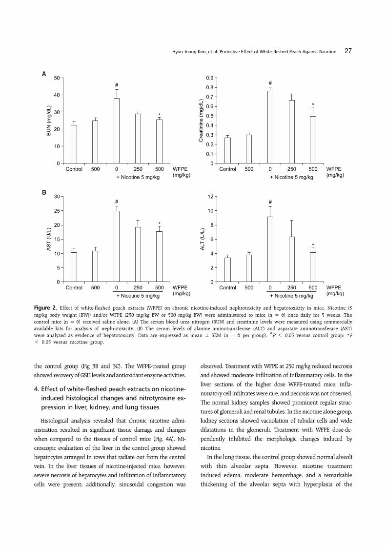
The protective effect of WFPE against chronic nicotine-in-
duced renal and hepatic toxicity was assessed by measuring
biochemical parameters in sera of the mice. We analyzed BUN and
creatinine levels for kidney function, and AST and ALT levels for
liver function. The mice treated with WFPE alone did not show
any kidney or liver dysfunction, but nicotine raised the levels of
all biochemical parameters in the mouse sera (Fig. 2). In contrast,
WFPE treatment dose-dependently lowered serum levels of BUN,
creatinine, AST, and ALT that were elevated by nicotine. In
particular, the serum BUN and ALT levels were recovered almost
to control levels at a WFPE dosage of 500 mg/kg.
3. Effect of white-fleshed peach extracts on oxidative damages in liver and kidney tissues of nicotine-in-jected mice
To investigate the effect of WFPE on chronic-induced oxidative
tissue damages, MDA, and GSH contents, as well as antioxidant
enzyme activities, were measured in the liver and kidney tissue
homogenates from all the mice. The level of MDA, which is a
major degradation product of lipid peroxidation, increased after
chronic nicotine treatment when compared to the control group
(Fig. 3A). However, administration of higher-dose WFPE to
nicotine-injected mice significantly reduced tissue MDA levels
and showed a more pronounced reduction effect in the liver than
the kidney.
The antioxidative status was assessed by measuring GSH
content and the activities of antioxidant enzymes in the liver and
kidney tissues. In the nicotine-injected mice, the level of GSH and
the activities of SOD, GPx, and CAT were significantly lower than
Hyun-Jeong Kim, et al: Protective Effect of White-fleshed Peach Against Nicotine 27
Figure 2. Effect of white-fleshed peach extracts (WFPE) on chronic nicotine-induced nephrotoxicity and hepatotoxicity in mice. Nicotine (5 mg/kg body weight [BW]) and/or WFPE (250 mg/kg BW or 500 mg/kg BW) were administered to mice (n = 6) once daily for 5 weeks. The control mice (n = 6) received saline alone. (A) The serum blood urea nitrogen (BUN) and creatinine levels were measured using commercially available kits for analysis of nephrotoxicity. (B) The serum levels of alanine aminotransferase (ALT) and aspartate aminotransferase (AST) were analyzed as evidence of hepatotoxicity. Data are expressed as mean ± SEM (n = 6 per group). #P < 0.05 versus control group; *P< 0.05 versus nicotine group.
the control group (Fig 3B and 3C). The WFPE-treated group
showed recovery of GSH levels and antioxidant enzyme activities.
4. Effect of white-fleshed peach extracts on nicotine- induced histological changes and nitrotyrosine ex-pression in liver, kidney, and lung tissues
Histological analysis revealed that chronic nicotine admi-
nistration resulted in significant tissue damage and changes
when compared to the tissues of control mice (Fig. 4A). Mi-
croscopic evaluation of the liver in the control group showed
hepatocytes arranged in rows that radiate out from the central
vein. In the liver tissues of nicotine-injected mice, however,
severe necrosis of hepatocytes and infiltration of inflammatory
cells were present; additionally, sinusoidal congestion was
observed. Treatment with WFPE at 250 mg/kg reduced necrosis
and showed moderate infiltration of inflammatory cells. In the
liver sections of the higher dose WFPE-treated mice, infla-
mmatory cell infiltrates were rare, and necrosis was not observed.
The normal kidney samples showed prominent regular struc-
tures of glomeruli and renal tubules. In the nicotine alone group,
kidney sections showed vacuolation of tubular cells and wide
dilatations in the glomeruli. Treatment with WFPE dose-de-
pendently inhibited the morphologic changes induced by
nicotine.
In the lung tissue, the control group showed normal alveoli
with thin alveolar septa. However, nicotine treatment
induced edema, moderate hemorrhage, and a remarkable
thickening of the alveolar septa with hyperplasia of the

28 Journal of Cancer Prevention Vol. 22, No. 1, 2017
Figure 3. Effect of white-fleshed peach extracts (WFPE) on chronic nicotine-induced oxidative stress in mouse tissues. After mice were adminis-trated nicotine (5 mg/kg body weight [BW]) and/or WFPE (250 mg/kg BW or 500 mg/kg BW) for 5 weeks, liver and kidney tissues were collectedand homogenized. (A) The level of malondialdehyde (MDA), as an indicator of lipid peroxidation, was measured in tissue homogenates. (B) Glutathione (GSH) content was evaluated by the reaction with Ellman’s reagent. (C) Among the enzymatic antioxidants, superoxide dismutase (SOD), glutathione peroxidase (GPx), and catalase (CAT) were analyzed by commercial available kits, respectively. Data are expressed as mean± SEM (n = 6 per group). #P < 0.05 versus control group; *P < 0.05, **P < 0.01 versus nicotine group.

Hyun-Jeong Kim, et al: Protective Effect of White-fleshed Peach Against Nicotine 29
Figure 4. Effect of white-fleshed peach extracts (WFPE) on histological changes and nitrotyrosine expression in the liver, kidney, and lung tissues of chronic nicotine-injected mice. The liver, kidney, and lung tissues from mice, which were administrated nicotine (5 mg/kg body weight [BW]) and/or WFPE (250 mg/kg BW or 500 mg/kg BW) for 5 weeks, were fixed and stained. (A) The morphological features of tissue sections were evaluated by Hematoxylin and eosin staining (×200). (B) Nitrotyrosine expressions were evaluated by immunohistochemistry (×200).
30 Journal of Cancer Prevention Vol. 22, No. 1, 2017
epithelial cells. WFPE treatment in nicotine-injected mice
reduced thickening of the alveolar septa and edema in a
dose-dependent manner.
We immunohistochemically analyzed the expression of nitro-
tyrosine in the liver, kidney, and lung tissues of the control mice
and nicotine-injected mice with or without WFPE treatment (Fig.
4B). Substantially enhanced levels of nitrotyrosine were observed
in three tissues of nicotine-injected mice. However, WFPE
treatment dose-dependently inhibited nicotine-induced nitro-
tyrosine formation.
DISCUSSION
Tobacco smoking was recognized as a major cause of mortality
and morbidity since environmental tobacco smoke was found to be
a human lung carcinogen by the US Environmental Protection
Agency in 1992. Over 4,000 chemicals have been identified in
tobacco smoke, including 69 known carcinogens and hundreds
that are hazardous.18 It has been reported that nicotine, a major
component of tobacco smoke and a highly addictive drug, plays an
important role in the development of cardiovascular and lung
diseases.5,19,20 As nicotine enters the human body through tobacco
smoking, it is efficiently absorbed into the bloodstream through
the lungs and rapidly delivered to the brain.21 Nicotine is also
absorbed through the mucosal lining of the mouth and nose, and
even through the skin. Nicotine is extensively metabolized to a
number of metabolites; cotinine, the primary metabolite of
nicotine, is formed after C-oxidation by hepatic cytochrome P450
(CYP2A6).22 Cotinine is further metabolized by the same enzyme
system as trans-3'-hydroxycotinine (hydroxycotinine) and other
minor metabolites including norcotinine.23 PAHs, including the
widely studied benzo[a]pyrene, are considered the main car-
cinogens of cigarette smoke. PAHs bind to aryl hydrocarbon
receptor after entering the body, and induce cytochrome P450
drug-metabolizing enzymes, such as CYP1A1, CYP1A2, CYP1Ba, and
CYP3A4, which metabolize PAHs into various PAH derivatives, such
as hydroxylated PAHs and PAH quinones.24 Urinary
1-hydroxypyrene, a monohydroxylated metabolite of pyrene, has
been widely used as a biomarker of total PAH uptake in smokers
and nonsmokers.25
The intake of fruits and vegetables, including flavonoids, is
closely related to the prevention and reduction in the risk of
chronic diseases, such as inflammation, various cancers, and
cardiovascular disorders. P. persica fruits have moderate effects
on blood circulation, recovery from exhaustion, detoxification,
reinforcement of immune ability, and cosmetic treatment of the
skin.26 However, the effect of P. persica fruits on the deto-
xification of smoking has not yet been reported. Thus, we firstly
investigated whether white-fleshed peaches have effects on the
excretion of nicotine metabolites and 1-hydroxypyrene in the
urine of smokers. Intake of white-fleshed peaches increased the
concentration of nicotine metabolites in the urine of 91.67% of
smokers. In addition, the concentration of urinary 1-hydro-
xypyrene, the major metabolite of PAH, increased in 83.33% of
smokers after intake of peach fruit. Thus, intake of
white-fleshed peaches increased the excretion of nicotine
metabolites and 1-hydroxypyrene in the smokers. Therefore,
intake of white-fleshed peaches and related processed foods
might be an effective method for the detoxification of nicotine
in smokers and nonsmokers who are exposed to environmental
tobacco smoke.
Nicotine, a major pharmacologically active component of
tobacco smoke, triggers an accumulation of free radicals or ROS,
and consequently causes oxidative stress, cytotoxicity, and tissue
damage. In our animal study, chronic nicotine exposure inhibited
a gain of body weight, but this weight loss was blocked in the mice
that were administered extracts of white-fleshed peach at 250
mg/kg or 500 mg/kg body weight. These results indicate that
chronic nicotine exposure eventually induces systemic toxicity,
and WFPE can suppress nicotine toxicity by being absorbed into
the bodies of animals.
The liver and kidney are highly susceptible to the oxidative
stress associated with the toxicity of nicotine. Our results showed
that chronic nicotine exposure caused hepatic and renal damage
associated with an accumulation of lipid peroxidation products.
However, WFPE treatment dose-dependently inhibited nephro-
toxicity and hepatotoxicity by reducing serum levels of BUN,
creatinine, AST, and ALT. A high-dose of WFPE did not affect the
functional index of the liver and kidney in normal mice, but did
reduce nicotine-increased levels of BUN and ALT to levels similar
to the control group. In addition, a marked increase in the MDA
level was identified in the liver and kidney homogenates of mice
chronically exposed to nicotine; oral administration of WFPE
blocked this increase. Furthermore, WFPE treatment blocked the
decrease of GSH content and inhibition of antioxidant enzymes
activities, including SOD, GPx, and CAT, in the liver and kidney
tissues of nicotine-injected mice. These results show that orally
administered WFPE attenuates oxidative stress by preventing
nicotine-induced reductions in GSH content and the activities of
antioxidant enzymes.
The lung, the primary site exposed to tobacco smoke, is highly
susceptible to free radical generation. Our histological data
Hyun-Jeong Kim, et al: Protective Effect of White-fleshed Peach Against Nicotine 31
showed nicotine-induced toxicity in the lung tissue, as well as in
the liver and kidney tissues, but organ damages caused by
nicotine were decreased by WFPE treatment. In immuno-
histochemistry, nitrotyrosine, a biomarker of oxidative stress,
was positively expressed in the liver, kidney, and lung tissues of
nicotine-injected mice, while its expression was dose-depen-
dently suppressed in the WFPE-treated group. Nitrotyrosine is
formed on tyrosine residues by toxic peroxynitrite derived from
nitric oxide (NO) and superoxide anion (O2−), which is elevated
in conditions of oxidative stress in many diseases, such as infla-
mmation, cytotoxicity, and cancer.27 Therefore, inhibition of
nicotine-induced nitrotyrosine formation by WFPE treatment
indicates a decrease of oxidative stress.
In conclusion, WFPE may mitigate nicotine toxicity by
promoting the excretion of nicotine metabolites. The
protective effect of WFPE against tissue damages by chronic
nicotine exposure, may be mediated through the inhibition of
oxidative stress by enhancing of antioxidant capacities. Thus,
white-fleshed peach may be a beneficial supplement for
smokers.
CONFLICTS OF INTEREST
No potential conflicts of interest were disclosed.
REFERENCES
1. Hecht SS. Tobacco carcinogens, their biomarkers and tobacco-in-duced cancer. Nat Rev Cancer 2003;3:733-44.
2. Bringuier PP, McCredie M, Sauter G, Bilous M, Stewart J, Mihatsch MJ, et al. Carcinomas of the renal pelvis associated with smoking and phenacetin abuse: p53 mutations and poly-morphism of carcinogen-metabolising enzymes. Int J Cancer 1998;79:531-6.
3. Joshi HN, Makaju R, Karmacharya A, Karmacharya RM, Shrestha B, Shrestha R, et al. Urinary bladder carcinoma: impact of smok-ing, age and its clinico-pathological spectrum. Kathmandu Univ Med J (KUMJ) 2013;11:292-5.
4. Xu Y, Qi Y, Zhang J, Lu Y, Song J, Dong B, et al. The impact of smoking on survival in renal cell carcinoma: a systematic review and meta-analysis. Tumour Biol 2014;35:6633-40.
5. Grando SA. Connections of nicotine to cancer. Nat Rev Cancer 2014;14:419-29.
6. Nakada T, Kiyotani K, Iwano S, Uno T, Yokohira M, Yamakawa K, et al. Lung tumorigenesis promoted by anti-apoptotic effects of cotinine, a nicotine metabolite through activation of PI3K/Akt pathway. J Toxicol Sci 2012;37:555-63.
7. Yuen ST, Gogo AR Jr, Luk IS, Cho CH, Ho JC, Loh TT. The effect of nicotine and its interaction with carbon tetrachloride in the rat liver. Pharmacol Toxicol 1995;77:225-30.
8. Tsutsumi K. Lipoprotein lipase and atherosclerosis. Curr Vasc Pharmacol 2003;1:11-7.
9. Latha MS, Vijayammal PL, Kurup PA. Effect of nicotine admin-istration on lipid metabolism in rats. Indian J Med Res 1993; 98:44-9.
10. Ashakumary L, Vijayammal PL. Additive effect of alcohol and nic-otine on lipid peroxidation and antioxidant defence mechanism in rats. J Appl Toxicol 1996;16:305-8.
11. Fukuda T, Ito H, Mukainaka T, Tokuda H, Nishino H, Yoshida T. Anti-tumor promoting effect of glycosides from Prunus persica seeds. Biol Pharm Bull 2003;26:271-3.
12. Shin TY, Park SB, Yoo JS, Kim IK, Lee HS, Kwon TK, et al. Anti-al-lergic inflammatory activity of the fruit of Prunus persica: role of calcium and NF-kappaB. Food Chem Toxicol 2010;48:2797-802.
13. Lee CK, Park KK, Hwang JK, Lee SK, Chung WY. The pericarp ex-tract of Prunus persica attenuates chemotherapy-induced acute nephrotoxicity and hepatotoxicity in mice. J Med Food 2008; 11:302-6.
14. Lee CK, Park KK, Hwang JK, Lee SK, Chung WY. Extract of Prunus persica flesh (PPFE) improves chemotherapeutic efficacy and pro-tects against nephrotoxicity in cisplatin-treated mice. Phytother Res 2009;23:999-1005.
15. Ohkawa H, Ohishi N, Yagi K. Assay for lipid peroxides in animal tissues by thiobarbituric acid reaction. Anal Biochem 1979;95: 351-8.
16. Sedlak J, Lindsay RH. Estimation of total, protein-bound, and nonprotein sulfhydryl groups in tissue with Ellman's reagent. Anal Biochem 1968;25:192-205.
17. Hecht SS, Carmella SG, Le KA, Murphy SE, Li YS, Le C, et al. Effects of reduced cigarette smoking on levels of 1-hydroxypyr-ene in urine. Cancer Epidemiol Biomarkers Prev 2004;13:834-42.
18. Hoffmann D, Hoffmann I, El-Bayoumy K. The less harmful ciga-rette: a controversial issue. a tribute to Ernst L. Wynder. Chem Res Toxicol 2001;14:767-90.
19. Heusch WL, Maneckjee R. Signalling pathways involved in nic-otine regulation of apoptosis of human lung cancer cells. Carcinogenesis 1998;19:551-6.
20. Balakumar P, Kaur J. Is nicotine a key player or spectator in the induction and progression of cardiovascular disorders? Pharma-col Res 2009;60:361-8.
21. Nakayama H. Nicotine metabolism in mammals. Drug Metabol Drug Interact 1988;6:95-122.
22. Nakajima M, Yamamoto T, Nunoya K, Yokoi T, Nagashima K, Inoue K, et al. Role of human cytochrome P4502A6 in C-oxida-tion of nicotine. Drug Metab Dispos 1996;24:1212-7.
23. Kyerematen GA, Vesell ES. Metabolism of nicotine. Drug Metab Rev 1991;23:3-41.
24. Bekki K, Toriba A, Tang N, Kameda T, Hayakawa K. Biological ef-fects of polycyclic aromatic hydrocarbon derivatives. J UOEH 2013;35:17-24.
25. Jacob P 3rd, Wilson M, Benowitz NL. Determination of phenolic metabolites of polycyclic aromatic hydrocarbons in human urine as their pentafluorobenzyl ether derivatives using liquid chroma-tography-tandem mass spectrometry. Anal Chem 2007;79:587-98.
26. Kono R, Okuno Y, Nakamura M, Inada K, Tokuda A, Yamashita M, et al. Peach (Prunus persica) extract inhibits angiotensin II-in-

32 Journal of Cancer Prevention Vol. 22, No. 1, 2017
duced signal transduction in vascular smooth muscle cells. Food Chem 2013;139:371-6.
27. Ischiropoulos H. Biological tyrosine nitration: a pathophysio-
logical function of nitric oxide and reactive oxygen species. Arch Biochem Biophys 1998;356:1-11.


![[Free Scores.com] Liszt Franz Hungarian Rhapsody No 2 3657](https://img.dokumen.tips/doc/110x75/55cf85bd550346484b90f235/free-scorescom-liszt-franz-hungarian-rhapsody-no-2-3657.jpg)