Embed Size (px)
Citation preview

Gut, 1964, 5, 27
Vitamin B12 deficiency in chronic gastritis1. J. WOOD', M. RALSTON', B. UNGAR, AND D. C. COWLING
From the Clinical Pathology Department of the Royal Melbourne Hospital,and the Clinical Research Unit of the Royal Melbourne Hospital and the
Walter and Eliza Hall Institute of Medical Research,Melbourne, Victoria, Australia
EDITORIAL SYNOPSIS This study covers a group of elderly persons who suffer from chronic atrophicgastritis and resulting vitamin B12 deficiency. Increasing weakness, loss of memory, and mentaldepression were common symptoms and flatulent dyspepsia was sometimes a troublesome recurringcomplaint. These symptoms were improved by treatment with vitamin B12. Even if untreated,persons in this group rarely develop severe pernicious anaemia.
The introduction of the flexible suction tube forgastric biopsy by members of our Unit (Wood,Doig, Motteram, and Hughes, 1949a) and itsextension for jejunal biopsy by Shiner (1956) hasenabled us to study chronic gastritis with specialreference to the part it may play in causing vitaminB12 deficiency. The clinical manifestations of chronicgastritis have been described elsewhere (Joske,Finckh, and Wood, 1955; Wood and Taft, 1958:Wood, Cowling, Ungar, and Gray, 1960).The introduction of methods for serum vitamin
B12 assay (Ross, 1952) and later the Schilling test forvitamin B12 absorption (Schilling, 1953) providedadditional knowledge of established perniciousanaemia. Subsequently Mollin and Ross (1953 and1954), Spray and Witts (1958), and Siurala, Eramaa,and Nyberg (1960) described the slow fall in theserum vitamin B12 level and its varying effects on thehealth of the patient.
Gastric biopsy has enabled us to make a conclusivediagnosis of chronic atrophic gastritis and to study agroup of cases for a period extending up to 14 years.In 1955 the case of a man aged 65 years, sufferingfrom chronic dyspepsia, achlorhydria, and gastricatrophy was reported: five years later he developedovert pernicious anaemia and subacute combineddegeneration of the cord (Robertson, Wood, andJoske, 1955).A study of 30 patients with chronic atrophic
gastritis is now reported, with special reference tothe clinical state, serum vitamin B12 levels, Schillingtests, and examination of the blood and central
'Working with the aid of a grant from the National Health and Med-ical Research Council of Australia
27
nervous system. A preliminary study of some ofthese patients was reported by Wood et al. in 1960,but Schilling tests were not performed as this testnecessitates the giving of a large dose of vitamin B12,thus interfering with the continuing study.
SELECTION OF PATIENTS
Those selected in the series were patients with achlor-hydria or hypochlorhydria in whom gastric biopsyrevealed moderate to severe gastritis, with partial tocomplete atrophy of the acid- and pepsinogen-secretingcells and varying degrees of cellular infiltration, pre-dominantly by plasma cells and lymphocytes.
In keeping with previous experience (Joske et al., 1955;Wood and Taft, 1958; Wood et al., 1960) the majority ofpatients suffered flatulent dyspepsia, but often this initself was not of sufficient severity to bring them to ourUnit. Thus many initially sought treatment for otherailments.
Patients were excluded from the series if they weresuffering from gastric or duodenal ulcer or cancer, aprevious gastric operation, or chronic alcoholism withhepatic cirrhosis (Cowling and Mackay, 1959).
METHODS
The method for serum vitamin B12 assay was that of Ross(1952) using Euglena gracilis Z stain and the medium ofHutner, Bach, and Ross (1956). The absorption andexcretion test was that of Schilling (1953) with minormodifications (Ungar and Cowling, 1962).
In the standard test meal 0 9 mg. of histamine acidphosphate was followed by gastric aspiration for 90minutes. In the majority of patients with low serumvitamin B,2 levels the maximum response test meal ofKay (1953) was used.
on 5 January 2019 by guest. Protected by copyright.
http://gut.bmj.com
/G
ut: first published as 10.1136/gut.5.1.27 on 1 February 1964. D
ownloaded from

8L J. Wood, M. Ralston, B. Ungar, and D. C. Cowling
Patients were considered to have 'achlorhydria' whenthe acidity of the gastric contents, as titrated againstN/10 sodium hydroxide using Topfer's reagent, was ofpH greater than 3-5 in all samples.
All patients were subjected to gastric biopsy with theflexible suction tube described by Wood et al. (1949a).Small bowel biopsies were performed by a modification ofthe Shiner tube (Shiner, 1956) to render it more flexible(Ralston, Wood, and Hughes, 1960) or by the Crosbycapsule (Crosby and Kugler, 1957).The erythrocyte count was performed with the E.E.L.
electronic cell counter and the haemoglobin by acolorimeter using oxyhaemoglobin.The autoimmune complement-fixation test and the
tanned red cell haemagglutination test were performed asdescribed by Mackay and Wood (1962) and Irvine,Davies, Delamore, and Williams (1962).
RESULTS
In the group of 30 cases of chronic atrophic gastritisthere were nine men and 21 women. The mean agewhen the diagnosis was first extablished by gastricbiopsy was 56 years (range 38 to 69 years). Themean duration of the study which followed was nineyears (range two to 14 years). Serial studies of the
Case No. Lowest B12 Level (lIlg.)
vitamin B12 level in the serum were made for a meanperiod of four years (range one to five years).
All of the 30 cases had achlorhydria to standardhistamine stimulation on occasion: 21 had itthroughout and, of the remainder, six had it at theend of the study. On no occasion was a level above25 units obtained. All of the seven cases with serumvitamin B12 levels less than 100 ,u,ug. per ml. hadachlorhydria to maximum histamine stimulation(Kay, 1953).
Gastric biopsy was performed on all of the 30patients, 28 being tested on two or more occasions,particularly at the beginning and end of the study(Table I). In 17 the atrophy of acid and pepsinogen-secreting cells was severe and in 13 it was moderate.In eight the atrophy became more pronouncedduring the study, in 19 it was unchanged, and in oneit was less pronounced (Figs. 1 to 4). Intestinalmetaplasia was often present when the atrophy waspronounced.The cellular infiltration of the tunica propria was
profuse in six cases and moderately intense in 14:over the years it became more pronounced in six, itwas unchanged in 14, and was less in eight. Itconsisted mostly of plasma cells and lymphocytes.
TABLE IFINDINGS IN THE 30 PATIENTS WITH GASTRITIS
Final Examination
R.B.C. (million per c.mm.) Lesions of Central Nervous System
20 Atrophic gastritis40 Atrophic gastritis
40 Atrophic gastritis50 Atrophic gastritis56 Gastric atrophy
80 Atrophic gastritis90 Atrophic gastritis100 Atrophic gastritis100 Atrophic gastritis100 Atrophic gastritis100 Atrophic gastritis110 Atrophic gastritis114 Atrophic gastritis130 Gastric atrophy130 Atrophic gastritis140 Atrophic gastritis140 Atrophic gastritis
140 Atrophic gastritis150 Atrophic gastritis
160 Atrophic gastritis170 Atrophic gastritis170 Atrophic gastritis180 Gastric atrophy180 Atrophic gastritis180 Atrophic gastritis210 Gastric atrophy210 Atrophic gastritis240 Atrophic gastritis240 Atrophic gastritis270 Atrophic gastritis
4-53-1
4-34.54.5
5-13.54-34-4403-93.93-64.95-24-04-0
3-74-5
4-74.53.94-04-24-44.54.34.54.54-9
NormalMinor subacute combined degeneration of thecordPeripheral neuritisSubacute combined degeneration of the cordMinor subacute combined degeneration of thecordNormalNormalNormalNormalNormalNormalNormalNormalNormalNormalNormalMinor subacute combined degeneration of thecordNormalMinor subacute combined degeneration of thecordPeripheral neuritisNormalNormalNormalNormalNormalPeripheral neuritisNormalNormalNormalPeripheral neuritis
Gastric Biopsy
2
345
67891011121314151617
1819
2021222324252627282930
28
on 5 January 2019 by guest. Protected by copyright.
http://gut.bmj.com
/G
ut: first published as 10.1136/gut.5.1.27 on 1 February 1964. D
ownloaded from

Vitamin B12 deficiency in chronic gastritis
FIG. 1. Case 3. Woman aged 58.Gastric biopsy by suction tube in1949 showing atrophic gastritis,there being moderate atrophyof the acid- andpepsinogen-secreting cells and intensecellular infiltration of the tunicapropria (higher magnification:plasma cells and lymphocytes).There is one large lymph folliclewith a germinal centre.Haematoxylin and eosin x 100.
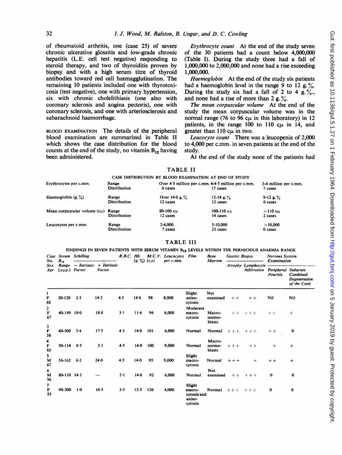
FIG. 2. Case 3 (same as Fig. 1).Gastric biopsy 13 years latershowing gross atrophy andpersisting cellular infiltration(plasma cells and lymphocytes).There were achlorhydria, serumlevel of vitamin B,2 reduced to40 pj,g., and an abnormalSchilling test, restored to normalby the addition of intrinsic factor.Haematoxylin and eosin x 100.
Polymorphonuclear cells were relatively sparse. Inthe normal gastric mucosa small lymphoid folliclesare present in the depth of the tunica propria, justabove the muscularis mucosae. In the present seriesthe lymph follicles were often enlarged and at thesesites the gastric glands were either absent or atrophic.
In four patients (cases 5, 14, 23, and 26) gastricatrophy (Motteram, 1951) was present at the endof the study, their lowest levels of serum vitamin B12being 56, 130, 180, and 210 ,tug. per ml.The lowest serum vitamin B12 levels and the range
in the 30 patients are shown in Figure 5. It will benoted that in seven cases the lowest level was in the'overt' or 'latent' pernicious anaemia range of lessthan 100 ,u,ig. per ml. (Wood et al., 1960), in 18patients it was in the 'pre-pernicious anaemia'range of 100 to 200 ,ug. per ml., and in five patientsit was in the normal range (200 to 700 ,u,g. per ml.in this laboratory).The levels fluctuated from month to month, but in
the majority of patients there was a downwardtrend. This pattern is shown in Figure 6, which
29
on 5 January 2019 by guest. Protected by copyright.
http://gut.bmj.com
/G
ut: first published as 10.1136/gut.5.1.27 on 1 February 1964. D
ownloaded from

I. J. Wood, M. Ralston, B. Ungar, and D. C. Cowling
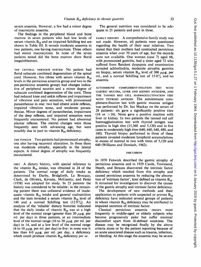
FIG. 3. Case 4. Woman aged 50with achlorhydria, serum vitaminB12 reduced to 50 p,.g.,abnormal Schilling test, hightitre of antibodies to gastricmucosa (16) and thyroid (5,120)and excess lymphoid accumulationin thyroid biopsy, and minorsubacute combined degenerationof the spinal cord. Gastric biopsyshows goblet cells, gross atrophy,and moderate cellular infiltration(lymphocytes and plasma cells).Haematoxylin and eosin x 90.
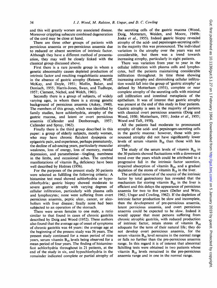
FIG. 4. Case 25. Woman aged 42with chronic hepatitis and chronicglossitis responding tocorticosteroids, and minorreduction ofserum vitamin B12level to 180 ,qug. Gastric biopsyshows gross atrophy andpronounced cellular infiltration(lymphocytes andplasma cells)with two enlarged lymph follicles,one having a germinal centre.Haematoxylin and eosin x 110.
records the progressive levels in six patients withserum vitamin B12 levels in the pernicious anaemiarange.
All patients had severe or moderate gastricatrophy, but the degree of atrophy did not correlatewith the serum vitamin B12 levels. Those with severegastric atrophy had a mean serum vitamin B12 levelof 136 ,tug. per ml. and those with moderate atrophy135 ,uqsg. per ml.The results of the Schilling test, performed without
the addition of intrinsic factor, are also shown in
Figure 5. It will be noted that the percentage ofabsorption and excretion of labelled vitamin B12 wasin the pernicious anaemia range of under 10% in sixof the seven patients whose serum vitamin B12 levelfell within the pernicious anaemia range (under 100,u,ug. per ml.), in only two of the 17 patients withinthe pre-pernicious anaemia range (100 to 200 ,u,g.per ml.), and in one of the four patients within thenormal range (over 200 ,ug. per ml.).
In nine of the patients in whom the Schilling testshowed impaired absorption of vitamin B12 the
30
on 5 January 2019 by guest. Protected by copyright.
http://gut.bmj.com
/G
ut: first published as 10.1136/gut.5.1.27 on 1 February 1964. D
ownloaded from

Vitaninl B12 deficiency in chronic gastritis
CASE NO.>400
3nr 350-1
1^1 X 300
1" 250-m
i i 200-IDoz
O 150-oa<-O > 100-
zO 00 50
La
PA. Range Pre - pernicdus Anoemia j Normal_ _-- - --I1 3 5
.II I
10 is 20 25
ij L
30
- o%D BoI! v
FIG. 5. The lowest level and range of serum vitamin B12in 30 patients with chronic atrophic gastritis: seven werein the pernicious anaemia range. 18 in the pre-perniciousanaemia range, and five in the normal range. The corre-sponding results of Schilling tests (without intrinsic factor)are shown in the lower panel: impaired absorption wasshown in six of the seven patients whose serum vitamin B12level was in the pernicious anaemia range.
300
200
100
0300
200
100
0300
200
100
0
MONTHSFIG. 6. Serial serum vitamin B12 levels in cases 1, 2, 3, 4,5, and 7 (panels 3, 5, 4, 1, 6 and 2 respectively). All hadlow levels, one or more being in the pernicious anaemiarange (hatched).
test was repeated with the addition of intrinsicfactor to determine whether the low level was due todeficient intrinsic factor being produced by thestomach. The results (Fig. 7) show that in eight ofthe nine patients tested the addition of intrinsicfactor produced a rise to the normal range. Theremaining patient (case 4). shown in panel I of Figure6, had achlorhydria to maximum stimulation, severeatrophic gastritis to biopsy, and biopsy of the upperjejunum and lower small bowel revealed well-formedvilli without excess lymphoid infiltration. However,she suffered occasional attacks of diarrhoea and fatabsorption was moderately impaired over a three-day period.
SYMPTOMS OF B12 DEFICIENCY The 30 patients wereelderly and the majority did not complain of majordistressing or debilitating symptoms which coulddefinitely be attributed to vitamin B12 deficiency.However, increasing weakness, loss of memory, andmental depression were frequently recorded, togetherwith the flatulent dyspepsia which is a feature ofchronic gastritis. Five patients had paraesthesiae inthe hands and feet.
I
-a:
ow3
1-
4 3'
Z0
C M111
Ii Jw
- I
-i00
002 3 4 S 6 7 8 9
0 - 9PERNICIOUS ANAEMIA RANGE50300
2010
PA.cou RANGEMI A
50lL__30 __
o WITH INTRINSIC FACTOR* WITHOUT INTRINSIC FACTOR
FIG. 7. The results of Schilling tests performed withoutand with the addition ofintrinsicfactor in cases 1, 2, 3, 4, 5,7, 20, 24, and 29 (panels 3, 8, 4, 1, 5, 2, 6, 9, and 7 respec-tively). There was evidence of deficiency of intrinsicfactor except in case 4 (panel 1), a patient who also hadintestinal malabsorption.
SYMPTOMS OF GASTRITIS AND ASSOCIATED DISEASES Ofthe 30 patients, 13 first reported because of dyspepsia,having periodic attacks varying from mild tomoderate epigastric discomfort with flatulence topain of moderate severity. Fifteen had associateddiseases: five of these could possibly be related to anautoimmune disease process, there being three cases
fl
n
I
a
i
v4J-
IA.0-ihli
-i
1 2
a
icious ANAEMIA RANG----3
5 6
-- -- A. 1-1
31
u
.0
Li
q19
iIt
Nc14cIc
Fc
w -
0 30 4010 0 GO 20 so.0%
on 5 January 2019 by guest. Protected by copyright.
http://gut.bmj.com
/G
ut: first published as 10.1136/gut.5.1.27 on 1 February 1964. D
ownloaded from
L. J. Wood, M. Ralston, B. Ungar, and D. C. Cowling
of rheumatoid arthritis, one (case 25) of severechronic ulcerative glossitis and low-grade chronichepatitis (L.E. cell test negative) responding tosteroid therapy, and two of thyroiditis proven bybiopsy and with a high serum titre of thyroidantibodies toward red cell haemagglutination. Theremaining 10 patients included one with thyrotoxi-cosis (test negative), one with primary hypertension,six with chronic cholelithiasis (one also withcoronary sclerosis and angina pectoris), one withcoronary sclerosis, and one with arteriosclerosis andsubarachnoid haemorrhage.
BLOOD EXAMINATION The details of the peripheralblood examination are summarized in Table IIwhich shows the case distribution for the bloodcounts at the end of the study, no vitamin B12 havingbeen administered.
Erythrocyte count At the end of the study sevenof the 30 patients had a count below 4,000,000(Table I). During the study three had a fall of1,000,000 to 2,000,000 and none had a rise exceeding1,000,000.Haemoglobin At the end of the study six patients
had a haemoglobin level in the range 9 to 12 g. %.During the study six had a fall of 2 to 4 g.%.,and none had a rise of more than 2 g. %.
The mean corpuscular volume At the end of thestudy the mean corpuscular volume was in thenormal range (76 to 96 c,t in this laboratory) in 12patients, in the range 100 to 110 c,t in 14, andgreater than 110 c,u in two.Leucocyte count There was a leucopenia of 2,000
to 4,000 per c.mm. in seven patients at the end of thestudy.At the end of the study none of the patients had
Erythrocytes per c.mm.
Haemoglobin (g. %)
TABLE IICASE DISTRIBUTION BY BLOOD EXAMINATION AT END OF STUDY
Range Over 4-5 million per c.mm. 4-4 5 million per c.mm.Distribution 6 cases 17 cases
RangeDistribution
Mean corpuscular volume (c,.) RangeDistribution
Leucocytes per c.mm. RangeDistribution
Over 14 0 g.%12 cases
80-100 c,u12 cases
2-4,0007 cases
12-14 g.%12 cases
100-110 c.U14 cases
5-10,00023 cases
3-4 million per c.mm.7 cases
9-12 g.%6 cases
>1 10 cs2 cases
>10,0000 cases
TABLE IIIFINDINGS IN SEVEN PATIENTS WITH SERUM VITAMIN B12 LEVELS WITHIN THE PERNICIOUS ANAEMIA RANGE
Case Serum Schilling R.B.C. Hb M.C. V. Leucocytes Film Bone Gastric Biopsy Nervous SystemNo. B,, (g. %) (cIL) per c.mm. Marrow ExaminationSex Range - Intrinsic + Intrinsic Atrophy LymphocyteAge (lsg g.) Factor Factor Infiltration Peripheral Subacute
Neuritis CombinedDegenerationof the Cord
F682F67
20-120 2-3
40-149 10-0
3F 40-300 5.4584F 50-114 0560SM 56-162 6-2676M
567F55
80-110 14-3
90-300 1-0
Slight Not14-2 4 5 14-6 98 8,000 aniso- examined
cytosisModerate
18-8 31 11-6 96 6,000 macro- Macro-cytosis normo-
blasts
4+ 1-4
A--F +1-4
17-5 4-3 14-0 101 6,000 Normal Normal + + + + + +
Macro-3-1 4-5 14-0 100 9,000 Normal normo- + + + + +
blastsSlight
24-0 4-5 14-0 95 9,000 macro- Normal + + + +cytosis
Not- 5-1 14-6 92 6,000 Normal examined + + ++ ±
Slight16-5 3-5 12-5 126 4,000 macro- Normal + + + ++
cytosis andaniso-cytosis
32
Nil Nil
++ +
++ 0
+ +
++ +
0 0
0 0
on 5 January 2019 by guest. Protected by copyright.
http://gut.bmj.com
/G
ut: first published as 10.1136/gut.5.1.27 on 1 February 1964. D
ownloaded from
Vitamin B12 deficiency in chronic gastritis
severe anaemia. However, a few had a minor degreeof macrocytic anaemia.The findings in the peripheral blood and bone
marrow in seven patients who had low levels ofserum vitamin B12 and an impaired Schilling test areshown in Table III. It reveals moderate anaemia intwo patients, one having macrocytosis. Three othershad minor macrocytosis. In none of the sevenpatients tested did the bone marrow show floridmegaloblastosis.
THE CENTRAL NERVOUS SYSTEM No patient hadflorid subacute combined degeneration of the spinalcord. However, five (three with serum vitamin B12levels in the pernicious anaemia group and two in thepre-pernicious anaemia group) had changes indica-tive of peripheral neuritis and a minor degree ofsubacute combined degeneration of the cord. Threehad reduced knee and ankle reflexes and impairmentof vibration and pain sensation, with pronouncedparaesthesiae in one: two had absent ankle reflexes,impaired vibration sense, and moderate paraes-thesiae. Minor degrees of paraesthesiae, impairmentof the deep reflexes, and impaired sensation werefrequently encountered. No patient had abnormalplantar reflexes. The mental changes were thosefrequently seen with advancing age, but werepossibly due in part to vitamin B12 deficiency.
THE TONGUE Two patients had pronounced atrophy,one also having recurrent ulceration. In three therewas moderate atrophy, especially in the lateralaspects. A minor degree of atrophy was frequentlyencountered.
DIET A dietary history, with special reference tothe vitamin B12 intake, was obtained in 24 of thepatients. The normal range of daily intake asdetermined by Darby, Bridgforth, Le Brocquy,Clark, de Oliveira, Kevany, McGanity, and Perez(1958) was adopted for study. In 23 patients thehistory was considered to be reliable: in the remain-ing patient there was collateral evidence of inade-quate vitamin B12 intake and general malnutritionand the tests revealed a serum vitamin B12 level of140 and a normal Schilling test (12-9y%). Ananalysis of the 'reliable' dietary histories indicatedthat the daily intake of vitamin B12 was at a highlevel of the normal range (greater than 20 ,u,ug. perml. per day) in three patients, at an intermediatelevel of the normal range (10 to 20 ,uug. per ml. perday) in 14, and at a low level of the normal range(6 to 10 ,utpg. per ml. per day) in five: in none was itless than 6-0 ,u,ug. per ml. per day, a deficiencywhich could produce vitamin B12 deficiency per se.
The general nutrition was considered to be ade-quate in 21 patients and poor in three.
FAMILY HISTORY A comprehensive family study wasnot made. However, all patients were questionedregarding the health of their near relatives. Twostated that their mothers had contracted perniciousanaemia when over 70 years of age, but the recordswere not available. One woman (case 7) aged 66,with pronounced gastritis, had a sister aged 72 whosuffered from flatulent dyspepsia and examinationrevealed achlorhydria, moderate atrophic gastritison biopsy, serum vitamin B12 level of 300 ,u,ug. perml., and a normal Schilling test of 15.0% and noanaemia.
AUTOIMMUNE COMPLEMENT-FIXATION TEST WITHGASTRIC MUCOSA, LIVER AND KIDNEY ANTIGENS, ANDTHE TANNED RED CELL HAEMAGGLUTINATION TESTWITH THYROID ANTIGEN The autoimmune com-plement-fixation test with gastric mucosa antigenwas performed by Dr. Ian Mackay on the serum of28 patients: six gave a significantly positive titre(16 or >16). None gave a positive reaction withliver or kidney. In two patients the tanned red cellhaemagglutination test with thyroid antigen waspositive in high titre (15,360 and 5,120) and in fivecases in moderately high titre (640, 640, 640, 480, and160). Thyroid biopsy performed in three of thesepatients revealed moderate lymphoid accumulationsin excess of normal in two with titres of 5,120 and640 (Williams and Doniach, 1962).
DISCUSSION
In 1870 Fenwick described the gastric atrophy ofpernicious anaemia and in 1929 Castle, Townsend,Heath, and Strauss discovered the intrinsic factordeficiency which resulted from this atrophy andcaused pernicious anaemia by reducing the absorp-tion of 'extrinsic factor', later defined as vitamin B12.It remained for investigators to discover the causesof the gastric atrophy and intrinsic factor deficiency.The development of new methods and their
application to patients with suspected or proven B12deficiency have indicated several groups of patientsin whom vitamin B12 deficiency may be attributed toimpaired secretion of intrinsic factor.
Classical pernicious anaemia occurs mostfrequently in middle-aged or elderly subjects whobecome progressively paler but suffer minimaldiscomfort apart from ill-defined weakness. Thedisease may be recognized finally by the abovecriteria alone or by the patient reporting because ofan acute associated disease such as trauma, infection,or bleeding. At this stage the anaemia may be severe
33
on 5 January 2019 by guest. Protected by copyright.
http://gut.bmj.com
/G
ut: first published as 10.1136/gut.5.1.27 on 1 February 1964. D
ownloaded from
3. J. Wood, M. Ralston, B. Ungar, and D. C. Cowling
and this will greatly worsen any associated disease.Moreover crippling subacute combined degenerationof the cord may be close at hand.There are three other groups of patients with
pernicious anaemia or pre-pernicious anaemia dueto reduced or absent secretion of intrinsic factor.Although they have a different method of presenta-ation, they may well be closely linked with theclassical group discussed above.
First there is a rare juvenile group in whom agenetic abnormality causes depleted production ofintrinsic factor and resulting megaloblastic anaemiain the absence of gastric atrophy (Reisner, Wolff,McKay, and Doyle, 1951; Mollin, Baker, andDoniach, 1955; Harris-Jones, Swan, and Tudhope,1957; Clement, Nichol, and Welch, 1961).Secondly there is a group of subjects, of widely
varying ages, in whom there is a strong geneticbackground of pernicious anaemia (Askey, 1940).The members of this group, which was identified byfamily studies, have achlorhydria, atrophy of thegastric mucosa, and latent or overt perniciousanaemia (Callender and Denborough, 1957;Callender and Spray, 1962).
Finally there is the third group described in thispaper: a group of elderly subjects, mostly women,who may have chronic flatulent dyspepsia orepigastric pain, and experience, perhaps prematurely,the decline of advancing years, particularly muscularweakness, loss of energy, loss of memory, mentaldepression, and paraesthesiae-tingling, numbnessin the limbs, and occasional aches. The cerebralmanifestations of vitamin B12 deficiency have beenwell described by Holmes (1956).For the purposes of the present study 30 patients
were selected as fulfilling the following criteria: Ahistamine test meal showed achlorhydria or hypo-chlorhydria; gastric biopsy showed moderate tosevere gastric atrophy with varying degrees ofcellular infiltration, particularly with plasma cellsand lymphocytes; none were suffering from overtpernicious anaemia, peptic ulcei-, cancer, or alco-holism with liver disease; finally none had beensubjected to an operation of the stomach.There were seven females to one male, a ratio
similar to that found in cases of chronic gastritisdescribed by Doig and Wood (1952). These authorsalso found that the average age of onset of symptomsof chronic gastritis was 44 years: the average age atthe beginning of the presenc study was 56 years. Thepresent study continued for a mean period of nineyears, serum vitamin B12 levels being observed for amean period of four years. The finding of histamine-fast achlorhydria throughout in 21 patients, at theend of the study in six, and hypochlorhydria in theremainder indicated complete or partial atrophy of
the secreting cells of the gastric mucosa (Wood,Doig, Motteram, Weiden, and Moore, l 949b;Joske et al., 1955). Indeed gastric biopsy revealedatrophy of the acid- and pepsinogen-secreting cells:in the majority this was pronounced. The individualvariation in the atrophy over the years was notconsiderable, but there was a trend towardsincreasing atrophy, particularly in eight patients.There was variation from year to year in the
cellular infiltration with plasma cells and lympho-cytes, half the patients maintaining considerableinfiltration throughout. In time those showingincreasing atrophy and diminishing cellular infiltra-tion would fall into the group of 'gastric atrophy' asdefined by Motterham (1951). complete or nearcomplete atrophy of the secreting cells with minimalcell infiltration and intestinal metaplasia of theepithelium. It was of interest that gastric atrophywas present at the end of this study in four patients.Gastric atrophy is seen in the majority of patientswith classical overt pernicious anaemia (Doig andWood, 1950; Motterham, 1951; Joske et al., 1955;Wood and Taft, 1958).
All the patients had moderate to pronouncedatrophy of the acid- and pepsinogen-secreting cellsin the gastric mucosa: however, those with pro-nounced atrophy did not have significantly lowerlevels of serum vitamin B12 than those with lessatrophy.The study of the serum levels of vitamin B12 in
the 30 patients showed fluctuations with a downwardtrend over the years which could be attributed to aprogressive fall in the intrinsic factor secretion,impaired absorption of vitamin B12, and a gradualdepletion of the stores of vitamin B12 in the liver.The artificial removal of the source of the intrinsic
factor by total gastrectomy has revealed that themechanism for storing vitamin B12 in the liver isefficient and this delays the appearance of perniciousanaemia for two to five years (Deller and Witts,1962; Ungar and Cowling, 1962). If the depletion ofintrinsic factor production be slow and incomplete,then the development of pre-pernicious anaemia,latent pernicious anaemia, and overt perniciousanaemia could be expected to be slow. Indeed itwould appear that most persons suffering fromchronic atrophic gastritis, with reduced productionof intrinsic factor, retain stores of vitamin B12adequate for the term of their natural life; they donot develop overt pernicious anaemia, for theserum vitamin B12 level remains in the normal rangeor falls no further than the pre-pernicious anaemiarange. In this regard it is of interest that abnormalSchilling tests were obtained in two patients whosevitamin B12 levels remained in the pre-perniciousanaemia range and in one in the normal range.
34
on 5 January 2019 by guest. Protected by copyright.
http://gut.bmj.com
/G
ut: first published as 10.1136/gut.5.1.27 on 1 February 1964. D
ownloaded from

Vitamin B12 deficiency in c/hronic gastritis
In the present study seven patients had vitamin B12levels in the pernicious anaemia range, 18 in thepre-pernicious anaemia range, and five in thenormal range. Although none of the seven in thepernicious anaemia range developed florid perniciousanaemia or subacute combined degeneration of thecord, five had findings indicative of vitamin B12deficiency, including mild macrocytic anaemia,macronormoblasts in the bone marrow, andparaesthesiae or other changes in the centralnervous system probably due to vitamin B12deficiency.The reason why pronounced vitamin B12 deficiency
may be present for many months without overtchanges in the peripheral blood or frank megalo-blastic changes in the bone marrow has beenascribed to adequate folic acid intake or lack ofsome precipitating factor (Mollin and Ross, 1953and 1954; Harrison, Booth, and Mollin, 1956;Callender and Spray, 1962).
Since Reizenstein (1959) found that the calculatedbiliary excretion of the radioactive label of cobalt-labelled (Co56 or Co58) vitamin B12 in humansubjects was greater than the daily faecal excretion,he postulated the existence of an enterohepaticcirculation of vitamin B12. Thus reabsorption in theintestine of vitamin B12 from the bile could partlyaccount for the apparently prolonged storage ofvitamin B12 in the liver. The intestinal reabsorptionof biliary vitamin B12 in gastrectomized rats isdependent on intrinsic factor (Grasbeck, Runeberg,and Simons, 1959), so that interruption of theenterohepatic circulation of vitamin B12 mightaggravate vitamin B12 deficiency in patients in whomproduction of intrinsic factor is impaired. Thispossibility was not investigated in the present study.That this vitamin B12 deficiency was due, at least
in part, to depleted production of intrinsic factorwas shown by the Schilling tests. The test wasabnormal in six of the seven patients with serumvitamin B12 levels in the pernicious anaemia range,in two of the 17 with levels in the pre-perniciousanaemia range, and in one of four with levels in thenormal range. Moreover, when the Schilling test wasrepeated with intrinsic factor in nine patients who hadabnormal Schilling tests without intrinsic factor,there was a rise to the normal range in eight, thusshowing that deficient secretion of intrinsic factorwas the cause, at least in part, of the vitamin B12deficiency. The remaining pdtient gave evidence ofintestinal malabsorption.
It may well be that diet plays a part in this group(Deller, Germar, and Witts, 1961): minor degrees ofdeficient secretion of intrinsic factor may require avitamin B12 intake in the food well above the lowlevel of the normal range quoted by Darby and his
colleagues (1958). A dietary history, considered to bereliable, was obtained in 23 of the patients, and theintake of vitamin B12 was assessed as being withinthe normal limits in all. However, in five patients itwas estimated as being in the range 6 to 10 ,u,ug. perml., a low level in the normal range. Two of thesepatients had levels of serum vitamin B12 in thepernicious anaemia range.
Genetic factors as a cause of gastritis were notexplored by family studies. However, familyhistories were obtained from all patients and thesedid not provide good evidence for a genetic factor:two stated that there had been possible perniciousanaemia in an elderly parent and one had a sisterwith proven chronic gastritis, but no perniciousanaemia.The cause of the atrophic gastritis is unknown.
However it is of interest that, of the 30 patientsstudied, five suffered from a disease which couldpossibly be caused by an autoimmune process: twohad chronic thyroiditis shown by biopsy and hightitre of thyroid antibodies; one had a moderatelyhigh titre of thyroid antibodies but normal thyroidbiopsy, low-grade hepatitis, and chronic ulcerativeglossitis responding to steroid therapy but not tovitamin B12 therapy; and three had rheumatoidarthritis.
In view of the recent finding by Irvine et al. (1962)and by Taylor, Roitt, Doniach, Couchman, andShapland (1962) that in cases of classical perniciousanaemia there is a high percentage with highantibody titre to gastric mucosa determined by theautoimmune complement-fixation test and to thyroidby the tanned red cell haemagglutination test, it is ofinterest that Dr. Ian Mackay of our Unit found thatsix of our patients (28 tested) had antibodies togastric mucosa, seven had antibodies to thyroid, andnone had antibodies to liver or kidney.
These findings and those of Irvine and hiscolleagues (1962) call for a wider investigation of thepart which may be played by autoimmune diseasein the production of atrophic gastritis. Dr. Mackayreports his methods and findings on page 23.The present study supports the view previously
held by the authors (Wood et al., 1960) and byothers (Siurala et al., 1960) that there is a group ofelderly subjects who should be considered to bepossible candidates for minor vitamin B12 deficiencybut who rarely develop classical pernicious anaemia.The members of this group are elderly and womenpredominate. Many have suffered from flatulentdyspepsia for many years and then, after the age of50, they may experience minor symptoms of vitaminB12 deficiency, particularly minor macrocyticanaemia, paraesthesiae, and mental and physicalweakness. A poor diet and a strong family history of
35
on 5 January 2019 by guest. Protected by copyright.
http://gut.bmj.com
/G
ut: first published as 10.1136/gut.5.1.27 on 1 February 1964. D
ownloaded from

36 I. J. Wood, M. Ralston, B. Ungar, and D. C. Cowling
pernicious anaemia are not necessary accompani-ments. Autoimmune disease of the gastric mucosa isa possible cause in some and may be associated withsimilar lesions elsewhere in the body.When a patient is suspected of being in this
elderly group with pre-pernicious anaemia or latentpernicious anaemia, a serum vitamin B12 assay is themost informative test. However, the present bio-logical assay is difficult to perform, time consuming,and expensive. It remains for investigators toprovide a less elaborate method of assay, readilyavailable to physicians. This would ensure moreefficient diagnosis and more rational therapy.Perhaps some of the mental and physical burdens ofadvancing age would be removed, much to thedelight of the patient.
SUMMARY
Twenty-one women and nine men (mean age 56years) with chronic atrophic gastritis on gastricbiopsy and depleted acid secretion were studied overa mean period of nine years, with special reference tothe progress of the gastritis, serum vitamin B12 levels,and abnormalities in the blood and nervous system.
Serial gastric biopsies showed that the atrophy ofacid and pepsinogen-secreting cells remained un-changed or increased, and infiltration of the tunicapropria with plasma cells and lymphocytes persisted.The serum vitamin B12 levels fluctuated, but in the
majority there was a downward trend. Analysis ofthe lowest levels showed that seven patients were inthe 'pernicious anaemia' range, 18 in the 'pre-pernicious anaemia' range, and five in the normalrange. The Schilling test proved varying degrees ofimpaired absorption of vitamin B12, particularly inthe first group.
There were varying changes in the central nervoussystem which were difficult to separate from theaging process. No patient developed severe subacutecombined degeneration of the spinal cord.No patient developed classical pernicious anaemia
as shown by peripheral blood and bone marrowexaminations. Minor degrees of anaemia andmacrocytosis were encountered.
Serological tests showed antibodies to gastricmucosa in six patients and to thyroid in seven.The study emphasizes that there is a group of
elderly persons who suffer chronic atrophic gastritisand resulting vitamin B12 deficiency. This causesdistressing symptoms which could be relieved bytreatment with vitamin B12. Even if untreated,persons in this group rarely develop severe perniciousanaemia or subacute combined degeneration of thecord.
Our sincere thanks are due to Dr. S. Weiden for thebiochemical tests, Dr. Ian Mackay for the serologicalstudies, Sister I. Langford for her skill with the patients,Mrs. A. Krupinska for the vitamin B12 estimations, andMiss G. Jessep for the nutrition survey. We are deeplygrateful for the help given by Dr. E. G. Robertson andother members of the medical staff of the RoyalMelbourne Hospital. Mr. E. Matthaei of the Universityof Melbourne prepared the photomicrographs.
REFERENCES
Askey, J. M. (1940). Prevention of pernicious anemia: recognitionof the latent stage in relatives. Ann. intern. Med., 14, 593-607.
Callender, S. T., and Denborough, M. A. (1957). A family study ofpernicious anaemia. Brit. J. Haemat., 3, 88-106.and Spray, G. H. (1962). Latent pernicious anaemia. Ibid., 8, 230-240.
Castle, W. B., Townsend, W. C., Heath, C. W., and Strauss, M. B.(1929). Observations on the etiological relationship of achyliagastrica to pernicious anemia. Part 1. Amer. J. med. Sci., 178,748-764.
Clement, D. H., Nichol, C. A., and Welch, A. D. (1961). A case ofjuvenile pernicious anemia: study of the effects of folic acidand vitamin Bl,. Blood, 17, 618-631.
Cowling, D. C., and Mackay, I. R. (1959). Serum vitamin B12 levels inliver disease. Med. J. Aust., 2, 558-562.
Crosby, W. H., and Kugler, H. W. (1957). Intraluminal biopsy of thesmall intestine. Amer. J. dig. Dis., 2, 236-241.
Darby, W. J., Bridgforth, E. B., Le Brocquy, J., Clark, S. L., Jr., deOliveira, J. D., Kevany, J., McGanity, W. J., and Perez, C.(1958). Vitamin B12 requirement of adult man. Amer. J. Med.,25, 726-732.
Deller, D. J., Germar, H., and Witts, L. J. (1961). Effect of food onabsorption of radioactive vitamin B12. Lancet, 1, 574-577.and Witts, L. J. (1962). Changes in the blood after partialgastrectomy with special reference to vitamin B12. Part 1. Quart.J. Med., 31, 71-78.
Doig, R. K., and Wood, I. J. (1950). Gastric biopsy in perniciousanaemia. Med. J. Aust., 2, 565-568.
-, (1952). Gastritis: a study of 112 cases diagnosed bygastric biopsy. Ibid., 1, 593-600.
Fenwick, S. (1870). On atrophy of the stomach. Lancet, 2, 78-80.Grasbeck, R., Runeberg, L., and Simons, K. (1959). Intrinsic factor
and radiovitamin B,, excretion in rats. Acta physiol. scand., 47,370-374.
Harris-Jones, J. N., Swan, H. T., and Tudhope, G. R. (1957).Pernicious anemia without gastric atrophy and in the presenceof free hydrochloric acid; report of a case. Blood, 12, 461-468.
Harrison, R. J., Booth, C. C., and Mollin, D. L. (1956). Vitamin B,,deficiency due to defective diet. Lancet. 1, 727-728.
Holmes, J. M. (1956). Cerebral manifestations of Vitamin Bl,deficiency. Brit. med. J., 2, 1394-1398.
Hutner, S. H., Bach, M. K., and Ross, G. I. M. (1956). A sugar-containing basal medium for vitamin B,2-assay with Euglena:application to body fluids. J. Protozool., 3, 101-112.
Irvine, W. J., Davies, S. H., Delamore, I. W., and Williams, A. W.(1962). Immunological relationship between perniciousanaemia and thyroid disease. Brit. med. J., 2, 454-456.
Joske, R. A., Finckh, E. S., and Wood, I. J. (1955). Gastric biopsy: astudy of 1,000 consecutive succcessful gastric biopsies. Quart. J.Med., 24, 269-294.
Kay, A. W. (1953). Effect of large doses of histamine on gastricsecretion of HCI: an augmented histamine test. Brit. med. J.,2, 77-80.
Mackay, I. R. (1964). Autoimmune serological studies in chronicgastritis and pernicious anaemia. Gut, 5, 23.and Wood, I. J. (1962). Lupoid hepatitis: a comparison of 22cases with other types of chronic liver disease. Quart. J. Med.,31, 485-507.
Mollin, D. L., Baker, S. J., and Doniach, I. (1955). Addisonianpernicious anaemia without gastric atrophy in a young manBrit. J. Haemat., 1, 278-290.
-, and Ross, G. I. M. (1953). Serum vitamin B,z concen-trations in patients with megaloblastic anaemia after treatmentwith vitamin B,,, folic acid, or folinic acid. Brit. med. J., 2, 640-645.
on 5 January 2019 by guest. Protected by copyright.
http://gut.bmj.com
/G
ut: first published as 10.1136/gut.5.1.27 on 1 February 1964. D
ownloaded from

Vitamin B12 deficiency in chronic gastritis 37
Mollin, ID. L., and Ross, G. I. M. (1954). Vitamin B,2 deficiency in themegaloblastic anaemias. Proc. roy. Soc. Med., 47, 428-431.
Motteram, R. (1951). A biopsy study of chronic gastritis and gastricatrophy. J. Path. Bact., 63, 389-394.
Ralston, M.. Wood, I. J., and Hughes, A. (1960). Small-bowel biopsywith the suction biopsy tube. Aust. Ann. Med., 9, 103-110.
Reisner, E. H., Jr., Wolf, J. A., McKay, R. J., Jr., and Doyle, E. F.(1951). Juvenile pernicious anemia. Pediatrics, 8, 88-106.
Reizenstein, P. G. (1959). Excretion, enterohepatic circulation, arnd re-tention of radiovitamin B,2 in pernicious anemia and incontrols. Proc. Soc. exp. Biol. (N. Y.), 101, 703-707.
Robertson, E. G., Wood, I. J., and Joske, R. A. (1955). Gastricatrophy with subsequent pernicious anaemia and pronouncedsubacute combined degeneration of the cord. Lancet, 2, 69-71.
Ross, G. I. M. (1952). Vitamin B1, assay in body fluids using Euglenagracilis. J. clin. Path., 5, 250-256.
Schilling, R. F. (1953). Intrinsic factor studies. It. The effect of gastricjuice on the urinary excretion of radioactivity after the oraladministration of radioactive vitamin B12. J. Lab. clin. Med.,42, 860-866.
Shiner, M. (1956). Jejunal-biopsy tube. Lancet, 1, 85.Siurala, M., Eramaa, E., and Nyberg, W. (1960). Pernicious anemia
and atrophic gastritis. Acta. med. scand., 166, 213-223.
Spray, G. H., and Witts, L. J. (1958). Results of three years' experiencewith microbiological assay of vitamin B12 in serum. Brit. med.J., 1, 295-298.
Taylor, K. B., Roitt, I. M., Doniach, D., Couchman, K. G., andShapland, C. (1962). Autoimmune phenomena in perniciousanaemia; gastric antibodies. Ibid., 2, 1347-1352.
Ungar, B., and Cowling, D. C. (1962). Vitamin B12 studies aftergastrectomy, Med. J. Aust., 2, 861-867.
Williams, E. D., and Doniach, 1. (1962). The post-mortem incidenceof focal thyroiditis. J. Path. Bact., 83, 255-264.
Wood, I. J., Doig, R. K., Motteram, R., and Hughes, A. (1949a).Gastric biopsy: report on 55 biopsies using a new flexiblegastric biopsy tube. Lancet, 1, 18-21.
Weiden, S., and Moore, A. (1949b). The relation-ship between the secretions of the gastric mucosa and itsmorphology as shown by biopsy specimens. Gastroenterology,12, 949-958.and Taft, L. I. (1958). Diffuse Lesions of the Stomach. Arnold,London.Cowling, D. C., Ungar, B., and Gray, A. (1960). Serum vitaminB1, levels in chronic atrophic gastritis. Aust. Ann. Med., 9, 309.317.
on 5 January 2019 by guest. Protected by copyright.
http://gut.bmj.com
/G
ut: first published as 10.1136/gut.5.1.27 on 1 February 1964. D
ownloaded from