Embed Size (px)
Citation preview

000000-1345-518-GA-SM-US-090916 Page 1
VisuMax Femtosecond Laser
Small Incision Lenticule Extraction (SMILE)
procedure for the correction of myopia
PROFESSIONAL USE INFORMATION

000000-1345-518-GA-SM-US-090916 Page 1
Contents
Notes on the Professional Use Information ........................................................................................................ 1
Purpose and availability of documentation ........................................................................................................ 1
Questions and comments ..................................................................................................................................... 2
Explanation of symbols used ............................................................................................................................... 2
Package checklist .................................................................................................................................................. 3
General Cautions .................................................................................................................................................. 4
Intended user profile ............................................................................................................................................ 5 User profile for the approval of treatment planning and execution ........................................................................................... 5
System description ................................................................................................................................................ 6 VisuMax Femtosecond Laser .................................................................................................................................................... 6 VisuMax SMILE Procedure...................................................................................................................................................... 7
Indications, contraindications, warnings, precautions, and potential risks .................................................... 9 Indication for use ...................................................................................................................................................................... 9 Contraindications ...................................................................................................................................................................... 9 Warnings ................................................................................................................................................................................... 9 Precautions .............................................................................................................................................................................. 10 Patient selection precautions ................................................................................................................................................... 11 Procedure-related precautions ................................................................................................................................................. 12 Potential risks .......................................................................................................................................................................... 12
Alternative Treatment Options ......................................................................................................................... 14
Clinical results .................................................................................................................................................... 15 Study objectives and methods ................................................................................................................................................. 15 Study design............................................................................................................................................................................ 15 Inclusion and exclusion criteria .............................................................................................................................................. 16 Results and data analysis ........................................................................................................................................................ 18
Demographics and baseline parameters ............................................................................................................................ 18 Accountability .................................................................................................................................................................. 20 Key safety outcomes ......................................................................................................................................................... 20 Secondary Surgical Interventions ..................................................................................................................................... 25 Contrast sensitivity outcomes ........................................................................................................................................... 25 Patient reported outcomes ................................................................................................................................................. 26 Additional safety outcomes .............................................................................................................................................. 31
Corneal topography .................................................................................................................................................... 31 Wavefront outcomes ................................................................................................................................................... 32
Key effectiveness outcomes .............................................................................................................................................. 34 Key effectiveness outcomes stratified by preoperative manifest refraction sphere (MRSPH) .......................................... 35 Postoperative UCVA versus preoperative BSCVA .......................................................................................................... 36 Accuracy of MRSE ........................................................................................................................................................... 37 Stability of MRSE ............................................................................................................................................................ 38
Surgical planning and procedures .................................................................................................................... 39 Laser activation, calibration, and surgical room environmental control ................................................................................. 39
General VisuMax procedure overview ............................................................................................................................. 39 Treatment license .................................................................................................................................................................... 40 Treatment parameters.............................................................................................................................................................. 40 Treatment planning ................................................................................................................................................................. 41 Laser treatment and SMILE procedure ................................................................................................................................... 45
Preparation ........................................................................................................................................................................ 47 Laser treatment ................................................................................................................................................................. 48 Lenticule removal ............................................................................................................................................................. 49 Completion of procedure .................................................................................................................................................. 50 List of Recommended Instruments for Lenticule Extraction: ........................................................................................... 50
Treatment interruption ............................................................................................................................................................ 51 Cap cut interruption .......................................................................................................................................................... 51

000000-1345-518-GA-SM-US-090916 Page 2
Postoperative care ................................................................................................................................................................... 52
User Manual ........................................................................................................................................................ 53
Technical data ..................................................................................................................................................... 54 Limit values of adjustment ranges .......................................................................................................................................... 54
Abbreviations/Glossary ...................................................................................................................................... 55
Figures/Tables ..................................................................................................................................................... 56
Index .................................................................................................................................................................... 57
000000-1345-518-GA-SM-US-090916 Page 1
Notes on the Professional Use Information
RESTRICTED DEVICE: U.S. Federal Law restricts this device to sale, distribution, and use by or on
the order of a physician or other licensed practitioner.
Use of this device is restricted to practitioners who have been trained in its calibration and operation
and who have knowledge of current therapy methods in refractive surgery and practical experience in
corneal surgery .
This document (Professional Use Information) provides information concerning the intended clinical
use of the Carl Zeiss Meditec VisuMax Femtosecond Laser. This manual must be used in
conjunction with the VisuMax Femtosecond Laser user manual that provides general use information
concerning system components, safety instructions, installation, maintenance, and troubleshooting for
this device.
The Professional Use Information booklet is provided to all users that have purchased the required
lenticule removal procedure license. The VisuMax Femtosecond Laser user manual is supplied with
the device at the time of purchase.
CAUTION
Carefully read all instructions prior to use. Observe all contraindications,
warnings, and precautions noted in these instructions. Failure to do so may
result in patient and/or user complications.
Purpose and availability of documents
This Professional Use Information booklet and the online help information of this instrument explain
the safety precautions, functions, usage and performance parameters of this option. In addition, the
VisuMax Femtosecond Laser user manual should be observed which contains information on the
operation of the device.
Correct operation of the device is imperative for its safe and successful function. You should
therefore ensure that you are thoroughly familiar with this Professional Use Information booklet
before setting up and using this option the first time.
The Professional Use Information booklet and other documents enclosed with this device should be
kept accessible to users at all times to ensure that the information required for use of this product is
readily available.

000000-1345-518-GA-SM-US-090916 Page 2
Questions and comments
If you have any questions or comments concerning this user manual or the device, please contact
Carl Zeiss Meditec customer service or your local dealer (Contact details see reverse).
Explanation of symbols used
The symbols used in this user manual refer to important safety information which may warn against
possible health risks or fatal injuries and contain useful notes. Whenever you see these symbols, read
the accompanying information carefully and observe all safety notes and information in this user
manual and on device labels.
WARNING
Indicates a hazardous situation which may result in fatal or serious injury if
the appropriate safety precautions are not heeded.
CAUTION
Indicates a situation in which special care should be exercised for the safe
and effective use of the device.
Information, hints and advice for a better understanding of the
instructions to be observed in the operation of the device.

000000-1345-518-GA-SM-US-090916 Page 3
Package checklist
The following documents are supplied with the purchase of the VisuMax SMILE module:
VisuMax Professional Use Information
License Certificate for VisuMax SMILE Procedure
*Note: The License Certificate serves as a proof of purchase for the VisuMax SMILE software
module. The certificate informs Zeiss personnel that they can proceed with activation of the VisuMax
SMILE module. Activation of this module is what allows Zeiss personnel to proceed with training.
Following successful completion of training, a Treatment License will be issued separately. This
Treatment License contains the required codes which, upon being entered into the laser, enable the
SMILE procedure to be performed.
Note: The following abbreviations are used on the License Certificate
SW ReLEx – Software for the SMILE procedure on the VisuMax laser (SW = Software;
ReLEx is the trademark name of the operating software).
VisuMax S/N – refers to the serial number for the particular VisuMax Femtosecond Laser.
000000-1345-518-GA-SM-US-090916 Page 4
General Cautions
Reading this Professional Use Information document is not a substitute for the need to carefully study
the VisuMax Femtosecond Laser user manual, or for the detailed training provided by
Carl Zeiss Meditec, nor does it release you from the obligation to update your own expertise in
keeping up with the latest results of general research in the field of refractive surgery on a regular
basis.
This device may only be set up, operated and used for the specified purpose. Observe all warnings,
precautions, and contraindications as described in the Professional Use Information booklet and the
VisuMax Femtosecond Laser user manual.
This device may only be installed, operated, used and maintained by persons who have been properly
trained or who have the required knowledge and experience to do so.
Only accessories, including software, conforming to the requirements stated in this user manual may
be used.
Use of the controls or settings in a manner other than described herein may result in exposure to
dangerous radiation.
The light dosage from the illumination system is a product of light intensity and exposure time. In
order to minimize radiation exposure, limit one of these parameters to the medically required level for
observing the patient’s eye. The optical radiation safety of VisuMax has been demonstrated for a
maximum observation and treatment time of 900 seconds.
Prior to use, examine the packaging of the Treatment Pack accessory to ensure there is no damage.
Do not use a Treatment Pack if you are not certain that it is sterile. Ensure that the Treatment Pack
accessory remains sterile during the procedure! Treatment Packs are single-use, disposable articles
and re-sterilization is not permitted. Considerable risk of injury to the patient exists in re-sterilization.

000000-1345-518-GA-SM-US-090916 Page 5
Intended user profile
User profile for the approval of treatment planning and execution
The following training, knowledge and experience prerequisites must be fulfilled:
Training as a physician or licensed practitioner specializing in the eye (ophthalmologist)
Training on the calibration and operation of this device
Experience with the Microsoft Windows operating system and applications based on it
Knowledge of current ophthalmic diagnostic procedures and their measurement results for proper
application in refractive surgery treatment planning
Experience with the accurate interpretation of diagnostic measurements
Knowledge of current therapy methods in refractive surgery
Practical experience in corneal surgery

000000-1345-518-GA-SM-US-090916 Page 6
System description
VisuMax Femtosecond Laser
The VisuMax Femtosecond Laser system (Figure 1) is a precision ophthalmic surgical laser designed
for the creation of incisions in the cornea. The action of the VisuMax and other femtosecond lasers
mimics the cutting action of mechanical or blade-based keratomes. The VisuMax accomplishes this
by scanning tightly focused patterns of femtosecond laser pulses in the cornea at precise and
predefined positions and depths. Each laser pulse produces a micro-photodisruption in tissue of only
a few microns in size. Patterns of contiguous, focused laser pulses results in the creation of
continuous cut surfaces in the cornea.
Figure 1. VisuMax Femtosecond Laser

000000-1345-518-GA-SM-US-090916 Page 7
The VisuMax Femtosecond Laser System consists of the following major components:
Laser Console The Laser Console houses the femtosecond laser source, the scanning
delivery system, the computer and software-hardware control system, an
uninterruptible electrical power supply, the power supply distribution
electronics, a visualization system and surgical microscope, two slit
illumination units, the interface hardware for the Treatment Pack, user
controls and user interface.
Patient
Supporting
System
The Patient Supporting System (PSS) is used to support the patient in a
supine position during corneal surgery with the VisuMax Femtosecond
Laser. The PSS is also used to properly position the patient with respect to
the Treatment Pack affixed to the treatment objective lens in the Laser
Console. The joystick control on the PSS is manipulated by the user to
position the patient with respect to the Treatment Pack, and to applanate and
immobilize the eye of the patient in preparation for laser treatment.
Accessories -
Treatment
Pack
The VisuMax Treatment Pack is a commercially available, pre-sterilized,
single-use disposable accessory to the VisuMax Femtosecond Laser. It
consists of disposable elements that allow for the laser beam to be properly
coupled onto a patient’s cornea in a precise and controlled manner. No
cleaning, disinfection or re-sterilization by the user is required or permitted.
The Treatment Pack is contained in the blister pack that has been tested to
maintain the sterility of the inner contents during the labeled shelf life using
accepted international standards and accelerated test conditions
accompanied by real life testing.
VisuMax SMILE Procedure
For the small incision lenticule extraction procedure, an intrastromal lenticule is created with the
femtosecond laser in a shape corresponding to the desired refractive correction in the intact cornea.
The femtosecond incisions for the SMILE procedure consist of four separate cuts (posterior cut, side
cut for the lenticule, cap cut, side cut for the opening incision) which are completed in succession in
the integrated procedure. The lenticule is subsequently accessed and removed by the surgeon through
the opening incision.
The geometry of the lenticule resection procedure is depicted below in Figure 2, and a schematic of
the procedure is provided in Figure 3. The VisuMax Femtosecond Laser is used to perform lenticule
resection for myopia by creating a series of femtosecond laser cuts. An initial cut (Cut 1 on Fig. 3)
defines the posterior surface of the lenticule. The first side cut (Cut 2 on Fig. 3) defines the diameter
of the resected lenticule. A shallower and larger diameter second lamellar cut (Cut 3 on Fig. 3)
defines both the anterior surface of the lenticule and the posterior surface of the attached cap. Finally,
a second side cut (Cut 4 on Fig. 4) defines the opening incision. The opening incision arc is used to
access and extract the resected lenticule (shown in dark grey on Fig. 2) from the stromal bed, without
disturbing the attached cap overlying the resected lenticule. Standard surgical instruments for corneal
refractive procedures (see List of Recommended Instruments for Lenticule Extraction, p. 49) are
utilized to access the opening, then separate and remove the lenticule. The procedure is very similar
to the keratoplasty and LASIK flap-cutting procedures. The principal difference is in the number and
geometry of the laser cut patterns. Refer to the Surgical planning and procedures section (p. 38) for
further details on the procedure.
000000-1345-518-GA-SM-US-090916 Page 8
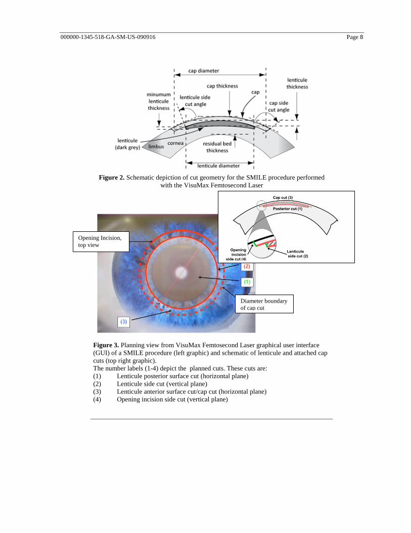
Figure 2. Schematic depiction of cut geometry for the SMILE procedure performed
with the VisuMax Femtosecond Laser
Figure 3. Planning view from VisuMax Femtosecond Laser graphical user interface
(GUI) of a SMILE procedure (left graphic) and schematic of lenticule and attached cap
cuts (top right graphic).
The number labels (1-4) depict the planned cuts. These cuts are:
(1) Lenticule posterior surface cut (horizontal plane)
(2) Lenticule side cut (vertical plane)
(3) Lenticule anterior surface cut/cap cut (horizontal plane)
(4) Opening incision side cut (vertical plane)
Diameter boundary
of cap cut
Opening Incision,
top view
000000-1345-518-GA-SM-US-090916 Page 9
Indications, contraindications, warnings, precautions, and potential risks
Indication for use
The VisuMax Femtosecond Laser is indicated for use in small incision lenticule extraction (SMILE)
for the reduction or elimination of myopia ≥ -1.00 D to ≤ -8.00 D, with ≤ -0.50 D cylinder and MRSE
≤ -8.25 D in the eye to be treated in patients who are 22 years of age or older with documentation of
stable manifest refraction over the past year as demonstrated by a change of ≤ 0.50 D MRSE.
Contraindications
VisuMax SMILE procedure for the correction of myopia is contraindicated in patients with:
a residual stromal bed thickness that is less than 250 microns from the corneal endothelium;
abnormal corneal topographic findings, e.g. keratoconus, pellucid marginal degeneration;
ophthalmoscopic signs of progressive or unstable myopia or keratoconus (or keratoconus suspect);
irregular or unstable (distorted/not clear) corneal mires on central keratometry images;
severe dry eye ;
active eye infection or inflammation;
recent herpes eye infection or problems resulting from past infection;
active autoimmune disease or connective tissue disease;
uncontrolled diabetes;
uncontrolled glaucoma.
Warnings
VisuMax SMILE procedure is not recommended for patients with:
controlled autoimmune or connective tissue disease;
controlled diabetes;
immunocompromised status (weakened immune system) due to medication or a disease condition,
e.g., immunosuppressive therapy, such as corticosteroids, or AIDS;
a history of Herpes simplex or Herpes zoster keratitis;
controlled glaucoma;
a history of taking isotretinoin (Accutane®);
epithelial basement membrane dystrophy;
amblyopia;
dry eyes;

000000-1345-518-GA-SM-US-090916 Page 10
deep orbits, strong blink, anxiety, pterygium, or any other finding suggesting difficulty in
achieving or maintaining suction;
eyelid malposition (e.g. severe lagophthalmos)
difficulty following directions or are unable to fixate.
Precautions
The safety and effectiveness of the VisuMax SMILE procedure have NOT been established for
patients:
with refractive error outside the range in the approved indications for use;
a difference between cycloplegic and manifest refractions of greater than or equal to 0.75 D
spherical equivalent in the eye to be treated;
with central corneal thickness of less than 500 microns in the eye to be treated;
with a family history of thinning of the cornea due to keratoconus, pellucid marginal degeneration,
or other conditions that may cause ectasia;
with uncorrected visual acuity (UCVA) better than or equal to 20/40 in the eye to be treated;
with best spectacle-corrected visual acuity (BSCVA) worse than 20/20 in the eye to be treated;
who wear contact lenses and did not discontinue use of contact lenses for at least 2 weeks (for hard
lenses) or 3 days (for soft lenses) prior to the preoperative examination, and through the day of
surgery;
who wear contact lenses and did not demonstrate a stable refraction (within ±0.5 D), as
determined by MRSE, on two consecutive examinations at least 1 week apart, in the eye to be
treated;
with mesopic pupil diameter >8.0 mm;
with eye to be treated targeted for monovision;
with BSCVA in the fellow eye worse than 20/40;
with previous corneal or intraocular surgery, or trauma to the intended ablation zone;
with corneal abnormalities including, but not limited to, scars, irregular astigmatism and corneal
warpage;
with severe blepharitis (e.g. ocular rosacea)
with elevated intraocular pressure (IOP), ocular hypertension or being followed for possible
glaucoma (glaucoma suspect);
with atopic syndrome;
with severe allergies and eye rubbing;
taking the medication sumatriptan succinate (Imitrex®);
who are taking the medication Amiodarone hydrochloride (Cordarone®);
under 22 years of age;

000000-1345-518-GA-SM-US-090916 Page 11
more than 12 months after surgery;
with media problems (corneal, lens, and/or vitreous opacities including, but not limited to,
cataract);
with a history of uveitis;
who are pregnant or nursing.
Patient selection precautions
All patients must be given the opportunity to read and understand the Patient Information Booklet and
to have all questions answered to their satisfaction prior to giving consent for the VisuMax SMILE
procedure. Consideration should be given to the following in determining the appropriate patients for
the procedure:
Complete examination, including but not limited to cycloplegic evaluation, must be performed.
Preoperative corneal mapping is essential to exclude any topographical abnormalities, such as
keratoconus. The lens must be evaluated, especially in older patients, to assure that nuclear
sclerosis or any other lens opacity is not present prior to laser surgery. Indirect ophthalmoscopy
through a dilated pupil is essential to rule out any retinal pathology.
To obtain accurate and stable refractive information, contact lens wearers must be examined after
a sufficient period of not wearing contact lenses. Additional precautions should be taken for rigid
gas permeable or hard contact lens wearers with respect to stable central keratometry readings.
Refractive stability is considered to be a change of ≤ 0.50 D in both MRSE and keratometric
meridian (either axis) as compared to the baseline measurements.
Evaluation of the optic nerve and measurement of intraocular pressure are necessary to rule out
glaucoma. If elevated intraocular pressure and/or evidence of glaucomatous damage are found,
topical steroids should only be used with careful medical supervision or the patient should not
undergo refractive surgery.
Pachymetry must be performed to obtain a baseline central corneal thickness measurement to
assure that the combination of the planned corneal cap thickness and the planned lenticule
thickness will not approach closer than 250 microns to the corneal endothelium.
The patient should have the ability to lie flat without difficulty and fixate steadily for the duration
of the procedure.
The patient should be clearly informed of all alternatives for the correction of his/her myopia
including, but not limited to, spectacles, contact lenses, and other refractive surgeries, prior to
consenting for the procedure.
Due to the importance of managing patient expectations in elective refractive surgery, it is
recommended that the physician:
– convey realistic expectations to the prospective patient;
– ensure patient comprehension of the risks and benefits at the start of the informed consent
process;
– discuss with patients how having the VisuMax SMILE procedure may affect the future
interpretation of intraocular pressure measurements; patients should be instructed to inform
future eye care providers that they have had a refractive procedure to correct their myopia;

000000-1345-518-GA-SM-US-090916 Page 12
– discuss the risk of decreased contrast sensitivity potentially affecting activities under low-light
conditions;
– provide a patient information card that has eye measurements from before the procedure.
Patients can keep this card to help their doctor calculate the lens implant power should they
need to have future cataract surgery; a form for the necessary information is available on the
internet.
Procedure-related precautions
The surgeon should share all expectations with the patient prior to initiating a procedure and to coach
and encourage the patient to continue fixating throughout the short duration of the VisuMax SMILE
procedure.
Surgeons should be vigilant for possible small eye movements through the operating microscope
during the procedure. There can be a relative shift of the pupil center during the operation and this
does not necessarily entail a shift of the cornea. Because the surgeon always retains direct control of
the delivery of laser energy, in the unlikely event these findings are observed, treatment can be
suspended or terminated by releasing the foot switch and disconnecting the suction. Follow the
instructions provided in the section for Treatment Interruption.
The formation of bubbles at the periphery of the suction zone is an indication of imminent suction
loss. In the event of a complete loss of suction, the VisuMax console detects the reduction in pressure
of the eye and the procedure is automatically halted. In this case, users are directed to follow the
instructions displayed on the graphic user interface (GUI) screen in accordance with instructions
provided below in the section for Treatment Interruption.
To ensure adequate suction prior to and throughout the laser procedure:
Do not use a contacting agent with the interface, as the desired result will not be achieved.
Ensure that no liquid is allowed to enter the vacuum system.
Take special care to ensure exact alignment of the patient’s eye. Continuously optimize the eye
position along the X and Y axes as the eye is brought closer to the contact glass.
Total surgery time (centering, suction time) should be kept as short as possible.
Ensure that conditions which may distract the patient (background noise, other activity in the
surgery) are kept to a minimum while the eye is under suction.
Note: The energy settings for the VisuMax SMILE procedure are programmable and adjustable only
by trained Zeiss personnel.
Potential risks
The potential risks associated with the VisuMax SMILE procedure include, but are not limited to:
Loss of BSCVA or contrast sensitivity;
Over-correction or under-correction;

000000-1345-518-GA-SM-US-090916 Page 13
Increase in refractive cylinder;
Difficulty with night driving;
Headache or eyestrain due to imbalance between the eyes;
Worsening of patient complaints such as glare, halos, starbursts, hazy or blurred vision, distortion,
double or ghost images, fluctuation of vision, focusing difficulty, difficulty with depth perception,
light sensitivity; grittiness, and ocular pain/soreness;
Transient light sensitivity syndrome;
Dry eye;
Ptosis;
Increase in IOP;
Lens opacity;
Conjunctivitis;
Iritis;
Corneal haze/scar/infection/inflammation/infiltrate/ulcer/epithelial defect/epithelium in the
interface/ edema/decompensation/striae or microstriae/ectasia;
Perforated, miscreated, or melting of the cap;
Treatment interruption, difficult lenticule removal with tissue damage or retained lenticule; ocular
penetration;
Retinal detachment/posterior vitreous detachment/vascular accidents.
For further discussion of adverse events and complications that occurred during the course of the
clinical trial, refer to the section on Key Safety Outcomes (p. 20).

000000-1345-518-GA-SM-US-090916 Page 14
Alternative Treatment Options
Alternatives to the small incision lenticule extraction (SMILE) available to a patient might include
spectacle correction (glasses), contact lenses, surgery with another FDA approved laser using PRK
(Photo Refractive Keratectomy) or LASIK (Laser-Assisted In Situ Keratomileusis), or a lens implant
surgically placed inside the eye. You should discuss with your patient whether they are a candidate
for these procedures as well as the risks/benefits of each alternative. Furthermore, for this discussion
important information about these alternative procedures is available at the following websites
(accessed August, 2016):
FDA:http://www.fda.gov/MedicalDevices/ProductsandMedicalProcedures/SurgeryandLifeSu
pport/LASIK/default.htm
NEI: https://nei.nih.gov/health/errors/myopia
AAO: http://www.aao.org/eye-health/treatments/lasik
FTC: https://www.consumer.ftc.gov/articles/0062-basics-lasik-eye-surgery#lasikbasics

000000-1345-518-GA-SM-US-090916 Page 15
Clinical results
Study objectives and methods
The objective of this study was to evaluate the safety and effectiveness of the Carl Zeiss Meditec
VisuMax SMILE procedure for the reduction or elimination of myopia from ≥ -1.00 D to ≤ -10.00 D
with ≤ -0.50 D cylinder and MRSE ≤ -10.25 D. Myopic spherical eyes and myopic eyes with
≤ 0.50 D of astigmatism were treated with a spherical treatment only.
Study design
This was a 12-month, prospective, multi-center, open-label, non-randomized clinical trial of up to
360 eyes of 360 consecutive subjects enrolled and treated with the VisuMax Femtosecond Laser.
Retreatments were not allowed during the study.
Follow-up examinations were scheduled at 1 day, 1 week, 1 month, 3 months, 6 months, 9 months,
and 12 months.
Postop Day 1: Day 1
Postop Week 1: Days 5 to 9
Postop Month 1: Days 21 to 35 (Weeks 3 to 5)
Postop Month 3: Days 70 to 98 (Weeks 10 to 14)
Postop Month 6: Days 147 to 182 (Weeks 21 to 26)
Postop Month 9: Days 245 to 301 (Weeks 35 to 43)
Postop Month 12: Days 330 to 420 (Months 11 to 14)
The key effectiveness variables for the study were:
Predictability: the percentage of eyes achieving MRSE within ± 1.00 D of the intended outcome,
and within ± 0.50 D of the intended outcome at the point at which stability is first achieved
Improvement in UCVA following treatment: the percentage of eyes that achieve uncorrected
visual acuity (UCVA) of 20/40 or better at the postoperative interval at which stability has been
established, as well as the percentage of eyes that achieve UCVA of 20/20 or better
Refractive stability was also evaluated:
Stability was considered to have been achieved at the latter of two postoperative refractions
performed at least 3 months apart or at 3 months after surgery when compared with the 1-month
interval, if at least three of the four stability criteria were met; these criteria were as follows:
1. At least 95% of the treated eyes should have a change ≤ 1.00 D of MRSE at the latter of two
postoperative refractions performed at least 3 months apart or at 3 months after surgery when
compared with the 1-month interval;
2. The mean rate of change in MRSE, as determined by paired analysis, is ≤ 0.5 D per year (0.04
D/month) over the same time period;

000000-1345-518-GA-SM-US-090916 Page 16
3. The mean rate of change of MRSE decreases monotonically over time, with a projected
asymptote of zero or a rate of change attributable to normal aging;
4. The 95% confidence interval for the mean rate of change includes zero or a rate of change
attributable to normal aging;
Stability was confirmed at least 3 months after the stability time point by a statistically adequate
subgroup.
The key safety variables for the study were:
Preservation of Best-Spectacle Corrected Visual Acuity (BSCVA)
– In eyes with preoperative BSCVA 20/20 or better, the percentage of eyes with BSCVA worse
than 20/40 at the postoperative interval at which stability has been established
– The percentage of eyes with ≥ 2 lines BSCVA loss
Induced manifest refractive astigmatism:
– the percentage of eyes with induced manifest refractive cylinder of >2.00 D at the
postoperative interval at which stability has been established
Loss of Contrast Sensitivity
– Mean "within-eye" loss of contrast sensitivity from baseline to 12 months with the 1-sided 95
% confidence interval for each spatial frequency
– The percentage of eyes showing ≥ 0.3 log units loss at two or more spatial frequencies
Incidence of Adverse Events
– The counts and percentages of eyes for each adverse event
Patient Reported Symptoms
– Patient reported symptoms were considered as a secondary safety variable and were stratified
by pupil size and fellow eye status
Additional safety variables for the study were:
Corneal Topography
Wavefront Aberrometry
Inclusion and exclusion criteria
In order to be enrolled in the study, patients needed to meet these conditions:
be 22 years of age and older;
have spherical myopia from ≥ -1.00 D to ≤ -10.00 D, with ≤ -0.50 D cylinder and MRSE
≤ -10.25 D, in the eye to be treated;
have a stable refraction for the past year, as demonstrated by a change in MRSE of ≤ 0.50 D in the
eye to be treated;

000000-1345-518-GA-SM-US-090916 Page 17
have a difference between cycloplegic and manifest refractions of < 0.75 D spherical equivalent
(SE) in the eye to be treated. (SE is the difference between cycloplegic and manifest refractions);
have UCVA worse than 20/40 in the eye to be treated;
have BSCVA of at least 20/20 in the eye to be treated;
discontinue use of contact lenses at least 2 weeks for hard contacts and 3 days for soft lenses prior
to the preoperative examination; all contact wearers must have two manifest refractions taken at
least one week apart that did not differ by more than 0.50 D;
have central corneal thickness of at least 500 microns in the eye to be treated;
be willing and able to return for scheduled follow-up examinations;
and provide written informed consent.
Patients not meeting the above inclusion criteria were excluded from the study.
In addition, subjects who exhibited any of the following conditions were excluded:
a mesopic pupil diameter > 8.0 mm;
cylinder of greater than 0.50 D;
treatment depth is less than 250 microns from the corneal endothelium;
eye to be to be treated is targeted for monovision;
fellow eye has BSCVA worse than 20/40;
abnormal corneal topographic findings, e.g. keratoconus, pellucid marginal degeneration, in either
eye;
history of anterior segment pathology, including cataracts in the treated eye;
clinically significant dry eye syndrome unresolved by treatment in either eye;
residual, recurrent, active ocular or uncontrolled eyelid disease, corneal scars or other corneal
abnormality such as recurrent corneal erosion or severe basement membrane disease in the eye to
be treated;
ophthalmoscopic signs of progressive or unstable myopia or keratoconus (or keratoconus suspect)
in either eye;
irregular or unstable (distorted/not clear) corneal mires on central keratometry images in either
eye;
history of ocular herpes zoster or herpes simplex keratitis;
have deep orbits, strong blink, anxiety, pterygium, or any other finding suggesting difficulty in
achieving or maintaining suction;
have difficulty following directions or unable to fixate;
have previous intraocular or corneal surgery of any kind in the eye to be treated, including any
type of surgery for either refractive or therapeutic purposes;
history of steroid-responsive rise in intraocular pressure, glaucoma, or preoperative
IOP > 21 mm Hg in either eye;

000000-1345-518-GA-SM-US-090916 Page 18
history of diabetes, diagnosed autoimmune disease, connective tissue disease or clinically
significant atopic syndrome;
be immunocompromised or requires chronic systemic corticosteroids or other immunosuppresive
therapy that may affect wound healing;
have a history of known sensitivity to planned study medications;
participating in any other ophthalmic drug or device clinical trial during the time of
this clinical investigation;
and pregnant, lactating, or child-bearing potential and not practicing a medically approved method
of birth control.
Results and data analysis
Demographics and baseline parameters
A total of 336 eyes were treated across five U.S. sites. Demographic information for all treated
subjects is provided in Table 1. Subjects ranged in age from 22 to 58 years, with a mean age of 33.3
for all treated eyes. More females (58.3 %) than males (41.7 %) were enrolled and treated
in the study, and the majority of subjects were Caucasian (92.0 %).
Table 1
Demographics
All Treated Eyes
Demographics All Treated Eyes
Number Percentage
NUMBER OF EYES & SUBJECTS 336 Eyes of 336 Subjects
GENDER
Male 140 41.7 %
Female 196 58.3 %
RACE
White 309 92.0 %
Black 10 3.0 %
Asian 6 1.8 %
Other 11 3.3 %
SURGICAL EYE
Right 152 45.2 %
Left 184 54.8 %
AGE (In Years)
Mean (SD) 33.3 ( 7.9)
Min., Max. 22.0, 58.0
Fellow-eye Status
Excimer Laser Refractive Surgery 333 99.1 %
Untreated 3 0.9 %

000000-1345-518-GA-SM-US-090916 Page 19
Preoperative refraction parameters are shown in Table 2. Mean manifest refraction sphere at baseline
for all treated eyes was -4.762 D, with a range of -1.00 D to -10.00 D. The mean manifest refraction
cylinder at baseline for all treated eyes was -0.194 D (SD = 0.20), with a range of 0.00 D to -0.50 D.
As specified in the study protocol, cylinder was not treated in the study eyes. Among all treated eyes,
the procedures were not completed for two subjects, due to intraoperative suction loss during the
posterior lamellar cut. Additionally, one subject had treatment on the wrong eye. These three
subjects were excluded from the effectiveness population, resulting in 333 total eyes.
Table 2
Preoperative Refraction Parameters
Manifest
All Treated Eyes Effectiveness
Population
Refraction Number % Number %
Sphere
0.00 to -1.00 D 4 1.2 4 1.2
-1.01 to -2.00 D 35 10.4 35 10.5
-2.01 to -3.00 D 54 16.1 53 15.9
-3.01 to -4.00 D 50 14.9 50 15.0
-4.01 to -5.00 D 50 14.9 49 14.7
-5.01 to -6.00 D 43 12.8 43 12.9
-6.01 to -7.00 D 44 13.1 44 13.2
-7.01 to -8.00 D 29 8.6 28 8.4
-8.01 to -9.00 D1 15 4.5 15 4.5
-9.01 D or higher1 12 3.6 12 3.6
Mean (SD) -4.762 (2.202) -4.763 (2.202)
Range -10.00 to -1.00 -10.00 to -1.00
Total 336 100.0 333 100.0
Cylinder
0.00 D 153 45.5 152 45.6
-0.25 D 105 31.3 105 31.5
-0.50 D 78 23.2 76 22.8
Mean (SD) -0.194 (0.200) -0.193 (0.199)
Range -0.50 to 0.00 -0.50 to 0.00
Total 336 100.0 333 100.0 1 Please note that treatment of these dioptric powers will present a flagged
warning to the users so that the user understands that correction of these
powers had not been substantiated by an adequate set of data.

000000-1345-518-GA-SM-US-090916 Page 20
Accountability
Accountability for all treated eyes through 12 months is presented in Table 3. Accountability over the
course of the entire study was excellent with 98.5 % (329/336) of eyes treated in the study available
for analysis at the 6-month visit, the point at which refractive stability was identified. The study
results presented below include all available outcomes through database lock in March, 2015.
Table 3
Accountability
All Treated Eyes
Enrolled (N = 336) Day
1
Week
1
Month
1
Month
3
Month
6
Month
9
Month
12
Available for analysis 335
(99.7 %)
334
(99.4 %)
335
(99.7 %)
333
(99.1 %)
329
(97.9 %)
320
(95.2 %)
311
(92.6 %)
Active 0
(0.0 %)
0
(0.0 %)
0
(0.0 %)
0
(0.0 %)
0
(0.0 %)
8
(2.4 %)
17
(5.1 %)
Missing 1
(0.3 %)
2
(0.6 %)
1
(0.3 %)
3
(0.9 %)
7
(2.1 %)
8
(2.4 %)
8
(2.4 %)
Discontinued 1
(0.3 %)
1
(0.3 %)
1
(0.3 %)
1
(0.3 %)
2
(0.6 %)
2
(0.6 %)
3
(0.9 %)
Death 0
(0.0 %)
0
(0.0 %)
0
(0.0 %)
0
(0.0 %)
0
(0.0 %)
0
(0.0 %)
1
(0.3 %)
Alternative treatment 1
(0.3 %)
1
(0.3 %)
1
(0.3 %)
1
(0.3 %)
2
(0.6 %)
2
(0.6 %)
2
(0.6 %)
Scheduled visit data
outstanding
0
(0.0 %)
1
(0.3 %)
0
(0.0 %)
1
(0.3 %)
2
(0.6 %)
2
(0.6 %)
0
(0.0 %)
Lost to follow-up 0
(0.0 %)
0
(0.0 %)
0
(0.0 %)
1
(0.3 %)
3
(0.9 %)
4
(1.2 %)
5
(1.5 %)
% Accountability 335
(100.0 %)
334
(99.7 %)
335
(100.0 %)
333
(99.4 %)
329
(98.5 %)
320
(98.2 %)
311
(98.4 %)
Status categories were based on ANSI-Z80.11-2012.
% = n N 100.
% Accountability = available (enrolled - discontinued - active) 100
Key safety outcomes
In Table 4, key safety variables are presented for all 329 available eyes at the point of stability, which
was established at 6 months (details provided on p. 38, Stability of the manifest refraction).
Additionally, key safety variables at the last available visits for each of the 336 treated eyes are
summarized in Table 5. No study subject presented with a loss of ≥ 2 lines BSCVA, with BSCVA
worse than 20/40, or with increased manifest refractive astigmatism > 2.00 D at 6 months or at the
last available visits. With regard to loss of ≥ 2 lines BSCVA at any point during the study, there were
19 study eyes at Week 1, 5 eyes at Month 1, and 3 eyes at interim visits with this degree of loss.
These are further presented and discussed below (Table 6).

000000-1345-518-GA-SM-US-090916 Page 21
Table 4
Summary of Key Safety Variables at 6-Month Point of Refractive Stability
All Treated Eyes
Key Safety Event n/N % 95 % CI1
Loss of ≥ 2 lines BSCVA 0/329 0.0 % (0.0 %, 1.1 %)
BSCVA worse than 20/40 if
20/20 or better preoperatively
0/329 0.0 % (0.0 %, 1.1 %)
Increased manifest refractive
astigmatism > 2.0 D
0/329 0.0 % (0.0 %, 1.1 %)
N = Number of case report forms received with non-missing values at each visit.
95 % CI was calculated based on Clopper-Pearson exact method.
Table 5
Summary of Key Safety Variables at Last Available Visit
All Treated Eyes
Key Safety Event n/N % 95 % CI1
Loss of ≥ 2 lines BSCVA 0/336 0.0 % (0.0 %, 1.1 %)
BSCVA worse than 20/40 if
20/20 or better preoperatively
0/336 0.0 % (0.0 %, 1.1 %)
Increased manifest refractive
astigmatism > 2.0 D
0/336 0.0 % (0.0 %, 1.1 %)
N = Number of case report forms received with non-missing values at each visit.
95 % CI was calculated based on Clopper-Pearson exact method.
The change in BSCVA postoperatively from baseline for all treated eyes is presented in Table 6. For
all scheduled visits from Month 1 and on, there were no BSCVA losses greater than one line, with 3.3
% (11/329) of eyes at 6 months and 2.6 % (8/311) of eyes at 12 months, showing a one line
decrement. With regard to loss of ≥ 2 lines BSCVA, 5.7% (19/334) of treated eyes at Week 1 and
1.5% (5/335) of treated eyes at Month 1 manifested this level of loss. Beyond Month 1, there were
three other instances, involving three separate subjects, of BSCVA loss ≥ 2 lines, all during interim
visits. Two of these occurred between Months 1 and 3, while one occurred between Months 6 and 9,
and each case was resolved by the subsequent visit. Further, following the Month 1 time point when
the proportions of eyes with losses versus gains in BSCVA were comparable, every subsequent visit
demonstrated a consistently and increasingly higher proportion of eyes with gains in BSCVA,
compared to losses.

000000-1345-518-GA-SM-US-090916 Page 22
Table 6
Change in Best Spectacle-Corrected Visual Acuity (BSCVA) from Preop
All Treated Eyes
Week 1 Month 1 Month 3 Month 6 Month 9 Month 12
BSCVA n (%) n (%) n (%) n (%) n (%) n (%)
Available (N) 334 335 333 329 319 311
Lost > 2 lines (>10 letters) 13 (3.9 %) 5 (1.5 %) 0 (0.0 %) 0 (0.0 %) 0 (0.0 %) 0 (0.0 %)
Lost 2 lines (10 letters) 6 (1.8 %) 0 (0.0 %) 0 (0.0 %) 0 (0.0 %) 0 (0.0 %) 0 (0.0 %)
Lost 1 line (5-9 letters) 75 (22.5 %) 36 (10.7 %) 21 (6.3 %) 11 (3.3 %) 10 (3.1 %) 8 (2.6 %)
Unchanged (< 5 letters) 222 (66.5 %) 255 (76.1 %) 246 (73.9 %) 243 (73.9 %) 239 (74.9 %) 224 (72.0 %)
Gained 1 line (5-9 letters) 15 (4.5 %) 38 (11.3 %) 59 (17.7 %) 66 (20.1 %) 64 (20.1 %) 71 (22.8 %)
Gained 2 lines (10 letters) 0 (0.0 %) 1 (0.3 %) 4 (1.2 %) 6 (1.8 %) 3 (0.9 %) 3 (1.0 %)
Gained > 2 lines (>10 letters) 3 (0.9 %) 0 (0.0 %) 3 (0.9 %) 3 (0.9 %) 3 (0.9 %) 5 (1.6 %)
Not reported 0 0 0 0 1 0
Total 334 335 333 329 320 311
N = Number of case report forms received with non-missing values at each visit.
Through the point of data lock, a total of 14 subjects were reported with 15 ocular adverse events
(AEs) over the course of the study. The intraoperative AEs are summarized in Table 7, while all
postoperative AEs are summarized in Table 8. In total, there were four intraoperative AEs: 2 cases of
difficult lenticule removal with tissue, 1 case with a cap perforation, and 1 case of retained tissue
following lenticule removal. With the exception of one subject, whose intraoperative AE was present
through the 3-month visit, none of the reported cases persisted beyond the 1-week visit. Importantly,
all four subjects completed the study with UCVA no worse than 20/16.
Table 7
Intraoperative Adverse Events
Intraoperative AE n
Difficult lenticule removal with tissue
damage
2 (0.6%)
Perforated cap 1 (0.3%)
Retained tissue, small 1 (0.3%)
The other 10 subjects experienced adverse events postoperatively which occurred at various time
points throughout the study. These events included 1 case of conjunctival carcinoma in situ; 1 case of
allergic conjunctivitis; 1 case of viral conjunctivitis; 1 case of decrease in BSCVA of greater than or
equal to 2 lines (10 letters) not due to irregular astigmatism as shown by hard contact lens refraction
at 3 months or later; 1 case of herpetic lid and corneal lesion; 2 cases of iritis (involving one subject);
1 case of posterior vitreous detachment (PVD); 1 case of pyogenic granuloma; 1 case of retinal
vasculitis, and 2 cases of retained tissue following lenticule removal, one of which is accounted for in
Table 7. With the exception of one subject whose UCVA at study exit was 20/25, the other 9 subjects
completed and exited the study with UCVA no worse than 20/20.

000000-1345-518-GA-SM-US-090916 Page 23
Table 8
Postoperative Ophthalmic Adverse Events — All Treated Eyes
D1 W1 M1 M3 M6 M9 M12 Uns Cum
AE N=335 N=334 N=335 N=333 N=329 N=320 N=311 N=24 N=336
Diffuse lamellar keratitis (Stage 3 or above) 0
0.0%
0
0.0%
0
0.0%
0
0.0%
0
0.0%
0
0.0%
0
0.0%
0 0
0.0%
Corneal infiltrate or ulcer 0
0.0%
0
0.0%
0
0.0%
0
0.0%
0
0.0%
0
0.0%
0
0.0%
0 0
0.0%
Any persistent corneal epithelial defect at 1
month or later
0
0.0%
0
0.0%
0
0.0%
0
0.0%
0
0.0%
0
0.0%
0
0.0%
0 0
0.0%
Corneal edema at 1 month or later 0
0.0%
0
0.0%
0
0.0%
0
0.0%
0
0.0%
0
0.0%
0
0.0%
0 0
0.0%
Epithelium in the interface with loss of 2
lines (10 letters) or more of BSCVA
0
0.0%
0
0.0%
0
0.0%
0
0.0%
0
0.0%
0
0.0%
0
0.0%
0 0
0.0%
Melting of the cap 0
0.0%
0
0.0%
0
0.0%
0
0.0%
0
0.0%
0
0.0%
0
0.0%
0 0
0.0%
IOP increase of > 10 mmHg above baseline
or IOP > 30 mmHg on 2 consecutive exams
0
0.0%
0
0.0%
0
0.0%
0
0.0%
0
0.0%
0
0.0%
0
0.0%
0 0
0.0%
Haze beyond 6 months with loss of 2 lines
or greater (10 letters) of BSCVA
0
0.0%
0
0.0%
0
0.0%
0
0.0%
0
0.0%
0
0.0%
0
0.0%
0 0
0.0%
Decrease in BSCVA of greater than or equal
to 2 lines (10 letters) not due to irregular
astigmatism as shown by hard contact lens
refraction at 3 months or later
0
0.0%
0
0.0%
0
0.0%
0
0.0%
0
0.0%
0
0.0%
0
0.0%
1 1
0.3%
Retinal Detachment 0
0.0%
0
0.0%
0
0.0%
0
0.0%
0
0.0%
0
0.0%
0
0.0%
0 0
0.0%
Retinal vascular accidents 0
0.0%
0
0.0%
0
0.0%
0
0.0%
0
0.0%
0
0.0%
0
0.0%
0 0
0.0%
Ocular penetration 0
0.0%
0
0.0%
0
0.0%
0
0.0%
0
0.0%
0
0.0%
0
0.0%
0 0
0.0%
Any other vision-threatening event
Retinal vasculitis 0
0.0%
0
0.0%
0
0.0%
0
0.0%
0
0.0%
0
0.0%
1
0.3%
0 1
0.3%
Other
Carcinoma in situ, conjunctival 0
0.0%
0
0.0%
0
0.0%
0
0.0%
0
0.0%
0
0.0%
1
0.3%
0 1
0.3%
Conjunctivitis, allergic 0
0.0%
0
0.0%
0
0.0%
0
0.0%
0
0.0%
1
0.3%
0
0.0%
0 1
0.3%
Conjunctivitis, viral 0
0.0%
0
0.0%
0
0.0%
0
0.0%
0
0.0%
0
0.0%
0
0.0%
1 1
0.3%
Herpetic lid and corneal lesion 0
0.0%
0
0.0%
0
0.0%
0
0.0%
0
0.0%
0
0.0%
0
0.0%
1 1
0.3%
Iritis 0
0.0%
0
0.0%
0
0.0%
0
0.0%
0
0.0%
0
0.0%
0
0.0%
1 1
0.3%
PVD 0
0.0%
0
0.0%
0
0.0%
0
0.0%
0
0.0%
1
0.3%
0
0.0%
0 1
0.3%
Pyogenic Granuloma 0
0.0%
0
0.0%
0
0.0%
0
0.0%
1
0.3%
0
0.0%
0
0.0%
1 1
0.3%
Retained tissue, small 2
0.6%
2
0.6%
1
0.3%
1
0.3%
0
0.0%
0
0.0%
0
0.0%
1 2*
0.6%
Multiple events could be reported for each subject.
Uns = interim visit, N is the number of eyes with interim visits, and incidence is the number of eyes with the reported events
during the interim visits.
Cum = cumulative, N is the number of all treated eyes with postoperative visits, and incidence is the number of eyes with the
reported events during the study.
*One of these subjects is also accounted for in Table 7, as the retained tissue was first observed intraoperatively.

000000-1345-518-GA-SM-US-090916 Page 24
In addition to the intraoperative and postoperative adverse events noted above, there were a total of
15 intraoperative events observed among the 336 procedures. These events, presented in Table 9
below, include 8 cases of difficult lenticule removal without tissue damage, 6 cases in which suction
was lost during the procedure, and 1 case of decentered treatment which was identified by
postoperative topography. It should be noted that none of these events led to clinically significant
sequelae.
Table 9
Intraoperative Events
All Treated Eyes
N = 336 Number Percent
Difficult lenticule removal without
tissue damage
8 2.4 %
Loss of suction: completed treatment 4 1.2 %
Loss of suction: discontinued
treatment
2 0.6 %
Decentered treatment* 1 0.3%
Any Events 15 4.5 % Multiple events could be reported for each subject.
Percent = Number/N 100.
* Identified by postoperative topography
Complications over the course of the study are summarized below in Table 10. The majority of these
reports involved questionnaire responses of moderate or severe glare or halos, at 10.4 % (35/336) and
6.0 % (20/336), respectively. The peak incidence of these reports occurred at 3 and 6 months, with a
significant reduction by the 9 and 12 month visits. At Month 12, in fact, there were four residual
reports of moderate or severe glare and one report of moderate or severe halos. The definitions for
glare and halo complications did not take into consideration whether the symptom was considered
bothersome, whether it was present at baseline with the use of contact lens or spectacle correction, or
whether it readily resolved when distance correction is worn.
Other findings included: clinical signs and/or subject symptoms consistent with dry eye (2.7 %,
9/336); diffuse lamellar keratitis (DLK) stage 2 or less (0.9 %, 3/336); epithelium in the interface
(0.9 %, 3/336); foreign body sensation at 1 month or later (0.3 %, 1/336); interface debris (2.7 %,
9/336); pain at 1 month or later (0.3 %, 1/336); striae/microstriae (0.3 %, 1/336); and transient light
sensitivity syndrome (0.3 %, 1/336).

000000-1345-518-GA-SM-US-090916 Page 25
Table 10
Complications
All Treated Eyes
D0 D1 W1 M1 M3 M6 M9 M12 Uns Cum
Complications N=336 N=335 N=334 N=335 N=333 N=329 N=320 N=311 N=24 N=336
Clinical signs and/or subject
symptoms consistent with dry eye
0
0.0 %
0
0.0 %
5
1.5 %
1
0.3 %
4
1.2 %
0
0.0 %
0
0.0 %
0
0.0 %
3 9
2.7 %
Corneal edema between 1 week and 1 month after procedure
0 0.0 %
0 0.0 %
0 0.0 %
0 0.0 %
0 0.0 %
0 0.0 %
0 0.0 %
0 0.0 %
0 0 0.0 %
Corneal scarring 0
0.0 %
0
0.0 %
0
0.0 %
0
0.0 %
0
0.0 %
0
0.0 %
0
0.0 %
0
0.0 %
0 0
0.0 %
Crystalline lens opacity 0 0.0 %
0 0.0 %
0 0.0 %
0 0.0 %
0 0.0 %
0 0.0 %
0 0.0 %
0 0.0 %
0 0 0.0 %
Diffuse lamellar keratitis (Stage 2 or
less)
0
0.0 %
1
0.3 %
3
0.9 %
0
0.0 %
0
0.0 %
0
0.0 %
0
0.0 %
0
0.0 %
0 3
0.9 %
Epithelium in the interface 0
0.0 %
1
0.3 %
2
0.6 %
0
0.0 %
0
0.0 %
0
0.0 %
0
0.0 %
0
0.0 %
0 3
0.9 %
Foreign body sensation at 1 month or later
0 0.0 %
0 0.0 %
0 0.0 %
0 0.0 %
0 0.0 %
1 0.3 %
1 0.3 %
0 0.0 %
0 1 0.3 %
Ghost/double images in the operative
eye
0
0.0 %
0
0.0 %
0
0.0 %
0
0.0 %
0
0.0 %
0
0.0 %
0
0.0 %
0
0.0 %
0 0
0.0 %
Interface debris, such as lint, pigment, air bubbles, and meibomian gland
secretions
0 0.0 %
5 1.5 %
5 1.5 %
2 0.6 %
2 0.6 %
0 0.0 %
0 0.0 %
0 0.0 %
1 9 2.7 %
Moderate or severe glare 0
0.0 %
0
0.0 %
0
0.0 %
1
0.3 %
21
6.3 %
15
4.6 %
7
2.2 %
4
1.3 %
0 35
10.4 %
Moderate or severe halos 0
0.0 %
0
0.0 %
0
0.0 %
0
0.0 %
10
3.0 %
11
3.3 %
4
1.3 %
1
0.3 %
0 20
6.0 %
Pain at 1 month or later 0
0.0 %
0
0.0 %
0
0.0 %
0
0.0 %
1
0.3 %
1
0.3 %
1
0.3 %
1
0.3 %
0 1
0.3 %
Striae/microstriae 0 0.0 %
0 0.0 %
0 0.0 %
1 0.3 %
0 0.0 %
0 0.0 %
0 0.0 %
0 0.0 %
0 1 0.3 %
Transient light sensitivity syndrome
(TLSS)
0
0.0 %
0
0.0 %
0
0.0 %
0
0.0 %
0
0.0 %
0
0.0 %
0
0.0 %
0
0.0 %
1 1
0.3 %
Multiple events could be reported for each subject.
Uns = interim visit, N is the number of eyes with interim visits, and incidence is the number of eyes with the reported events during the interim visits.
Cum = cumulative, N is the number of all treated eyes with postoperative visits, and incidence is the number of eyes with the reported events during the study.
One subject did not complete VisuMax treatment and had an alternative treatment at the operative visit. Since the data after the alternative treatment were not
included, the total number of subjects with postoperative visits was 335.
Secondary Surgical Interventions
Three secondary interventions for epithelial ingrowth or interface debris were performed at or before
the 1-week time point, one involving an irrigation to remove interface debris and two involving
irrigation with BSS to remove epithelial cells in the interface.
Contrast sensitivity outcomes
Mesopic (monocular) contrast sensitivity in the study eye was assessed at a calibrated luminance of
3 cd/m2 with no glare,
using sine wave gratings at spatial frequencies of 1.5, 3.0, 6.0, and 12.0 cycles
per degree (cpd). Subjects were dark-adapted for 10 minutes prior to mesopic contrast sensitivity
testing.
As shown in Table 11, the mean change in monocular mesopic contrast sensitivity (CS) were positive
at all postoperative time points for 1.5, 3.0, and 6.0 cpd and at 12 months for 12 cpd, indicating a

000000-1345-518-GA-SM-US-090916 Page 26
consistent sensitivity gain for the cohort. At 12 months the proportion of subjects with clinically
significant gains was 23.5%, compared to 1.6% with clinically significant losses. “Clinically
significant” was defined as ≥ 0.3 log units of change at two or more spatial frequencies.
Table 11
Log Contrast Sensitivity Change from Preoperative Visit
All Treated Eyes
Frequency Statistics Preop Month 3 Month 6 Month 9 Month 12
A (1.5 cpd) N 335 333 329 320 311
Mean (SD) 1.584 (0.226) 1.606 (0.230) 1.658 (0.212) 1.653 (0.222) 1.665 (0.224)
Q1, Q2, Q3 1.40, 1.56, 1.70 1.40, 1.56, 1.85 1.56, 1.70, 1.85 1.48, 1.56, 1.85 1.56, 1.70, 1.85
Min., Max. 0.95, 2.00 0.85, 2.00 0.95, 2.00 0.95, 2.00 0.95, 2.00
< 0.85 0 (0.0%) 0 (0.0%) 0 (0.0%) 0 (0.0%) 0 (0.0%)
Not Reported 1 0 0 0 0
B (3 cpd) N 335 333 329 320 311
Mean (SD) 1.800 (0.211) 1.839 (0.214) 1.882 (0.215) 1.886 (0.203) 1.907 (0.209)
Q1, Q2, Q3 1.76, 1.76, 1.90 1.76, 1.90, 2.06 1.76, 1.90, 2.06 1.76, 1.90, 2.06 1.76, 1.90, 2.06
Min., Max. 1.18, 2.20 1.00, 2.20 1.00, 2.20 1.00, 2.20 1.00, 2.20
< 1.00 0 (0.0%) 0 (0.0%) 0 (0.0%) 0 (0.0%) 0 (0.0%)
Not Reported 1 0 0 0 0
C (6 cpd) N 335 333 329 320 311
Mean (SD)1 < 1.749 (> 0.240) < 1.785 (> 0.254) < 1.826 (> 0.252) < 1.846 (> 0.245) < 1.883 (> 0.250)
Q1, Q2, Q3 1.52, 1.81, 1.95 1.65, 1.81, 1.95 1.65, 1.81, 2.11 1.65, 1.81, 2.11 1.65, 1.95, 2.11
Min., Max.1 < 1.08, 2.26 < 1.08, 2.26 < 1.08, 2.26 < 1.08, 2.26 < 1.08, 2.26
< 1.08 5 (1.5%) 4 (1.2%) 1 (0.3%) 3 (0.9%) 1 (0.3%)
Not Reported 1 0 0 0 0
D (12 cpd) N 335 333 329 320 311
Mean (SD)1 < 1.349 (> 0.305) < 1.353 (> 0.303) < 1.408 (> 0.323) < 1.424 (> 0.335) < 1.469 (> 0.339)
Q1, Q2, Q3 1.18, 1.34, 1.63 1.18, 1.34, 1.48 1.18, 1.48, 1.63 1.18, 1.48, 1.63 1.18, 1.48, 1.78
Min., Max.1 < 0.90, 2.08 < 0.90, 2.08 < 0.90, 2.08 < 0.90, 2.08 < 0.90, 2.08
< 0.90 24 (7.2%) 35 (10.5%) 27 (8.2%) 23 (7.2%) 22 (7.1%)
Not Reported 1 0 0 0 0
0 patch at one or more cpds 24 (7.2%) 35 (10.5%) 27 (8.2%) 23 (7.2%) 22 (7.1%)
One subject had an alternative treatment after the 3-month visit and one subject had an alternative treatment at the operative visit.
Records after alternative treatment were excluded. Both were followed for safety after the alternative treatment.
One subject had the incorrect eye treated. The treated OS did not have the contrast sensitivity test preoperatively.
N = Number of case report forms received with non-missing values at each visit. Not Reported = Number of case report forms
received with missing values at each visit. Q1 = first quartile, Q2 = second quartile (median), and Q3 = third quartile. 1 Number of subjects that could not read any patch at the respective spatial frequency. 0.85, 1.00, 1.08, and 0.90 are the lowest
measurable contrast sensitivity values at 1.5, 3, 6, and 12 cpd, respectively. These lowest values were used for statistical
calculation. In case of no patches could be read, a "<" sign was included in the Mean and Minimum, and ">" sign was
included in the SD.
Patient reported outcomes
The patient reported outcomes (PRO) instrument used in IDE clinical study consisted of the full
Quality of Vision (QoV) questionnaire with accompanying photographs, and 2 of the 3 domains of
the Ocular Surface Disease Index (OSDI). The QoV instrument had three domains (frequency,
severity, and bothersome) each consisting of 10 items which evaluate glare, halos, starbursts, hazy
vision, blurred vision, distortion, double or multiple images, fluctuation, focusing, and judging

000000-1345-518-GA-SM-US-090916 Page 27
distance or depth perception. The two domains of the OSDI included all questions related to ocular
symptoms and all questions related to environmental triggers.
Table 12 provides the QoV score changes from baseline to each postoperative visit stratified by
whether the score was "worse", "same", or "improved".
The data suggest that, on average, subjects noted less severity and were less bothered by symptoms
at 12 months following the procedure compared to the preoperative visit, during which subjects were
using spectacle and contact lens correction for myopia.
Table 12
QoV Score Change from Preoperative
All Treated Eyes
Sub-scale Month 3 Month 6 Month 9 Month 12
Frequency N 332 328 319 309
Worse 176/332 (53%) 150/328 (46%) 133/319 (42%) 116/309 (38%)
Same 70/332 (21%) 74/328 (23%) 74/319 (23%) 71/309 (23%)
Improved 86/332 (26%) 104/328 (32%) 112/319 (35%) 122/309 (39%)
Not Reported 1 1 1 2
Severity N 332 328 319 309
Worse 160/332 (48%) 131/328 (40%) 108/319 (34%) 93/309 (30%)
Same 81/332 (24%) 97/328 (30%) 93/319 (29%) 95/309 (31%)
Improved 91/332 (27%) 100/328 (30%) 118/319 (37%) 121/309 (39%)
Not Reported 1 1 1 2
Bothersome N 332 328 319 309
Worse 138/332 (42%) 106/328 (32%) 95/319 (30%) 79/309 (26%)
Same 102/332 (31%) 123/328 (38%) 119/319 (37%) 119/309 (39%)
Improved 92/332 (28%) 99/328 (30%) 105/319 (33%) 111/309 (36%)
Not Reported 1 1 1 2
Change = Postop - Preop (pairwise).
Worse: Change > 0. Same: Change = 0. Improved: Change < 0.
Not Reported = Number of eyes with missing values at each visit.
As shown in Table 13, the proportion of subjects at Month 12 with an improvement of at least two
grades from baseline (with contact lenses and/or spectacle wear) was consistently the same or larger
than the proportion of subjects with at least a two-grade worsening for the majority of QoV symptoms
and their domains. In total, the overall proportion of subjects that experienced improvement in QoV
symptoms from baseline at 12 months was greater than the proportion of subjects who experienced
worsening of PRO symptoms, with 12.6% of subjects experiencing improvement versus 8.7%
experiencing worsening.
Table 13 also highlights the QoV symptoms with the highest rate of worsening of 2-grades or more at
12 months, with respect to frequency, severity, and bothersomeness. Starbursts (1.6%) and blurred
vision (2.6%) represented the symptoms with the highest proportion of subjects with a 2-grade or
more worsening in frequency from baseline at 12 months. With respect to severity, double or
multiple images and blurred vision, both at 1.3%, had the highest proportion of reported worsening by
two grades or more at 12 months. Starbursts, blurred vision, and judging distance or depth
perception, each with 1.3%, were the symptoms that had the highest proportion of subjects with a 2-
grade or more worsening at 12 months in terms of bothersomeness.

000000-1345-518-GA-SM-US-090916 Page 28
Note: There were minor differences in instructions, method of choosing the response option
formatting, and directions associated with choosing the responses for the QoV questionnaire used in
this trial compared to the original QoV questionnaire. The impact of these differences on the reported
frequency, bothersome-ness, and severity of symptoms is unknown.
Table 13
Changes of 2 or More Grades in QoV Symptoms at 12 Months
Better Worse
Symptom Outcomes n/N (%) n/N (%)
Glare Frequency 5/309 (1.6%) 3/309 (1.0%)
Severity 11/309 (3.6%) 3/309 (1.0%)
Bothersome 7/309 (2.3%) 2/309 (0.6%)
# of Subjects 17/309 (5.5%) 7/309 (2.3%)
Halos Frequency 7/309 (2.3%) 4/309 (1.3%)
Severity 4/309 (1.3%) 0/309 (0.0%)
Bothersome 2/309 (0.6%) 0/309 (0.0%)
# of Subjects 8/309 (2.6%) 4/309 (1.3%)
Starbursts Frequency 1/309 (0.3%) 5/309 (1.6%)
Severity 2/309 (0.6%) 3/309 (1.0%)
Bothersome 0/309 (0.0%) 4/309 (1.3%)
# of Subjects 3/309 (1.0%) 7/309 (2.3%)
Hazy Frequency 4/309 (1.3%) 1/309 (0.3%)
Vision Severity 2/309 (0.6%) 0/309 (0.0%)
Bothersome 2/309 (0.6%) 0/309 (0.0%)
# of Subjects 5/309 (1.6%) 1/309 (0.3%)
Blurred Frequency 3/309 (1.0%) 8/309 (2.6%)
Vision Severity 4/309 (1.3%) 4/309 (1.3%)
Bothersome 5/309 (1.6%) 4/309 (1.3%)
# of Subjects 5/309 (1.6%) 8/309 (2.6%)
Distortion Frequency 1/309 (0.3%) 0/309 (0.0%)
Severity 0/309 (0.0%) 0/309 (0.0%)
Bothersome 1/309 (0.3%) 0/309 (0.0%)
# of Subjects 1/309 (0.3%) 0/309 (0.0%)
Double or Frequency 0/309 (0.0%) 4/309 (1.3%)
Multiple Images Severity 0/309 (0.0%) 4/309 (1.3%)
Bothersome 0/309 (0.0%) 3/309 (1.0%)
# of Subjects 0/309 (0.0%) 5/309 (1.6%)
Fluctuation Frequency 0/309 (0.0%) 2/309 (0.6%)
Severity 2/309 (0.6%) 1/309 (0.3%)
Bothersome 1/309 (0.3%) 1/309 (0.3%)
# of Subjects 2/309 (0.6%) 2/309 (0.6%)
Focusing Frequency 0/309 (0.0%) 2/309 (0.6%)
Severity 7/309 (2.3%) 2/309 (0.6%)
Bothersome 5/309 (1.6%) 2/309 (0.6%)
# of Subjects 11/309 (3.6%) 3/309 (1.0%)
Judging Distance or Frequency 6/309 (1.9%) 0/309 (0.0%)
Depth Perception Severity 5/309 (1.6%) 1/309 (0.3%)
Bothersome 4/309 (1.3%) 4/309 (1.3%)
# of Subjects 9/309 (2.9%) 4/309 (1.3%)
# of Subjects 39/309 (12.6%) 27/309 (8.7%)
N = Number of eyes with non-missing values the 12-Month visit. % = n/N 100.
The symptoms with the two highest rates of 2-grades of worsening or more within each
subscale are shaded.

000000-1345-518-GA-SM-US-090916 Page 29
Table 14 presents the two highest reported categories (i.e., symptoms reported as being “quite” or
“very” bothersome, as well as those reported with severity of “moderate” or “severe”) of bothersome-
ness and severity for each symptom at 12 months. The table does not, however, take into
consideration the corresponding reports at baseline. As shown, there were very few reports overall,
with the large majority being “quite” bothersome and of “moderate” severity. There was a single
report each of “very” bothersome and “severe” involving the symptom of double or multiple images,
and there was one report of “severe” for the symptom of difficulty focusing.
Table 14
Two Highest Categories of Bothersome and Severity
for Each QoV Symptom at 12 Months
Visual Symptom Number of patients out of 310 Total
Bothersome Severity
Glare Quite 3 (1.0%) Moderate 4 (1.3%)
Very 0 (0.0%) Severe 0 (0.0%)
Total 3 (1.0%) Total 4 (1.3%)
Halos Quite 1 (0.3%) Moderate 1 (0.3%)
Very 0 (0.0%) Severe 0 (0.0%)
Total 1 (0.3%) Total 1 (0.3%)
Starbursts Quite 6 (1.9%) Moderate 6 (1.9%)
Very 0 (0.0%) Severe 0 (0.0%)
Total 6 (1.9%) Total 6 (1.9%)
Hazy vision Quite 0 (0.0%) Moderate 0 (0.0%)
Very 0 (0.0%) Severe 0 (0.0%)
Total 0 (0.0%) Total 0 (0.0%)
Blurred vision Quite 4 (1.3%) Moderate 4 (1.3%)
Very 0 (0.0%) Severe 0 (0.0%)
Total 4 (1.3%) Total 4 (1.3%)
Distortion Quite 0 (0.0%) Moderate 0 (0.0%)
Very 0 (0.0%) Severe 0 (0.0%)
Total 0 (0.0%) Total 0 (0.0%)
Double or
Multiple Images
Quite 2 (0.6%) Moderate 3 (1.0%)
Very 1 (0.3%) Severe 1 (0.3%)
Total 3 (1.0%) Total 4 (1.3%)
Fluctuation Quite 1 (0.3%) Moderate 1 (0.3%)
Very 0 (0.0%) Severe 0 (0.0%)
Total 1 (0.3%) Total 1 (0.3%)
Focusing Quite 3 (1.0%) Moderate 2 (0.6%)
Very 0 (0.0%) Severe 1 (0.3%)
Total 3 (1.0%) Total 3 (1.0%)
Judging Distance or
Depth Perception
Quite 5 (1.6%) Moderate 2 (0.6%)
Very 0 (0.0%) Severe 0 (0.0%)
Total 5 (1.6%) Total 2 (0.6%)
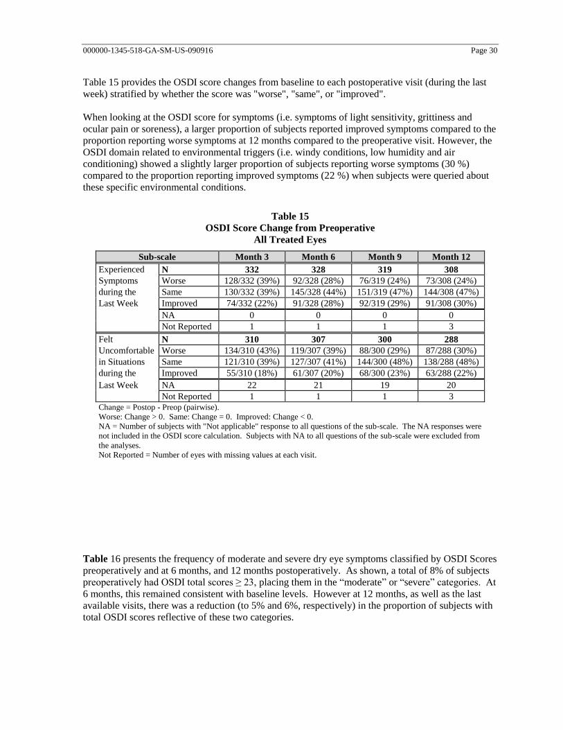
000000-1345-518-GA-SM-US-090916 Page 30
Table 15 provides the OSDI score changes from baseline to each postoperative visit (during the last
week) stratified by whether the score was "worse", "same", or "improved".
When looking at the OSDI score for symptoms (i.e. symptoms of light sensitivity, grittiness and
ocular pain or soreness), a larger proportion of subjects reported improved symptoms compared to the
proportion reporting worse symptoms at 12 months compared to the preoperative visit. However, the
OSDI domain related to environmental triggers (i.e. windy conditions, low humidity and air
conditioning) showed a slightly larger proportion of subjects reporting worse symptoms (30 %)
compared to the proportion reporting improved symptoms (22 %) when subjects were queried about
these specific environmental conditions.
Table 15
OSDI Score Change from Preoperative
All Treated Eyes
Sub-scale Month 3 Month 6 Month 9 Month 12
Experienced N 332 328 319 308
Symptoms Worse 128/332 (39%) 92/328 (28%) 76/319 (24%) 73/308 (24%)
during the Same 130/332 (39%) 145/328 (44%) 151/319 (47%) 144/308 (47%)
Last Week Improved 74/332 (22%) 91/328 (28%) 92/319 (29%) 91/308 (30%)
NA 0 0 0 0
Not Reported 1 1 1 3
Felt N 310 307 300 288
Uncomfortable Worse 134/310 (43%) 119/307 (39%) 88/300 (29%) 87/288 (30%)
in Situations Same 121/310 (39%) 127/307 (41%) 144/300 (48%) 138/288 (48%)
during the Improved 55/310 (18%) 61/307 (20%) 68/300 (23%) 63/288 (22%)
Last Week NA 22 21 19 20
Not Reported 1 1 1 3
Change = Postop - Preop (pairwise).
Worse: Change > 0. Same: Change = 0. Improved: Change < 0.
NA = Number of subjects with "Not applicable" response to all questions of the sub-scale. The NA responses were
not included in the OSDI score calculation. Subjects with NA to all questions of the sub-scale were excluded from
the analyses.
Not Reported = Number of eyes with missing values at each visit.
Table 16 presents the frequency of moderate and severe dry eye symptoms classified by OSDI Scores
preoperatively and at 6 months, and 12 months postoperatively. As shown, a total of 8% of subjects
preoperatively had OSDI total scores ≥ 23, placing them in the “moderate” or “severe” categories. At
6 months, this remained consistent with baseline levels. However at 12 months, as well as the last
available visits, there was a reduction (to 5% and 6%, respectively) in the proportion of subjects with
total OSDI scores reflective of these two categories.

000000-1345-518-GA-SM-US-090916 Page 31
Table 16
Frequency of Moderate and Severe Dry Eye Symptoms Classified by OSDI Scores
All Treated Eyes
Severity of Dry Eye
Symptoms
Preop Month 6 Month 12 Last Available
Visit
N 335 329 309 336
Moderate 16 (5%) 21 (6%) 7 (2%) 10 (3%)
Severe 10 (3%) 6 (2%) 8 (3%) 9 (3%)
Not Reported 1 0 2 0
OSDI score = (sum of scores) x 25/(# of questions answered). The responses of N/A were excluded.
“Moderate”: OSDI score 23 to < 33. “Severe”: OSDI score 33.
Scoring based on Miller et al. Minimal Clinically Important Difference for the Ocular Surface Disease
Index Arch Ophthalmol. 2010;128(1):94-101.
Additional safety outcomes
Corneal topography
Computerized corneal topography was performed in all study subjects preoperatively and at the
3, 6, 9, and 12-month visits using the ATLAS 9000 system. A corneal axial curvature map was
generated for each subject using the standard scale of 38.5 D to 49.5 D. Difference corneal power
maps, i.e. 3 months minus baseline and 6 months minus 3 months, were also generated.
Other than the seven instances during the study, where topographies were mistakenly not performed,
the remaining topographic scans were of high quality in terms of the interpretability of these
topographic maps. As shown in Table 17, there were no postoperative findings suggesting the
development of irregular astigmatism or ectasia or any “other” topographic findings outside of the
specified categories, such as flattening over the superior opening incision, abnormal curvature
changes over the peripheral treatment zone corresponding to the lenticule edge, or localized areas of
steepening corresponding to retained lenticule fragments. Over the course of the study, there were 23
reports of tear film artifacts, involving 17 subjects. Each of these subjects had BSCVA of 20/25 or
better at the respective visits. There were also seven reports of decentration greater than 1 mm,
involving three subjects, but only one subject showed consistent decentration at all four scheduled
postoperative visits indicating true decentration. All three subjects completed the study with BSCVA
of 20/16 or better, but the subject with true decentration also experienced overcorrection of myopia,
induced astigmatism, reduced contrast sensitivity, dry eye symptoms, and temporary loss of more
than two lines of BSCVA. However, there is no definitive relationship of this subject’s post-operative
course to the finding of decentration on topography.

000000-1345-518-GA-SM-US-090916 Page 32
Table 17
Topography Findings
All Treated Eyes
Preop Month 3 Month 6 Month 9 Month 12 n/N (%) n/N (%) n/N (%) n/N (%) n/N (%)
Evaluable 335 332 325 319 311
Irregular Astigmatism 0/335 (0.0%) 0/332 (0.0%) 0/325 (0.0%) 0/319 (0.0%) 0/311 (0.0%)
Ectasia 0/335 (0.0%) 0/332 (0.0%) 0/325 (0.0%) 0/319 (0.0%) 0/311 (0.0%)
Tear Film Artifacts 3/335 (0.9%) 3/332 (0.9%) 9/325 (2.8%) 3/319 (0.9%) 5/311 (1.6%)
Decentration NA 1/332 (0.3%) 2/325 (0.6%) 2/319 (0.6%) 2/311 (0.6%)
Other 0/335 (0.0%) 0/332 (0.0%) 0/325 (0.0%) 0/319 (0.0%) 0/311 (0.0%)
Topography not performed 1 1 4 1 0
Total 336 333 329 320 311
N = Number of eyes with non-missing values at each visit. % = n/N 100.
Wavefront outcomes
Wavefront aberrometry measurements were obtained at preoperatively, then at 3 and 12 months
postoperatively, using the Tracey™ iTrace aberrometer, which has an infrared laser wavelength of
785 nm. A Zernike analysis was performed to evaluate the effect of the SMILE procedure on
aberrations in the treated eyes. Calculations of the total higher order-order aberrations root mean
square (HORMS) on the iTrace encompass the 6th order of Zernike polynomial terms. Since
quantification of wavefront aberrations are dependent on pupil size, wavefront aberrometry was
assessed at fixed pupil sizes. Depending on each subject’s mesopic pupil size, wavefront images were
obtained at 4.0 mm, 5.0 mm, and/or 6.0 mm, so that reliable data at the largest pupil size obtainable
for each given subject could be compared preoperatively and postoperatively. If the pupil of any
subject did not reach the minimum size necessary to obtain a reliable image at 4.0 mm, no
comparisons of wavefront scans were performed.
Table 18 summarizes the change in wavefront aberrometry findings from baseline at the 3-month and
12-month visits, stratified by the largest scan size (i.e., 4.0, 5.0, or 6.0 mm) obtained preoperatively
and postoperatively at these time points. These are specified in terms of the RMS changes of Zernike
coefficients from baseline for the total HORMS, as well as for coma (microns) and spherical
aberration (microns), specifically. Of the 311 eyes with 12-month visits at the time of the database
lock, wavefront data for 250 eyes at the preoperative, 3-month, and 12-month visits were available for
the comparison presented in Table 18.
Total HORMS for wavefront images obtained with an image size of 4.0 mm was essentially the same
at baseline (0.175) and 12 months (0.173). Only a small increase in total HORMS was observed for
5.0 mm scan sizes (from 0.289 at baseline to 0.340), with slightly larger increases for 6.0 mm scan
sizes (from 0.450 at baseline to 0.698), as expected. The mean changes in total HORMS from
baseline to 12 months for scan sizes of 5.0 and 6.0 mm were 0.051 and 0.248, respectively.

000000-1345-518-GA-SM-US-090916 Page 33
With regard to postoperative changes in coma and spherical aberration for 4.0 mm scan sizes at 3 and
12 months, there was essentially no change. At the 5.0 mm scan size, the postoperative changes at
both time points with respect to coma and spherical aberration were slightly more evident, with
changes less than 0.07 microns for coma and less than 0.05 microns for spherical aberration. Not
unexpected were the more pronounced corresponding changes at the 6.0 mm scan size, though only
16 scans were available for analysis.
Table 18
Change in Wavefront Aberrometry from Preoperative
Stratified by Largest Scan Size (mm)
Treated Eyes with Preoperative, 3-Month, and 12-Month Visits
Scan Size Parameters Statistics Month 3 Month 12
4.0 Change in Wavefront from Preoperative (micron)
Total Higher N 106 106
Order RMS Mean (SD) -0.001 (0.086) -0.002 (0.079)
Min, Max -0.310, 0.234 -0.341, 0.142
Coma Mean (SD) 0.010 (0.087) 0.013 (0.079)
Min, Max -0.268, 0.258 -0.274, 0.199
Spherical Mean (SD) -0.002 (0.047) -0.002 (0.047)
Min, Max -0.201, 0.120 -0.180, 0.112
5.0 Change in Wavefront from Preoperative (micron)
Total Higher N 128 128
Order RMS Mean (SD) 0.049 (0.141) 0.051 (0.149)
Min, Max -0.293, 0.397 -0.311, 0.365
Coma Mean (SD) 0.070 (0.170) 0.064 (0.170)
Min, Max -0.288, 0.515 -0.420, 0.467
Spherical Mean (SD) 0.048 (0.100) 0.046 (0.101)
Min, Max -0.184, 0.400 -0.174, 0.339
6.0 Change in Wavefront from Preoperative (micron)
Total Higher N 16 16
Order RMS Mean (SD) 0.220 (0.268) 0.248 (0.314)
Min, Max -0.239, 0.766 -0.248, 0.806
Coma Mean (SD) 0.232 (0.289) 0.261 (0.326)
Min, Max -0.255, 0.793 -0.392, 0.874
Spherical Mean (SD) 0.129 (0.196) 0.149 (0.173)
Min, Max -0.285, 0.511 -0.255, 0.414
Overall Change in Wavefront from Preoperative (micron)
Total Higher N 250 250
Order RMS Mean (SD) 0.039 (0.143) 0.041 (0.153)
Min, Max -0.310, 0.766 -0.341, 0.806
Coma Mean (SD) 0.055 (0.161) 0.055 (0.165)
Min, Max -0.288, 0.793 -0.420, 0.874
Spherical Mean (SD) 0.032 (0.098) 0.032 (0.097)
Min, Max -0.285, 0.511 -0.255, 0.414
N = Number of case report forms received with non-missing values at each visit.
The largest scan size was 4.0, 5.0, or 6.0 mm, depending on the largest scan size obtained at all the
preoperative and postoperative visits
.

000000-1345-518-GA-SM-US-090916 Page 34
Key effectiveness outcomes
A summary of the key effectiveness outcomes for all 333 eyes in the effectiveness population is
shown in Table 19. While there were no eyes preoperatively with UCVA of 20/40 or better, at the 6-
month visit, 99.7 % (327/328) and 87.5 % (287/328) of treated eyes achieved UCVA levels of 20/40
or better and 20/20 or better, respectively. With respect to MRSE predictability, 93.0% and 98.5%
achieved MRSE within ±0.50D and ±1.00D, respectively, of the attempted correction.
Table 19
Summary of Key Effectiveness Variables
Effectiveness Cohort Eyes
Key Week 1 Month 1 Month 3 Month 6 Month 9 Month 12
Effectiveness
Variables
n/N (%)
95 % CI
n/N (%)
95 % CI
n/N (%)
95 % CI
n/N (%)
95 % CI
n/N (%)
95 % CI
n/N (%)
95 % CI
Effectiveness Variables (Effectiveness Population)
UCVA, 20/16 or better 95/332 (28.6 %)
(23.8 %, 33.8 %)
155/333 (46.5 %)
(41.1 %, 52.1 %)
192/331 (58.0 %)
(52.5 %, 63.4 %)
197/328 (60.1 %)
(54.5 %, 65.4 %)
196/319 (61.4 %)
(55.9 %, 66.8 %)
198/310 (63.9 %)
(58.2 %, 69.2 %)
UCVA, 20/20 or better 210/332 (63.3 %)
(57.8 %, 68.5 %)
262/333 (78.7 %)
(73.9 %, 83.0 %)
282/331 (85.2 %)
(80.9 %, 88.8 %)
287/328 (87.5 %)
(83.4 %, 90.9 %)
281/319 (88.1 %)
(84.0 %, 91.4 %)
273/310 (88.1 %)
(83.9 %, 91.5 %)
UCVA, 20/25 or better 286/332 (86.1 %)
(82.0 %, 89.7 %)
308/333 (92.5 %)
(89.1 %, 95.1 %)
317/331 (95.8 %)
(93.0 %, 97.7 %)
313/328 (95.4 %)
(92.6 %, 97.4 %)
309/319 (96.9 %)
(94.3 %, 98.5 %)
301/310 (97.1 %)
(94.6 %, 98.7 %)
UCVA, 20/32 or better 315/332 (94.9 %)
(91.9 %, 97.0 %)
324/333 (97.3 %)
(94.9 %, 98.8 %)
324/331 (97.9 %)
(95.7 %, 99.1 %)
322/328 (98.2 %)
(96.1 %, 99.3 %)
315/319 (98.7 %)
(96.8 %, 99.7 %)
305/310 (98.4 %)
(96.3 %, 99.5 %)
UCVA, 20/40 or better 325/332 (97.9 %)
(95.7 %, 99.1 %)
333/333 (100.0 %)
(98.9 %, 100.0 %)
329/331 (99.4 %)
(97.8 %, 99.9 %)
327/328 (99.7 %)
(98.3 %, 100.0 %)
318/319 (99.7 %)
(98.3 %, 100.0 %)
309/310 (99.7 %)
(98.2 %, 100.0 %)
MRSE, Attempted vs.
Achieved, ±0.25D
262/331 (79.2 %)
(74.4 %, 83.4 %)
264/333 (79.3 %)
(74.5 %, 83.5 %)
262/331 (79.2 %)
(74.4 %, 83.4 %)
261/328 (79.6 %)
(74.8 %, 83.8 %)
258/319 (80.9 %)
(76.1 %, 85.0 %)
250/310 (80.6 %)
(75.8 %, 84.9 %)
MRSE, Attempted vs.
Achieved, ±0.50D
308/331 (93.1 %)
(89.8 %, 95.5 %)
310/333 (93.1 %)
(89.8 %, 95.6 %)
304/331 (91.8 %)
(88.4 %, 94.6 %)
305/328 (93.0 %)
(89.7 %, 95.5 %)
303/319 (95.0 %)
(92.0 %, 97.1 %)
291/310 (93.9 %)
(90.6 %, 96.3 %)
MRSE, Attempted vs.
Achieved, ±1.00D
328/331 (99.1 %)
(97.4 %, 99.8 %)
331/333 (99.4 %)
(97.8 %, 99.9 %)
328/331 (99.1 %)
(97.4 %, 99.8 %)
323/328 (98.5 %)
(96.5 %, 99.5 %)
316/319 (99.1 %)
(97.3 %, 99.8 %)
306/310 (98.7 %)
(96.7 %, 99.6 %)
MRSE, Attempted vs.
Achieved, ±2.00D
331/331
(100.0 %)
(98.9 %, 100.0 %)
333/333
(100.0 %)
(98.9 %, 100.0 %)
331/331
(100.0 %)
(98.9 %, 100.0 %)
328/328
(100.0 %)
(98.9 %, 100.0 %)
319/319
(100.0 %)
(98.9 %, 100.0 %)
310/310
(100.0 %)
(98.8 %, 100.0 %)
N = Number of case report forms received with non-missing values at each visit.
95 % CI was calculated based on Clopper-Pearson exact method.
Stratification by age revealed differences in proportions of eyes achieving UCVA of 20/20 or better,
with lower proportions of eyes in the 40 to 49 and 50+ age groups achieving UCVA of 20/20 or better
at the point of stability (72.0% for the 40 to 49 years subgroup and 71.4% for the 50 years & above
subgroup). Despite these slight differences, the older age groups still experienced a clinically
significant visual benefit as 98.4% of the subjects in these age bins achieved UCVA of 20/40 or better
postoperatively.

000000-1345-518-GA-SM-US-090916 Page 35
Key effectiveness outcomes stratified by preoperative manifest refraction sphere (MRSPH)
The key effectiveness variables at 6 months stratified by preoperative MRSPH are presented below
in Table 20. As shown below, there was no obvious clinically significant impact of preoperative
MRSPH on 6-month outcomes for UCVA of 20/40 or better or on outcomes for achieved MRSE
within ± 0.50 D of attempted MRSPH.
Table 20
Summary of Key Effectiveness Variables at 6 Months
Stratified By Preoperative MRSPH
Effectiveness Population
Key Preop MRSPH
Effectiveness 0.00 to -1.00 D -1.01 to -2.00 D -2.01 to -3.00 D -3.01 to -4.00 D -4.01 to -5.00 D -5.01 to -6.00 D
Variables n/N (%) n/N (%) n/N (%) n/N (%) n/N (%) n/N (%)
UCVA, 20/16 or
better
2/4
(50.0%)
20/35
(57.1%)
35/52
(67.3%)
37/50
(74.0%)
32/48
(66.7%)
23/42
(54.8%)
UCVA, 20/20 or
better
3/4
(75.0%)
32/35
(91.4%)
48/52
(92.3%)
48/50
(96.0%)
44/48
(91.7%)
35/42
(83.3%)
UCVA, 20/25 or
better
4/4
(100.0%)
33/35
(94.3%)
51/52
(98.1%)
48/50
(96.0%)
47/48
(97.9%)
40/42
(95.2%)
UCVA, 20/32 or
better
4/4
(100.0%)
35/35
(100.0%)
51/52
(98.1%)
48/50
(96.0%)
48/48
(100.0%)
40/42
(95.2%)
UCVA, 20/40 or
better
4/4
(100.0%)
35/35
(100.0%)
52/52
(100.0%)
49/50
(98.0%)
48/48
(100.0%)
42/42
(100.0%)
MRSE, Attempted vs.
Achieved, ±0.25D
3/4
(75.0%)
29/35
(82.9%)
45/52
(86.5%)
46/50
(92.0%)
40/48
(83.3%)
34/42
(81.0%)
MRSE, Attempted vs.
Achieved, ±0.50D
4/4
(100.0%)
34/35
(97.1%)
50/52
(96.2%)
48/50
(96.0%)
45/48
(93.8%)
38/42
(90.5%)
MRSE, Attempted vs.
Achieved, ±1.00D
4/4
(100.0%)
35/35
(100.0%)
52/52
(100.0%)
49/50
(98.0%)
48/48
(100.0%)
41/42
(97.6%)
MRSE, Attempted vs.
Achieved, ±2.00D
4/4
(100.0%)
35/35
(100.0%)
52/52
(100.0%)
50/50
(100.0%)
48/48
(100.0%)
42/42
(100.0%)
Key Preop MRSPH Total
Effectiveness -6.01 to -7.00 D -7.01 to -8.00 D -8.01 to -9.00 D1 > -9.01 D
1
Variables n/N (%) n/N (%) n/N (%) n/N (%) n/N (%)
UCVA, 20/16 or
better
25/42
(59.5%)
16/28
(57.1%)
4/15
(26.7%)
3/12
(25.0%)
197/328
(60.1%)
UCVA, 20/20 or
better
36/42
(85.7%)
24/28
(85.7%)
9/15
(60.0%)
8/12
(66.7%)
287/328
(87.5%)
UCVA, 20/25 or
better
39/42
(92.9%)
27/28
(96.4%)
13/15
(86.7%)
11/12
(91.7%)
313/328
(95.4%)
UCVA, 20/32 or
better
42/42
(100.0%)
28/28
(100.0%)
15/15
(100.0%)
11/12
(91.7%)
322/328
(98.2%)
UCVA, 20/40 or
better
42/42
(100.0%)
28/28
(100.0%)
15/15
(100.0%)
12/12
(100.0%)
327/328
(99.7%)
MRSE, Attempted vs.
Achieved, ±0.25D
31/42
(73.8%)
16/28
(57.1%)
11/15
(73.3%)
6/12
(50.0%)
261/328
(79.6%)
MRSE, Attempted vs.
Achieved, ±0.50D
38/42
(90.5%)
24/28
(85.7%)
14/15
(93.3%)
10/12
(83.3%)
305/328
(93.0%)
MRSE, Attempted vs.
Achieved, ±1.00D
41/42
(97.6%)
27/28
(96.4%)
15/15
(100.0%)
11/12
(91.7%)
323/328
(98.5%)
MRSE, Attempted vs.
Achieved, ±2.00D
42/42
(100.0%)
28/28
(100.0%)
15/15
(100.0%)
12/12
(100.0%)
328/328
(100.0%)
N = Number of case report forms received with non-missing values for each group. 1 Please note that treatment of -8.01 to -10.0 D will present a flagged warning to the users so that the user
understands that correction of these powers had not been substantiated by an adequate set of data.

000000-1345-518-GA-SM-US-090916 Page 36
Postoperative UCVA versus preoperative BSCVA
Postoperative UCVA results, as compared to preoperative BSCVA at all scheduled visits, are
presented in Table 21. Over the course of the study from Day 1 through Month 12, there was a steady
increase in the proportion of eyes with UCVA equal to or better than preoperative BSCVA.
Table 21
Postoperative Uncorrected Visual Acuity (UCVA) Compared
to Preoperative Best Spectacle Corrected Visual Acuity (BSCVA)
Effectiveness Cohort Eyes
Day 1 Week 1 Month 1 Month 3
UCVA n (%) n (%) n (%) n (%)
Available (N) 333 332 333 331
UCVA >2 Lines Better
than Preop BSCVA
0 (0.0 %) 0 (0.0 %) 0 (0.0 %) 0 (0.0 %)
UCVA 2 Lines Better than
Preop BSCVA
1 (0.3 %) 1 (0.3 %) 5 (1.5 %) 12 (3.6 %)
UCVA 1 Line Better than
Preop BSCVA
10 (3.0 %) 20 (6.0 %) 56 (16.8 %) 71 (21.5 %)
UCVA Equal to Preop
BSCVA
51 (15.3 %) 106 (31.9 %) 125 (37.5 %) 137 (41.4 %)
UCVA 1 Line Worse than
Preop BSCVA
111 (33.3 %) 109 (32.8 %) 94 (28.2 %) 73 (22.1 %)
UCVA 2 Lines Worse than
Preop BSCVA
86 (25.8 %) 56 (16.9 %) 31 (9.3 %) 25 (7.6 %)
UCVA >2 Lines Worse
than Preop BSCVA
74 (22.2 %) 40 (12.0 %) 22 (6.6 %) 13 (3.9 %)
UCVA Better than or
Equal to Preop BSCVA
62 (18.6 %) 127 (38.3 %) 186 (55.9 %) 220 (66.5 %)
Not reported 0 0 0 0
Total 333 332 333 331
Month 6 Month 9 Month 12
UCVA n (%) n (%) n (%)
Available (N) 328 319 310
UCVA >2 Lines Better
than Preop BSCVA
1 (0.3 %) 1 (0.3 %) 3 (1.0 %)
UCVA 2 Lines Better than
Preop BSCVA
12 (3.7 %) 4 (1.3 %) 13 (4.2 %)
UCVA 1 Line Better than
Preop BSCVA
83 (25.3 %) 93 (29.2 %) 93 (30.0 %)
UCVA Equal to Preop
BSCVA
133 (40.5 %) 132 (41.4 %) 119 (38.4 %)
UCVA 1 Line Worse than
Preop BSCVA
67 (20.4 %) 59 (18.5 %) 57 (18.4 %)
UCVA 2 Lines Worse than
Preop BSCVA
19 (5.8 %) 20 (6.3 %) 18 (5.8 %)
UCVA >2 Lines Worse
than Preop BSCVA
13 (4.0 %) 10 (3.1 %) 7 (2.3 %)
UCVA Better than or
Equal to Preop BSCVA
229 (69.8 %) 230 (72.1 %) 228 (73.5 %)

000000-1345-518-GA-SM-US-090916 Page 37
Not reported 0 0 0
Total 328 319 310
N = Number of case report forms received with non-missing values at each visit.
Accuracy of MRSE
Accuracy of the intended refractive correction, with respect to MRSE, is shown in Table 22 for the 6-
month consistent effectiveness cohort. This cohort consists of all eyes from the effectiveness cohort
with every follow-up exam from 1 week onward to the 6-month point of stability. The deviation of
MRSE is considered in terms of a refractive target that is not necessarily emmetropia, due to the
astigmatic components of 0.25 D and 0.50 D that were not treated in the study.
The MRSE was within ±1.00 D of attempted correction in over 98 % of eyes at all study visits.
No less than 79 % of eyes were within ±0.25 D, and no less than 92 % of eyes were within ±0.50 D
of the targeted MRSE correction from the 1-week through 12-month visits. There were no reports of
overcorrection > 1.00 D MRSE at any point in the study.
Table 22
Accuracy of MRSE — Attempted vs. Achieved
6-Month Consistent Effectiveness Cohort
MRSE Week 1 Month 1 Month 3
Deviation n/N (%) n/N (%) n/N (%)
Available (N) 326 327 327
± 0.25 D 257/326 (78.8%) 259/327 (79.2%) 258/327 (78.9%)
± 0.50 D 303/326 (92.9%) 304/327 (93.0%) 300/327 (91.7%)
± 1.00 D 323/326 (99.1%) 325/327 (99.4%) 324/327 (99.1%)
± 2.00 D 326/326 (100.0%) 327/327 (100.0%) 327/327 (100.0%)
Overcorrected > 1.00 D 0/326 (0.0%) 0/327 (0.0%) 0/327 (0.0%)
Overcorrected > 2.00 D 0/326 (0.0%) 0/327 (0.0%) 0/327 (0.0%)
Undercorrected > 1.00 D 3/326 (0.9%) 2/327 (0.6%) 3/327 (0.9%)
Undercorrected > 2.00 D 0/326 (0.0%) 0/327 (0.0%) 0/327 (0.0%)
Mean (SD) 0.035 (0.311) 0.054 (0.301) 0.062 (0.328)
Range -1.000, 1.375 -1.000, 1.375 -0.875, 1.875
Not reported 1 0 0
Total 327 327 327
MRSE Month 6 Month 9 Month 12
Deviation n/N (%) n/N (%) n/N (%)
Available (N) 327 316 307
± 0.25 D 260/327 (79.5%) 256/316 (81.0%) 248/307 (80.8%)
± 0.50 D 304/327 (93.0%) 300/316 (94.9%) 288/307 (93.8%)
± 1.00 D 322/327 (98.5%) 313/316 (99.1%) 303/307 (98.7%)
± 2.00 D 327/327 (100.0%) 316/316 (100.0%) 307/307 (100.0%)
Overcorrected > 1.00 D 0/327 (0.0%) 0/316 (0.0%) 0/307 (0.0%)
Overcorrected > 2.00 D 0/327 (0.0%) 0/316 (0.0%) 0/307 (0.0%)
Undercorrected > 1.00 D 5/327 (1.5%) 3/316 (0.9%) 4/307 (1.3%)
Undercorrected > 2.00 D 0/327 (0.0%) 0/316 (0.0%) 0/307 (0.0%)
Mean (SD) 0.041 (0.325) 0.023 (0.292) 0.017 (0.309)
Range -0.750, 1.750 -0.875, 1.250 -1.000, 1.750
Not reported 0 0 0
Total 327 316 307
N = Number of case report forms received with non-missing values at each visit.

000000-1345-518-GA-SM-US-090916 Page 38
Stability of MRSE
As presented in Table 23, MRSE stability was identified at the 3 to 6 month interval and confirmed at
the 6 to 9 month interval.
Table 23
Stability of Manifest Refraction Spherical Equivalent (MRSE)
Effectiveness Population
Change in MRSE Between 1 and
3 Months
Between 3 and
6 Months
Between 6 and
9 Months
Between 9 and
12 Months
Pairwise Sequential Visits
Eyes within 0.50 D change
(n/N, %, [% CI]1)
319/331 (96.4%)
(93.8%, 98.1%)
317/327 (96.9%)
(94.4%, 98.5%)
311/317 (98.1%)
(95.9%, 99.3%)
301/308 (97.7%)
(95.4%, 99.1%)
Eyes within 1.00 D change
(n/N, %, [% CI]1)
331/331 (100.0%)
(98.9%, 100.0%)
327/327 (100.0%)
(98.9%, 100.0%)
316/317 (99.7%)
(98.3%, 100.0%)
307/308 (99.7%)
(98.2%, 100.0%)
Mean change between visits (D)
SD
95% CI
-0.008
0.241
(-0.034, 0.018)
0.021
0.232
(-0.005, 0.046)
0.013
0.213
(-0.011, 0.037)
0.005
0.198
(-0.017, 0.027)
Mean change per month (D) -0.004 0.007 0.004 0.002
Mean change per year (D)
(change per month 12)
-0.048 0.083 0.052 0.019
12-Month Consistent Cohort
Eyes within 0.50 D change
(n/N, %, [% CI]1)
296/305 (97.0%)
(94.5%, 98.6%)
298/305 (97.7%)
(95.3%, 99.1%)
301/305 (98.7%)
(96.7%, 99.6%)
298/305 (97.7%)
(95.3%, 99.1%)
Eyes within 1.00 D change
(n/N, %, [% CI]1)
305/305 (100.0%)
(98.8%, 100.0%)
305/305 (100.0%)
(98.8%, 100.0%)
304/305 (99.7%)
(98.2%, 100.0%)
304/305 (99.7%)
(98.2%, 100.0%)
Mean change between visits (D)
SD
95% CI
0.002
0.231
(-0.024, 0.028)
0.014
0.219
(-0.011, 0.039)
0.013
0.205
(-0.010, 0.036)
0.005
0.198
(-0.018, 0.027)
Mean change per month (D) 0.001 0.005 0.004 0.002
Mean change per year (D)
(change per month 12)
0.010 0.056 0.051 0.018
Pairwise Sequential Visits = Eyes that had two consecutive exams, but not necessarily every follow-up exam.
Consistent Cohort = All eyes examined at 1, 3, 6, 9 and 12 months. 1 95% CI was calculated based on Clopper-Pearson method.

000000-1345-518-GA-SM-US-090916 Page 39
Surgical planning and procedures
Laser activation, calibration, and surgical room environmental control
Detailed information on general operation of the VisuMax is provided
in the VisuMax Femtosecond Laser user manual
Detailed information on general operation of the VisuMax as well as
the planning and execution of treatment is to be found in the VisuMax
Femtosecond Laser user manual
The stromal thickness shown is a theoretical anticipated value. If
changes occur in the course of the operation the achieved residual
stromal thickness may deviate from the value shown. Please take into
account that the calculation of residual stromal thickness is based on
the pachymetry value entered, which is subject to measurement
uncertainty.
General VisuMax procedure overview
Note that the following procedure overview is general for all VisuMax procedures. See
Treatment Planning, page 45 and following, for complete procedure instructions.
1. The surgeon or other suitably trained personnel switches on the VisuMax Femtosecond Laser and
initiates the surgical start-up routine.
2. The monitor on the left-hand side in combination with the keyboard/trackball is used to enter
patient data and patient related treatment parameters as part of the treatment planning process.
Treatment parameters entered by the surgeon may include patient eye manifest refraction,
pachymetry, lenticule diameter, cap diameter, etc. After the appropriate treatment parameters have
been entered, the particular resection parameters for the procedure are displayed to the surgeon on
the video display. The surgeon must review and verify the parameters (i.e. the residual stromal
thickness for corneal caps, or the lenticule diameter for lenticule removal).
3. When the treatment planning is complete, the keyboard tray is pushed back into the VisuMax
Femtosecond Laser console housing. This activates the surgery controls on the touch screen
(located on the right hand side of the laser arm).
4. The patient is then positioned on the Patient Supporting System (PSS). The PSS is manually
rotated into the observation position underneath the laser arm.
5. The PSS with the patient situated properly is then moved using the PSS translation control joystick
to center the eye to be treated in the observation position under the surgical microscope.
6. Using the surgical microscope, the surgeon prepares the eye to be treated.
7. The touch screen display is used to start the surgical routine. The treatment parameters previously
entered should be verified.

000000-1345-518-GA-SM-US-090916 Page 40
8. Instructions are displayed on the touch screen display by a treatment wizard, and should be
followed by the surgeon.
9. The surgeon places the sterile contact glass of the Treatment Pack onto the treatment objective
lens and connects the filter and vacuum connector to the Vacuum Connection fixture on the laser
console when indicated by the treatment wizard on the touch screen display.
10. The treatment objective lens is then slightly lifted by the physician when indication is shown on
the touchscreen display. The VisuMax Femtosecond Laser then automatically initiates a system
test.
11. After the system test has been completed, the surgeon checks the preparation of the eye once
again and moves the Patient Supporting System into the treatment position.
12. In the treatment position, the surgeon positions the eye to be treated against the contact glass so
that the lens applanates the patient’s cornea. Taking care to center the contact glass with respect to
the optical axis of the patient’s eye, the cornea is then completely pressed against the contact glass.
The surgeon then presses the vacuum suction control which produces appropriate suction force to
cause the eye to adhere to the contact glass.
13. When the suction pressure is sensed by the laser console hardware to reach the acceptable range,
the READY mode indicator becomes active.
14. The laser surgery procedure is initiated by pressing and holding down the foot switch.
Carefully monitor centration and suction throughout the laser treatment
initiation.
15. Upon completion of the laser procedure, the vacuum is automatically released and the patient’s
eye is separated from the contact glass. The surgeon returns the PSS and patient to the observation
position using the PSS joystick.
16. The physician uses the VisuMax Femtosecond Laser surgical microscope to complete the
treatment under direct visualization, as required.
17. When the entire surgical procedure has been completed, the surgeon moves the PSS into the
patient exit position.
18. Finally, the physician clears the surgery area and closes the treatment routine in the user
interface/Touch Screen Display. The software returns the display to the main screen, and is now
ready for a new surgical procedure.
Treatment license
The VisuMax SMILE procedure requires activation by a treatment license to carry out the surgery.
The treatment license will be provided by Carl Zeiss Meditec or its authorized representatives based
on the commercial agreement.
Treatment parameters
Treatment parameters available for the VisuMax lenticule procedure are provided in the Technical
Data section of this document.

000000-1345-518-GA-SM-US-090916 Page 41
Treatment planning
Treatment License - The VisuMax SMILE procedure requires
activation by a treatment license for the specific procedure to carry
out the surgery. The treatment license will be provided by Carl Zeiss
Meditec or its authorized representatives based on the commercial
agreement.
For system start-up, patient record management, and general laser
operating instructions, see the VisuMax Femtosecond Laser's user
manual.
Treatment planning steps are provided in this section.
1. Switch the laser system on. The computer will execute several internal test routines and then
automatically start the main menu.
2. Select the small incision lenticule extraction procedure.
3. To enter procedure parameters, use the two separate tabs displayed in the graphical user interface
(GUI) window. The window shows two separate tabs: the Lenticule Tab and the Cap Tab.
The Lenticule tab contains procedure parameters associated with the refractive aspect of the
procedure. The Cap tab contains parameters related to the creation of the opening and lenticule access
cuts.
Parameters may be entered directly using the trackball and cursor, or by simply using the cursor to
increment the up and down arrows. Some parameters are not adjustable.
NOTE: Laser parameters cannot be modified by the users.
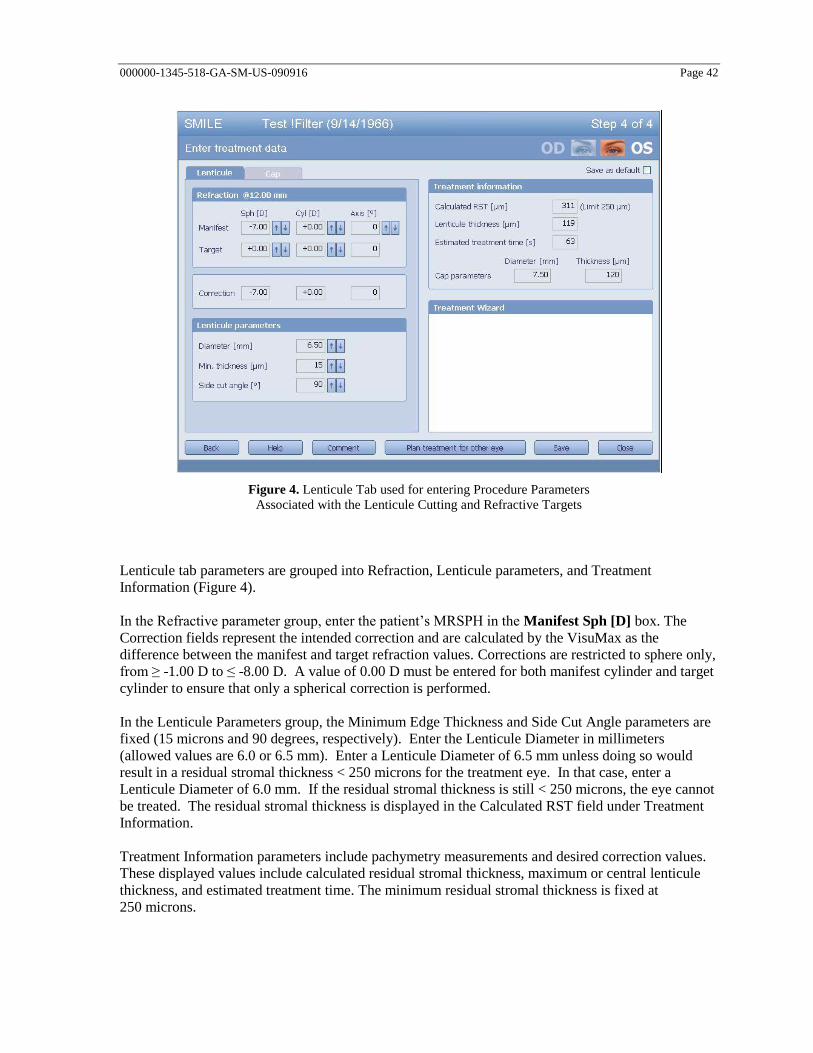
000000-1345-518-GA-SM-US-090916 Page 42
Figure 4. Lenticule Tab used for entering Procedure Parameters
Associated with the Lenticule Cutting and Refractive Targets
Lenticule tab parameters are grouped into Refraction, Lenticule parameters, and Treatment
Information (Figure 4).
In the Refractive parameter group, enter the patient’s MRSPH in the Manifest Sph [D] box. The
Correction fields represent the intended correction and are calculated by the VisuMax as the
difference between the manifest and target refraction values. Corrections are restricted to sphere only,
from ≥ -1.00 D to ≤ -8.00 D. A value of 0.00 D must be entered for both manifest cylinder and target
cylinder to ensure that only a spherical correction is performed.
In the Lenticule Parameters group, the Minimum Edge Thickness and Side Cut Angle parameters are
fixed (15 microns and 90 degrees, respectively). Enter the Lenticule Diameter in millimeters
(allowed values are 6.0 or 6.5 mm). Enter a Lenticule Diameter of 6.5 mm unless doing so would
result in a residual stromal thickness < 250 microns for the treatment eye. In that case, enter a
Lenticule Diameter of 6.0 mm. If the residual stromal thickness is still < 250 microns, the eye cannot
be treated. The residual stromal thickness is displayed in the Calculated RST field under Treatment
Information.
Treatment Information parameters include pachymetry measurements and desired correction values.
These displayed values include calculated residual stromal thickness, maximum or central lenticule
thickness, and estimated treatment time. The minimum residual stromal thickness is fixed at
250 microns.

000000-1345-518-GA-SM-US-090916 Page 43
Cap tab parameters (Figure 5) are grouped into Anatomical Parameters, Treatment Pack size, Cap
Parameters, Incision Parameters, and Treatment Information.
The Cap tab is used to select or indicate the eye to be treated. A graphic of both eyes with
a designation is shown in the upper right part of the window. Use the cursor to select the appropriate
eye. The graphic will highlight your choice.
In the Anatomical parameters group, biometric values are entered. Enter the pachymetry values in the
appropriate box. The average corneal radius may be entered directly into the Corneal Radius field.
Alternatively, K-readings may be entered. To enter K-readings, select the K-readings button and enter
values for K min and K max. The corneal radius is calculated using the index of refraction stored in
the Settings window and the formula:
1/Kmean(mm) = (1/Kmax(mm) + 1/Kmin(mm) )/2.0
In the Treatment Pack size group, only the small size can be selected.
In the Cap Parameters group, enter a Cap Diameter of 7.5 mm (if a 6.5 mm lenticule diameter
was specified) or 7.0 mm (if a 6.0 mm lenticule diameter was specified). Cap Thickness is
fixed at 120 microns and Side Cut Angle is fixed at 90 degrees.
In the Incision Parameters group, only a single incision can be selected. The Position
parameter refers to the opening incision clock position, and is fixed at the superior location.
The Angle parameter refers to the angular width of the procedure, and is fixed at 90 degrees.
The width parameter is calculated based on the Angle parameter and the Cap Diameter
parameter and is not independently selected.
Treatment Information parameters are again summarized/displayed for convenience. The
Treatment Wizard box indicates any problems associated with selected treatment parameters.
If any parameters entered are incorrect or inconsistent, appropriate fields will be highlighted
in red and the user can select and adjust the indicated fields to correct any errors.

000000-1345-518-GA-SM-US-090916 Page 44
Figure 5. Cap Tab used for entering Procedure Parameters
Associated with the Opening Cut and Cap Cut
The user is to proceed as per the following instructions:
Click Save to store treatment data. Click Close to return to step 1 - Select patient.
To cancel the treatment planning without storing the data, click on Close without previously
saving.
Once the planning is complete, click on Close to return to step 1. Click on Cancel to quit
planning and return to the main dialog.
Once the treatment planning has been completed, push the keyboard back into the VisuMax
housing. This procedure activates the treatment check on the touchscreen on the right-hand
side.

000000-1345-518-GA-SM-US-090916 Page 45
Laser treatment and SMILE procedure
WARNING
Upon connection of the Treatment Pack you will be asked via the
graphical user interface to perform an excursion test. When testing
the excursion by lifting the treatment objective, the treatment
objective must travel smoothly. If you feel that the treatment
objective does not move freely, shut down the laser and contact
Carl Zeiss Meditec customer service. As soon as the treatment
objective is moved, the system test will be started, a status bar on the
display shows the progress and end of the test.

000000-1345-518-GA-SM-US-090916 Page 46
CAUTION
Use only Treatment Packs expressly approved by Carl Zeiss Meditec. If
Treatment Packs not expressly approved by Carl Zeiss Meditec are used,
there is a risk of treatment errors.
Examine the wrappings of the Treatment Pack to ensure there is no damage
before removing the Treatment Pack. Do not use a Treatment Pack if you
are not certain that it is sterile. In the subsequent procedure take steps to
ensure that the Treatment Pack remains sterile! Treatment Packs are
disposable articles. The re-sterilization of Treatment Packs is not permitted.
Considerable risk of injury to the patient exists in re-sterilization.
Ensure sterility, especially of the contact glass!
Ensure that the filter is correctly attached.
Ensure that no liquid is allowed to enter the vacuum system to avoid
suction loss with termination of treatment.
Do not use a contacting agent, as the desired result will otherwise not be
achieved.
Take special care to ensure precise alignment of the patient's eye.
Continuously optimize the eye position along the X and Y axes as the eye is
brought closer to the contact glass.
Surgeons should be vigilant for possible small eye movements through the
operating microscope during the procedure. There can be a relative shift of
the pupil center during the operation, which does not necessarily entail a
shift of the cornea. The process must be stopped immediately if the size and
position of the incision deviate from the intended treatment.
Prior to and throughout treatment, ensure adequate suction. Total surgery
time should be kept as short as possible, minimizing conditions which may
distract the patient.
Observe the entire surgical procedure through the surgical microscope. The
process must be stopped immediately if the size and position of the incision
deviate from the intended treatment. This may otherwise result in treatment
errors. Do not perform surgery if the incisions are incorrectly positioned!
The formation of bubbles at the periphery of the suction zone is an
indication of imminent suction loss. In the event of a complete loss of
suction, the VisuMax console detects the reduction in pressure of the eye
and the procedure is automatically halted.

000000-1345-518-GA-SM-US-090916 Page 47
Preparation
1. Adjust the microscope positioning controls and adjust the microscope magnification as required.
2. Bring the patient into the operating room and have the patient lie comfortably supine on the patient
supporting system (PSS) such that his/her eye is appropriately positioned.
3. Ready the Treatment Pack by first examining the packaging to verify that there is
no damage. Remove the sterile Treatment Pack from the packaging. Do not use a Treatment Pack
if you are not certain that it is sterile.
4. Connect the filter from the Treatment Pack to the vacuum connection on the laser console.
5. Place the sterile contact glass on the laser aperture. The contact glass will now be held to the
treatment objective by suction pressure.
6. The GUI instructs the user to manually lift the treatment objective. If the treatment objective does
not move freely, abort the procedure, shut down the VisuMax Femtosecond Laser and contact
customer service.
7. Verify patient, eye, and prescription to be treated.
8. Approximately two minutes prior to the laser initiation, administer topical proparacaine or
tetracaine anesthetic to the conjunctival sac of the operative eye. The patient is monitored as
appropriate for the degree of anesthesia.
9. Swivel the PSS into the observation position.
10. Prep and drape the operating area for corneal refractive surgery.
11. Using the illumination controls, illuminate the eye and surgical field appropriately.
12. Insert a lid speculum to provide adequate corneal exposure.
13. Open the lid speculum to comfortably accommodate the contact glass using the PSS control
joystick, fine align the PSS such that the iris is in the center of the palpebral fissure. Rotate the
patient head as necessary to allow for contact glass to touch cornea without impinging on nose or
other face structures.
14. Remove excess fluid from the cornea and area where the suction surface of the Treatment Pack
will be applied. The target corneal surface should be moist but not wet.
15. Initiate the treatment routine by selecting patient/eye mode on the right hand touch screen monitor
and follow the steps indicated by the treatment wizard. Selecting the Start button initiates the PSS
to move the patient into the treatment position. The operator is able to stop the movement with the
joystick at any time. The automatic motion will stop as the eye approaches the contact glass that
has been affixed to the treatment objective by the system vacuum pressure.
16. Use the surgical microscope to visualize and inspect patient’s eye and surgical field.
17. Instruct the patient to maintain a fixed gaze on the internal fixation light during the procedure.
18. Raise the operative eye into position by elevating the PSS through the use of the PSS joystick.
Move the PSS laterally and vertically using the PSS joystick to properly center the cornea under
the treatment objective. Observe all PSS movements and repeatedly check the positions of the eye
and contact glass in the microscope or by direct visualization until initial contact is made with the
contact glass. Use the concentric circles of the ocular reticule in the surgical microscope as an aid
in aligning the pupil with respect to the contact glass center.
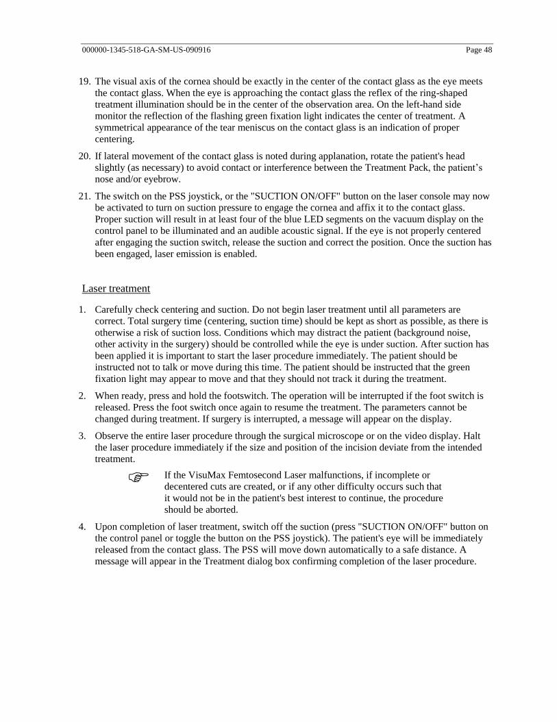
000000-1345-518-GA-SM-US-090916 Page 48
19. The visual axis of the cornea should be exactly in the center of the contact glass as the eye meets
the contact glass. When the eye is approaching the contact glass the reflex of the ring-shaped
treatment illumination should be in the center of the observation area. On the left-hand side
monitor the reflection of the flashing green fixation light indicates the center of treatment. A
symmetrical appearance of the tear meniscus on the contact glass is an indication of proper
centering.
20. If lateral movement of the contact glass is noted during applanation, rotate the patient's head
slightly (as necessary) to avoid contact or interference between the Treatment Pack, the patient’s
nose and/or eyebrow.
21. The switch on the PSS joystick, or the "SUCTION ON/OFF" button on the laser console may now
be activated to turn on suction pressure to engage the cornea and affix it to the contact glass.
Proper suction will result in at least four of the blue LED segments on the vacuum display on the
control panel to be illuminated and an audible acoustic signal. If the eye is not properly centered
after engaging the suction switch, release the suction and correct the position. Once the suction has
been engaged, laser emission is enabled.
Laser treatment
1. Carefully check centering and suction. Do not begin laser treatment until all parameters are
correct. Total surgery time (centering, suction time) should be kept as short as possible, as there is
otherwise a risk of suction loss. Conditions which may distract the patient (background noise,
other activity in the surgery) should be controlled while the eye is under suction. After suction has
been applied it is important to start the laser procedure immediately. The patient should be
instructed not to talk or move during this time. The patient should be instructed that the green
fixation light may appear to move and that they should not track it during the treatment.
2. When ready, press and hold the footswitch. The operation will be interrupted if the foot switch is
released. Press the foot switch once again to resume the treatment. The parameters cannot be
changed during treatment. If surgery is interrupted, a message will appear on the display.
3. Observe the entire laser procedure through the surgical microscope or on the video display. Halt
the laser procedure immediately if the size and position of the incision deviate from the intended
treatment.
If the VisuMax Femtosecond Laser malfunctions, if incomplete or
decentered cuts are created, or if any other difficulty occurs such that
it would not be in the patient's best interest to continue, the procedure
should be aborted.
4. Upon completion of laser treatment, switch off the suction (press "SUCTION ON/OFF" button on
the control panel or toggle the button on the PSS joystick). The patient's eye will be immediately
released from the contact glass. The PSS will move down automatically to a safe distance. A
message will appear in the Treatment dialog box confirming completion of the laser procedure.

000000-1345-518-GA-SM-US-090916 Page 49
Lenticule removal
1. Move the patient from the femtosecond laser position to the observation position under the
surgical microscope using the PSS controls. The VisuMax Femtosecond Laser surgical
microscope position can be used for the extraction portion of the procedure.
2. Inspect the complete cap and lenticule cut. Confirm that there are no obvious abnormalities in the
interface or sidecuts before attempting removal of the lenticule.
3. Insert a sterilized Sinsky hook into the small superior incision to open the incision and expose the
superior portion of the stroma beneath it.
4. Once stroma has been exposed, the lenticule edge should be identified using the Sinsky hook and
separating the anterior corneal layer from anterior surface of the lenticule (the cap cut) followed
by separation of posterior layer of lenticule from posterior layer of cornea. This initial separation
should be performed for approximately 1 to 1.5 mm from the edge of lenticule for the purpose of
identification of lenticule.
5. Using sterilized hand held instruments, use a thin blunt corneal spatula or iris spatula to separate
the anterior corneal layer from anterior surface of the lenticule (the cap cut). The tissue should be
separated to the full extent of the Cap cut which is 0.5mm larger in the periphery than the
refractive cut.
a. The anterior surface of the lenticule cut should be addressed first, otherwise an overextension
of the cap, striae or even penetration of the cap may result. It is more difficult to separate the
lenticule from the cap than from the bed, which makes the superficial separation best
performed initially followed by the deeper tissue separation. During the dissection of the cuts,
it is important to avoid pulling on or distorting the lenticule.
b. The lip of the opening incision should be gently reflected and the internal side cut visually
identified and then separated superiorly with the Sinsky hook.
6. Separate the lenticule posterior surface from the stromal bed. This is best performed with a broad
tipped rounded spatula which is blunt but thin of which there are many examples. Confirm that the
tissue has been adequately separated from the entire lenticule and from the lenticule internal side
cut.
7. Once the surfaces of the lenticule have been freed, grasp the resected tissue with sterilized forceps
and carefully extract it through the small incision. This is best performed by grasping the lenticule
with a smooth tipped non-toothed forceps and gently applying force in a circular direction similar
to performing a capsulorhexis maneuver.
8. Once the lamellar tissue has been removed, it should be placed on the corneal surface under the
microscope to be certain that the entire lenticule has been removed. The tissue is then discarded or
saved with consideration to appropriate treatment of medical waste.
CAUTION
External perforation of the cap is possible even with the use of a blunt
corneal spatula due to excessive pressure at the tip of the instrument or due
to a sudden eye movement by the patient during tissue separation.

000000-1345-518-GA-SM-US-090916 Page 50
9. Inspect the surgical field and irrigate the interface with filtered BSS through a disposable blunt
tipped 27 or 30 gauge cannula to allow the anterior cap to come into contact with the stroma
without microstriae. The incision site should be well approximated, and can be swabbed closed
with an ophthalmic surgical sponge, if necessary.
10. A final inspection of the surgical site completes the procedure with removal of the lid speculum.
Completion of procedure
1. Move the PSS to the exit position.
2. Allow the patient to stand up and exit the PSS.
3. Remove and dispose of the Treatment Pack. Remove surgical instruments from operating field.
4. Close the surgical management software by clicking the Finish button. The main dialog box will
open again. The VisuMax Femtosecond Laser is ready for the next laser procedure or treatment
planning activity.
List of Recommended Instruments for Lenticule Extraction:
1. Sinsky Hook
2. LASIK Flap “unzipper”
3. Cyclodialysis Spatula
4. Iris Spatula
5. LASIK Spatula
6. Platinum Spatula
7. LASIK Cannula blunt tipped irrigating 27G or 30G
8. LASIK Forceps
9. Capsulorhexis or other fine smooth tipped, non-toothed forceps
10. Weck-Cel sponge

000000-1345-518-GA-SM-US-090916 Page 51
Treatment interruption
If treatment is interrupted during laser incision generation, the user interface will automatically
display the treatment approach offered by the system for resuming the laser procedure. The
procedures automatically recommended by the system are based on progress information acquired
from electronic process monitoring. This process monitoring does not substitute for monitoring by the
physician and the automatic recommendation does not substitute for the physician's decision
regarding the method of continuation of treatment. If the surgeon decides to proceed with
continuation of treatment as recommended by the system, treatment should be resumed promptly after
the treatment interruption has occurred.
For any treatment interruption, when resuming the laser treatment, centering of the initial treatment
should be reproduced as accurately as possible.
In the event of an intraoperative treatment interruption, the following procedures are recommended:
Interruption of the lenticule cut in the first 10 %:
If laser treatment is interrupted during the first 10 % of lenticule cut (underside of lenticule), the
entire procedure should be repeated.
Interruption of the lenticule cut between 10 % and 100 % or interruption of a lenticule side
cut:
If laser treatment is interrupted between 10 % and 100 % of the lenticule cut or during the lenticule
side cut, the case should be aborted.
Cap cut interruption
If treatment interruption occurs during the cap cut, this portion of the treatment should be repeated.
The cap cut will default to the same thickness as originally programmed. Since the cap cut has no
effect on the refractive correction, the diameter of the cap cut can be increased to ensure that the cap
completely covers the lenticule.
WARNING
When resuming the laser treatment following treatment interruption,
strict care must be taken to ensure that the centering of the initial
treatment is reproduced as accurately as possible. Lack of caution
during this step may result in misalignment of incisions and
misaligned corneal cuts as a result.

000000-1345-518-GA-SM-US-090916 Page 52
Postoperative care
A regimen of postoperative topical ophthalmic medications (antibiotic and steroid) is recommended.
Use of a clear shield is also recommended; the shield should not be disturbed except for lifting the
shield to instill drops.
A slit-lamp examination should be performed on postoperative day one and as needed thereafter to
ensure that healing of the cornea is complete.

000000-1345-518-GA-SM-US-090916 Page 53
User Manual
Please reference the VisuMax Femtosecond Laser User Manual.

000000-1345-518-GA-SM-US-090916 Page 54
Technical data
Limit values of adjustment ranges
PARAMETER UNIT RANGE
LASER PARAMETER
Laser energy nJ 125-170
Track distance µm 2.0 – 3.0
Spot distance µm 2.0 – 3.0
SURGICAL
Cap diameter mm 7.0 or 7.5
Cap thickness um 120
Lenticule diameter mm 6.0 or 6.5
Lenticule edge minimum thickness µm 15
Residual bed minimum thickness µm 250
Incision position – opening position deg 90
Incision angle - cap opening size deg 90
Side cut angle – opening cut deg 90
Side cut angle – lenticule cut deg 90
REFRACTIVE
Intended spherical corrections D - 1.00 to -8.00*
Intended cylindrical corrections D - - -
Intended cylindrical axis deg - - -
* Please note that treatment of -8.01 through -10.00 D will present a flagged warning to the
users so that the user understands that correction of these powers had not been
substantiated by an adequate set of data.

000000-1345-518-GA-SM-US-090916 Page 55
Abbreviations
D Diopter
Deg Degree
µm Micrometer
mm Millimeter
MRSE Manifest refractive spherical equivalent
nJ nanojoule
OSDI Ocular Surface Disease Index
PRO Patient reported outcomes
QoV Quality of Vision
HORMS Higher-order aberrations root mean square
SMILE Small Incision Lenticule Extraction

000000-1345-518-GA-SM-US-090916 Page 56
Figures/Tables
Figure 1. VisuMax Femtosecond Laser 6 Figure 2. Schematic depiction of cut geometry for the SMILE procedure performed with the VisuMax
Femtosecond Laser 8 Figure 3. Planning view from VisuMax Femtosecond Laser graphical user interface (GUI) of a
SMILE procedure (left graphic) and schematic of lenticule and attached cap cuts (top right
graphic). 8 Figure 4. Lenticule Tab used for entering Procedure Parameters Associated with the Lenticule Cutting
and Refractive Targets 42 Figure 5. Cap Tab used for entering Procedure Parameters Associated with the Opening Cut and Cap
Cut 44
Table 1 Demographics All Treated Eyes 18 Table 2 Preoperative Refraction Parameters 19 Table 3 Accountability All Treated Eyes 20 Table 4 Summary of Key Safety Variables at 6-Month Point of Refractive Stability All Treated Eyes21 Table 5 Summary of Key Safety Variables at Last Available Visit All Treated Eyes 21 Table 6 Change in Best Spectacle-Corrected Visual Acuity (BSCVA) from Preop All Treated Eyes 22 Table 7 Intraoperative Adverse Events 22 Table 8 Postoperative Ophthalmic Adverse Events — All Treated Eyes 23 Table 9 Intraoperative Events All Treated Eyes 24 Table 10 Complications All Treated Eyes 25 Table 11 Log Contrast Sensitivity Change from Preoperative Visit All Treated Eyes 26 Table 12 QoV Score Change from Preoperative All Treated Eyes 27 Table 13 Changes of 2 or More Grades in QoV Symptoms at 12 Months 28 Table 14 Two Highest Categories of Bothersome and Severity for Each QoV Symptom at 12 Months29 Table 15 OSDI Score Change from Preoperative All Treated Eyes 30 Table 16 Frequency of Moderate and Severe Dry Eye Symptoms Classified by OSDI Scores 31 Table 17 Topography Findings All Treated Eyes 32 Table 18 Change in Wavefront Aberrometry from Preoperative Stratified by Largest Scan Size (mm)
Treated Eyes with Preoperative, 3-Month, and 12-Month Visits 33 Table 19 Summary of Key Effectiveness Variables Effectiveness Cohort Eyes 34 Table 20 Summary of Key Effectiveness Variables at 6 Months Stratified By Preoperative MRSPH 35 Table 21 Postoperative Uncorrected Visual Acuity (UCVA) Compared to Preoperative Best Spectacle
Corrected Visual Acuity (BSCVA) Effectiveness Cohort Eyes 36 Table 22 Accuracy of MRSE — Attempted vs. Achieved 6-Month Consistent Effectiveness Cohort 37 Table 23 Stability of Manifest Refraction Spherical Equivalent (MRSE) Effectiveness Population 38

000000-1345-518-GA-SM-US-090916 Page 57
Index
A
Abbreviations .................................................................................................... 55
Adjustment ranges ............................................................................................. 54
C
Contraindications ................................................................................................. 9
G
Glossary ............................................................................................................. 55
I
Indication for use ................................................................................................. 9
P
Package check list ............................................................................................... 3
S
Symbols ............................................................................................................... 2
T
Technical data .................................................................................................... 54
Treatment license .............................................................................................. 40
U
User profile .......................................................................................................... 5
Manufacturer
Carl Zeiss Meditec AG
Goeschwitzer Str. 51-52
07745 Jena
Germany
Phone: +49 3641 220 444
Fax: +49 3641 220 442
Email: [email protected]
Internet: www.zeiss.com/med
Distributor
Carl Zeiss Meditec, Inc.
5160 Hacienda Drive
Dublin, CA 94568
USA
Toll Free: +1 800 341 6968
Phone: +1 925 557 4100
Fax: +1 925 557 4101
Email: [email protected]
Internet: www.zeiss.com/med/us
000000-1345-518-GA-SM-US-090916
VisuMax SMILE procedure
Specifications subject to change