Embed Size (px)
Citation preview
Vm
TFD2PT
OG5BT
SN
D1TT
JCD2TT
JNDNTT
WTG2TT
CND
NTT
*ApTcsJ2
Journal of Biomedical Optics 14�2�, 024034 �March/April 2009�
J
isualizing laser-skin interaction in vivo by multiphotonicroscopy
sung-Hua Tsai*ar Eastern Memorial Hospitalepartment of Dermatology1, Nan-Ya South Road, Section 2an-Chiao, Taipei 220aiwan
andriental Institute of Technologyeneral Education Center8, Section 2, Sihchuan Roadan-Chiao City, Taipei County 220aiwan
hiou-Hwa Jee*ational Taiwan University Hospital
and College of Medicineepartment of Dermatology, Section 1, Jen-Ai Roadaipei 100aiwan
ung-Yi Chanathay General Hospitalepartment of Dermatology80, Section 4, Jen-Ai Roadaipei 106aiwan
in-Ning Leeational Taiwan Universityepartment of Physicsumber 1, Section 4, Roosevelt Road
aipei 106aiwan
oan-Ruoh Leeaipei Medical Universityraduate Institute of Medical Sciences50, Wu-Xin Streetaipei 100aiwan
hen-Yuan Dongational Taiwan Universityepartment of Physics
and Center for Quantum Science and Engineeringumber 1, Section 4, Roosevelt Road
aipei 106aiwan
Authors contributed equally to this work.ddress all correspondence to: Chen-Yuan Dong, PhD, Associate Professor, De-artment of Physics National Taiwan University, No. 1, Sec. 4, Roosevelt Road,aipei 106, Taiwan; Tel: 886-2-3366-5155; Fax: 886-2-3366-5244; E-mail:[email protected]; and Sung-Jan Lin, MD, PhD, Assistant Professor, In-titute of Biomedical Engineering, National Taiwan University, No. 1, Sec. 1,en-Ai Road, Taipei 100, Taiwan; Tel: 886-2-23123456 ext 5323; Fax: 886-2-392-5351; E-mail: [email protected].
ournal of Biomedical Optics 024034-
Sung-Jan LinNational Taiwan UniversityInstitute of Biomedical Engineering
andNational Taiwan University Hospital
and College of MedicineDepartment of Dermatology1, Section 1, Jen-Ai RoadTaipei 100Taiwan
Abstract. Recently, multiphoton microscopy has gainedmuch popularity as a noninvasive imaging modality inbiomedical research. We evaluate the potential of multi-photon microscopy for monitoring laser-skin reaction invivo. Nude mouse skin is irradiated with an erbium:YAGlaser at various fluences and immediately imaged by amultiphoton microscope. The alterations of cutaneousnonlinear optical properties including multiphoton autof-luorescence and second-harmonic generation associatedwith laser irradiation are evaluated morphologically andquantitatively. Our results show that an erbium:YAG laserat a low fluence can selectively disrupt the stratum cor-neum, and this alteration may account for the penetrationenhancing effect of laser-assisted transcutaneous drug de-livery. At a higher fluence, the zone of tissue ablation aswell as the disruption of the surrounding stratum corneum,keratinocytes, and dermal extracellular matrix can be bet-ter characterized by multiphoton microscopy as comparedwith conventional histology. Furthermore, the degree ofcollagen damage in the residual thermal zone can bequantified by second-harmonic generation signals, whichhave significant difference between control skin, skin irra-diated with a 1.5-, 8-, and 16-J /cm2 erbium:YAG laser�P�0.05�. We show that multiphoton microscopy can bea useful noninvasive imaging modality for monitoringlaser-skin reaction in vivo. © 2009 Society of Photo-Optical Instru-mentation Engineers. �DOI: 10.1117/1.3116711�
Keywords: multiphoton; autofluorescence; second-harmonicgeneration; laser; laser-assisted drug delivery; collagen.Paper 08296R received Aug. 27, 2008; revised manuscript receivedDec. 4, 2008; accepted for publication Feb. 11, 2009; published on-line Apr. 22, 2009.
1 IntroductionVarious light therapies have been employed in clinical prac-tice. Advances in these treatment modalities, such as varioustypes of laser, intense pulsed light �IPL�, and light emittingdiodes, have led to their wide clinical applications in treatingcutaneous diseases and performing cosmetic procedures.1
The results of laser treatment depend on the immediateinteraction of incident light with tissues well as the subse-quent tissue regeneration and remodeling. As in many areas ofmedicine, histological examinations are necessary to evaluate
1083-3668/2009/14�2�/024034/8/$25.00 © 2009 SPIE
March/April 2009 � Vol. 14�2�1
tcoateevvpaafittte
ooocmnTil
tnbntmtrtaooshcbi
efsrlUdmcehpeTe
Tsai et al.: Visualizing laser-skin interaction in vivo by multiphoton microscopy
J
he reaction of epidermal, dermal, vascular, and appendagealomponents of skin after laser therapy.2 The major drawbackf conventional histological examination is its invasive naturend the inability to examine the light-tissue interaction in realime. The use of inappropriate parameters can result in inad-quate treatment outcome and may lead to serious sideffects.3 To be specific, skin conditions can vary among indi-iduals as well as among the treated areas in the same indi-idual. To achieve a favorable result, the parameters em-loyed should be adjusted accordingly. Despite the widepplication of light therapy, the association between immedi-te light-tissue reaction, subsequent tissue remodeling, andnal clinical outcome has not been investigated in detail due
o the lack of an appropriate real-time noninvasive monitoringool. As a result, validation of appropriate adjustable clinicalreatment parameters often relies on repetition of trial-and-rror practice.
Investigators have resorted to noninvasive objective meth-ds to assess laser-tissue reaction, such as sonography andptical coherence tomography.4,5 However, limited by the res-lution, these techniques do not enable detailed analysis ofutaneous structural alterations. Recently, reflectant confocalicroscopy has gained popularity as a useful clinical tool for
oninvasive imaging by providing subcellular resolution.6
hough it is powerful in analyzing the cellular morphologiesn epidermis, its ability to resolve the turbid dermal extracel-ular matrical structures is limited.
Recently, the nonlinear optical imaging technique of mul-iphoton microscopy �MPM� has emerged as a useful tech-ique for skin imaging.7–10 In MPM, fluorophores are excitedy simultaneous absorption of two or more photons in theear-IR spectrum as opposed to absorption of one photon inhe UV or visible light spectrum in conventional florescence
icroscopy. To achieve efficient multiphoton excitation, ex-remely high peak photon input from lasers with pulses in theange of tens to hundreds of femtoseconds is required.7 Due tohe nonlinearity, the excitation occurs only at the focal pointnd images from a thin optical section can be acquired with-ut using a pinhole. MPM holds several unique advantagesver confocal scanning laser microscopy, such as reducedpecimen photobleaching, higher contrast images, and en-anced penetration depth.7,9,11 Since endogenous autofluores-ent substances including NAD�P�H, keratin, and elastic fi-ers can be excited by multiphoton excitation, label-freemaging can be achieved.9,11
By use of a femtosecond laser, the nonlinear polarizationffect of second-harmonic generation �SHG� can also be usedor visualizing biological structures lacking inversionymmetry.11,12 For SHG, the interaction of noncentrosymmet-ic structures with incident light yields photons with a wave-ength of exactly half the wavelength of the incident light.nlike fluorescence excitation, the polarization effect of SHGoes not involve absorption of the incident photons by theolecules. Therefore, photobleaching and thermal damage
an be avoided. Collagen fibers, myosin, and microtubules areffective biological SHG signal generators.12 As SHG isighly sensitive to structures, SHG signals can be used forrobing modification of collagen structures in cancer andvaluating the thermal structural transitions of collagen.13,14
he wavelength of SHG is shorter than the fluorescence gen-rated by multiphoton excitation, and hence, SHG signals can
ournal of Biomedical Optics 024034-
be easily separated from fluorescence for further analysis andimage processing. The combination of multiphoton autofluo-rescence �AF� and SHG imaging has been applied to evaluat-ing cutaneous aging and differentiating basal cell carcinomafrom normal dermal stroma.15,16 Multiphoton microscopy hasbeen used to detect corneal architecture and intratissue fem-tosecond laser effect in a rabbit eye model.17
In this paper, we assessed the usefulness of MPM in moni-toring laser-skin reactions in vivo. An erbium:YAG �Er:YAG�laser was chosen in this study due to its wide dermatologicalapplications, such as dermabrasion, skin tumor removal, skinrejuvenation, and enhancement of transcutaneous drugdelivery.18,19 We investigated the morphological and quantita-tive changes of AF and SHG signals associated with Er:YAGlaser irradiation and evaluated the potential of MPM in real-time evaluation of laser-skin interaction.
2 Materials and Methods2.1 Multiphoton MicroscopeThe multiphoton microscope is similar to the one we previ-ously described with modification.20 A femtosecond titanium-sapphire �ti-sa� laser �Tsunami; Spectra Physics, MountainView, California� pumped by a diode-pumped solid state�DPSS� laser �Millennia X; Spectra Physics, Mountain View,California� was used as the excitation source. The 780-nmoutput of the laser system was guided toward a modified com-mercial upright microscope �E800; Nikon, Tokyo, Japan�.Prior to entering the microscope, the excitation source wasangularly deflected by an x-y scanning system �Model 6220;Cambridge Technology, Cambridge, Massachusetts�. The in-put of the upright microscope was modified to accommodate abeam expander. The excitation source was beam expandedand reflected toward the focusing objective �S Fluor 40�,numerical aperture �NA�=1.30; Nikon, Tokyo, Japan� by ashort-pass primary dichroic �700DCSPXRUV-3p; ChromaTechnology, Rockingham, Vermont�. The power at the samplewas 35 mW. The nonlinear AF and SHG signals were gener-ated at the sample focal plane and collected in the epi-illuminated or back-scattering geometry by the same focusingobjective. After passing through the primary dichroic, the AFand SHG signals were separated into two separate channelswhere they were detected by independent photomultipliertubes �R7400P; Hamamatsu, Hamamatsu City, Japan�. TheAF and SHG signals were separated by a secondary dichroic�435DCXR; Chroma Technology Rockingham, Vermont�. Thesecondary dichroic is a long-pass filter that reflects below andtransmits above 435 nm. The SHG signal was further filteredby a SHG filter �HQ390/20; Chroma Technology, Rocking-ham, Vermont� centered at 390 nm with a bandwidth of20 nm, while the AF was detected by a bandpass filter�E700sp-2P-E435lp; ChromaTechnology, Rockingham, Ver-mont� for broadband fluorescence detection between 435 and700 nm. To acquire a global image of the thermally treatedspecimens at high resolution, an x-y sample positioning stage�H101; Prior Scientific Instruments, Cambridge, UK� wasused to translate the skin specimen after each imaged frame.An array of overlapping and individually beam-scanned im-ages, each with an area of 110�110 �m2, was thenassembled.
March/April 2009 � Vol. 14�2�2
2TstvlATfldacfSw
2AEaass
2
Tbntpif
2FT
Frd3ds�cfi
Tsai et al.: Visualizing laser-skin interaction in vivo by multiphoton microscopy
J
.2 Image Processing and Analysishe MPM images were analyzed and reconstructed by theoftware ImageJ 1.36b �National Institutes of Health, Be-hesda, Maryland�. The toolbox enabled visual inspection ofarious morphologic features and fluorescence patterns of cel-ular components within normal skin and laser-irradiated skin.F and SHG signals of each site were quantitatively acquired.o determine whether treatment with Er:YAG laser at variousuences influences dermal SHG, average SHG intensity at aepth of more than 60 �m of untreated and treated skin werenalyzed with a general linear model. A value of P=0.05,orresponding to the 95% confidence interval, was selectedor analysis of significant difference of SHG in this study.PSS 15.0 statistical software �SPSS Inc., Chicago, Illinois�as used.
.3 Er:YAG Laser Assemblyn Er:YAG laser �Burane; WaveLight Laser Technologie AG,rlangen, Germany� was used to irradiate skin. The laser hadwavelength of 2940 nm and pulse duration of 250 �s. An
rticulated arm was used to deliver the laser beam onto thekin. Laser beam spot size was 1.5 mm in diameter. Each skinite was exposed once.
.4 Accommodation of the Mouse on theMicroscopic Stage
he experimental protocol involving animals was approvedy our Institutional Animal Care and Use Committee. Maleude mice �Balb/c-nu strain, 8 to 10 weeks old� were anes-hetized by intraperitoneal phenobarbitol injection and pre-ared for Er:YAG laser irradiation. Immediately after laserrradiation, mouse dorsal skin were stabilized in a dorsal skinold chamber, coverslipped, and observed under the MPM.
.5 Histological Examination of Skinull-thickness dorsal skin after MPM imaging was excised.he skin specimens were fixed in 10% buffered formalin so-
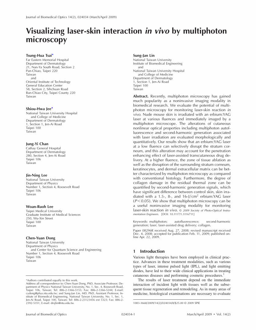
ig. 1 MPM images and histology of unirradiated nude mouse skin �aepresents SHG. �a� MPM image of the skin surface. The stratum coremonstrated. Hair is also intensely autofluorescent �arrows�. �b� Ma2 �m from the surface. Keratinocytes with abundant autofluorescenemonstrated. �d� Basal layer and the dermal-epidermal junction, 80canty cytoplasm as compared with that in stratum spinosum. SHG eme� MPM image of the dermis, 160 �m from skin surface. Fibrous collorresponding histological image. Compared with MPM images, the stxation and staining process. �Bars: 20 �m.�
ournal of Biomedical Optics 024034-
lution and further processed for histological examinationswith hematoxylin and eosin stains. The adjacent normal skinwas assessed as the control.
3 Results3.1 Multiphoton Image of Unirradiated Nude Mouse
Skin
Figure 1 shows the multiphoton images of unirradiated nudemouse skin. MPM images of skin surface reveal compactanucleated corneocytes. Autofluorescent pentagonal or hex-agonal corneocytes are tightly packed and the cell borders areclearly delineated �Figs. 1�a� and 1�b��. Hair is also intenselyautofluorescent and can be seen on the skin surface �arrows�.In the optical sections corresponding to the stratum spinosumand stratum basale, the cells are characterized by fluorescentcellular cytoplasm surrounding nonfluorescent round nuclei.A gradual decrease in cell size can be observed when serialimages are taken from the stratum spinosum down to the stra-tum basale. The ratio of the nucleus to the cytoplasm alsoincreases with depth �Figs. 1�c� and 1�d��. The dermal-epidermal junction with intermingled AF signals from basalkeratinocytes and SHG signals from collagen fibers can beseen in Fig. 1�d�. Collagen fibers in dermis emit strong SHGsignals �Fig. 1�e��.
In the corresponding histological image, in contrast tocompact structures revealed in the MPM images, stratum cor-neum is loose with a basket-weave pattern �Fig. 1�f��. Thisdiscrepancy is due to the artifacts caused by fixation andstaining procedures in histological examinations that disruptthe normal intercellular packing of corneocytes.21–23 In addi-tion, the epidermis is thinner measured histologically �about60 �m� than that measured by multiphoton microscopy�about 80 �m�. This indicates that tissue shrinkage duringprocessing of the specimen for histological examination canbe a significant artifact that interferes with the precise mea-surement of tissue thickness.
MPM images. The green channel represents AF and the red channels autofluorescent and the morphology of corneocytes can be clearlypicture of the skin surface. �c� MPM image of the stratum spinosum,plasm surrounding round nonfluorescent nuclei �arrowhead� can bem the skin surface. Basal keratinocytes �arrowhead� are smaller withom fibrous collagen starts to appear in the dermal-epidermal junction.ndles with intense SHG signals are penetrating in the dermis. �f� Theorneum is loose with a basket-weave pattern due to artifacts from the
� to �e�neum ignifiedt cyto
�m froitted fragen buratum c
March/April 2009 � Vol. 14�2�3
3
Netcohchn�aenhl
mss2arSpodt
3
Asd
Fchmc�swi
Tsai et al.: Visualizing laser-skin interaction in vivo by multiphoton microscopy
J
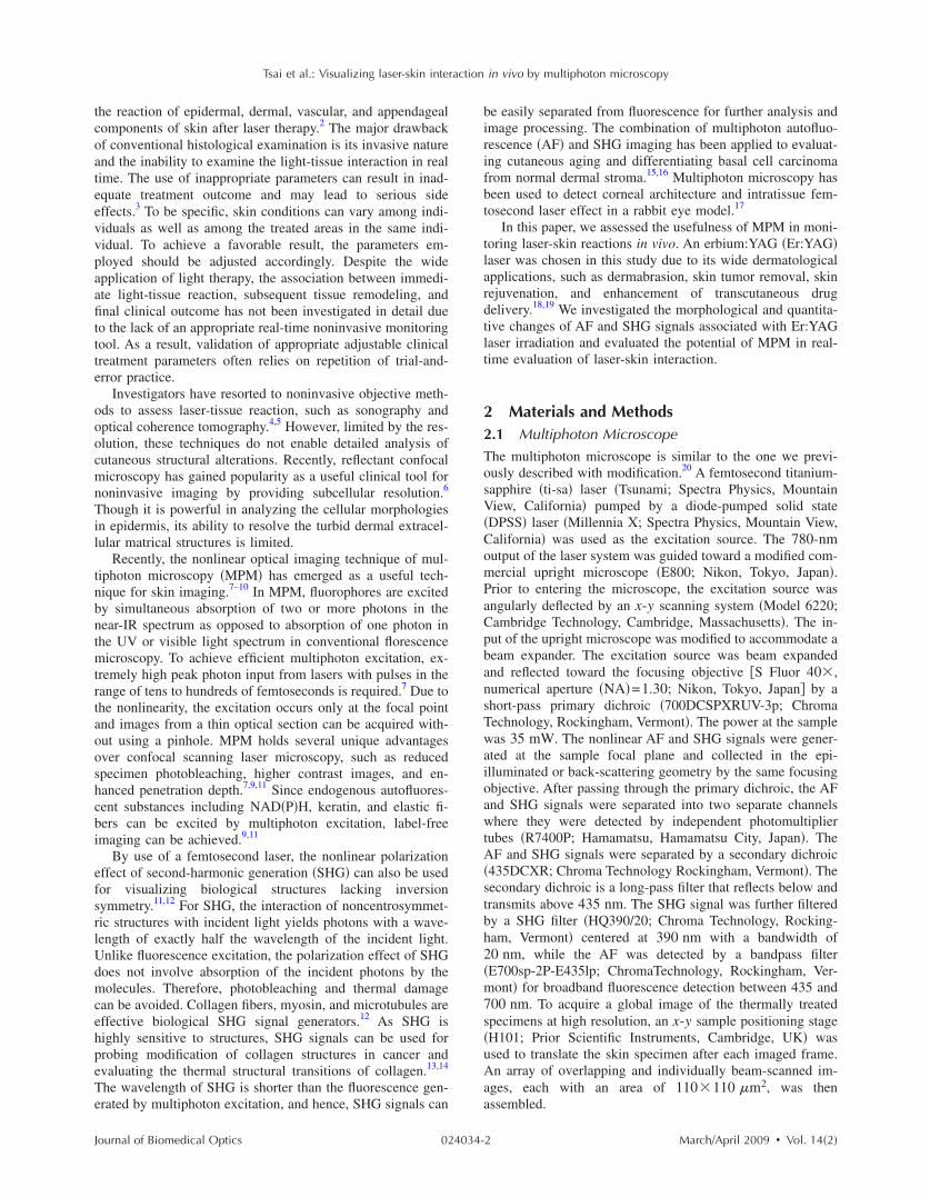
.2 Selective Disruption of Stratum Corneum byEr:YAG Laser at a Low Fluence of 1.5 J /cm2
Revealed by Multiphoton Imagingude mouse skin was irradiated with Er:YAG laser at a flu-
nce of 1.5 J /cm2. In the MPM images, compared with theightly packed hexagonal corneocytes with distinctive inter-ellular borders in control group �Fig. 1�a��, the morphologyf a large proportion of corneocytes has been disrupted into aomogenized pattern and intercellular borders can not belearly delineated �Figs. 2�a� and 2�b��. In addition to theomogenization change, selective loosening of stratum cor-eum and loss of corneocytes can also be observed focallyFig. 2�a�, asteroids�. Brightly fluorescent hairs are still intactnd are relatively resistant to laser irradiation �arrows�. How-ver, in the corresponding histological image, only scanty cor-eocytes are still preserved �Fig. 2�f��. We speculate that theomogenized or loosened corneocytes are lost during histo-ogical processing.
When images are taken farther down the skin surface, theorphology of keratinocytes in the stratum spinosum and
tratum basale is still intact �Figs. 2�c� and 2�d��. The SHGignals can still be detected from the dermal collagen �Fig.�e��. In the histological image, epidermal keratinocytes arelso still intact and no significant change of dermal collagen isevealed �Fig. 2�f��. Since the AF from the epidermal cells andHG from the dermis are all decreased and the cellular mor-hology and collagen fibers are relatively intact, the decreasef the intensity of AF and SHG from epidermal cells andermal collagen may be caused by the shielding effect fromhe intensely fluorescent disrupted corneocytes.
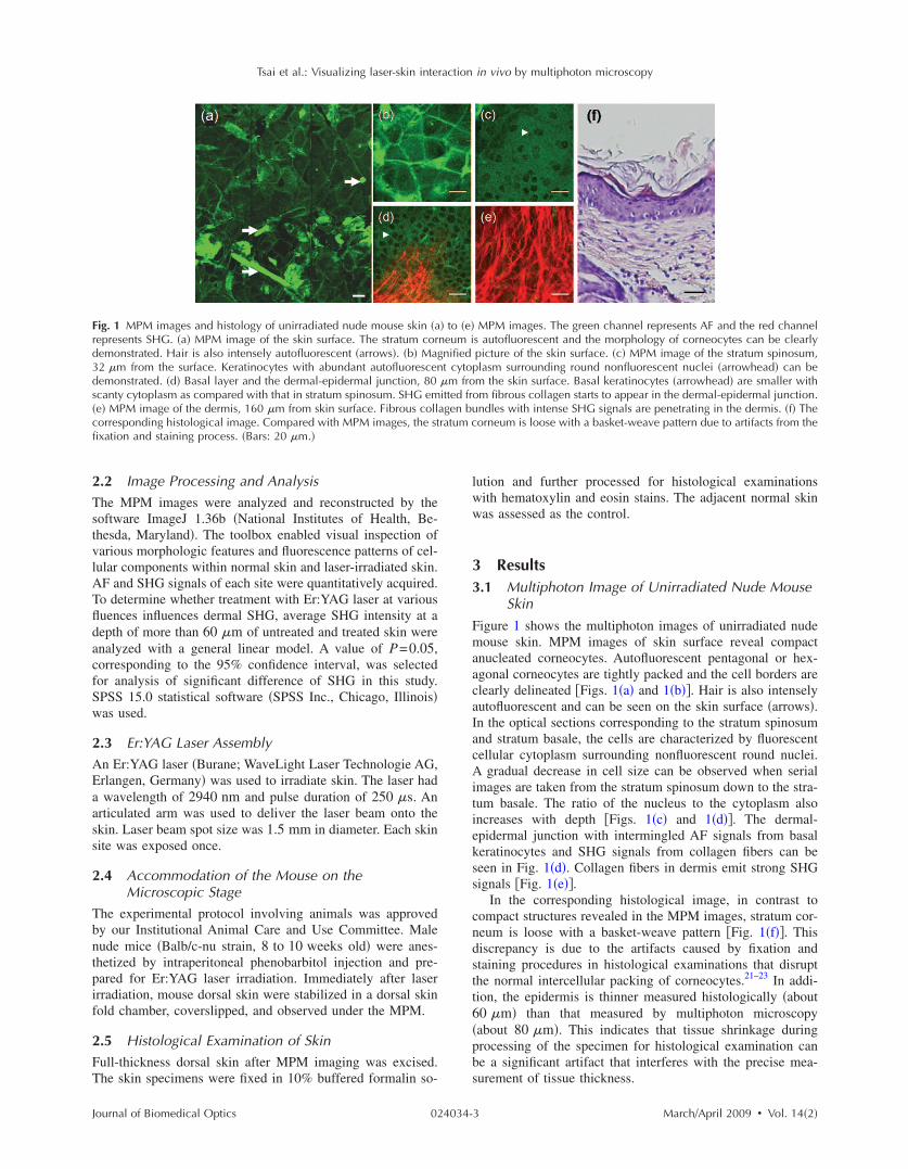
.3 Ablation of Stratum Corneum and Thermal Injuryto Underlying Structures by Er:YAG Laser atModerate Fluence of 8 J /cm2
fter irradiation at the fluence of 8 J /cm2, partial ablation oftratum corneum is revealed and the residual stratum corneumisplays a loose reticular pattern �Figs. 3�a� and 3�b��. The
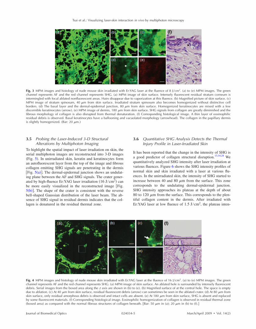
ig. 2 MPM images and histology of nude mouse skin irradiated withhannel represents AF and the red channel represents SHG. �a� MPomogenized morphology. The focal stratum corneum is loosened oagnified picture of skin surface. �c� MPM image of stratum spinosum
an still be observed �arrowhead�. �d� Basal layer and the dermal-eparrowhead� and SHG-emitting collagen can be seen. �e� MPM imagetill be observed but the detected SHG intensity is decreased compaith the MPM image, the stratum corneum is almost totally detached d
n the underlying structures. �Bars: 20 �m.�
ournal of Biomedical Optics 024034-
stratum spinosum starts to become a homogenous fluorescentmorphology without discernable intercellular borders �Fig.3�c��. Residual discernable keratinocytes are mixed with ho-mogenized cells in basal layer �Fig. 3�d��. In the correspond-ing histological image, intact corneocytes can hardly be seen.There is a thin layer of eosinophilic debris in which upperepidermal cells show a condensed nucleus and more eosino-philic cytoplasm. Some of these cells are partially detachedfrom the underlying epidermal cells. Many residual basal cellsbecome vacuolated �Fig. 3�f�, arrowhead�.
When the images are taken farther down to the dermis, theintensity of SHG signals from the dermis is greatly dimin-ished and the fibrous morphology of collagen is further dete-riorated into an amorphous structure due to the residual ther-mal damage from laser irradiation �Fig. 3�e��. Compared withMPM images, this denatured collagen zone can not be easilyappreciated in the histology. It presents as a thin layer ofslightly homogenized collagen immediately beneath dermal-epidermal junction �Fig. 3�f��.
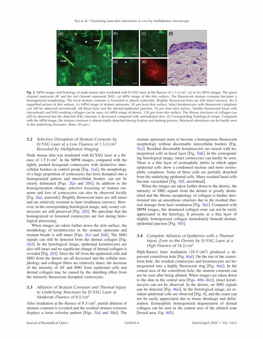
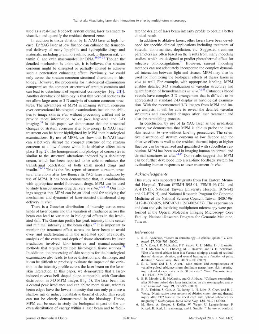
3.4 Complete Ablation of Epidermis with a ThermalInjury Zone to the Dermis by Er:YAG Laser at aHigh Fluence of 16 J /cm2
High-fluence laser irradiation �16 J /cm2� produced a de-pressed crateriform hole �Fig. 4�a��. On the rim of the crateri-form hole, the residual corneocytes and keratinocytes are ho-mogenized into a highly fluorescent ring �Fig. 4�a��. In thecentral area of the crateriform hole, the stratum corneum cannot be seen after being ablated. When images are taken downto the skin in the central area �Figs. 4�b�–4�e��, intact kerati-nocytes can not be observed. In the dermis, no SHG signalscan be detected �Fig. 4�e��. In the histological image, no re-sidual epidermal cells are observed �Fig. 4�, and the crater cannot be easily appreciated due to tissue shrinkage and defor-mation. Eosinophilic homogenized degeneration of dermalcollagen can be seen in the central area of the ablated zone�boxed area, Fig. 4�f��.
laser at the fluence of 1.5 J /cm2. �a� to �e� MPM images. The greenge of the skin surface. The fluorescent stratum corneum becomes at �asteroids�. Brightly fluorescent hairs are still intact �arrows�. �b� Am from skin surface. Intact keratinocytes with fluorescent cytoplasml junction, 70 �m from skin surface. Smaller fluorescent basal cellsis, 130 �m from skin surface. The fibrous structures of collagen can
h unirradiated skin. �f� Corresponding histological image. Comparedfixation and staining process. Structural alterations can be hardly seen
Er:YAGM imar absen
, 30 �idermaof derm
red wituring
March/April 2009 � Vol. 14�2�4
3
Ts�ac�iab5bsl
FciMbdfiri
Fcddsb�
Tsai et al.: Visualizing laser-skin interaction in vivo by multiphoton microscopy
J
.5 Probing the Laser-Induced 3-D StructuralAlterations by Multiphoton Imaging
o highlight the spatial impact of laser irradiation on skin, theerial multiphoton images are reconstructed into 3-D imagesFig. 5�. In unirradiated skin, keratin and keratinocytes formn autofluorescent layer from the top of the image and fibrousollagen emitting SHG signals are penetrating in the dermisFig. 5�a��. The dermal-epidermal junction shows an undulat-ng plane between the AF and SHG signals. The crater gener-ted by high-fluence Er:YAG laser irradiation �16 J /cm2� cane more easily visualized in the reconstructed image �Fig.�b��. The shape of the crater is consistent with the reverseell-shaped Gaussian distribution of the laser beam. The ab-ence of SHG signal in residual dermis indicates that the col-agen is denatured in the residual thermal zone.
ig. 3 MPM images and histology of nude mouse skin irradiated withhannel represents AF and the red channel represents SHG. �a� MPntermingled with focal ablated nonfluorescent areas. Hairs disappear
PM image of stratum spinosum, 40 �m from skin surface. Irradiaorders. �d� The basal layer and the dermal-epidermal junction, 80iscernible keratinocytes �arrow�. �e� MPM image of dermis, 180 �mbrous morphology of collagen is also disrupted from thermal denaesidual debris is observed. Basal keratinocytes have a ballooning ands slightly homogenized. �Bar: 20 �m.�
ig. 4 MPM images and histology of nude mouse skin irradiated withhannel represents AF and the red channel represents SHG. �a� MPMebris. Serial images from the boxed area along the z axis are shownue to ablation. �c� At 40 �m from skin surface, residual fluorescent dkin surface, only residual amorphous debris is observed and intact cey some fluorescent materials. �f� Corresponding histological image. Eboxed area� as compared with the normal fibrous structures of colla
ournal of Biomedical Optics 024034-
3.6 Quantitative SHG Analysis Detects the ThermalInjury Profile in Laser-Irradiated Skin
It has been reported that the change in the intensity of SHG isa good predictor of collagen structural disruption.13,14,24 Wequantitatively analyzed SHG intensity after laser irradiation atvarious fluences. Figure 6 shows the SHG intensity profiles ofnormal skin and skin irradiated with a laser at various flu-ences. In the unirradiated skin, the intensity of SHG started toincrease between 40 and 80 �m from the surface. This zonecorresponds to the undulating dermal-epidermal junction.SHG intensity approaches its plateau at the depth of about80 to 120 �m from the surface. This corresponds to the plen-tiful collagen content in the dermis. After irradiated withEr:YAG laser at low fluence of 1.5 J /cm2, the plateau inten-
G laser at the fluence of 8 J /cm2. �a� to �e� MPM images. The greene of skin surface. Intensely fluorescent residual stratum corneum isvaporization at this fluence. �b� Magnified picture of skin surface. �c�tum spinosum also becomes homogenized without distinctive cell
om skin surface. Homogenized keratinocytes are mixed with a fewin surface. SHG signals from collagen are greatly diminished and the. �f� Corresponding histological image. A thin layer of eosinophiliclated morphology �arrowhead�. The collagen in the papillary dermis
laser at the fluence of 16 J /cm2. �a� to �e� MPM images. The greenf skin surface. An ablated hole is surrounded by intensely fluorescent
o �e�. �b� Magnified surface of at the central hole. The space is emptyrrow� can sometimes be seen in the ablated crater. �d� At 80 �m fromabsent. �e� At 180 �m from skin surface, SHG is absent and replacedilic homogenization of collagen is observed in residual thermal zoneeath. �Bar: 50 �m in �a�; 20 �m in �b� to �f�.�
Er:YAM imagdue toted stra�m frfrom skturation
vacuo
Er:YAGimage oin �b� tebris �alls areosinophgen ben
March/April 2009 � Vol. 14�2�5

sttsommeajhSt
4Tm8aapoguowttntn
c
FsSladsapGsp
Tsai et al.: Visualizing laser-skin interaction in vivo by multiphoton microscopy
J
ity of SHG signals in the dermis decreases compared withhe control group. However, the increase of SHG intensity athe dermal-epidermal junction of depth about 40 to 80 �m istill observed. Since the fibrous structures of collagen are stillbserved, the decrease in the plateau SHG intensity in dermisay be caused by the shielding effect of the overlying ho-ogenized highly fluorescent stratum corneum. At the mod-
rate fluence of 8 J /cm2, SHG decreases more significantlynd the increasing trend of intensity at the dermal-epidermalunction can only be barely observed. In skin exposed to aigh fluence of 16 J /cm2, the SHG signals are undetectable.HG signals of dermal collagen are significantly different be-
ween four groups with paired comparisons �P�0.05�.
Discussionhe Er:YAG laser and the 10,600-nm CO2 laser are two com-only used resurfacing lasers. The epidermis is composed of
0% water. In these two lasers, the major chromophore forbsorbance is water. At high fluences of the CO2 laser forblation, instant heating of water to 100 °C occurs and va-orization of tissue ensues. However, it can also yields a zonef coagulation beyond the vaporization zone to a variable de-ree, depending on the mode and parameters of CO2 lasersed.25 The residual thermal effects in the temperature rangef 40 to 100 °C can result in denaturation of proteins, un-inding of DNA, and disruption of cell membranes. Though
he residual thermal zone of the CO2 laser can contribute toissue shrinkage for the purpose of tightening in skin rejuve-ation, it can also lead to undesirable effects. For example,his residual thermal effect can compromise cell function andecrosis ensues.26
With the Er:YAG laser at 2940 nm, the energy is so effi-iently absorbed by water �12 to 18 times that of the
ig. 5 Three-dimensional MPM images of control and laser irradiatedkin. The green channel represents AF and the red channel representsHG. �a� Normal skin has keratinocytes that emit AF in the epidermalayer and fibrous collagen that emits SHG in the dermal layer. Hex-gonal tightly packed corneocytes can be seen from the top view. Theermal-epidermal junction can be delineated by the undulating wave-haped junction of AF and SHG. �b� After irradiation with Er:YAG lasert fluence of 16 J /cm2, a slope of hole appears. The gradually de-ressed shape of the crater is consistent with the reverse bell-shapedaussian distribution of light intensity across the laser beam. SHG
ignals are absent in the dermis due to thermal denaturation. �x-ylane: surface of skin 110�110 �m2; z axis: skin depth 180 �m.�
ournal of Biomedical Optics 024034-
10,600 nm wavelength� that its shorter penetration depth re-sults in a more precise zone of tissue ablation with reducedresidual thermal injury compared with CO2 laser.27 TheEr:YAG laser is currently widely used in dermatology as anablative tool for skin tumor removal, acne scar treatment, andfacial rejuvenation. The vaporizing effect of Er:YAG laserablates superficial tissue, whereas the residual heat conductioncan still lead to tissue coagulation and collagen denaturation.2
Though the residual thermal zone can induce collagen shrink-age and neogenesis,28 too much thermal damage can also leadto unwanted complications, including postinflammatory hy-perpigmentation and scars.3 The evaluation of residual ther-mal zones previously depended on histological examinations,but the procedure does not allow for real-time imaging. Whenthe collagen is totally coagulated, eosinophilic homogeniza-tion can be seen in histological examination. However, if col-lagen is not heated enough to become totally coagulated, itcan be difficult to detect the subtle changes in histology.24,29
Thermal denaturation results in the loss of molecular order inthe collagen fibers, which can have a profound influence onSHG.13,14,24 Consistent with our previous reports,24,29 thermaltreatment can disrupt regular collagen packing and hence theSHG signal in dermis decreases. Decrease in intensity of theSHG signals from collagen, which have statistically signifi-cant difference between each group, is correlated to the flu-ence of the laser �Fig. 6�. The findings in this study supportthat fact that the SHG signal in MPM is a more sensitive toolin identifying the degree and extent of residual thermal dam-age in laser-irradiated skin. Hence, the SHG signals may be
Fig. 6 SHG intensity profiles of skin irradiated with Er:YAG laser atvarious fluences as a function of skin depth �z� from the skin surface�at z=0 �m�. Unirradiated control skin has higher SHG along thedepth since collagen appears in the dermal-epidermal junction at thedepth of about 40 to 80 �m. SHG intensity rapidly increases to a pla-teau value at the depth of 80 to 120 �m from the surface. AfterEr:YAG laser irradiation at a low fluence of 1.5 J /cm2, the plateauintensity of the SHG signals in the dermis decreases compared withthe control group. At a moderate fluence of 8 J /cm2, the SHG signalfurther decreases and the increasing trend at the depth of 40 to 80 �mis also greatly diminished. At a high fluence of 16 J /cm2, the SHGsignals decrease to an undetectable level. Error bars represent 1 SD�standard deviation� in the average value of five skin samples. Singleasterisks � *� indicate regions of significant difference �P�0.05� be-tween each group.
March/April 2009 � Vol. 14�2�6
uv
emmtdcsotccAntotpicteccpsctituwtimd
kbaamoaimIeitsidabscMe
Tsai et al.: Visualizing laser-skin interaction in vivo by multiphoton microscopy
J
sed as a real-time feedback system during laser treatment toisualize and quantify the residual thermal zone.
In addition to tissue ablation by Er:YAG laser at high flu-nce, Er:YAG laser at low fluence can enhance the transder-al delivery of many lipophilic and hydrophilic drugs andaterials, including 5-aminolevulinic acid, 5-fluorouracil, vi-
amin C, and even macromolecular DNA.19,30–32 Though theetailed mechanism is unknown, it is believed that stratumorneum might be disrupted or partially ablated to achieveuch a penetration enhancing effect. Previously, we couldnly assess the stratum corneum structural alterations in his-ology. However, the processing for histological examinationompromises the compact structures of stratum corneum andan lead to detachment of superficial corneocytes �Fig. 2�f��.nother drawback of histology is that thin vertical sections doot allow large-area or 3-D analysis of stratum corneum struc-ures. The advantages of MPM in imaging stratum corneumver conventional histological examinations include the abili-ies to image skin in vivo without processing artifact and torovide more information by en face large-area and 3-Dmaging.33 In this paper, we demonstrate that the structuralhanges of stratum corneum after low-energy Er:YAG laserreatment can be better highlighted by MPM than histologicalxaminations. By use of MPM, we show that Er:YAG laseran selectively disrupt the compact structure of the stratumorneum at a low fluence while little ablative effect takeslace �Fig. 2�. The homogenized morphology of cornecytes isimilar to the structural alterations induced by a depilatoryream, which has been reported to be able to enhance theransdermal penetration of both small model drugs andnsulin.33–35 This is the first report of stratum corneum struc-ural alterations after low-fluence Er:YAG laser irradiation byse of MPM. It has been demonstrated that, in combinationith appropriate model fluorescent drugs, MPM can be used
o study transcutaneous drug delivery in vitro.33,36–38 Our find-ngs suggest that MPM can be an ideal tool for studying the
echanism and dynamics of laser-assisted transdermal drugelivery in vivo.
There is a Gaussian distribution of intensity across mostinds of laser beams and this variation of irradiance across theeam can lead to variation in biological effects in the irradi-ted skin. The Gaussian profile has peak intensity in the centernd minimal intensity at the beam edges.39 It is important toonitor the treatment effect across the laser beam to avoid
ver- and undertreatment in the irradiated spot. Previously,nalysis of the extent and depth of tissue alterations by laserrradiation involved labor-intensive and manual-counting
ethods that required multiple histological tissue sections.40
n addition, the processing of skin samples for the histologicalxamination also leads to tissue distortion and shrinkage, andt can be difficult to precisely evaluate the impact of the varia-ion in the intensity profile across the laser beam on the laser-kin interaction. In this paper, we demonstrate that a laser-nduced reverse bell-shaped slope compatible with Gaussianistribution in 3-D MPM images �Fig. 5�. The laser beam hascentral peak irradiance and can ablate more tissue, whereas
eam edges have the lowest intensity that can only produce ahallow rim or induce nonablative thermal effects. This resultan not be clearly demonstrated in the histology. Hence,PM can be used to study the biological impact of the un-
ven distribution of energy within a laser beam and to facili-
ournal of Biomedical Optics 024034-
tate the design of laser beam intensity profile to obtain a betterclinical result.
In addition to ablative lasers, other lasers have been devel-oped for specific clinical applications including treatment ofvascular abnormalities, depilation, etc. Suggested treatmentparameters are often based on the results from many modelingstudies, which are designed to predict photothermal effect forselective photocoagulation.41 However, current modelingmethods can not adequately incorporate the complex dynami-cal interaction between light and tissues. MPM may also beused for monitoring the biological effects of theses lasers invivo as well. For example, with appropriate labeling, MPMenables detailed 3-D visualization of vascular structures andquantification of hemodynamics in vivo.42,43 Cutaneous bloodvessels have complex 3-D arrangement that is difficult to beappreciated in standard 2-D display in histological examina-tion. With the reconstructed 3-D images from MPM and im-age analysis, it will be able to reveal the detailed vascularstructures and associated changes after laser treatment andalso the remodeling process.
In conclusion, by use of Er:YAG laser as the irradiationsource, we demonstrate that MPM is able to probe the laser-skin reaction in vivo without labeling procedures. The selec-tive disruption of stratum corneum at low fluence and theablative effects as well as the residual thermal injury at higherfluences can be visualized and quantified with subcellular res-olution. MPM has been used in imaging human epidermal anddermal structures in vivo.44,45 Our results suggest that MPMcan be further developed into a real-time feedback system forevaluating human responses to laser treatment in vivo.
AcknowledgmentsThis study was supported by grants from Far Eastern Memo-rial Hospital, Taiwan �FEMH-B95-01, FEMH-96-C29, and97-FTN15�, National Taiwan University Hospital �97S-842and 97-FTN15�, and National Research Program for GenomicMedicine of the National Science Council, Taiwan �NSC-96-3112-B-002-025, NSC-97-3112-B-002-037�. The experimentsand data analysis involving multiphoton microscopy were per-formed at the Optical Molecular Imaging Microscopy CoreFacility, National Research Program for Genomic Medicine,Taiwan.
References1. R. R. Anderson, “Lasers in dermatology—a critical update,” J. Der-
matol. 27, 700–705 �2000�.2. E. V. Ross, J. R. McKinlay, F. P. Sajben, C. H. Miller, D. J. Barnette,
K. J. Meehan, N. P. Chhieng, M. J. Deavers, and B. D. Zelickson,“Use of a novel erbium laser in a Yucatan minipig: a study of residualthermal damage, ablation, and wound healing as a function of pulseduration,” Lasers Surg. Med. 30, 93–100 �2002�.
3. E. L. Tanzi and T. S. Alster, “Side effects and complications ofvariable-pulsed erbium:yttrium-aluminum-garnet laser skin resurfac-ing: extended experience with 50 patients,” Plast. Reconstr. Surg.111, 1524–1529 �2003�.
4. B. R. Moody, J. E. McCarthy, and G. J. Hruza, “Collagen remodelingafter 585-nm pulsed dye laser irradiation: an ultrasonographic analy-sis,” Dermatol. Surg. 29, 997–999 �2003�.
5. B. A. Torkian, S. Guo, A. W. Jahng, L. H. Liaw, Z. Chen, and B. J.Wong, “Noninvasive measurement of ablation crater size and thermalinjury after CO2 laser in the vocal cord with optical coherence to-mography,” Otolaryngol.-Head Neck Surg. 134, 86–91 �2006�.
6. M. Horn, A. Gerger, S. Koller, W. Weger, U. Langsenlehner, P.Krippl, H. Kerl, H. Samonigg, and J. Smolle, “The use of confocal
March/April 2009 � Vol. 14�2�7
1
1
1
1
1
1
1
1
1
1
2
2
2
2
2
2
2
Tsai et al.: Visualizing laser-skin interaction in vivo by multiphoton microscopy
J
laser-scanning microscopy in microsurgery for invasive squamouscell carcinoma,” Br. J. Dermatol. 156, 81–84 �2007�.
7. W. Denk, J. H. Strickler, and W. W. Webb, “Two-photon laser scan-ning fluorescence microscopy,” Science 248, 73–76 �1990�.
8. K. Konig and I. Riemann, “High-resolution multiphoton tomographyof human skin with subcellular spatial resolution and picosecond timeresolution,” J. Biomed. Opt. 8, 432–439 �2003�.
9. S.-J. Lin, S.-H. Jee, and C.-Y. Dong, “Multiphoton microscopy: anew paradigm in dermatological imaging,” Eur. J. Dermatol. 17,361–366 �2007�.
0. P. T. So, C. Y. Dong, B. R. Masters, and K. M. Berland, “Two-photonexcitation fluorescence microscopy,” Annu. Rev. Biomed. Eng. 2,399–429 �2000�.
1. W. R. Zipfel, R. M. Williams, R. Christie, A. Y. Nikitin, B. T. Hyman,and W. W. Webb, “Live tissue intrinsic emission microscopy usingmultiphoton-excited native fluorescence and second harmonic gen-eration,” Proc. Natl. Acad. Sci. U.S.A. 100, 7075–7080 �2003�.
2. P. J. Campagnola and L. M. Loew, “Second-harmonic imaging mi-croscopy for visualizing biomolecular arrays in cells, tissues and or-ganisms,” Nat. Biotechnol. 21, 1356–1360 �2003�.
3. A. T. Yeh, B. Choi, J. S. Nelson, and B. J. Tromberg, “Reversibledissociation of collagen in tissues,” J. Invest. Dermatol. 121, 1332–1335 �2003�.
4. Y. Sun, W.-L. Chen, S.-J. Lin, S.-H. Jee, Y.-F. Chen, L.-C. Lin, P. T.C. So, and C.-Y. Dong, “Investigating mechanisms of collagen ther-mal denaturation by high resolution second-harmonic generation im-aging,” Biophys. J. 91, 2620–2625 �2006�.
5. S.-J. Lin, R.-. Wu, Jr., H.-Y. Tan, W. Lo, W.-C. Lin, T.-H. Young,C.-J. Hsu, J.-S. Chen, S.-H. Jee, and C.-Y. Dong, “Evaluating cuta-neous photoaging by use of multiphoton fluorescence and second-harmonic generation microscopy,” Opt. Lett. 30, 2275–2277 �2005�.
6. S.-J. Lin, S.-H. Jee, C.-J. Kuo, R. Wu, Jr., W.-C. Lin, J.-S. Chen,Y.-H. Liao, C.-J. Hsu, T.-F. Tsai, Y.-F. Chen, and C.-Y. Dong, “Dis-crimination of basal cell carcinoma from normal dermal stroma byquantitative multiphoton imaging,” Opt. Lett. 31, 2756–2758 �2006�.
7. B.-G. Wang, I. Riemann, H. Schubert, D. Schweitzer, K. Konig, andK.-J. Halbhuber, “Multiphoton microscopy for monitoring intratissuefemtosecond laser surgery effects,” Lasers Surg. Med. 39, 527–533�2007�.
8. T. S. Alster and J. R. Lupton, “Erbium:YAG cutaneous laser resur-facing,” Dermatol. Clin. 19, 453–466 �2001�.
9. W.-R. Lee, S.-C. Shen, C.-R. Liu, C.-L. Fang, C.-H. Hu, and J.-Y.Fang, “Erbium:YAG laser-mediated oligonucleotide and DNA deliv-ery via the skin: an animal study,” J. Controlled Release 115, 344–353 �2006�.
0. M.-G. Lin, T.-L. Yang, C.-T. Chiang, H.-C. Kao, J.-N. Lee, W. Lo,S.-H. Jee, Y.-F. Chen, C.-Y. Dong, and S.-J. Lin, “Evaluation of der-mal thermal damage by multiphoton autofluorescence and second-harmonic-generation microscopy,” J. Biomed. Opt. 11, 064006�2006�.
1. D. C. Swartzendruber, A. Manganaro, K. C. Madison, M. Kremer, P.W. Wertz, and C. A. Squier, “Organization of the intercellular spacesof porcine epidermal and palatal stratum corneum: a quantitativestudy employing ruthenium tetroxide,” Cell Tissue Res. 279, 271–276�1995�.
2. D. C. Swartzendruber, I. H. Burnett, P. W. Wertz, K. C. Madison, andC. A. Squier, “Osmium tetroxide and ruthenium tetroxide arecomplementary reagents for the preparation of epidermal samples fortransmission electron microscopy,” J. Invest. Dermatol. 104, 417–420 �1995�.
3. K. C. Madison, “Barrier function of the skin: ‘la raison d’etre’ of theepidermis,” J. Invest. Dermatol. 121, 231–241 �2003�.
4. S. J. Lin, W. Lo, H. Y. Tan, J. Y. Chan, W. L. Chen, S. H. Wang, Y.Sun, W. C. Lin, J. S. Chen, C. J. Hsu, J. W. Tjiu, H. S. Yu, S. H. Jee,and C. Y. Dong, “Prediction of heat-induced collagen shrinkage byuse of second harmonic generation microscopy,” J. Biomed. Opt. 11,34020 �2006�.
5. E. V. Ross, Y. Domankevitz, M. Skrobal, and R. R. Anderson, “Ef-fects of CO2 laser pulse duration in ablation and residual thermaldamage: implications for skin resurfacing,” Lasers Surg. Med. 19,123–129 �1996�.
6. S. Thomsen, “Pathologic analysis of photothermal and photome-
ournal of Biomedical Optics 024034-
chanical effects of laser-tissue interactions,” Photochem. Photobiol.53, 825–835 �1991�.
27. E. V. Ross, G. S. Naseef, J. R. McKinlay, D. J. Barnette, M. Skrobal,J. Grevelink, and R. R. Anderson, “Comparison of carbon dioxidelaser, erbium:YAG laser, dermabrasion, and dermatome: a study ofthermal damage, wound contraction, and wound healing in a live pigmodel: implications for skin resurfacing,” J. Am. Acad. Dermatol. 42,92–105 �2000�.
28. T. Kuo, M. T. Speyer, W. R. Ries, and L. Reinisch, “Collagen thermaldamage and collagen synthesis after cutaneous laser resurfacing,”Lasers Surg. Med. 23, 66–71 �1998�.
29. S.-J. Lin, C.-Y. Hsiao, Y. Sun, W. Lo, W.-C. Lin, G.-J. Jan, S.-H. Jee,and C.-Y. Dong, “Monitoring the thermally induced structural transi-tions of collagen by use of second-harmonic generation microscopy,”Opt. Lett. 30, 622–624 �2005�.
30. W.-R. Lee, S.-C. Shen, W. Kuo-Hsien, C.-H. Hu, and J.-Y. Fang,“Lasers and microdermabrasion enhance and control topical deliveryof vitamin C,” J. Invest. Dermatol. 121, 1118–1125 �2003�.
31. J. Y. Fang, W. R. Lee, S. C. Shen, Y. P. Fang, and C. H. Hu, “En-hancement of topical 5-aminolaevulinic acid delivery by erbi-um:YAG laser and microdermabrasion: a comparison with ionto-phoresis and electroporation,” Br. J. Dermatol. 151, 132–140 �2004�.
32. S.-C. Shen, W.-R. Lee, Y.-P. Fang, C.-H. Hu, and J.-Y. Fang, “In vitropercutaneous absorption and in vivo protoporphyrin IX accumulationin skin and tumors after topical 5-aminolevulinic acid applicationwith enhancement using an erbium:YAG laser,” J. Pharm. Sci. 95,929–938 �2006�.
33. J. N. Lee, S. H. Jee, C. C. Chan, W. Lo, C. Y. Dong, and S. J. Lin,“The effects of depilatory agents as penetration enhancers on humanstratum corneum structures,” J. Invest. Dermatol. 128, 2240–7�2008�.
34. N. Kanikkannan, J. Singh, and P. Ramarao, “Transdermal ionto-phoretic delivery of bovine insulin and monomeric human insulinanalogue,” J. Controlled Release 59, 99–105 �1999�.
35. C. A. Zakzewski, J. Wasilewski, P. Cawley, and W. Ford, “Transder-mal delivery of regular insulin to chronic diabetic rats: effect of skinpreparation and electrical enhancement,” J. Controlled Release 50,267–272 �1998�.
36. B. Yu, K. H. Kim, P. T. C. So, D. Blankschtein, and R. Langer,“Visualization of oleic acid-induced transdermal diffusion pathwaysusing two-photon fluorescence microscopy,” J. Invest. Dermatol.120, 448–455 �2003�.
37. Y. Sun, W. Lo, S.-J. Lin, S.-H. Jee, and C.-Y. Dong, “Multiphotonpolarization and generalized polarization microscopy reveal oleic-acid-induced structural changes in intercellular lipid layers of theskin,” Opt. Lett. 29, 2013–2015 �2004�.
38. F. Stracke, B. Weiss, C.-M. Lehr, K. Konig, U. F. Schaefer, and M.Schneider, “Multiphoton microscopy for the investigation of dermalpenetration of nanoparticle-borne drugs,” J. Invest. Dermatol. 126,2224–2233 �2006�.
39. M. H. Niemz, Laser-Tissue Interactions: Fundamentals and Applica-tions, Springer-Verlag, New York �2004�.
40. B. Majaron, S. M. Srinivas, H. E. Huang, and J. S. Nelson, “Deepcoagulation of dermal collagen with repetitive Er:YAG laser irradia-tion,” Lasers Surg. Med. 26, 215–222 �2000�.
41. M. J. van Gemert, D. J. Smithies, W. Verkruysse, T. E. Milner, and J.S. Nelson, “Wavelengths for port wine stain laser treatment: influenceof vessel radius and skin anatomy,” Phys. Med. Biol. 42, 41–50�1997�.
42. J. C. Malone, A. F. Hood, T. Conley, J. Nurnberger, L. A. Baldridge,J. L. Clendenon, K. W. Dunn, and C. L. Phillips, “Three-dimensionalimaging of human skin and mucosa by two-photon laser scanningmicroscopy,” J. Cutan Pathol. 29, 453–458 �2002�.
43. B. Choi, W. Jia, J. Channual, K. M. Kelly, and J. Lotfi, “The impor-tance of long-term monitoring to evaluate the microvascular responseto light-based therapies,” J. Invest. Dermatol. 128, 485–488 �2008�.
44. M. J. Koehler, K. Konig, P. Elsner, R. Buckle, and M. Kaatz, “In vivoassessment of human skin aging by multiphoton laser scanning to-mography,” Opt. Lett. 31, 2879–2881 �2006�.
45. K. Konig, A. Ehlers, F. Stracke, and I. Riemann, “In vivo drugscreening in human skin using femtosecond laser multiphoton tomog-raphy,” Skin Pharmacol. Physiol. 19, 78–88 �2006�.
March/April 2009 � Vol. 14�2�8